Review

Gut Bacteria-Based Cancer Therapy and Anti-Solid Tumor Mechanisms

1
College of Biosystems Engineering and Food Science, Zhejiang University, Hangzhou 310058, China
2
College of Pharmacy, Chongqing Medical and Pharmaceutical College, Chongqing 401331, China
3
William G. Lowrie Department of Chemical and Biomolecular Engineering, The Ohio State University, Columbus, OH 43210, USA
*
Author to whom correspondence should be addressed.
These authors contributed equally to this work.
Microbiol. Res. 2025, 16(5), 92; https://doi.org/10.3390/microbiolres16050092
Submission received: 11 February 2025 / Revised: 21 April 2025 / Accepted: 23 April 2025 / Published: 26 April 2025

Abstract

Cancer constitutes a significant global health challenge, ranking among the leading contributors to worldwide mortality. The inherent limitations of conventional oncologic interventions, particularly their frequent inability to induce durable remissions in advanced malignancies, continue to drive transformative explorations into novel therapeutic paradigms. In recent years, bacteria-based therapies have gained recognition in the management of solid tumors. Compared to traditional therapeutic modalities, extensive research has demonstrated that bacteria possess remarkable anticancer properties. Gut bacteria, which naturally coexist within the human body, represent a unique category of living cells with inherent advantages for solid tumor treatment. These microorganisms are characterized by their relative safety, ease of cultivation, and potential for use in precision medicine through genetic modifications. Furthermore, gut bacteria exhibit diverse mechanisms of action against tumor cells, with different bacterial species potentially exerting synergistic effects. However, the precise anticancer mechanisms of these bacteria, particularly those of gut microbiota, require further detailed investigation. This review categorizes anticancer gut bacteria according to their effects on cancer cells and elucidates their anticancer mechanisms across five domains: modification of the tumor microenvironment, competitive inhibition, activation of immune cells, vectors for gene therapy, and production of bacterial anticancer biomolecules. Additionally, we discuss the potential challenges of utilizing different gut bacteria for cancer treatment, highlight their anticancer advantages, and suggest promising directions for future research. Ultimately, this review serves as a comprehensive guide for utilizing both natural and engineered gut bacteria as therapeutic agents against solid tumors in cancer treatment.

1. Introduction

Cancer continues to represent a major global health challenge, exacerbated by increasing stress levels in modern work and life. It is anticipated that cancer will become the leading cause of death and the most significant impediment to improvements in life expectancy worldwide during the 21st century. Recent estimates suggest approximately 18.1 million new cancer cases and 9.6 million cancer-related deaths annually across 20 world regions [1]. In 2023, 1,958,310 new cancer cases and 609,820 cancer deaths were projected to occur in the United States [2]. Global statistics indicate lung cancer is the most commonly diagnosed cancer, accounting for 11.6% of all cases, and remains the leading cause of cancer-related mortality, responsible for 18.4% of total cancer deaths. Female breast cancer also represents 11.6% of cases, followed by prostate cancer at (7.1%) and colorectal cancer (CRC,6.1%). In terms of mortality, CRC, stomach cancer, and liver cancer contribute to 9.2%, 8.2%, and 8.2% of deaths, respectively [1]. While conventional cancer treatments, such as surgery, radiotherapy, and chemotherapy, remain the cornerstone of therapeutic strategies, particularly for malignant tumors, their efficacy is often limited and unsatisfactory, with increasing evident constraints [3]. A major hurdle in cancer treatment is the development of drug resistance. Tumor cells can gradually acquire resistance through genetic mutations or adaptive mechanisms during treatment, leading to a significant decline in therapeutic efficacy [4]. Furthermore, conventional therapies often induce unavoidable damage to normal cells, resulting in severe side effects such as hair loss, immunosuppression, nausea, and vomiting [5]. These adverse effects diminish the patient’s quality of life and may limit the duration and overall effectiveness of the treatment. Consequently, there is an urgent need to develop more targeted and less toxic therapeutic strategies. Recent studies have highlighted the potential of live bacteria to interact with the tumor microenvironment (TME), enhancing tumor localization and stimulating the host immune response, thereby inducing tumor regression [6]. The role of gut bacteria in health and disease is increasingly recognized. Although certain bacteria and viruses contribute to cellular dysplasia and carcinogenesis, including Salmonella typhi and Helicobacter spp. in biliary cancer [7,8], Helicobacter pylori in gastric cancer [9], and Fusobacterium nucleatum in CRC [10], a balanced gut microbiota has been shown to support cancer therapy and prevention. Dysbiosis of the gut microbiota has been implicated in tumorigenesis and tumor progression through various mechanisms [11]. Fecal microbiota transplantation has been explored in a series of disease therapies and clinical trials. However, comprehensive mechanistic insights remain limited due to the complexity of the gut microbiota. Over the past decade, certain gut bacteria have been found to specifically target tumors, actively penetrate tissues, and controllably induce cytotoxicity, making the bacterial therapy for cancer an increasingly promising approach with more clearly defined mechanism of action [12].
Bacteria, particularly gut bacteria, have emerged as promising candidates for cancer treatment, functioning as biological “microrobots” capable of performing six key functions: targeting tumors, self-propelling, producing cytotoxic molecules, sensing the local environment, responding to triggering signals, and producing externally detectable signals [13]. These capabilities rely on their inherent biological mechanisms, including gene translation machinery for producing anticancer proteins, flagella for chemotaxis, molecular detection systems, and specific gene promoter regions that respond to environmental cues [14]. Bacteria-based cancer therapies can build upon conventional treatment strategies, integrating insights from modern cancer biology. Genetically engineered live bacteria can be employed to treat human tumors, either as direct oncolytic agents or as vectors for delivering gene therapies, cytotoxic agents, prodrugs, antitumor proteins, therapeutic peptides, antibodies, and microRNAs (miRNAs) [15]. This approach has the potential to enhance host immunity, induce tumor vascular contraction, and promote tumor cell death.

2. Gut Bacteria Used in Cancer Therapy

The human gut harbors approximately 1014 symbiotic microorganisms spanning around 1000 species with microbial density and diversity increasing from the stomach to the colorectum [16]. The human microbiome, which colonizes the surface of the human body’s epithelial barrier, is composed of varieties of bacteria, archaea, fungi, protozoa, and viruses. The gut microbiota contains approximately 3 × 10¹³ bacteria, most of which exist in a symbiotic relationship with the host [17]. Gut bacteria are generally classified into three categories based on their functional roles within the host: (1) symbionts, which provide mutual benefits and account for nearly 90% of the microbiota; (2) conditional pathobionts, normally harmless but pathogenic resulting from fortuitous events that affect the host; and (3) pathobionts, which are pathogenic biomes [18]. It has been studied that the gut bacteria of humans and other creatures play a much greater role in keeping host health than previously thought, such as preventing weight gain and resisting influenza virus infection by Clostridium [19,20].
The use of gut bacteria in tumor therapy has emerged as a prominent topic in cancer treatment, with some gut bacteria—both probiotics and harmful bacteria—demonstrating efficacy in controlling tumor growth. Bacterial genera such as Lactobacillus, Clostridium, Salmonella, Listeria, Escherichia, Bifidobacterium, and Bacillus have shown considerable potential in regulating tumor growth and promoting survival in animal models. Research has shown that these bacterial species can preferentially replicate and accumulate within tumors. While the precise anticancer mechanisms remain unclear, the potential of bacteria in cancer therapy has been recognized for nearly a century.
These gut bacteria, belonging to different genera, exhibit distinct characteristics that may directly influence their anticancer effects. Lactobacillus, a Gram-positive, rod-shaped, non-sporing, non-motile, and anaerobic bacterium, is commonly found in various environments, including raw or fermented dairy products, fresh or fermented plant products, reproductive systems, and the intestinal tracts of humans and animals. The lactic acid produced by Lactobacillus during fermentation plays a crucial role in the production of cheese and yogurt, lowering cholesterol levels, boosting immune responses, managing diarrhea, inhibiting intestinal pathogens, and alleviating lactose intolerance [19]. Lactobacillus strains protect the host from CRC by producing antioxidant and anticarcinogenic metabolites, such as short-chain fatty acids (SCFAs) and vitamins. Additionally, they can bind to and deactivate carcinogenic compounds like heterocyclic aromatic amines and N-nitroso compounds present in feces. Chronic intestinal inflammation is directly linked to an increased risk of colon cancer, and probiotics play a role in modulating the immune response by inducing anti-inflammatory cytokines and enhancing the production of defensins, mucins, and immunoglobulin A (IgA) [20].
The application of anaerobic bacteria, such as Clostridium, in cancer therapy faces several significant challenges, particularly in terms of toxicity, targeted delivery, and the regulation of bacterial proliferation. These bacteria may produce toxic metabolites that can negatively affect healthy cells, while efficient delivery to tumor sites is hindered by the complexities of the tumor microenvironment. Moreover, controlling bacterial growth within the body, preventing unintended dissemination, and modulating immune responses to avoid bacterial clearance are crucial considerations. Additionally, the inherent heterogeneity of tumors complicates bacterial colonization and growth, underscoring the need for advanced bacterial engineering and refined delivery strategies to optimize therapeutic efficacy [21,22].
Salmonella, a rod-shaped, motile bacterium equipped with flagella, is naturally part of the bacterial flora of reptiles and amphibians and is commonly found on raw egg shells, red meat, and poultry. Salmonella preferentially accumulates in tumors due to the unique characteristics of the tumor microenvironment, such as hypoxia, high lactate levels, and immune evasion, which enhance bacterial adaptability. Additionally, the poor blood supply to tumors increases vascular permeability, facilitating bacterial accumulation. Salmonella also exhibits chemotaxis and tumor-targeting properties, enabling it to accumulate and proliferate at tumor sites [23].
Listeria, a non-spore-forming, Gram-positive, rod-shaped bacterium, includes some pathogenic strains that primarily infect humans and ruminants, causing septicemic diseases. It can enter vertebrate hosts by way of the oral route from contaminated food [24]. Listeria monocytogenes activates the host immune system through interactions with surface receptors, triggering innate immune responses. The bacterium engages pattern recognition receptors on dendritic cells and macrophages, stimulating cytokine production and promoting adaptive immunity. Furthermore, Listeria induces an interferon response by upregulating IRF1 (Interferon regulatory factor 1), thereby enhancing antibacterial defense mechanisms and aiding in bacterial clearance. This multifaceted immune activation is crucial to the host’s defense against infection [25].
Escherichia, particularly Escherichia coli, is widely used as a model organism due to its simple nutritional requirements and rapid growth. Extensive research on E. coli genetics has led to the sequencing of numerous strains, providing valuable insights into bacterial physiology and biotechnology. E. coli typically colonizes an infant’s gastrointestinal tract within hours of birth and is the predominant facultative anaerobe in the human colonic flora [26].
Bifidobacterium is a common inhabitant of the mammalian gut and can also be found in human blood, food products, and sewage. As natural symbionts of the human microbiota, Bifidobacterium species play a beneficial role in host health. Bifidobacterium longum produces lactic and acetic acids, promotes digestion, and enhances immune function. Its presence has been shown to inhibit the growth of pathogenic microorganisms, including E. coli and Candida albicans [27,28].
Bacillus species, characterized as Gram-positive, spore-forming bacteria with exceptional environmental resilience. Some Bacillus strains capable of synthesizing important antibiotics are used as pesticides to control fungal diseases in postharvest fruits and seedling plants. Surfactin and iturin secreted by B. subtilis, B. amyloliquefaciens, and others have antimicrobial effects by disrupting cell membrane permeability and inhibiting biofilm formation [29,30]. The bioactive compounds bacicyclicin XIN-1 (Xanthomonas inhibitory peptide-1), and mersacidin can be used to combat multiple-resistant Staphylococcus aureus in food preservation [31,32]. Additionally, its metabolites bacillibactin, propionate, and butyrate are beneficial for colorectal cancer therapy and gut health [33,34]. Meanwhile, some Bacillus strains demonstrate significant potential in oncology due to their tumor microenvironment-targeting capabilities. These microorganisms exhibit preferential colonization of hypoxic tumor regions, a trait linked to their extremophilic adaptations. Their therapeutic impact diverges based on ecological roles: commensal variants exert antitumor effects through apoptosis induction and immune pathway activation, while pathobiont strains promote malignancy via barrier disruption and genomic instability mechanisms. This functional dichotomy appears regulated by microenvironmental redox gradients, with commensals maintaining therapeutic efficacy through oxidative stress adaptation, contrasting with pathobiont activation of virulence pathways [24,35].

3. Anticancer Mechanism of Gut Bacteria

Cancer remains one of the most complex and pressing global health challenges [1,29]. Over the past century, significant research efforts have been dedicated to identifying and evaluating novel therapeutic approaches, as well as elucidating the mechanisms underlying cancer development, processes, progression, and regulatory mechanisms. The gut microbiome has an established role in the development of numerous human diseases, including cancer, and significantly influences responses to various cancer therapies [30]. Certain gut bacteria, whether beneficial or pathogenic, can colonize and proliferate within the human body, and some have been developed as therapeutic agents for cancer treatment. This review focuses on the mechanisms by which gut bacteria exert anticancer effects, summarizing gut bacteria-based therapies, their roles, and their tumor-suppressive mechanisms. The anticancer mechanisms of gut bacteria primarily involve five key aspects: modulation of the tumor microenvironment (TME), competitive inhibition of pathogenic microbes, activation of immune cells, use as vectors for gene therapy, and the production of bacterial anticancer biomolecules.

3.1. Modulation of the Tumor Microenvironment

The TME refers to the complex ecosystem surrounding a tumor, which includes blood vessels, fibroblasts, immune cells, signaling molecules, and the extracellular matrix [31]. The tumor and its surrounding microenvironment are in constant, tightly regulated interaction. Tumors affect their microenvironment by releasing extracellular signaling molecules, promoting angiogenesis, and inducing peripheral immune tolerance, while immune cells located within the microenvironment influence tumor growth and evolution [1,31,32]. Therefore, disrupting the interactions between these cells and their microenvironment could present an effective strategy for modifying the TME and eliminating cancer cells [33]. Emerging therapeutic strategies target microbial mediators within the TME, with Lactobacillus species modulating immune responses in oxygenated tumor regions and Clostridium species exploiting hypoxic niches for selective antitumor activity.

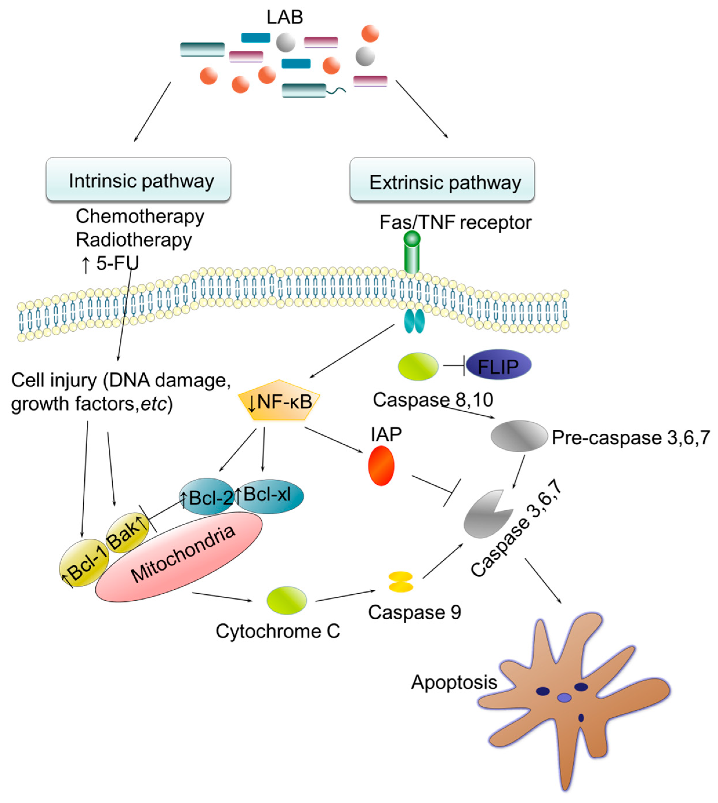
3.1.1. TME Modulation by Lactobacillus

Recent studies suggest that microorganisms play a pivotal role in cancer development and progression. For example, in cervical cancer, chronic infection with high-risk human papillomaviruses (HPVs) is a primary pathogenic factor [34]. Lactobacillus is the predominant bacterial genus in a healthy vaginal microbiome, where it plays a significant role in protecting the female reproductive system. Similarly, Lactobacillus and Bifidobacterium are common probiotics in the gastrointestinal tract, with substantial evidence suggesting that the relationship between diet and CRC may result from an imbalanced gut microbiota, particularly the depletion of Lactobacillus acidophilus and Bifidobacterium bifidum [35].
Lactobacillus exerts its anticancer effects through multiple mechanisms. By competing for epithelial binding sites, it prevents the adhesion of invasive pathogenic bacteria, thereby reducing the risk of malignant transformation. In addition, Lactobacillus secretes glycogen-derived organic acids, which maintain an acidic microenvironment that inhibits the growth of pathogenic bacteria and supports the activity of hydrogen peroxide (H2O2) and bacteriocins. The secretion of bioactive metabolites, including exopolysaccharides, peptidoglycans, phosphorylated polysaccharides, and bacteriocins, further contributes to tumor suppression [34,36]. Another key mechanism by which Lactobacillus influences cancer progression is through immune system modulation and the induction of apoptosis. It enhances immune responses by promoting the activation and proliferation of B cells and T cells [37]. Emerging research indicates that various Lactobacillus strains induce the production of pro-inflammatory cytokines, such as interleukin (IL)-1 and IL-6, as well as anti-inflammatory cytokines, including IL-10 and IL-12, in animal models [38]. Several of these cytokines play a pivotal role in promoting cancer cell apoptosis. Notably, the combination of Lactobacillus with sulforaphane, a compound known to activate the TNF-related apoptosis-inducing ligand (TRAIL) pathway, has demonstrated beneficial effects in cancer prevention [1].

3.1.2. TME Modulation by Clostridium

Clostridium is a genus of rod-shaped, anaerobic, spore-forming, Gram-positive bacteria. Pathogenic species such as Clostridium botulinum, Clostridium tetani, Clostridium perfringens, and Clostridium novyi can germinate and produce potent toxins, which are responsible for botulism, tetanus, gas gangrene, and hemolysis, respectively. However, the majority of Clostridium species found in soil are non-pathogenic [39]. Tumor necrosis results in an anaerobic microenvironment that facilitates the proliferation of Clostridium, presenting a unique opportunity to harness this characteristic for targeted cancer therapy. Certain Clostridium species also produce butyrate, which may be beneficial for controlling tumor growth and development. A comparative analysis of the fecal microbiome and metabolome in CRC patients and healthy individuals revealed that butyrate-producing bacteria were underrepresented in cancer samples [40], suggesting a possible anticancer role for butyrate-producing Clostridium.
The oncolytic potential of Clostridium species was initially documented in late 19th-century clinical observations, where spontaneous tumor regression occurred in patients with gas gangrene caused by natural infection of C. perfringens. Subsequent studies have exploited genetically engineered clostridial strains, such as Clostridium sporogenes (ATCC 13732), which retain tumor-targeting capabilities while exhibiting significantly reduced systemic toxicity compared to wild-type species. The hypoxic and necrotic microenvironment prevalent in solid tumors enables selective colonization by intravenously administered clostridial spores. Following germination, these anaerobic bacteria proliferate preferentially within tumor tissues, achieving localized oncolytic activity without harming normoxic healthy cells [41]. Most solid cancers contain necrotic tissue regions, which allows the infiltration and selective germination of intravenously injected anaerobic bacterial spores from the Clostridium genus, enabling cancer-specific colonization. The anaerobic bacteria belonging to the Clostridium genus can proliferate within anoxic tumor regions, consuming necrotic cancer tissue. Upon encountering oxygenated areas at the tumor periphery, these bacteria cease proliferation and die, thereby minimizing systemic toxicity and ensuring safety for surrounding healthy tissues (Figure 1).
In addition to direct oncolysis, Clostridium-based gene-directed enzyme prodrug therapy (GDEPT) constitutes a promising cancer gene therapy approach. This strategy involves injecting genetically engineered non-pathogenic Clostridium spores that express non-mammalian prodrug-activating enzymes within tumors (Figure 1). For instance, Clostridium acetobutylicum has been engineered to express tumoricidal agents such as functional TNF-α and cytosine deaminase, the latter of which converts 5-fluorocytosine (5-FC) into the chemotherapeutic agent 5-fluorouracil (5-FU), with the active exogenous enzyme successfully delivered to tumors [44].

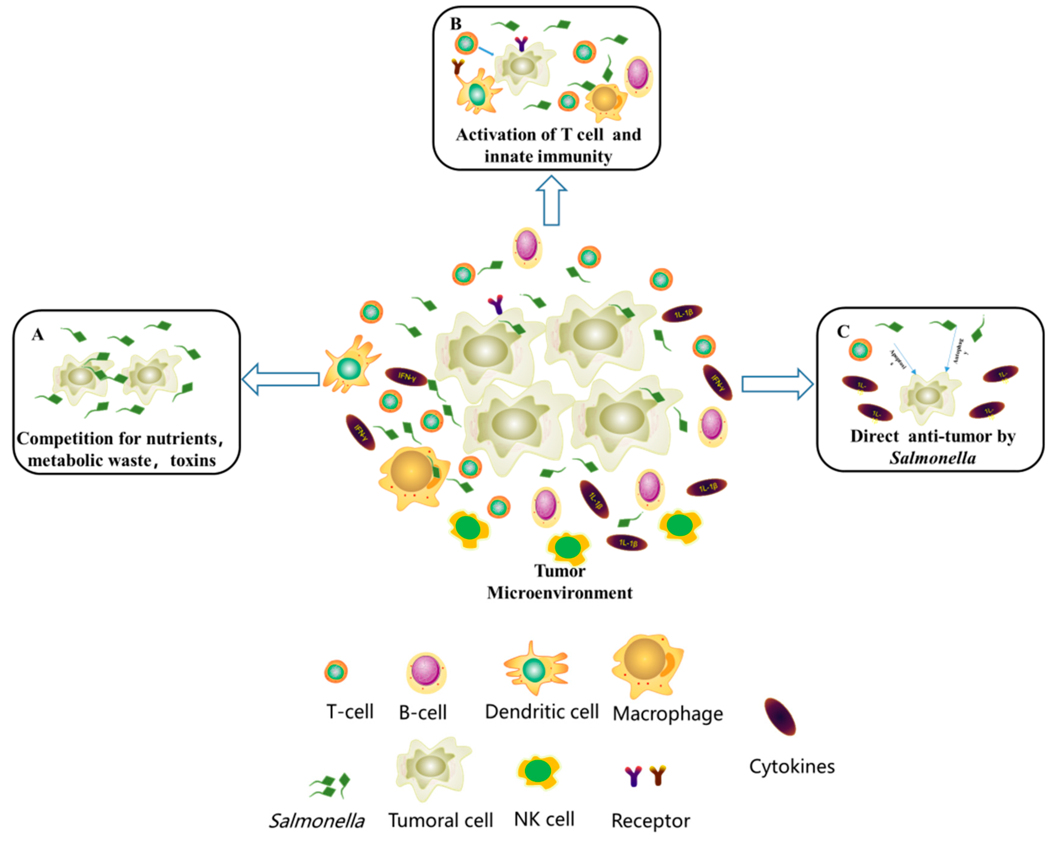
3.2. Competitive Inhibition

In solid tumors, tumor cell density is influenced by multiple factors, including nutrient availability, spatial constraints due to contact inhibition, chemical signaling, and other environmental conditions. Certain gut bacteria can thrive within tumor tissues, enabling competitive inhibition as a potential strategy for tumor control. The primary mechanisms of bacterial tumor inhibition involve competition for nutrients and oxygen (O2). Among these microbial competitors, Salmonella species exhibit tumor-specific tropism through metabolic interference and hypoxia-driven chemotaxis, offering targeted nutrient deprivation while concurrently stimulating antitumor immune surveillance.

Competitive Inhibition by Salmonella

A key advantage of Salmonella over Clostridium or Bifidobacterium is its facultative anaerobic capability, making it effective in targeting small tumors [45]. Research has demonstrated that Salmonella strains are attracted to primary and metastatic tumor cells through chemotaxis mediated by serine, galactose/ribose, and aspartate receptors on their surface [46]. Proper flagellum structure, active motility, and signal transduction proteins are necessary for Salmonella migration to tumor regions via gravitaxis [43]. In addition, host macrophages and bacterial metabolism are involved in bacterial distribution and colonization within tumors. Compared to healthy tissues, Salmonella typhimurium accumulates in tumor tissues at levels up to 10,000-fold higher [15]. Once colonized, Salmonella deprives tumor cells of essential nutrients, activates antitumor immunity, and enhances antitumor chemokine expression, ultimately inducing cancer cell death (Figure 2) [3,47].
Several Salmonella strains have been investigated for cancer therapy. Laboratory-engineered S. typhimurium strains, including VNP20009, A1-R, and ΔppGpp, have been designed to reduce cytotoxicity in normal tissues while enhancing tumor specificity and antitumor efficacy [3]. Hoffman further proposed that these mechanisms could be enhanced through genetic modifications, such as engineering S. typhimurium A1-R to carry auxotrophic mutations for arginine and leucine, improving tumor targeting in lymphatic and pulmonary tissues [48]. S. typhimurium A1-R releases therapeutic molecules to the tumor via quorum sensing or lytic release of amino acids, which increases target specificity and penetration.
Salmonella-mediated tumor inhibition involves multiple mechanisms, such as the induction of the immune response and cell death (Figure 2). The innate immune response can be triggered via the Toll-like receptor (TLR)-myeloid differentiation primary response gene signaling pathway. In vivo studies have demonstrated increased expression of CXCL10 (IP-10), IFN-γ, and IFN-induced chemokines CXCL9 (MIG) in tumor cells during Salmonella treatment. These factors are responsible for recruiting peripheral natural killer (NK) cells, macrophages, neutrophils, and T cells into tumors [49,50]. Bacterial components, including lipoteichoic acid, flagellin, and lipopolysaccharide, can also activate T cells to recognize and kill tumor cells at the primary site while preventing metastasis formation [51]. Subsequently, CD8+ and CD4+ T cell infiltration occurs, enhancing immune responses against tumor cells [52]. Salmonella may suppress negative regulators of apoptosis and autophagy, thereby killing cancer cells via the AKT/mTOR signaling pathway, as demonstrated using apoptosis and autophagy inhibitors. Additionally, Salmonella induces tumor cell apoptosis by stimulating caspase-1 through inflammasomes, which controls tumors by cleaving pro-IL-1β and pro-IL-18 to produce active IL-1β and IL-18 [3].

3.3. Activation of Immune Cells

Many anticancer bacteria control the growth of solid tumors by activating immune cells. Tumor-immune cell interactions can be conceptualized as a series of events known as the cancer-immunity cycle [53] (Figure 3B). Somatic mutations generate tumor-specific peptides that can function as neoantigens. The mutational burden of individual tumors varies substantially across malignancies, ranging from dozens to tens of thousands of somatic mutations per genome. This genomic diversity drives extensive molecular heterogeneity in tumor cells, primarily mediated by neoantigen production. These neoantigens are presented on antigen-presenting cells via highly polymorphic major histocompatibility complex (MHC) molecules, termed human leukocyte antigens (HLAs) in humans, which further amplifies intertumoral heterogeneity through allele-specific antigen presentation. Following tumor cell death, released neoantigens initiate cascades of immune activation. This process stimulates clonal expansion of T cells exhibiting diverse T cell receptor (TCR) repertoires, which recognize neoantigen-MHC complexes through conformation-specific interactions. Beyond molecular heterogeneity, intratumoral immune landscapes are characterized by pronounced cellular heterogeneity. This encompasses dynamically interacting innate and adaptive immune cell subpopulations, including macrophages, DCs, cytotoxic T lymphocytes (CTLs), and regulatory T cells (Tregs; Figure 3A). Notably, tumors with elevated mutational loads, such as microsatellite instability-high (MSI-H) CRCs and melanoma, exhibit heightened neoantigen abundance. This correlates with robust infiltration of heterogeneous immune cell populations, a hallmark of immunologically “hot” tumors. In contrast, microsatellite-stable tumors with low mutation loads show significantly decreased infiltration. Consequently, tumor phenotype development during progression occurs through a process termed cancer immunoediting. These molecular and cellular heterogeneities create an intricate system of tumor-immune cell interactions that challenges researchers attempting to unravel the system and identify anti-tumor immune mechanisms.
The captured antigens are presented on MHC molecules to T cells by DCs, triggering and activating effector T cell responses against cancer-specific antigens. Guided by chemokine gradients, the activated T cells migrate to and infiltrate the tumor site. T cells specifically recognize and bind to cancer cells through interactions between the TCR and the neoantigen-MHC complex, subsequently killing the cancer cells in a process defined as cytolytic activity. A variety of molecular and genomic tools can assess each stage of these cancer-immune cell interactions and their stimulatory or inhibitory elements. Importantly, phase I–II clinical trials have shown that interventions such as fecal microbiota transplantation can overcome resistance to immune checkpoint blockade in melanoma patients, improve therapeutic outcomes in treatment-naïve patients, and reduce therapy-induced immunotoxicities [54].

3.3.1. Activation of Immune Cells by Listeria

L. monocytogenes, an intracellular pathogen, represents a promising therapeutic vector for delivering DNA, RNA, or protein to cancer cells or for activating immune responses to tumor-specific antigens. Multiple biological capabilities make L. monocytogenes a favorable substrate for utilization as a vector for anti-cancer vaccines or gene therapy [55]. L. monocytogenes can invade and distribute within tumors, producing specific virulence factors that cleave the vacuole membrane, enabling cytoplasmic escape [56]. The process by which L. monocytogenes escapes into the host cell cytoplasm relies on several key virulence factors. Initially, the bacterium utilizes internalins (InlA and InlB) to bind to specific receptors on the host cell surface, facilitating internalization. Once internalized, Listeria employs listeriolysin O (LLO) to create pores in the phagosomal membrane, allowing the bacterium to escape into the cytoplasm. Additionally, phospholipases C (PlcA and PlcB) further hydrolyze phospholipids in the phagosomal membrane, aiding the escape process [57]. Once in the cytoplasm, Listeria utilizes the ActA protein to hijack the host cell’s actin polymerization machinery, driving bacterial movement within the cytoplasm and facilitating the spread to neighboring cells. These virulence factors work in concert to enable Listeria to effectively evade host defenses and proliferate within host cells. The bacterium achieves actin-based motility and intercellular diffusion without an extracellular phase. This cytoplasmic localization of L. monocytogenes is essential as it enhances antigen entry into the MHC Class I antigen processing pathway, resulting in the activation of specific CD8+ T cell responses.
In the host cell cytoplasm, L. monocytogenes stimulates cytosolic surveillance pathways, inducing IFN-β expression [58]. The host immune response to L. monocytogenes occurs in two distinct phases: an ‘early response’ triggered by the initial interaction between the pathogen and host cell TLRs, followed by a separate ‘late response’ that depends on the entry of L. monocytogenes into the cytosol. Cytoplasmic infection triggers NOD1 and other NLR-family proteins that detect pathogen-associated molecular patterns (PAMPs) related to L. monocytogenes infection [43,59]. Cytosolic growth of L. monocytogenes in DCs activates proinflammatory cytokine secretion and upregulates co-stimulatory molecules, including CD40, CD80, and CD86, which subsequently initiate adaptive immune responses (Figure 4). The cytoplasmic location ensures that antigens expressed by L. monocytogenes enter the MHC Class I processing pathway, thus driving CD8+ T cell-mediated responses (i.e., production of granzymes, IFN-γ, and perforin) necessary for sterilizing immunity. CD4+ T cells are also produced during the adaptive immune response, providing additional defense against the pathogen. L. monocytogenes induces an intense Th1 response, with IFN-γ being necessary for Th cell immune function. The pathogen also induces Tregs (CD4+, CD25+, Foxp3+) that limit CD8+ cell expansion. While wild-type infections typically do not induce antibody reactions, administration of large doses of attenuated L. monocytogenes mutants can induce high antibody titers against the pathogen. Overall, the cytoplasmic position of L. monocytogenes is significant as it facilitates antigen entry into the cytosolic processing pathway for presentation via MHC Class I molecules.

3.3.2. Activation of Immune Cells by the Gut Microbiome

The gut microbiome exerts profound effects on human immunity, both locally and systemically, including in the context of cancer [60]. Preclinical models have demonstrated that bacteria and their metabolites can activate proinflammatory cytokine production, enhance antigen presentation, and thereby augment antitumor immune function. The gut microbiome also affects the efficacy of cancer immunotherapies, particularly immune checkpoint inhibitors (ICIs) involving the blockade of PD-L1/PD-1 and CTLA-4. Intestinal microbial diversity in patients increases significantly following PD-1 therapy, with certain microorganisms showing notable expansion, including Ruminococcaceae, Clostridiales, and Faecalibacterium [61]. Bacterial metabolites, such as SCFAs, or the bacteria themselves can activate local DCs to migrate to the mesenteric lymph nodes (mLNs). Under the influence of mature DCs, naive T cells subsequently differentiate into effector T cells, Th17 cells, or Tregs. When DCs present antigens from symbiotic bacteria in the gut mLNs, T cells and B cells, including Th17 cells and Tregs, can enter the systemic circulation and facilitate immune responses against identical remote antigens or against other antigens via cross-reactivity with similar epitopes.
Recent studies have established a link between the microbiome and immunotherapy efficacy. Fecal samples from patients responding to anti-PD-1 therapy showed a relative abundance of Akkermansia muciniphila [62]. The efficacy of ICIs in germ-free (GF) mice can be enhanced by fecal microbiota transfer (FMT) from responders [30]. The microbiome has also been shown to influence CTLA-4 blockade-based treatments. The response to CTLA-4 blockade was diminished in axenic mice or those treated with antibiotics, while therapeutic efficacy could be restored by supplementing mice with enriched bacterial species found in responder animals [63]. Remarkably, there was only moderate overlap in these checkpoint-blockade-responsive microbiome signatures across cohorts. Diet, probiotics, and prebiotics can regulate the abundance of beneficial bacteria in the intestinal flora.
In addition, other bacteria have been employed as immune activators to control cancer. In the late 19th century, William Coley developed a safer vaccine composed of two inactivated bacterial species, Serratia marcescens and Streptococcus pyogenes [64]. These strains stimulate an infection-like response accompanied by fever without actual infection risk. This vaccine was successfully applied to treat various malignancies, including carcinomas, sarcomas, melanomas, myelomas, and lymphomas [45].

3.4. Vectors for Gene Therapy

Gene therapy, also known as human gene transfer, is a medical field focusing on delivering nucleic acids into patient cells as therapeutic agents [65,66]. The primary challenge in cancer-targeted gene therapy is achieving specific delivery to solid tumors [45]. To overcome this limitation, bacteria, particularly genetically engineered strains, represent an optimal approach, serving as vectors that can express specific therapeutic genes. These bacterial vectors provide powerful alternative therapies by preferentially delivering anticancer agents, therapeutic proteins, cytotoxic peptides, or prodrug-converting enzymes to solid tumors.

3.4.1. Escherichia as a Vector for Gene Therapy

Gene therapy-based strategies are being developed for various cancers to improve therapeutic efficacy, decrease side effects, and address quality-of-life concerns. These approaches include suicide gene therapy [67], restoration of inactivated or poorly expressed tumor suppressor genes [68], and cytokine-based immunotherapy [69]. In prostate cancer treatment, herpes simplex virus thymidine kinase/Ganciclovir and purine nucleoside phosphorylase/Fludara are commonly expressed in E. coli for suicide gene therapy [67]. E. coli purine nucleoside phosphorylase (PNP) demonstrates potent tumor-killing capacity and an intensive bystander effect on surrounding tumor cells. E. coli PNP differs from human purine nucleoside phosphorylase in its ability to accept adenosine-based nucleosides as substrates [68]. This bacterial enzyme converts adenosine analogs into highly cytotoxic metabolites that incorporate into both DNA and RNA, effectively inhibiting DNA, RNA, and protein synthesis, leading to the death of both dividing and non-dividing cells [70]. The antitumor efficacy of the PNP/Fludara system has been investigated using the prostate-specific, rat probasin-based promoter to drive E. coli PNP/Fludara expression [67].
E. coli also produces L-asparaginase, an enzyme that catalyzes the hydrolysis of L-asparagine to L-aspartate and ammonia. Type II E. coli asparaginase, located in the bacterial periplasmic region, demonstrates a significantly higher affinity for asparagine than Type I E. coli asparaginase [71]. Type II E. coli asparaginase is used clinically to treat acute lymphoblastic leukemia. Its enzymatic mechanism involves nucleophilic attack on the β-amide group of asparagine, forming an acyl-enzyme intermediate. Based on the proximity of their side chains to the aspartate ligand, Thr-12, Tyr-25, Ser-58, and Thr-89 are potential nucleophiles in this reaction.

3.4.2. Bifidobacterium as a Vector for Gene Therapy

Since the 1899 discovery of Bifidobacteria dominance in the feces of breast-fed infants, numerous studies have investigated their role in regulating intestinal flora and other potential health benefits [72]. Bifidobacterium adolescentis, Bifidobacterium bifidum, and Bifidobacterium longum have been employed as gene-therapeutic systems to deliver the antiangiogenic protein endostatin to solid tumors [73,74,75,76]. Systemic administration of transfected Bifidobacterium via tail vein in tumor-bearing mice effectively inhibits angiogenesis and reduces tumor growth [73]. Furthermore, endostatin gene-enriched Bifidobacterium treatment enhances T cell and NK cell activity while stimulating TNF-α and IL-2 activity [74]. Additionally, Bifidobacterium demonstrates a competitive advantage in the cancer microenvironment, competing with pathogens by altering pH and inducing metabolic changes to prevent cancers such as CRC [75,77].
Bifidobacterium and Lactobacillus, as representative lactic acid bacteria and common probiotics, exert anticancer effects through several mechanisms, including TME regulation, immune activation, and gene therapy, ultimately leading to tumor apoptosis. Apoptosis, defined as genetically programmed cell death, is frequently dysregulated in cancers, where decreased apoptotic capacity occurs alongside altered cell proliferation control [78]. Research has demonstrated that lactic acid bacteria can regulate cell apoptosis through both endogenous and exogenous pathways, representing potential key mechanisms for CRC prevention (Figure 5).

3.5. Microbial Anticancer Biomolecules

Bacteria are known to produce diverse and potent bioactive compounds, including antitumor agents, immunosuppressive agents, antibiotics, and enzymes. These bioactive substances have an extensive range of applications as chemotherapeutic agents for disease therapies, and novel bioactive substances remain in constant demand [79]. These bioactive metabolites trigger specific gene expressions, leading to bioluminescence, swarming motility, biofilm formation, virulence factor production, and antibiotic biosynthesis [80].

Production of Anticancer Biomolecules by Bacillus

Bacteria produce over 3800 bioactive metabolites, with Bacillus spp. and Pseudomonas spp. being the most prominent sources, each producing approximately 800–900 metabolites [81]. Polyketides, macro-lactones, fatty acids, lipoamides, isocoumarins, lipopeptides, polypeptides, and microbial enzymes represent the most abundant bioactive compounds from terrestrial and marine Bacillus spp. [82]. Bacillus exhibits a wide range of biological activities, including antifungal, antibacterial, anti-algal, antioxidant, antifouling, and most significantly, anticancer activity. Numerous identified enzymes and biologically active molecules with anticancer activity from Bacillus are summarized in Table 1, with their modes of action listed in Table 2. Consequently, Bacillus species represent appropriate sources for diverse bioactive molecules in developing novel cancer therapeutics.
In 1976, the engineered bacterium Bacillus Calmette-Guérin was successfully implemented to treat superficial bladder tumors, a therapy that remains unsurpassed to date [126]. Beyond anticancer functional proteins and other molecules, Bacillus Calmette-Guérin serves as an immunotherapy for metastatic bladder cancer. Infection of bladder and urothelial tumor cells by Bacillus Calmette-Guérin results in bacterial internalization, enhancing the expression of antigen-presenting molecules.
The immune reaction is induced through the release of Th1 cytokines, including IL-2, IL-12, IFN-γ, and TNF. Th2 cytokines (IL-4, IL-5, IL-6, and IL-10) are implicated alongside IL-8 and IL-17. This immune cascade induces antitumor activity mediated by macrophages, NK cells, CTLs, and neutrophils [126].

4. Future Prospects

The relationship between the gut microbiota and malignant solid tumors has received increasing attention in recent years. Understanding the interactions between different bacterial species and tumor tissues is essential for developing more effective strategies for cancer prevention, management, and treatment. Gut microbes contribute to cancer treatment primarily through modulation of the TME, competitive inhibition, immune cell activation, serving as vectors for gene therapy, and secretion of anticancer biomolecules. While this review highlights these primary anticancer mechanisms, it is important to acknowledge that additional mechanisms likely play significant roles. Notably, microbes exhibiting robust anticancer properties often operate through multiple complementary mechanisms.
Currently, bacterial therapy for malignant solid tumors remains predominantly in the experimental research stage, with numerous challenges to overcome before clinical implementation. These challenges include incomplete tumor clearance, variability in immune cell activation across different cancer types, suboptimal cytotoxicity profiles of anticancer molecules, and inappropriate expression of antitumor complexes in gene therapy vectors. Furthermore, achieving targeted colonization of intestinal bacteria within solid tumors presents significant technical difficulties. Although these gut bacteria coexist harmoniously within the human gastrointestinal tract, they may exhibit toxicity when present in other tissues and organs. Currently, there are no well-established strategies to mitigate the adverse effects associated with microbe-based cancer therapies.
To transition gut bacteria, including certain conditional pathogens, from laboratory research to clinical applications, several critical issues must be addressed. These include establishing controlled bacterial growth within solid tumors, enhancing intelligent recognition of diverse cancer cells and improving target specificity, facilitating bacterial entry into solid tumors and subsequent release of active factors, and stimulating immune cell aggregation and activation of the host immune system. As an emerging approach to solid tumor treatment, gut microbiota-based therapies show promise for cancer prevention and management. Emerging technologies that integrate principles from microbiology, molecular biology, synthetic biology, materials science, and engineering will be essential for enhancing the precision and efficacy of bacteria-based cancer therapies while mitigating adverse effects, representing a promising direction in solid tumor treatment.

Author Contributions

T.Z. and X.-M.Y.: writing—review and editing, methodology, S.-T.Y. and W.-W.Z.: supervision and project administration, T.Z. and W.-W.Z.: funding acquisition. All authors have read and agreed to the published version of the manuscript.

Funding

This research was supported by National Key R & D Program of China (2018YFA0903200) and Science and Technology Research Program of Chongqing Municipal Education Commission (KJQN202402809). The APC was funded by KJQN202402809.

Conflicts of Interest

The authors have no financial conflicts of interest to declare.

References

  1. Bray, F.; Ferlay, J.; Soerjomataram, I.; Siegel, R.L.; Torre, L.A.; Jemal, A. Global cancer statistics 2018: GLOBOCAN estimates of incidence and mortality worldwide for 36 cancers in 185 countries. CA Cancer J. Clin. 2018, 68, 394–424. [Google Scholar] [CrossRef] [PubMed]
  2. Siegel, R.L.; Miller, K.D.; Wagle, N.S.; Jemal, A. Cancer statistics, 2023. CA Cancer J. Clin. 2023, 73, 17–48. [Google Scholar] [CrossRef] [PubMed]
  3. Mi, Z.; Feng, Z.-C.; Li, C.; Yang, X.; Ma, M.-T.; Rong, P.-F. Salmonella-mediated cancer therapy: An innovative therapeutic strategy. J. Cancer 2019, 10, 4765. [Google Scholar] [CrossRef] [PubMed]
  4. Gurbatri, C.R.; Arpaia, N.; Danino, T. Engineering bacteria as interactive cancer therapies. Science 2022, 378, 858–864. [Google Scholar] [CrossRef]
  5. Kaur, R.; Bhardwaj, A.; Gupta, S. Cancer treatment therapies: Traditional to modern approaches to combat cancers. Mol. Biol. Rep. 2023, 50, 9663–9676. [Google Scholar] [CrossRef]
  6. Duong, M.T.-Q.; Qin, Y.; You, S.-H.; Min, J.-J. Bacteria-cancer interactions: Bacteria-based cancer therapy. Exp. Mol. Med. 2019, 51, 1–15. [Google Scholar] [CrossRef]
  7. Di Domenico, E.G.; Cavallo, I.; Pontone, M.; Toma, L.; Ensoli, F. Biofilm producing Salmonella typhi: Chronic colonization and development of gallbladder cancer. Int. J. Mol. Sci. 2017, 18, 1887. [Google Scholar] [CrossRef]
  8. Huang, Y.; Fan, X.; Wang, Z.; Zhou, J.; Tian, X.; Li, N. Identification of helicobacter species in human liver samples from patients with primary hepatocellular carcinoma. J. Clin. Pathol. 2004, 57, 1273–1277. [Google Scholar] [CrossRef]
  9. Helmink, B.A.; Khan, M.W.; Hermann, A.; Gopalakrishnan, V.; Wargo, J.A. The microbiome, cancer, and cancer therapy. Nat. Med. 2019, 25, 377–388. [Google Scholar] [CrossRef]
  10. Wang, N.; Fang, J.-Y. Fusobacterium nucleatum, a key pathogenic factor and microbial biomarker for colorectal cancer. Trends Microbiol. 2023, 31, 159–172. [Google Scholar] [CrossRef]
  11. Kenneth, M.J.; Tsai, H.-C.; Fang, C.-Y.; Hussain, B.; Chiu, Y.-C.; Hsu, B.-M. Diet-mediated gut microbial community modulation and signature metabolites as potential biomarkers for early diagnosis, prognosis, prevention and stage-specific treatment of colorectal cancer. J. Adv. Res. 2023, 52, 45–57. [Google Scholar] [CrossRef] [PubMed]
  12. Park, E.M.; Chelvanambi, M.; Bhutiani, N.; Kroemer, G.; Zitvogel, L.; Wargo, J.A. Targeting the gut and tumor microbiota in cancer. Nat. Med. 2022, 28, 690–703. [Google Scholar] [CrossRef] [PubMed]
  13. Schmidt, C.K.; Medina-Sánchez, M.; Edmondson, R.J.; Schmidt, O.G. Engineering microrobots for targeted cancer therapies from a medical perspective. Nat. Commun. 2020, 11, 5618. [Google Scholar] [CrossRef] [PubMed]
  14. Forbes, N.S. Engineering the perfect (bacterial) cancer therapy. Nat. Rev. Cancer 2010, 10, 785–794. [Google Scholar] [CrossRef]
  15. Curran, C.S.; Rasooly, A.; He, M.; Prickril, B.; Thurin, M.; Sharon, E. Report on the NCI microbial-based cancer therapy conference. Cancer Immunol. Res. 2018, 6, 122–126. [Google Scholar] [CrossRef]
  16. Tao, J.; Li, S.; Gan, R.-Y.; Zhao, C.-N.; Meng, X.; Li, H.-B. Targeting gut microbiota with dietary components on cancer: Effects and potential mechanisms of action. Crit. Rev. Food Sci. Nutr. 2020, 60, 1025–1037. [Google Scholar] [CrossRef]
  17. Li, W.; Deng, Y.; Chu, Q.; Zhang, P. Gut microbiome and cancer immunotherapy. Cancer Lett. 2019, 447, 41–47. [Google Scholar] [CrossRef]
  18. Shui, L.; Yang, X.; Li, J.; Yi, C.; Sun, Q.; Zhu, H. Gut microbiome as a potential factor for modulating resistance to cancer immunotherapy. Front. Immunol. 2020, 10, 2989. [Google Scholar] [CrossRef]
  19. Bradley, K.C.; Finsterbusch, K.; Schnepf, D.; Crotta, S.; Llorian, M.; Davidson, S.; Fuchs, S.Y.; Staeheli, P.; Wack, A. Microbiota-driven tonic interferon signals in lung stromal cells protect from influenza virus infection. Cell Rep. 2019, 28, 245–256.e244. [Google Scholar] [CrossRef]
  20. Barlow, G.M.; Yu, A.; Mathur, R. Role of the gut microbiome in obesity and diabetes mellitus. Nutr. Clin. Pract. 2015, 30, 787–797. [Google Scholar] [CrossRef]
  21. Ghorbani, E.; Avan, A.; Ryzhikov, M.; Ferns, G.; Khazaei, M.; Soleimanpour, S. Role of Lactobacillus strains in the management of colorectal cancer: An overview of recent advances. Nutrition 2022, 103, 111828. [Google Scholar] [CrossRef] [PubMed]
  22. Yaghoubi, A.; Ghazvini, K.; Khazaei, M.; Hasanian, S.M.; Avan, A.; Soleimanpour, S. The use of Clostridium in cancer therapy: A promising way. Rev. Res. Med. Microbiol. 2022, 33, 121–127. [Google Scholar] [CrossRef]
  23. Al-Saafeen, B.H.; Fernandez-Cabezudo, M.J.; Al-Ramadi, B.K. Integration of Salmonella into combination cancer therapy. Cancers 2021, 13, 3228. [Google Scholar] [CrossRef]
  24. Selvanesan, B.C.; Chandra, D.; Quispe-Tintaya, W.; Jahangir, A.; Patel, A.; Meena, K.; Alves Da Silva, R.A.; Friedman, M.; Gabor, L.; Khouri, O. Listeria delivers tetanus toxoid protein to pancreatic tumors and induces cancer cell death in mice. Sci. Transl. Med. 2022, 14, eabc1600. [Google Scholar] [CrossRef]
  25. Osek, J.; Wieczorek, K. Listeria monocytogenes—How this pathogen uses its virulence mechanisms to infect the hosts. Pathogens 2022, 11, 1491. [Google Scholar] [CrossRef]
  26. Thakur, B.K.; Malaise, Y.; Choudhury, S.R.; Neustaeter, A.; Turpin, W.; Streutker, C.; Copeland, J.; Wong, E.O.; Navarre, W.W.; Guttman, D.S. Dietary fibre counters the oncogenic potential of colibactin-producing Escherichia coli in colorectal cancer. Nat. Microbiol. 2025, 10, 855–870. [Google Scholar] [CrossRef]
  27. Kaźmierczak-Siedlecka, K.; Roviello, G.; Catalano, M.; Polom, K. Gut microbiota modulation in the context of immune-related aspects of Lactobacillus spp. and Bifidobacterium spp. in gastrointestinal cancers. Nutrients 2021, 13, 2674. [Google Scholar] [CrossRef]
  28. Oscarsson, J.; Bao, K.; Shiratsuchi, A.; Grossmann, J.; Wolski, W.; Aung, K.M.; Lindholm, M.; Johansson, A.; Mowsumi, F.R.; Wai, S.N. Bacterial symbionts in oral niche use type VI secretion nanomachinery for fitness increase against pathobionts. iScience 2024, 27, 109650. [Google Scholar] [CrossRef]
  29. Maroufi, N.F.; Rashidi, M.R.; Vahedian, V.; Akbarzadeh, M.; Fattahi, A.; Nouri, M. Therapeutic potentials of Apatinib in cancer treatment: Possible mechanisms and clinical relevance. Life Sci. 2020, 241, 117106. [Google Scholar] [CrossRef]
  30. Gopalakrishnan, V.; Helmink, B.A.; Spencer, C.N.; Reuben, A.; Wargo, J.A. The influence of the gut microbiome on cancer, immunity, and cancer immunotherapy. Cancer Cell 2018, 33, 570–580. [Google Scholar] [CrossRef]
  31. Alfarouk, K.O.; Muddathir, A.K.; Shayoub, M.E. Tumor acidity as evolutionary spite. Cancers 2011, 3, 408–414. [Google Scholar] [CrossRef] [PubMed]
  32. Korneev, K.V.; Atretkhany, K.-S.N.; Drutskaya, M.S.; Grivennikov, S.I.; Kuprash, D.V.; Nedospasov, S.A. TLR-signaling and proinflammatory cytokines as drivers of tumorigenesis. Cytokine 2017, 89, 127–135. [Google Scholar] [CrossRef] [PubMed]
  33. Kim, M.-G.; Shon, Y.; Kim, J.; Oh, Y.-K. Selective activation of anticancer chemotherapy by cancer-associated fibroblasts in the tumor microenvironment. J. Natl. Cancer Inst. 2017, 109, djw186. [Google Scholar] [CrossRef]
  34. Yang, X.; Da, M.; Zhang, W.; Qi, Q.; Zhang, C.; Han, S. Role of Lactobacillus in cervical cancer. Cancer Manag. Res. 2018, 10, 1219–1229. [Google Scholar] [CrossRef]
  35. Heydari, Z.; Rahaie, M.; Alizadeh, A.M.; Agah, S.; Khalighfard, S.; Bahmani, S. Effects of Lactobacillus acidophilus and Bifidobacterium bifidum probiotics on the expression of microRNAs 135b, 26b, 18a and 155, and their involving genes in mice colon cancer. Probiotics Antimicrob. Proteins 2019, 11, 1155–1162. [Google Scholar] [CrossRef]
  36. Yasuda, S.; Horinaka, M.; Sakai, T. Sulforaphane enhances apoptosis induced by Lactobacillus pentosus strain S-PT84 via the TNFα pathway in human colon cancer cells. Oncol. Lett. 2019, 18, 4253–4261. [Google Scholar] [CrossRef]
  37. Jacouton, E.; Chain, F.; Sokol, H.; Langella, P.; Bermudez-Humaran, L.G. Probiotic strain Lactobacillus casei BL23 prevents colitis-associated colorectal cancer. Front. Immunol. 2017, 8, 1553. [Google Scholar] [CrossRef]
  38. Rajoka, M.S.R.; Zhao, H.; Lu, Y.; Lian, Z.; Li, N.; Hussain, N.; Shao, D.; Jin, M.; Li, Q.; Shi, J. Anticancer potential against cervix cancer (HeLa) cell line of probiotic Lactobacillus casei and Lactobacillus paracasei strains isolated from human breast milk. Food Funct. 2018, 9, 2705–2715. [Google Scholar] [CrossRef]
  39. Mowday, A.M.; Guise, C.P.; Ackerley, D.F.; Minton, N.P.; Lambin, P.; Dubois, L.J.; Theys, J.; Smaill, J.B.; Patterson, A.V. Advancing clostridia to clinical trial: Past lessons and recent progress. Cancers 2016, 8, 63. [Google Scholar] [CrossRef]
  40. Paul, B.; Barnes, S.; Demark-Wahnefried, W.; Morrow, C.; Salvador, C.; Skibola, C.; Tollefsbol, T.O. Influences of diet and the gut microbiome on epigenetic modulation in cancer and other diseases. Clin. Epigenetics 2015, 7, 112. [Google Scholar] [CrossRef]
  41. Möse, J.R.; Möse, G. Oncolysis by clostridia. I. Activity of Clostridium butyricum (M-55) and other nonpathogenic clostridia against the Ehrlich carcinoma. Cancer Res. 1964, 24, 212–216. [Google Scholar]
  42. Mohr, U.; Boldingh, W.H.; Emminger, A.; Behagel, H. Oncolysis by a new strain of Clostridium. Cancer Res. 1972, 32, 1122–1128. [Google Scholar] [PubMed]
  43. Agrawal, N.; Bettegowda, C.; Cheong, I.; Geschwind, J.-F.; Drake, C.G.; Hipkiss, E.L.; Tatsumi, M.; Dang, L.H.; Diaz, L.A., Jr.; Pomper, M. Bacteriolytic therapy can generate a potent immune response against experimental tumors. Proc. Natl. Acad. Sci. USA 2004, 101, 15172–15177. [Google Scholar] [CrossRef] [PubMed]
  44. Theys, J.; Landuyt, W.; Nuyts, S.; Van Mellaert, L.; Van Oosterom, A.; Lambin, P.; Anné, J. Specific targeting of cytosine deaminase to solid tumors by engineered Clostridium acetobutylicum. Cancer Gene Ther. 2001, 8, 294–297. [Google Scholar] [CrossRef]
  45. Patyar, S.; Joshi, R.; Byrav, D.P.; Prakash, A.; Medhi, B.; Das, B. Bacteria in cancer therapy: A novel experimental strategy. J. Biomed. Sci. 2010, 17, 21. [Google Scholar] [CrossRef]
  46. Jiang, S.-N.; Park, S.-H.; Lee, H.J.; Zheng, J.H.; Kim, H.-S.; Bom, H.-S.; Hong, Y.; Szardenings, M.; Shin, M.G.; Kim, S.-C. Engineering of bacteria for the visualization of targeted delivery of a cytolytic anticancer agent. Mol. Ther. 2013, 21, 1985–1995. [Google Scholar] [CrossRef]
  47. Sznol, M.; Lin, S.L.; Bermudes, D.; Zheng, L.-M.; King, I. Use of preferentially replicating bacteria for the treatment of cancer. J. Clin. Investig. 2000, 105, 1027–1030. [Google Scholar] [CrossRef]
  48. Hoffman, R.M. Tumor-targeting Salmonella typhimurium A1-R: An overview. Bact. Ther. Cancer Methods Protoc. 2016, 1409, 1–8. [Google Scholar]
  49. Barak, Y.; Schreiber, F.; Thorne, S.H.; Contag, C.H.; DeBeer, D.; Matin, A. Role of nitric oxide in Salmonella typhimurium-mediated cancer cell killing. BMC Cancer 2010, 10, 146. [Google Scholar] [CrossRef]
  50. Grille, S.; Moreno, M.; Bascuas, T.; Marqués, J.M.; Muñoz, N.; Lens, D.; Chabalgoity, J.A. Salmonella enterica serovar T yphimurium immunotherapy for B-cell lymphoma induces broad anti-tumour immunity with therapeutic effect. Immunology 2014, 143, 428–437. [Google Scholar] [CrossRef]
  51. Lee, C.-H.; Wu, C.-L.; Shiau, A.-L. Toll-like receptor 4 mediates an antitumor host response induced by Salmonella choleraesuis. Clin. Cancer Res. 2008, 14, 1905–1912. [Google Scholar] [CrossRef] [PubMed]
  52. Avogadri, F.; Martinoli, C.; Petrovska, L.; Chiodoni, C.; Transidico, P.; Bronte, V.; Longhi, R.; Colombo, M.P.; Dougan, G.; Rescigno, M. Cancer immunotherapy based on killing of Salmonella-infected tumor cells. Cancer Res. 2005, 65, 3920–3927. [Google Scholar] [CrossRef] [PubMed]
  53. Hackl, H.; Charoentong, P.; Finotello, F.; Trajanoski, Z. Computational genomics tools for dissecting tumour–immune cell interactions. Nat. Rev. Genet. 2016, 17, 441–458. [Google Scholar] [CrossRef]
  54. Blake, S.J.; Wolf, Y.; Boursi, B.; Lynn, D.J. Role of the microbiota in response to and recovery from cancer therapy. Nat. Rev. Immunol. 2024, 24, 308–325. [Google Scholar] [CrossRef]
  55. Tangney, M.; Gahan, C.G. Listeria monocytogenes as a vector for anti-cancer therapies. Curr. Gene Ther. 2010, 10, 46–55. [Google Scholar] [CrossRef]
  56. Van Pijkeren, J.P.; Morrissey, D.; Monk, I.R.; Cronin, M.; Rajendran, S.; O’Sullivan, G.C.; Gahan, C.G.; Tangney, M. A novel Listeria monocytogenes-based DNA delivery system for cancer gene therapy. Hum. Gene Ther. 2010, 21, 405–416. [Google Scholar] [CrossRef]
  57. Pillich, H.; Puri, M.; Chakraborty, T. ActA of Listeria monocytogenes and its manifold activities as an important listerial virulence factor. Actin Cytoskelet. Bact. Infect. 2017, 399, 113–132. [Google Scholar]
  58. O’Riordan, M.; Yi, C.H.; Gonzales, R.; Lee, K.-D.; Portnoy, D.A. Innate recognition of bacteria by a macrophage cytosolic surveillance pathway. Proc. Natl. Acad. Sci. USA 2002, 99, 13861–13866. [Google Scholar] [CrossRef]
  59. Sivan, A.; Corrales, L.; Hubert, N.; Williams, J.B.; Aquino-Michaels, K.; Earley, Z.M.; Benyamin, F.W.; Man Lei, Y.; Jabri, B.; Alegre, M.-L. Commensal Bifidobacterium promotes antitumor immunity and facilitates anti–PD-L1 efficacy. Science 2015, 350, 1084–1089. [Google Scholar] [CrossRef]
  60. Chen, D.S.; Mellman, I. Elements of cancer immunity and the cancer–immune set point. Nature 2017, 541, 321–330. [Google Scholar] [CrossRef]
  61. Gopalakrishnan, V.; Spencer, C.N.; Nezi, L.; Reuben, A.; Andrews, M.C.; Karpinets, T.V.; Prieto, P.; Vicente, D.; Hoffman, K.; Wei, S.C. Gut microbiome modulates response to anti–PD-1 immunotherapy in melanoma patients. Science 2018, 359, 97–103. [Google Scholar] [CrossRef] [PubMed]
  62. Routy, B.; Le Chatelier, E.; Derosa, L.; Duong, C.P.; Alou, M.T.; Daillère, R.; Fluckiger, A.; Messaoudene, M.; Rauber, C.; Roberti, M.P. Gut microbiome influences efficacy of PD-1–based immunotherapy against epithelial tumors. Science 2018, 359, 91–97. [Google Scholar] [CrossRef] [PubMed]
  63. Vétizou, M.; Pitt, J.M.; Daillère, R.; Lepage, P.; Waldschmitt, N.; Flament, C.; Rusakiewicz, S.; Routy, B.; Roberti, M.P.; Duong, C.P. Anticancer immunotherapy by CTLA-4 blockade relies on the gut microbiota. Science 2015, 350, 1079–1084. [Google Scholar] [CrossRef] [PubMed]
  64. Richardson, M.A.; Ramirez, T.; Russell, N.C.; Moye, L.A. Coley toxins immunotherapy: A retrospective review. Altern. Ther. Health Med. 1999, 5, 42. [Google Scholar]
  65. Ermak, G. Emerging Medical Technologies; World Scientific Publishing Company: Chennai, India, 2015. [Google Scholar]
  66. Kaji, E.H.; Leiden, J.M. Gene and stem cell therapies. JAMA 2001, 285, 545–550. [Google Scholar] [CrossRef]
  67. Xie, X.; Guo, J.; Kong, Y.; Xie, G.X.; Li, L.; Lv, N.; Xiao, X.; Tang, J.; Wang, X.; Liu, P. Targeted expression of Escherichia coli purine nucleoside phosphorylase and Fludara® for prostate cancer therapy. J. Gene Med. 2011, 13, 680–691. [Google Scholar] [CrossRef]
  68. Watanabe, M.; Nasu, Y.; Kashiwakura, Y.; Kusumi, N.; Tamayose, K.; Nagai, A.; Sasano, T.; Shimada, T.; Daida, H.; Kumon, H. Adeno-associated virus 2-mediated intratumoral prostate cancer gene therapy: Long-term maspin expression efficiently suppresses tumor growth. Hum. Gene Ther. 2005, 16, 699–710. [Google Scholar] [CrossRef]
  69. Onion, D.; Patel, P.; Pineda, R.G.; James, N.; Mautner, V. Antivector and tumor immune responses following adenovirus-directed enzyme prodrug therapy for the treatment of prostate cancer. Hum. Gene Ther. 2009, 20, 1249–1258. [Google Scholar] [CrossRef]
  70. Tai, C.-K.; Wang, W.; Lai, Y.-H.; Logg, C.R.; Parker, W.B.; Li, Y.-F.; Hong, J.S.; Sorscher, E.J.; Chen, T.C.; Kasahara, N. Enhanced efficiency of prodrug activation therapy by tumor-selective replicating retrovirus vectors armed with the Escherichia coli purine nucleoside phosphorylase gene. Cancer Gene Ther. 2010, 17, 614–623. [Google Scholar] [CrossRef]
  71. Swain, A.L.; Jaskólski, M.; Housset, D.; Rao, J.; Wlodawer, A. Crystal structure of Escherichia coli L-asparaginase, an enzyme used in cancer therapy. Proc. Natl. Acad. Sci. USA 1993, 90, 1474–1478. [Google Scholar] [CrossRef]
  72. Lee, J.-H.; O’Sullivan, D.J. Genomic insights into bifidobacteria. Microbiol. Mol. Biol. Rev. 2010, 74, 378–416. [Google Scholar] [CrossRef] [PubMed]
  73. Li, X.; Fu, G.-F.; Fan, Y.-R.; Liu, W.-H.; Liu, X.-J.; Wang, J.-J.; Xu, G.-X. Bifidobacterium adolescentis as a delivery system of endostatin for cancer gene therapy: Selective inhibitor of angiogenesis and hypoxic tumor growth. Cancer Gene Ther. 2003, 10, 105–111. [Google Scholar] [CrossRef]
  74. Fu, G.-F.; Li, X.; Hou, Y.-Y.; Fan, Y.-R.; Liu, W.-H.; Xu, G.-X. Bifidobacterium longum as an oral delivery system of endostatin for gene therapy on solid liver cancer. Cancer Gene Ther. 2005, 12, 133–140. [Google Scholar] [CrossRef] [PubMed]
  75. Kimura, N.T.; Taniguchi, S.; Aoki, K.; Baba, T. Selective localization and growth of Bifidobacterium bifidum in mouse tumors following intravenous administration. Cancer Res. 1980, 40, 2061–2068. [Google Scholar]
  76. Yazawa, K.; Fujimori, M.; Amano, J.; Kano, Y.; Taniguchi, S. Bifidobacterium longum as a delivery system for cancer gene therapy: Selective localization and growth in hypoxic tumors. Cancer Gene Ther. 2000, 7, 269–274. [Google Scholar] [CrossRef]
  77. Wei, H.; Chen, L.; Lian, G.; Yang, J.; Li, F.; Zou, Y.; Lu, F.; Yin, Y. Antitumor mechanisms of bifidobacteria. Oncol. Lett. 2018, 16, 3–8. [Google Scholar]
  78. Zhong, L.; Zhang, X.; Covasa, M. Emerging roles of lactic acid bacteria in protection against colorectal cancer. World J. Gastroenterol. WJG 2014, 20, 7878. [Google Scholar] [CrossRef]
  79. Jouda, J.-B.; Mbazoa, C.D.; Sarkar, P.; Bag, P.K.; Wandji, J. Anticancer and antibacterial secondary metabolites from the endophytic fungus Penicillium sp. CAM64 against multi-drug resistant Gram-negative bacteria. Afr. Health Sci. 2016, 16, 734–743. [Google Scholar] [CrossRef]
  80. Kumavath, R.N. Novel antimicrobial and anticancer drugs from bacteria. In Antimicrobials; CRC Press: Boca Raton, FL, USA, 2015; pp. 160–171. [Google Scholar]
  81. Berdy, J. Bioactive microbial metabolites. J. Antibiot. 2005, 58, 1–26. [Google Scholar] [CrossRef]
  82. Tamanna, U.; Asaduzzaman, M.; Nargis, S.; Mozammel, M. Bacillus spp.: Attractive sources of anti-cancer and anti-proliferative biomolecules. Microb. Bioact. 2018, 1, 33–45. [Google Scholar]
  83. Chen, Y.-T.; Yuan, Q.; Shan, L.-T.; Lin, M.-A.; Cheng, D.-Q.; Li, C.-Y. Antitumor activity of bacterial exopolysaccharides from the endophyte Bacillus amyloliquefaciens sp. isolated from Ophiopogon japonicus. Oncol. Lett. 2013, 5, 1787–1792. [Google Scholar] [CrossRef] [PubMed]
  84. Vijaya Kumar, M.; Thippeswamy, B.; Vasanth Raj, P. Cytotoxicity and anticancer studies of Bacillus cereus and Bacillus pumilus metabolites targeting human cancer cells. Appl. Biochem. Microbiol. 2014, 50, 619–623. [Google Scholar] [CrossRef]
  85. Seerangaraj, V.; Suruli, K.; Vijayakumar, U.; Meganathan, B.; Seerangara, V.; Selvam, S.; Rajendran, V.; Selvaraj, J. Isolation and characterization of bioactive compounds for Bacillus cereus and Bacillus subtilis from Oreochromis mossambicus and Labeo rohita. Int. J. Pharm. Sci. Rev. Res 2017, 43, 71–77. [Google Scholar]
  86. Aboul-Ela, H.M.; Shreadah, M.A.; Abdel-Monem, N.M.; Yakout, G.A.; van Soest, R.W. Isolation, cytotoxic activity and phylogenetic analysis of Bacillus sp. bacteria associated with the red sea sponge Amphimedon ochracea. Adv. Biosci. Biotechnol. 2012, 3, 815–823. [Google Scholar] [CrossRef]
  87. Li, Y.; Xu, Y.; Liu, L.; Han, Z.; Lai, P.Y.; Guo, X.; Zhang, X.; Lin, W.; Qian, P.-Y. Five new amicoumacins isolated from a marine-derived bacterium Bacillus subtilis. Mar. Drugs 2012, 10, 319–328. [Google Scholar] [CrossRef]
  88. Ramasubburayan, R.; Sumathi, S.; Bercy, D.M.; Immanuel, G.; Palavesam, A. Antimicrobial, antioxidant and anticancer activities of mangrove associated bacterium Bacillus subtilis subsp. subtilis RG. Biocatal. Agric. Biotechnol. 2015, 4, 158–165. [Google Scholar] [CrossRef]
  89. El-Sersy, N.A.; Abdelwahab, A.E.; Abouelkhiir, S.S.; Abou-Zeid, D.M.; Sabry, S.A. Antibacterial and anticancer activity of ε-poly-L-lysine (ε-PL) produced by a marine Bacillus subtilis sp. J. Basic Microbiol. 2012, 52, 513–522. [Google Scholar] [CrossRef]
  90. Kameda, Y.; Matsui, K.; Kato, H.; Yamada, T.; Sagai, H. Antitumor activity of Bacillus natto. III. Isolation and characterization of a cytolytic substance on Ehrlich ascites carcinoma cells in the culture medium of Bacillus natto KMD 1126. Chem. Pharm. Bull. 1972, 20, 1551–1557. [Google Scholar] [CrossRef]
  91. Kameda, Y.; Ouhira, S.; Matsui, K.; Kanatomo, S.; Hase, T.; Atsusaka, T. Antitumor activity of Bacillus natto. V. Isolation and characterization of surfactin in the culture medium of Bacillus natto KMD 2311. Chem. Pharm. Bull. 1974, 22, 938–944. [Google Scholar] [CrossRef]
  92. Dahech, I.; Belghith, K.S.; Belghith, H.; Mejdoub, H. Partial purification of a Bacillus licheniformis levansucrase producing levan with antitumor activity. Int. J. Biol. Macromol. 2012, 51, 329–335. [Google Scholar] [CrossRef]
  93. Tareq, F.S.; Kim, J.H.; Lee, M.A.; Lee, H.-S.; Lee, Y.-J.; Lee, J.S.; Shin, H.J. Ieodoglucomides A and B from a marine-derived bacterium Bacillus licheniformis. Org. Lett. 2012, 14, 1464–1467. [Google Scholar] [CrossRef] [PubMed]
  94. Mahajan, R.V.; Kumar, V.; Rajendran, V.; Saran, S.; Ghosh, P.C.; Saxena, R.K. Purification and characterization of a novel and robust L-asparaginase having low-glutaminase activity from Bacillus licheniformis: In vitro evaluation of anti-cancerous properties. PLoS ONE 2014, 9, e99037. [Google Scholar] [CrossRef] [PubMed]
  95. Abdelnasser, S.M.; Yahya, S.M.; Mohamed, W.F.; Asker, M.M.; Shady, H.M.A.; Mahmoud, M.G.; Gadallah, M.A. Antitumor exopolysaccharides derived from novel marine bacillus: Isolation, characterization aspect and biological activity. Asian Pac. J. Cancer Prev. APJCP 2017, 18, 1847. [Google Scholar]
  96. Chatterjee, P.; Kouzi, S.A.; Pezzuto, J.M.; Hamann, M.T. Biotransformation of the antimelanoma agent betulinic acid by Bacillus megaterium ATCC 13368. Appl. Environ. Microbiol. 2000, 66, 3850–3855. [Google Scholar] [CrossRef]
  97. Makkar, R.S.; Cameotra, S.S.; Banat, I.M. Advances in utilization of renewable substrates for biosurfactant production. AMB Express 2011, 1, 5. [Google Scholar] [CrossRef]
  98. Parthiban, K.; Vignesh, V.; Thirumurugan, R. Characterization and in vitro studies on anticancer activity of exopolymer of Bacillus thuringiensis S13. Afr. J. Biotechnol. 2014, 13, 2137–2144. [Google Scholar]
  99. Ma, Z.; Wang, N.; Hu, J.; Wang, S. Isolation and characterization of a new iturinic lipopeptide, mojavensin A produced by a marine-derived bacterium Bacillus mojavensis B0621A. J. Antibiot. 2012, 65, 317–322. [Google Scholar] [CrossRef]
  100. Pettit, G.R.; Arce, P.M.; Chapuis, J.-C.; Macdonald, C.B. Antineoplastic agents. 600. from the south pacific ocean to the silstatins. J. Nat. Prod. 2015, 78, 510–523. [Google Scholar] [CrossRef]
  101. Jeong, S.Y.; Park, S.Y.; Kim, Y.H.; Kim, M.; Lee, S.J. Cytotoxicity and apoptosis induction of Bacillus vallismortis BIT-33 metabolites on colon cancer carcinoma cells. J. Appl. Microbiol. 2008, 104, 796–807. [Google Scholar] [CrossRef]
  102. Ma, E.L.; Choi, Y.J.; Choi, J.; Pothoulakis, C.; Rhee, S.H.; Im, E. The anticancer effect of probiotic Bacillus polyfermenticus on human colon cancer cells is mediated through ErbB2 and ErbB3 inhibition. Int. J. Cancer 2010, 127, 780–790. [Google Scholar] [CrossRef]
  103. Kumar, S.N.; Nambisan, B.; Kumar, B.D.; Vasudevan, N.G.; Mohandas, C.; Cheriyan, V.T.; Anto, R.J. Antioxidant and anticancer activity of 3, 5-dihydroxy-4-isopropylstilbene produced by Bacillus sp. N strain isolated from entomopathogenic nematode. Arch. Pharmacal Res. 2013, 36, 1–11. [Google Scholar] [CrossRef] [PubMed]
  104. Zhang, H.L.; Hua, H.M.; Pei, Y.H.; Yao, X.S. Three new cytotoxic cyclic acylpeptides from marine Bacillus sp. Chem. Pharm. Bull. 2004, 52, 1029–1030. [Google Scholar] [CrossRef] [PubMed]
  105. Trischman, J.A.; Jensen, P.R.; Fenical, W. Halobacillin: A cytotoxic cyclic acylpeptide of the iturin class produced by a marine Bacillus. Tetrahedron Lett. 1994, 35, 5571–5574. [Google Scholar] [CrossRef]
  106. Alrumman, S.; Mostafa, Y.; Al-Izran, K.A.; Alfaifi, M.; Taha, T.; Elbehairi, S. Production and anticancer activity of an L-asparaginase from Bacillus licheniformis isolated from the Red Sea, Saudi Arabia. Sci. Rep. 2019, 9, 3756. [Google Scholar] [CrossRef] [PubMed]
  107. Lee, J.H.; Nam, S.H.; Seo, W.T.; Yun, H.D.; Hong, S.Y.; Kim, M.K.; Cho, K.M. The production of surfactin during the fermentation of cheonggukjang by potential probiotic Bacillus subtilis CSY191 and the resultant growth suppression of MCF-7 human breast cancer cells. Food Chem. 2012, 131, 1347–1354. [Google Scholar] [CrossRef]
  108. Ramamoorthy, S.; Gnanakan, A.; Lakshmana, S.S.; Meivelu, M.; Jeganathan, A. Structural characterization and anticancer activity of extracellular polysaccharides from ascidian symbiotic bacterium Bacillus thuringiensis. Carbohydr. Polym. 2018, 190, 113–120. [Google Scholar] [CrossRef]
  109. Shakambari, G.; Birendranarayan, A.K.; Lincy, M.J.A.; Rai, S.K.; Ahamed, Q.T.; Ashokkumar, B.; Saravanan, M.; Mahesh, A.; Varalakshmi, P. Hemocompatible glutaminase free L-asparaginase from marine Bacillus tequilensis PV9W with anticancer potential modulating p53 expression. RSC Adv. 2016, 6, 25943–25951. [Google Scholar] [CrossRef]
  110. Venkatachalam, P.; Nadumane, V.K. Overexpression of p53 and Bax mediating apoptosis in cancer cell lines induced by a bioactive compound from Bacillus endophyticus JUPR15. Process Biochem. 2018, 73, 170–179. [Google Scholar] [CrossRef]
  111. Prokhorova, I.V.; Akulich, K.A.; Makeeva, D.S.; Osterman, I.A.; Skvortsov, D.A.; Sergiev, P.V.; Dontsova, O.A.; Yusupova, G.; Yusupov, M.M.; Dmitriev, S.E. Amicoumacin A induces cancer cell death by targeting the eukaryotic ribosome. Sci. Rep. 2016, 6, 27720. [Google Scholar] [CrossRef]
  112. Arnold, L., Jr.; Dagan, A.; Gutheil, J.; Kaplan, N. Antineoplastic activity of poly (L-lysine) with some ascites tumor cells. Proc. Natl. Acad. Sci. USA 1979, 76, 3246–3250. [Google Scholar] [CrossRef]
  113. Wu, Y.-S.; Ngai, S.-C.; Goh, B.-H.; Chan, K.-G.; Lee, L.-H.; Chuah, L.-H. Anticancer activities of surfactin and potential application of nanotechnology assisted surfactin delivery. Front. Pharmacol. 2017, 8, 761. [Google Scholar] [CrossRef] [PubMed]
  114. Queiroz, E.A.; Fortes, Z.B.; da Cunha, M.A.; Sarilmiser, H.K.; Dekker, A.M.B.; Öner, E.T.; Dekker, R.F.; Khaper, N. Levan promotes antiproliferative and pro-apoptotic effects in MCF-7 breast cancer cells mediated by oxidative stress. Int. J. Biol. Macromol. 2017, 102, 565–570. [Google Scholar] [CrossRef] [PubMed]
  115. Shrivastava, A.; Khan, A.A.; Khurshid, M.; Kalam, M.A.; Jain, S.K.; Singhal, P.K. Recent developments in L-asparaginase discovery and its potential as anticancer agent. Crit. Rev. Oncol. Hematol. 2016, 100, 1–10. [Google Scholar] [CrossRef] [PubMed]
  116. Harris, F.; Pierpoint, L. Photodynamic therapy based on 5-aminolevulinic acid and its use as an antimicrobial agent. Med. Res. Rev. 2012, 32, 1292–1327. [Google Scholar] [CrossRef]
  117. Katayama, H.; Kusaka, Y.; Yokota, H.; Akao, T.; Kojima, M.; Nakamura, O.; Mekada, E.; Mizuki, E. Parasporin-1, a novel cytotoxic protein from Bacillus thuringiensis, induces Ca2+ influx and a sustained elevation of the cytoplasmic Ca2+ concentration in toxin-sensitive cells. J. Biol. Chem. 2007, 282, 7742–7752. [Google Scholar] [CrossRef]
  118. Abe, Y.; Inoue, H.; Ashida, H.; Maeda, Y.; Kinoshita, T.; Kitada, S. Glycan region of GPI anchored-protein is required for cytocidal oligomerization of an anticancer parasporin-2, Cry46Aa1 protein, from Bacillus thuringiensis strain A1547. J. Invertebr. Pathol. 2017, 142, 71–81. [Google Scholar] [CrossRef]
  119. Ito, A.; Sasaguri, Y.; Kitada, S.; Kusaka, Y.; Kuwano, K.; Masutomi, K.; Mizuki, E.; Akao, T.; Ohba, M. A Bacillus thuringiensis crystal protein with selective cytocidal action to human cells. J. Biol. Chem. 2004, 279, 21282–21286. [Google Scholar] [CrossRef]
  120. Yamashita, S.; Katayama, H.; Saitoh, H.; Akao, T.; Park, Y.S.; Mizuki, E.; Ohba, M.; Ito, A. Typical three-domain Cry proteins of Bacillus thuringiensis strain A1462 exhibit cytocidal activity on limited human cancer cells. J. Biochem. 2005, 138, 663–672. [Google Scholar] [CrossRef]
  121. Nagamatsu, Y.; Okamura, S.; Saitou, H.; Akao, T.; Mizuki, E. Three Cry toxins in two types from Bacillus thuringiensis strain M019 preferentially kill human hepatocyte cancer and uterus cervix cancer cells. Biosci. Biotechnol. Biochem. 2010, 74, 494–498. [Google Scholar] [CrossRef]
  122. Makarov, A.A.; Kolchinsky, A.; Ilinskaya, O.N. Binase and other microbial RNases as potential anticancer agents. BioEssays 2008, 30, 781–790. [Google Scholar] [CrossRef]
  123. Hajare, S.N.; Subramanian, M.; Gautam, S.; Sharma, A. Induction of apoptosis in human cancer cells by a Bacillus lipopeptide bacillomycin D. Biochimie 2013, 95, 1722–1731. [Google Scholar] [CrossRef] [PubMed]
  124. Zhao, H.; Xu, X.; Lei, S.; Shao, D.; Jiang, C.; Shi, J.; Zhang, Y.; Liu, L.; Lei, S.; Sun, H. Iturin A-like lipopeptides from Bacillus subtilis trigger apoptosis, paraptosis, and autophagy in Caco-2 cells. J. Cell. Physiol. 2019, 234, 6414–6427. [Google Scholar] [CrossRef] [PubMed]
  125. Cabrera-Fuentes, H.A.; Aslam, M.; Saffarzadeh, M.; Kolpakov, A.; Zelenikhin, P.; Preissner, K.T.; Ilinskaya, O.N. Internalization of Bacillus intermedius ribonuclease (BINASE) induces human alveolar adenocarcinoma cell death. Toxicon 2013, 69, 219–226. [Google Scholar] [CrossRef] [PubMed]
  126. Fuge, O.; Vasdev, N.; Allchorne, P.; Green, J.S. Immunotherapy for bladder cancer. Res. Rep. Urol. 2015, 7, 65–79. [Google Scholar]
Figure 1. Clostridium spores in tumor therapy [39]. Clostridium novyi-NT is one of the most clinically advanced Clostridium species. It is an experimental, highly mobile anaerobic bacterium genetically modified to lack the lethal α-toxin gene. Intravenous injection of Clostridium novyi-NT spores can selectively locate and germinate within solid tumors but do not colonize nonmalignant hypoxic lesions, leading to extensive tumor lysis (progressing from initial softening to liquefaction) [15]. Similar observations have been reported for other non-pathogenic Clostridium species, expanding their potential as oncolytic agents [42]. The antitumor effects of Clostridium novyi-NT involve the secretion of lytic factors, including lipase and phospholipase C, as well as the induction of a robust CD8+ T cell-mediated adaptive immune response [43].
Microbiolres 16 00092 g001
Figure 2. Anticancer mechanisms induced by Salmonella [3]. Salmonella can inhibit tumor growth and cause cell death through three primary mechanisms: (A) Competing for nutrients and altering the cancer microenvironment. (B) Recruiting NK and T cells by upregulating IFN-γ and IFN-inducible chemokines, while activating DCs and macrophages through bacterial molecules (e.g., lipopolysaccharide, lipoteichoic acid, and flagellin), leading to cytokine expression and T cell activation. (C) Inducing apoptosis and autophagy by generating IL-18 and IL-1β through the downregulation of the AKT/mTOR signaling pathway or activation of caspases.
Microbiolres 16 00092 g002
Figure 3. Overview of the cancer immunity cycle and tumor immune configuration [53]. (A) Tumors are frequently infiltrated by immune cells of the adaptive immune system, including CTLs, B cells, helper T cells, memory T cells, and Tregs. Tumors are also infiltrated by cells of the innate immune system, such as DCs, mast cells, macrophages, myeloid-derived suppressor cells (MDSCs), and NK cells. (B) The cancer immunity cycle consists of several successive steps: neoantigens produced by cancer cells are released following cancer cell death and captured by DCs. The antigens are then presented on MHC molecules to T cells by DCs, triggering and activating effector T cell responses against cancer-specific antigens. Guided by chemokine gradients, activated T cells migrate to and infiltrate the tumor site. T cells specifically recognize and bind to cancer cells via the interaction between the TCR and the neoantigen-MHC complex, killing the cancer cells in a process defined as cytolytic activity. Various molecular and genomic tools can be used to assess each stage of these cancer-immune cell interactions and factors associated with stimulation or inhibition. [APC, antigen-presenting cell; CD80, T-lymphocyte activation antigen CD80; CTLA4, cytotoxic T lymphocyte-associated protein 4; PD1, programmed cell death 1; PDL1, PD1 ligand 1].
Microbiolres 16 00092 g003
Figure 4. Immunotherapies with fusion protein of Listeria monocytogenes [15]. L. monocytogenes infects mononuclear cells and secretes pore-forming lysin listeriolysin O (LLO) into phagosomes, enabling escape into the cytosol where antigens are processed and presented via MHC-I and MHC-II. Antigens fused with truncated LLO (tLLO) prime CD8+ and CD4+ T cells for antigen-specific immunity. The attenuated L. monocytogenes, engineered with a D-alanine synthesis gene (dal) deletion, irreversible deletions in an actin polymerization gene (ActA), and fractions of the epidermal growth factor receptor HER2/neu fused to tLLO, are administered intravenously to osteosarcoma dogs after amputation and adjuvant carboplatin chemotherapy.
Microbiolres 16 00092 g004
Figure 5. Potential mechanisms of action of lactic acid bacteria via intrinsic and extrinsic pathways of apoptosis [78]. Lactic acid bacteria activate apoptosis signaling through both internal and external pathways. The intrinsic pathway requires mitochondrial localization and activation of Bax and Bak, which can be blocked by anti-apoptotic Bcl-2 family proteins or drug inhibitors. Lactic acid bacteria enhance 5-fluorouracil (5-FU) apoptosis-inducing abilities, directly induce Beclin-1 and GRP78, and indirectly induce Bcl-2 and Bak to activate autophagic cell death. The exogenous pathway induces caspase-related pathways via Fas/tumor necrosis factor receptors or other factors. Lactobacillus may prevent cancer through downregulation of nuclear factor-kappaB (NF-κB)-dependent gene products that regulate cell proliferation, including Cox-2 and cyclin D1, and survival, including Bcl-2 and Bcl-xL.
Microbiolres 16 00092 g005
Table 1. Anticancer bioactive compounds produced by Bacillus species.
SpeciesCell LinesDosage, IC50 (μg/mL)Bioactive Agents [82]References
Bacillus Amyloliquefaciens (MD-bl)i. MC-4
ii. SGC-7901
i. 19,600
ii. 26,800
Exopolysaccharide[83]
Bacillus cereusi. HepG2
ii. Hep2
i. 225.4
ii. 152.2
ND[84]
Bacillus cereus SVSK2i. MCF7
ii. HeLa
i. 150
ii. 300
Silicic acid, diethyl bis (trimethylsilyl) ester[85]
Bacillus subtilis FS05i. HepG2
ii. HCT
iii. MCF
i. 10.42
ii. 4.3
iii. 75.5
ND[86]
Bacillus subtilis SVSK5i. MCF7
ii. HeLa
i. 150
ii. 300
Eicosane, Pentacosane, Phthalic Acid[85]
Bacillus subtilis B1779HeLai. 33.60 μM
ii. 4.32 μM
i. Amicoumacin A
ii. Bacilosarcin B
[87]
Bacillus subtilis subsp. subtilis RGMCF-746.64ND[88]
Bacillus subtilis SDNSi. HelaS3
ii. HepG2
i. 77.2%
ii. 56.2%
ε-Poly-L-lysine[89]
B. subtilis var. natto KMD 1126EAC10%Surfactin[90]
B. subtilis var. KMD 2311EAC20%Surfactin[91]
Bacillus licheniformisHepG2200 mg/mLLevan[92]
Bacillus licheniformis 09IDYM23i. NCI-H23
ii. NUGC3
i. 25.18
ii. 17.78
Ieodoglucomide B[93]
Bacillus licheniformis RAM-8i. Jurkat clone E6-1
ii. MCF-7
iii. K-562
i. 0.22 IU
ii. 0.78 IU
iii. 0.153 IU
L-asparaginase[94]
Bacillus megaterium SAmt17HepG2218EPS[95]
Bacillus megaterium ATCC 13368Mel-20.1–0.3Betulinic acid metabolites[96]
Bacillus flexusHepG2372Exopolysaccharide[95]
Bacillus sp. BS3Mammary epithelial carcinoma0.25Biosurfactant[97]
Bacillus safensis PDRVi. HepG2
ii. HCT
iii. MCF
i. 46.9
ii. 28.6
iii. 721.3
ND[86]
Bacillus thuringiensis S13A549133.27Exopolymer[98]
Bacillus mojavensis B0621AHL-60100
100
1.6 mM
Andanteiso-C17 fengycin B
Mojavensin A iso-C16 fengycin B
[99]
Bacillus silvestrisi. BXPC-3
ii. MCF-7
iii. SF-268
iv. NCI-H460
v. KM20L2
10−4–10−5Bacillistatins 1 and 2[100]
Bacillus vallismortis BIT-33i. HT-29,
ii. SW480
iii. HCT116
10PCC[101]
Bacillus polyfermenticusi. HT-29
ii. DLD-1
iii. Caco-2
i. 56%
ii. 33%
iii. 95%
ND[102]
Bacillus sp. NHeLa253,5-Dihydroxy-4-isopropylstilbene[103]
Marine Bacillus sp.HCT-1160.68, 1.6, 1.3 mg/mLMixirins A, B and C[104]
Marine Bacillus sp. CND-914HCT-1160.98Halobacillin[105]
Marine Bacillus sp. BF1-3i. HepG2
ii. HCT
iii. MCF-7
i. 13.2
ii. 9.3
iii. 12.2
ND[86]
Bacillus licheniformis KKU-KH14i. HepG-2
ii. MCF-7
iii. HCT-116
i. 11.66
ii. 14.55
iii. 17.02
L-asparaginase[106]
Bacillus subtilis CSY191MCF-710Surfactin[107]
Bacillus thuringiensis RSK CAS4i. HEp-2
ii. A549
iii. Vero cell lines
i. 480
ii. 115
iii. 320
EPS[108]
Bacillus tequilensis PV9WHeLa0.036 ± 0.009 IUL-asparaginase[109]
Bacillus endophyticus JUPR15i. HeLa
ii. HepG2
iii. MCF-7
i. 13.21
ii. 6.53
iii. 8.21
ND[110]
Table 2. Molecular mechanisms of action for Bacillus-derived anticancer compounds.
Bioactive AgentsMode of ActionReferences
Exopolysaccharide Causes morphological abnormalities and mitochondrial dysfunction in tumor cells leading to apoptosis[83]
Amicoumacin AInhibits mRNA translation[111]
ε-Poly-L-lysineCauses morphological changes and growth inhibition[112]
SurfactinInhibits tumor growth, cell cycle arrest, apoptosis, and metastasis arrest[113]
LevanIncreases oxidative stress and apoptosis[114]
L-asparaginaseCauses nutritional deficiencies and inhibits protein synthesis resulting in apoptosis[115]
Ieodoglucomide BInhibits tumor cell growth[93]
Bacillistatins 1 and 2Inhibits tumor cell growth[100]
Mixirins A, B, and CInhibits tumor cell growth[116]
Parasporin 1Activates apoptotic signaling pathway by binding with Beclin 1 receptor and increasing Ca2+ influx[117]
Parasporin 2Permeabilizes the plasma membrane through GPI-anchored protein[118]
Parasporin 3Causes pore formation[119]
Parasporin 4Induces cholesterol-independent pore formation[120]
Parasporin 6Causes swelling of cells and vacuole formation[121]
RNaseCatalyzes RNA degradation and inhibits protein synthesis [122]
Bacillomycin DIncreases apoptosis[123]
Iturin A-like lipopeptideUpregulates apoptotic genes bax and bad expression and downregulates antiapoptotic gene bcl-2 expression[124]
BinaseIncreases cellular permeability for macromolecules and induces apoptosis[125]
Disclaimer/Publisher’s Note: The statements, opinions and data contained in all publications are solely those of the individual author(s) and contributor(s) and not of MDPI and/or the editor(s). MDPI and/or the editor(s) disclaim responsibility for any injury to people or property resulting from any ideas, methods, instructions or products referred to in the content.

