Article

In Utero Exposure to 2,3,7,8-Tetrachlorodibenzo-p-dioxin Impairs the Ability of Mice to Clear a Pseudomonas aeruginosa Infection in Adulthood

by
Victoria R. Stephens
1,2,
Julia K. Bohannon
2,3,
Kaylon L. Bruner-Tran
1,
Xenia D. Davis
3,
Mary A. Oliver
2,
Margaret A. McBride
2,
Sharareh Ameli
2,
Jelonia T. Rumph
1,4,
Jennifer A. Gaddy
2,5,6,7,
Edward R. Sherwood
2,3,8,9 and
Kevin G. Osteen
1,2,7,*
1
Department of Obstetrics and Gynecology, Women’s Reproductive Health Research Center, Vanderbilt University School of Medicine, Nashville, TN 37232, USA
2
Department of Pathology, Microbiology and Immunology, Vanderbilt University School of Medicine, Nashville, TN 37232, USA
3
Department of Anesthesiology, Vanderbilt University Medical Center, Nashville, TN 37232, USA
4
Department of Microbiology and Immunology, Meharry Medical College, Nashville, TN 37208, USA
5
Department of Medicine, Division of Infectious Diseases, Vanderbilt University Medical Center, Nashville, TN 37232, USA
6
Department of Medicine, Health, and Society, Vanderbilt University, Nashville, TN 37235, USA
7
Tennessee Valley Health Systems, Department of Veterans Affairs, Nashville, TN 37212, USA
8
Department of Surgery, Quillen College of Medicine, East Tennessee State University, 325 North State of Franklin Road, Johnson City, TN 37614, USA
9
Center for Inflammation, Infectious Disease and Immunity, Quillen College of Medicine, East Tennessee State University, 325 North State of Franklin Road, Johnson City, TN 37614, USA
*
Author to whom correspondence should be addressed.
Microbiol. Res. 2025, 16(5), 91; https://doi.org/10.3390/microbiolres16050091
Submission received: 6 March 2025 / Revised: 17 April 2025 / Accepted: 19 April 2025 / Published: 26 April 2025

Abstract

:
Exposure to endocrine-disrupting chemicals (EDCs) has been linked to several pathologies in human health, especially those involving the immune system. The vast majority of studies have focused on cells and functions of the adaptive immune system with little investigation of the impact of EDCs on innate immunity. While EDC exposure remains a threat throughout the lifetime of an individual, the most detrimental effects on human health occur during critical stages of development, such as in utero. Fetal development is not only associated with growth and tissue remodeling but also with the establishment of key processes, including those of the immune system. Unfortunately, due to fetal plasticity, developmental exposure to certain EDCs, including 2,3,7,8-tetrachlorodibenzo-p-dioxin (TCDD), can affect mammalian health well into adulthood by altering fetal programming. Herein, we hypothesize that in utero exposure to TCDD induces developmental reprogramming of the innate immune system that subsequently impacts the adult response to infection. To interrogate our hypothesis, we challenged adult mice with and without a history of in utero TCDD exposure with 1 × 10⁸ CFU Pseudomonas aeruginosa via intraperitoneal injection. Results revealed a significant decrease in the number of innate leukocytes at the site of infection six hours after inoculation in toxicant-exposed mice compared to unexposed mice. The reduction in the number of phagocytes correlated with a reduction in bacterial clearance in toxicant-exposed mice. We also noted a decreased ability of peritoneal immune cells from toxicant-exposed mice to produce chemokines necessary for immune cell recruitment. Taken together, our results indicate that in utero EDC exposure impairs the innate immune response to a bacterial infection in adult offspring, particularly in males.

1. Introduction

Endocrine-disrupting chemicals (EDCs) are recognized for their ability to interfere with the synthesis, secretion, transport, metabolism, binding action, or elimination of endogenous hormones [1]. Consequently, in utero EDC exposure in mammals disrupts fetal development by manipulating hormone-coordinated developmental processes and hormone-mediated signaling for gene activation and silencing. In addition to their endocrine effects, EDCs have been found to negatively affect an array of biological systems (i.e., reproductive, nervous, metabolic, and immune systems) with the potential of inducing adverse effects on human health [2,3,4,5,6].
Numerous EDCs have been shown to alter host disease susceptibility, and experimental evidence suggests that the etiology and pathology of several diseases are consequential of the fetal environment in which the host formed. The concept of “Fetal Origins of Adult Disease (FOAD)” was first postulated by Dr. David Barker, and it calls attention to the developmental plasticity of the fetus [7]. In Barker’s initial study, a strong correlation was observed between low birth weight (LBW) and metabolic syndrome in adults who were born to women who experienced famine during their pregnancy [8]. In anticipation of experiencing similar stressors in the extrauterine environment, fetal growth was adapted via developmental reprogramming and epigenetic modifications to promote survival in postnatal life. Unfortunately, fetal plasticity is lost after birth, and the developmental programming (whether appropriate or not) becomes permanent, potentially increasing susceptibility to disease.
FOAD, now more commonly known as Developmental Origins of Health and Disease (DOHaD), originally focused on how nutritional stresses during fetal development contribute to chronic diseases later in life. However, this concept has now expanded to include additional environmental factors, such as toxicant exposures. Exposure to environmental toxicants, many of which are EDCs, is an ongoing threat, especially in industrialized communities, due to their widespread use as plasticizers, preservatives, antimicrobial compounds, pharmaceutical agents, and more. Unfortunately, few of these man-made compounds undergo rigorous toxicity testing prior to use; thus, harmful effects on human health are often only determined after exposure has occurred. Numerous EDCs have been banned from production after studies revealed their adverse effects on human health; however, humans are still faced with exposures throughout their lifetime [9,10]. Chronic, low-level toxicant exposures can occur in association with exhaust from automobiles, the use of plastic water bottles, plastic children’s toys, cosmetics, personal care items, and pharmaceutical products. High-dose, acute exposures are less common but can occur due to volcanic eruptions and forest fires as well as in association with military service. Military personnel are at increased risk of acute toxic exposures from a variety of sources. For example, smoke from burn pits used on military bases for the destruction of refuse has been found to contain a variety of toxic compounds, including dioxins [11,12].
Our laboratory has largely focused on developmental exposure to 2,3,7,8-tetrachlorodibenzo-p-dioxin (TCDD), a potent aryl hydrocarbon receptor (AhR) agonist, and our previous studies have demonstrated that a single in utero exposure of mice to TCDD leads to both multigenerational and transgenerational effects on fertility, pregnancy outcomes, and neonatal health [2,3,13,14,15,16]. Given that each of our findings using this mouse model is suggested to contain an inflammatory component, we aimed to investigate how in utero EDC exposure affects the immune response to infection in adult offspring in this study. Literature regarding the effects of EDCs on innate immune cells remains fairly scarce; however, studies have shown extensive toxic effects on cells of the adaptive immune system [17,18]. Findings of this study demonstrated that, compared to unexposed naïve wild-type animals (CT mice), a history of in utero TCDD exposure (F1 mice) leads to adult animals with a reduced ability to clear a Pseudomonas aeruginosa infection due to an impaired innate immune response.

2. Materials and Methods

2.1. Mice

Adult C57BL/6 mice were obtained from Envigo (Indianapolis, IN, USA) or born in-house. Animals were housed in Vanderbilt University’s Barrier Animal Care Facility (free of common mouse pathogens) according to National Institutes of Health and institutional guidelines for laboratory animals. Fresh food and water were provided ad libitum. Animal rooms were maintained at a temperature of 22–24 °C and a relative humidity of 40–50% on a 12 h light:dark schedule. Experiments described herein were approved by Vanderbilt University’s Institutional Animal Care and Use Committee in accordance with the Animal Welfare Act.

2.2. Chemicals

TCDD (99% in nonane #ED-901) was obtained from Cambridge Isotope Laboratories (Andover, MA). Lipopolysaccharide (LPS) from E. coli, Serotype O55:B5 S-form, is purchased from Enzo Life Sciences.

2.3. Mouse Model of Toxicant Exposure

Virgin C57BL/6 females, >10 weeks of age, were mated with C57BL/6 males of similar age. Upon observation of a vaginal plug, females were separated and denoted as day 0.5 of pregnancy (E0.5). Pregnant mice (F0) were exposed to TCDD (10 μg/kg) in corn oil by gavage before 1200 h local time on E15.5 (when organogenesis is complete). This in utero plus lactational exposure paradigm results in direct exposure of the fetuses (F1 mice). The selected dose of TCDD reflects the more rapid clearance of this toxicant in mice compared to humans and is well below the LD50 for adult mice of this strain (230 μg/kg). TCDD given at this time and dose is not overtly teratogenic, and gestation length was not affected in the F0 animals; pups (F1 mice) were born on E20 ± 0.5 days. Naïve, wild-type C57BL/6 mice were used as controls (CT mice).

2.4. Pseudomonas Aeruginosa Intraperitoneal (i.p.) Infection Model

Mice were infected with P. aeruginosa via an i.p. injection. P. aeruginosa was purchased from American Type Culture and Collection (ATCC 19660; Manassas, VA, USA). Bacterial cultures were grown in tryptic soy broth for 22 h at 37 °C, washed, and diluted in sterile saline. Mice were inoculated i.p. with 1 × 10⁸ CFUs of P. aeruginosa in 0.5 mL saline. Six hours after inoculation, body temperatures were measured by rectal thermometer, and mice were anesthetized. Whole blood was collected by carotid artery laceration under isoflurane anesthesia into heparinized microcentrifuge tubes for complete blood count (CBC) analysis, which was conducted by the Translational Pathology Shared Resource Core at Vanderbilt University Medical Center. Following cervical dislocation, the peritoneal cavity was lavaged with 5 mL of cold sterile PBS. A portion of lavage fluid was diluted and plated on tryptic soy agar overnight, and bacterial colonies were counted to determine CFUs per milliliter recovered. The remaining peritoneal lavage fluid was centrifuged at 300× g for 6 min at 4 °C, and cells were resuspended appropriately for flow cytometric analyses.

2.5. Flow Cytometry

Cells collected from the bone marrow and by peritoneal lavage were resuspended in PBS at a concentration of 1 × 10⁷ cells/mL and incubated with 1 mg/mL anti-mouse CD16/32 (eBioscience, San Diego, CA, USA) prior to addition of fluorochrome-conjugated antibodies (0.5 mg/106 cells) and incubation for 30 min at 4 °C. Antibodies used to differentiate peritoneal leukocytes included anti-F4/80-FITC (clone BM8; eBioscience), anti-Ly6G-PE (clone 1A8; BD Biosciences, San Jose, CA, USA), and anti-Ly6C-PE Cy5.5 (clone HK1.4; eBioscience) alongside respective isotype controls. Monocytes were identified as F4/80+Ly6C+, macrophages as F4/80+Ly6C−, and neutrophils as Ly6G+F4/80−. Data were collected using an Accuri C6 flow cytometer and analyzed using Accuri C6 software (BD Biosciences).

2.6. Cytokine Analysis

Peritoneal content was harvested from the mice via peritoneal lavage. Samples were frozen at −80 °C or on dry ice until analyses were performed. Samples were analyzed by Eve Technologies (Calgary, Canada) via a multiplex cytokine array.

2.7. Statistical Analysis

Analyses were performed with GraphPad Prism (version 10.4.1) and presented as mean ± SEM. The statistical difference between two samples was determined using Student’s t-test, while comparisons between 3 or more samples/treatment groups were analyzed using one-way ANOVA on ranks followed by Tukey’s post-hoc multiple comparison test. Outliers were determined using the ROUT (Q = 1%) method in GraphPad Prism.

2.8. Ethical Approval

All animal experiments were performed with approval from the Vanderbilt University Institutional Animal Care and Use Committee (IACUC) under protocols M1800068 and M2300037.

3. Results

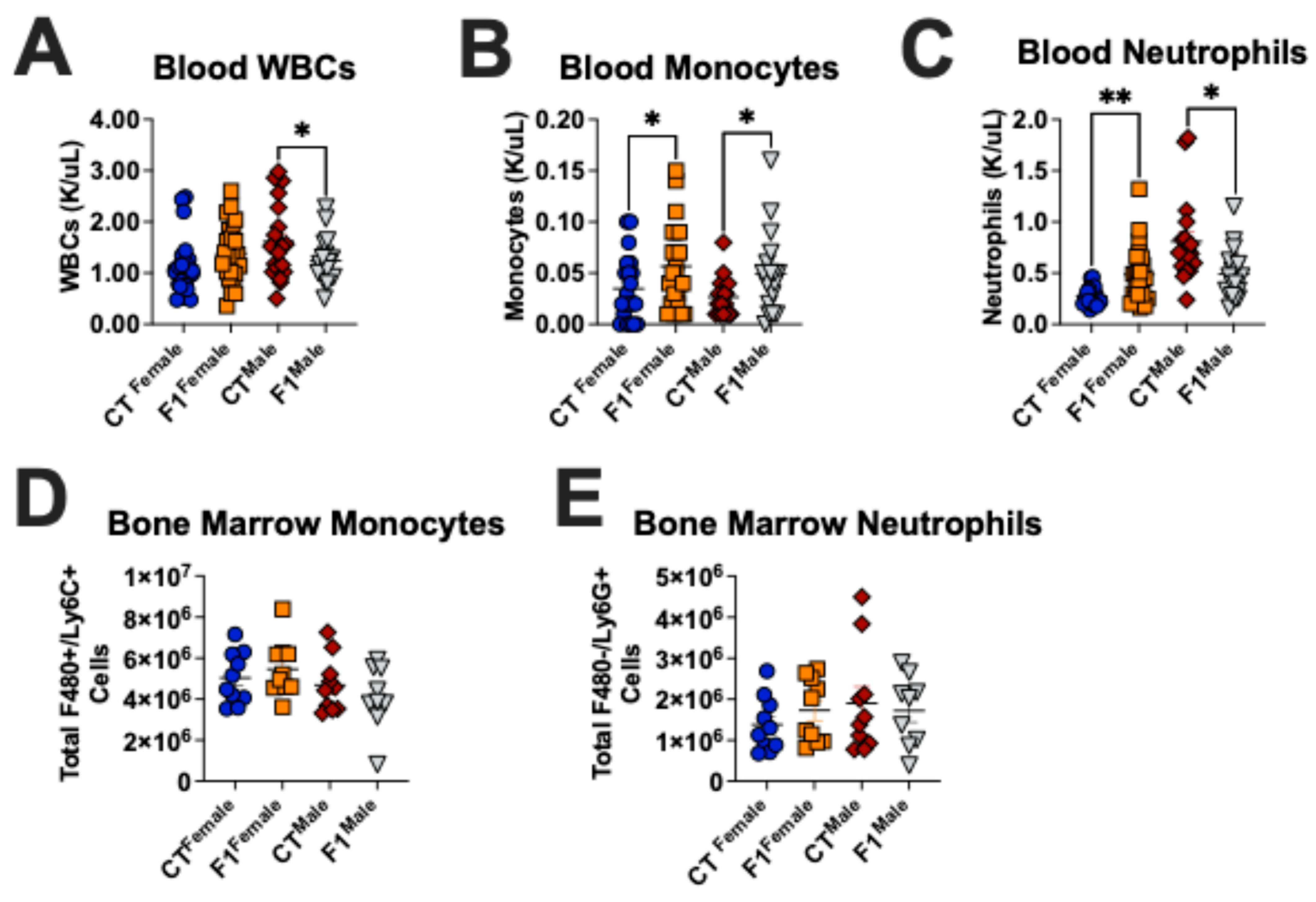
3.1. Prenatal TCDD Exposure Alters the Immune Cell Composition in Adult Mice

Using mice with and without a history of TCDD exposure, we first assessed the presence of sentinel innate immune cells within the bone marrow, blood, and peritoneal cavity in the absence of infection. As shown in Figure 1A and Figure S1, we did not observe significant differences in the numbers of monocytes, macrophages, and neutrophils in the bone marrow of F1 mice compared to CT mice. Using a complete blood count (CBC) analysis, we observed a significant decrease in circulating neutrophils in the blood of male, but not female, F1 mice (Figure 1B). Analysis of peritoneal cell populations revealed a reduction of monocytes and macrophages in F1 male mice (Figure 1C and Figure S2). Interestingly, we specifically observed a decrease in large peritoneal macrophages (LPMs) in F1 males. These data suggest that in utero toxicant exposure induces alterations in innate immune cells that persist into adulthood.

3.2. Prenatal TCDD Exposure Exacerbates Pseudomonas aeruginosa Infection in Adult Mice

To grasp a more in-depth understanding of the immunological changes that are derived from an in utero toxicant exposure, we used an established Pseudomonas aeruginosa infection model to compare the innate immune response in our toxicant-exposed F1 mice to our naïve wild-type CT mice. Using both male and female mice, we examined the potential for sex-specific differences in homeostasis and bacterial clearance in our experimental groups. Adult F1 and CT mice were inoculated with 1 × 10⁸ CFU P. aeruginosa via intraperitoneal (i.p.) injection; 6 h later, we harvested blood and peritoneal fluid from these mice in addition to obtaining their core body temperatures (Figure 2A). Our studies revealed that male, but not female, F1 mice had a significantly higher bacterial burden than CT mice 6 h post-infection (Figure 2B). All mice showed a significant decrease in core body temperature at 6 h after infection. However, F1 male mice showed a significantly larger decrease in core body temperature compared to CT male mice, an effect that was not observed in the female cohort (Figure 2C,D). These observations indicate that an in utero toxicant exposure hinders the ability of mice to effectively and efficiently clear a bacterial infection well into adulthood, even more so in male mice.

3.3. Prenatal TCDD Exposure Stunts Innate Immune Cell Recruitment During Infection in Adult Mice

To begin to parse out TCDD-mediated immunotoxicities in relation to the innate immune system, we used flow cytometry to quantify key populations of innate immune cells in the peritoneal cavity of mice 6 h after infection. The number of monocytes recruited to the site of infection was significantly lower in F1 female and male mice compared to CT mice (Figure 3A). F1 males also exhibited a significantly lower number of neutrophils in the peritoneal cavity compared to their CT counterparts (Figure 3B). Regardless of sex, macrophage numbers were not significantly different between the groups (Figure 3C). Here, we see a clear difference in the immune cell composition between male and female mice; furthermore, we observed an overall reduction in the percentage of innate immune cells in the peritoneal cavity of F1 mice compared to CT mice (Figure 3D). While we have noted significant changes between F1 male and female mice compared to the CT mice, each of the immune cell populations behaved as expected in response to the infection. The number of monocytes and neutrophils increased after infection, whereas the number of macrophages decreased, which is consistent with current findings in the literature (Figure 3E).
After noting the decrease in innate immune cells at the site of infection, we sought to determine if in utero TCDD exposure altered immune cell trafficking by affecting the circulation of immune cells in the blood. Complete blood count (CBC) analysis revealed that F1 males exhibited a significant decrease in circulating white blood cells (WBCs) compared to CT males (Figure 4A and Figure S3A show data including outliers). We next looked specifically at monocytes and found that F1 mice had increased concentrations of circulating monocytes compared to CT mice (Figure 4B and Figure S3B show data including outliers). F1 female mice also had an increased concentration of circulating neutrophils compared to CT females (Figure 4C and Figure S3C show data including outliers). Conversely, F1 males exhibited a significantly decreased concentration of circulating neutrophils compared to CT males. We observed no difference in the total number of monocytes and neutrophils in the bone marrow of F1 mice compared to CT mice (Figure 4D,E). These data suggest that the lack of innate leukocytes at the site of infection may be due to the inability of EDC-exposed immune cells to properly respond to migratory cues.

3.4. Prenatal TCDD Exposure Alters Cytokine Production in Adult Mice

We next sought to investigate how in utero TCDD exposure affects the production and secretion of inflammatory mediators. Six hours after inoculating CT and F1 mice with Pseudomonas aeruginosa, we collected peritoneal fluid from all mice in our experiment to assess the concentration of an array of cytokines. Of the 31 cytokines that were tested, we observed insignificant differences in the majority of the cytokines that were related to monocyte and neutrophil activities. However, we did observe a significant decrease in tumor necrosis factor alpha (TNFα) and monocyte chemoattractant protein (MCP)-1 and an increase in granulocyte-macrophage colony-stimulating factor (GM-CSF) in F1 males compared to CT males (Figure 5A,B). After assessing the local cytokine production, we examined the systemic effect of the infection by assessing the concentration of numerous cytokines in the plasma of CT and F1 mice. Data revealed a significant increase in the concentration of interleukin (IL)-1α, IL-1β, IL-10, macrophage inflammatory protein (MIP)-1α, and MIP-1β in the plasma of F1 males compared to CT males 6 h after infection (Figure 6A,B). Interestingly, we also observed a decrease in cytokine production in CT males compared to CT females, further highlighting sex-specific differences in inflammatory responses.

4. Discussion

A major source of EDC exposure is through dietary intake of contaminated foods; hence our use of an oral gavage mouse model of in utero toxicant exposure for translational relevance. Public health agencies, including the United States Environmental Protection Agency, World Health Organization, and National Toxicology Program, have expressed immense concern for the effects of EDCs on human health, especially during pregnancy and critical stages of development. Determining the direct contribution of EDCs to adverse human health would require purposefully exposing humans to chemicals and monitoring their outcomes, which is highly unethical. Therefore, our current understanding of the effects of EDC exposure on mammalian health is mostly derived from epidemiology studies following incidents of mass exposure and animal studies.
In vivo and ex vivo studies show that EDCs bioaccumulate at the maternal-fetal interface, especially in the placenta, likely due to the abundance of hormone receptors [19,20,21]. Studies have also shown that the placenta is not effective at preventing EDCs from entering fetal circulation [22,23]. In fact, due to the biochemical features of EDCs, they can reach the fetus through transplacental absorption and transport, where they have been reported to have a prolonged half-life [24].
Since environmental toxicants can cause maladaptive changes to endocrine signaling within multiple cell types, it is not surprising that TCDD can negatively affect cells of the innate immune system. The innate immune system is comprised of cells, proteins, and mechanisms that serve as the body’s first line of defense against invading pathogens. Unlike the adaptive immune system, the innate immune system does not require pathogen-specific recognition to elicit a response. While literature regarding the effects of EDCs on innate immune cells remains fairly scarce, studies have shown extensive toxic effects on cells of the adaptive immune system [17,18]. The prenatal innate immune system is exceptionally vulnerable, and the trajectory of postnatal inflammatory responses is largely established at this stage of development [25,26]. In the current study, we combined an established mouse model of infection with an established mouse model of toxicant exposure to explore the potential impact of in utero TCDD exposure on the adult innate immune system.
Prior to infection, we observed a decrease in monocytes and LPMs in the peritoneal cavity of F1 males compared to CT males. Peritoneal macrophages are comprised of two populations: small peritoneal macrophages (SPMs) and LPMs. LPMs are tissue-resident macrophages that are thought to originate from embryonic precursors. At steady state, LPMs are the most abundant subset, and their function is to maintain homeostasis by performing repair and defense functions. In the presence of an inflammatory challenge, LPMs exit the peritoneal cavity, and the inflammatory SPMs become the most abundant macrophage population [27]. Recent evidence suggests that LPMs are replaced by monocyte-derived, tissue-resident macrophages [28]. Collectively, the decrease we observed in peritoneal monocytes and LPMs may provide evidence to explain the increased risk of inflammatory diseases previously observed in our mouse model of in utero TCDD exposure compared to unexposed mice.
After establishing a Pseudomonas aeruginosa infection in adult mice, we found that toxicant-exposed F1 male mice exhibited a significantly compromised ability to clear the infection compared to their unexposed counterparts. Previous studies have indicated a robust immune response within 6 h of a Pseudomonas aeruginosa infection [29]; thus, we chose to use this timepoint for our studies. In support of these findings, Lawrence and colleagues have reported that a similar developmental TCDD exposure inhibits successful resolution of an influenza A virus infection in adult mice [17]. Human studies have also identified a positive association between prenatal exposure to polychlorinated biphenyls (PCBs), a family of environmental toxicants, and an increased incidence of postnatal infections (i.e., lower respiratory tract infections) [30,31].
Herein, we demonstrated that, in response to infection, both male and female F1 mice exhibit changes in the composition of circulating and peritoneal immune cells compared to CT mice. Interestingly, F1 males had a significantly lower concentration of WBCs compared to CT males, which correlates with their increased infectious phenotypes and reduced ability to clear the infection. Although we observed minimal and inconsistent trends in the changes in cytokine production in the peritoneal cavity of F1 mice compared to CT mice in response to the peritoneal infection, analyses of cytokines in the plasma revealed more interesting results (Figures S4 and S5). We observed a significant increase in the concentration of numerous cytokines in the plasma of F1 males compared to CT males. This increase is likely attributed to the elevated bacterial burden in these animals, which leads to the increased production, secretion, and activity of inflammatory mediators. These data also revealed an obvious sex-specific difference in cytokine production, especially when comparing CT females to CT males. Here, we observed a significant reduction in the concentration of several cytokines in the plasma of male mice compared to female mice. Perhaps these findings can be explained by the sex differences in genetic and hormonal mediators that contribute to the observed dimorphic innate immune responses, particularly cytokine production [32]. Overall, we demonstrate that in utero TCDD exposure contributes to an impaired innate immune cell response to infection in adult mice. Consistent with these data, several studies have concluded that the premature activation of immune cells in the absence of antigens by EDC exposures results in a blunted immune cell presence and response to infection [33,34].
Taken together, our study indicates that in utero EDC exposure compromises the innate immune response to a common bacterial infection. Similarly, previous work has demonstrated that TCDD exposure results in immunosuppression, and it has been correlated with diminished host resistance to an alternate infection [35,36]. Other studies have highlighted the ability of EDCs to activate immune cells to produce cytokines and co-stimulatory molecules and upregulate major histocompatibility complex (MHC) class II molecules in the absence of infectious and/or stimulating pathogens [33,37,38,39]. Thus, studies from multiple laboratories, including this study, demonstrate a reduced ability of toxicant-exposed immune cells to respond to a subsequent infection.

5. Conclusions

In conclusion, our results demonstrate that developmental toxicant exposure results in a significant decrease in the number of innate leukocytes at the site of infection in adult mice compared to unexposed mice. This reduction in immune cells, particularly phagocytes, correlated with increased infectious phenotypes in toxicant-exposed animals, particularly in males. Furthermore, toxicant-exposed mice displayed an altered ability to produce cytokines necessary for a robust immune response. Taken together, our results indicate that in utero EDC exposure induces immunomodulatory effects that impair the innate immune response to a bacterial infection in adult offspring, particularly in males. Although determining the cause of sex-specific differences in TCDD-associated immune effects is beyond the scope of the current manuscript, this will be an important avenue of investigation in future studies. Future work will interrogate the contribution of cellular signaling pathways (such as the aryl hydrocarbon receptor) to these phenotypes. Investigating endocrine signaling pathways will also be important in understanding the sex-specific differences observed in this study.

Supplementary Materials

The following supporting information can be downloaded at: https://www.mdpi.com/article/10.3390/microbiolres16050091/s1, Figure S1: Prenatal TCDD exposure does not alter the composition of innate immune cells in the bone marrow of adult mice; Figure S2: Prenatal TCDD exposure alters the composition of innate immune cells in the peritoneal cavity of adult mice; Figure S3: Prenatal TCDD exposure alters the concentration of blood circulating cells in response to infection in adult mice; Figure S4: Prenatal TCDD exposure alters the concentration of cytokines in the peritoneal cavity of adult mice in response to infection; Figure S5: Prenatal TCDD exposure alters the concentration of cytokines in the plasma of adult mice in response to infection.

Author Contributions

Conceptualization, V.R.S., J.K.B., K.L.B.-T., J.A.G., E.R.S. and K.G.O.; methodology, V.R.S., J.K.B., K.L.B.-T., E.R.S. and K.G.O.; validation, V.R.S., J.K.B. and E.R.S.; formal analysis, V.R.S., J.K.B., X.D.D., M.A.O., M.A.M., J.A.G. and E.R.S.; investigation, V.R.S., J.K.B., X.D.D., M.A.O., M.A.M., S.A., J.T.R., J.A.G. and E.R.S.; resources, J.K.B., K.L.B.-T., J.A.G., E.R.S. and K.G.O.; data curation, V.R.S.; writing—original draft preparation, V.R.S.; writing—review and editing, V.R.S., J.K.B., K.L.B.-T., S.A., J.T.R., J.A.G., E.R.S. and K.G.O.; visualization, V.R.S. and S.A.; supervision, J.K.B., J.A.G., E.R.S. and K.G.O.; project administration, V.R.S.; funding acquisition, J.A.G., E.R.S. and K.G.O. All authors have read and agreed to the published version of the manuscript.

Funding

This work was supported by the National Institutes of Health (NIH) Training Program in Environmental Toxicology under award numbers TOX T32 ES007028 and F31ES034957 (to V.R.S.), the Bill and Melinda Gates Foundation, U.S. Environmental Protection Agency #R83950101, R01HD096147 (to K.G.O.), Department of Veterans Affairs Merit Award I01BX005352 Office of Research (to J.A.G. and K.G.O.), and the National Institute of Allergy and Infectious Diseases under the award number R01AI151210 (E.R.S.). In Utero exposure to 2,3,7,8-Tetrachlorodibenzo-p-dioxin impairs the ability of mice to clear a Pseudomonas aeruginosa infection in adulthood, which was developed under the Assistance Agreement No. RD83950101 awarded by the U.S. Environmental Protection Agency (to K.G.O.). It has not been formally reviewed by the EPA. The views expressed in this document are solely those of the authors and do not necessarily reflect those of the agency. EPA does not endorse any products or commercial services mentioned in this publication.

Institutional Review Board Statement

The animal study protocol was approved by the Institutional Review Board (or Ethics Committee) of Vanderbilt University Medical Center (protocol code: M1800068 and date of approval: 04 April 2024; protocol code: M2300037 and date of approval: May 2023).

Informed Consent Statement

Not applicable.

Data Availability Statement

The original contributions presented in this study are included in the article/Supplementary Material. Further inquiries can be directed to the corresponding author(s).

Conflicts of Interest

The authors declare no conflict of interest.

References

  1. Diamanti-Kandarakis, E.; Bourguignon, J.P.; Giudice, L.C.; Hauser, R.; Prins, G.S.; Soto, A.M.; Zoeller, R.T.; Gore, A.C. Endocrine-disrupting chemicals: An Endocrine Society scientific statement. Endocr. Rev. 2009, 30, 293–342. [Google Scholar] [CrossRef] [PubMed]
  2. Bruner-Tran, K.L.; Osteen, K.G. Developmental exposure to TCDD reduces fertility and negatively affects pregnancy outcomes across multiple generations. Reprod. Toxicol. 2011, 31, 344–350. [Google Scholar] [CrossRef] [PubMed]
  3. Bruner-Tran, K.L.; Duleba, A.J.; Taylor, H.S.; Osteen, K.G. Developmental Toxicant Exposure Is Associated with Transgenerational Adenomyosis in a Murine Model. Biol. Reprod. 2016, 95, 73. [Google Scholar] [CrossRef] [PubMed]
  4. Adegoke, E.O.; Rahman, M.S.; Park, Y.-J.; Kim, Y.J.; Pang, M.-G. Endocrine-Disrupting Chemicals and Infectious Diseases: From Endocrine Disruption to Immunosuppression. Int. J. Mol. Sci. 2021, 22, 3939. [Google Scholar] [CrossRef]
  5. Kajta, M.; Wójtowicz, A.K. Impact of endocrine-disrupting chemicals on neural development and the onset of neurological disorders. Pharmacol. Rep. 2013, 65, 1632–1639. [Google Scholar] [CrossRef]
  6. Papalou, O.; Kandaraki, E.A.; Papadakis, G.; Diamanti-Kandarakis, E. Endocrine Disrupting Chemicals: An Occult Mediator of Metabolic Disease. Front. Endocrinol. 2019, 10, 112. [Google Scholar] [CrossRef]
  7. Calkins, K.; Devaskar, S.U. Fetal origins of adult disease. Curr. Probl. Pediatr. Adolesc. Health Care 2011, 41, 158–176. [Google Scholar] [CrossRef]
  8. Barker, D.J. The developmental origins of adult disease. J. Am. Coll. Nutr. 2004, 23 (Suppl. 6), 588s–595s. [Google Scholar] [CrossRef] [PubMed]
  9. Zamora-León, P. Are the Effects of DES Over? A Tragic Lesson from the Past. Int. J. Environ. Res. Public Health 2021, 18, 10309. [Google Scholar] [CrossRef]
  10. Othman, N.; Ismail, Z.; Selamat, M.I.; Sheikh Abdul Kadir, S.H.; Shibraumalisi, N.A. A Review of Polychlorinated Biphenyls (PCBs) Pollution in the Air: Where and How Much Are We Exposed to? Int. J. Environ. Res. Public Health 2022, 19, 13923. [Google Scholar] [CrossRef]
  11. Kim, Y.H.; Warren, S.H.; Kooter, I.; Williams, W.C.; George, I.J.; Vance, S.A.; Hays, M.D.; Higuchi, M.A.; Gavett, S.H.; DeMarini, D.M.; et al. Chemistry, lung toxicity and mutagenicity of burn pit smoke-related particulate matter. Part. Fibre Toxicol. 2021, 18, 45. [Google Scholar] [CrossRef] [PubMed]
  12. Aurell, J.; Gullett, B.K.; Yamamoto, D. Emissions from open burning of simulated military waste from forward operating bases. Environ. Sci. Technol. 2012, 46, 11004–11012. [Google Scholar] [CrossRef]
  13. Mokshagundam, S.; Ding, T.; Rumph, J.T.; Dallas, M.; Stephens, V.R.; Osteen, K.G.; Bruner-Tran, K.L. Developmental 2,3,7,8-tetrachlorodibenzo-p-dioxin exposure of either parent enhances the risk of necrotizing enterocolitis in neonatal mice. Birth Defects Res. 2020, 112, 1209–1223. [Google Scholar] [CrossRef] [PubMed]
  14. Nayyar, T.; Bruner-Tran, K.L.; Piestrzeniewicz-Ulanska, D.; Osteen, K.G. Developmental exposure of mice to TCDD elicits a similar uterine phenotype in adult animals as observed in women with endometriosis. Reprod. Toxicol. 2007, 23, 326–336. [Google Scholar] [CrossRef]
  15. Rumph, J.T.; Rayford, K.J.; Stephens, V.R.; Ameli, S.; Nde, P.N.; Osteen, K.G.; Bruner-Tran, K.L. A Preconception Paternal Fish Oil Diet Prevents Toxicant-Driven New Bronchopulmonary Dysplasia in Neonatal Mice. Toxics 2021, 10, 7. [Google Scholar] [CrossRef] [PubMed]
  16. Rumph, J.T.; Stephens, V.R.; Ameli, S.; Brown, L.K.; Rayford, K.J.; Nde, P.N.; Osteen, K.G.; Bruner-Tran, K.L. A Paternal Fish Oil Diet Preconception Reduces Lung Inflammation in a Toxicant-Driven Murine Model of New Bronchopulmonary Dysplasia. Mar. Drugs 2023, 21, 161. [Google Scholar] [CrossRef]
  17. Post, C.M.; Myers, J.R.; Winans, B.; Lawrence, B.P. Postnatal administration of S-adenosylmethionine restores developmental AHR activation-induced deficits in CD8+ T cell function during influenza A virus infection. Toxicol. Sci. 2023, 192, 233–246. [Google Scholar] [CrossRef]
  18. Marshall, N.B.; Kerkvliet, N.I. Dioxin and immune regulation: Emerging role of aryl hydrocarbon receptor in the generation of regulatory T cells. Ann. N. Y Acad. Sci. 2010, 1183, 25–37. [Google Scholar] [CrossRef]
  19. Augustowska, K.; Gregoraszczuk, E.E.; Grochowalski, A.; Milewicz, T.; Mika, M.; Krzysiek, J.; Chrzaszcz, R. Comparison of accumulation and altered steroid secretion by placental tissue treated with TCDD and natural mixture of PCDDs-PCDFs. Reproduction 2003, 126, 681–687. [Google Scholar] [CrossRef]
  20. Abbott, B.D.; Birnbaum, L.S.; Diliberto, J.J. Rapid distribution of 2,3,7,8-tetrachlorodibenzo-p-dioxin (TCDD) to embryonic tissues in C57BL/6N mice and correlation with palatal uptake in vitro. Toxicol. Appl. Pharmacol. 1996, 141, 256–263. [Google Scholar] [CrossRef]
  21. Gingrich, J.; Ticiani, E.; Veiga-Lopez, A. Placenta Disrupted: Endocrine Disrupting Chemicals and Pregnancy. Trends Endocrinol. Metab. 2020, 31, 508–524. [Google Scholar] [CrossRef] [PubMed]
  22. Li, L.X.; Chen, L.; Meng, X.Z.; Chen, B.H.; Chen, S.Q.; Zhao, Y.; Zhao, L.F.; Liang, Y.; Zhang, Y.H. Exposure levels of environmental endocrine disruptors in mother-newborn pairs in China and their placental transfer characteristics. PLoS ONE 2013, 8, e62526. [Google Scholar] [CrossRef]
  23. Chen, M.L.; Chang, C.C.; Shen, Y.J.; Hung, J.H.; Guo, B.R.; Chuang, H.Y.; Mao, I.F. Quantification of prenatal exposure and maternal-fetal transfer of nonylphenol. Chemosphere 2008, 73 (Suppl. 6), S239–S245. [Google Scholar] [CrossRef]
  24. Gingrich, J.; Pu, Y.; Ehrhardt, R.; Karthikraj, R.; Kannan, K.; Veiga-Lopez, A. Toxicokinetics of bisphenol A, bisphenol S, and bisphenol F in a pregnancy sheep model. Chemosphere 2019, 220, 185–194. [Google Scholar] [CrossRef]
  25. Sabic, D.; Koenig, J.M. A perfect storm: Fetal inflammation and the developing immune system. Pediatr. Res. 2020, 87, 319–326. [Google Scholar] [CrossRef]
  26. Apostol, A.C.; Jensen, K.D.C.; Beaudin, A.E. Training the Fetal Immune System Through Maternal Inflammation—A Layered Hygiene Hypothesis. Front. Immunol. 2020, 11, 123. [Google Scholar] [CrossRef]
  27. Cassado Ados, A.; D’Império Lima, M.R.; Bortoluci, K.R. Revisiting mouse peritoneal macrophages: Heterogeneity, development, and function. Front. Immunol. 2015, 6, 225. [Google Scholar] [CrossRef] [PubMed]
  28. Ardavín, C.; Alvarez-Ladrón, N.; Ferriz, M.; Gutiérrez-González, A.; Vega-Pérez, A. Mouse Tissue-Resident Peritoneal Macrophages in Homeostasis, Repair, Infection, and Tumor Metastasis. Adv. Sci. (Weinh) 2023, 10, e2206617. [Google Scholar] [CrossRef] [PubMed]
  29. McBride, M.A.; Caja, K.R.; Patil, T.K.; Owen, A.M.; Luan, L.; Bohannon, J.K.; Hernandez, A.; Stothers, C.L.; Trenary, I.A.; Rahim, M.; et al. Immunoresponsive gene 1 facilitates TLR4 agonist-induced augmentation of innate antimicrobial immunity. J. Leukoc. Biol. 2025, 117, qiae198. [Google Scholar] [CrossRef]
  30. Dallaire, F.; Dewailly, E.; Vézina, C.; Muckle, G.; Weber, J.P.; Bruneau, S.; Ayotte, P. Effect of prenatal exposure to polychlorinated biphenyls on incidence of acute respiratory infections in preschool Inuit children. Environ. Health Perspect. 2006, 114, 1301–1305. [Google Scholar] [CrossRef]
  31. Stølevik, S.B.; Nygaard, U.C.; Namork, E.; Haugen, M.; Kvalem, H.E.; Meltzer, H.M.; Alexander, J.; van Delft, J.H.; Loveren, H.; Løvik, M.; et al. Prenatal exposure to polychlorinated biphenyls and dioxins is associated with increased risk of wheeze and infections in infants. Food Chem. Toxicol. 2011, 49, 1843–1848. [Google Scholar] [CrossRef] [PubMed]
  32. Klein, S.L.; Flanagan, K.L. Sex differences in immune responses. Nat. Rev. Immunol. 2016, 16, 626–638. [Google Scholar] [CrossRef] [PubMed]
  33. Stephens, V.R.; Moore, R.E.; Spicer, S.K.; Talbert, J.A.; Lu, J.; Chinni, R.; Chambers, S.A.; Townsend, S.D.; Manning, S.D.; Rogers, L.M.; et al. Environmental Toxicant Exposure Paralyzes Human Placental Macrophage Responses to Microbial Threat. ACS Infect. Dis. 2023, 9, 2401–2408. [Google Scholar] [CrossRef]
  34. Domínguez-Acosta, O.; Vega, L.; Estrada-Muñiz, E.; Rodríguez, M.S.; Gonzalez, F.J.; Elizondo, G. Activation of aryl hydrocarbon receptor regulates the LPS/IFNγ-induced inflammatory response by inducing ubiquitin-proteosomal and lysosomal degradation of RelA/p65. Biochem. Pharmacol. 2018, 155, 141–149. [Google Scholar] [CrossRef] [PubMed]
  35. Fiorito, F.; Santamaria, R.; Irace, C.; De Martino, L.; Iovane, G. 2,3,7,8-tetrachlorodibenzo-p-dioxin and the viral infection. Environ. Res. 2017, 153, 27–34. [Google Scholar] [CrossRef]
  36. Wheeler, J.L.; Martin, K.C.; Resseguie, E.; Lawrence, B.P. Differential consequences of two distinct AhR ligands on innate and adaptive immune responses to influenza A virus. Toxicol. Sci. 2014, 137, 324–334. [Google Scholar] [CrossRef]
  37. Ruby, C.E.; Funatake, C.J.; Kerkvliet, N.I. 2,3,7,8 Tetrachlorodibenzo-p-Dioxin (TCDD) Directly Enhances the Maturation and Apoptosis of Dendritic Cells In Vitro. J. Immunotoxicol. 2005, 1, 159–166. [Google Scholar] [CrossRef]
  38. Vorderstrasse, B.A.; Kerkvliet, N.I. 2,3,7,8-Tetrachlorodibenzo-p-dioxin affects the number and function of murine splenic dendritic cells and their expression of accessory molecules. Toxicol. Appl. Pharmacol. 2001, 171, 117–125. [Google Scholar] [CrossRef]
  39. Simones, T.; Shepherd, D.M. Consequences of AhR activation in steady-state dendritic cells. Toxicol. Sci. 2011, 119, 293–307. [Google Scholar] [CrossRef]
Figure 1. Prenatal TCDD exposure alters the innate immune cell composition in adult mice. (A) Total number of monocytes, neutrophils, and macrophages in the bone marrow of CT and F1 mice. (B) Total number of monocytes and neutrophils circulating in the blood of CT and F1 mice. (C) Total number of monocytes, neutrophils, and macrophages in the peritoneal cavity of CT and F1 mice. Data shown as mean ± SEM with 5 biological replicates. * p < 0.05 and ** p < 0.01 by Student’s t-test.
Microbiolres 16 00091 g001
Figure 2. Prenatal TCDD exposure exacerbates Pseudomonas aeruginosa infection in adult mice. (A) Schematic of experimental workflow. (B) Colony-forming units of Pseudomonas aeruginosa per mL of peritoneal fluid 6 h after inoculation. Core body temperature of control, unexposed (CT), and TCDD-exposed (F1) (C) female and (D) male mice pre- and post-Pseudomonas aeruginosa infection. Data shown as mean ± SEM with 17–23 biological replicates. ** p < 0.01 and **** p < 0.0001 by Student’s t-test.
Microbiolres 16 00091 g002
Figure 3. Prenatal TCDD exposure stunts innate immune cell recruitment during an acute Pseudomonas aeruginosa infection in adult mice. Total number of (A) monocytes, (B) neutrophils, and (C) macrophages in the peritoneal cavity of CT and F1 mice 6 h post-infection. (D) The composition of immune cells as the total percentage in each experimental group. (E) Comparison of the total number of leukocytes in the peritoneal cavity with and without infection. Data shown as mean ± SEM with 14–18 biological replicates. * p < 0.05, ** p < 0.01, *** p < 0.001, and **** p < 0.0001 by Student’s t-test.
Microbiolres 16 00091 g003
Figure 4. Prenatal TCDD exposure alters the concentration of cells circulating in the blood in response to infection in adult mice. Concentration of (A) white blood cells (WBCs), (B) monocytes, and (C) neutrophils in blood of CT and F1 mice 6 h post-infection. Total number of (D) monocytes and (E) neutrophils in the bone marrow of CT and F1 mice 6 h post-infection. Data shown as mean ± SEM with 9–23 biological replicates. * p < 0.05 and ** p < 0.01 by Student’s t-test.
Microbiolres 16 00091 g004
Figure 5. Prenatal TCDD exposure alters the concentration of cytokines in the peritoneal cavity of adult mice in response to infection. (A) Heatmap of cytokines in the peritoneal cavity of CT and F1 mice 6 h after a Pseudomonas aeruginosa infection. (B) Individual bar graphs of cytokines significantly altered by Pseudomonas aeruginosa infection and/or TCDD exposure. Data shown as mean ± SEM with 5 biological replicates. * p < 0.05 and ** p < 0.01 by one-way ANOVA with Tukey’s post-hoc multiple comparison test. # p < 0.05 and ## p < 0.01 by Student’s t-test.
Microbiolres 16 00091 g005
Figure 6. Prenatal TCDD exposure alters the concentration of cytokines in the plasma of adult mice in response to infection. (A) Heatmap of cytokines in the plasma of CT and F1 mice 6 h after a Pseudomonas aeruginosa infection. (B) Individual bar graphs of cytokines significantly altered by Pseudomonas aeruginosa infection and/or TCDD exposure. Data shown as mean ± SEM with 5 biological replicates. * p < 0.05 by one-way ANOVA with Tukey’s post-hoc multiple comparison test. # p < 0.05, ## p < 0.01, and ### p < 0.001 by Student’s t-test.
Microbiolres 16 00091 g006
Disclaimer/Publisher’s Note: The statements, opinions and data contained in all publications are solely those of the individual author(s) and contributor(s) and not of MDPI and/or the editor(s). MDPI and/or the editor(s) disclaim responsibility for any injury to people or property resulting from any ideas, methods, instructions or products referred to in the content.

