Brief Report

Effect of Tilapia Parvovirus (TiPV) on Fish Health: An In Vitro Approach

Aquatic Environmental Biotechnology Division (AEB), ICAR-Central Inland Fisheries Research Institute, Kolkata 700120, India
*
Author to whom correspondence should be addressed.
Microbiol. Res. 2025, 16(3), 68; https://doi.org/10.3390/microbiolres16030068
Submission received: 24 January 2025 / Revised: 25 February 2025 / Accepted: 10 March 2025 / Published: 17 March 2025
(This article belongs to the Special Issue Veterinary Microbiology and Diagnostics)

Abstract

:
Tilapia Parvovirus (TiPV) is a rising pathogen responsible for high mortality in tilapia aquaculture. Understanding TiPV’s pathogenesis is crucial for developing effective management strategies. This study aimed to elucidate TiPV pathogenesis by evaluating its cytotoxic effects on Danio rerio gill (DRG) cell monolayers and its impact on host immune responses. PCR-confirmed TiPV-infected DRG cell monolayers were subjected to an MTT (3-(4,5-dimethylthiazol-2-yl)-2,5-diphenyltetrazolium bromide) assay at 24, 48, 72, and 96 h post-infection to assess cell viability and cytotoxicity. The MTT assay revealed a progressive decline in DRG cell viability over time, with viable cell percentages decreasing from 66.71% at 24 h to 31.28% at 96 h in TiPV-infected cultures, compared to consistently high viability in controls. Simultaneously, quantitative real-time PCR (qPCR) was used to assess the expression of key immune-related genes, including Interleukins (IL-1β, IL-8), Toll-like receptor 7 (TLR7), Major Histocompatibility Complex II (MHC-II), Tumor Necrosis Factor α (TNF-α), Nuclear Factor Kappa B (NF-κB), and Chemokine Receptors (CRs).qPCR analysis showed an upregulation of IL-8, IL-1β, TNF-α, and CRs, indicating an early inflammatory response. However, significant downregulation of TLR7, MHC-II, and NF-κB suggests TiPV’s ability to modulate host immune responses. The results highlight that TiPV induces significant cytotoxicity in DRG cells, leading to severe cellular damage. The virus also alters host immune responses by modulating the expression of key immune genes, which may contribute to its virulence and persistence. These findings enhance our understanding of TiPV pathogenesis and highlight the need for targeted research to develop effective control strategies for TiPV in aquaculture systems.

1. Introduction

Tilapia (Oreochromis spp.) aquaculture is being practiced in more than 100 nations, making it the second-most popular fish species farmed globally, and millions of people benefit from it in terms of their dietary requirements and socio-economic development [1,2]. The tilapia global production is estimated to be 5.88 million metric tonnes per year, and the fish market’s worth is currently more than USD 11 billion. Along with the intensive tilapia farming and commerce, the extensive transportation of fresh fish and associated products between nations fosters the establishment of infections that may cause disease to appear and spread [3,4]. Even though the fish species is resilient and easily able to adapt to the harsh farming environments and modifications to the environment, disease outbreaks, especially the ones prompted by viruses, continue to endanger tilapia productivity [5,6].
Tilapia parvovirus (TiPV) has a single-stranded DNA (ssDNA) genome and a non-enveloped virus encompassing 4269 nucleotides whose complete characterization was performed in China way back in 2019 [7]. The genome of the virus is characterized by two primary ORFs (open reading frames): ORF1 and ORF2. ORF1 encodes the non-structural protein 1 (NS1), while ORF2 is responsible for coding the structural protein VP1. Initially, the virus was assumed to be non-transmissible, as it was first identified in crocodile feces following the consumption of tilapia and in gut samples obtained from healthy tilapia specimens [8]. However, it was later discovered that the virus can be fatal to tilapia, resulting in mortality rates of up to 90% [6,7]. TiPV infections can pose substantial adverse financial consequences owing to the elevated mortality and morbidity rates associated with the outbreak of the disease [9]. Consequently, to ascertain the detrimental impacts of TiPV, elaborative research on the virulence of TiPV and associated pathobiology is also recommended.
Cell lines derived from fish serve as a cutting-edge in vitro toolkit, offering a streamlined and highly efficient platform for evaluating the impact of viruses and toxicants, including nanoparticles, pesticides, xenobiotics, and industrial effluents. Cell lines are indispensable tools for the propagation, purification, and detailed characterization of viruses. For example, the RSIV (Red Sea Bream Iridovirus) and the RBIV (Rock Bream Iridovirus) have been shown to proliferate effectively in BF-2, a fibroblastic cell line derived from the caudal fin of bluegill sunfish, as well as CRF-1, a cell line from the red sea bream Pagrus major [10,11]. These cell lines have been instrumental in studying host susceptibility, conducting functional genomic analyses, and unraveling the complex interactions between hosts and viruses. Recently, TiLV (Tilapia Lake Virus), a significant viral pathogen affecting tilapia, was successfully isolated using the Channa striata E11 cell line. This breakthrough facilitated investigations into whether broodstock experimentally infected could transmit the virus to their reproductive organs, eggs, sperm, and even in vitro fertilized embryos [12,13].
The zebrafish has emerged as a globally recognized scientific model, valued for its genetic similarity to the human genome and an array of advantageous traits, including low maintenance cost, small body size, optical transparency, and high reproductive output [14]. Additionally, zebrafish-derived cell lines have become a classical system for screening viruses and assessing the ecological impacts of harmful compounds such as biocides, nanoplastics, and heavy metals. The Danio rerio gill (DRG) cell line has been employed to gain deeper insights into the intricate dynamics of host–pathogen interactions [15]. Compared to other in vitro models, fish cell lines require only minimal sample volumes for virus proliferation and characterization studies, making them exceptionally efficient [16,17]. Fish cell lines also serve as invaluable tools for evaluating preliminary virulence responses and are widely recognized as reliable alternative models to whole-organism toxicity assessments. Among these methods, the MTT assay (3-(4,5-dimethyl-2-thiazolyl)-2,5-diphenyl-2H-tetrazolium bromide) stands out as one of the most widely employed colorimetric techniques for quantifying cell density. The assay’s principle is reducing yellow soluble MTT into purple insoluble formazan via mitochondrial, non-mitochondrial, and plasma membrane reductases in viable cells [18]. The assay is safe, simple, cost-effective, and compatible with several animal cell lines. Recently, a significant outbreak of viral disease caused by Tilapia Parvovirus (TiPV) was reported, resulting in extensive losses and large-scale mortality among farmed tilapia populations [9].
Although there are few works that reported the isolation and characterization of TiPV [19,20], very limited information is available on the characterization of TiPV response in aquatic organisms using in vitro model fish cell lines. In vitro models have proven indispensable in virological research, offering a robust platform to investigate host–pathogen responses at both cellular and molecular levels [21]. Gills, being a primary target organ for numerous microbial pathogens in fish, are particularly significant in such studies. Hence, in this study, using the DRG cell line, the virus was isolated and characterized. We studied the effects of TiPV on DrG cells by measuring TiPV proliferation and its impact on cell viability and host immune response. This study further understands the host–virus interaction at a molecular level.

2. Materials and Methods

2.1. Isolation of Virus from Infected Samples

During August–September 2023, severe disease outbreaks with high mortality rates were reported from two aquaculture farms in the Indian states of West Bengal and Odisha. These farms, which stocked tilapia (Oreochromis niloticus) along with other fish species, including Indian Major Carps (IMCs), minor carps, and non-native carp species, observed significant mortality exclusively in tilapia. In contrast, the cohabiting species remained unaffected [9]. Fish farmers reported an alarming 75% mortality rate in tilapia, based on questionnaires and passive data collection from fishers during the outbreak period. Clinically, the affected tilapia exhibited pronounced symptoms, including hemorrhages, ulcers, discoloration, and redness around the fins and body surface. To investigate the etiology, fish samples comprising healthy and symptomatic individuals were collected and transported to the Fish Pathology Laboratory at ICAR-Central Inland Fisheries Research Institute (ICAR-CIFRI), Kolkata. Upon arrival, fresh gill and skin smears were prepared to detect ectoparasites using light microscopy, following standard protocols [22]. Tilapia were humanely euthanized using an overdose of MS-222 (Sigma-Aldrich, St. Louis, MO, USA) in accordance with ethical guidelines [23], and post-mortem examinations were immediately conducted to assess gross pathological lesions in internal organs, such as the liver, kidney, spleen, and intestines. Tissue samples from these organs were collected aseptically for further analysis. Tissue samples of infected and non-infected fish were preserved in RNAlater (Sigma-Aldrich, St. Louis, MO, USA) and stored at −80 °C for subsequent molecular studies. Clinical and post-mortem findings were recorded and analyzed following established fish health and disease investigation protocols [24]. All experimental procedures, including animal handling and sampling, were conducted under the approval of the Animal Ethics Committee of the institute, ICAR-CIFRI, Kolkata, India (CIFRI-IAEC/17/2023-24), adhering strictly to institutional ethical guidelines.

2.2. Fish Cell Line Infection

A confluent monolayer of the Danio rerio gill (DRG) cell line, sourced from the National Repository of Fish Cell Lines (NRFC), ICAR-NBFGR, was utilized for virus isolation and susceptibility assays. The cells of DRG were cultured in Lebovitz’s L-15 medium enriched with 10% fetal bovine serum (FBS; Gibco Invitrogen, Waltham, MA, USA) and supplemented with antibiotic–antimycotic solution (1%) (Gibco Life Technologies, Grand Island, NY, USA). The cultures were maintained in 25 cm2 cell culture flasks (Thermo Fisher Scientific, Waltham, MA, USA) at an optimized temperature of 28 °C, adhering to standardized cell culture methodologies [25]. For fish cell line infection, tissue extracts were prepared and inculcated from infected and non-infected tilapia samples (Control). In the infected group, the brain, spleen, and kidney tissue samples were aseptically collected from infected tilapia exhibiting clinical signs. Control samples were collected from apparently healthy, PCR TiPV-negative tilapia. Tissue extracts were prepared using a modified protocol based on George et al. [26]. The tissues were mixed in L-15 media and aseptically homogenized. Later, the suspension was subjected to clarification via refrigerated centrifugation at 4000× g for 15 min. A 0.22 μm membrane filter (Millipore, St. Louis, MO, USA) was used to filter the supernatants in order to eliminate any possible bacterial contamination and cell debris. The clarified tissue extracts from both infected and non-infected tilapia were then inoculated onto DRG monolayers in 25 cm2 culture flasks, while control flasks, which were not inoculated with tissue extracts, were maintained to monitor background cytopathic effects (CPE), including cell shrinkage, rounding, and cell fusion with cytoplasmic vacuolization [7]. All flasks were incubated (28 °C) and observed daily using an inverted microscope for the development of CPE, which was indicative of viral infection. CPE was observed, and cell culture supernatants were collected and passaged onto fresh DRG monolayers to confirm viral replication and the reproducibility of CPE. This second passage helped verify the presence of a viral agent, as consistent CPE after repeated inoculation is a strong indicator of viral activity [27]. The entire procedure was conducted under sterile conditions to prevent contamination and ensure the accuracy of virus isolation.

2.3. Confirmation of Virus Presence in Cell Line by PCR

Freeze–thaw lysis, low-speed centrifugation (400× g for 5 min), and filtration through a 0.2 μm pore-size syringe filter were used to extract virus stocks from infected DRG monolayers. Next, using the QIAamp DNA Mini Kit (QIAGEN, Hilden, Germany) in accordance with the manufacturer’s instructions, the total genomic DNA was extracted from the virus stocks. A UV–visible spectrophotometer was employed to quantify the DNA content (A51119700C Multiskan SkyHigh, Thermo Scientific, Waltham, MA, USA). A nanodrop device (Eppendorf, Hamburg, Germany) was used to evaluate the quality of the extracted DNA after it had been analyzed on a 1.2% agarose gel. PCR amplification was performed using primers specific to Tilapia Parvovirus on a GeneAmp PCR System 9700 thermal cycler (Applied Biosystems, Carlsbad, CA, USA) (Table 1). For Tilapia Parvovirus, the anticipated PCR products at 534 bp showed positive results. A total of 5 μL of 10× PCR buffer, 1 μL of 50 mM MgCl2, 1 μL of 10 mM dNTP (Sigma, USA), 1 μL of 10 pmol of each primer, 100 ng of extracted genomic DNA, and 1 U Taq DNA polymerase (Sigma, St. Louis, MO, USA) made up the final volume of the PCR reaction mixture, which was kept at 50 μL. A total of 35 cycles of denaturation (94 °C for 30 s), annealing (52 °C for 1 min), extension (72 °C for 1.50 min), and final extension (7 min at 72 °C) comprised the thermal condition. A 1.2% agarose gel was used to visualize the amplified products. An ABI 3730 xl capillary sequencer (Applied Biosystem, MA, USA) was used to sequence the amplified gene both forward and backward. Forward and reverse sequences were aligned using DNA Baser version 7.0.0 to generate a contig, which was subsequently uploaded to NCBI GenBank under the accession number OR714781.

2.4. Cell Viability Assay (MTT Assay) to Determine the Virus Virulence

The cell viability assay was performed at different time intervals at 24, 48, 72, and 96 h after infection. Using the supernatant from the second virus passage with a titer of 104.⁰ TCID50/mL (tissue culture infectious dose 50%). In brief, the virus production in the infected supernatant was determined by measuring the TCID50 using the endpoint dilution assay. In short, DRG cells were seeded with 10% FBS in 96-well plates at densities that reached near 100% confluence in 24 h. The cells were then washed with DPBS, and the medium was exchanged for Advanced MEM. The virus suspension was serially diluted with at least 8 replicates per concentration. The 96-well plates were incubated at 28 °C with 5% CO2 and observed daily for CPE. Once a clear CPE was visible, positive wells were identified, and titers were calculated based on the Spearman and Kärber algorithm [29,30]. The MTT assay kit (Abcam, Cambridge, UK) was employed, following the manufacturer’s protocol. In brief, both control and infected DRG cell monolayers (1 × 10⁴ cells) in L-15 medium were seeded in triplicate into a 96-well microtiter plate and incubated for 24 h at 28 °C. On a subsequent day, the medium was removed, and 50 µL of serum-free L-15 medium along with 50 µL of MTT reagent was added to each well. The plate was incubated for 3 h at 28 °C. Following incubation, each well received 50 µL of MTT solvent. The plate was then covered with foil and gently agitated for 15 min on an orbital shaker. Lastly, a microplate reader (Bio-Rad, Hercules, CA, USA) was used to measure the absorbance at 590 nm.

2.5. Investigation of Cellular Immune Gene Response Against Viral Infection

Following virus infection of the DRG monolayers, total RNA was isolated at 24, 48, 72, and 96 h (different time points) to assess immune gene expression. Concurrently, control DRG monolayers were also processed for RNA extraction to serve as a comparative baseline. RNA from both the control and virus-infected DRG cells was extracted using TRI-reagent (Sigma-Aldrich, St. Louis, MO, USA), adhering to the manufacturer’s guidelines. The RNA concentration of each sample was determined after DNase I treatment (Turbo DNA-free kit, Thermo Scientific, Waltham, MA, USA) using a NanoDrop spectrophotometer (Eppendorf AG 22331, Hamburg, Germany). RNA integrity was evaluated by performing electrophoresis on a 1% agarose gel. To synthesize cDNA, the RNA samples were reverse transcribed using a First Strand cDNA Synthesis Kit (K1622, Thermo Scientific, USA), following the manufacturer’s instructions. The synthesized cDNA samples were stored at −20 °C until further use. The housekeeping β-actin gene was used for the initial amplification of the cDNA, followed by the quantification of immune-related genes, including Toll-like receptors (TLR7), Interleukins (IL-8, IL-1β), Nuclear Factor Kappa B (NF-κB), Major Histocompatibility Complex (MHC-II), Chemokine Receptors (CRs), and Tumor Necrosis Factor α (TNF-α) using Real-Time PCR on the StepOnePlus system (Applied Biosystems, Woburn, MA, USA). Each amplification reaction was performed with a 20 μL reaction volume containing 2 μL of cDNA, 1 μL of forward and reverse primers (5 pmol each), 10 μL of 2 × SYBR Green I mix, and 6 μL of NFW (nuclease-free water). The PCR cycling conditions included an initial denaturation at 95 °C for 5 min, followed by 40 amplification cycles (95 °C for 10 s, gene-specific annealing temperatures for 10 s, and extension at 72 °C for 10 s). A melt curve analysis was performed to confirm the amplification specificity, with a heating protocol of 95 °C for 5 s, 65 °C for 1 min, and 97 °C for 1 min. The samples were then cooled to 40 °C for 10 s. Gene expression levels were calculated as fold changes relative to the housekeeping β-actin gene using the 2−ΔΔCT method.

2.6. Statistical Analysis

Cell viability data were subjected to an arcsine transformation to meet the assumptions of homoscedasticity and normality. The transformed data were subsequently analyzed using a one-way analysis of variance (ANOVA), followed by Duncan’s multiple range test, with the Statistical Package for the Social Sciences (SPSS) version 24.0. Gene expression results were presented as fold changes relative to the housekeeping gene (β-actin). The expression level of the control group (cells inoculated with tissue extracts from non-infected tilapia) was assigned a value of 1.0, and all infected group expression ratios were calculated relative to this control. A one-tailed Student’s t-test was conducted to evaluate significant differences in expression levels between the control and infected groups. A significance threshold was established at p ≤ 0.05.

3. Results

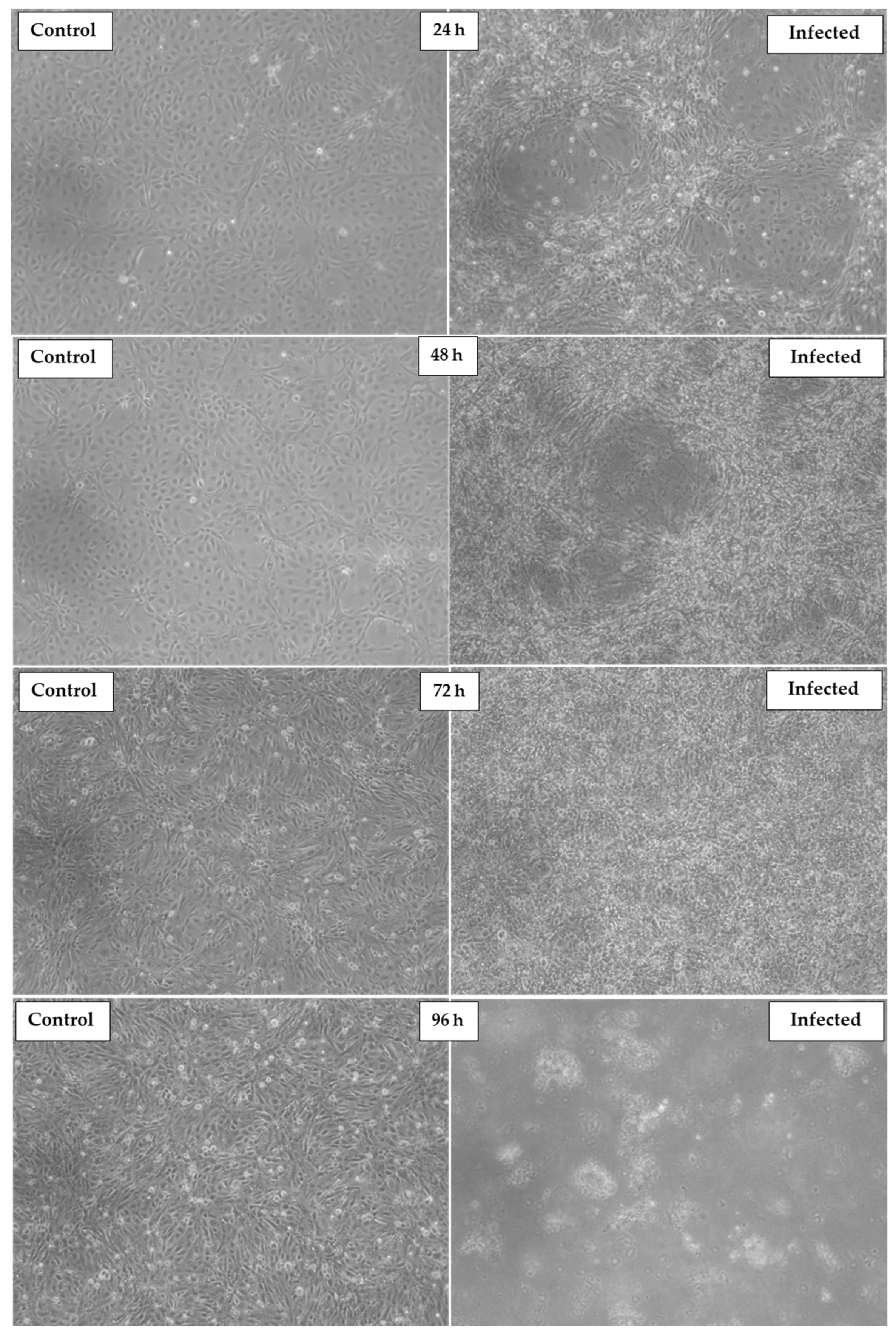
3.1. Virus Isolation in Cell Line

Tissue homogenates were added to the DRG cell line during early virus isolation, resulting in noticeable changes in cell morphology within two days post-infection (dpi). By four dpi, a pronounced cytopathic effect (CPE) was observed, characterized by cell shrinkage, rounding, cell fusion, and the presence of cytoplasmic vacuolization. The CPE progressively intensified, leading to significant monolayer disruption, with over 60% of the cells detaching by four dpi. These morphological changes align with typical viral cytopathic responses as described in similar studies [26]. DNA was extracted from the infected cell pellet to confirm the presence of the viral agent, and PCR analysis was performed. The PCR yielded a specific positive amplicon for Tilapia Parvovirus (TiPV), confirming the successful isolation of the virus from the infected tissues. The DRG cell line, when inoculated with tissue extracts from non-infected tilapia, tested negative for TiPV in PCR analysis. These results substantiate the susceptibility of the DRG cell line to TiPV infection, offering a valuable in vitro model for studying TiPV pathogenesis and host–virus interactions.

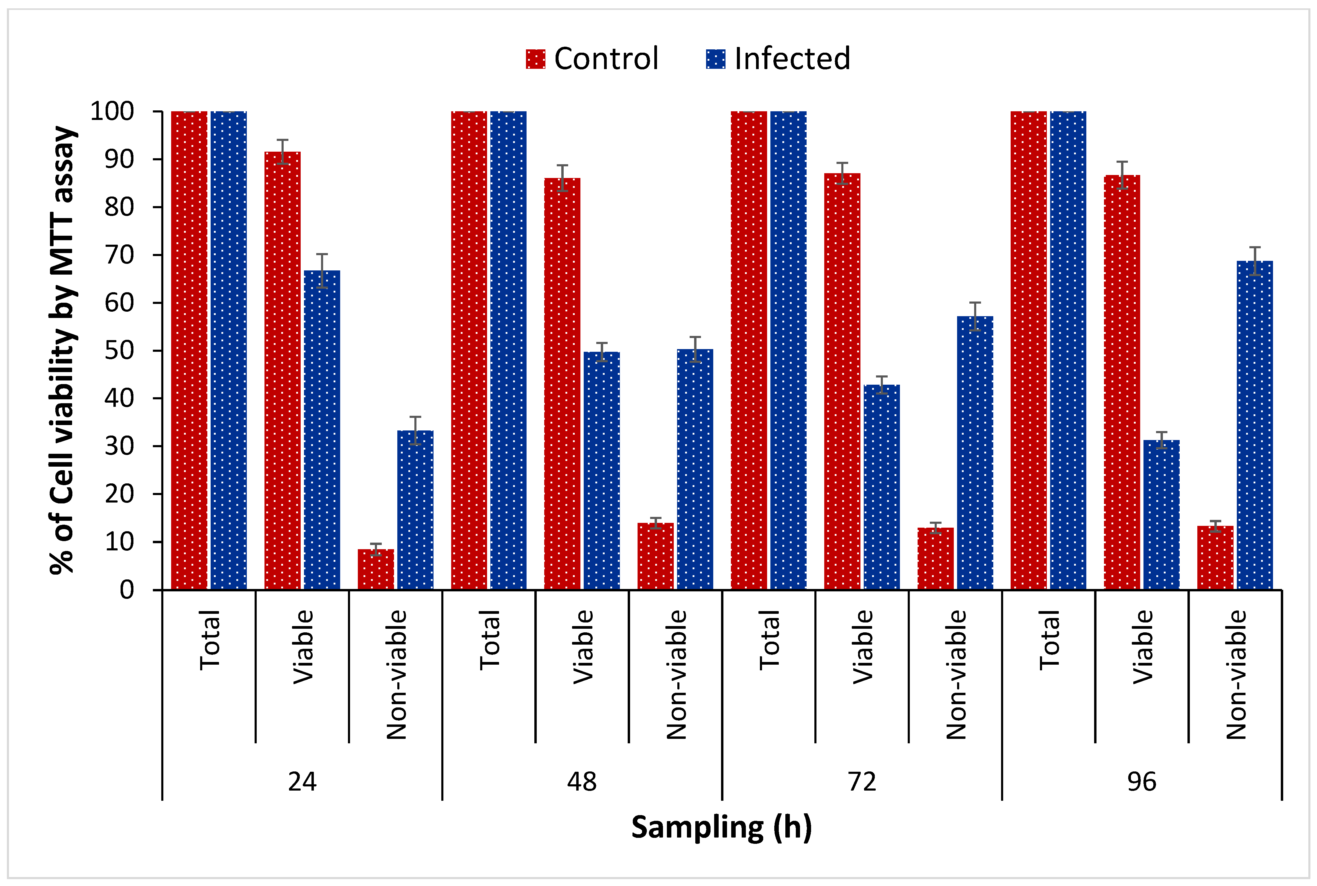
3.2. Assessment of TiPV Virulence by MTT

The MTT assay further quantified TiPV’s virulence on DRG cells. Figure 1 shows the absorbance values at 590 nm, reflecting the metabolic activity of DRG cells post-exposure. The control group maintained an absorbance of 1.50 ± 0.05, indicating robust cellular metabolism. A time-dependent decline in absorbance was observed upon infection with TiPV suspension. After 24 h, the viable/non-viable cells were 67/33% in virus-isolated cell lines. The number of non-viable cells subsequently increased, and at 48, 72, and 96 h post-infection, the viable/non-viable cells were 49/51, 42/58, and 31/69% in the infected group (Figure 1). The decreased viability of the DRG cell line in virus-infected cells highlighted enhanced virulence of TiPV with increased infection time (Figure 2).

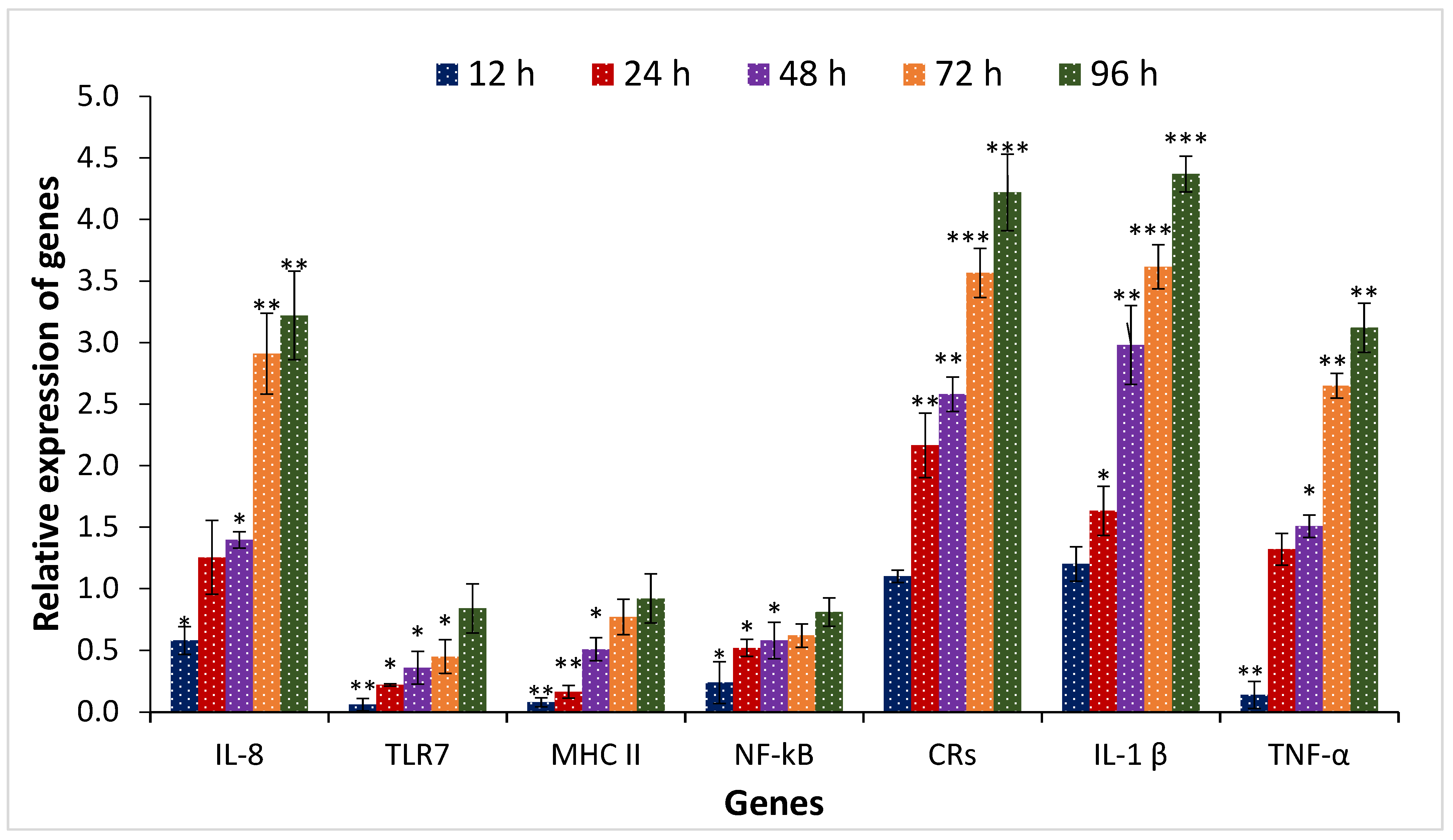
3.3. Effect of TiPV on Gene Expression of Danio rerio Gill (DRG) Cell Samples

We then conducted a thorough study to examine the relationship between viral pathogenesis and fish health, focusing on the dynamic alterations in the transcriptional expression of essential immune genes. These genes include TLR7 (Toll-like receptor 7), IL-8 (Interleukin-8), MHC-II (Major Histocompatibility Complex II), NF-κB (Nuclear Factor Kappa B), CRs (Chemokine Receptors), IL-1β (Interleukin-1β), and TNF-α (Tumor Necrosis Factor α). According to the results, the condition of Danio rerio gill (DRG) cells in their immunocompromised state in the case of Tilapia Parvovirus infection has been shown in Figure 3. The studied immune-related genes, such as IL-8, CRs, IL-1β, and TNF-α, were significantly upregulated in the DRG cell line. A ~3-fold upregulation in transcription levels was observed for IL-8, CRs, IL-1β, and TNF-α at 72 h following TiPV infection. Moreover, maximum transcription of genes, ~4 folds, were observed post 96 h post-TiPV infection in IL-8, CRs, IL-1β, and TNF-α genes (Figure 3). In contrast, TLR7, MHC-II, and NF-κB expression was consistently downregulated at all time points in the TiPV-infected DRG cell line compared to controls. Despite some variations, the results highlight a significant interaction between the immune system and TiPV infection. It appears that the virus may be actively suppressing immune responses to facilitate its establishment within the host, while the immune system, in turn, may also be attempting to counteract the infection. These observations warrant further investigation to fully elucidate the underlying mechanisms.

4. Discussion

Over the past three decades, tilapia aquaculture has experienced unprecedented growth, fueled by the rising global demand for this species as a vital source of protein [2]. This expansion, compounded by the frequent transboundary movement of live fish and their derivatives, as well as the escalating effects of climate change, has accelerated the emergence and dissemination of novel diseases that threaten the sustainability of aquaculture systems [31,32]. Recent disease outbreaks in tilapia farms across the globe, including in India, have led to catastrophic mortality events, with pathogens such as TiLV (Tilapia Lake Virus) and ISKNV (Infectious Spleen and Kidney Necrosis Virus) inflicting substantial damage on production systems [33,34,35]. Among these emerging threats, Tilapia Parvovirus (TiPV) has recently been identified as a significant pathogen in several aquaculture-producing countries [7]. Initially reported in bait Nile tilapia, TiPV has been associated with high mortality rates and severe economic losses in tilapia aquaculture systems [8]. Hence, to examine the relationship between viral pathogenesis and host–virus interaction, we studied the effects of TiPV on DrG cells by measuring TiPV proliferation and its impact on cell viability and host immune response.
Our study aimed to investigate TiPV outbreaks in multiple tilapia farms in India, focusing on characterizing the virus and its pathogenic effects. The results show a strong correlation between TiPV infection and widespread demise in cage and pond farms, underscoring the seriousness of this new virus. Through cell line assays, we confirmed the susceptibility of Danio rerio gill (DRG) cells to TiPV infection, observing marked cytopathic effects (CPE), including cell rounding, fusion, and vacuolization, culminating in significant monolayer destruction (Figure 1 and Figure 2). These observations align with the CPE patterns reported in previous studies of TiPV [7]. Additionally, our investigation into the host immune response showed a complex interaction between TiPV and the immune system, with early upregulation of pro-inflammatory cytokines such as IL-1β and suppression of key immune pathways (Figure 3). This immune modulation likely facilitates viral persistence and evasion of host defenses. Our study thus underscores the urgent need for targeted research on TiPV’s molecular pathogenesis and the development of effective management strategies to mitigate its impact on tilapia aquaculture.
The pathogenesis of Tilapia Parvovirus (TiPV) was comprehensively characterized through an integrated approach, combining cell metabolic activity with immune gene expression analyses. This study provides critical insights into TiPV’s virulence and intricate interactions with host immune responses, revealing the mechanisms underlying TiPV-induced disease. The MTT assay results demonstrated a significant decline in the viability of Danio rerio gill (DRG) cell monolayers following TiPV infection. At 24 h post-infection, viable cell percentages in TiPV-infected cultures were notably reduced to 66.71%, declining further to 31.28% by 96 h. In contrast, control cells maintained high viability throughout the experimental period, with viable cell percentages consistently above 86% (Figure 1). This progressive loss of cell viability in TiPV-infected cells is indicative of the virus’s potent cytotoxic effects, which are consistent with the findings reported by Liu et al. [7], who observed similar cytopathic effects of parvoviruses in various fish species. The increase in non-viable cell percentages from 33.29% at 24 h to 68.72% at 96 h corroborates the substantial cytotoxicity of TiPV. These results highlight the viral host evasion mechanisms and the virus’s ability to induce severe cellular damage, aligning with the extensive cell death observed in previous studies of viral infections in aquatic organisms [36]. The observed decline in metabolic activity (viable and non-viable cells), measured via absorbance at 590 nm, confirms the progressive impairment of cellular functions and underscores TiPV’s potent virulence.
Quantitative real-time PCR (qPCR) analysis of immune gene expression revealed significant alterations in the host’s immune response to TiPV infection. Interleukin-1β (IL-1β) and Chemokine Receptors (CRs) initial upregulation at 24 h indicates an early inflammatory response and immune cell recruitment. IL-1β is a crucial pro-inflammatory cytokine involved in activating innate immunity, while CRs play a role in directing immune cell migration [37]. The observed fold changes (3.62 for IL-1β and 2.16 for CRs) suggest a robust initial immune response consistent with findings in other viral infections [38]. However, the subsequent decrease in CRs expression at later time points (0.26-fold at 48 h) suggests potential viral interference with chemotactic signaling pathways. This downregulation could reflect an adaptive response by TiPV to dampen immune cell recruitment, potentially aiding in viral persistence [39]. On the other hand, TiPV’s possible defense mechanisms against immune detection are highlighted by the downregulation of Interleukin-8 (IL-8), Toll-like receptor 7 (TLR7), Major Histocompatibility Complex II (MHC-II), and Nuclear Factor Kappa B (NF-κB). IL-8 is pivotal in recruiting neutrophils to infection sites, while TLR7 is essential for recognizing viral RNA and initiating antiviral responses [40]. The substantial reductions in IL-8 (0.11 at 24 h) and TLR7 (0.06 at 24 h) suggest TiPV’s interference with early innate immune responses, impairing pathogen recognition and subsequent activation of immune defenses. The observed downregulation of MHC-II and NF-κB further indicates antigen presentation and inflammatory signaling pathways impairment. MHC-II is critical for presenting antigens to CD4 + T cells, while NF-κB is a key transcription factor involved in regulating inflammatory responses [41]. The significant reductions in these molecules (MHC-II to 0.14 at 24 h and NF-κB to 0.09 at 24 h) suggest that TiPV may suppress adaptive immune responses by impeding antigen presentation and inflammatory signaling.
TiPV’s ability to upregulate pro-inflammatory cytokines while downregulating critical immune modulators underscores its sophisticated strategy to evade host immune surveillance. The differential regulation of immune genes suggests that TiPV may employ a multifaceted approach to modulate host immune responses. By enhancing specific inflammatory pathways and suppressing key components of pathogen recognition and antigen presentation, TiPV manipulates host immune functions to facilitate viral persistence and evade immune clearance [42]. These findings are consistent with the observation that parvoviruses often induce extensive cellular damage while simultaneously employing mechanisms to evade immune detection [43]. The virus’s ability to impair cellular functions and modulate immune responses highlights the complex interplay between viral pathogenesis and host defense mechanisms, as described by Kembou-Ringert et al. [37]. This study comprehensively characterizes TiPV pathogenesis, revealing its capacity to induce significant cellular damage and modulate host immune responses. The observed cytotoxic effects and alterations in immune gene expression underscore the need for further research to elucidate the molecular mechanisms employed by TiPV. Understanding these interactions is crucial for developing targeted strategies to manage and mitigate the impact of TiPV in aquaculture systems. The exact viral components that cause cellular damage and immune evasion should be the main focus of future study, along with both therapeutic and preventative strategies. Continued efforts in viral diagnostics, as highlighted by recent reviews and advancements in detection technologies [44,45], will be essential for advancing our understanding of TiPV pathogenesis and improving disease control measures in tilapia farming.
Our study provides significant insights into the pathogenesis of Tilapia Parvovirus (TiPV) through a detailed analysis of immune responses in Danio rerio gill (DRG) cell monolayers. The observed upregulation of pro-inflammatory cytokines, such as Interleukin-1β and Tumor Necrosis Factor α, in the early stages of infection underscores an initial robust immune response. However, the subsequent downregulation of crucial immune components, including Interleukin-8, Toll-like receptor 7, Major Histocompatibility Complex II, and Nuclear Factor Kappa B, reveals TiPV’s sophisticated strategy to subvert host immune defenses. This dual-phase response illustrates TiPV’s ability to initiate an inflammatory response while concurrently evading immune surveillance by suppressing key pathways involved in pathogen recognition and immune activation. These findings enhance our understanding of TiPV-induced disease mechanisms and highlight the virus’s capacity to modulate host immune responses. Future research should focus on elucidating the precise molecular interactions between TiPV and host immune cells to develop targeted therapeutic and management strategies for controlling this emerging pathogen in aquaculture systems. Understanding these interactions is crucial for advancing disease control measures and improving fish health management practices.

Supplementary Materials

The following supporting information can be downloaded at: https://www.mdpi.com/article/10.3390/microbiolres16030068/s1.

Author Contributions

Conceptualization, V.K. and B.K.D.; methodology, V.K., A.A., K.B. and B.M.; software, V.K.; formal analysis, V.K., A.A., K.B. and B.M.; investigation, V.K. and K.B.; writing—original draft preparation, V.K.; writing—review and editing, B.K.D.; project administration, B.K.D.; funding acquisition, B.K.D. All authors have read and agreed to the published version of the manuscript.

Funding

This work was funded by the National Surveillance Program for Aquatic Animal Disease, Pradhan Mantri Matsya Sampada Yojana (PMMSY), Govt. of India file no. (G/Nat. Surveil-lance/2022-23).

Data Availability Statement

All data generated or analyzed during this study are included in this published article and its Supplementary Information Files.

Acknowledgments

The authors are also thankful to the Director, ICAR-CIFRI, for supporting the experiment trials and analysis.

Conflicts of Interest

The authors declare no conflicts of interest.

References

  1. FAO. The State of World Fisheries and Aquaculture (SOFIA). In Contributing to Food Security and Nutrition for All; FAO: Rome, Italy, 2016; p. 204. [Google Scholar]
  2. FAO. The State of World Fisheries and Aquaculture. In Sustainability in Action; FAO: Rome, Italy, 2020; p. 244. [Google Scholar]
  3. Keawcharoen, J.; Techangamsuwan, S.; Ponpornpisit, A.; Lombardini, E.D.; Patchimasiri, T.; Pirarat, N. Genetic characterization of a betanodavirus isolated from a clinical disease outbreak in farm-raised tilapia Oreochromis niloticus (L.) in Thailand. J. Fish Dis. 2015, 38, 49–54. [Google Scholar] [CrossRef]
  4. Figueiredo, H.C.; Tavares, G.C.; Dorella, F.A.; Rosa, J.C.C.; Marcelino, S.A.C.; Pierezan, F.; Pereira, F.L. First report of infectious spleen and kidney necrosis virus in Nile tilapia in Brazil. Transbound. Emerg. Dis. 2022, 69, 3008–3015. [Google Scholar] [CrossRef]
  5. Nicholson, P.; Mon-On, N.; Jaemwimol, P.; Tattiyapong, P.; Surachetpong, W. Coinfection of tilapia lake virus and Aeromonas hydrophila synergistically increased mortality and worsened the disease severity in tilapia (Oreochromis sp.). Aquaculture 2020, 520, 734746. [Google Scholar] [CrossRef]
  6. Yamkasem, J.; Piewbang, C.; Techangamsuwan, S.; Pierezan, F.; Soto, E.; Surachetpong, W. Susceptibility of ornamental African cichlids Aulonocara spp. to experimental infection with Tilapia lake virus. Aquaculture 2021, 542, 736920. [Google Scholar] [CrossRef]
  7. Liu, W.; Zhang, Y.; Ma, J.; Jiang, N.; Fan, Y.; Zhou, Y.; Cain, K.; Yi, M.; Jia, K.; Wen, H.; et al. Determination of a novel parvovirus pathogen associated with massive mortality in adult tilapia. PLoS Pathog. 2020, 16, e1008765. [Google Scholar] [CrossRef]
  8. Du, J.; Wang, W.; Chan, J.F.W.; Wang, G.; Huang, Y.; Yi, Y.; Zhu, Z.; Peng, R.; Hu, X.; Wu, Y.; et al. Identification of a Novel Ichthyic Parvovirus in Marine Species in Hainan Island, China. Front. Microbiol. 2019, 10, 2815. [Google Scholar] [CrossRef]
  9. Das, B.K.; Kumar, V.; Roy, S.; Malick, R.C.; Bisai, K.; Jana, A.K.; Dhar, S. Pathological effects and immune modulation in host during Tilapia Parvovirus (TiPV) outbreak in cage and wetland Tilapia farms. Sci. Rep. 2024, 14, 28689. [Google Scholar] [CrossRef]
  10. Imajoh, M.; Ikawa, T.; Oshima, S.I. Characterization of a new fibroblast cell line from a tail fin of red sea bream, Pagrus major, and phylogenetic relationships of a recent RSIV isolate in Japan. Virus Res. 2007, 126, 45–52. [Google Scholar] [CrossRef]
  11. Kim, S.J.; Lee, S.J.; Jung, S.J.; Jung, M.H. Effect of rock bream iridovirus (RBIV) contained tissue intake on rock bream (Oplegnathus fasciatus) mortality and blood cell distribution. Fish Shellfish. Immunol. 2024, 153, 109858. [Google Scholar] [CrossRef]
  12. Dong, H.T.; Senapin, S.; Gangnonngiw, W.; Nguyen, V.V.; Rodkhum, C.; Debnath, P.; Delamare-Deboutteville, J.; Mohan, C.V. Experimental infection reveals transmission of tilapia lake virus (TiLV) from tilapia broodstock to their reproductive organs and fertilized eggs. Aquaculture 2020, 515, 734541. [Google Scholar] [CrossRef]
  13. Jaemwimol, P.; Rawiwan, P.; Tattiyapong, P.; Saengnual, P.; Kamlangdee, A.; Surachetpong, W. Susceptibility of important warm water fish species to tilapia lake virus (TiLV) infection. Aquaculture 2018, 497, 462–468. [Google Scholar] [CrossRef]
  14. Dai, Y.J.; Jia, Y.F.; Chen, N.; Bian, W.; Li, Q.K.; Ma, Y.B.; Chen, Y.L.; Pei, D.S. Zebrafish as a model system to study toxicology. Environ. Toxicol. Chem. 2014, 33, 11–17. [Google Scholar] [CrossRef]
  15. MJ, A.W.; S, A.M.; S, M.; V, R.; AS, S.H. A comparative study on targeted gene expression in zebrafish and its gill cell line exposed to chlorpyrifos. Vitr. Cell. Dev. Biol. Anim. 2024, 60, 397–410. [Google Scholar]
  16. Ajitha, V.; Sarasan, M.; Sreevidya, C.; Aswathy, C.; Kachiprath, B.; Mohandas, A.; Singh, I.S.B.; Hameed, A.S.; Schlenk, D.; Magnuson, J.T.; et al. Cytotoxic impacts of treated electroplating industrial effluent and the comparative effect of their metal components (Zn, Hg, and Zn + Hg) on Danio rerio gill (DrG) cell line. Sci. Total Environ. 2021, 793, 148533. [Google Scholar] [CrossRef]
  17. Ashfaque, M.; Ahmed, A.N.; Safiullah, S.M.; Taju, G.; Majeed, S.A.; Hameed, A.S.; Basha, K.A. Toxicological assessment of functional polymer with single-walled carbon nanotubes in zebrafish embryos and its gill cell line. Chemosphere 2022, 303, 134891. [Google Scholar] [CrossRef]
  18. Nga, N.T.H.; Ngoc, T.T.B.; Trinh, N.T.M.; Thuoc, T.L.; Thao, D.T. Optimization and application of MTT assay in determining density of suspension cells. Anal. Biochem. 2020, 610, 113937. [Google Scholar] [CrossRef]
  19. Rajendran, K.V.; Sood, N.; Rao, B.M.; Valsalam, A.; Bedekar, M.K.; Jeena, K.; Pradhan, K.; Paria, A.; Swaminathan, T.R.; Verma, D.K.; et al. Widespread occurrence of Tilapia parvovirus in farmed Nile tilapia Oreochromis niloticus from India. J. Fish Dis. 2023. [Google Scholar] [CrossRef]
  20. Badhusha, A.; Mithra, S.; Taju, G.; Rajkumar, V.; Abdul Majeed, S.; Suryakodi, S.; Haridas, L.; Haridas, D.; Sahoo, K.; Mohanty, J.; et al. Detection of Tilapia parvovirus in farm-reared tilapia in India and its isolation using fish cell lines. Vitr. Cell. Dev. Biol. Anim. 2025, 1–13. [Google Scholar] [CrossRef]
  21. Wang, Z.; Nie, L.; Fei, C.; Chen, J. Establishment of Gill-Derived Primary Cell Cultures from Largemouth Bass (Micropterus salmoides) as an Alternative Platform for Studying Host–Virus Interactions. Fishes 2025, 10, 18. [Google Scholar] [CrossRef]
  22. Brian, A.; Dawn, A. Bacterial Fish Pathogens; Springer: Berlin/Heidelberg, Germany, 2016. [Google Scholar] [CrossRef]
  23. Schreck, C.B.; Moyle, B. Methods for Fish Biology; American Fisheries Society: Bethesda, MD, USA, 1990. [Google Scholar]
  24. Roberts, C. Information structure: Towards an integrated formal theory of pragmatics. Semant. Pragmat. 2012, 5, 1–69. [Google Scholar] [CrossRef]
  25. Freshney, R.I. Database of misidentified cell lines. Int. J. Cancer 2010, 126, 302. [Google Scholar] [CrossRef] [PubMed]
  26. George, M.R.; John, K.R.; Mansoor, M.M.; Saravanakumar, R.; Sundar, P.; Pradeep, V. Isolation and characterization of a ranavirus from koi, Cyprinus carpio L., experiencing mass mortalities in India. J. Fish Dis. 2015, 38, 389–403. [Google Scholar] [CrossRef]
  27. Lorenzen, N.; Lorenzen, E.; Einer-Jensen, K.; Heppell, J.; Wu, T.; Davis, H. Protective immunity to VHS in rainbow trout (Oncorhynchus mykiss, Walbaum) following DNA vaccination. Fish Shellfish. Immunol. 1998, 8, 261–270. [Google Scholar]
  28. Elbahnaswy, S.; Elshopakey, G.E. Differential gene expression and immune response of Nile tilapia (Oreochromis niloticus) challenged intraperitoneally with Photobacterium damselae and Aeromonas hydrophila demonstrating immunosuppression. Aquaculture 2020, 526, 735364. [Google Scholar]
  29. Spearman, C. The method of right and wrong cases (constant stimuli) without Gauss’s formulae. Br. J. Psychol. 1908, 2, 227. [Google Scholar] [CrossRef]
  30. Kärber, G. Contribution to the collective treatment of pharmacological serial experiments. Naunyn Schmiedebergs Arch. Exp. Pathol. Pharmacol. 1931, 162, 480–483. [Google Scholar] [CrossRef]
  31. Kibenge, F.S.B. Emerging viruses in aquaculture. Curr. Opin. Virol. 2019, 34, 97–103. [Google Scholar] [CrossRef] [PubMed]
  32. Surachetpong, W.; Roy, S.R.K.; Nicholson, P. Tilapia lake virus: The story so far. J. Fish Dis. 2020, 43, 1115–1132. [Google Scholar] [CrossRef]
  33. Behera, B.K.; Pradhan, K.; Swaminathan, T.R.; Sood, N.; Paria, P.; Das, A.; Verma, D.K.; Kumar, R.; Yadav, M.K.; Dev, A.K.; et al. Emergence of Tilapia Lake Virus associated with mortalities of farmed Nile Tilapia Oreochromis niloticus (Linnaeus 1758) in India. Aquaculture 2018, 484, 168–174. [Google Scholar] [CrossRef]
  34. Acharya, V.; Chakraborty, H.J.; Rout, A.K.; Balabantaray, S.; Behera, B.K.; Das, B.K. Structural Characterization of Open Reading Frame-Encoded Functional Genes from Tilapia Lake Virus (TiLV). Mol. Biotechnol. 2019, 61, 945–957. [Google Scholar] [CrossRef]
  35. Ramírez-Paredes, J.G.; Paley, R.K.; Hunt, W.; Feist, S.W.; Stone, D.M.; Field, T.R.; Haydon, D.J.; Ziddah, A.; Nkansa, M.; Guilder, J.; et al. First detection of infectious spleen and kidney necrosis virus (ISKNV) associated with massive mortalities in farmed tilapia in Africa. Transbound. Emerg. Dis. 2021, 68, 1550–1563. [Google Scholar] [CrossRef]
  36. Piewbang, C.; Tattiyapong, P.; Khemthong, M.; Lachroje, S.; Boonrungsiman, S.; Kasantikul, T.; Surachetpong, W.; Techangamsuwan, S. Dual infections of tilapia parvovirus (TiPV) and tilapia lake virus (TiLV) in multiple tilapia farms: Their impacts, genetic diversity, viral tropism, and pathological effects. Aquaculture 2022, 550, 737887. [Google Scholar] [CrossRef]
  37. Kembou-Ringert, J.E.; Steinhagen, D.; Thompson, K.D.; Daly, J.M.; Adamek, M. Immune responses to Tilapia lake virus infection: What we know and what we don’t know. Front. Immunol. 2023, 14, 1240094. [Google Scholar] [CrossRef] [PubMed]
  38. Dong, H.T.; Chaijarasphong, T.; Barnes, A.C.; Delamare-Deboutteville, J.; Lee, A.; Senapin, S.; Mohan, C.V.; Tang, K.F.; McGladdery, S.E.; Bondad-Reantaso, M.G. From the basics to emerging diagnostic technologies: What is on the horizon for tilapia disease diagnostics? Rev. Aquac. 2023, 15, 186–212. [Google Scholar] [CrossRef]
  39. Xue, S.; Liu, X.; Liu, Y.; Lu, C.; Jia, L.; Yu, Y.; Liu, H.; Yang, S.; Zeng, Z.; Li, H.; et al. Determination and Characterization of Novel Papillomavirus and Parvovirus Associated with Mass Mortality of Chinese Tongue Sole (Cynoglossus semilaevis) in China. Viruses 2024, 16, 705. [Google Scholar] [CrossRef]
  40. Zhao, H.; Zhou, Y.; Fan, Y.; Jiang, N.; Meng, Y.; Li, Y.; Xue, M.; Xu, C.; Guo, W.; Liu, W. Development and application of a sensitive droplet digital PCR-based method to detect tilapia parvovirus. J. Fish Dis. 2023, 46, 239–245. [Google Scholar] [CrossRef]
  41. Suresh, T.; Nithin, M.S.; Kushala, K.B.; Girisha, S.K.; Shivakumar, V.B.; Dheeraj, S.B.; Puneeth, T.G.; Kishan, K.; Vinay, T.N. Largescale mortality of Oreochromis mossambicus in lakes and reservoirs of Karnataka due to coinfection of Tilapia Lake virus (TiLV) and multidrug-resistant Aeromonas veronii: An emerging fish disease in India. Aquaculture 2023, 565, 739077. [Google Scholar] [CrossRef]
  42. Phusantisampan, T.; Yamkasem, J.; Tattiyapong, P.; Sriariyanun, M.; Surachetpong, W. Specific and rapid detection of tilapia parvovirus using loop-mediated isothermal amplification (LAMP) method. J. Fish Dis. 2022, 45, 1893–1898. [Google Scholar] [CrossRef]
  43. Raksaseri, P.; Lertwanakarn, T.; Tattiyapong, P.; Kijtawornrat, A.; Klomkleaw, W.; Surachetpong, W. Tilapia lake virus causes mitochondrial damage: A proposed mechanism that leads to extensive death in fish cells. PeerJ 2023, 11, e16190. [Google Scholar] [CrossRef]
  44. Johny, T.K.; Swaminathan, T.R.; Sood, N.; Pradhan, K.; Lal, K.K. A panoptic review of techniques for finfish disease diagnosis: The status quo and future perspectives. J. Microbiol. Methods 2022, 196, 106477. [Google Scholar] [CrossRef]
  45. Ranjitha, H.B.; Ramesh, M.; Behera, S.; ValiyaValappil, D.; Basagoudanavar, S.H.; Sherasiya, A. Genetic engineering tools and techniques in livestock production. In Sustainable Agriculture Reviews 57: Animal Biotechnology for Livestock Production 2; Springer International Publishing: Cham, Switzerland, 2022; pp. 175–207. [Google Scholar]
Figure 1. The metabolic activity of DRG cells was measured via an MTT assay post-exposure to Tilapia Parvovirus (TiPV). DRG cells were exposed to TiPV, and samples were collected after 24, 48, 72, and 96 h for further analysis. Later, the effect of TiPV on cell viability was measured by taking absorbance in a microplate reader at 590 nm. Based on the absorbance, the % of cell viability was calculated, and DRG cell lines were classified as viable and non-viable cells in the control and infected groups. The results are presented as the mean ± standard error of the mean (n = 3).
Microbiolres 16 00068 g001
Figure 2. Danio rerio gill (DRG) cell line viability post-exposure to Tilapia Parvovirus (TiPV). The control group was maintained without TiPV exposure, while the infected group was exposed to TiPV, and samples were collected after 24, 48, 72, and 96 h for further analysis.
Microbiolres 16 00068 g002
Figure 3. Fold change in gene expression in Danio rerio gill (DRG) cell samples from both control and infected groups. Gene expression of TLR7 (Toll-like receptor 7), IL-8 (Interleukin-8), MHC-II (Major Histocompatibility Complex II), NF-κB (Nuclear Factor Kappa B), CRs (Chemokine Receptors), IL-1β (Interleukin-1β), and TNF-α (Tumor Necrosis Factor α) was quantified as fold changes relative to the housekeeping β-actin gene using quantitative real-time PCR. Temporal gene expression profiles were compared between control and virus-infected cell samples. The control group’s expression level was set to 1.0, and the gene expression ratio in the infected group was calculated relative to the control (cell line inoculated with tissue extracts from non-infected tilapia). Data are presented as the mean ± standard error (SE), n = 3. Asterisk (*) indicates statistically significant differences between infected and control groups at p < 0.05. * p < 0.05, ** p < 0.01, *** p < 0.001.
Microbiolres 16 00068 g003
Table 1. List of primers used in the study.
GenePrimer Sequence 5′ to 3′References
Tilapia Parvovirus-FGAGATGGTGTGAAAATGAACGGG[7]
Tilapia Parvovirus-RCTATCTCCTCGTTGCTCGGTGTATC
Tilapia Parvovirus-FqGCACCACAGCTGAGTACAAC
Tilapia Parvovirus-RqAACTGCTCGGCTATCTCCTC
Beta-Actin-FCAGCAAGCAGGAGTACGATGAG[28]
Beta-Actin-RTGTGTGGTGTGTGGTTGTTTTG
Interleukin8-FGCACTGCCGCTGCATTAAG
Interleukin88-RGCAGTGGGAGTTGGGAAGAA
Toll-like receptor7-FTCAGCAGGGTGAGAGCATAC
Toll-like receptor7-RACATATCCCAGCCGTAGAGG
Major Histocompatibility Complex-II-FTGGCCCTGACTGAACCACTG
Major Histocompatibility Complex-II-RTCAGACCCACGCCACAGAAC
Nuclear factorκB-FAACGACGGTGATGACAACGAC
Nuclear factorκB-RAAATTCAGGCTCCACACTGACC
CRs-chemokine receptors -FACAGAGCCGATCTTGGGTTACTTG
CRs-chemokine receptors-RTGAAGGAGAGGCGGTGGATGTTAT
Interleukin1β -FTGCACTGTCACTGACAGCCAA
Interleukin1β -RATGTTCAGGTGCACTATGCGG
Tumor necrosis factor-α -FCCAGAAGCACTAAAGGCGAAGA
Tumor necrosis factor-α -RCCTTGGCTTTGCTGCTGATC
Disclaimer/Publisher’s Note: The statements, opinions and data contained in all publications are solely those of the individual author(s) and contributor(s) and not of MDPI and/or the editor(s). MDPI and/or the editor(s) disclaim responsibility for any injury to people or property resulting from any ideas, methods, instructions or products referred to in the content.

