Case Report

First Isolation, Molecular Identification, and Phylogenetic Characterization of A3B5 Very Virulent Infectious Bursal Disease Virus in Pullets in Chile

1
Núcleo de Investigación en One Health (NIOH), Facultad de Medicina Veterinaria y Agronomía, Universidad de las Américas, Campus Maipú, 5 de Abril 620, Santiago 7500975, Chile
2
Laboratory of Avian Pathology, Department of Animal Pathology, Faculty of Veterinary and Animal Sciences, Universidad de Chile, Santiago 8820808, Chile
*
Author to whom correspondence should be addressed.
Microbiol. Res. 2025, 16(2), 31; https://doi.org/10.3390/microbiolres16020031
Submission received: 28 October 2024 / Revised: 3 January 2025 / Accepted: 20 January 2025 / Published: 23 January 2025

Abstract

:
Infectious bursal disease virus (IBDV) is an important pathogen affecting the poultry industry worldwide. IBDV serotype 1, including classical virulent strains (cvIBDV), variant strains (varIBDV), and very virulent strains (vvIBDV), is pathogenic for chickens. IBDV mainly infects immature B-lymphocytes in the bursa of Fabricius, weakening the humoral immune response and leading to secondary infections and increased morbidity and mortality. The Laboratory of Avian Pathology received ten live 8-week-old pullets from a laying hen operation experiencing increased mortality, prostration, diarrhea, and sudden death. Upon necropsy, the affected birds presented swollen, hemorrhagic, and edematous bursa of Fabricius, as well as hemorrhage in the breast and thigh muscles. RT-PCR confirmed that the samples from the bursa of Fabricius were positive for IBDV. Phylogenetic analysis of the VP1 and VP2 gene nucleotide sequences classified the strain, isolated in embryonated chicken eggs, as the A3B5 genotype. Amino acid sequence analysis of the VP2 hypervariable region revealed the presence of amino acid residues commonly found in vvIBDV. Additional studies are required to investigate the epidemiological situation of this genotype in Chile and to evaluate current vaccination plans and their effectiveness against new variants.

1. Introduction

Infectious bursal disease (IBD) or “Gumboro Disease” is an acute, highly contagious, immunosuppressive disease affecting young chicks. It causes significant economic losses worldwide due to mortality, increased susceptibility to infection, and poor vaccination response [1,2,3]. The disease is caused by the infectious bursal disease virus (IBDV), a small non-enveloped viral particle with a 60 to 70 nm diameter that belongs to the Avibirnavirus genus within the Birnaviridae family [4]. The virus genome comprises double-stranded RNA divided into A and B segments. The longer segment A (3.2 kb) encodes VP2–VP3–VP4 polyprotein and a nonstructural protein VP5, while the shorter segment B (2.9 kb) encodes VP1, which has RNA-dependent RNA polymerase activity [2]. The capsid consists of 260 VP2 trimers. Each VP2 monomer has three distinct domains: base domain (B), shell domain (S), and projection domain (P) [5]. The P domain contains a hypervariable (hv) region between aa positions 206 and 350 [6], which includes four exposed loops identified as PBC, PDE, PFG, and PHI [7]. The VP2 protein is the main structural protein related to virulence, cellular tropism, production of neutralizing antibodies, and prediction of phylodynamic relationships [8]. Cross-neutralization assays have identified two IBDV serotypes [9]. Serotype 2 is nonpathogenic for chickens, and there are no cross-reactions between the two serotypes [10]. Serotype 1 includes classical virulent strains (cvIBDV) [11], variant strains (varIBDV) [12], and very virulent strains (vvIBDV) [13]. As a non-enveloped virus, IBDV is highly resistant to environmental conditions, facilitating its persistence and spread in poultry farms [14].
The virus primarily targets the bursa of Fabricius (BF), infecting immature B lym-phocytes in the target organ, inducing inflammation and subsequent atrophy and de-creasing the B lymphocyte count. The resulting weakened immune response [15] reduces vaccine-induced immunity and predisposes birds to secondary infections, increasing morbidity and mortality in infected birds [16]. Macroscopically, on day 3–4 PI, the bursa increased in size and weight due to edema and hyperemia. Necrotic foci and petechial or ecchymotic hemorrhages are usually observed on the mucosal surface, resulting in a “purple grape” appearance. Microscopically, severely reduced lymphocytes, heterophil infiltrates, and interstitial hyperplasia are observed, along with erythrocyte infiltration, which produces atrophy and bleeding of the bursa [17].
IBDV first appeared in Gumboro, Delaware, USA, in 1957 and is commonly known as the classical virulent strain (cvIBDV). The disease was called “avian nephrosis” due to kidney damage in affected birds [18]. In 1984, variant IBDV (varIBDV) strains emerged in Gumboro County [19]. Very virulent strains (vvIBDV) were later identified in Holland [11], Belgium [20], Brazil [21], and the USA [22]. These vvIBDV strains cause 60% to 70% mortality in susceptible 3- to 10-week-old layer pullets [1]. According to molecular epidemiology studies, approximately 60–76% of IBDV isolates in several countries worldwide can be classified as very virulent [23].
The emergence of new strains with different characteristics led to proposals for new classification schemes, some of which are based exclusively on changes in the hypervariable region of VP2 (hvVP2), making it possible to classify IBDV strains into seven genogroups [24]. These were subsequently complemented with an analysis of the VP1 (segment B) sequence since both contribute to IBDV pathogenicity [25]. The combined classification scheme detects reassortment in the IBDV segments. It identifies nine genogroups for Segment A [A1 (classical), A2 (US antigenic variant), A3 (very virulent), A4 (dIBDV), A5 (atypical Mexican), A6 (atypical Italian), A7 (early Australian), A8 (Australian variant), and A9 (attenuated)] and five genogroups for Segment B [B1 (classical-like), B2 (very virulent-like), B3 (early Australian-like), B4 (Polish and Tanzanian), and B5 (Nigerian)]. Given IBDV’s bi-segmented genome, up to 45 different genotypes could be identified if all genogroups are present [26,27,28].
The vvIBDV strains have been identified in Poland [29], China [30], Finland [31], Nigeria [32], Egypt [33], and Western European countries [34]. In Latin America, they have been identified in Brazil, the Dominican Republic, Venezuela [35], and Argentina [36]. In Chile, cvIBDV strains of the A1B1 genotype and a varIBDV strain of the A2B1 genotype have been identified [37]. This is the first report on the isolation, molecular identification, and phylogenetic characterization of a very virulent strain of infectious bursal disease virus in Chile.

2. Materials and Methods

2.1. Examination and Sampling of the Pullets

The Universidad de Chile’s Avian Pathology Laboratory received ten live, 8-week-old pullets from a laying hen farm experiencing increased mortality, prostration, diarrhea, and sudden death in Chile’s Metropolitan Region. The birds were humanely euthanized by cervical dislocation for subsequent necropsy. The flocks had been vaccinated at the hatchery for Salmonella, infectious bronchitis, and Marek’s disease. No IBD vaccine had been administered. Bursa samples were collected, sliced, and homogenized in phosphate-buffered saline (PBS; pH 7.2) 10% containing 200 U/mL penicillin and 0.2 mg/mL streptomycin. Homogenates were vortexed for 10 s, subjected to three freeze–thaw cycles, and centrifuged at 3000× g for 10 min at 4 °C. The supernatant was filtered through a 0.22 μm membrane, transferred to a sterile tube and stored at −80 °C for further analysis.

2.2. Viral Isolation and Propagation in Specific Pathogen-Free Embryonated Chicken Eggs (SPF-ECE)

Fertile 9- to 11-day-old SPF-ECE were inoculated via chorioallantoic membrane (CAM) using a dose of 0.1 mL per egg with bursa homogenate. They were subsequently incubated at 37 °C and 60–70% humidity. The chicken embryos were candled daily; those that died within the first 48 h post-inoculation were discarded. Then, the CAM from the affected ECE was collected and homogenized in sterile PBS. The resulting supernatant was filtered through a 0.22 μm membrane, placed in sterile tubes, and stored at −80 °C for molecular analysis [38].

2.3. Reverse Transcription Polymerase Chain Reaction (RT-PCR)

Total RNA from bursa homogenates and CAM was extracted using the PureLink™ Viral RNA/DNA Mini Kit (Invitrogen, Waltham, MA, USA) according to the manufacturer’s instructions. RNA was eluted in 50 µL nuclease-free water to amplify IBDV gene fragments from segments A and B. For viral identification and phylogenetic analyses, cDNA was obtained using SuperScript III Reverse Transcriptase (Invitrogen, Waltham, MA, USA) according to the manufacturer’s instructions. The PCR was conducted in a final volume of 25 μL containing 10 mM of each primer, 2.5 μL of 10x High Fidelity PCR buffer, 10 mM dNTP Mix, 50 mM MgSO4, 0.5 U of Platinum™ Taq DNA Polymerase High Fidelity (Invitrogen, Waltham, MA, USA), 5 μL of cDNA and ultrapure water under the following conditions: 2 min at 94 °C, 35 cycles of 1 min at 94 °C, 30 s at 61 °C, 45 s at 72 °C; and, finally, an additional extension step at 72 °C for 10 min. The PCR products were analyzed by 2% agarose gel electrophoresis using GelRed® Nucleic Acid Stain (Millipore, Burlington, MA, USA) and visualized using a Trans-Lum SOLO transilluminator (Biotop, Jing’an District, Shanghai, China). The primer sequences used are listed in Table 1.

2.4. VP1 and VP2 Sequence Analysis and Phylogenetic Analysis

The amplified PCR products from the VP1 and VP2 genes were purified using a PureLink™ Quick Gel Extraction Kit (Invitrogen, Waltham, MA, USA) according to the manufacturer’s instructions. Austral Omics (Valdivia, Los Ríos Region, Chile) provided the sequencing services. Sanger sequencing was performed using the chain termination method with the BigDye® Terminator v3.1 Cycle Sequencing Kit (Invitrogen, Waltham, MA, USA) according to the manufacturer’s instructions. Sequences from this study’s VP1 and VP2 genes were assembled and translated into amino acid sequences using Bi-oedit v.7.2.5 software [41]. VP1 and VP2 gene sequences were retrieved from GenBank and included in the analysis. The datasets were aligned using the MAFFT v.7 online software (http://mafft.cbrc.jp/alignment/server/large.html (accessed on 25 June 2024)) [42]. The VP2 dataset included 74 sequences, whereas the VP1 dataset consisted of 57 representative sequences from the previously described genogroups for segments A and B [26]. These figures include sequences from reported Chilean isolates [37]. A maximum-likelihood tree was created using PhyML 3.0 [43], and a bootstrap expectation (TBE) process with 1000 replicates supported the robustness of the nodes. The tree was visualized and edited using FigTree v. 7.4.4 software [44] (https://code.google.com/archive/p/beast-mcmc/ (accessed on 28 June 2024)).
MEGA 11 software [45] estimated evolutionary divergence in the VP1 and VP2 sequences. The data are expressed as a relationship between the two groups listed in the headers of each table in terms of the average difference (by group) in the amino acids per site. A bootstrapping procedure (500 replicates) produced standard error estimates, and analysis used the Poisson correction model. The rate of variation between the two sites was modeled with gamma distribution (shape parameter = 4).
To analyze the VP2 hypervariable region (hvVP2), the sequence of the strain isolated in this study was compared with previously published Chilean IBDV sequences, GenBank sequences for the A1, A2, A3, A4, A5, A6, A7, A8, and A9 genogroups (accession numbers AY012683, JX134483, MF996499, MN313611, MF142574, JN852985, AF381001, and AF148081, MZ687401, respectively), and sequences from live attenuated vaccines: D78 (accession number AJ586963) and ViBursa CE (accession number EU162089).

3. Results

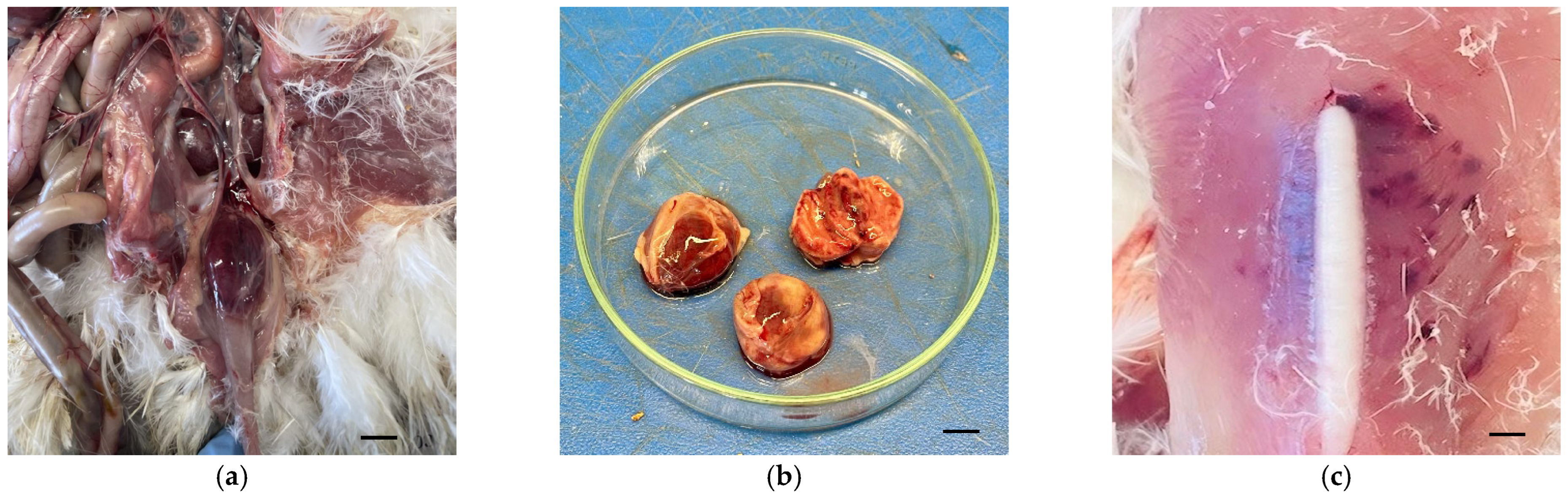
3.1. Examination of the Pullets

The postmortem analysis of the pullets in the laboratory reveals dehydration and macroscopic lesions in the bursa of Fabricius, which appear swollen, hemorrhagic, and edematous (Figure 1a,b). Hemorrhages were also found in the breast and thigh muscles of some affected birds (Figure 1c). The kidneys were swollen, whitish, and contained urate deposits. Lesions were compatible with infectious bursal disease. No other significant lesions were observed in any other organ.

3.2. Viral Isolation

Bursal samples were propagated via the chorioallantoic route on embryonated chicken eggs to isolate the IBDV. Forty-eight hours post-inoculation, the dead embryos exhibited lesions of dwarfing (Figure 2a), congestion, edema, hemorrhage, a pale appearance of the heart, and a swollen, greenish liver (Figure 2b). The chicken embryo negative controls showed no significant lesions (Figure 2c).

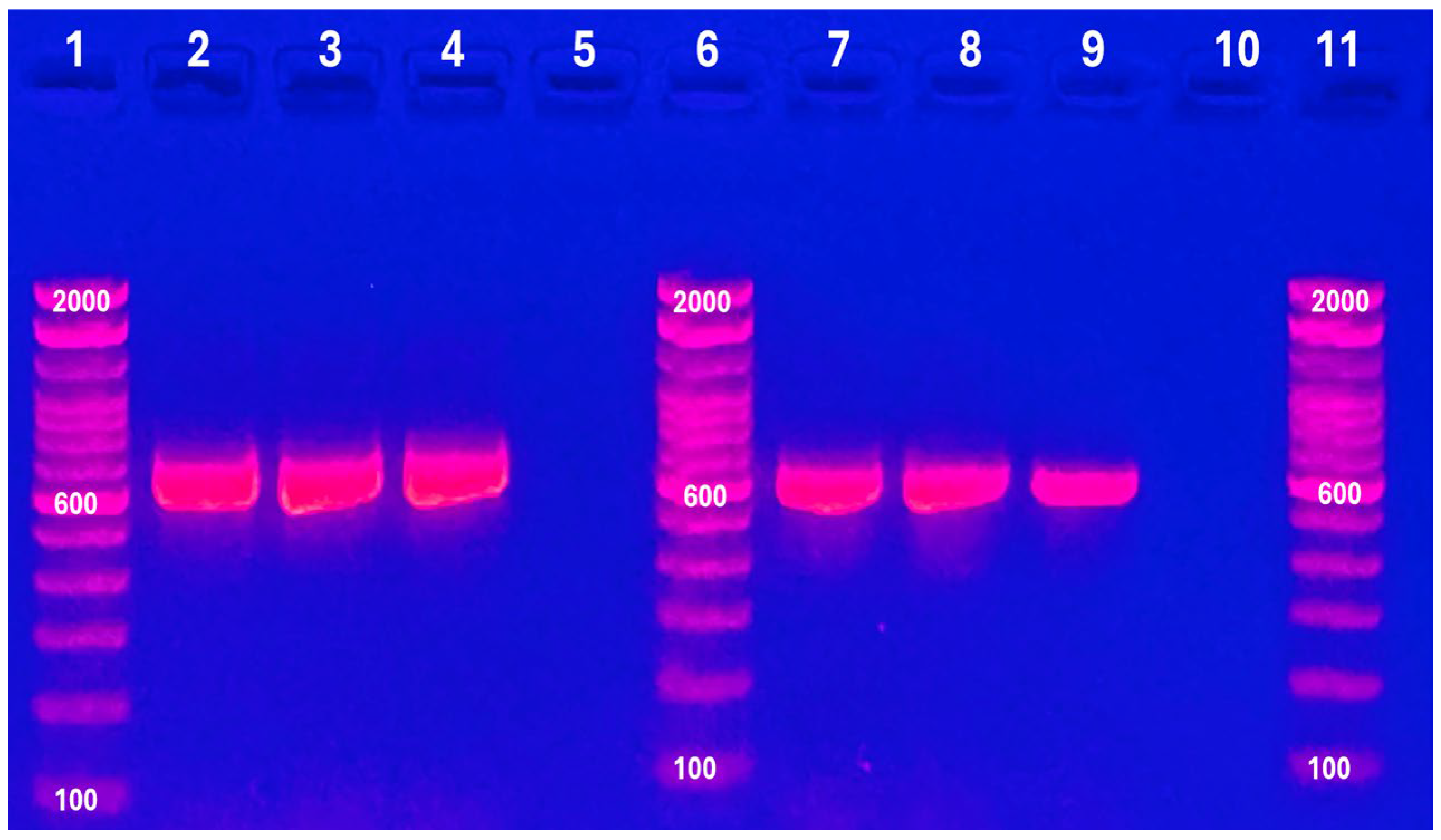
3.3. Identification of the IBDV

The samples from the pullets’ BF were positive for IBDV infection, amplifying 642 and 604 bp corresponding to VP1 and VP2 genes, respectively. The samples obtained from the CAM of SPF-ECE infected with bursa homogenate were also positive for both genes (Figure 3), confirming the presence of the virus in the affected bursa and its successful isolation in embryonated chicken eggs.

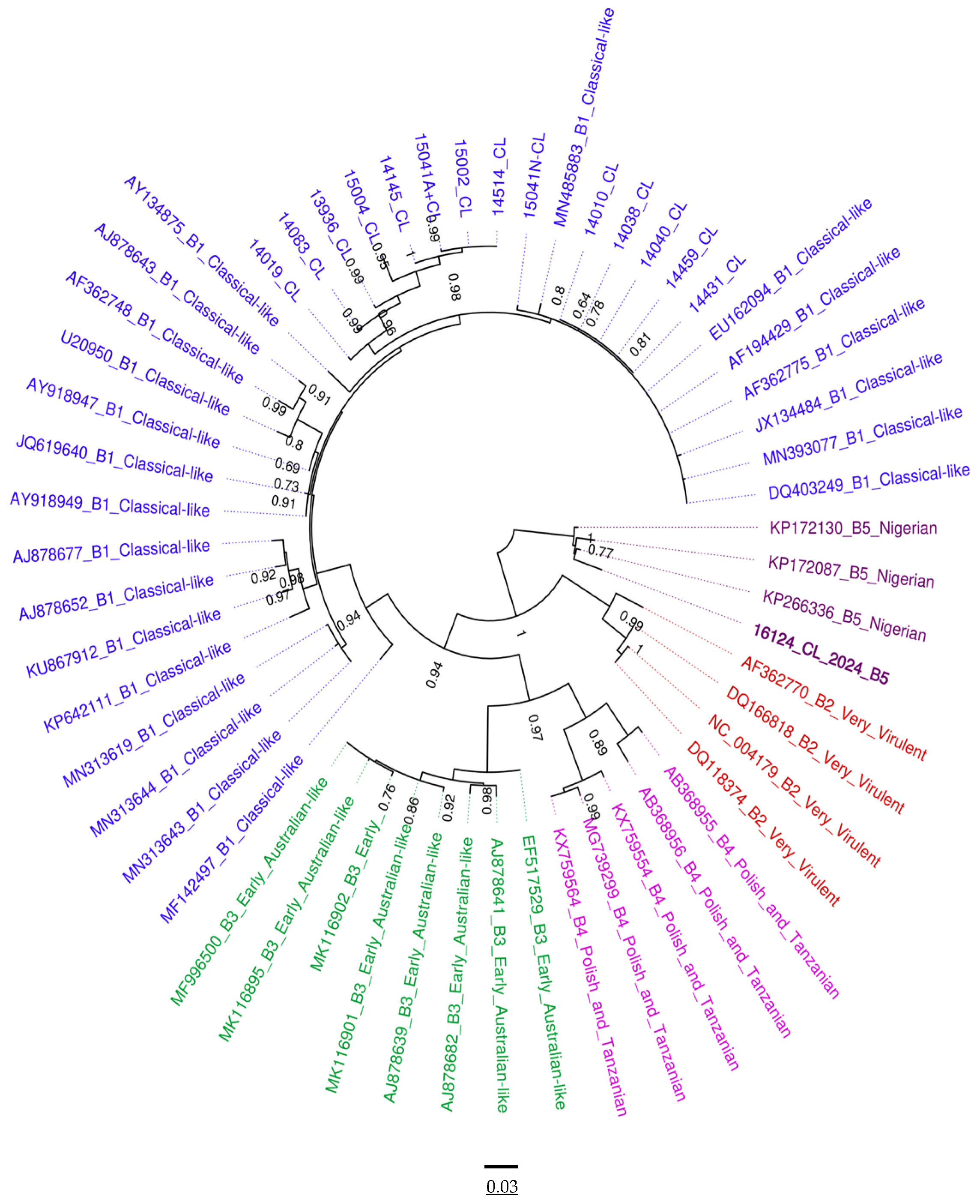
3.4. Phylogenetic Analysis of VP1 and VP2

Analysis of the VP1 sequence shows that the isolated virus could be classified as genogroup B5, which contains previously characterized strains from Nigeria (Figure 4). Analysis of the VP2 sequence classifies it within genogroup A3, which contains strains previously characterized as very virulent in several countries [20] (Figure 5). Genotyping analysis based on the partial sequences of both segments classified the detected IBDV strain as the A3B5 genotype. The sequences obtained in this study were designated as 16124_CL_2024 and submitted to GenBank. The accession numbers assigned to VP1 and VP2 were PP824543 and PP824544, respectively.

3.5. Nucleotide Sequence Analysis of Segments A and B

Continuing nucleotide sequence analysis for the VP1 and VP2 genes, the consensus sequences obtained for segments A and B were compared to the GenBank database using the BLAST algorithm (https://blast.ncbi.nlm.nih.gov/Blast.cgi (accessed on 26 July 2024)).
This analysis determined that the nucleotide sequences shared significant identity with those from strains obtained primarily in Brazil and the USA. A summary of the main results is presented in Table 2.

3.6. Amino Acid Sequence Analysis for Segments A and B

When comparing the amino acid distance between the different genogroups and the strain isolated in this study, based on segment A, less divergence was observed with genogroup A3 (0.0102) than with other genogroups (A1 to A2, A4 to A9 average = 0.1342) (Table 3). When comparing the amino acid distances of different genogroups based on segment B and the strain isolated in this study, a close relationship was observed with genogroup B5 (0.0365), with greater divergence from the rest of the genogroups (B1 to B4 average = 0.1648) (Table 4).

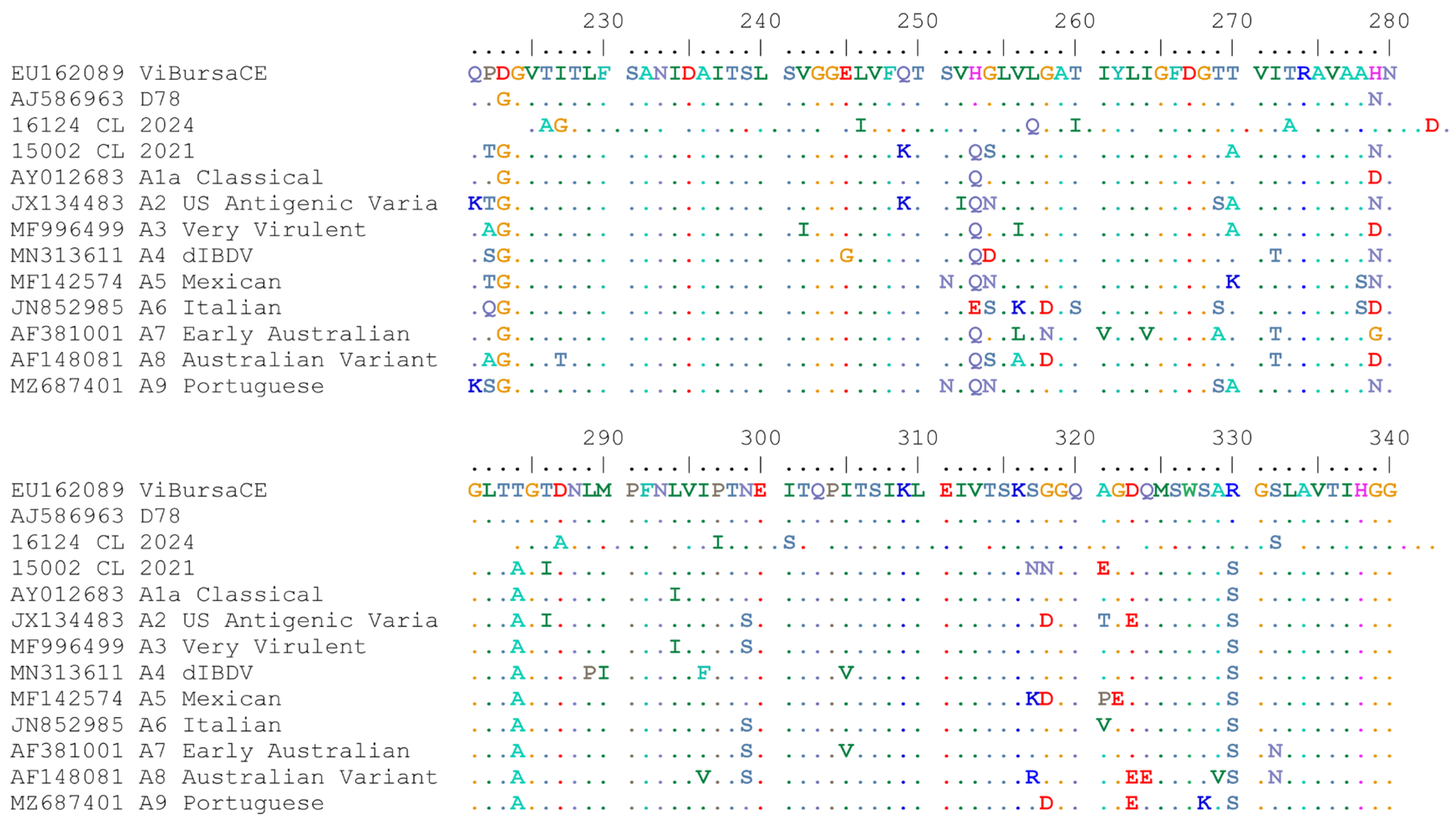
3.7. Analysis of VP2 Hypervariable Region (hvVP2)

The amino acid sequence for the VP2 hypervariable region of the strain isolated in this study was compared with previously published Chilean IBDV sequence; representative sequences recovered from GenBank of A1, A2, A3, A4, A5, A6, A7, A8, and A9 genogroups; and sequences from live attenuated vaccines D78 and ViBursa CE. The VP2 amino acid sequences of the strain isolated in this study had the markers typical of vvIBDV (222A, 242I, 256I, 279D, 294I, and 299S) [20] (Figure 6).

4. Discussion

IBD causes significant losses for the poultry industry. The cvIBDV, varIBDV, and vvIBDV strains have spread worldwide, and the presence of IBDV in Chile, specifically the classic (A1B1) and variant (A2B1) strains, has been previously confirmed [37]. However, this is the first report of a very virulent IBDV (vvIBDV). IBDV vaccination is widely used in poultry production worldwide, and live-attenuated vaccines based on classical-type virus strains are common. Despite vaccination, IBD is continuously observed [46].
The pullets examined in this study showed swollen, hemorrhagic, and edematous bursa of Fabricius, similar to that previously characterized in vvIBDV strains [47]. The vvIBDV isolated in fertile 9- to 11-day-old SPF-ECE resulted in dwarfing and greenish liver in the inoculated dead embryos, similar to previous observations [48]. The presence of IBDV was confirmed in the bursa of Fabricius sample and the CAM by RT-PCR using a set of primers previously designed to amplify the hv region of the VP2 gene and the VP1 gene of IBDV strains [39,40].
According to the phylogenetic analysis of the nucleotide sequences of the VP2 and VP1 genes (segment A and B, respectively), the isolated IBDV strain was classified within the A3B5 genotype using the scheme proposed by Islam et al. [26] and modified by Wang et al. [28]. Genogroup A3 (previously G3) has been observed in strains from the Netherlands, France, Poland, China, Finland, Vietnam, and other countries [48]. In Latin America, it has also been characterized in Argentina [36], Brazil [49], and Uruguay [50]. The hvVP2 sequences of this virus are genetically similar to those of the vvIBDV strains EU042139 and AY134874 of Chinese origin [51]. Meanwhile, genogroup B5 has only been identified in Nigeria [52]. The VP1 sequence of this virus is genetically similar to the vvIBDV strains KP172130, KP172087, and KP266336 of Nigerian origin. The A3B5 genotype identified in this study is part of a monophyletic cluster formed exclusively by Nigerian strains [53]. The origin of this new A3B5 genotype in Chile remains unknown. There are no data on the occurrence of this genotype in Latin America. More studies and greater GenBank database availability of sequences originating in the region are required to clarify this issue.
The high percentage identity of the VP1 and VP2 genes (average of 97.36% and 95.31%, respectively) between the virus isolated in this study and other IBDVs previously reported (mainly in Brazil) could indicate a possible origin, probably due to the importation of sub-clinically infected birds from nearby countries. This needs to be properly determined. Previous reports have indicated that vvIBDV strains have been introduced in Brazil, mainly from the Netherlands [54] and that very virulent IBDV isolates in Latin America emerged from virulent strains from Europe or Asia. This could partly explain the transmission route of the virus from Africa and Asia to America [35].
Based on segments A and B, the estimated amino acid distance between IBDV genogroups confirmed that this isolated IBDV strain belongs to the A3B5 genotype and was classified as very virulent. Amino acid analysis of the hvVP2 region revealed that the 16,124 CL strain contained the conserved residues (A222, I242, I256, I294, and S299) typical of vvIBDV strains [24]. Residues at positions 222, 242, 256, 279, 294, and 299 are involved in antigenicity, virulence, cell tropism, ability to replicate in chicken embryonic fibroblasts, and pathogenicity [39,54]. The hypervariable region of VP2 (aa 210–350) in 16,124 CL was completely identical to the vvIBDV strain MF996499 from Pakistan [55], confirming observations in the pathological findings of the affected birds, phylogenetic analysis, and analysis of the nucleotide sequences of the VP1 and VP2 genes. To the best of our knowledge, this is the first report of vvIBDV infection in Chile.

5. Conclusions

The presence of very virulent IBDV strain in Chile has been confirmed for the first time. Phylogenetic analysis based on segments A and B classified the strain within the A3B5 genotype, previously identified only in strains isolated from Nigeria. However, the high percentage of nucleotide identity in both genes relative to strains isolated in America suggests that the strain may have been introduced in Chile from nearby countries via the importation of sub-clinically infected birds. The flock was not adequately vaccinated against IBD; the emergence of new variants reinforces the importance of vaccination to prevent the spread, mutation, and reassortment of IBDV strains. Despite the widely distributed immunoprophylaxis, live-attenuated vaccines based on classic strains, as commonly used in Chile, may not adequately protect against vvIBDV strains. As a result, developing next-generation vaccines—e.g., approaches based on bioinformatics and reverse vaccinology [56]—is essential to effectively and safely controlling the disease in susceptible birds. The presence of three genotypes of IBDV in Chile (A1B1, A2B1, and A3B5) has been confirmed. Given the IBDV strain’s reassortment capacity, co-circulation of several different genotypes in the same region, simultaneous co-infection of the same host, and vaccination failures could favor the emergence of a new reassortment of viruses. Epidemiological surveillance and pathogenicity studies of vvIBDV strains are necessary to evaluate the effectiveness of the vaccines currently available in Chile.

Author Contributions

Conceptualization, L.C.; methodology, L.C., P.R., M.G. and F.N.; investigation, L.C., P.T. and H.H.; writing—original draft preparation, L.C.; writing—review and editing, L.C., M.G., P.T. and H.H.; visualization, L.C.; supervision, H.H. All authors have read and agreed to the published version of the manuscript.

Funding

This research was funded with internal funds from the Avian Pathology Laboratory, Department of Animal Pathology, Faculty of Veterinary and Animal Sciences, Universidad de Chile.

Institutional Review Board Statement

Not applicable.

Informed Consent Statement

Not applicable.

Data Availability Statement

In this study, all data analyzed were collected as part of routine diagnosis; therefore, according to national legislation, ethical approval and written informed consent were not required.

Conflicts of Interest

The authors declare no conflicts of interest.

References

  1. Dobos, P.; Hill, B.J.; Hallett, R.; Kells, D.T.; Becht, H.; Teninges, D. Biophysical and biochemical characterization of five animal viruses with bisegmented double-stranded RNA genomes. J. Virol. 1979, 32, 593–605. [Google Scholar] [CrossRef] [PubMed] [PubMed Central]
  2. Mahgoub, H.A.; Bailey, M.; Kaiser, P. An overview of infectious bursal disease. Arch. Virol. 2012, 157, e47–e57, Erratum in Arch. Virol. 2012, 157, 2059. [Google Scholar] [CrossRef] [PubMed]
  3. Faragher, J.T.; Allan, W.H.; Wyeth, P.J. Immunosuppressive effect of infectious bursal agent on vaccination against Newcastle disease. Vet. Rec. 1974, 95, 385–388. [Google Scholar] [CrossRef] [PubMed]
  4. Delmas, B.; Attoui, H.; Ghosh, S.; Malik, Y.S.; Mundt, E.; Vakharia, V.N.; ICTV Report Consortium. ICTV virus taxonomy profile: Birnaviridae. J. Gen. Virol. 2019, 100, 5–6. [Google Scholar] [CrossRef] [PubMed]
  5. Lee, C.C.; Ko, T.P.; Chou, C.C.; Yoshimura, M.; Doong, S.R.; Wang, M.Y.; Wang, A.H. Crystal structure of infectious bursal disease virus VP2 subviral particle at 2.6A resolution: Implications in virion assembly and immunogenicity. J. Struct. Biol. 2006, 155, 74–86. [Google Scholar] [CrossRef] [PubMed]
  6. Bayliss, C.D.; Spies, U.; Shaw, K.; Peters, R.W.; Papageorgiou, A.; Müller, H.; Boursnell, M.E. A comparison of the sequences of segment A of four infectious bursal disease virus strains and identification of a variable region in VP2. J. Gen. Virol. 1990, 71 Pt 6, 1303–1312. [Google Scholar] [CrossRef] [PubMed]
  7. Delmas, B. Birnaviruses. In Encyclopedia of Virology, 3rd ed.; Mahy, B.W.J., van Regenmortel, M.H.V., Eds.; Elsevier: Oxford, UK, 2008; Volume 1, pp. 321–328. [Google Scholar]
  8. Brandt, M.; Yao, K.; Liu, M.; Heckert, R.A.; Vakharia, V.N. Molecular determinants of virulence, cell tropism, and pathogenic phenotye of infectious bursal disease virus. J. Virol. 2001, 75, 11974–11982. [Google Scholar] [CrossRef] [PubMed] [PubMed Central]
  9. Coulibaly, F.; Chevalier, C.; Gutsche, I.; Pous, J.; Navaza, J.; Bressanelli, S.; Delmas, B.; Rey, F.A. The birnavirus crystal structure reveals structural relationships among icosahedral viruses. Cell 2005, 120, 761–772. [Google Scholar] [CrossRef] [PubMed]
  10. Jackwood, D.J.; Saif, Y.M.; Moorhead, P.D. Immunogenicity and antigenicity of infectious bursal disease virus serotypes I and II in chickens. Avian Dis. 1985, 29, 1184–1194. [Google Scholar] [CrossRef] [PubMed]
  11. Chettle, N.; Stuart, J.C.; Wyeth, P.J. Outbreak of virulent infectious bursal disease in East Anglia. Vet. Rec. 1989, 125, 271–272. [Google Scholar] [CrossRef]
  12. Jackwood, D.H.; Saif, Y.M. Antigenic diversity of infectious bursal disease viruses. Avian Dis. 1987, 31, 766–770. [Google Scholar] [CrossRef] [PubMed]
  13. Jackwood, D.J.; Sommer-Wagner, S.E.; Crossley, B.M.; Stoute, S.T.; Woolcock, P.R.; Charlton, B.R. Identification and pathogenicity of a natural reassortant between a very virulent serotype 1 infectious bursal disease virus (IBDV) and a serotype 2 IBDV. Virology 2011, 420, 98–105. [Google Scholar] [CrossRef] [PubMed]
  14. Van den Berg, T.P.; Morales, D.; Eterradossi, N.; Rivallan, G.; Toquin, D.; Raue, R.; Zierenberg, K.; Zhang, M.F.; Zhu, Y.P.; Wang, C.Q.; et al. Assessment of genetic, antigenic and pathotypic criteria for the characterization of IBDV strains. Avian Pathol. 2004, 33, 470–476. [Google Scholar] [CrossRef] [PubMed]
  15. Saif, Y.M. Immunosuppression induced by infectious bursal disease virus. Vet. Immunol. Immunopathol. 1991, 30, 45–50. [Google Scholar] [CrossRef] [PubMed]
  16. Spackman, E.; Stephens, C.B.; Pantin-Jackwood, M.J. The Effect of Infectious Bursal Disease Virus-Induced Immunosuppression on Vaccination Against Highly Pathogenic Avian Influenza Virus. Avian Dis. 2018, 62, 36–44. [Google Scholar] [CrossRef] [PubMed]
  17. Li, K.; Niu, X.; Jiang, N.; Zhang, W.; Wang, G.; Li, K.; Huang, M.; Gao, Y.; Qi, X.; Wang, X. Comparative Pathogenicity of Three Strains of Infectious Bursal Disease Virus Closely Related to Poultry Industry. Viruses 2023, 15, 1257. [Google Scholar] [CrossRef] [PubMed] [PubMed Central]
  18. Firquet, S.; Beaujard, S.; Lobert, P.E.; Sané, F.; Caloone, D.; Izard, D.; Hober, D. Survival of Enveloped and Non-Enveloped Viruses on Inanimate Surfaces. Microbes Environ. 2015, 30, 140–144. [Google Scholar] [CrossRef] [PubMed] [PubMed Central]
  19. Cosgrove, A.S. An Apparently New Disease of Chickens: Avian Nephrosis. Avian Dis. 1962, 6, 385. [Google Scholar] [CrossRef]
  20. Rosenberger, J.K.; Cloud, S.S. Isolation and characterization of variant infectious bursal disease viruses. J. Am. Vet. Med. Assoc. 1986, 189, 357. [Google Scholar]
  21. Berg, T.P.; Gonze, M.; Morales, D.; Meulemans, G. Acute infectious bursal disease in poultry: Immunological and molecular basis of antigenicity of a highly virulent strain. Avian Pathol. 1996, 25, 751–768. [Google Scholar] [CrossRef] [PubMed]
  22. Di Fabio, J.; Rossini, L.I.; Eterradossi, N.; Toquin, M.D.; Gardin, Y. European-like pathogenic infectious bursal disease viruses in Brazil. Vet. Rec. 1999, 145, 203–204. [Google Scholar] [PubMed]
  23. Stoute, S.T.; Jackwood, D.J.; Sommer-Wagner, S.E.; Cooper, G.L.; Anderson, M.L.; Woolcock, P.R.; Bickford, A.A.; Sentíes-Cué, C.G.; Charlton, B.R. The diagnosis of very virulent infectious bursal disease in California pullets. Avian Dis. 2009, 53, 321–326. [Google Scholar] [CrossRef] [PubMed]
  24. Michel, L.O.; Jackwood, D.J. Classification of infectious bursal disease virus into genogroups. Arch. Virol. 2017, 162, 3661–3670. [Google Scholar] [CrossRef] [PubMed] [PubMed Central]
  25. Escaffre, O.; Le Nouën, C.; Amelot, M.; Ambroggio, X.; Ogden, K.M.; Guionie, O.; Toquin, D.; Müller, H.; Islam, M.R.; Eterradossi, N. Both genome segments contribute to the pathogenicity of very virulent infectious bursal disease virus. J. Virol. 2013, 87, 2767–2780. [Google Scholar] [CrossRef] [PubMed] [PubMed Central]
  26. Islam, M.R.; Nooruzzaman, M.; Rahman, T.; Mumu, T.T.; Rahman, M.M.; Chowdhury, E.H.; Eterradossi, N.; Müller, H. A unified genotypic classification of infectious bursal disease virus based on both genome segments. Avian Pathol. 2021, 50, 190–206. [Google Scholar] [CrossRef] [PubMed]
  27. Wang, Y.; Fan, L.; Jiang, N.; Gao, L.; Li, K.; Gao, Y.; Liu, C.; Cui, H.; Pan, Q.; Zhang, Y.-P.; et al. An improved scheme for infectious bursal disease virus genotype classification based on both genome-segments A and B. J. Integr. Agric. 2021, 20, 1372–1381. [Google Scholar] [CrossRef]
  28. Legnardi, M.; Franzo, G.; Tucciarone, C.M.; Koutoulis, K.; Duarte, I.; Silva, M.; Le Tallec, B.; Cecchinato, M. Detection and molecular characterization of a new genotype of infectious bursal disease virus in Portugal. Avian Pathol. 2022, 51, 97–105. [Google Scholar] [CrossRef] [PubMed]
  29. Pikuła, A.; Lisowska, A.; Domańska-Blicharz, K. Epidemiology of Infectious Bursal Disease Virus in Poland During 2016–2022. Viruses 2023, 15, 289. [Google Scholar] [CrossRef] [PubMed] [PubMed Central]
  30. Lian, J.; Wang, Z.; Xu, Z.; Pang, Y.; Leng, M.; Tang, S.; Zhang, X.; Qin, J.; Chen, F.; Lin, W. Pathogenicity and molecular characterization of infectious bursal disease virus in China. Poult. Sci. 2022, 101, 101502. [Google Scholar] [CrossRef] [PubMed] [PubMed Central]
  31. Tammiranta, N.; Ek-Kommonen, C.; Rossow, L.; Huovilainen, A. Circulation of very virulent avian infectious bursal disease virus in Finland. Avian Pathol. 2018, 47, 520–525. [Google Scholar] [CrossRef] [PubMed]
  32. Arowolo, O.A.; George, U.E.; Luka, P.D.; Maurice, N.A.; Atuman, Y.J.; Shallmizhili, J.J.; Shittu, I.; Oluwayelu, D.O. Infectious bursal disease in Nigeria: Continuous circulation of reassortant viruses. Trop. Anim. Health Prod. 2021, 53, 271. [Google Scholar] [CrossRef] [PubMed]
  33. Samy, A.; Courtillon, C.; Briand, F.X.; Khalifa, M.; Selim, A.; Arafa, A.E.S.; Hegazy, A.; Eterradossi, N.; Soubies, S.M. Continuous circulation of an antigenically modified very virulent infectious bursal disease virus for fifteen years in Egypt. Infect. Genet. Evol. 2020, 78, 104099. [Google Scholar] [CrossRef] [PubMed]
  34. Legnardi, M.; Franzo, G.; Tucciarone, C.M.; Koutoulis, K.; Cecchinato, M. Infectious bursal disease virus in Western Europe: The rise of reassortant strains as the dominant field threat. Avian Pathol. 2023, 52, 25–35. [Google Scholar] [CrossRef] [PubMed]
  35. Banda, A.; Villegas, P. Genetic characterization of very virulent infectious bursal disease viruses from Latin America. Avian Dis. 2004, 48, 540–549. [Google Scholar] [CrossRef] [PubMed]
  36. Remorini, P.; Calderón, M.G.; Aguirre, S.; Periolo, O.; La Torre, J.; Mattion, N. Characterization of infectious bursal disease viruses from Argentina. Avian Dis. 2006, 50, 245–251. [Google Scholar] [CrossRef] [PubMed]
  37. Guzmán, M.; Cádiz, L.; Guerrero-Moncayo, A.; Cáceres, F.; Vidal, S.; Lapierre, L.; Sáenz, L.; Hidalgo, H. Molecular characterization of Infectious Bursal Disease Virus isolated in Chile reveals several mutations in VP2 coding region and a reassortment in its genome. Vet. Res. Commun. 2022, 46, 1281–1289. [Google Scholar] [CrossRef] [PubMed] [PubMed Central]
  38. World Organization of Animal Health (WOAH). Chapter 3.3.12—Infect Bursal Disease (Gumboro Disease). In Terrestrial Manual; WOAH: Paris, France, 2024; pp. 1–23. [Google Scholar]
  39. Eterradossi, N.; Arnauld, C.; Toquin, D.; Rivallan, G. Critical amino acid changes in VP2 variable domain are associated with typical and atypical antigenicity in very virulent infectious bursal disease viruses. Arch. Virol. 1998, 143, 1627–1636. [Google Scholar] [CrossRef] [PubMed]
  40. Nouën, C.L.; Rivallan, G.; Toquin, D.; Darlu, P.; Morin, Y.; Beven, V.; de Boisseson, C.; Cazaban, C.; Comte, S.; Gardin, Y.; et al. Very virulent infectious bursal disease virus: Reduced pathogenicity in a rare natural segment-B-reassorted isolate. J. Gen. Virol. 2006, 87 Pt 1, 209–216. [Google Scholar] [CrossRef] [PubMed]
  41. Alzohairy, A. BioEdit: An important software for molecular biology. GERF Bull. Biosci. 2011, 2, 60–61. [Google Scholar]
  42. Katoh, K.; Standley, D.M. MAFFT multiple sequence alignment software version 7: Improvements in performance and usability. Mol. Biol. Evol. 2013, 30, 772–780. [Google Scholar] [CrossRef] [PubMed] [PubMed Central]
  43. Guindon, S.; Dufayard, J.F.; Lefort, V.; Anisimova, M.; Hordijk, W.; Gascuel, O. New algorithms and methods to estimate maximum-likelihood phylogenies: Assessing the performance of PhyML 3.0. Syst Biol. 2010, 59, 307–321. [Google Scholar] [CrossRef] [PubMed]
  44. Drummond, A.J.; Rambaut, A. BEAST: Bayesian evolutionary analysis by sampling trees. BMC Evol. Biol. 2007, 7, 214. [Google Scholar] [CrossRef] [PubMed] [PubMed Central]
  45. Tamura, K.; Stecher, G.; Kumar, S. MEGA11: Molecular Evolutionary Genetics Analysis Version 11. Mol. Biol. Evol. 2021, 38, 3022–3027. [Google Scholar] [CrossRef] [PubMed]
  46. Setta, A.; Yehia, N.; Shaheen, M.; Shami, A.; Al-Saeed, F.A.; Alsamghan, A.; Amin, R.; El-Saadony, M.T.; El-Tarabily, K.A.; Salem, H.M. Continuous clinicopathological and molecular recognition of very virulent infectious bursal disease virus in commercial broiler chickens. Poult. Sci. 2024, 103, 103306. [Google Scholar] [CrossRef] [PubMed] [PubMed Central]
  47. Mosad, S.M.; Eladl, A.H.; El-Tholoth, M.; Ali, H.S.; Hamed, M.F. Molecular characterization and pathogenicity of very virulent infectious bursal disease virus isolated from naturally infected turkey poults in Egypt. Trop. Anim. Health Prod. 2020, 52, 3819–3831. [Google Scholar] [CrossRef] [PubMed]
  48. Le, X.T.K.; Do, R.T.; Doan, H.T.T.; Nguyen, K.T.; Pham, L.T.K.; Le, T.H. Phylogenotyping of infectious bursal disease virus in Vietnam according to the newly unified genotypic classification scheme. Arch. Virol. 2023, 168, 201. [Google Scholar] [CrossRef] [PubMed]
  49. De Fraga, A.P.; Gräf, T.; Coltro, V.P.; Ikuta, N.; Fonseca, A.S.K.; Majó, N.; Lunge, V.R. Phylodynamic analyses of Brazilian antigenic variants of infectious bursal disease virus. Infect. Genet. Evol. 2019, 73, 159–166. [Google Scholar] [CrossRef] [PubMed]
  50. Hernández, M.; Banda, A.; Hernández, D.; Panzera, F.; Pérez, R. Detection of very virulent strains of infectious bursal disease virus (vvIBDV) in commercial broilers from Uruguay. Avian Dis. 2006, 50, 624–631. [Google Scholar] [CrossRef] [PubMed]
  51. Sun, J.H.; Lu, P.; Yan, Y.X.; Hua, X.G.; Jiang, J.; Zhao, Y. Sequence and analysis of genomic segment A and B of very virulent infectious bursal disease virus isolated from China. J. Vet. Med. B Infect. Dis. Vet. Public Health 2003, 50, 148–154. [Google Scholar] [CrossRef] [PubMed]
  52. Nwagbo, I.; Milani, A.; Salviato, A.; Zamperin, G.; Sulaiman, L.; Maurice, N.; Meseko, C.; Fusaro, A.; Shittu, I. Genomic Analysis of Infectious Bursal Disease Virus in Nigeria: Identification of Unique Mutations of Yet Unknown Biological Functions in Both Segments A and B. Vaccines 2023, 11, 867. [Google Scholar] [CrossRef] [PubMed] [PubMed Central]
  53. Silva, F.M.; Vidigal, P.M.; Myrrha, L.W.; Fietto, J.L.; Silva, A., Jr.; Almeida, M.R. Tracking the molecular epidemiology of Brazilian Infectious bursal disease virus (IBDV) isolates. Infect. Genet. Evol. 2013, 13, 18–26. [Google Scholar] [CrossRef] [PubMed]
  54. Mundt, E. Tissue culture infectivity of different strains of infectious bursal disease virus is determined by distinct amino acids in VP2. J. Gen. Virol. 1999, 80 Pt 8, 2067–2076. [Google Scholar] [CrossRef] [PubMed]
  55. Shabbir, M.Z.; Ali, M.; Abbas, M.; Chaudhry, U.N.; Zia-Ur-Rehman, M.M. Molecular characterization of infectious bursal disease viruses from Pakistan. Arch. Virol. 2016, 161, 2001–2006. [Google Scholar] [CrossRef] [PubMed]
  56. Gul, I.; Hassan, A.; Muneeb, J.M.; Akram, T.; Haq, E.; Shah, R.A.; Ganai, N.A.; Ahmad, S.M.; Chikan, N.A.; Shabir, N. A multiepitope vaccine candidate against infectious bursal disease virus using immunoinformatics-based reverse vaccinology approach. Front. Vet. Sci. 2023, 9, 1116400. [Google Scholar] [CrossRef] [PubMed] [PubMed Central]
Figure 1. Gross lesions in affected pullets. (a) Swollen, hemorrhagic, and edematous bursa of Fabricius; (b) swollen, hemorrhagic, and edematous bursae of Fabricius removed from affected birds; (c) hemorrhages in pectoral muscle. Bar = 1 cm.
Microbiolres 16 00031 g001
Figure 2. Isolation of the IBDV strain in 10-day-old chick embryo. (a) Chicken embryo with dwarfism; (b) swollen, greenish-colored liver; (c) chicken embryo negative control. Bar = 1 cm.
Microbiolres 16 00031 g002
Figure 3. PCR amplicons of VP1 (lanes 2–4) and VP2 (lanes 7–9). Lane 1: 100 bp DNA ladder. Lane 2: Bursa homogenate sample. Lane 3: CAM sample. Lane 4: Positive control. Lane 5: Negative control. Lane 6: 100 bp DNA ladder. Lane 7: Bursa homogenate sample. Lane 8: CAM sample. Lane 9: Positive control. Lane 10: Negative control. Lane 11: 100 bp DNA ladder.
Microbiolres 16 00031 g003
Figure 4. Maximum-likelihood tree (polar format) with 1000 bootstrap replicates. The tree was constructed using 57 VP1 sequences, including the one isolated in this study and other Chilean IBDV sequences. Genogroups were highlighted as follows: B1 = blue, B2 = red, B3 = green, B4 = light violet, B5 = dark violet.
Microbiolres 16 00031 g004
Figure 5. Maximum-likelihood tree (polar format) with 1000 bootstrap replicates. The tree was constructed using 79 VP2 sequences, including the one isolated in this study and other Chilean IBDV sequences. Genogroups were highlighted as follows: A0 = black, A1 = blue, A2 = light blue, A3 = red, A4 = yellow, A5 = dark red, A6 = light purple, A7 = green, A8 = dark purple, A9 = orange.
Microbiolres 16 00031 g005
Figure 6. Amino acid differences in hvVP2 (aa 220–340) between the virus isolated in this study, representative viruses of each genogroup, and two vaccines currently used in Chile. The dots indicate that the sample had the same amino acid as the query sequence. Abbreviations: A—alanine, D—aspartic acid, E—glutamic acid, F—phenylalanine, G—glycine, H—histidine, I –isoleucine, K—lysine, L—leucine, Q—glutamine, N—asparagine, P—proline, S—serine, R—arginine, T—threonine, V—valine, Y—tyrosine.
Microbiolres 16 00031 g006
Table 1. Primers used in this study.
SegmentGenPrimerSequence (5′ to 3′)PCR Product (bp)Reference
AVP2U3TGTAAAACGACGGCCAGTGCATGCGGTATGTGAGGCTTGGTGAC604[39]
L3CAGGAAACAGCTATGACCGAATTCGATCCTGTTGCCACTCTTTC
BVP1+290TGTAAAACGACGGCCAGTGAATTCAGATTCTGCAGCCACGGTCTCT642[40]
−861CAGGAAACAGCTATGACCCTGCAGTTGATGACTTGAGGTTGATTTTG
Table 2. Closest BLAST matches against NCBI database of IBDV segments A and B.
SegmentGeneAccession Number% IdentityAccession NumberCountryYear
AVP2PP82454495.31%JN982259Brazil2013
95.31%JF811919Brazil2016
95.31%DQ286035Brazil2008
BVP1PP824543 97.64%JN982250Brazil2013
97.30%JN982249Brazil2013
97.13%DQ679811USA2008
Table 3. Amino acid distance between IBDV genogroups based on segment A 1.
A0A1A2A3A4A5A6A7A8A9CL1CL2
A0 0.06740.07790.07140.07150.07690.07330.07190.06410.07780.07010.0717
A10.4360 0.02540.02100.02430.02840.02700.02670.03390.02790.01240.0206
A20.49240.1110 0.02810.03150.02810.03310.03460.03730.02320.02830.0282
A30.46790.07940.1157 0.02990.03320.02610.02980.03070.03250.02520.0035
A40.47080.09570.13240.1207 0.03330.03290.03000.03580.03290.02680.0299
A50.49930.11060.10210.13140.1307 0.03380.03880.04090.02780.03050.0329
A60.45350.11150.13960.10040.13880.1308 0.03250.03350.03470.02990.0265
A70.47690.11300.16010.12230.12200.16950.1347 0.03210.03790.02880.0300
A80.38570.15690.17670.13400.15380.18750.13810.1366 0.04050.03660.0311
A90.49060.10710.07080.12380.12100.08720.13680.16100.1823 0.03110.0321
CL10.44790.04810.11500.09210.10090.11330.11460.12020.16200.1146 0.0252
CL20.46430.07160.10780.01020.11530.12490.09680.11690.12970.11530.0860
1 The number of per-site amino acid substitutions based on the average of all inter-group sequence pairs. Standard error estimates are shown above the diagonal. “CL1” indicates the group of Chilean viruses previously isolated. “CL2” indicates the virus isolated in this study.
Table 4. Amino acid distance between IBDV genogroups based on segment B 1.
B1B2B3B4B5CL2CL1
B1 0.02200.01990.01990.02440.02670.0075
B20.1273 0.02210.02380.02310.02440.0227
B30.12410.1361 0.01650.02710.02950.0205
B40.13100.14920.1036 0.02490.02870.0204
B50.14840.13850.16330.1498 0.00900.0249
CL20.16250.14660.18040.16990.0365 0.0272
CL10.04700.13050.13200.13690.15400.1642
1 The number of per-site amino acid substitutions based on the average of all inter-group sequence pairs. Standard error estimates are shown above the diagonal. “CL1” indicates the group of Chilean viruses previously isolated. “CL2” indicates the virus isolated in this study.
Disclaimer/Publisher’s Note: The statements, opinions and data contained in all publications are solely those of the individual author(s) and contributor(s) and not of MDPI and/or the editor(s). MDPI and/or the editor(s) disclaim responsibility for any injury to people or property resulting from any ideas, methods, instructions or products referred to in the content.

