Communication

Development of a Sensitive and Selective Fluorescent Substrate for the Detection of Chitinase Activity in Entomopathogenic Fungi

by
Estefan Miranda-Miranda
,
César A. Arreguín-Pérez
,
Hugo Aguilar-Díaz
and
Raquel Cossío-Bayúgar
*
Centro Nacional de Investigación Disciplinaria en Salud Animal, Instituto Nacional de Investigaciones Forestales, Agrícolasy Pecuarias (INIFAP), Boulevard Cuauhnahuac 8534, Jiutepec 62574, Morelos, Mexico
*
Author to whom correspondence should be addressed.
Microbiol. Res. 2025, 16(11), 243; https://doi.org/10.3390/microbiolres16110243
Submission received: 21 October 2025 / Revised: 5 November 2025 / Accepted: 11 November 2025 / Published: 19 November 2025

Abstract

The identification and quantification of chitinolytic activity in microorganisms is critical for advancing biological control strategies against arthropod pests and fungal pathogens. However, current laboratory methods designed for fast detection of chitinolytic microorganisms are often time-consuming, produce low-quality results and lack sensitivity. Here, we report the development of a novel fluorogenic culture medium incorporating a chemically modified chitinase substrate, N-fluoresceyl poly-D-glucosamine, which allows for a highly sensitive chitinase assay, enabling both qualitative and quantitative fluorescent detection of chitinase activity in situ. This substrate is synthesized through covalent conjugation of poly-D-glucosamine with fluorescein isothiocyanate under alkaline conditions, resulting in an insoluble polymer that becomes fluorescent upon enzymatic hydrolysis by chitinases. When supplemented with culture media, the modified fluorogenic substrate serves as the sole carbon source, selectively supporting the growth of chitinolytic microorganisms. Enzymatic activity is visualized under longwave UV light and can be quantitatively measured via spectrophotometric (493 nm) or fluorometric (530 nm) methods. Validation using characterized entomopathogenic chitinolytic strains of the fungi Aspergillus flavus, Beauveria bassiana, and Metarhizium anisopliae demonstrated a detection sensitivity that was at least three orders of magnitude greater than that of conventional methods. In contrast, the non-chitinolytic fungi Penicillium notatum and Fusarium venenatum presented no detectable fluorescent signals. This fluorogenic medium provides a rapid, cost-effective, and highly sensitive tool for screening chitinolytic microorganisms with potential applications in agriculture, veterinary parasitology, and environmental microbiology.

1. Introduction

Chitin is the second most abundant natural biopolymer on Earth after cellulose and is composed of N-acetyl-D-glucosamine units linked by β-1,4 glycosidic bonds [1]. It is a structural component of arthropod exoskeletons, fungal cell walls, and particular algal species [2]. Microorganisms capable of degrading chitin, collectively known as chitinolytic microorganisms, produce chitinases (EC 3.2.1.14), a group of enzymes that hydrolyze chitin into oligomers and monomers of N-acetyl-D-glucosamine. These organisms play pivotal roles in nutrient recycling, pathogenesis, and, more importantly, the biological control of arthropod vectors and phytopathogens [3].
Chitinases are found in several microorganisms, perform a variety of roles in nature, and are well documented in bacteria and fungi [4]. Fungal chitinases exhibit exoactivity based on the cleavage pattern of chitin, a molecular mass varying from 40 to 60 kDa, and exhibiting one catalytic domain [5].
In addition to bio pest control, chitinases have industrial value as enzymes in the biotechnology industry, with a constant search for new chitinases useful in various chitin fermentation procedures designed for different fungal species [6]. Chitinases can be applied in several sectors, such as the food and pharmaceutical industries, medicine, agriculture, and environmental management [7]. The treatment of residues containing chitin, such as shrimp, lobster, and crab shells, allows for the obtainment of products with added value that minimize the impact on the natural environment [8].
Chitinolytic fungi, particularly those with entomopathogenic properties, have been increasingly explored as eco-friendly alternatives to chemical pesticides in agriculture and public health [9]. However, one of the significant limitations in the screening and characterization of new chitinolytic microorganisms isolated from natural sources is the lack of sensitive, rapid, and quantitative detection methods [10]. Classic chitinase assays are designed as a medium for the culture of living microorganisms, based on colloidal chitin agar and visualization of hydrolysis halos or simply by observing the actual growth on chitin-based media; these methods are labor-intensive, and often yield false negatives, particularly for slow-growing or low-activity strains of fungi [11,12].
There are colorimetric and fluorescent laboratory methods suitable for the assessment of chitinase enzymatic activity in partially purified extracts from microorganism cultures [13,14]; however, these procedures rely on laboratory methods and specialized equipment performed on partially characterized fungal strains and are not suitable for screening unknown fungi from natural sources [15,16]. To address this gap, we developed a novel fluorogenic substrate, N-fluoresceyl poly-D-glucosamine, by covalently linking fluorescein isothiocyanate to poly-D-glucosamine. This substrate allows the selective growth of chitinolytic microorganisms from natural sources without prior characterization. This method enables the simultaneous detection of chitinase activity through the release of a fluorescent signal upon enzymatic hydrolysis of the modified chitinase substrate. The resulting culture medium enables both visual detection under UV light and quantitative measurements via fluorometry and spectrophotometry, significantly improving the sensitivity of chitinolytic microorganism identification. This study describes the synthesis, formulation, and application of a novel fluorogenic medium, demonstrating its utility when model entomopathogenic fungi are cultured in it. This approach offers a practical and cost-effective tool for screening chitinolytic microorganisms from natural sources with potential applications in microbial ecology, biocontrol agent discovery, and industrial biotechnology.

2. Materials and Methods

2.1. Tick and Fungal Strains

Engorged female Rhipicephalus microplus ticks of the INIFAP strain were obtained as previously described [17]. Some of the collected ticks presented natural and recurrent fungal infections, as previously reported [18]. Fungi-infected ticks were identified by their dark cuticle color and the presence of fungal mycelium and conidiophore growth (Figure 1). Fungal spores were collected and cultured on Sabouraud agar in Petri dishes as reported previously [15]. The isolated fungi were identified by morphology and genome sequencing and added to the fungal collection on INIFAP as previously reported [10]. Other fungal strains used during these experiments and provided by the INIFAP fungal collection included Penicillium notatum, Beauveria bassiana, Fusarium venenatum, and Metarhizium anisopliae.

2.2. Microorganisms and Inoculation

Aspergillus flavus INIFAP-2021 strain, an entomopathogenic strain previously isolated from Rhipicephalus microplus [15], was used as a model organism. The spore suspensions were prepared in sterile distilled water containing 0.05% Tween 20 and adjusted to 1 × 10⁵ spores/mL. One hundred microliters of the spore suspension were inoculated into 10 mL of fluorogenic medium and incubated at 25 °C for 48 h. Penicillium notatum, Beauveria bassiana, Fusarium venenatum, and Metarhizium anisopliae were also inoculated under the same conditions. No inoculated media served as negative controls for fluorescence readings.

2.3. Chemicals and Reagents

Poly-D-glucosamine (≥95% purity), fluorescein isothiocyanate (FITC), sodium bicarbonate (NaHCO3), chitinase from Streptomyces griseus and lysozyme from chicken egg, and all other analytical-grade reagents were purchased from Sigma-Aldrich (St. Louis, MO, USA). Phosphate-buffered saline (PBS, pH 7.2) was prepared with Na2HPO4, NaH2PO4, and NaCl. Peptone was obtained from Becton Dickinson (Franklin Lakes, NJ, USA).

2.4. Synthesis of N-Fluoresceyl Poly-D-Glucosamine
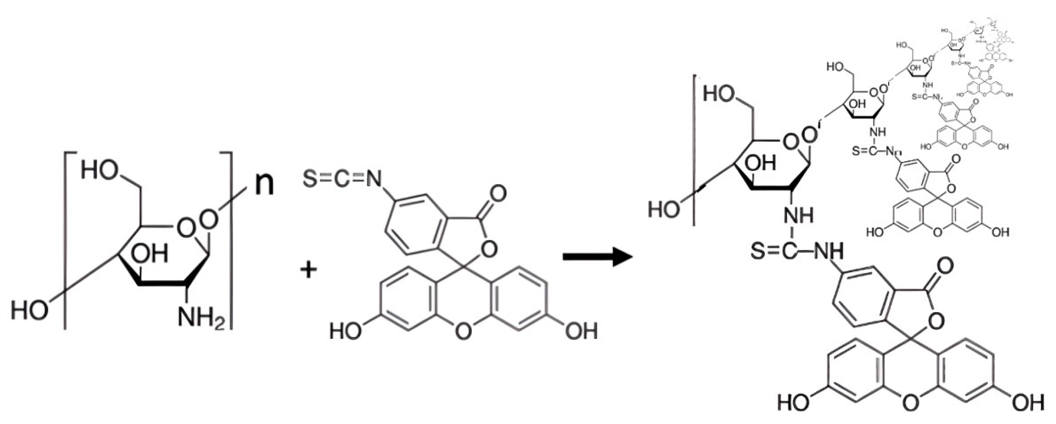
The fluorogenic substrate N-fluoresceyl poly-D-glucosamine was synthesized by dissolving poly-D-glucosamine in 0.1 M sodium bicarbonate buffer (pH 8.3) at room temperature. FITC was added at a weight-mass ratio of 1:200 (FITC 5 mg: poly-D-glucosamine 1000 mg), and the mixture was stirred gently for 1 h in the dark. The resulting product was centrifuged at 3000 rpm for 5 min and washed three times with 0.1 M NaHCO3, and the final sediment was collected and stored at 4 °C in the dark (Figure 1).
Figure 1. Schematic representation of the covalent conjugation reaction between poly-D-glucosamine and fluorescein isothiocyanate (FITC) under alkaline conditions to generate the fluorogenic substrate N-fluoresceyl poly-D-glucosamine. The isothiocyanate group of FITC reacts with the free amino group of poly-D-glucosamine, forming a stable thiourea bond. The resulting modified polymer remains insoluble and becomes fluorescent upon enzymatic hydrolysis by chitinases, enabling selective growth and fluorescence-based detection of chitinolytic microorganisms in culture media.
Microbiolres 16 00243 g001

2.5. Preparation of the Fluorogenic Culture Medium Domposition

The fluorescent fungal culture medium substrate was resuspended in phosphate-buffered saline (PBS, pH 7.2). The final composition served as the base additive for the selective fungal culture medium prepared with base compositions containing N-fluoresceyl D-glucosamine at a final concentration of 5 g per 100 mL supplemented with peptone at a 1:1 (v/w) ratio. The resulting media were sterilized by autoclaving under conditions compatible with the stability of the fluorogenic compound.

2.6. Qualitative Detection of Chitinolytic Activity

After incubation, the cultures were visually inspected under a longwave UV lamp (365 nm) to assess fluorescence in the supernatant as a proxy for chitinase activity. The fluorescence intensity was qualitatively graded on a scale from “none” (–) to “strong” (+++).

2.7. Quantitative Analysis of Chitinase Activity

Aliquots (5 µL) of culture supernatant were analyzed via a NABI UV/VIS microvolume spectrophotometer (MicroDigital Co., Seoul, Republic of Korea) at 493 nm for absorbance and 530 nm for fluorescence emission via a Quatus® fluorometer (Promega; Madison, WI, USA). The enzymatic activity was expressed as units per mL (U/mL), where one unit was defined as the amount of enzyme releasing one µmol of fluorescein per hour.

2.8. Statistical Analysis

The experiments were performed in quadruplicate. The results are reported as the means ± standard deviations (SDs). Statistical comparisons were made via Student’s t test, with significance set at p < 0.05.

3. Results

3.1. Experimental Fungal Infection of Engorged Female Ticks

To reproduce and document the infection process of Aspergillus flavus in Rhipicephalus microplus, adult engorged females were artificially inoculated in the laboratory as previously described [10]. Ticks were immersed for 10 min in a suspension containing 1 × 10⁷ spores/mL of A. flavus and incubated for 15 days at 28 °C and 80% relative humidity. Progressive fungal infection was observed on the tick cuticle and eggs, resembling the natural aspergillosis previously described in field-collected samples. Early infection stages were characterized by the appearance of small, homogeneously distributed plaques on the chitinous cuticle of ovipositing females (Figure 2A). As the infection advanced, extensive mycelial growth and conidiophore formation became evident, and the conidia propagated to the egg masses (Figure 2B,C). Microscopic examination confirmed mycelial penetration through the chitin shell of infected eggs, revealing the complete developmental cycle of A. flavus on tick tissues (Figure 2D).

3.2. Visual Detection of Chitinolytic Activity

After 48 h of incubation at 25 °C, cultures of Aspergillus flavus INIFAP-2021 in the fluorogenic medium presented a distinct fluorescent signal in the supernatant when exposed to longwave UV light (365 nm), indicating enzymatic hydrolysis of the N-fluoresceyl poly-D-glucosamine substrate. The fluorescence intensity was scored as strong (+++), whereas the noninoculated control media showed no detectable fluorescence (–), confirming the specificity of the substrate for microbial chitinase activity (Table 1).

3.3. Quantitative Measurement of Chitinase Activity

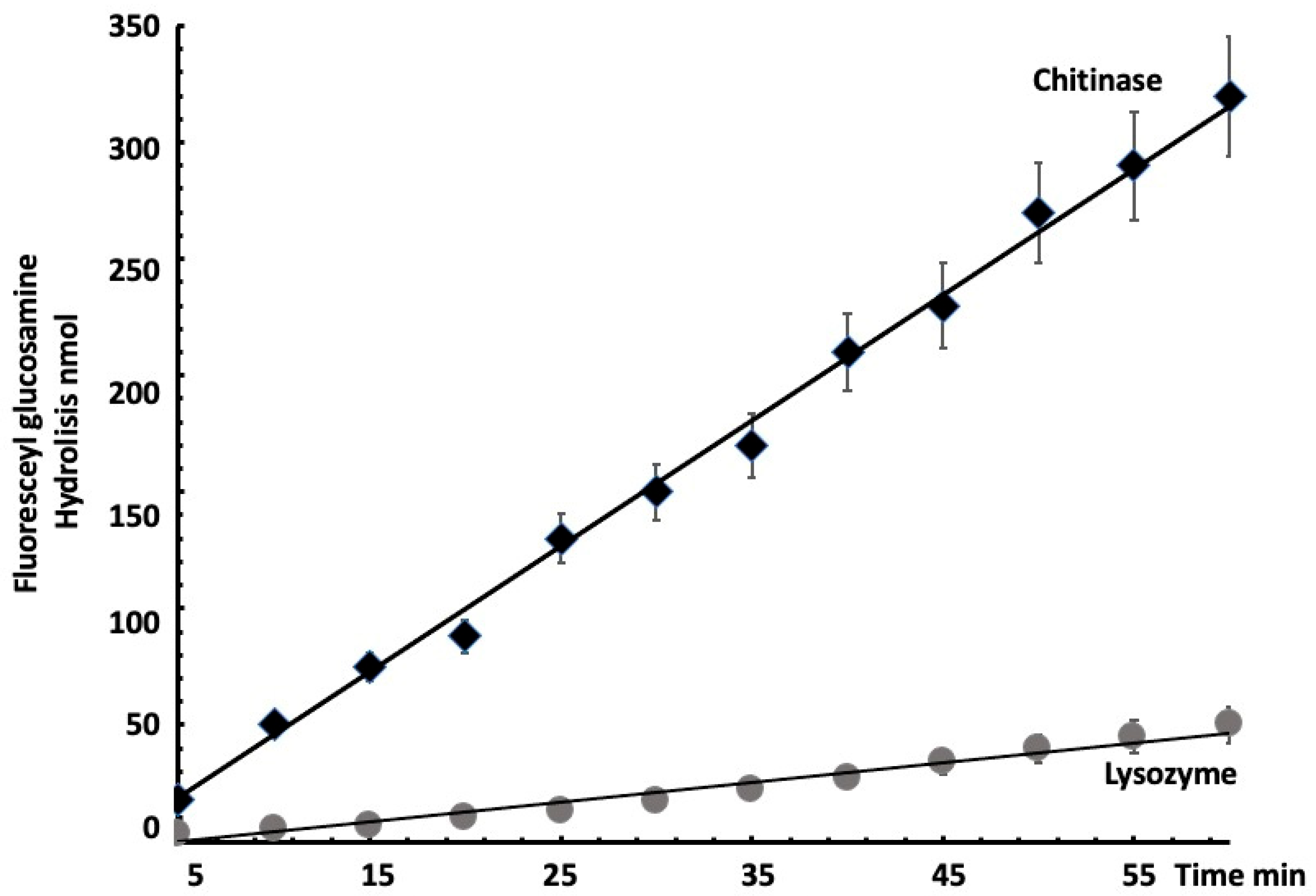
Spectrophotometric analysis of the culture supernatants revealed a marked increase in optical density at 493 nm and fluorescence emission at 530 nm in inoculated samples compared with those in controls. The calculated enzymatic activity reached 5.14 U/mL for A. flavus INIFAP-2021 cultures, whereas that of the control samples remained below 0.001 U/mL. These results demonstrate a high signal-to-noise ratio and a sensitivity threshold of at least three orders of magnitude compared with those of traditional halo-based agar methods. These results demonstrate a high signal-to-noise ratio and a sensitivity threshold that is at least three orders of magnitude greater than that of traditional halo-based agar methods, confirming the specificity and catalytic efficiency of the fluorogenic substrate when hydrolyzed by chitinases (Figure 3).

3.4. Reproducibility and Sensitivity

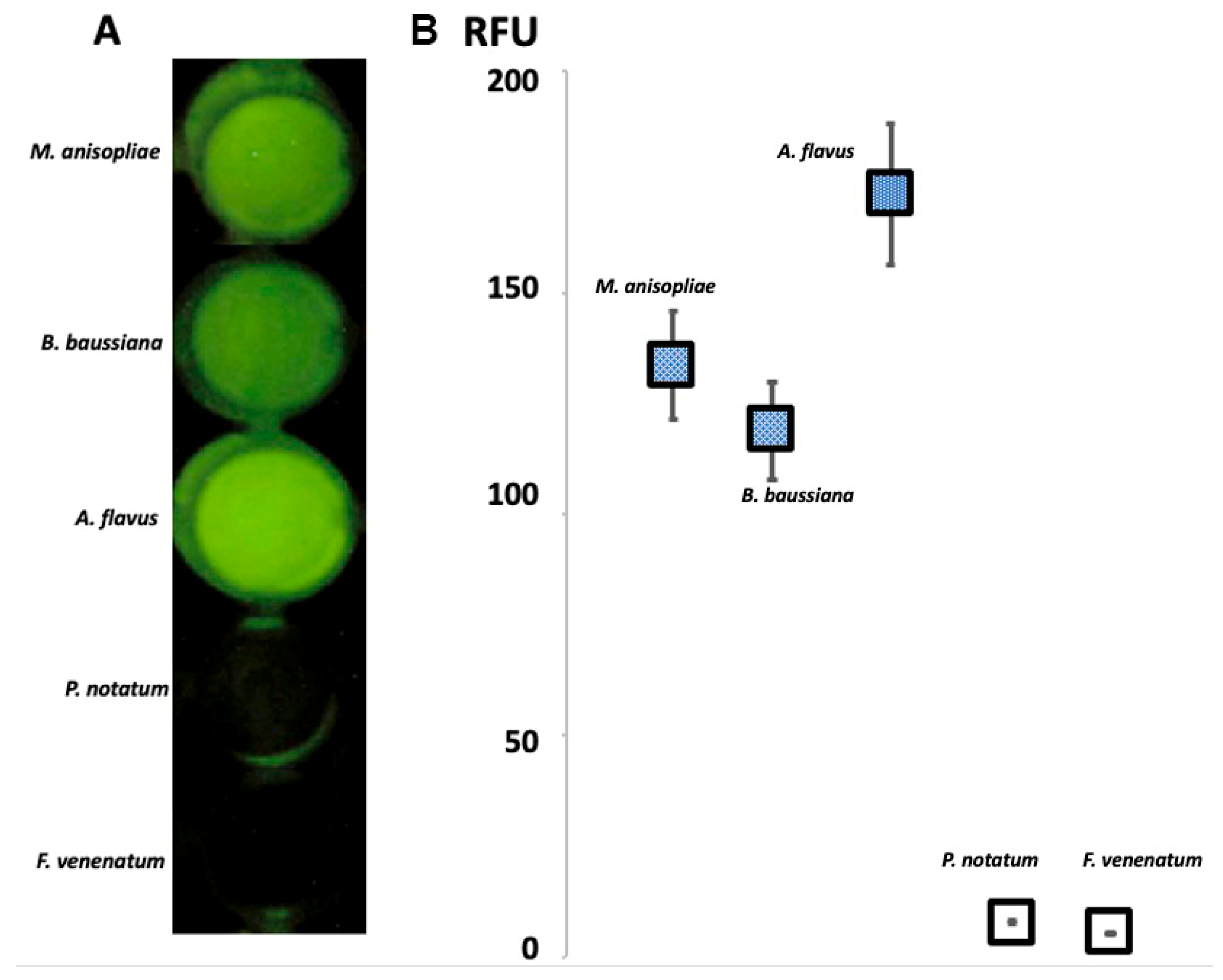
The results of the fluorogenic assay were consistent across all the replicates (n = 4), with a low standard deviation (±0.012 OD units). The statistical significance in every case exceeded two standard errors p > 0.005. Additionally, the method could detect chitinase activity in microbial suspensions as low as 103 chitinolytic cells/mL, indicating high analytical sensitivity suitable for screening low-density or slow-growing chitinolytic strains. The fluorogenic medium consistently detected chitinase activity across all replicates and fungal species tested, highlighting its robustness and analytical precision for screening chitinolytic microorganisms from diverse origins (Figure 4).

4. Discussion

Chitinase-producing microorganisms have long been recognized as valuable agents for the biodegradation of chitin-containing substrates and for their potential use in the biological control of pests and phytopathogens [3,19]. However, the development of practical tools for their identification and enzymatic characterization remains a significant bottleneck in both applied microbiology and industrial biotechnology. The fluorogenic medium described in this study provides a novel and effective solution to this problem by enabling qualitative and quantitative detection of chitinolytic activity in microbial cultures.
The conjugation of fluorescein isothiocyanate to poly-D-glucosamine results in a fluorogenic substrate that is highly sensitive to enzymatic hydrolysis by chitinases. Unlike traditional methods that rely on the visual detection of clearance halos on colloidal chitin agar—which are often subjective and lack sensitivity—this system allows direct detection of fluorescence in the supernatant with minimal background interference. The observed increase in the signal-to-noise ratio (≥1000-fold over control) represents a substantial improvement over conventional assays, making it suitable for screening slow-growing or weakly chitinolytic organisms that may be overlooked by standard techniques. A direct comparison with conventional N-acetyl-D-glucosamine enzymatic assays was beyond the scope of the present communication. However, this evaluation is planned for future work currently in development by our group.
The ability to quantify chitinase activity in culture supernatants through either absorbance at 493 nm or fluorescence emission at 530 nm adds further analytical utility. This dual-mode quantification allows researchers to adapt the assay to the available instrumentation while maintaining high reproducibility. Furthermore, the assay was sensitive enough to detect enzyme activity from inocula at concentrations as low as 103 cells/mL, suggesting its application in early-stage microbial screenings or environmental sample analysis.
Notably, the model strain Aspergillus flavus demonstrated robust chitinolytic activity under the conditions tested, validating the practical utility of the substrate for entomopathogenic fungi. Given that many biocontrol agents act through cuticle degradation mechanisms, the method described here may serve as a functional screening tool for selecting candidate organisms for agricultural, veterinary, or ecological applications.
Overall, this study introduces a low-cost, high-sensitivity assay that integrates microbial selectivity with precise enzymatic quantification. Beyond its immediate applications in microbiological screening, the approach could be adapted to high-throughput platforms or microfluidic devices for industrial or diagnostic purposes. Further studies could explore its compatibility with other chitinase-producing taxa and test its utility in environmental or clinical samples.

5. Patents

A patent application related to this work was filed by and in favor of the Instituto Nacional de Investigaciones Forestales, Agrícolas y Pecuarias (INIFAP) as the sole beneficiary. The patent is currently under second formal examination at the Mexican Patent Office, under Application No. MX/a/2024/001507, filed on April 22, 2024, entitled “Medio fluorogénico selectivo de microorganismos quitinolíticos.”

Author Contributions

E.M.-M.: conceptualization, sample isolation, writing—original draft, writing—review and editing; C.A.A.-P.: investigation, formal analysis; H.A.-D.: writing—review and editing, data analysis; R.C.-B.: conceptualization, writing—original draft, writing—review and editing, funding acquisition, project administration. All authors have read and agreed to the published version of the manuscript.

Funding

This research was funded by INIFAP-México (grant No. 1322633028), CONACYT (now SECHITI; grant No. 248049), and a CONACYT (now SECHITI) scholarship (No. 827786).

Institutional Review Board Statement

Not applicable.

Informed Consent Statement

Not applicable.

Data Availability Statement

The genome assembly dataset analyzed in this study has been deposited in GenBank under accession number PRJNA758689 (last accessed on 31 October 2025).

Conflicts of Interest

The authors declare that they have no conflicts of interest. The funders had no role in the design of the study; in the collection, analyses, or interpretation of data; in the writing of the manuscript; or in the decision to publish the results.

Abbreviations

The following abbreviations are used in this manuscript:
FITCFluorescein isothiocyanate
INIFAPInstituto Nacional de Investigaciones Forestales, Agrícolas y Pecuarias
PBSPhosphate-buffered saline
RFURelative fluorescence units
SDStandard deviation
UVUltraviolet
CONACYTConsejo Nacional de Humanidades, Ciencia y Tecnología

References

  1. Lv, J.; Lv, X.; Ma, M.; Oh, D.-H.; Jiang, Z.; Fu, X. Chitin and Chitin-Based Biomaterials: A Review of Advances in Processing and Food Applications. Carbohydr. Polym. 2023, 299, 120142. [Google Scholar] [CrossRef] [PubMed]
  2. Nunes, C.S.; Kumar, V. Enzymes in Human and Animal Nutrition; Elsevier: Amsterdam, The Netherlands, 2018; ISBN 978-0-12-805419-2. [Google Scholar]
  3. Unuofin, J.O.; Odeniyi, O.A.; Majengbasan, O.S.; Igwaran, A.; Moloantoa, K.M.; Khetsha, Z.P.; Iwarere, S.A.; Daramola, M.O. Chitinases: Expanding the Boundaries of Knowledge beyond Routinized Chitin Degradation. Environ. Sci. Pollut. Res. 2024, 31, 38045–38060. [Google Scholar] [CrossRef] [PubMed]
  4. Dukariya, G.; Kumar, A. Distribution and Biotechnological Applications of Chitinase: A Review. Int. J. Biochem. Biophys. 2020, 8, 17–29. [Google Scholar] [CrossRef]
  5. Ornela, P.H.; Guimarães, L.H.S. Purification, Characterization and Antifungal Activity of the Aspergillus niveus Chitinase Produced Using Shrimp Shells. Appl. Biosci. 2024, 3, 220–232. [Google Scholar] [CrossRef]
  6. Dahiya, N.; Tewari, R.; Hoondal, G.S. Biotechnological Aspects of Chitinolytic Enzymes: A Review. Appl. Microbiol. Biotechnol. 2006, 71, 773–782. [Google Scholar] [CrossRef] [PubMed]
  7. Sunny, N.E.; Kumar, S.R.; Kumar, S.V. A Review on Chitinase Synthesis from Varied Sources and Its Applications towards Environment. Res. J. Pharm. Technol. 2018, 11, 4200. [Google Scholar] [CrossRef]
  8. Zhang, Z.; Ma, Z.; Song, L.; Farag, M.A. Maximizing Crustaceans (Shrimp, Crab, and Lobster) by-Products Value for Optimum Valorization Practices: A Comparative Review of Their Active Ingredients, Extraction, Bioprocesses and Applications. J. Adv. Res. 2024, 57, 59–76. [Google Scholar] [CrossRef] [PubMed]
  9. Deka, B.; Baruah, C.; Babu, A. Entomopathogenic Microorganisms: Their Role in Insect Pest Management. Egypt. J. Biol. Pest Control. 2021, 31, 121. [Google Scholar] [CrossRef]
  10. Arreguin-Perez, C.A.; Miranda-Miranda, E.; Folch-Mallol, J.; Ferrara-Tijera, E.; Cossio-Bayugar, R. Complete Genome Sequence Dataset of Enthomopathogenic Aspergillus Flavus Isolated from a Natural Infection of the Cattle-Tick Rhipicephalus microplus. Data Brief 2023, 48, 109053. [Google Scholar] [CrossRef] [PubMed]
  11. Homthong, M.; Kubera, A.; Srihuttagum, M.; Hongtrakul, V. Isolation and Characterization of Chitinase from Soil Fungi, Paecilomyces sp. Agric. Nat. Resour. 2016, 50, 232–242. [Google Scholar] [CrossRef]
  12. Gonfa, T.G.; Negessa, A.K.; Bulto, A.O. Isolation, Screening, and Identification of Chitinase-Producing Bacterial Strains from Riverbank Soils at Ambo, Western Ethiopia. Heliyon 2023, 9, e21643. [Google Scholar] [CrossRef] [PubMed]
  13. Boller, T.; Mauch, F. Colorimetric Assay for Chitinase. In Methods in Enzymology; Elsevier: Amsterdam, The Netherlands, 1988; Volume 161, pp. 430–435. ISBN 978-0-12-182062-6. [Google Scholar]
  14. Ferrari, A.R.; Gaber, Y.; Fraaije, M.W. A Fast, Sensitive and Easy Colorimetric Assay for Chitinase and Cellulase Activity Detection. Biotechnol. Biofuels 2014, 7, 37. [Google Scholar] [CrossRef] [PubMed]
  15. Arreguin-Perez, C.A.; Miranda-Miranda, E.; Folch-Mallol, J.L.; Cossío-Bayúgar, R. Identification of Virulence Factors in Entomopathogenic Aspergillus flavus Isolated from Naturally Infected Rhipicephalus microplus. Microorganisms 2023, 11, 2107. [Google Scholar] [CrossRef] [PubMed]
  16. Tikhonov, V.E.; Lopez-Llorca, L.V.; Salinas, J.; Monfort, E. Endochitinase Activity Determination Using N-Fluorescein-Labeled Chitin. J. Biochem. Biophys. Methods 2004, 60, 29–38. [Google Scholar] [CrossRef] [PubMed]
  17. Martinez-Ibañez, F.; Miranda-Miranda, E.; Jasso-Villazul, C.E.; Cossio-Bayugar, R. Chapter 9. Reference Tick Strains as an Important Biological Material for Acaricide-Resistance Characterization. In The Entomological Guide to Rhipicephalus; Insects and Other Terrestrial Arthopods: Biology, Chemistry and Behavior; Nova Science Publishers, Inc.: New York, NY, USA, 2021; pp. 177–200. ISBN 978-1-5361-9635-1. [Google Scholar]
  18. Miranda-Miranda, E.; Cossio-Bayugar, R.; Martínez-Ibañez, F.; Casasanero-Orduña, R.; Folch-Mallol, J. Natural Occurrence of Lethal Aspergillosis in the Cattle Tick Rhipicephalus (Boophilus) microplus (Acari:Ixodidae). Parasitology 2012, 139, 259–263. [Google Scholar] [CrossRef] [PubMed]
  19. Veliz, E.A.; Martínez-Hidalgo, P.; Hirsch, A.M. Chitinase-Producing Bacteria and Their Role in Biocontrol. AIMS Microbiol. 2017, 3, 689–705. [Google Scholar] [CrossRef] [PubMed]
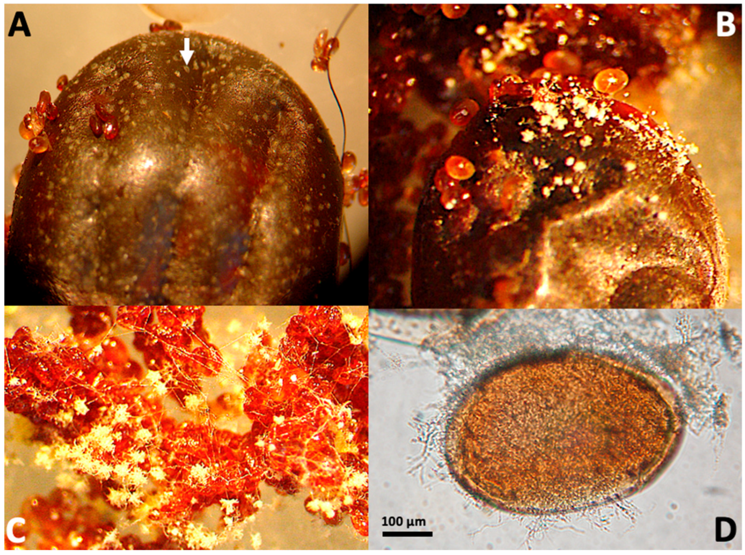
Figure 2. Different developmental stages of Aspergillus flavus infection in Rhipicephalus microplus. (A) Early signs of infection on ovipositing engorged females show small, homogeneously distributed plaques on the tick’s chitinous cuticle (arrow). Numerous eggs are visible in the image. (B) As infection progresses, plaques evolve into extensive mycelial growth with conidiophores exhibiting the characteristic morphology of the Aspergillus genus and propagating to the egg masses. (C) Tick egg masses can sustain fungal development, showing visible mycelial and conidiophore structures. (D) Light microscopy image of an infected egg showing mycelial penetration through the chitin shell (objective 20×).
Microbiolres 16 00243 g002
Figure 3. Hydrolysis rate of monomeric N-fluoresceyl D-glucosamine by 1 IU of purified chitinase from Streptomyces griseus (Sigma Cat. No. C6137) and by 1 IU of lysozyme from chicken egg white (Sigma Cat. No. L6976), which was used as a specificity control. Only the chitinase treatment produced a measurable fluorescent signal, confirming the substrate’s selectivity for chitinolytic enzymes.
Microbiolres 16 00243 g003
Figure 4. Growth and detection of different fungal species in the selective fluorogenic medium. (A) Culture plates inoculated with Metarhizium anisopliae, Beauveria bassiana, Aspergillus flavus, Penicillium notatum, and Fusarium venenatum inspected under UV light after 72 h of incubation showing distinct fluorescence only in chitinolytic strains. (B) Quantitative measurement of relative fluorescence units (RFUs) in culture supernatants confirming high enzymatic activity in entomopathogenic species compared with non-chitinolytic controls.
Microbiolres 16 00243 g004
Table 1. Visual and Quantitative Assessment of Chitinase Activity in Culture Supernatants.
Inoculum Type Fluorescence Relative Units Optical Density at 493 nm (±SD)Chitinase Activity (U/mL)
Aspergillus flavus1760.36 ± 0.0125.14
Bauveria bassiana1350.32 ± 0.0114.02
Metarhizium anisopliae1220.29 ± 0.0103.77
Penicillum notatum30.005 ± 0.0040.02
Fusarium venenatum10.002 ± 0.0010.01
Negative Control (no inoculum)0.001 ± 0.00010.001
Disclaimer/Publisher’s Note: The statements, opinions and data contained in all publications are solely those of the individual author(s) and contributor(s) and not of MDPI and/or the editor(s). MDPI and/or the editor(s) disclaim responsibility for any injury to people or property resulting from any ideas, methods, instructions or products referred to in the content.