
Effect of Buffer Room Configuration on Isolation of Bacteriophage phi6 and Micrococcus Luteus Emissions

 , , , , ,
Abstract
1. Introduction
2. Materials and Methods
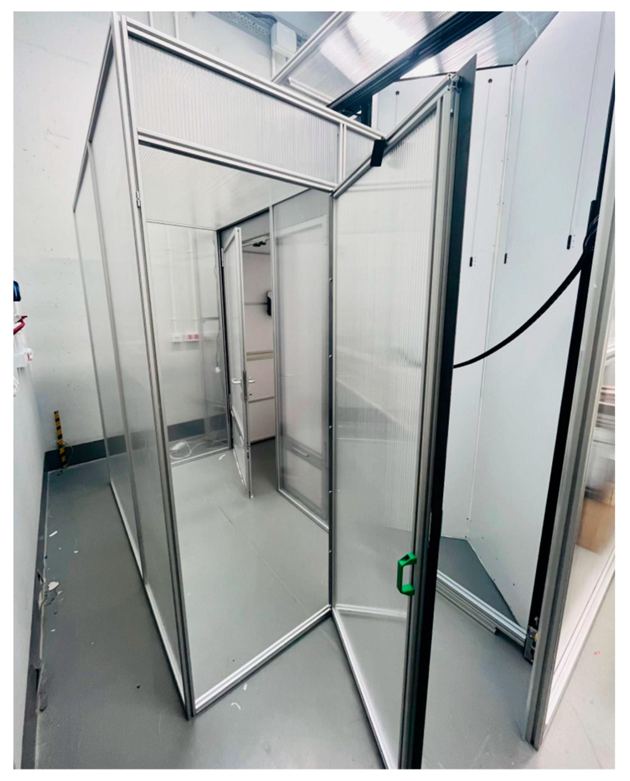
2.1. Layout of the Tested Rooms
2.2. Test Apparatus
2.3. Test Procedures
- Determining the effectiveness of airlock prototype I assembled as a single-chamber airlock with passage time of 30 s (the basic variant);
- Determining the effectiveness of airlock prototype I as double-chamber airlock with passage time 30 s;
- Determining the effectiveness of airlock prototype I assembled as a single-chamber airlock with passage time of 5 s;
- Determining the effectiveness of airlock prototype I assembled as a single-chamber airlock with passage time of 120 s.
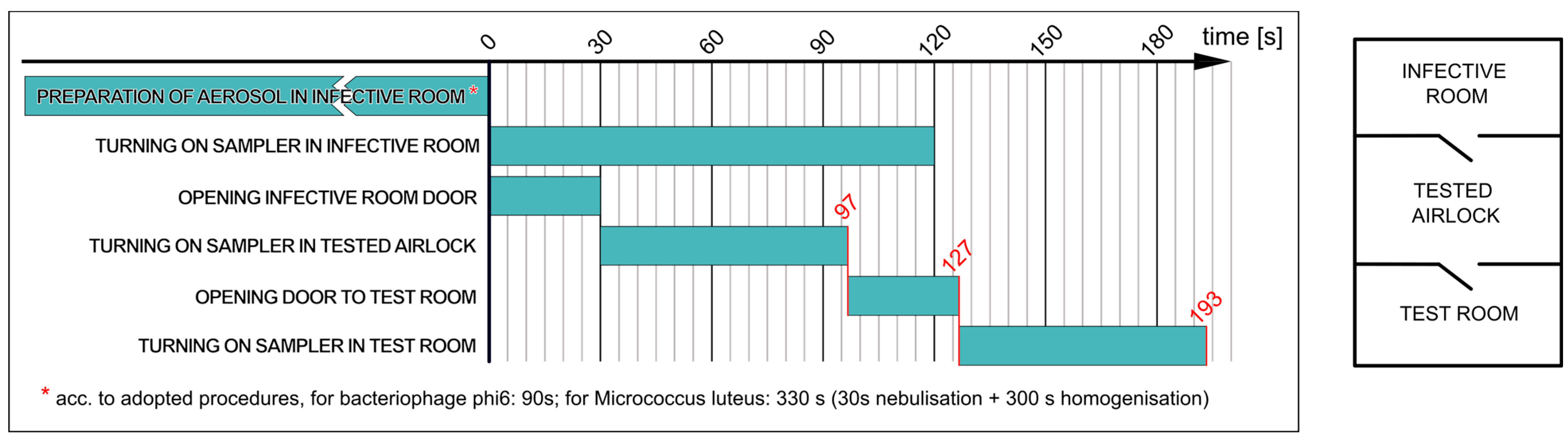
2.3.1. Determining Effectiveness of Airlock Prototype I Assembled as Single-Chamber Airlock with Passage Time of 30 s (Variant a)
- ▪
- The nebulisation of the M. luteus suspension in the infective room for 30 s, followed by bioaerosol homogenisation for 5 min;
- ▪
- Collecting 200 L of air from the infective room and simultaneously opening the door to the airlock for 30 s;
- ▪
- Collecting 200 L of air from the airlock and then opening the door to the test room for 30 s;
- ▪
- Collecting 200 L of air in the test room.
- ▪
- The nebulisation of the phi6 suspension in the infective room for 90 s (without homogenisation—without switching on fans in the infective room);
- ▪
- Collecting 200 L of air from the infective room and simultaneously opening the door to the airlock for 30 s;
- ▪
- Collecting 200 L of air from the airlock and then opening the door to the test room for 30 s;
- ▪
- Collecting 200 L of air in the test room.
2.3.2. Determining Effectiveness of Airlock Prototype I Assembled as Double-Chamber Airlock with Passage Time of 30 s (Variant b)
- ▪
- The nebulisation of the M. luteus suspension in the infective room for 30 s, followed by bioaerosol homogenisation for 5 min;
- ▪
- Collecting 200 L of air from the infective room and simultaneously opening the door to the airlock for 30 s;
- ▪
- Collecting 200 L of air from the contaminated room and then opening the door to the clean room for 30 s;
- ▪
- Passage to the airlock; then opening the door to the test room;
- ▪
- Collecting 200 L of air in the test room.
- ▪
- The nebulisation of the phage suspension in the infective room for 90 s (without homogenisation);
- ▪
- Collecting 200 L of air from the infective room and simultaneously opening the door to the contaminated room for 30 s;
- ▪
- Collecting 200 L of air from the contaminated room and then opening the door to the clean room for 30 s;
- ▪
- Passage to the airlock; then opening the door to the test room;
- ▪
- Collecting 200 L of air in the test room.
2.3.3. Determining Effectiveness of Airlock Prototype I Assembled as Single-Chamber Airlock Depending on Time of Passage through Door (Variants c and d)
3. Results
3.1. Determining Effectiveness of Airlock Prototype I Assembled as Single-Chamber Airlock
3.2. Determining Effectiveness of Airlock Prototype I Assembled as Double-Chamber Airlock in Laboratory Conditions
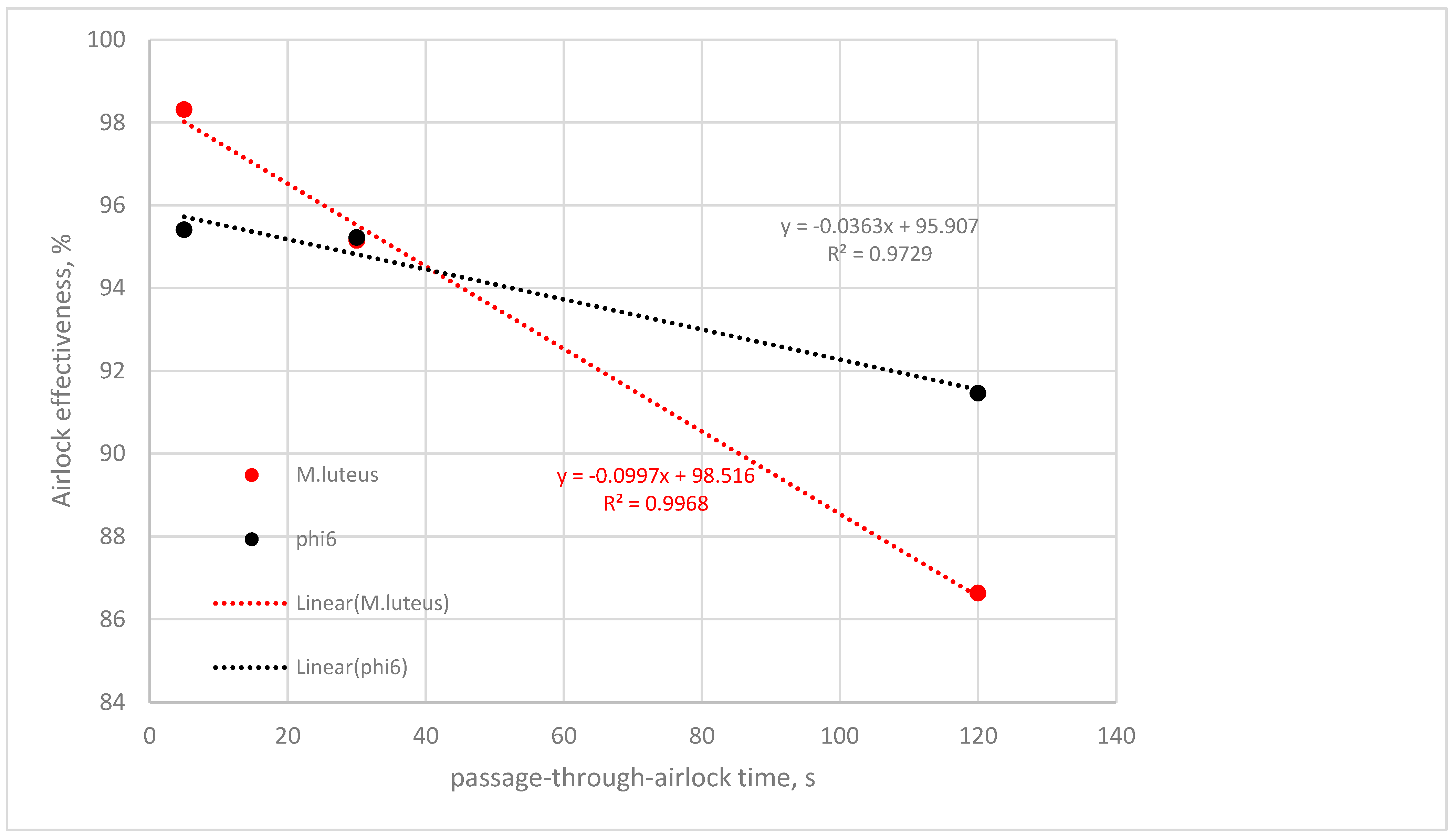
3.3. Determining Effectiveness of Airlock Prototype I Assembled as Single-Chamber Airlock, Depending on Passage-through-Airlock Time
4. Discussion
Author Contributions
Funding
Institutional Review Board Statement
Informed Consent Statement
Data Availability Statement
Conflicts of Interest
References
- Priolo Filho, S.R.; Chae, H.; Bhakta, A.; Moura, B.R.; Correia, B.B.; da Silva Santos, J.; Sieben, T.L.; Goldfarb, D. A qualitative analysis of child protection professionals’ challenges during the COVID-19 pandemic. Child. Abuse Negl. 2023, 143, 106229. [Google Scholar] [CrossRef]
- Finger, J.A.; Lima, E.M.; Coelho, K.S.; Behrens, J.H.; Landgraf, M.; Franco, B.D.; Pinto, U.M. Adherence to food hygiene and personal protection recommendations for prevention of COVID-19. In Trends in Food Science and Technology; Elsevier Ltd.: Amsterdam, The Netherlands, 2021; Volume 112, pp. 847–852. [Google Scholar]
- Herselman, R.; Lalloo, V.; Ueckermann, V.; van Tonder, D.J.; de Jager, E.; Spijkerman, S.; Van der Merwe, W.; Du Pisane, M.; Hattingh, F.; Stanton, D.; et al. Adapted full-face snorkel masks as an alternative for COVID-19 personal protection during aerosol generating procedures in South Africa: A multi-centre, non-blinded in-situ simulation study. Afr. J. Emerg. Med. 2021, 11, 436–441. [Google Scholar] [CrossRef] [PubMed]
- Katz, C.; Glucklich, T.; Attrash-Najjar, A.; Jacobson, M.A.; Cohen, N.; Varela, N.; Priolo-Filho, S.R.; Bérubé, A.; Chang, O.D.; Collin-Vézina, D.; et al. The global impact of COVID-19 on child protection professionals: A scoping review and thematic analysis. Child. Abus. Negl. 2023, 106347. [Google Scholar] [CrossRef]
- King, E.C.; Zagrodney, K.A.P.; McKay, S.M.; Holness, D.L.; Nichol, K.A. Determinants of nurse’s and personal support worker’s adherence to facial protective equipment in a community setting during the COVID-19 pandemic in Ontario, Canada: A pilot study. Am. J. Infect. Control 2023, 51, 490–497. [Google Scholar] [CrossRef]
- Bettari, L.; Pero, G.; Maiandi, C.; Messina, A.; Saccocci, M.; Cirillo, M.; Troise, G.; Conti, E.; Cuccia, C.; Maffeo, D. Exploring Personal Protection During High-Risk PCI in a COVID-19 Patient. JACC Case Rep. 2020, 2, 1279–1283. [Google Scholar] [CrossRef]
- Martin, C.; Kloka, J.; Lotz, G.; Zacharowski, K.; Raimann, F.J. The Frankfurt COVid aErosol pRotEction Dome–COVERED–a consideration for personal protective equipment improvement and technical note. In Anaesthesia Critical Care and Pain Medicine; Elsevier Masson SAS: Amsterdam, The Netherlands, 2020; Volume 39, pp. 373–374. [Google Scholar]
- Abkar, L.; Zimmermann, K.; Dixit, F.; Kheyrandish, A.; Mohseni, M. COVID-19 pandemic lesson learned-critical parameters and research needs for UVC inactivation of viral aerosols. J. Hazard. Mater. Adv. 2022, 8, 100183. [Google Scholar] [CrossRef]
- Wathore, R.; Gupta, A.; Bherwani, H.; Labhasetwar, N. Understanding air and water borne transmission and survival of coronavirus: Insights and way forward for SARS-CoV-2. Sci. Total Environ. 2020, 749, 141486. [Google Scholar] [CrossRef]
- Wang, Q.; Gu, J.; An, T. The emission and dynamics of droplets from human expiratory activities and COVID-19 transmission in public transport system: A review. Build. Environ. 2022, 219, 109224. [Google Scholar] [CrossRef]
- Arslan, M.; Xu, B.; Gamal El-Din, M. Transmission of SARS-CoV-2 via fecal-oral and aerosols–borne routes: Environmental dynamics and implications for wastewater management in underprivileged societies. Sci. Total Environ. 2020, 743, 140709. [Google Scholar] [CrossRef]
- Zhang, H.; Li, D.; Xie, L.; Xiao, Y. Documentary Research of Human Respiratory Droplet Characteristics. In Procedia Engineering; Elsevier Ltd.: Amsterdam, The Netherlands, 2015; pp. 1365–1374. [Google Scholar]
- Kearney, P.M.; Stamenic, D.; Gajewska, K.; O’Sullivan, M.B.; Doyle, S.; O’Reilly, O.; Buckley, C.M. Cross-sectional survey of compliance behaviour, knowledge and attitudes among cases and close contacts during COVID-19 pandemic. Public Health Pract. 2023, 5, 100370. [Google Scholar] [CrossRef]
- Jones, R.M.; Brosseau, L.M. Aerosol transmission of infectious disease. J. Occup. Environ. Med. 2015, 57, 501–508. [Google Scholar] [CrossRef]
- Eames, I.; Tang, J.W.; Li, Y.; Wilson, P. Airborne transmission of disease in hospitals. J. R. Soc. Interface 2009, 6, S697–S702. [Google Scholar] [CrossRef]
- Fan, X.; Zhang, X.; Weerasuriya, A.U.; Hang, J.; Zeng, L.; Luo, Q.; Li, C.Y.; Chen, Z. Numerical investigation of the effects of environmental conditions, droplet size, and social distancing on droplet transmission in a street canyon. Build. Environ. 2022, 221, 109261. [Google Scholar] [CrossRef]
- Mirza, S.; Niwalkar, A.; Gupta, A.; Gautam, S.; Anshul, A.; Bherwani, H.; Biniwale, R.; Kumar, R. Is safe distance enough to prevent COVID-19? Dispersion and tracking of aerosols in various artificial ventilation conditions using OpenFOAM. Gondwana Res. 2023, 114, 40–54. [Google Scholar] [CrossRef] [PubMed]
- Khaled Ahmed, S.; Mohammed Ali, R.; Maha Lashin, M.; Fayroz Sherif, F. Designing a new fast solution to control isolation rooms in hospitals depending on artificial intelligence decision. Biomed. Signal Process Control 2023, 79, 104100. [Google Scholar] [CrossRef]
- Liu, H.; Liu, Z.; He, J.; Hu, C.; Rong, R.; Han, H.; Wang, L.; Wang, D. Reducing airborne transmission of SARS-CoV-2 by an upper-room ultraviolet germicidal irradiation system in a hospital isolation environment. Environ. Res. 2023, 237, 116952. [Google Scholar] [CrossRef]
- Saravia, S.A.; Raynor, P.C.; Streifel, A.J. A performance assessment of airborne infection isolation rooms. Am. J. Infect. Control 2007, 35, 324–331. [Google Scholar] [CrossRef] [PubMed]
- Bhattacharyya, S.; Dey, K.; Paul, A.R.; Biswas, R. A novel CFD analysis to minimize the spread of COVID-19 virus in hospital isolation room. Chaos Solitons Fractals 2020, 139, 110294. [Google Scholar] [CrossRef]
- Zhang, Y.; Han, O.; Li, A.; Olofsson, T.; Zhang, L.; Lei, W. Adaptive Wall-Based Attachment Ventilation: A Comparative Study on Its Effectiveness in Airborne Infection Isolation Rooms with Negative Pressure. Engineering 2022, 8, 130–137. [Google Scholar] [CrossRef]
- Kalliomäki, P.; Hagström, K.; Itkonen, H.; Grönvall, I.; Koskela, H. Effectiveness of directional airflow in reducing containment failures in hospital isolation rooms generated by door opening. Build. Environ. 2019, 158, 83–93. [Google Scholar] [CrossRef]
- Chang, P.K.; Chuang, H.H.; Hsiao, T.C.; Chuang, H.C.; Chen, P.C. Investigating the invisible threat: An exploration of air exchange rates and ultrafine particle dynamics in hospital operating rooms. Build. Environ. 2023, 245, 110870. [Google Scholar] [CrossRef]
- Wróbel, R.; Andrych-Zalewska, M.; Matla, J.; Molska, J.; Sierzputowski, G.; Szulak, A.; Włostowski, R.; Włóka, A.; Rutkowska-Gorczyca, M. Assessment of the Possibility of Using Bacterial Strains and Bacteriophages for Epidemiological Studies in the Bioaerosol Environment. Microbiol. Res. 2024, 15, 236–246. [Google Scholar] [CrossRef]
- Sierzputowski, G.; Piechota, N.; Krajewski, T.; Szewc, M. Patent application No P442153 (30 August 2022) “Adaptive Geometric Dimensions Mobile Buffer Space” (Szczegóły PAT—P.442153). Available online: https://uprp.gov.pl/pl (accessed on 3 June 2024).


| Bacterium | Bacteriophage | |
|---|---|---|
| Kind | Micrococcus luteus ATCC 7468 | phage phi6 |
| Control samples | bacterial strain streak inoculation | host strain streak inoculation |
| surface inoculation of stabilised suspension before nebulisation | surface inoculation of phage before nebulisation | |
| surface inoculation of stabilised suspension after nebulisation | surface inoculation of phage suspension after nebulisation | |
| Samples of air in test room | after disinfection | after disinfection |
| after nebulisation | after nebulisation |
| Kind of Bioaerosol | Effectiveness without Airlock, %R | Effectiveness with Airlock, %R |
|---|---|---|
| M. luteus | 77.27% (N = 8; SD = 10%) | 95.15% (N = 8; SD = 2.2%) |
| phage phi6 | 72.48% (N = 6; SD = 26.3%) | 95.22% (N = 8; SD = 3.9%) |
| Kind of Bioaerosol | Effectiveness without Airlock, %R | Effectiveness with Airlock, %R |
|---|---|---|
| M. luteus | 85.94% (N = 7; SD = 3.2%) | 98.17% (N = 7; SD = 1%) |
| phage phi6 | 82.99% (N = 8; SD = 8.3%) | 98.37% (N = 8; SD = 2%) |
| Kind of Bioaerosol | Passage-through-Airlock Time | Effectiveness without Airlock, %R | Effectiveness with Airlock, %R |
|---|---|---|---|
| M. luteus | 5 s | 86.32% (N = 8; SD = 7.1%) | 98.31% (N = 8; SD = 0.9%) |
| 30 s | 77.27% (N = 8; SD = 10%) | 95.15% (N = 8; SD = 2.2%) | |
| 120 s | 59.25% (N = 6; SD = 17.5%) | 86.63% (N = 6; SD = 4%) | |
| phage phi6 | 5 s | 67.75% (N = 6; SD = 23.5%) | 95.41% (N = 6; SD = 3.1%) |
| 30 s | 72.48% (N = 6; SD = 26.3%) | 95.22% (N = 8; SD = 3.9%) | |
| 120 s | 65.69% (N = 3; SD = 38.7%) | 91.46% (N = 3; SD = 10.1%) |
| Version | M. luteus ATCC 7468 | Phage phi6 |
|---|---|---|
| Single-chamber version, passage time 30 s | 95.15% | 95.22% |
| Double-chamber version, passage time 30 s | 98.17% | 98.37% |
Disclaimer/Publisher’s Note: The statements, opinions and data contained in all publications are solely those of the individual author(s) and contributor(s) and not of MDPI and/or the editor(s). MDPI and/or the editor(s) disclaim responsibility for any injury to people or property resulting from any ideas, methods, instructions or products referred to in the content. |
© 2024 by the authors. Licensee MDPI, Basel, Switzerland. This article is an open access article distributed under the terms and conditions of the Creative Commons Attribution (CC BY) license (https://creativecommons.org/licenses/by/4.0/).
Share and Cite
Wróbel, R.; Andrych-Zalewska, M.; Matla, J.; Molska, J.; Sierzputowski, G.; Szulak, A.; Włostowski, R.; Włóka, A.; Rutkowska-Gorczyca, M. Effect of Buffer Room Configuration on Isolation of Bacteriophage phi6 and Micrococcus Luteus Emissions. Microbiol. Res. 2024, 15, 1099-1109. https://doi.org/10.3390/microbiolres15030073
Wróbel R, Andrych-Zalewska M, Matla J, Molska J, Sierzputowski G, Szulak A, Włostowski R, Włóka A, Rutkowska-Gorczyca M. Effect of Buffer Room Configuration on Isolation of Bacteriophage phi6 and Micrococcus Luteus Emissions. Microbiology Research. 2024; 15(3):1099-1109. https://doi.org/10.3390/microbiolres15030073
Chicago/Turabian StyleWróbel, Radosław, Monika Andrych-Zalewska, Jędrzej Matla, Justyna Molska, Gustaw Sierzputowski, Agnieszka Szulak, Radosław Włostowski, Adriana Włóka, and Małgorzata Rutkowska-Gorczyca. 2024. "Effect of Buffer Room Configuration on Isolation of Bacteriophage phi6 and Micrococcus Luteus Emissions" Microbiology Research 15, no. 3: 1099-1109. https://doi.org/10.3390/microbiolres15030073
APA StyleWróbel, R., Andrych-Zalewska, M., Matla, J., Molska, J., Sierzputowski, G., Szulak, A., Włostowski, R., Włóka, A., & Rutkowska-Gorczyca, M. (2024). Effect of Buffer Room Configuration on Isolation of Bacteriophage phi6 and Micrococcus Luteus Emissions. Microbiology Research, 15(3), 1099-1109. https://doi.org/10.3390/microbiolres15030073