Article

Response of Biofortified Green Bean Plants to Colletotrichum lindemuthianum

by
Luis G. Hernández-Montiel
1,
Juan P. Ciscomani-Larios
2,
Esteban Sánchez-Chávez
2,3,
Irasema Vargas-Arispuro
4,
Abeer Hashem
5,
Elsayed F. Abd_Allah
6 and
Graciela D. Avila-Quezada
2,*
1
Nanotechnology & Microbial Biocontrol Group, Centro de Investigaciones Biológicas del Noroeste, La Paz 23096, Mexico
2
Facultad de Ciencias Agrotecnológicas, Universidad Autónoma de Chihuahua, Campus 1, Chihuahua 31000, Mexico
3
Centro de Investigación en Alimentación y Desarrollo A.C., Delicias 33088, Mexico
4
Centro de Investigación en Alimentación y Desarrollo A.C., Hermosillo 83304, Mexico
5
Botany and Microbiology Department, College of Science, King Saud University, Riyadh 11451, Saudi Arabia
6
Plant Production Department, College of Food and Agricultural Sciences, King Saud University, Riyadh 11451, Saudi Arabia
*
Author to whom correspondence should be addressed.
Microbiol. Res. 2023, 14(4), 2067-2078; https://doi.org/10.3390/microbiolres14040139
Submission received: 22 September 2023 / Revised: 13 November 2023 / Accepted: 17 November 2023 / Published: 6 December 2023

Abstract

:
Enhancing crop nutrition though biofortification with essential minerals can, in some circumstances, increase the resistance of plants to the attack by pathogens. As a result, plants activate their defense mechanisms and produce bioactive compounds (BCs) in response. To date, there has been no investigation into the response of green bean plants fortified with magnesium (Mg) salts to the presence of Colletotrichum lindemuthianum. This research involved two Mg sources applied by the edaphic route. The pathogen was inoculated on green bean pods, and subsequent analysis was conducted on the accumulation of BCs, including total anthocyanins, total phenols, and total flavonoids, within both symptomatic and healthy tissues. Remarkably, the plant’s defense system was activated, as evidenced by the significantly higher concentration of anthocyanins (p ≤ 0.05) observed in the symptomatic tissues following treatments with both MgCl2 and MgSO4. Further, green bean plants treated with MgSO4 displayed notably elevated concentrations of phenols (p ≤ 0.05) in the inoculated tissues of the pods, suggesting a plausible plant defense mechanism. The levels of BCs were considerably higher in green bean pods of the biofortified plants compared to those which were nonbiofortified. However, perhaps one of the most noteworthy findings is that there were no discernible differences between biofortified and nonbiofortified treatments in stopping anthracnose in green bean pods. These results provide valuable insights contributing to a deeper understanding of this interaction.

1. Introduction

The fungal pathogen, Colletotrichum spp., is responsible for anthracnose in numerous plants species, such as avocado, potato, mango, grape, citrus, among others [1,2]. In particular, Colletotrichum lindemuthianum (teleomorph Glomerella cingulata f. sp. phaseoli) has a significant impact on bean production, leading to yield losses of up to 90% [3]. Diets that primarily rely on beans, grains, and cereals are commonly deficient in magnesium (Mg+), which is a mineral element important to every human body cell [4]. The diseases or disorders associated with magnesium deficiency in the human are glaucoma, renal calculi, muscle cramps, diabetes mellitus, osteoporosis, coronary artery disease, preeclampsia, migraine, among others [5,6].
Therefore, biofortification technologies are applied to increase the Mg+ content through the application of mineral fertilizers [7]. Adequate magnesium nutrition, regardless of the Mg source supplied, has been shown to reduce the incidence of certain plant diseases. For instance, the study conducted by Huber and Jones [8] highlights that, in conditions where levels of Mg in the soil are sufficient, Fusarium wilt is less severe because Mg enhances the resistance of plant tissues against degradation by pectolytic enzymes. Recent studies further emphasize that biofortification with minerals and nanominerals increases the concentration of essential elements within plants, thereby bolstering their resistance to attack by plant pathogens [9,10].
In a previous study, it was documented that the biofortification of bean plants led to an increase in the content of phenolic compounds [11]. Phenolic compounds represent a subset of bioactive compounds (BCs). These are a group of natural compounds found in plants; they play a significant role in the plant’s defense mechanisms against pathogens. When a plant is attacked by a pathogen (such as a disease-causing microorganism), it needs to protect itself. Phenolic compounds are one of the ways plants respond to this threat [12].
When a plant is attacked by a pathogen, it recognizes the presence of the intruder. This recognition triggers a series of defense mechanisms within the plant. As part of its defense strategy, the plant starts producing phenolic compounds. These compounds are bioactive, which means they have a biological impact on the plant and its surroundings; additionally, the plant actively accumulates phenolic compounds during interactions with pathogens [13]. Furthermore, the concentration of specific phenolic compounds has been utilized to discern healthy tissue from diseased tissue in response to pathogen attack [14]. In addition, phenolic compounds serve the dual role of inhibiting the germination of fungal spores and inactivating particular enzymes produced by phytopathogens as part of the plant’s defense mechanisms. One of the primary roles of phenolic compounds is to act as barriers against fungal invaders. When fungal spores attempt to germinate and establish infection within the plant, phenolic compounds act. These compounds create an inhospitable environment, hindering the germination of fungal spores. By impeding this critical initial step in the fungal life cycle, phenolic compounds serve as a line of defense, preventing the pathogen invasion within the plant. In addition, phenolic compounds can inactivate specific enzymes produced by plant pathogens. Many pathogens rely on enzymes to breach plant cell walls, facilitate nutrient acquisition by haustoriums, and counteract the plant’s defense responses. Phenolic compounds can disrupt these enzymatic processes, rendering the pathogen’s efforts less effective. By this, they hinder the pathogen’s ability to colonize, spread, and cause damage to the plant [7,15].
Mikulic-Petkovsek et al. [16] conducted a study on sweet peppers and observed phenolic changes induced by the inoculation of Colletotrichum coccodes. Their findings revealed an increase in the total phenolic content, especially at the symptomatic area, and even more pronouncedly in the surrounding border zone near the diseased tissue. Particularly, anthocyanins were identified as key components in the defense mechanism of plants. Slatnar et al. [17] also noted a high concentration of anthocyanins in and around the symptomatic tissue in apples affected with Venturia inaequalis, in contrast to healthy tissue. Consequently, these anthocyanins have the potential to delay fungal infections or cause the fungi to become inactive, demonstrating their role in plant defense against pathogenic threats [18].
An elevated concentration of anthocyanins is closely linked to plants that face pathogenic threats. In the research of Somalraju et al. [19] with potato plants of the ‘Russian Blue’ variety biofortified with selenium (Se) and exposed to Phytophthora infestans inoculation, they exhibited notably higher anthocyanin levels in their tubers compared to the nonbiofortified, uninoculated potatoes. This underscores how the combination of pathogen exposure and biofortification stimulates the synthesis of anthocyanins, contributing to enhanced plant protection.
In pursuit of sustainable bean production to address global food security and nutrition improvement, this study aims to elucidate the response of the magnesium-biofortified green bean plants to C. lindemuthianum. By doing so, it contributes valuable insight to the current knowledge regarding the interaction between green beans and C. lindemuthianum.

2. Materials and Methods

2.1. Biofortification with Magnesium

In August 2018, we conducted an outdoor experiment at the Autonomous University of Chihuahua in Chihuahua, Mexico (28°39′19″ N; 106°05′14″ W). We began by planting seeds of the green bean (Phaseolus vulgaris L.) cv “Strike” at a depth of 3 cm in pots measuring 30 cm in diameter and 30 cm in height. The sowing date was August 30th. These pots were filled with a mixture of vermiculite and perlite in a ratio of 2:1 (vol/vol). The ambient temperature during the day averaged 28 °C. Starting 26 days after germination, the plants received the nutrient solution as recommended by Márquez-Quiroz et al. [20] without Mg, administrated every three days.
To investigate the impact of magnesium on the plants, we established two Mg treatments using either MgCl2 or MgSO4 at a concentration of 100 ppm from 99.99% pure Mg, as previously reported for biofortification [11,21]. The control treatment lacked Mg. Each treatment consisted of four plants per replicate, with two replicates per treatment, following a completely randomized experimental design. The Mg treatments were applied to the soil, commencing 26 days after germination, and continuing for a period of 40 days.

2.2. C. lindemuthianum Inoculation and Disease Assessment

We employed a monoconidial strain of C. lindemuthianum, which was isolated from common beans (Phaseolus vulgaris L.) in the State of Mexico and generously donated by the Collection of Phytopathogenic Fungi at the Department of Agricultural Parasitology of the Chapingo Autonomous University.
During the 40-day period of Mg application, a suspension containing 106 conidia mL−1 was inoculated on day 26 onto four green bean pods per plant. This inoculation process was carried out on four plants for each treatment group. Five µL of the conidial suspension were applied to three specific points on each green bean pod, as can be seen in Figure 1, ensuring that the pod´s tissue remained undamaged. Subsequently, the inoculated plants were kept at a temperature of 18 °C for a period of 14 days, from October 27th to November 10th.
Fourteen days after inoculation, the diameter of the anthracnose lesion was measured. To document the symptoms, an HP Scanjet M377dw (Hewlett-Packard) was utilized. For further analysis, both healthy and symptomatic green bean pods that had been observed 14 days postinoculation were cut and then stored by freezing them at −20 °C.

2.3. Bioactive Compounds (BCs)

2.3.1. Total Anthocyanins

To extract total anthocyanins from both the healthy and diseased tissue of the frozen green bean pods, we followed the methodology outlined by Ciscomani-Larios et al. [11]. Subsequently, the absorbance of the first phase was measured at 460 nm, and that of the second phase at 710 nm using a JENWAY spectrophotometer (Jenway Limited®, Essex, UK). The results are reported in mg cyanidin-3-glucoside g−1.

2.3.2. Total Phenols

For the extraction of total phenols from the macerated tissues of both healthy and diseased green bean pods, we employed the method of Singleton and Rossi [22] and Singleton et al. [23], with slight modifications, as reported by Ciscomani-Larios et al. [11]. The absorbance of the reaction mixture was recorded at 725 nm. The results are reported in mg gallic acid equivalent g−1 (mg GAE g−1) fresh weight (FW).

2.3.3. Total Flavonoids

The quantification of total flavonoids in the healthy and diseased tissues were carried out using the colorimetric method developed by Zhishen et al. [24]. We measured the absorbance of the mixture at 510 nm, with catechin serving as the standard compound for reference. The results were expressed in mg of catechin equivalents (CE) g−1 of fresh weight (FW) of the sample. All the previous analyses were performed in triplicate for precision.

2.4. Statistical Analysis

The obtained results were subjected to statistical analysis, including analysis of variance, followed by a comparison of means utilizing the least-significant-difference (LSD) method, all performed within the SAS program (SAS Inst. Inc., Cary, NC, USA). Significance was attributed to differences in means when the p-value was equal to or less than 0.05 (p ≤ 0.05).
To facilitate the grouping of means pertaining to BCs and antioxidant capacity in both inoculated and noninoculated green bean pods, a Tukey test was performed (p ≤ 0.05). The data presented in this study are expressed as means ± standard error (SE) for precision.

3. Results and Discussion

3.1. Symptoms on Green Bean Pods

In this study, Colletotrichum lindemuthianum induced the formation of small brown spots measuring 5 to 6 mm in diameter on beans 14 days after inoculation, regardless of the Mg treatment applied (Figure 1). The symptoms were typical of anthracnose in beans. No symptoms were observed on the green bean pods inoculated with water. This symptomatic expression suggests that, regardless of the Mg biofortification, the pathogenicity process continued.
Both of the Mg salts did not affect the conidia applied to green bean pods, nor did they disrupt the specialized infection structures from entering the cuticle. It is known that to successfully infect the host plant, this fungus has developed many specialized infection strategies and structures, among which the fungal appressorium plays an important role.
According to the literature, biofortification would be expected to stop or slow the development of the appressorium at the point of contact, where it detects physical and chemical signals from the host, such as surface hydrophobicity and chemical signals. This is because some authors point out that biofortification induces defense mechanisms in plants. Then, it can be concluded that, under the conditions of this study, the fungus is so aggressive that Colletotrichum appressoria have the ability to penetrate the host cuticle, crossing the barriers of waxes and other hydrophobic compounds independently of the Mg.
Thus, for a fungus with these highly specialized structures, such as appressoria with their pegs, which exert turgor pressure and a high mechanical force that pierces the host cuticle [25], fortification alone will not be enough. Since various specialized pathogenic factors have been documented in Colletotrichum spp., which include the production of pectolytic enzymes responsible for maceration of the plant cell wall and the ability to regulate the tissue pH as a means of host attack [26], they must be added to the biofortification with Mg, other minerals, and other control strategies for the fungus.
Additionally, the fungus has evolved an ability to regulate the tissue pH, which is crucial for successful host colonization. It can manipulate the pH of the host tissue by secreting alkaline compounds, such as ammonia and amines. This alkalization of the surrounding plant tissue creates an environment that is conducive to fungal growth and inhibits the plant’s defense responses [26].
The interaction between the fungal pathogen C. lindemuthianum and its host, Phaseolus vulgaris, is driven by an interplay of signals and genes from both the pathogen and the host, which collectively determine the outcome of the interaction. The pathogen begins by recognizing specific cues and signals emitted by the host plant. These cues may include chemical signals released by damaged plant tissue, as well as surface molecules that are recognized by the pathogen. In response to pathogen recognition, P. vulgaris activates its own set of defense mechanisms. These include the recognition of conserved molecular patterns on the pathogen’s surface, known as pathogen-associated molecular patterns (PAMPs), and the subsequent initiation of signaling cascades [27].

3.2. Bioactive Compounds in Healthy and Diseased Tissues

3.2.1. Total Anthocyanin Content

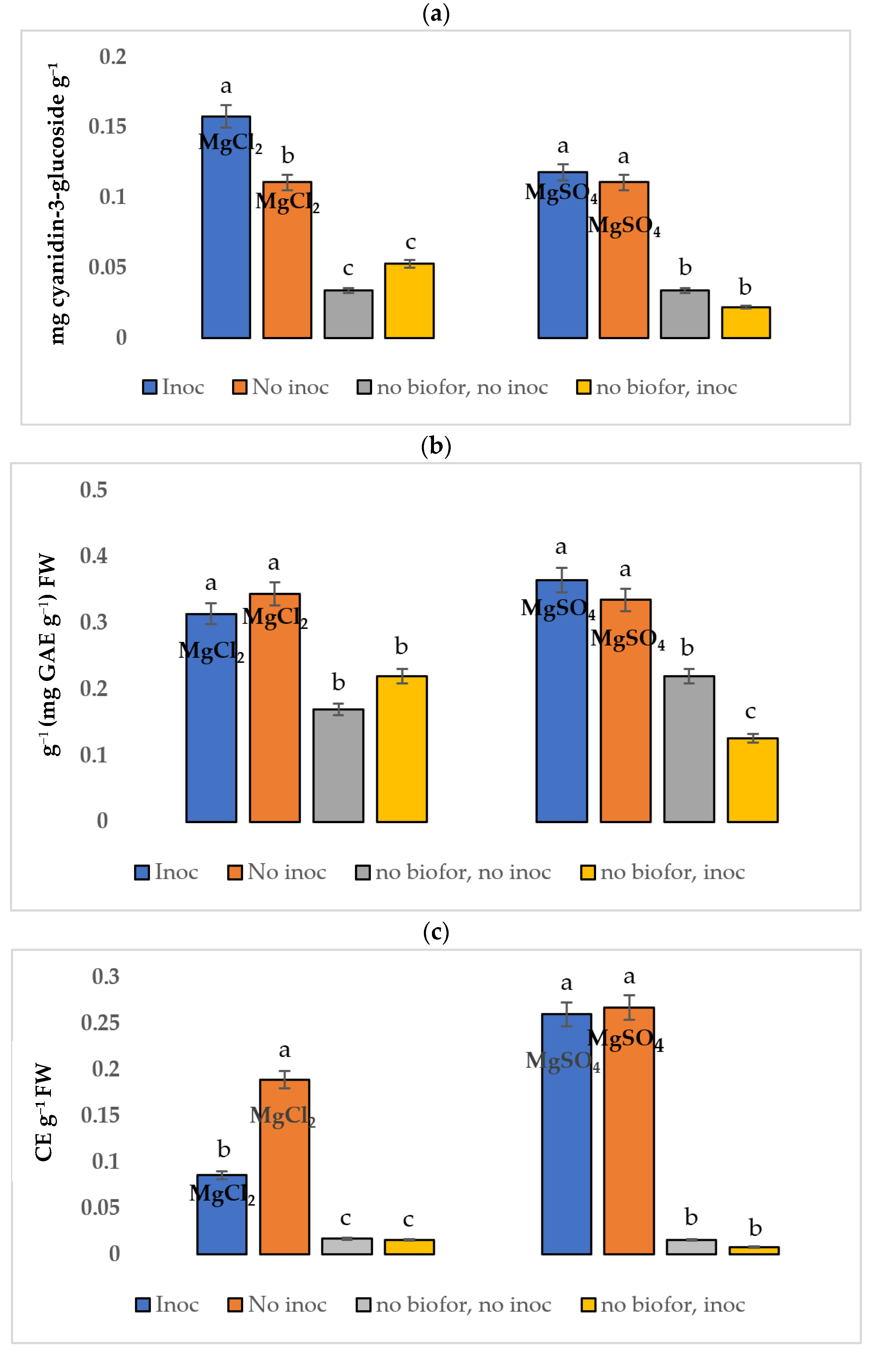
The highest concentration of total anthocyanins was observed in the symptomatic tissues of both Mg-treated groups (Figure 2a). Notably, significant statistical differences (p ≤ 0.05) were detected in anthocyanin levels between the inoculated and noninoculated MgCl2 treatments in the biofortified plants.
Anthocyanins, as natural pigments responsible for the red, purple, and blue colors in various fruits and vegetables, develop their increased presence through biofortification. Additionally, anthocyanins play a crucial role in the plant’s defense mechanism and have been shown to potentially delay fungal infections. In plants, they act as free radical scavengers, neutralizing harmful molecules that can damage cellular structures. Anthocyanins have been extensively studied for their ability to deter fungal infections. When a plant is under attack by fungi, it often responds by increasing the production of anthocyanins in the affected areas.
These compounds can inhibit the growth of fungi or make it more challenging for them to establish an infection [18]. Supporting this, Harshman et al. [28] provided evidence for the correlation between the degree of pigmentation in raspberries and the reduced severity of B. cinerea infection. This observation aligns with the findings of Bassolino et al. [29], whose research revealed a similar phenomenon in transgenic tomato engineering to accumulate anthocyanins, leading to a noticeable reduction in the severity of B. cinerea infections.
In the study conducted by Slatnar et al. [17], it was noticed that the highest levels of anthocyanins were detected in the symptomatic tissue and the areas surrounding lesions caused by Venturia inaequalis, in contrast to the comparatively lower levels present in healthy ‘Golden Delicious’ apple tissue. This observation highlights the dynamic response of anthocyanin production to stress and infection. Similarly, in separate research focusing on Malus crabapple, it was revealed that anthocyanins accumulated predominantly in leaf tissue, serving as an essential antioxidant defense mechanism against rust infection [30]. This underscores the versatile role of anthocyanins in plant defense. Anthocyanins, a subclass of flavonoids, act as a crucial component of the plant’s defense system against pathogens. As exemplified in a study involving mangoes, an increase in both anthocyanins and flavonoids was noted in the fruit peel, and this increase was associated with heightened resistance to C. gloeosporioides [31]. These findings collectively emphasize the significance of anthocyanins in plant resistance against pathogens.
Furthermore, when plants undergo biofortification and face the pathogen, there is a notable surge in anthocyanin levels, as observed in the study conducted by Somalraju et al. [19] involving potato plants biofortified with selenium and subsequently inoculated with Phytophthora infestans.

3.2.2. Total Phenols

In this study, we observed slightly elevated concentrations of total phenols in the inoculated (diseased) tissues of pods from plants biofortified with MgSO4 than in healthy tissues. Although these differences did not reach statistical significance (Figure 2b), it is a notable fact due to the pathogenicity of Colletotrichum and the reaction of the plant tissue.
However, a statistically significant difference (p ≤ 0.05) was observed when comparing the concentration of total phenols between biofortified and nonbiofortified plants, with biofortified plants exhibiting substantially higher concentrations.
Notably, various previous studies have consistently reported elevated levels of phenolic compounds within symptomatic spots caused by various pathogens, such as Colletotrichum spp. [16], Pyrenophora tritici-repentis race 1 [32], Gloeosporium spp., Penicillium expansum, Monilinia fructigena [33], Glomerella cingulata [34], and Phytophthora cinnamomi [35].
Phenolic compounds, known for their role in plant defense, frequently exhibit higher concentrations in the boundary zone surrounding infected tissue. This phenomenon suggests a strategic plant defense mechanism aimed at stopping the advance of fungal pathogens into healthy tissue. In response to fungal invasion, the plant appears to deploy phenolic compounds, creating a barrier at the interface between the infected and healthy regions. The presence of phenolic compounds in this zone underscores their significance as a critical component of the plant’s response, showcasing the dynamic nature of plant–pathogen interactions. This defense strategy not only protects the plant’s structural integrity but also preserves its ability to thrive and reproduce [16]. These compounds exhibit capabilities when it comes to combatting fungal pathogens. Notably, they possess the remarkable ability to stop fungal spore germination and incapacitate specific pathogenic enzymes. During fungal defense, phenolic compounds serve as potent antagonists, impeding the crucial first step in fungal infection by hindering the germination of spores. This early interception disrupts the fungus’s life cycle, preventing it from establishing on the host plant. They also exert their biochemical activities against the invaders by targeting specific pathogenic enzymes. These enzymes are essential for the fungus’s ability to infiltrate and colonize the host plant [36]. Among the phenolic phytoalexins, several compounds stand out as prominent elements in the plant defense activities.
These include phenolic compounds, as well as isoflavonoids and phenylpropanoids, all of which have been well-documented as key compounds produced by plants in their defense mechanisms [37]. According to Marschner [38], these compounds represent essential components of the plant´s strategy for safeguarding itself against potential threats. Plant metabolic responses to specific nutrients exhibit variability dependent on the type of ions present, including ions like Cl and SO4=. This variability can be linked to the oxidative burst, which is a rapidly induced response by plants when under pathogen attack. The type and combination of ions in the nutrient environment can trigger unique metabolic cascades, highlighting the complexity of the plant’s response mechanisms [39]. Defensive phenolic compounds are recognized for their ability to contribute to an overall reduction in the production of ROS, as well as being involved in more direct interactions with the signal transport and transduction pathways within the plant. In terms of ROS regulation, phenolic compounds act as molecular pacifiers, calming the potentially harmful of oxidative stress that can result from pathogen attacks or environmental stressors. By limiting the overproduction of ROS, these compounds help prevent other damage to the plant’s own tissues, thereby maintaining cellular integrity and function [40].
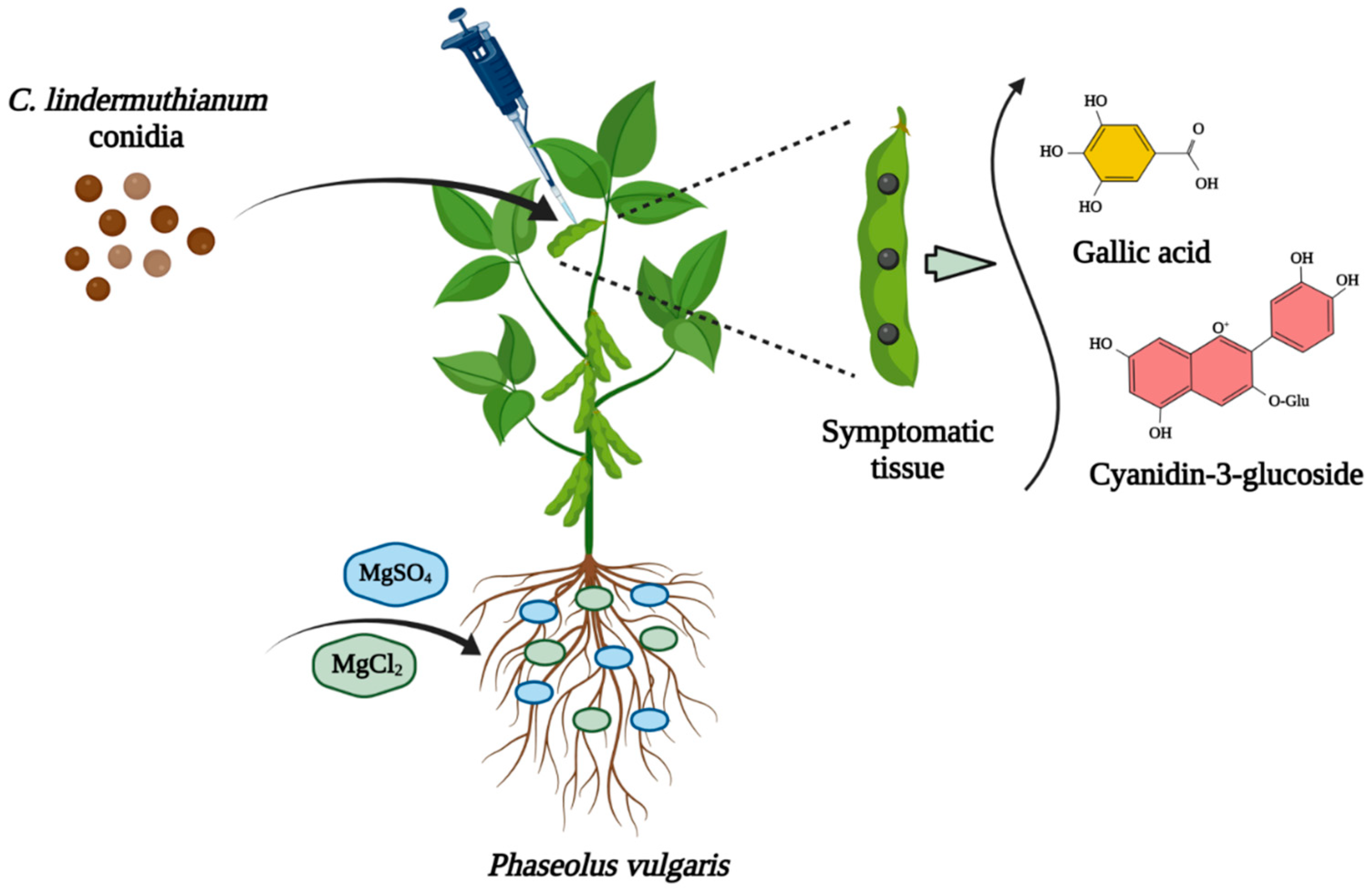
Our findings can be elucidated through the recognition of signals emitted by diseased cells when C. lindemuthianum is deposited onto green bean pods. This recognition initiates a signaling cascade that leads to the production of phenolic compounds [41]. Plants engage the synthesis of phenolic metabolites, a diverse group of organic compounds [42]. These phenolic compounds serve multiple purposes, extending beyond their essential role in defensive mechanisms. They also function as crucial adaptations to various environmental conditions, aiding the plant´s ability to thrive and respond effectively to its surroundings. Consequently, phenolic compounds within plants play an important role in shaping the adaptability and responsiveness of microorganisms. The compounds serve as influential mediators, promoting the dynamic interplay between microorganisms and their plant host, contributing to this microbial plasticity, allowing them to survive to various environmental stresses [43,44]. Figure 3 illustrates the process, from pathogen inoculation to biofortification with two Mg salts, the appearance of symptoms, and the production of metabolites in the symptomatic tissue.

3.2.3. Total Flavonoids

Among the various phenolic phytoalexins, flavonoids are typically the least abundant compounds utilized by plants for defensive purposes. There are some exceptions to this trend, particularly with catechins and proanthocyanidins. Interestingly, the biofortified treatment using MgSO4 exhibited notably higher concentrations of total flavonoids, regardless of whether the plants were inoculated with the fungus, and these levels were statistically superior to those observed with MgCl2 (p ≤ 0.05) (Figure 2c).
The presence of MgSO4 may be linked to enhanced pathogen resistance due to the elevated concentrations of both preformed and induced total flavonoids. Notably, biofortified plants with both salts demonstrated statistically significant differences when compared to nonbiofortified plants (p ≤ 0.05).
Initially, we hypothesized that the areas affected by inoculation would exhibit higher concentrations of flavonoids, as these secondary metabolites synthesized in plants in response to physical injury, infection, or stress in plants serve a range of functions, including protection against herbivores, pathogens, and UV radiation [45,46]. However, our results yielded unexpected outcomes. Fungal quiescence is a multifaceted process influenced by factors beyond the mere accumulation of phenolic compounds. In summary, even though the diseased pods did not present notably high concentrations of flavonoids, our findings remain significant, as biofortification with Mg salts induced the production of flavonoids in bean pods.

3.3. C. lindemuthianum in Mg-Biofortified Green Bean Pods

In our research, we did not observe statistically significant differences (p ≤ 0.05) between the biofortified treatments and the nonbiofortified control groups concerning the prevention of anthracnose in green bean pods. This outcome could be attributed to the inherent severity of the fungus in the common bean crops, as it is known to exhibit high aggressiveness [47]. Variability in cultivar resistance from one year to another could also be a contributing factor.
We conducted a study on the biofortification of Mg, since this element has a vital role in maintaining human health. Furthermore, this biofortification approach has shown promise in fortified plants against attacks by various plant pathogens, as highlighted in previous research [38]. For instance, Cakmak [48] conducted research focused on biofortification using zinc to enhance plant resistance to pathogens. The findings of this study concluded that, when there are adequate levels of available zinc in the soil, plants gain protection against numerous soil-borne pathogens. Additionally, the accumulation of high concentrations of zinc in seeds has been demonstrated to play a significant role for safeguarding germinating seeds and seedlings against pathogenic threats.
In a similar way, Siddiqui and Shaukat´s research [49] successfully suppressed infections caused by Fusarium solani and Rhizoctonia solani through the use of an iron chelator. The suppression can likely be attributed to plant´s response to nutrient deficiency, leading to the production of root exudates that attract beneficial microorganisms. Consequently, it is important to note that mineral or nutrient deficiencies can lead to an increase in the production of these exudates, rendering the plant more susceptible to pathogenic attacks, as discussed in previous studies [38].
In our study, no significant differences were observed in the lesion diameter between the two Mg salts, nor between these treatments and the nonbiofortified plants. It is worth noting that some minerals play a beneficial role in responding to attacks by plant pathogens, and many of these mechanisms remain still unknown [50]. In summary, our study revealed that the concentration of bioactive compounds in beans was higher in biofortified plants compared to the nonbiofortified ones. In kale (Brassica oleracea L. var. sabellica), the application of iodoquinolines significantly increased the iodine content in the plants [51]. The biofortification of faba bean (Vicia faba L.) with selenate, selenite, and nanoselenium increased the yield, seed weight, pod number, and protein accumulation in the plants [52]. The application foliar of sodium selenate and zinc oxide in two pea (Pisum sativum L.) seed varieties increased the concentration of protein and chlorophyll [53].
For future studies, we recommend investigating biofortification under conditions of nutrient deficiency to ascertain whether it offers protection against pathogens in such conditions. Furthermore, we suggest exploring genetic signaling and the synthesis of biochemical products during the interaction between biofortified green bean plants and C. lindemuthianum at various time intervals following their encounter.

4. Conclusions

In summary, our findings indicate that green bean plants treated with MgSO4 exhibit significantly elevated concentrations of total phenols in the inoculated tissue of the green bean pods, suggesting a potential defense mechanism against C. lindemuthianum. Furthermore, the plant´s defense system is activated, as evidenced by the substantial concentration of total anthocyanins synthesized in the symptomatic (inoculated) tissue of green bean pods of both treatments biofortified with Mg measured 14 days after inoculation.
Contrary to prior assumptions, this study´s results reveal that biofortification with Mg salts 100 ppm, applied over a 40-day period, does not impede the development of anthracnose caused by C. lindemuthianum. In conclusion, while biofortification programs for green bean plants may be promising in addressing malnutrition concerns, ensuring pathogen-free agricultural production and a stable food supply for the global population will require the adoption of cultivars resistant to C. lindemuthianum.

Author Contributions

Conceptualization, J.P.C.-L., E.S.-C. and G.D.A.-Q.; methodology, J.P.C.-L., I.V.-A. and G.D.A.-Q.; software, E.S.-C.; validation, G.D.A.-Q.; formal analysis, L.G.H.-M. and I.V.-A.; investigation, J.P.C.-L., E.S.-C. and G.D.A.-Q.; writing—original draft preparation, L.G.H.-M. and I.V.-A.; writing—review and editing, A.H., E.F.A. and G.D.A.-Q.; project administration, G.D.A.-Q.; funding acquisition A.H., E.F.A. and G.D.A.-Q. All authors have read and agreed to the published version of the manuscript.

Funding

This study was funded by the Facultad de Ciencias Agrotecnológicas, Universidad Autónoma de Chihuahua, Mexico, and the Centro de Investigación en Alimentación y Desarrollo A.C., Coordinación Delicias, Chihuahua, Mexico. The authors would like to extend their sincere appreciation to the Researchers Supporting Project (RSP2023R356), King Saud University, Riyadh, Saudi Arabia.

Institutional Review Board Statement

Not applicable.

Informed Consent Statement

Not applicable.

Data Availability Statement

The datasets used and analyzed during this study are available from the corresponding authors upon reasonable request.

Conflicts of Interest

The authors declare no conflict of interest.

References

  1. Silva-Rojas, H.V.; Ávila-Quezada, G.D. Phylogenetic and morphological identification of Colletotrichum boninense: A novel causal agent of anthracnose in avocado. Plant Pathol. 2011, 60, 899–908. [Google Scholar] [CrossRef]
  2. García-Ávila, C.; Valenzuela-Tirado, G.A.; Florencio-Anastasio, J.G.; Ruiz-Galván, I.; Moreno-Velázquez, M.; Hernández-Macías, B.; López-Buenfil, J.A.; Bravo-Pérez, D.; Pineda-Ríos, J.M.; Quezada-Salinas, A.; et al. Organisms associated with damage to post-harvest potato tubers. Mex. J. Phytopathol. 2018, 36, 308–320. [Google Scholar]
  3. Martins, S.J.; de Faria, A.F.; Pedroso, M.P.; Cunha, M.G.; da Rocha, M.R.; de Medeiros, F.H.V. Microbial volatiles organic compounds control anthracnose (Colletotrichum lindemuthianum) in common bean (Phaseolus vulgaris L.). Biol. Control. 2019, 131, 36–42. [Google Scholar] [CrossRef]
  4. Cakmak, I.; White, P.J. Magnesium in crop production and food quality. Plant Soil 2020, 457, 1–3. [Google Scholar] [CrossRef]
  5. Mathew, A.A.; Panonnummal, R. Magnesium the master cation as a drug possibilities and evidences. Biometals 2021, 34, 955–986. [Google Scholar] [CrossRef]
  6. Arancibia-Hernández, Y.L.; Hernández-Cruz, E.Y.; Pedraza-Chaverri, J. Magnesium (Mg2+) deficiency, not well-recognized non-infectious pandemic: Origin and consequence of chronic inflammatory and oxidative stress-associated diseases. Cell Physiol. Biochem. 2023, 57, 1–23. [Google Scholar]
  7. Buturi, C.V.; Mauro, R.P.; Fogliano, V.; Leonardi, C.; Giuffrida, F. Mineral biofortification of vegetables as a tool to improve human diet. Foods 2021, 10, 223. [Google Scholar] [CrossRef]
  8. Huber, D.M.; Jones, J.B. The role of magnesium in plant disease. Plant Soil 2013, 368, 73–85. [Google Scholar] [CrossRef]
  9. Marra, R.; Lombardi, N.; Piccolo, A.; Bazghaleh, N.; Prashar, P.; Vandenberg, A.; Woo, S. Mineral biofortification and growth stimulation of lentil plants inoculated with Trichoderma strains and metabolites. Microorganisms 2021, 10, 87. [Google Scholar] [CrossRef]
  10. Avila-Quezada, G.D.; Ingle, A.P.; Golińska, P.; Rai, M. Strategic applications of nano-fertilizers for sustainable agriculture: Benefits and bottlenecks. Nanotech. Rev. 2022, 11, 2123–2140. [Google Scholar] [CrossRef]
  11. Ciscomani-Larios, J.P.; Sánchez-Chávez, E.; Jacobo-Cuellar, J.L.; Sáenz-Hidalgo, H.K.; Orduño-Cruz, N.; Cruz-Alvarez, O.; Avila-Quezada, G.D. Biofortification efficiency with magnesium salts on the increase of bioactive compounds and antioxidant capacity in snap beans. Ciência Rural 2021, 51, e20200442. [Google Scholar] [CrossRef]
  12. Ali, M.; Li, Q.H.; Zou, T.; Wei, A.M.; Gombojab, G.; Lu, G.; Gong, Z.H. Chitinase gene positively regulates hypersensitive and defense responses of pepper to Colletotrichum acutatum infection. Int. J. Mol. Sci. 2020, 21, 6624. [Google Scholar] [CrossRef]
  13. Schulman, P.; Ribeiro, T.H.; Fokar, M.; Chalfun-Junior, A.; Lally, R.D.; Paré, P.W.; de Medeiros, F.H. A microbial fermentation product induces defense-related transcriptional changes and the accumulation of phenolic compounds in Glycine max. Phytopathol. 2022, 112, 862–871. [Google Scholar] [CrossRef]
  14. Abbey, J.; Jose, S.; Percival, D.; Jaakola, L.; Asiedu, S.K. Modulation of defense genes and phenolic compounds in wild blueberry in response to Botrytis cinerea under field conditions. BMC Plant Biol. 2023, 23, 117. [Google Scholar] [CrossRef]
  15. El-Baky, N.A.; Amara, A.A.A.F. Recent approaches towards control of fungal diseases in plants: An updated review. J. Fungi 2021, 7, 900. [Google Scholar] [CrossRef]
  16. Mikulic-Petkovsek, M.; Schmitzer, V.; Jakopic, J.; Cunja, V.; Veberic, R.; Munda, A.; Stampar, F. Phenolic compounds as defence response of pepper fruits to Colletotrichum coccodes. Physiol. Mol. Plant Pathol. 2013, 84, 138–145. [Google Scholar] [CrossRef]
  17. Slatnar, A.; Petkovsek, M.M.; Halbwirth, H.; Stampar, F.; Stich, K.; Veberic, R. Polyphenol metabolism of developing apple skin of a scab resistant and a susceptible apple cultivar. Trees 2012, 26, 109–119. [Google Scholar] [CrossRef]
  18. Yu, D.; Wei, W.; Fan, Z.; Chen, J.; You, Y.; Huang, W.; Zhan, J. VabHLH137 promotes proanthocyanidin and anthocyanin biosynthesis and enhances resistance to Colletotrichum gloeosporioides in grapevine. Hortic. Res. 2023, 10, uhac261. [Google Scholar] [CrossRef]
  19. Somalraju, A.; Mccallum, J.L.; Main, D.; Peters, R.D.; Fofana, B. Foliar selenium application reduces late blight severity and incidence in potato and acts as a pathogen growth inhibitor and elicitor of induced plant defence. Can. J. Plant Pathol. 2022, 44, 39–55. [Google Scholar] [CrossRef]
  20. Márquez-Quiroz, C.; De-la-Cruz-Lázaro, E.; Osorio-Osorio, R.; Sánchez-Chávez, E. Biofortification of cowpea beans with iron: Iron’s influence on mineral content and yield. J. Soil Sci. Plant Nutr. 2015, 15, 839–847. [Google Scholar] [CrossRef]
  21. Sida-Arreola, J.P.; Sánchez-Chávez, E.; Ávila-Quezada, G.D.; Zamudio-Flores, P.B.; Acosta, M.C. Iron biofortification and its impact on antioxidant system, yield and biomass in common bean. Plant Soil Environ. 2015, 61, 573–576. [Google Scholar] [CrossRef]
  22. Singleton, V.L.; Rossi, J.A. Colorimetry of total phenolics with phosphomolybdic phosphotungstic acid reagents, Am. J. Enol. Vitic. 1965, 16, 144–158. [Google Scholar] [CrossRef]
  23. Singleton, V.L.; Salgues, M.; Zaya, J.; Trousdale, E. Caftaric acid disappearance and conversion to products of enzymatic oxidation in grape must and wine. Am. J. Enol. Vitic. 1985, 36, 50–56. [Google Scholar] [CrossRef]
  24. Zhishen, J.; Mengcheng, T.; Jianming, W. The determination of flavonoid contents in mulberry and their scavenging effects on superoxide radicals. Food Chem. 1999, 64, 555–559. [Google Scholar] [CrossRef]
  25. Wang, X.; Lu, D.; Tian, C. Mucin Msb2 cooperates with the transmembrane protein Sho1 in various plant surface signal sensing and pathogenic processes in the poplar anthracnose fungus Colletotrichum gloeosporioides. Mol. Plant Pathol. 2021, 22, 1553–1573. [Google Scholar] [CrossRef]
  26. Miranda-Gómez, B.; García-Hernández, A.; Muñoz-Castellanos, L.; Ojeda-Barrios, D.L.; Avila-Quezada, G.D. Pectate lyase production at high and low pH by Colletotrichum gloeosporioides and Colletotrichum acutatum. Afr. J. Microbiol. Res. 2014, 8, 1948–1954. [Google Scholar]
  27. Ferreira, J.J.; Campa, A.; Kelly, J.D. Organization of genes conferring resistance to anthracnose in common bean. In Translational Genomics for Crop Breeding; Varshney, R.K., Tuberosa, R., Eds.; John Wiley & Sons Inc.: Iowa City, IA, USA, 2013; pp. 151–181. [Google Scholar]
  28. Harshman, J.M.; Jurick, W.M.; Lewers, K.S.; Wang, S.Y.; Walsh, C.S. Resistance to Botrytis cinerea and quality characteristics during storage of raspberry genotypes. HortScience 2014, 49, 311–319. [Google Scholar] [CrossRef]
  29. Bassolino, L.; Zhang, Y.; Schoonbeek, H.-J.; Kiferle, C.; Perata, P.; Martin, C. Accumulation of anthocyanins in tomato skin extends shelf life. New Phytol. 2013, 200, 650–655. [Google Scholar] [CrossRef]
  30. Duan, Y.; Hao, S.; Luo, R.; Lu, Y.; Li, G.; Zhang, J.; Tian, J.; Yao, Y. Antioxidant defense against rust infection in the leaf tissue of Malus crabapple. Acta Physiol. Plant. 2019, 41, 58. [Google Scholar] [CrossRef]
  31. Sivankalyani, V.; Feygenberg, O.; Diskin, S.; Wright, V.; Alkan, N. Increased anthocyanin and flavonoids in mango fruit peel are associated with cold and pathogen resistance. Postharvest Biol. Technol. 2016, 111, 132–139. [Google Scholar] [CrossRef]
  32. Ghorbi, M.; Momeni, H.; Rashidi, V.; Ahmadzadeh, A.; Yarnia, M. Changes in phenolic acid levels in wheat cultivars inoculated with Pyrenophora tritici-repentis race 1. J. Agric. Sci. Technol. 2023, 25, 185–198. [Google Scholar] [CrossRef]
  33. Schovankova, J.; Opatova, H. Changes in phenols composition and activity of phenylalanine-ammonia lyase in apples after fungal infections. Hortic. Sci. 2011, 38, 1–10. [Google Scholar] [CrossRef]
  34. Liu, Y.; Xu, R.; Tian, Y.; Wang, H.; Ma, F.; Liu, C.; Liang, W.; Li, C. Exogenous chitosan enhances the resistance of apple to Glomerella leaf spot. Sci. Hortic. 2023, 309, 111611. [Google Scholar]
  35. Morcillo, M.; Sales, E.; Corredoira, E.; Martínez, M.T.; Segura, J.; Arrillaga, I. Effect of methyl jasmonate in gene expression, and in hormonal and phenolic profiles of Holm oak embryogenic lines before and after infection with Phytophthora cinnamomi. Front. Plant Sci. 2022, 13, 824781. [Google Scholar] [CrossRef]
  36. Pérez Gómez, L.; Mendoza Rodríguez, J.; Quirós Molina, Y.; Leiva-Mora, M.; Martinez-Montero, M.E.; Acosta-Suarez, M.; Ferrer Serrano, A.; Trujillo Sánchez, R.; Pérez Martínez, A.T. Antifungal activity of Mosiera bullata (Britton & P. Wilson) extract against phytopathogenic fungi. Vegetos 2022, 1–10. [Google Scholar] [CrossRef]
  37. Rana, B.; Chahal, K. Phenolic compounds under stress. In Plant Metabolites under Environmental Stress; Desai, N.M., Patil, M., Pawar, U.R., Eds.; Apple Academic Press: New York, NY, USA, 2023; pp. 203–218. [Google Scholar]
  38. Marschner, H. Mineral Nutrition of Higher Plants; Academic Press: London, UK; Elsevier: London, UK, 2012; 650p. [Google Scholar]
  39. Zhang, M.; Zhang, Y.; Li, Y.; Bi, Y.; Mao, R.; Yang, Y.; Jiang, Q.; Prusky, D. Cellular responses required for oxidative stress tolerance of the necrotrophic fungus Alternaria alternata, causal agent of pear black spot. Microorganisms 2022, 10, 621. [Google Scholar] [CrossRef]
  40. Raza, A.; Salehi, H.; Rahman, M.A.; Zahid, Z.; Madadkar Haghjou, M.; Najafi-Kakavand, S.; Charagh, S.; Osman, H.S.; Albaqami, M.; Zhuang, Y.; et al. Plant hormones and neurotransmitter interactions mediate antioxidant defenses under induced oxidative stress in plants. Front. Plant Sci. 2022, 13, 961872. [Google Scholar] [CrossRef]
  41. Khan, M.; Ali, S.; Al Azzawi, T.N.I.; Saqib, S.; Ullah, F.; Ayaz, A.; Zaman, W. The key roles of ROS and RNS as a signaling molecule in plant–microbe interactions. Antioxidants 2023, 12, 268. [Google Scholar] [CrossRef]
  42. Jha, Y.; Mohamed, H.I. Plant secondary metabolites as a tool to investigate biotic stress tolerance in plants: A review. Gesunde Pflanzen 2022, 74, 771–790. [Google Scholar] [CrossRef]
  43. Kawa, D.; Brady, S.M. Root cell types as an interface for biotic interactions. Trends Plant Sci. 2022, 11, 1173–1186. [Google Scholar] [CrossRef]
  44. Xia, Z.; He, Y.; Korpelainen, H.; Niinemets, Ü.; Li, C. Sex-specific interactions shape root phenolics and rhizosphere microbial communities in Populus cathayana. For. Ecol. Manag. 2022, 504, 119857. [Google Scholar] [CrossRef]
  45. Mendoza-Wilson, A.M.; Ávila-Quezada, G.D.; Balandrán-Quintana, R.R.; Glossman-Mitnik, D.; Ruiz-Cruz, S. Characterization of the semiquinones and quinones of (−)-epicatechin by means of computational chemistry. J. Mol. Struct. Theochem 2009, 897, 6–11. [Google Scholar] [CrossRef]
  46. Sgherri, C.; Pérez-López, U.; Micaelli, F.; Miranda-Apodaca, J.; Mena-Petite, A.; Muñoz-Rueda, A.; Quartacci, M.F. Elevated CO2 and salinity are responsible for phenolics-enrichment in two differently pigmented lettuces. Plant Physiol. Biochem. 2017, 115, 269–278. [Google Scholar] [CrossRef]
  47. Gonçalves-Vidigal, M.C.; Cruz, A.S.; Lacanallo, G.F.; Vidigal Filho, P.S.; Sousa, L.L.; Pacheco, C.M.; McClean, P.; Gepts, P.; Pastor-Corrales, M.A. Co-segregation analysis and mapping of the anthracnose Co-10 and angular leaf spot Phg-ON disease-resistance genes in the common bean cultivar Ouro Negro. Theor. Appl. Genet. 2013, 126, 2245–2255. [Google Scholar] [CrossRef]
  48. Cakmak, I. Enrichment of cereal grains with zinc: Agronomic or genetic biofortification? Plant Soil 2008, 302, 1–17. [Google Scholar] [CrossRef]
  49. Siddiqui, I.A.; Shaukat, S.S. Role of iron in rhizobacteria-mediated suppression of root-infecting fungi and root-knot nematodes in tomato. Nematol. Mediterr. 2003, 31, 11–14. [Google Scholar]
  50. Guerrero-Prieto, V.M.; Berlanga-Reyes, D.I.; Jacobo-Cuéllar, J.L.; Guigón-López, C.; Ojeda-Barrios, D.; Ávila-Quezada, G.D.; Núñez-Barrios, A.; Hernández-Rodríguez, A. Calcium content on apple fruit influences the severity of Penicillium expansum. Phyton-Int. J. Experim. Bot. 2017, 86, 74–78. [Google Scholar]
  51. Krawczyk, K.K.; Smoleń, S.; Wisła-Świder, A.; Kowalska, I.; Kiełbasa, D.; Pitala, J.; Krzeminska, J.; Wasniowska, J.; Koronowicz, A. Kale (Brassica oleracea L. var. sabellica) biofortified with iodoquinolines: Effectiveness of enriching with iodine and influence on chemical composition. Sci. Hortic. 2024, 323, 112519. [Google Scholar] [CrossRef]
  52. Sindireva, A.; Golubkina, N.; Bezuglova, H.; Fedotov, M.; Alpatov, A.; Erdenotsogt, E.; Sekara, A.; Murariu, O.C.; Caruso, G. Effects of high doses of selenate, selenite and nano-selenium on biometrical characteristics, yield and biofortification levels of Vicia faba L. cultivars. Plants 2023, 12, 2847. [Google Scholar] [CrossRef]
  53. Malka, M.; Du Laing, G.; Bohn, T. Separate effects of foliar applied selenate and zinc oxide on the accumulation of macrominerals, macronutrients and bioactive compounds in two pea (Pisum sativum L.) seed varieties. Plants 2022, 11, 2009. [Google Scholar] [CrossRef]
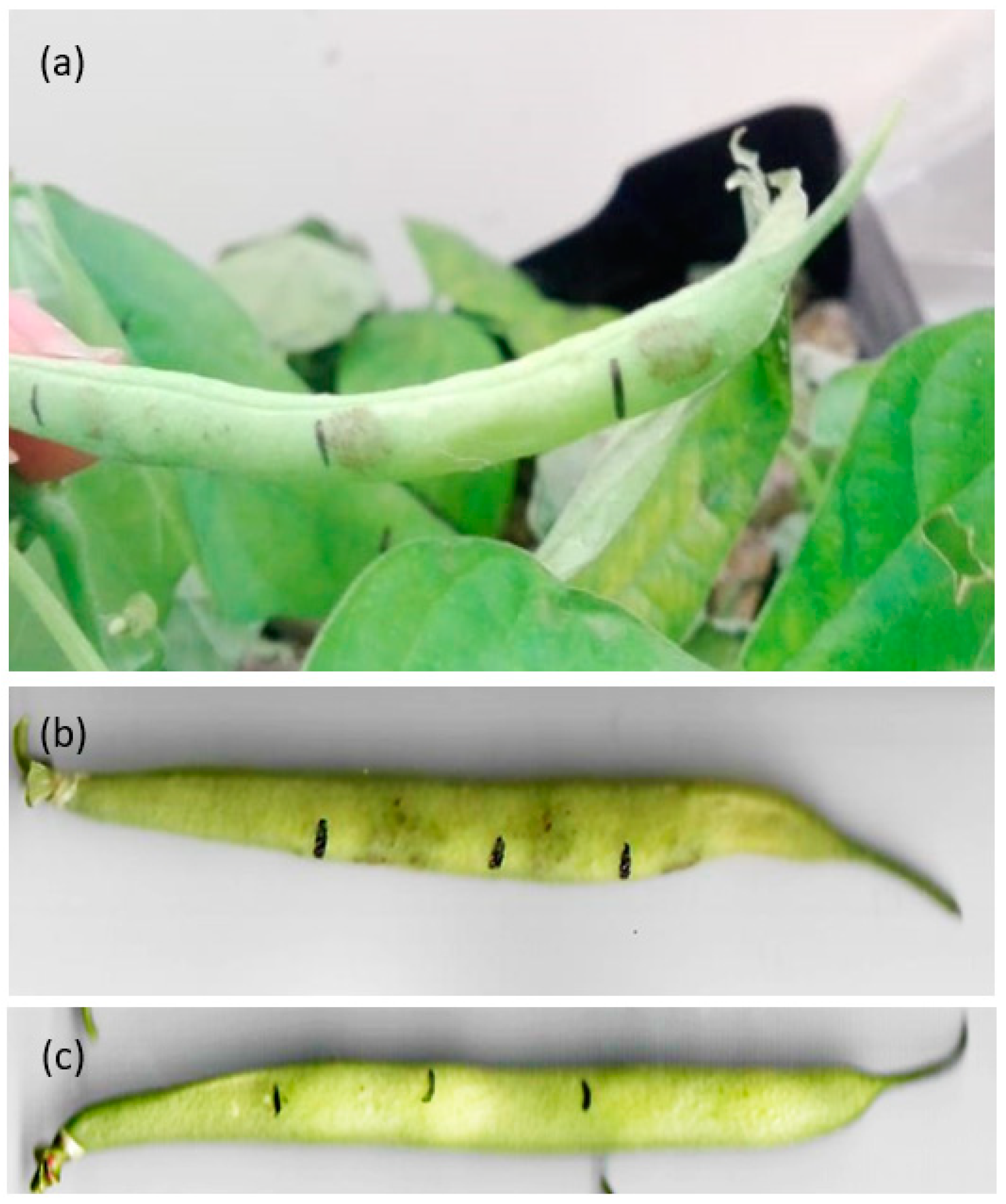
Figure 1. Common symptoms of anthracnose on the green bean pod (Phaseolus vulgaris L.) cultivar “Strike”, induced by C. lindemuthianum. (a) After 14 days of inoculation, biofortified plants treated with MgCl2 exhibited the emergence of oval-shaped brown necrotic lesions. (b) Inoculated with C. lindemuthianum, nonbiofortified. (c) Control, inoculated with water.
Microbiolres 14 00139 g001
Figure 2. Bioactive compound (BCs) analysis in green bean pods. (a) Total anthocyanins (mg of cyanidin-3-glucoside g−1), (b) total phenols (mg of gallic acid equivalents g−1), and (c) total flavonoids (mg catechin equivalents [CE] g−1). Assessment of green bean pods subjected to inoculated (Inoc) and noninoculated (No Inoc) treatments with MgCl2 and MgSO4. Statistically significant differences were determined at p ≤ 0.05. Grouping of means for BCsin inoculated and noninoculated green bean pods was conducted using a Tukey test (p ≤ 0.05). Data presented as means ± standard errors (n = 4).
Microbiolres 14 00139 g002
Figure 3. Illustration of biofortification in green bean using two Mg salts. This scheme represents a biofortification process of green bean plants with two magnesium salts, where Colletotrichum lindemuthianum inoculated onto green bean pods theoretically causes the plant to react by synthesizing metabolites in both symptomatic and healthy tissues.
Microbiolres 14 00139 g003
Disclaimer/Publisher’s Note: The statements, opinions and data contained in all publications are solely those of the individual author(s) and contributor(s) and not of MDPI and/or the editor(s). MDPI and/or the editor(s) disclaim responsibility for any injury to people or property resulting from any ideas, methods, instructions or products referred to in the content.