Article

Preliminary Data on Escherichia coli, Yersinia enterocolitica, and Other Bacteria, as Well as Absent African Swine Fever Virus in the Gut Microbiota of Wild Mice and Voles from Bulgaria

1
Department of Infectious Microbiology, The Stephan Angeloff Institute of Microbiology, Bulgarian Academy of Sciences, 1113 Sofia, Bulgaria
2
Department of Ecosystem Research, Environmental Risk Assessment and Conservation Biology, Institute of Biodiversity and Ecosystem Research, Bulgarian Academy of Sciences, 1000 Sofia, Bulgaria
3
Evgeni Budevski Institute of Electrochemistry and Energy Systems, Bulgarian Academy of Sciences, 1113 Sofia, Bulgaria
*
Author to whom correspondence should be addressed.
Microbiol. Res. 2023, 14(4), 1788-1819; https://doi.org/10.3390/microbiolres14040123
Submission received: 24 August 2023 / Revised: 27 October 2023 / Accepted: 30 October 2023 / Published: 4 November 2023

Abstract

:
Small mammals are bioindicator organisms, and, through their gut microbiota (GM), could be carriers of pathogens and resistant bacteria. Also, wild GM composition has been suggested to have large implications for conservation efforts. Seventeen bacterial species were obtained from intestinal samples of Bulgarian yellow-necked mice (Apodemus flavicollis) and bank voles (Myodes glareolus) via classic microbiological cultivation and biochemical identification. Twelve Gram-negative—Escherichia coli, Yersinia enterocolitica, Yersinia kristensenii, Hafnia alvei, Serratia liquefaciens, Serratia marcescens, Serratia proteamaculans, Pseudescherichia vulneris, Klebsiella pneumoniae ssp. ozaenae, Enterobacter cloacea, Pantoea agglomerans, Pseudomonas fluorescens group—and five Gram-positive bacteria, Enterococcus faecium, Enterococcus faecalis, Enterococcus hirae, Bacillus thuringiensis, and Lysinibacillus sphaericus, were discovered. Enterobacteriaceae was the most abundant family. The isolates belonged to one of the major reported taxa in rodents, Firmicutes (the Gram-positive species) and to the less abundant, but still among the first, phyla, Proteobacteria (the Gram-negative strains). We did not find any members of the other major phylum, Bacteroidetes, likely due to lack of metagenomic techniques. E. coli and Y. enterocolitica were confirmed with polymerase chain reaction. Almost all strains had pathogenic potential, but the good condition of the test animals suggests their commensal role. The Y. enterocolitica strains did not have the ail pathogenicity gene. There was high prevalence of multi-drug resistance (MDR), but for the expected species with high level of intrinsic resistance, such as the enterococci and S. marcescens. E. coli and some other species had very low antimicrobial resistance (AMR), in line with other studies of wild rodents. Many of the strains had biotechnological potential; e.g., B. thuringiensis is the most used biological insecticide, with its proteins incorporated into the Bt genetically modified maize. The GM of the tested wild mice and voles from Bulgaria proved to be a source of bacterial diversity; many of the strains were promising in terms of biotechnology, and, in addition, the samples did not contain the African swine fever virus.

1. Introduction

When studying gut microbiota (GM) of wild rodents, numerous interconnected factors come into play, which make that research especially important. First of all, causal claims in the microbiome field are often overstated because of incentives that result in ignoring negative results and stimulating hype. Human diseases are increasingly linked with an altered or “dysbiotic” GM, but whether such changes are causal, consequential, or bystanders is mostly unresolved. Only Helicobacter pylori and Clostridium difficile, two members of the human microbiome, are known to have established causality in human disease. They, however, do not colonize the guts of mice enough to cause reliable pathology in conventional murine models. There are many other similar limitations after the transplantation of fecal GM from humans, with and without a disease, into germ-free rodents, mainly mice, in order to produce human microbiota-associated (HMA) rodents [1].
Nevertheless, still there is no doubt that the GM has a profound impact on the health of the host. Vertebrate GM are often complex communities that provide essential nutrients and metabolites, modulate the host’s immune system, and limit the niche space available for colonization by pathogens [2]. It is well known that E. coli produces vitamin K, but less known is that enterococci produce vitamin B12 and the antimicrobial compound enterocin [3]. A bacterial peptidoglycan (PGN) is found in the developing mouse brain, suggesting that this bacterial product penetrates the blood-brain barrier. This PGN in the brain modulates the expression of pathogen pattern recognition molecules, such as toll-like receptor 2, while knock-down of proteins involved in PGN translocation to the brain leads to increased expression of genes related to a higher risk factor of autism (c-Met). According to the authors, bacterial product translocation into the CNS is key for establishing adequate social behaviors [4]. The use of germ-free animal models has provided data that strongly support the idea of early life gut colonization as a key event in brain development, which in turn affects behavior in later life. For example, germ-free mice exhibit a change in dopamine turnover. Therefore, maintenance of GM should be considered to prevent factors that may trigger mental illness [5].
As mentioned, laboratory rodents, especially mice (admixture between the house mice Mus musculus of different subspecies such as M. musculus domesticus) [6], have been the major study system for microbiota research. However, laboratory animals and their microbiota often poorly model aspects of the biology of wild animals and their microbiota [7]. As unwanted bacterial, viral, protozoal, and parasitic agents have gradually been eliminated, mice have developed an immune system more akin to that of a neonate than an adult human. Probably for that reason, standard laboratory mice have been killed by an experimental virus infection, whereas laboratory mice that had received wild mouse microbiota were resistant. These different outcomes were due to reduced inflammatory responses in the latter [6], thus showing that laboratory mice, while paramount for understanding basic biological phenomena, are limited in modeling complex diseases of humans and other free-living mammals [6,7], and point to the necessity to study wild rodent GM.
In terrestrial ecosystems, small mammals represent one of the best groups as ecological and physiological bioindicators—organisms which, with reference to chemical, physiological, ethological or ecological factors, provide information on the state of ecosystems [8]. In regard to antimicrobial resistance (AMR), small rodents are sentinel species—biological monitors that accumulate a pollutant (in the case—bacteria with AMR) in their tissues without significant adverse effects [9]. Wild microbial community composition also has been suggested to have large implications for conservation biology efforts [10].
There are even more factors that are associated with the research of the intestinal microflora of wild rodents, and they determine the importance of that work. We can understand why an ever-expanding body of work is dedicated to the GM of wild small mammals, including rodents. The research on their skin, oral, vaginal, etc. microbiomes is also increasing.
However, the research of GM in particular species is scarce. Such a species is the yellow-necked mouse (Apodemus flavicollis) [11], with studies on helminth [12] and radiation impact [13]. Work on the bank vole (Myodes glareolus) is more abundant [2,14,15] and also includes unicellular co-infections [16] and anthopogenic pressure such as radiation [17,18,19] and heavy metals [20]. Studies on AMR in these two species show low prevalence, still a little higher in farm areas [21,22,23,24,25]. To our knowledge, the microbiota of these two rodent species from Bulgaria has never been studied.
African swine fever (ASF) is a hemorrhagic infection caused by the African swine fever virus (ASFV). Just as in many other countries, it continues until the present date, and has resulted in a true disaster in recent years in Bulgaria, leading to significant economic losses, i.e., the killing of a vast number of uninfected farm pigs and wild boars for disease control, apart from dying wild boars. The most unfortunate consequence, however, is the near extinction of the East Balkan Swine, the last remaining native pig breed in Bulgaria. It is raised by extensive foraging, and hence is called “half-wild”. From nearly 55,000 East Balkan pigs, only approximately 300 have so far survived the epidemic [26]. In 2019, while the disease had occurred only in the Northern Bulgaria, some officials raised questions whether exterminating wild rodents in the Balkan Mountains would prevent the spread of the virus to Southern Bulgaria.
The aim of our work was to conduct a pilot study of the GM of two Bulgarian wild rodent species by culturing methods and to check whether they contain and could be passive mechanistic carriers of ASFV. We found 17 bacterial species and no traces of ASFV.

2. Materials and Methods

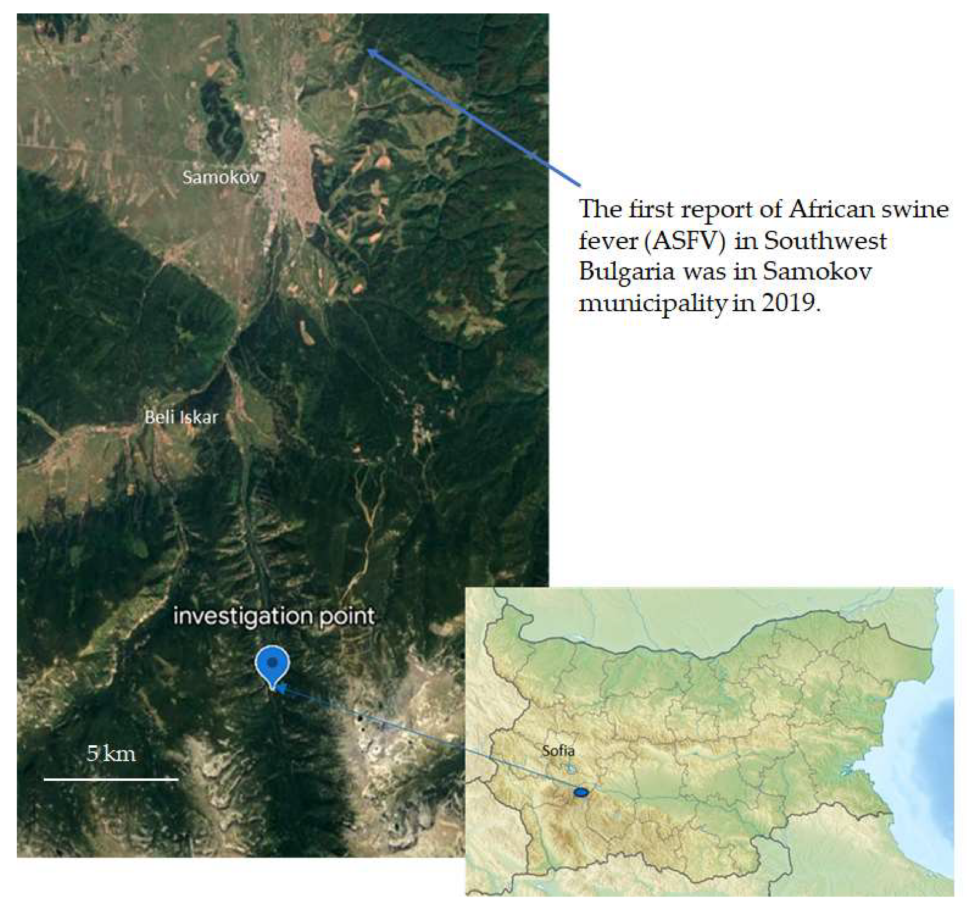
2.1. Sample Collection

Seventy live-bait traps set for about seven nights were intended to catch small mammals in August 2019 in the Skakavtsite area (Rila Mountain, 1500 m above sea level, Figure 1). The trap number and field stay aimed at, and were in accordance with, the methodology for determining the population size of the animals. The process involved non-endangered species, without a protected status (CITES lists and IUCN). Two male M. glareolus as well as five male and two female Ap. flavicollis were caught during the trapping period. The location (mixed forests) was representative of the region. These two species are common and permanent inhabitants of deciduous and mixed forests at this altitude in Bulgaria and at these latitudes. All small rodents caught were in normal physiological condition. All of them were parasitized with ticks, which is frequent and does not affect the normal physiological condition of the rodents. The collected animals were dissected to remove the intestinal tract, and fecal matter was taken from the rectum in close proximity to a flame from a spirit lamp. The fecal matter was suspended in brain-heart infusion (BHI) broth (M210, Himedia, Mumbai, India) and in tryptone soya broth (TSB, soyabean casein digest medium) (GM2011, Himedia, Mumbai, India), and refrigerated as soon as possible until further processing. A part of them were frozen for the purpose of DNA isolation.
The samples were likely taken after circulation of ASFV in the area had been established, as it was after the first report of ASFV in Southern Bulgaria, at the end of July 2019 [27], and approximately at the time of the first report of ASFV near the sampling area (Southwest Bulgaria), in the beginning of August. The virus was detected in wild boars in the Samokov municipality (state hunting farm “Iskar”), Figure 1 [28,29].

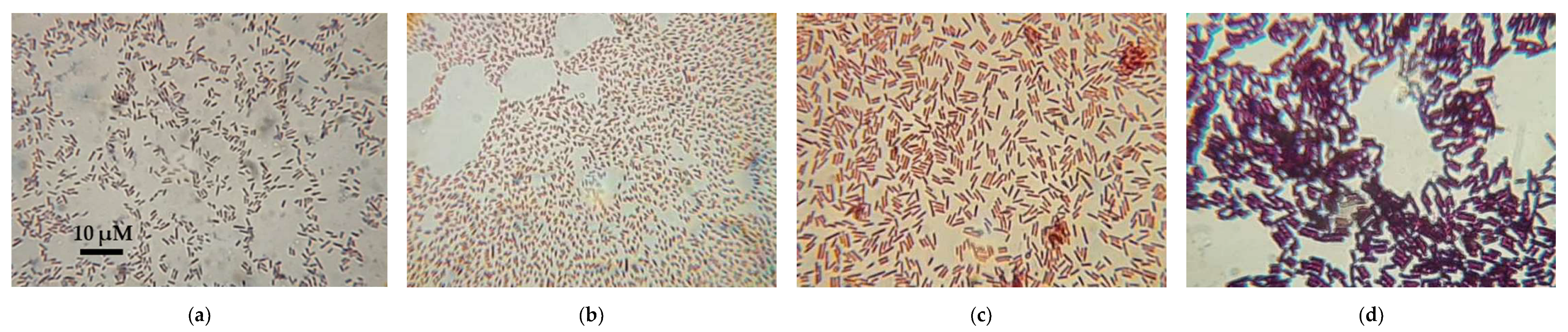
2.2. Isolation of Single Bacterial Cultures

Samples from the BHI broth were subjected to serial dilutions, and the concentrations of 102 and 103 colony-forming units/mL (CFU/mL) were inoculated on Nutrient agar (GM002, Himedia, Mumbai, India) and MacConkey agar (1002050500, Sigma-Aldrich, St. Louis, MO, USA) petri dishes. Colonies were selected for further work and were photographed under a magnifier. Microscopic slide preparations were made, and they were Gram-stained. The strains were subjected to biochemical identification.

2.3. Biochemical Identification

2.3.1. Identification with BD PhoenixTM

An automatic BD PhoenixTM M50 system (443624, Becton, Dickinson and Company, Franklin Lakes, NJ, USA) was used for a full biochemical characterization of selected isolates. Firstly, we determined whether they were Gram-positive or Gram-negative via Gram-staining and potassium hydroxide. Cultures inoculated on Columbia Blood Agar with 5% Sheep Blood (BDTM Columbia Agar, PA-254005.06) were made into a 0.5–0.6 McFarland bacterial suspension at hours 18 to 24, by taking material from approximately 5 single colonies and dissolving it directly in the vial of identification (ID) broth buffer (246001, Becton, Dickinson and Company, Franklin Lakes, NJ, USA). For the antimicrobial susceptibility testing (AST), 1 drop of indicator solution (246004, Becton, Dickinson and Company, Franklin Lakes, NJ, USA), warmed to room temperature, is placed in a bottle of the AST broth buffer (246003, Becton, Dickinson and Company, Franklin Lakes, NJ, USA). The bottle is stirred by inversion 1–2 times (without forming bubbles) and 25 µL of the ID broth buffer solution is added, then stirred again. The suspensions of the Gram-negative strains were poured in NMIC/ID panels (448103, Becton, Dickinson and Company, Franklin Lakes, NJ, USA); the suspensions of the Gram-positive strains were poured in PMIC/ID panels (448796, Becton, Dickinson and Company, Franklin Lakes, NJ, USA). The ID and AST buffers were poured into the ID and AST sides of the panels, respectively. The panels were loaded into the instrument at 35 °C for 24 ± 4 h. The obtained data were analyzed by EpiCentre™ software (V7.45A/V6.71A).

2.3.2. Identification with API 20 E of the Gram-Negative Bacteria

An API 20 E standardized identification system for Enterobacteriaceae and other non-fastidious Gram-negative rods (bioMérieux, SA, Marcy-l’Étoile, France) was used to corroborate the results from BD PhoenixTM for the Gram-negative bacteria. The API 20 E strip consists of 20 microtubes containing dehydrated substrates. These tests were inoculated with a bacterial suspension according to the kit instructions. During incubation, metabolism produced color changes that were either spontaneous or revealed by the addition of reagents. The reactions were read according to the Reading Table and the identification was obtained using the identification software at http://apiweb.mediclim.ro:81/, accessed on 2 October 2021.

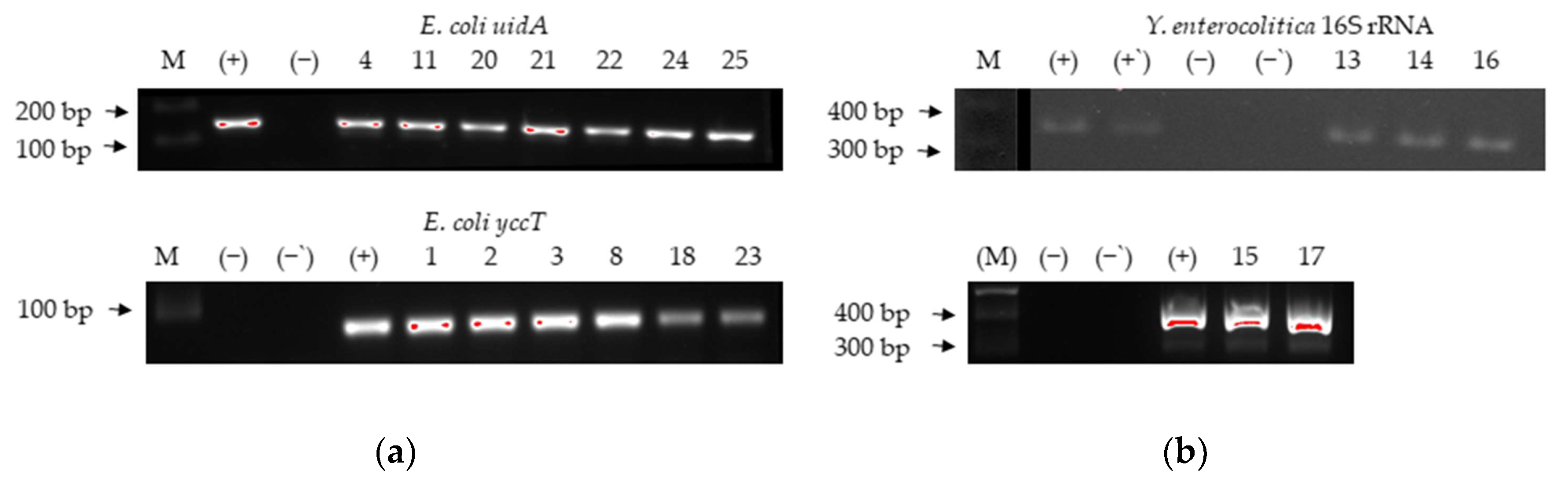
2.3.3. Confirmation of Identification of Escherichia coli and Yersinia enterocolitica by Traditional Polymerase Chain Reaction (PCR)

Total genomic DNA from all isolates was extracted through crude lysate preparation. The lysates were made by dissolving one bacterial colony in 100 µL of lysis buffer of 0.05 M NaOH and 0.125% sodium dodecyl sulfate (final concentrations), and samples were incubated for 17 min at 90 °C. The DNA from the identified E. coli and Y. enterocolitica strains was subjected to conventional PCR with gene-specific primers for E. coli (gene uidA, coding β-glucuronidase, and gene yccT, coding a conserved protein with unknown function) and for Y. enterocolitica (the gene for 16S rRNA), respectively (Table 1). For PCR amplification, we used the Taq PCR Master Mix (2×) (E2520, EURx Ltd., Gdańsk, Poland) and its protocol optimized with temperature of annealing tailored to the temperature of melting (annealing) (Tm) of the primers, which had final concentrations of 0.5 µM in the reaction mix. The BioRad Thermo-cycler was used. The PCR products were visualized in 1.5% agarose gels. For positive controls, we used E. coli American Type Culture Collection (ATCC, Manassas, VA, USA) 35218 and Y. enterocolitica strain IP8081, the highly pathogenic bioserotype 1B/O:8 (Institut Pasteur Collection, Paris, France).

2.4. Antibiotic Resistance/Susceptibility Elucidation

2.4.1. BD PhoenixTM

The antimicrobial susceptibility testing was performed as described in Section 2.3.1, because the AST side of the panel contains antimicrobial agents. The minimum inhibitory concentrations (MIC) and the respective susceptibility were determined according to the preset parameters and intrinsic (expert) rules of the instrument and of the EpiCentre™ software version V7.45A. The instrument does not do an AST for every species of bacteria it can identify.

2.4.2. Disk Diffusion Method

A standard Kirby–Bauer method, according to the protocols of the CLSI, was performed for two Pseudomonas isolates which had problematic AST at BD PhoenixTM [34]. We used antibiotics for Pseudomonas spp. from Breakpoint tables for interpretation of MICs and zone diameters of the European Committee on Antimicrobial Susceptibility Testing (EUCAST) [35]. One antibiotic from each given class was selected, namely, ticarcillin (75 µg, SD074-1CT), ceftazidime (10 µg, SD062A-1CT), Himedia, Mumbai, India), meropenem (10 µg, MEM10C Oxoid Ltd., Basingstoke, Hampshire, UK), aztreonam (30 µg, SD212-1CT), ciprofloxacin (5 µg, SD060-1PK), and tobramycin (10 µg, SD044-1CT) (HiMedia, Mumbai India). The results were evaluated according to the cut-off breakpoint values of EUCAST version 12.0, 2022.

2.5. Searching for the Ail Gene of Pathogenicity in Y. enterocolitica

2.5.1. Traditional PCR

The crude lysates from the Y. enterocolitica strains were subjected to conventional PCR with gene-specific primers for the gene for pathogenicity ail (attachment and invasion locus) (Table 1). PCR amplification was performed as in Section 2.3.3.

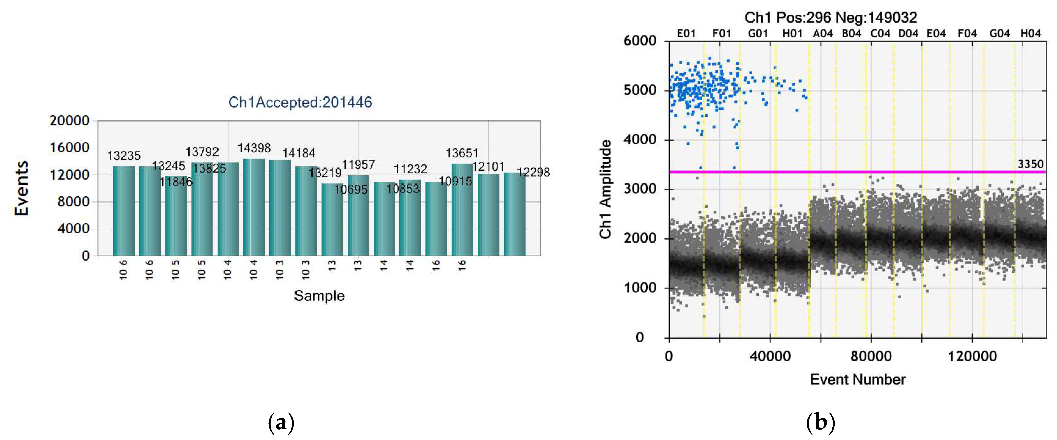
2.5.2. Droplet Digital PCR (ddPCR)

Pure genomic DNA was extracted from the single colonies of some Y. enterocolitica strains with a GeneMATRIX Tissue & Bacterial DNA Purification Kit (E3551, EURx Ltd., Gdańsk, Poland).
The ddPCR was performed on the BioRad QX200 Droplet Digital PCR System. The primers and TaqMan probe were previously applied in our laboratory for the detection of Y. enterocolitica with real-time PCR (Table 1). The TaqMan probe was 5′-6-FAM-TCT-ATG-GCA-GTA-ATA-AGT-TTG-GTC-ACG-GTG-ATC-T-TAMRA-3′. The reaction was carried out with ”ddPCR™ Supermix for Probes” (#1863010, Bio-Rad, Hercules, CA, USA). Purified DNA extracted from the Y. enterocolitica strain IP8081 was used as a positive control. Based on the concentration of the DNA template in [ng/µL] measured with a NanoDropTM Lite UV Spectrophotometer (ThermoFisher Scientific, Waltham, MA, USA), the concentration of the positive control in molecules/µL was calculated using the following equation:
m   g = n b p × 1   m o l e   D N A 6.02214076 × 10 23 = n b p × 660 6.02214076 × 10 23 = n b p × 1.096 × 10 21
where m is the mass of the genome of the investigated microorganism in [g] and n is the genome size in base pairs [bp] multiplied by the average molecular weight of a double-stranded DNA molecule divided by the Avogadro’s number. The DNA template was diluted to obtain positive controls with two concentrations: 102 and 101 DNA molecules/5 µL.
The master mix was prepared according to the recommendations of the master mix kit pointed above. The final concentration of the primers was 1 µM, and that of the probe, 0.5 µM. Briefly, 5 µL of the positive controls, the negative control (DNase and RNase free water), and the unknown samples were added to 15 µL master mix to obtain final volume of 20 µL. The ddPCR program was also designed following the instruction in the manual whereby the Tm of the primers was 60 °C and the cycles of amplification were 40.

2.6. Searching for ASFV with Conventional PCR

Total DNA was extracted from the primary frozen rectal samples via the GeneMATRIX Stool DNA Purification Kit (E3575, EURx Ltd., Gdańsk, Poland). The primers designed were from the C-terminal end of the ASFV VP72 gene (virus ptotein 72, major capsid protein), a well-characterized and highly conserved region of the ASFV genome in the different viral genotypes [36]. The complete genome of the viral isolate ASFV/Kaliningrad was taken from GenBank: OM799941.1 (National Center for Biotechnology Information (NCBI)) and the sequence of the gene was confirmed by GenBank: AY578697.1 [37]. Appropriate primers (Table 1) were selected from the sequence of the gene. The forward primer encompassed nucleotides 178–198 from the VP72 gene and the reverse primer encompassed nucleotide positions 577–597. PCR amplification was performed as in Section 2.3.3 except that PCR products were visualized in 2.5% agarose gels.

3. Results

3.1. Isolation of Single Bacterial Cultures

Fifty-three isolates (pure cultures) were isolated from selected single bacterial colonies. They were divided into 39 groups by morphological similarity of the colonies and were subjected to biochemical identification.

3.2. Biochemical Identification

The selected strains were identified as 17 bacterial species. Most of them (12) were Gram-negative: E. coli, Y. enterocolitica, Yersinia kristensenii, Hafnia alvei, Serratia liquefaciens, Serratia marcescens, Serratia proteamaculans, Pseudescherichia vulneris, Klebsiella pneumoniae ssp. ozaenae, Enterobacter cloacea, Pantoea agglomerans, and Pseudomonas fluorescens complex (group).
Gram-positive isolates were Enterococcus faecium, Enterococcus faecalis, Enterococcus hirae, Bacillus thuringiensis, and Lysinibacillus sphaericus.
The results are presented in Table 2. All identified isolates gave a confidence value of 99% by BD PhoenixTM, unless otherwise specified in parentheses. The API test confirmed the bacterial identification for the Gram-negative bacteria, except for Pantoea agglomerans strains, which were not put through the test because they looked like a symbiont microrganism. With a few exceptions, most of the isolates within one morphological group turned out to be the same species.
Upon reculturing, isolates 29 and 32 turned out to grow much faster and better at 26 °C than at 37 °C, and they turned to be P. fluorescens, a psychrophile microorganism. Actually, the average nucleotide identities of P. fluorescens have not fulfilled the criteria of a species, since they were very diverse. It has been concluded that P. fluorescens is not a species in the strict sense, but should be considered as a wider evolutionary group, or a relaxed species complex, that includes other species within it, also [38].
It can be observed that E. coli, L. sphaericus, and E. faecalis were the only species that were found in more than one animal; the rest of the species were limited to one individual rodent.
The results show that the Enterobacteriaceae family was the most abundant, comprising all the Gram-negative bacteria, except P. agglomerans (Erwiniaceae)—Enterobacteriaceae and Erwiniaceae are in the order Enterobacterales—and P. fluorescens (Pseudomonadaceae, order Pseudomonadales). The Enterococcaceae family (Lactobacillales), including the enterococci and the Bacillaceae family (order Bacillales), including B. thuringiensis and L. sphaericus, comprised the Gram-positive bacteria found. All Gram-negative bacteria were members of the class Gammaproteobacteria and phylum Proteobacteria (synonym Pseudomonadota) and all the Gram-positive ones were in the class Bacilli and phylum Firmicutes (synonym Bacillota). We did not find any members of the Bacteroidetes phylum (synonim Bacteroidota), which, together with Firmicutes, is the major phyla in rodents and the studied two species and has reached from 27% (Ap. flavicollis) [12] to 73% (M. glareolus) [20]. Figure S1 in the Supplementary Materials presents an example of the morphology of colonies from this study. Figure 2 presents examples of the photodocumented microscopic preparations (the morphology of the microorganisms)—two Gram-negative and two Gram-positive isolates. All the microscopic slide preparations are given in Figure S2 in the Supplementary Materials.

3.3. Confirmation of Identification of E. coli and Y. enterocolitica by Traditional PCR

All the strains that were biochemically identified as E. coli and Y. enterocolitica were confirmed to belong to these species with classic PCR (Figure 3). PCR product sequencing was not performed.
The original designers of the 16S rRNA Y. enterocolitica primers, Neubauer et al. [32], stated that those primers can confirm the bacterial species only in combination with biochemical identification as a member of the genus Yersinia because the target sequence of the primer pair is also present in some other bacteria from Enterobacteriaceae. Later, other researchers confirmed that they gave false positive results with Serratia, Citrobacter [39], and (from our experience) Morganella. It should be stressed that the Cefsulodin–Irgasan–Novobiocin (CIN) agar is not selective enough to exclude other bacteria and some of them also have the typical bull’s eye appearance on it. Therefore, growth on CIN agar of a bull`s eye colony and a positive PCR with the aforementioned 16S rRNA primers does not necessarily mean Y. enterocolitica or even Yersinia. In our case, a double identification—with BD PhoenixTM (biochemical) and with a 16S rRNA PCR—leaves no doubt that the isolates are Y. enterocolitica [40].
Having identified the isolates, we can say that in terms of being enteral bacteria and their pathogenicity, many of them turned out to be opportunistic pathogens, meaning that they affect mostly morbid patients with compromised immune systems.
Escherichia coli is a normal enteric commensal of animals and humans but also has pathogenic clones and causes the fourth-most commonly reported foodborne gastrointestinal infection in humans. It also can cause hemolytic-uremic syndrome, urinary tract and surgical site infection, sepsis, and meningitis, and is reported with increasing frequency, severity, and mortality [41,42].
Hafnia alvei is a facultative anaerobe found in water, soil, dairy products, and sewage, and is also a commensal in human and animal gut flora. An uncommon human pathogen, it may cause disease in immunocompromised patients occasionally isolated from clinical sources, and has received increased attention from the medical community over the past decade due to its possible association with gastroenteritis [43].
Yersinia enterocolitica is widespread in nature, in the intestinal tracts of many mammals, birds, and cold-blooded species, and from terrestrial and aquatic niches. Most environmental isolates are avirulent, but isolates from porcine sources contain human pathogenic serogroups. In addition, dogs, sheep, notably wild rodents, and environmental water may also be a reservoir of pathogenic strains. Human pathogenic strains are usually confined to the intestinal tract and lead to yersiniosis, which is mostly reported from Scandinavia. The symptoms are enteritis/diarrhea but complications may occur, such as arthritis and even septicemia [44]. Y. kristensenii is isolated from soil and environmental sources but rarely from animal or human feces. It is an opportunistic pathogen in extraintestinal specimens, though it is very rarely pathogenic to humans. It is potentially infectious to mice [41].
Lysinibacillus sphaericus is found on soil and is rarely a pathogen, with one or two cases of bacteremia, endocarditis, meningitis, pseudotumor and food poisoning reported clinically.
Serratia spp. are frequently isolated from environmental sources, including in animals, as they are facultatively anaerobic. S. marcescens is actually the most frequently isolated Serracia sp. in human infections as diverse as respiratory infections and septicemia, and is a prominent opportunistic pathogen in hospital epidemics and in animals. S. liqefaciens may be commensal in the human respiratory tract and has caused nosocomial infections, such as osteomyelitis and endocarditis [45]. Serratia proteamaculans is an opportunistic plant pathogen with only one report of human fatal pneumonia due to a subspecies of it [46].
Pseudomonas fluorescens group can be found in the soil and water; however, in the last few years, its isolation as the causative agent of nosocomial infections has rapidly increased. They usually affect patients with compromised immune systems [47].
Enterococci are ubiquitous, resilient, and human gut and oral commensal, and are facultative anaerobes, which means that they can proliferate under the anaerobic conditions found inside the gut or rumen. Moreover, they are resistant to gastric juices and bile salts. E. faecalis is one of the most common species of them, and, together with E. faecium, they are the most common human isolates among enterococci, including in clinical samples. They are opportunistic pathogens and etiologic agents of infections, mostly nosocomial—again, from wound and urinary tract infections to meningitis and septicemia. Their high AMR makes these infections problematic. E. faecalis and E. faecium comprise 80–90% and 5–10%, respectively, of the clinically isolated enterococci, and MDR E. faecium is responsible for the majority of device-associated infections [48]. It is the third-most common cause of nosocomial bloodstream infections [42]. Infection with E. hirae with low virulence has rarely been reported in humans, but it is not uncommon in mammals and birds.
Pantoea agglomerans is ubiquitous and found in environmental sources and in animals and humans and their feces. It is also an occasional opportunistic pathogen, causing wound, blood, and urinary-tract infections. Infections are typically acquired from infected vegetation parts penetrating the skin, and bloodstream infection can lead to disseminated disease and end-organ infections, such as septic arthritis and endocarditis [49].
Pseudescherichia vulneris is found in a few animal and environmental samples such as apple leaves, wastewater, and wild birds, but also in human clinical specimens, mainly in wounds (vulneris is Latin for wound), but also in sputum, blood, urine, etc., and is infrequently reported in cases of meningitis [41].
Klebsiella pneumoniae ssp. ozaenae is mainly a colonizer of the oral and nasopharyngeal mucosa, and is an opportunistic pathogen that may be responsible for a chronic atrophic rhinitis with resorption of the underlying bone, called ozena. These have become uncommon in developed countries. The species is rarely associated with infections—from wound infections to septic pulmonary emboli associated with bacteremia [50].
Enterobacter cloacae is the most frequently isolated Enterobacter species, which are facultative anaerobes and cause increasing MDR nosocomial infections. It is found in water, soil, meat, and sewage, and is animal and human gut and skin commensal. It as an important opportunistic and nosocomial pathogen in urologic, respiratory, trauma, and intensive care units and is also isolated from sputum, burns, and occasionally spinal fluid and blood [51].
Therefore, E. coli, H. alvei, Y. enterocolitica, Y. kristensenii, E. faecalis, E. faecium, E. cloacae, and P. agglomerans are mammal gut commensals, or at least often found in feces. In addition, all the strains in this study have pathogenic potential in vertebrates, except only B. thuringiensis and most likely S. proteamaculans.

3.4. Antibiotic Resistance/Susceptibility Elucidation

The results show that most of the isolates from the same bacterial species had very similar resistance profiles, for some even identical, likely because most of them were isolated from one individual rodent. One of the few exceptions was one strain of S. marcescens, and another was one of the two isolates of Y. kristensenii, which differed significally in their profile. Among all isolates, a total of 30 strains had multi-drug resistance (MDR), defined as resistance to at least three agents. They were all of the H. alvei, all of the Yersinia strains, S. proteamaculans, E. cloacae, all E. faecalis, and E. faecium, and one of the P. fluorescens strains. It is notable that both Y. kristensenii and one of the P. fluorescens strains had resistance to carbapenems. This may be due to carbapenemase presence, hyperproduction of AmpC, or other extended-spectrum beta-lactamase (ESBL). It is also noteworthy that isolate 48 (S. marcescens) was confirmed positive for ESBL by the BD PhoenixTM but that enzyme did not influence the resistance to aztreonam and third-generation cephalosporins (cefotaxime and ceftazidime), and it was susceptible to them. These phenomena increase the pathogenic potential of the strains so much that, according to the BD PhoenixTM manual, if the case was in clinical settings, a clinician and an infection control practitioner should have been alerted.

3.4.1. BD PhoenixTM

The data about the antibiotic resistance of isolates for which BD PhoenixTM is able to do an AST in addition to identification are given in Table 3, Table 4 and Table 5.
The strain that had the highest level of AMR was one of the two isolates of Y. kristensenii (19). It had resistance to eight agents, including a third-generation cephalosporin (ceftazidime). This phenomenon in Enterobacterales indicates a likely potent beta-lactamase and explains the resistance to first- and second-generation cephalosporins, as it was the case. The other strain of Y. kristensenii had resistance only to three agents.
Next, AMR was most prevalent among most of the S. marcescens strains and the enterococci, with resistance to seven agents.
Enterococcus spp. are reported as resistant to trimethoprim (including a combination with sulfamethoxazole) because it is not effective clinically. The results are of no surprise because enterococci are intrinsically resistant to bacteriostatic and bactericidal activities of the most of the commonly used agents. For that reason, recommended therapy for serious infections is a synergism between a cell-wall active agent, such as penicillin, combined with an aminoglycoside. E. faecalis is intrinsically resistant to fusidic acid, cephalosporins, macrolides (e.g., clindamycin), and sulfonamides [41]. As expected, the four isolates were resistant to those of the listed agents (the panels did not have sulfonamides alone). It is also intrinsically resistant to low-level aminoglycosides and quinipristin-dalfopristin; however, the isolates were sensitive to the combination of the two. E. faecium is increasingly resistant to vancomycin, but it was sensitive in that study.
According to the BD PhoenixTM manual and other sources [41], S. marcescens is intrinsically resistant to ampicillin, amoxicillin-clavulanate, cefazolin, cephalexin, cefuroxime, tetracycline, nitrofurantoin, colistin, and other polymixins such as polymixin B. Two of the three strains had resistance to all these agents (except for tetracyclines, which are only in the panels for Gram-positive strains). Surprisingly, the third S. marcescens had resistance only to colistin. S. proteamaculans had resistance to six agents.
Next in the level of AMR were H. alvei, E. cloacae, and Y. enterocolitica, with resistance to four agents. H. alvei and Y. enterocolitica are intrinsically resistant to ampicillin, amoxicillin-clavulanate, cefazolin, cephalexin, cefoxitin, and ticarcillin. Y. enterocolitica is resistant to penicillins and cephalosporins due to beta-lactamase production [41]. The results showed that they were indeed resistant to the first four agents (the system could not give interpretation for cephalexin only for the Hafnia strains) and the other two agents were not in the respective panels. In addition, H. alvei was resistant to colistin, just as S. proteamaculans was, and this is notable because resistance to it is uncommon for most Enterobacterales, except S. marcescens. E. cloacae complex is intrinsically resistant to ampicillin, amoxicillin-clavulanate, cefazolin, cefoxitin, cephalexin, and other cephalosporins [41,52], which is due to the cephalosporinases and ESBLs, in addition to the carbapenemases, of Enterobacter species.
E. coli strains had very little resistance level—only to some aminoglycosides of some the isolates. This is noteworthy, since E. coli is an ESBL producer and easily gains resistance to carbapenems, polymyxins, and quinolones through horizontal gene transfer [53,54].
The rest of the strains, i.e., S. liquefaciens, P. agglomerrans, K. pneumoniae ssp. ozaenae, and P. vulneris, had resistance to very few antibiotics. That is a good sign, as the K. pneumoniae species, which are also increasing in frequency, severity, and mortality, have ESBLs and carbapenemases, and the latter cause severe infections in long-term care facilities [42]. Studies have shown that P. vulneris has resistance to penicillin and clindamycin, and here, it was resistant to ampicillin [41].

3.4.2. Disk Diffusion Method

Although, in theory, BD PhoenixTM can perform AST on Pseudomonas fluorescens, in practice, the AST of our isolates failed even after repeating. Although BD PhoenixTM is more reliable than the disk diffusion or broth macrodilution methods [55], in order to obtain the AST of all bacteria that BD PhoenixTM could, in theory, provide, we had to implement the disk diffusion method. Table 6 gives the results for the two P. fluorescens isolates.
Glucose-nonfermenting Gram-negative rods such as Pseudomonas are generally intrinsically resistant to penicillin. However, ticarcillin (as well as piperacillin) are the so-called pseudomonas penicillins, as they are expected to be more effective, but we see that the two Pseudomonas isolates were resistant to it (zone diameters 0 mm). In contrast, these rods are generally intrinsically resistant to first- and second-generation cephalosporins; however, the results here show intermediate resistance.

3.5. No Ail Gene of Pathogenicity in Y. enterocolitica Was Found

The ail gene, which is involved in eukaryotic cell invasion and serum resistance, was not detected in isolates 13, 14, and 16 (Y. enterocolitica) with the help of traditional or ddPCR. The results from the ddPCR are presented in Figure 4 and Table 7. The generated droplets in the samples varied between 10,000 and more than 14,000, which is statistically acceptable for calculation of the results. The exact concentration of both positive controls corresponded to the preliminary calculations (Table 7).

3.6. No ASFV Was Detected

The virus of the African swine fever disease was not detected in the intestinal samples.

4. Discussion

Biological indicators (or bioindicators) must be sensitive enough to indicate environmental changes as soon as possible. The main reasons why non-endangered small mammals are one of the best groups as bioindicators are their basic position in the food chains as primary consumers, short life cycle, rapid maturity, large number of generations annually, large population number, wide distribution area all over the world except Antarctica, and rapid biological reaction to any kind of biotic and abiotic environmental changes [21,56,57,58]. Small animals, including mice, voles, and insectivores, can be effectively used as sentinels of antimicrobial-resistant microorganisms and their antibiotic-resistance genes (ARG) [9].
Conservation efforts such as breeding or rehabilitation include captivity and thus have a significant impact on the lives of the animals concerned. Captivity conditions such as altered diet may influence the composition of the GM, thus causing negative effects on animal health. The GM retained regardless of captivity status is hypothesized to cover important symbiotic relationships with the host, while the remaining part reflects the artificial living conditions and can therefore be used as a future tool for conservation biologists. Identification of keystone microbes in the GM can serve as indicators of the host’s adaptation to the surrounding environment. Monitoring of keystone organisms of high importance for the GM metabolic functional potential can provide important new tools in conservation efforts. The GM can provide additional information about different aspects of the host’s welfare and health, thus justifying the application of microbiome data in animal welfare and conservation processes [10]. For example, a captive breeding colony of a highly endangered Amargosa vole was created. It turned out that captive vole foregut microbiomes were dominated by Allobaculum sp., while wild foreguts were dominated by Lactobacillus sp. If these differences result in beneficial functional differences in digestion, then captive-reared Amargosa voles should be prepared prior to release into the wild [59]. Studies on non-endangered species like ours can still be translated in terms of endangered ones and be helpful to some extent in the conservation process. For example, they could determine whether a change in the GM is an adaptation to a stressor or a harmful consequence of it [20].
We see that some bacterial species are unique for voles in this study (e.g., P. fluorescens group), while others, as expected, are found in both voles and mice (e.g., Serratia spp.). Mice are omnivorous rodents, while voles are strictly herbivorous, eating insects by mistake if they are stuck to plant parts [60], and this is likely the reason for the differences in their GM.
Studies show that in comparison to carnivory and omnivory, herbivory appears to be associated with increased bacterial diversity and preponderance of microorganisms capable of breaking down complex plant carbohydrates such as resistant starches and celluloses. Ruminococcus flavefaciens expresses a complex cellulolytic machinery (cellusome) and has been found as one of the main cellulolytic species in ruminant and non-ruminant herbivores, including voles. The genus Ruminococcus, mostly represented by R. flavefaciens, was one of the most abundant taxa found in prairie voles [61]. In the capybara, besides cellulose-degrading bacteria, a diverse set of carbohydrate-active enzymes from Bacteroidetes, organized in polysaccharide utilization loci, tackles complex hemicelluloses typically found in gramineous and aquatic plants that capybaras eat [62]. Interestingly, Ruminococcaceae and Lachnospiraceae were the most abundant families in the GM of wild M. musculus, while they exhibited lower abundance in inbred mice [63]. Even the GM of omnivorous rodents such as rats is proven to digest cellulose [64].
In fact, there are data that most of the bacterial species from this study are cellulolytic [65,66,67,68,69,70,71,72,73]. Only Y. enterocolitica does not have such activity [74] and there are no data for that ability for H. alvei and Y. kristensenii. Most E. faecium strains are not cellulolytic, but there are rare reports of strains with that ability [3,75]. Only one strain of E. coli, from bovine rumen, has shown that activity [71]. An E. hirae strain with 93% certainty of identity was cellulolytic [76].
Most research on GM is performed via metagenomic analysis—16S rRNA amplicon sequencing of samples, also called 16S rRNA gene catalogues (metataxonomics). Compared to classic microbiological cultivation, these techniques have great advantages, since they encompass virtually all the bacteria in the tested sample, including the anaerobic and uncultivable ones. On the other hand, metagenomics does not completely differentiate between resident microbiota and non-resident microbes ingested transiently with food. The DNA ingested from animal cells is not digested and degraded completely, and is present in the feces, and even more is the DNA from plant, fungus, and bacterial cells, which have thick cell walls [77,78,79].
This is not the first report on the microbiota of wild rodents in Bulgaria. There were 21% PCR positive for Francisella tularensis rodents (viscera of M. musculus and Rattus rattus) in an endemic for tularemia region (Pernik area) [80]. Nevertheless, the current work is the first screening on their gut flora and is an attempt to assess the species composition of Bulgarian rodents. In addition, pathogenic Y. enterocolitica and Yersinia pseudotuberculosis have been isolated from many wild mammal species in Bulgaria, e.g., mouflon and polecat. Rodents were not included in the study [81].
Nine samples (animals) is not a very large sample size, as statisticians prefer to have at least 100 samples (n = 100), e.g., when processing microbiological samples. But the trap number and field stay aimed at and had to be in accordance with the methodology for determining the population size of the animals. Therefore, the sample size depended on the population density of the animals at the time of investigation. Therefore, this microbiological work can be considered a qualitative, not quantitative study. In addition, from a humane standpoint, less is better, even for non-endangered species. The authors of this study welcome the idea of future research involving low- or non-invasive methods. Moreover, even with that small representative number of samples, we find that the bacterial diversity we were able to uncover is impressive.

4.1. Previous Studies on the GM of Ap. flavicollis (Yellow-Necked Mice)

The research of GM in this species is scarce. The typical community composition for GM of studied members of the Apodemus genus consists of three bacterial phyla that account for >97% of the GM community: Bacteroidetes (67%), Firmicutes (27%), and Proteobacteria (3%). Firmicutes consist mainly of three families: Lachnospiraceae (7%), Lactobacillaceae (6.5%), and Ruminococcaceae (6%) [82,83].
Ap. flavicollis, together with five other wild species, e.g., deer, had clinical paratuberculosis in 2002 in South Austria, and one of 18 mice was positive for Mycobacterium avium subsp. paratuberculosis [11].
Intestinal helminths are host immunomodulators and have evolved both temporally and spatially in close association with the GM, resulting in potential mechanistic interplay. Research showed that helminths clearly have the potential to alter gut homeostasis. Three populations were studied; 93% of the mice had a single infection, and over half were infected with two or more helminth species. Overall, the GM was dominated by Firmicutes (67%) and Bacteroidetes (27%), while Proteobacteria represented 4%, and other bacterial phyla represented more than 1%. We see that Ap. flavicollis is less abundant in Bacteroidetes than other Apodemus spp. The stomach, small intestine, and mucosa were dominated by members of the class Bacilli (78%), whereas the colon and caecum contained more Bacteroidia (49%) and Clostridia (34%). The nematode Mastophorus muris, as well as the two nematodes ubiquitous in each population, Heligmosomoides polygyrus and Syphacia spp., and cestodes Hymenolepis spp. were studied. Each helminth colonized distinct parts of the gut; Hymenolepis spp. and H. polygyrus were detected exclusively in the small intestine, M. muris in the stomach, and Syphacia spp. in the caecum and, to a lesser extent, in the colon. In general, natural helminth (co-)infections were linked with high microbiota diversity, which may confer health benefits to the host. Variation in the composition and abundance of GM taxa associated with helminths was specific to each helminth species and occurred both up- and downstream of a given helminth’s niche (gut position). The most pronounced helminth–microbiota association was between the presence of tapeworms in the small intestine and increased Bacteroidetes family in the stomach [12].
Studies on two species pairs of Apodemus spp. mice that occur in sympatry in habitats affected by the Chernobyl and Fukushima nuclear accidents examined Ap. speciosus and Ap. argenteus from Japan, and Ap. flavicollis and Ap. sylvaticus from Ukraine. Overall, in all species of mice, the GM was typical of the Apodemus genus [82,83], albeit with different proportions among host species. Species from Japan were characterized by significantly more diverse GM than their congeners in Ukraine. The country of origin accounted for ~17% of variation in GM structure. Ap. argenteus harbored significantly higher gut bacterial diversity compared with Ap. speciosus, but there was no significant difference in diversity of the GM of Ap. flavicollis and Ap. sylvaticus. The results showed that radiation exposure altered the GM composition and structure in Ap. flavicollis, as well as in Ap. sylvaticus, and Ap. speciosus. Members of the Bacteroidetes family Muribaculaceae were over-represented in the GM of mice from contaminated areas, so they are particularly responsive to radiation exposure. Similar responses to radiation were found within other taxa in the GM of the two Ukrainian mouse species. Firmicutes exhibit a significant difference in relative abundance among contaminated and uncontaminated areas in Ap. flavicollis and Ap. speciosus (but not in Ap. sylvaticus). At the lower taxonomy levels, these ASVs were assigned to Lactobacillaceae, Ruminococcacae, Lachnospiraceae, and other families within the Clostridiales. Hence, members of the Firmicutes and Bacteroidetes phyla drive compositional changes in response to radiation in wild mice. Contrary to the author’s prediction, the Firmicutes:Bacteroidetes (F:B) ratio is not a general biomarker of radiation exposure, as it was increased only in Ap. speciosus. Hence, effects of radiation on mouse GM composition appear to be taxonomically widespread across many bacterial families, with members of each family both positively and negatively associated with radiation. The GM of Ap. argenteus was unaltered, probably because this wild mouse species is tree-dwelling and does “radiation escape”, i.e., avoiding soil radionuclide contamination [13].

4.2. Previous Studies on GM of M. glareolus (Bank Voles)

The GM and its seasonal variation have been studied by W Kunicki-Goldfinger and W Kunicka-Goldfinger in the early 1960s but unfortunately, the works seem to be available only to associates of the University of Guelph, Canada [84,85,86]. More recently, the order Bacteroidales (52% of the microbiome) was estimated to be the most abundant group, followed by the phyla Firmicutes (31%), Spirochaetes (6%), and Proteobacteria (4%) [2]. Hanhimäki et al., 2022, found that bank vole GM is mediated by physiological modifications. They artificially selected for fast metabolism, but that had minimal impact on the GM in laboratory conditions. A lab-to-field transition resulted in an increase in species diversity (α-diversity), a significant increase in Actinobacteria, and a decrease in Patescibacteria (both being generally less-abundant bacteria). Notably, young voles born from the selection lines had significantly higher α-diversity in comparison with those born from control lines [14]. Kohl et al., 2016 performed a 15-generation selection experiment with selection for high swim-induced aerobic metabolism, predatory behavior toward crickets, and the ability to maintain body mass on a high-fiber, herbivorous diet. The cecal chambers of voles selected for herbivorous capability had higher diversity than control lines, and their foregut communities were also distinct, which suggested that differences in GM across herbivores may be evolved, and not solely driven by current diet [15]. Brila et al., 2023, coinfected Finnish voles with bacterial and protozoan pathogens. Animals with a single infection had slightly higher phylogenetic diversity than animals with no infections, but that aspect did not differ between single and coinfected, or even between uninfected and coinfected, voles. Still, the GM of coinfected animals differed from that of single-pathogen-infected individuals, and all pathogens affected GM ß-diversity in a pathogen-specific way, affecting both rare and abundant gut bacteria. The authors concluded that the effects of coinfection (compared with those of single infection) are idiosyncratic (i.e., pathogen-specific) and that excluding co-infection information from statistical models masks pathogen-specific patterns and confounds interpretations [16].
Regarding anthropogenic environmental impacts, bank voles inhabiting areas contaminated by radionuclides (at Chernobyl, Ukraine) were characterized by no detectable effect on the gut community richness but an increase in Firmicutes and a reduction in Bacteroidetes (an almost two-fold increase in the F:B ratio). Radiation-associated bacteria had distinct inferred functional profiles, including pathways involved in degradation, assimilation, and transport of carbohydrates, xenobiotic biodegradation, and DNA repair [19]. In another study, Lavrinienko et al., 2020, found that the GM of all voles was comprised of 10 bacterial phyla, dominated by Firmicutes, Bacteroidetes, and Spirochaetes (9%). Similarly, members of the Bacteroidetes family were abundant in uncontaminated areas again, while an increase in Ruminococcaceae, Lachnospiraceae (Firmicutes, Clostridiales order), and Desulfovibrionaceae (Proteobacteria) families was observed in contaminated areas. The authors also found that exposure to radiation was also associated with a reduction the degree of GM temporal changes and in GM inter-individual variation, i.e., lower dispersion. The GM may be constrained by the physiology of a stressed host, and this prevented its dynamic response to natural spatio-temporal variation in resources. Most likely, the distinct GM profile provided beneficial services under chronic radiation exposure. For example, members of the Ruminococcaceae and Lachnospiraceae families could provide direct benefit to the host in the form of short-chain fatty acids. As a summary, exposure to radiation impacts composition, but not α-diversity of bank vole GM [18]. The same authors also found that, besides being affected by the level of radioactivity, GM could be strikingly similar among distant (80 km of separation) uncontaminated locations, while skin microbiome communities were structured more by geography than the level of soil radionuclides. In addition, the level of (dis)similarity between the skin and gut microbiome communities from the same individuals was contingent on the potential for exposure to radionuclides [17].
Here is the place to note one hypothesized outcome of (anthropogenic) stress on animal microbiomes, called the “Anna Karenina principle” (AKP). The outcome expects a destabilized microbial community that is characterized by an increase in inter-individual differences compared with microbiomes of healthy animals, which are expected to be temporally stable and relatively similar among individuals. AKP parallels Leo Tolstoy’s dictum that “all happy families look alike; each unhappy family is unhappy in its own way” [87]. However, the cited practical studies on rodents show that, for instance, radiation on M. glareolus, as well as a metal load stressor in laboratory mice, shift the community composition, leading to more uniform GM, less prone to natural temporal changes. Wild rodents have more inter-individual differences; thus, here, the AKP turned to not be valid [7,17,18].
Bank voles inhabiting areas with elevated levels of metals from two locations in Finland had, contrary to the hypothesis of Brila et al., 2021, higher GM α-diversity and subtly altered community composition (β-diversity), but there was no change in the level of community dispersion. The bank vole GM was composed of 11 bacterial phyla, the three major being, again, Bacteroidetes (73%), Firmicutes (22%), and Proteobacteria (2 ± 3%). At the family level, GM was dominated by Muribaculaceae (71%), Lachnospiraceae (13 ± 11%), and Ruminococcaceae (7 ± 5%).
The core GM of voles from the high-pollution group consisted of 108 amplicon sequence variants (ASVs), and from the low-pollution group, consisted of 88 ASVs, with animals from both groups sharing 80 ASVs. Positively associated with multi-metal exposure were members of genera Anaerostipes, Odoribacter, and Lachnospiraceae NK4A136 group, while the negatively-associated members were from the genera Desulfovibrio, Treponema, and Ruminococcus. Moreover, many families had ASVs both positively and negatively associated with multi-metal exposure. For example, the Clostridiales vadinBB60 group had 9 ASVs positively and 3 ASVs negatively associated, and Lachnospiraceae had 20 positively and 22 negatively associated ASVs. ASVs from the genus Ruminococcaceae NK4A214 group were negatively associated, while the genera Ruminiclostridium and Ruminiclostridium had, again, members both positively and negatively associated with multi-metal exposure ASVs. Multi-metal exposure and increased levels of several metals (Cd, Hg, Pb and Se) were mostly associated with differences in the abundance within the families Clostridiales vadinBB60 group, Desulfovibrionaceae, Lachnospiraceae, Muribaculaceae, and Ruminococcaceae. Hence, even low-level metal pollution is associated with altered GM of wild mammals. The authors argued that the trend of higher α-diversity after multi-metal exposure showed similarities with environmental exposure to multiple metals in humans and differed from the findings of single metal exposure in laboratory animals. Thus, it seemed likely that exposure to multiple metals is associated with a different microbiota response than single-metal exposure and that the effects of environmental metal exposure on GM of wild animals cannot be extrapolated from laboratory studies. They assumed that the results were consistent with the intermediate disturbance hypothesis (IDH) [88] in GM. IDH predicts that species diversity is expected to be highest at intermediate levels of disturbance. Translated to that study, it might be plausible that low-intermediate metal exposure allows coexistence of many species of GM, whereas no pollution would result in less diverse and more stable, “core” GM, while high total metal load would favour metal-resistant microbial taxa. It was unclear whether the higher α-diversity was a favorable adaptation to the stressor or a consequence of it, leading to less stable GM containing more opportunistic bacteria [20].

4.3. Previous Studies on the Antimicrobial Resistance of M. glareolus and Ap. flavicollis

E. coli isolated from colon, cecal, and fecal samples are commonly used as indicator organisms to monitor antimicrobial resistance (AMR) in wild animals [89].
A study of AMR in fecal probes of bank voles and two other closely related species from England showed an unexpected correlation between the abundancy of antimicrobial-resistant (AR) E. coli in the rodents and their habitats. Samples from near river edges showed 79% AMR strains, in contrast to 35% of inland-dwelling animals; the phylotype diversity (groups with shared sequence similarity of a particular gene marker) was also twice as much in coastal isolates than in inland-obtained ones. The most frequent antibiotic resistance was to ampicillin, though ciprofloxacin and cefotaxime resistance was also detected [21]. Our personal experience also shows that AMR is very prevalent in river bacteria. A pilot study to get a first insight into the occurrence of AR E. coli in the guts of eight rodent and one shrew species originating from Germany in the frame of different studies within the network rodent-borne pathogens was conducted, by testing 188 fecal isolates. The rodent objects of the network were Ap. Flavicollis, M. glareolus, and other common rodents—other Apodemus spp., other mice, the Norwegian rat, the common and field vole, etc. The prevalence of AR isolates was low, with only 5.5% of the isolates exhibiting resistant phenotypes against at least one antimicrobial compound, including beta-lactams, tetracyclines, aminoglycosides, and sulfonamides. Nevertheless, MDR E. coli were significantly more often detected in wild rodents originating from areas with high livestock density, suggesting a possible transmission from livestock to wild rodents [22]. Only around 5% of isolated E. coli from different mice and voles, including 121 samples of Ap. flavicollis and three samples of M. glareolus in Poland, had any AMR. New unique sequence types were found, showing a significant genomic heterogeneity. One of the strains with tetracycline resistance had the tet(B) gene. An interesting fact is that all colistin-resistant strains were recovered from female Ap. Flavicollis, captured at different sites and in different periods of time. Two colistin-resistant isolates possessed unique mutations in the pmrB gene. Even in the small percentage of resistant E. coli, a high diversity of virulence factors was obtained, including carriers of eilA, astA, ibeA, etc. [23].
There were a few other AR species of bacteria detected in samples from the two species. For example, three methicillin-resistant Staphylococcus aureus (MRSA) strains containing the mecA gene, as well as other species of staphylococci with ampicillin and erythromycin resistance, were found in Ap. flavicollis samples from Slovakia. Some specific genes detected were the beta-lactamase (blaZ) in all ampicillin-resistant strains and the ermC with the efflux msrA in some erythromycin-resistant strains [24]. Among eight samples of Ap. Flavicollis, eight samples of Ap. Agrarius, and one sample of Microtus arvalis, the AMR level within all gut bacterial species was very low in comparison with poultry, pigs, wild boars, and foxes. Rodents did not have abundant ARGs for tetracyclines, macrolides, or aminoglycosides, and did not have ARGs for quinolones or plasmid-mediated colistin resistance at all. They only had moderately abundant ARGs for beta-lactams and low entire plasmid content. Evidence of plasmids associated with AMR transfer was noted within plasmid profiles of all of the tested animals [25].
All these studies suggest a minor role of wild small rodents from rural areas in the cycle of transmission and spread of AR E. coli and other bacteria into the environment. Often, only traces of AMR compared with other animals is observed. Collected at forest and meadow areas, their probable contact with human settlements is occasional, and therefore the animals have little contact with antimicrobials. However, AMR is more prevalent in wild rodents from river areas and even more from areas with high livestock density, suggesting a possible transmission from livestock to wild rodents.

4.4. Biotechnological and Practical Potential of the Isolated Bacteria

Notably, some of the bacteria that we isolated could be of practical use.
H. alvei is used as a lactic ferment by the dairy industry and more recently as a probiotic included in a dietary supplement product. At the end of Camembert manufacturing, it remains as the dominant species. H. alvei is a psychrotrophic strain, which can develop at low temperatures, meaning that it doesn’t stop growing during the storage phase of cheese, unlike E. coli [90].
B. thuringiensis (Bt) is the most-commonly used biological insecticide worldwide, as it is the source of crystal δ-endotoxins, a.k.a Cry proteins. These proteinaceous inclusions are incorporated into genetically modified maize. They are not toxic to humans and all mammals but can also kill nematodes such as Caenorhabditis elegans. Cry proteins are made by many Bt strains during sporulation, the cry genes are mostly located on plasmids but not all strains and crystals have insecticidal properties [91]. In 2000, a novel subgroup of Cry proteins, parasporin, was discovered from non-insecticidal Bt strains. Notably, they and related bacterial parasporal proteins turned out to preferentially kill cancer cells but are not hemolytic [92].
L. sphaericus is commonly found on soil and is also frequently used in commercial insecticides. It is of particular interest to the World Health Organization, as some strains have a larvicidal effect for disease-carrying mosquitos (Culex and Anopheles), more effective than Bt. It is of important use in mosquito control programs worldwide and is safe for vertebrates and nondipterean insects [93]. Moreover, cells in a vegetative state are also effective against Aedes aegypti larvae [94], an important vector of yellow fever and dengue viruses.
L. sphaericus also has bioremediation potential. Cells have a proteinaceous surface covering, called the S-layer, which is able to bind high quantities of heavy metals in saline solutions. Strains with chromate reduction capacity have been isolated from contaminated environments and naturally metal-rich soils [95]. Other strains bind aluminium, cadmium, copper, lead, and uranium [96], and are resistant to up to 200 mM of arsenic, which may be due to an arsenate reductase gene [97].
Some strains of P. fluorescens group protect the roots of some plant species against parasitic fungi such as Fusarium or oomycetes and phytophagous nematodes. The species complex produces the antibiotics obafluorin and mupirocin. The latter treats skin, ear, and eye disorders. Mupirocin free acid and its salts and esters are agents used in creams, ointments, and sprays for MRSA [98].
P. agglomerans also has potential to produce antibiotics and can serve as a plant pathogen competitor for the management of plant diseases. It is toxic to the bacterium Erwinia amylovora which causes fire blight, a plant disease commonly found in pear and apple crops [99,100].

4.5. Can Other Animals Spread ASFV?

The native origin of ASFV is sub-Saharan Africa, where it circulates in a sylvatic cycle between wild warthogs—which carry it asymptomatically—and Ornithodoros ticks such as O. moubata. Unfortunately, it also spreads via direct transmission between Suidae and is lethal to domestic swine, including in Africa, and to other species of wild hogs such as the bushpig and the European wild boar [101,102,103,104]. Ornithodoros ticks transmit ASFV—by active biological or mechanical transmission or by passive transport or ingestion—also in Southern Europe and the United States [105]. For example, the O. erraticus tick transmits it only in Spain and Portugal. However, most of the soft tick species in Europe and the Caucasus region do not infest domestic and wild swine; therefore, there is no risk of tick-mediated ASF spread there [103].
As to other arthropods, Bonnet et al., 2020 find their role as vectors to be generally unstudied [104], although there is research on different species. Only two insects have been found to be possible vectors. ASFV has been detected in Haematopinus suis, swine lice prevalent in temperate regions, collected from experimentally infected domestic pigs [106].
Stomoxys flies are the so-called stable flies. They are hematophagous and can fly up to ten miles in search of animals on which to feed. Flies infected with the virus by engorging on a pad soaked with viraemic blood have been shown to be experimentally competent for mechanically transmitting ASFV to domestic pigs for 24 h by feeding on them, and the titre of the virus was constant in the flies for two days. In contrast, the virus was recovered from one out of ten of the flies infected by feeding directly from an infected host at two days after infection, and they failed to transmit it to pigs at that time [107]. If they are able to pass it to pigs, abundances of 20 and 50 stable flies per pig would mean that the vector-borne transmission would likely be responsible for almost 30% and 50% of transmission events, respectively [108]. However, Stomoxys flies collected on ASF-affected farms in Lithuania tested negative for ASFV [109].
While some Bulgarian farm owners suspect mosquitoes to be another vector, Hakobyan et al., 2022, using real-time PCR and hemadsorbtion analysis, show that the insect does not provide significant support for the persistence of the ASF virus in the environment, with no evidence for transmission to their offspring or pigs that ingested mosquitoes [110]. A nymphal stage of the blood-sucking insect Triatoma gerstaeckeri, with range in the North American continent, is able to carry the virus for 40 days and retain it through one molt, but is unable to transmit it to susceptible pigs [111]. Seasonality of the disease in the summers and outbreaks in farms with high biosecurity levels impelled Yoon et al., 2021, to test 28,729 arthropods in the vicinity of the farms. They turned out to be PCR negative. Still, the authors do not rule out an ability to passively infect. They admit the chance that the results may reflect the effects of immediate control measures in the early phase of infection in Korea [112]. To date, there have been few studies investigating the potential passive transport of ASFV by non-hematophagous arthropods, e.g., domestic flies. Calliphoridae (blowfly) larvae feeding on an infected carcass quickly inactivated ASFV [113].
Thus, the infection of wild boar or pigs by an arthropod mechanical vector pathway (hematophagous and non-blood sucking insects, as well as ingestion of infected arthropods) seems possible, but likely corresponds to exceptional events without epidemiological importance [101,104]. Nevertheless, The European Food Safety Authority (EFSA) declared vector studies of arthropods such as Stomoxys a research priority in 2021 [114].
Attempts to artificially infect other animals have been made, and the only species found to be possible vectors are three.
ASFV persisted for a certain period of time in air-breathing land snails (Xeropicta derbentina), and transcription of viral genes was maintained within them, although the question of full-fledged viral replication is still open. The virus was likely to be localized in the intestines of snails as it was regularly excreted from their feces. In addition, the active movements of snails make their role in spreading the virus possible [110].
As described in [115,116], several studies from as early as the 1950s and 1960s demonstrated that ASFV could be propagated in rabbits and goats, however, only after the agent had been modified through multiple experimental infections. Neitz and Alexander, cited by [117], succeeded in maintaining the virus in rabbits for a limited number of serial passages. The method of blind passage was used, meaning unapparent infection and disease. In another example, the virus appeared to be unaltered in virulence after 85 passages in rabbits [118]. Others reported substantial attenuation after 100 passages in rabbits [119]. Another attenuated lapinized strain of ASF virus recovered the initial virulence when passaged a number of times in pigs [120].
Kovalenko et al., 1965, have shown that kids (juvenile goats) 4 to 5 months old could be infected with ASFV by intraperitoneal inoculation of infected blood. The animals developed symptoms within 6 to 25 days, and one kid died after 36 days. The virus was found in the blood 6 days after infection but was no longer present after 30 days. It was present in the spleen after 36 days but not after 70 days. The disease was characterized by hyperthermia, diarrhea, severe emaciation, and lesions in the reticuloendothelial system. The virus was passaged 19 times in kids and appeared to adapt progressively to these animals, causing damage to the reticuloendothelial system and accumulating in the spleen.
Levels of ASFV in earthworms (Dendrobaena alpina) and soil declined at similar rates, suggesting that earthworms likely have no influence on the ecology of the ASFV. Ciliates (Paramecium caudatum) significantly increased the rate of ASFV disappearance from the aquatic environment, probably using the virus as a food source [110].
Efforts to artificially infect other animals have been tried, as early as 1921 [121] for cattle, calves, horses, sheep, and dogs, and as early as 1971 [115] for cats, guinea pigs, oxen, hedgehogs, hamsters, rats, mice, and various fowls, but have failed. Therefore, they cannot transmit the virus. A more recent study from Vietnam shows that rats are not only unsusceptible to challenge with ASFV, but they do not have a mechanical vector potential (the capacity for passive transmission), as different sample types turned to be PCR-negative [122].
As described in [101], blood samples from other live animals, such as rodents and birds, have been collected from ASF-affected farms in Lithuania [109] and Russia [123], but have tested negative for ASFV.
It is likely that some European authorities claim rodents to be ASFV vectors because all that described research was made many years ago or is still not published as articles in international scientific journals. EFSA still declared in 2021 the investigation of birds as ASFV vectors as research priority [114].
As a summary, we agree that investigating invertebrates and non-Suidae vertebrates is important for understanding ASFV [110], but we also corroborate the general conclusion that only Suidae and Ornithodoros spp. can productively be naturally infected [103] and other animals do not appear to be of a significant risk and significance in the epidemiology of the disease [122]. Some officials raised questions whether exterminating wild rodents would prevent the spread of the virus. We can answer them that it would not.
Officials know that feeding pigs kitchen scraps is a major route of spreading the disease, since they contain pork or other infected foodstuff. Direct transmission between pigs, and contact with corpses and infected feed, equipment, delivery trucks, tools, clothes, and shoes are the other main transmission routes. In Bulgaria, voices were heard that the explanatory campaigns that the mayors were supposed to lead about the danger of ASF were underestimated and, despite the plans developed years ago, many of the measures in them were not implemented [124]. Therefore, policies should continue to focus on biosecurity, control of import, export, and logistics, and on correcting the mistakes from the past.

4.6. Future Directions

Future directions for this research likely include PCR confirmation, where possible, of all bacterial species, especially for P. agglomerans. Although this species gave 99% certainty here, there are data suggesting that, using the biochemical panels commonly employed in medical diagnostics, it is difficult to differentiate P. agglomerans from other Pantoea spp. or from related genera, such as Phytobacter, Enterobacter, Klebsiella, and Serratia spp. [125], and DNA sequencing has disproven the identity of several clinical isolates initially reported as P. agglomerans [126]. Virulence genes and ARGs for aminoglycosides can be searched in E. coli. We cannot state that we uncovered all the bacterial species in the GM, not even the cultivable ones, since we selected to keep many, but not all, of the colonies that grew. For that reason, another important direction would be a metagenomic analysis of the DNA of the original fecal samples. The B. thuringiensis strain could be checked for insecticidal activity and, if active, its toxins can be characterized. The potential of the other isolates in biotechnology can also be elucidated. Testing samples for ASFV from these or other small mammals taken later on during the flare of the Bulgarian ASFV epidemic is another goal, since it can shed even more light as to whether they can be passive carriers.

5. Conclusions

Fifty-three isolates were obtained from intestinal samples of Bulgarian bank voles and yellow-necked mice; they were identified by biochemical panels. Twelve Gram-negative and 5 Gram-positive bacterial species were identified. Enterobacteriaceae was the most abundant family. The phylum Firmicutes, to which all our Gram-positive species (ten isolates) belonged, is reported as one of the major taxa in rodents, but we did not find any members of the other major phylum, Bacteroidetes. All Gram-negative strains (43 isolates) were in the, generally, third major phylum in rodents, Proteobacteria, which is reported as much less abundant. These differences may be due to the utilization of cultivation techniques vs. the metagenomic analysis in most of the published studies. E. coli and Y. enterocolitica were confirmed with PCR. Almost all strains had pathogenic potential, but the good condition of the test animals suggests their commensal role. Wild rodents are a known reservoir of pathogenic Y. enterocolitica strains, but the isolates we obtained did not have the ail pathogenicity gene. This does not exclude the possibility of the presence of other virulence genes. In addition, there was high prevalence of MDR only for the expected species, i.e., those with high level of intrinsic resistance, such as the enterococci and S. marcescens. E. coli and some other species had very low AMR, in line with other studies of rodents in natural habitats. Most of the species were cellulolytic, according to literature data. Many of the obtained strains had biotechnological potential as insecticide and antibiotic producers, ferments in the dairy industry, for plant pathogen protection, and for bioremediation. No ASFV DNA was detected in the rodent intestines, which is another corroboration that wild animals, except boars, are not of significance in the epidemiology of the disease, and, in order to limit African swine fever and the euthanizing of uninfected pigs for disease control, policies should continue to focus on biosecurity. Wild GM composition has been suggested to have large implications for conservation efforts. Therefore, collectively, our results determine our hope that they would be useful for biotechnologists and conservation biologists.

Supplementary Materials

The following supporting information can be downloaded at: https://www.mdpi.com/article/10.3390/microbiolres14040123/s1, Figure S1: Example of the macro morphology of colonies. (a) A control E. coli American Type Culture Collection (ATCC) 35218; (b) E. coli, isolate 4; (c) E. coli, isolate 8.; Figure S2: Microscopic preparations of the isolates arranged by bacterial species.

Author Contributions

Conceptualization, Y.I., M.B., H.N. and M.M.Z.; methodology, H.N., M.M.Z., M.B. and Y.M.; software, not applicable; validation, Y.I. and M.D.K.; formal analysis, Y.M.; investigation, Y.I., J.J., M.D., M.B., I.A., L.D., M.D.K., M.M.Z., Y.M. and Y.G.; resources, Y.I., L.D., H.N., M.B., I.A. and Y.M.; data curation, not applicable; writing—original draft preparation, Y.I., Y.G., M.B. and M.M.Z.; writing—review and editing, Y.I. and Y.G.; visualization, Y.I., M.M.Z. and M.B.; supervision, H.N., M.M.Z. and L.D.; project administration, Y.I.; funding acquisition, not applicable. All authors have read and agreed to the published version of the manuscript.

Funding

This research received no external funding.

Institutional Review Board Statement

The sampling process involved species without a protected status (CITES lists and IUCN) and was fully compliant with the OECD guidelines of 17 December 2001 and 29 July 2016.

Informed Consent Statement

Not applicable.

Data Availability Statement

The data presented in this study are available on request from the corresponding author.

Acknowledgments

The authors would like to express their gratitude to microbiologist Tanya Kim and to the Ministry of Education and Science of Bulgaria, Competition “Junior researchers and postdoctoral students “—III stage, module “Junior researchers”, acronym of the project: ΠMC №353/10.12.2020, III-321/18.03.2020, Study of the intestinal microflora of wild rodents.

Conflicts of Interest

The authors declare no conflict of interest.

References

  1. Walter:, J.; Armet, A.M.; Finlay, B.B.; Shanahan, F. Establishing or Exaggerating Causality for the Gut Microbiome: Lessons from Human Microbiota-Associated Rodents. Cell 2020, 180, 221–232. [Google Scholar] [CrossRef]
  2. Lavrinienko, A.; Tukalenko, E.; Mousseau, T.A.; Thompson, L.R.; Knight, R.; Mappes, T.; Watts, P.C. Two Hundred and Fifty-Four Metagenome-Assembled Bacterial Genomes from the Bank Vole Gut Microbiota. Sci. Data 2020, 7, 312. [Google Scholar] [CrossRef] [PubMed]
  3. Mamuad, L.L.; Kim, S.H.; Biswas, A.A.; Yu, Z.; Cho, K.-K.; Kim, S.-B.; Lee, K.; Lee, S.S. Rumen Fermentation and Microbial Community Composition Influenced by Live Enterococcus faecium Supplementation. AMB Express 2019, 9, 123. [Google Scholar] [CrossRef]
  4. Arentsen, T.; Qian, Y.; Gkotzis, S.; Femenia, T.; Wang, T.; Udekwu, K.; Forssberg, H.; Diaz Heijtz, R. The Bacterial Peptidoglycan-Sensing Molecule Pglyrp2 Modulates Brain Development and Behavior. Mol. Psychiatry 2017, 22, 257–266. [Google Scholar] [CrossRef] [PubMed]
  5. González-Arancibia, C.; Urrutia-Piñones, J.; Illanes-González, J.; Martinez-Pinto, J.; Sotomayor-Zárate, R.; Julio-Pieper, M.; Bravo, J.A. Do Your Gut Microbes Affect Your Brain Dopamine? Psychopharmacology 2019, 236, 1611–1622. [Google Scholar] [CrossRef]
  6. Rosshart, S.P.; Vassallo, B.G.; Angeletti, D.; Hutchinson, D.S.; Morgan, A.P.; Takeda, K.; Hickman, H.D.; McCulloch, J.A.; Badger, J.H.; Ajami, N.J.; et al. Wild Mouse Gut Microbiota Promotes Host Fitness and Improves Disease Resistance. Cell 2017, 171, 1015–1028.e13. [Google Scholar] [CrossRef] [PubMed]
  7. Viney, M. The Gut Microbiota of Wild Rodents: Challenges and Opportunities. Lab. Anim. 2019, 53, 252–258. [Google Scholar] [CrossRef]
  8. Pearce, J.; Venier, L. Small Mammals as Bioindicators of Sustainable Boreal Forest Management. For. Ecol. Manag. 2005, 208, 153–175. [Google Scholar] [CrossRef]
  9. Gwenzi, W.; Chaukura, N.; Muisa-Zikali, N.; Teta, C.; Musvuugwa, T.; Rzymski, P.; Abia, A.L.K. Insects, Rodents, and Pets as Reservoirs, Vectors, and Sentinels of Antimicrobial Resistance. Antibiot. Basel Switz. 2021, 10, 68. [Google Scholar] [CrossRef]
  10. de Jonge, N.; Carlsen, B.; Christensen, M.H.; Pertoldi, C.; Nielsen, J.L. The Gut Microbiome of 54 Mammalian Species. Front. Microbiol. 2022, 13, 886252. [Google Scholar] [CrossRef]
  11. Deutz, A. Paratuberkulose Bei Wildtieren—(Ernährungsbedingte) Krankheit Der Zukunft? In Proceedings of the Tagung für die Jägerschaft, Federal Institute for Alpine Agriculture Gumpenstein, A-8952 Irdning, Gumpenstein, Austria, 16–17 February 2004. [Google Scholar]
  12. Kreisinger, J.; Bastien, G.; Hauffe, H.C.; Marchesi, J.; Perkins, S.E. Interactions between Multiple Helminths and the Gut Microbiota in Wild Rodents. Philos. Trans. R. Soc. B Biol. Sci. 2015, 370, 20140295. [Google Scholar] [CrossRef] [PubMed]
  13. Lavrinienko, A.; Hämäläinen, A.; Hindström, R.; Tukalenko, E.; Boratyński, Z.; Kivisaari, K.; Mousseau, T.A.; Watts, P.C.; Mappes, T. Comparable Response of Wild Rodent Gut Microbiome to Anthropogenic Habitat Contamination. Mol. Ecol. 2021, 30, 3485–3499. [Google Scholar] [CrossRef] [PubMed]
  14. Hanhimäki, E.; Watts, P.C.; Koskela, E.; Koteja, P.; Mappes, T.; Hämäläinen, A.M. Evolved High Aerobic Capacity Has Context-Specific Effects on Gut Microbiota. Front. Ecol. Evol. 2022, 10, 934164. [Google Scholar] [CrossRef]
  15. Kohl, K.D.; Sadowska, E.T.; Rudolf, A.M.; Dearing, M.D.; Koteja, P. Experimental Evolution on a Wild Mammal Species Results in Modifications of Gut Microbial Communities. Front. Microbiol. 2016, 7, 634. [Google Scholar] [CrossRef] [PubMed]
  16. Brila, I.; Lavrinienko, A.; Tukalenko, E.; Kallio, E.R.; Mappes, T.; Watts, P.C. Idiosyncratic Effects of Coinfection on the Association between Systemic Pathogens and the Gut Microbiota of a Wild Rodent, the Bank Vole Myodes glareolus. J. Anim. Ecol. 2023, 92, 826–837. [Google Scholar] [CrossRef]
  17. Lavrinienko, A.; Tukalenko, E.; Mappes, T.; Watts, P.C. Skin and Gut Microbiomes of a Wild Mammal Respond to Different Environmental Cues. Microbiome 2018, 6, 209. [Google Scholar] [CrossRef]
  18. Lavrinienko, A.; Tukalenko, E.; Kesäniemi, J.; Kivisaari, K.; Masiuk, S.; Boratyński, Z.; Mousseau, T.A.; Milinevsky, G.; Mappes, T.; Watts, P.C. Applying the Anna Karenina Principle for Wild Animal Gut Microbiota: Temporal Stability of the Bank Vole Gut Microbiota in a Disturbed Environment. J. Anim. Ecol. 2020, 89, 2617–2630. [Google Scholar] [CrossRef]
  19. Lavrinienko, A.; Mappes, T.; Tukalenko, E.; Mousseau, T.A.; Møller, A.P.; Knight, R.; Morton, J.T.; Thompson, L.R.; Watts, P.C. Environmental Radiation Alters the Gut Microbiome of the Bank Vole Myodes glareolus. ISME J. 2018, 12, 2801–2806. [Google Scholar] [CrossRef]
  20. Brila, I.; Lavrinienko, A.; Tukalenko, E.; Ecke, F.; Rodushkin, I.; Kallio, E.R.; Mappes, T.; Watts, P.C. Low-Level Environmental Metal Pollution Is Associated with Altered Gut Microbiota of a Wild Rodent, the Bank Vole (Myodes glareolus). Sci. Total Environ. 2021, 790, 148224. [Google Scholar] [CrossRef]
  21. Furness, L.E.; Campbell, A.; Zhang, L.; Gaze, W.H.; McDonald, R.A. Wild Small Mammals as Sentinels for the Environmental Transmission of Antimicrobial Resistance. Environ. Res. 2017, 154, 28–34. [Google Scholar] [CrossRef]
  22. Guenther, S.; Grobbel, M.; Heidemanns, K.; Schlegel, M.; Ulrich, R.G.; Ewers, C.; Wieler, L.H. First Insights into Antimicrobial Resistance among Faecal Escherichia coli Isolates from Small Wild Mammals in Rural Areas. Sci. Total Environ. 2010, 408, 3519–3522. [Google Scholar] [CrossRef] [PubMed]
  23. Skarżyńska, M.; Zając, M.; Kamińska, E.; Bomba, A.; Żmudzki, J.; Jabłoński, A.; Wasyl, D. Salmonella and Antimicrobial Resistance in Wild Rodents-True or False Threat? Pathog. Basel Switz. 2020, 9, 771. [Google Scholar] [CrossRef] [PubMed]
  24. Kmet’, V.; Čuvalová, A.; Stanko, M. Small Mammals as Sentinels of Antimicrobial-Resistant Staphylococci. Folia Microbiol. 2018, 63, 665–668. [Google Scholar] [CrossRef]
  25. Skarżyńska, M.; Leekitcharoenphon, P.; Hendriksen, R.S.; Aarestrup, F.M.; Wasyl, D. A Metagenomic Glimpse into the Gut of Wild and Domestic Animals: Quantification of Antimicrobial Resistance and More. PLoS ONE 2020, 15, e0242987. [Google Scholar] [CrossRef] [PubMed]
  26. Available online: https://bnr.bg/post/101521898/poroda-na-raba-na-izchezvaneto (accessed on 8 August 2023).
  27. Available online: https://www.vesti.bg/bulgaria/svinskata-chuma-stigna-do-iuzhna-bylgariia-6098131 (accessed on 21 August 2023).
  28. Available online: https://btvnovinite.bg/bulgaria/parvi-sluchaj-na-afrikanska-chuma-po-svinete-v-sofijska-oblast.html (accessed on 21 August 2023).
  29. Available online: https://bnr.bg/horizont/post/101152445/potvardiha-nalichieto-na-afrikanska-chuma-v-lovno-stopanstvo-iskar (accessed on 21 August 2023).
  30. Bej, A.K.; DiCesare, J.L.; Haff, L.; Atlas, R.M. Detection of Escherichia coli and Shigella spp. in Water by Using the Polymerase Chain Reaction and Gene Probes for Uid. Appl. Environ. Microbiol. 1991, 57, 1013–1017. [Google Scholar] [CrossRef] [PubMed]
  31. Clifford, R.J.; Milillo, M.; Prestwood, J.; Quintero, R.; Zurawski, D.V.; Kwak, Y.I.; Waterman, P.E.; Lesho, E.P.; Mc Gann, P. Detection of Bacterial 16S rRNA and Identification of Four Clinically Important Bacteria by Real-Time PCR. PLoS ONE 2012, 7, e48558. [Google Scholar] [CrossRef]
  32. Neubauer, H.; Hensel, A.; Aleksic, S.; Meyer, H. Identification of Yersinia enterocolitica within the Genus Yersinia. Syst. Appl. Microbiol. 2000, 23, 58–62. [Google Scholar] [CrossRef]
  33. Lambertz, S.T.; Nilsson, C.; Hallanvuo, S.; Lindblad, M. Real-Time PCR Method for Detection of Pathogenic Yersinia enterocolitica in Food. Appl. Environ. Microbiol. 2008, 74, 6060–6067. [Google Scholar] [CrossRef] [PubMed]
  34. CLSI. Performance Standards for Antimicrobial Disk and Dilution Susceptibility Tests for Bacteria Isolated from Animals, 3rd ed.; Approved Standard; Clinical and Laboratory Standards Institute: Wayne, PA, USA, 2009. [Google Scholar]
  35. The European Committee on Antimicrobil Susceptibility Testing. Breakpoint tables for interpretation of MICs and zone diameters. Version 12.0. 2022. Available online: https://www.eucast.org/ (accessed on 1 August 2023).
  36. Fernández-Pinero, J.; Gallardo, C.; Elizalde, M.; Robles, A.; Gómez, C.; Bishop, R.; Heath, L.; Couacy-Hymann, E.; Fasina, F.O.; Pelayo, V.; et al. Molecular Diagnosis of African Swine Fever by a New Real-Time PCR Using Universal Probe Library. Transbound. Emerg. Dis. 2013, 60, 48–58. [Google Scholar] [CrossRef]
  37. GenBank Overview. Available online: https://www.ncbi.nlm.nih.gov/genbank/ (accessed on 1 August 2023).
  38. Mulet, M.; Lalucat, J.; García-Valdés, E. DNA Sequence-Based Analysis of the Pseudomonas Species. Environ. Microbiol. 2010, 12, 1513–1530. [Google Scholar] [CrossRef]
  39. Arnold, T.; Neubauer, H.; Nikolaou, K.; Roesler, U.; Hensel, A. Identification of Yersinia enterocolitica in Minced Meat: A Comparative Analysis of API 20E, Yersinia Identification Kit and a 16S rRNA-Based PCR Method. J. Vet. Med. B Infect. Dis. Vet. Public Health 2004, 51, 23–27. [Google Scholar] [CrossRef] [PubMed]
  40. Kwaga, J.; Iversen, J.O.; Misra, V. Detection of Pathogenic Yersinia enterocolitica by Polymerase Chain Reaction and Digoxigenin-Labeled Polynucleotide Probes. J. Clin. Microbiol. 1992, 30, 2668–2673. [Google Scholar] [CrossRef] [PubMed]
  41. Cohen, J.; Powderly, W.G.; Opal, S.M. Infectious Diseases, 4th ed.; Elsevier: London, UK, 2017. [Google Scholar]
  42. Richwagen, N.; Lyles, J.T.; Dale, B.L.F.; Quave, C.L. Antibacterial Activity of Kalanchoe mortagei and K. fedtschenkoi Against ESKAPE Pathogens. Front. Pharmacol. 2019, 10, 67. [Google Scholar] [CrossRef]
  43. Cooney, S.; O’Brien, S.; Iversen, C.; Fanning, S. Bacteria: Other Pathogenic Enterobacteriaceae—Enterobacter and Other Genera; Elsevier: Amsterdam, The Netherlands, 2014; pp. 433–441. [Google Scholar]
  44. Fàbrega, A.; Vila, J. Yersinia enterocolitica: Pathogenesis, Virulence and Antimicrobial Resistance. Enfermedades Infecc. Microbiol. Clínica 2012, 30, 24–32. [Google Scholar] [CrossRef]
  45. Mahlen, S.D. Serratia Infections: From Military Experiments to Current Practice. Clin. Microbiol. Rev. 2011, 24, 755–791. [Google Scholar] [CrossRef]
  46. Bollet, C.; Grimont, P.; Gainnier, M.; Geissler, A.; Sainty, J.M.; De Micco, P. Fatal Pneumonia Due to Serratia proteamaculans Subsp. Quinovora. J. Clin. Microbiol. 1993, 31, 444–445. [Google Scholar] [CrossRef]
  47. Scales, B.S.; Dickson, R.P.; LiPuma, J.J.; Huffnagle, G.B. Microbiology, Genomics, and Clinical Significance of the Pseudomonas fluorescens Species Complex, an Unappreciated Colonizer of Humans. Clin. Microbiol. Rev. 2014, 27, 927–948. [Google Scholar] [CrossRef] [PubMed]
  48. Mascini, E.M.; Troelstra, A.; Beitsma, M.; Blok, H.E.M.; Jalink, K.P.; Hopmans, T.E.M.; Fluit, A.C.; Hene, R.J.; Willems, R.J.L.; Verhoef, J.; et al. Genotyping and Preemptive Isolation to Control an Outbreak of Vancomycin-Resistant Enterococcus faecium. Clin. Infect. Dis. Off. Publ. Infect. Dis. Soc. Am. 2006, 42, 739–746. [Google Scholar] [CrossRef]
  49. Dutkiewicz, J.; Mackiewicz, B.; Kinga Lemieszek, M.; Golec, M.; Milanowski, J. Pantoea agglomerans: A Mysterious Bacterium of Evil and Good. Part III. Deleterious Effects: Infections of Humans, Animals and Plants. Ann. Agric. Environ. Med. AAEM 2016, 23, 197–205. [Google Scholar] [CrossRef] [PubMed]
  50. Russo, T.A.; Marr, C.M. Hypervirulent Klebsiella pneumoniae. Clin. Microbiol. Rev. 2019, 32, 3. [Google Scholar] [CrossRef]
  51. Keller, R.; Pedroso, M.Z.; Ritchmann, R.; Silva, R.M. Occurrence of Virulence-Associated Properties in Enterobacter cloacae. Infect. Immun. 1998, 66, 645–649. [Google Scholar] [CrossRef]
  52. Davin-Regli, A.; Pagès, J.-M. Enterobacter aerogenes and Enterobacter cloacae; Versatile Bacterial Pathogens Confronting Antibiotic Treatment. Front. Microbiol. 2015, 6, 392. [Google Scholar] [CrossRef]
  53. Kaleva, M.D.; Ilieva, Y.; Zaharieva, M.M.; Dimitrova, L.; Kim, T.C.; Tsvetkova, I.; Georgiev, Y.; Orozova, P.; Nedev, K.; Najdenski, H. Antimicrobial Resistance and Biofilm Formation of Escherichia coli Isolated from Pig Farms and Surroundings in Bulgaria. Microorganisms 2023, 11, 1909. [Google Scholar] [CrossRef] [PubMed]
  54. Dimitrova, L.; Kaleva, M.; Zaharieva, M.M.; Stoykova, C.; Tsvetkova, I.; Angelovska, M.; Ilieva, Y.; Kussovski, V.; Naydenska, S.; Najdenski, H. Prevalence of Antibiotic-Resistant Escherichia coli Isolated from Swine Faeces and Lagoons in Bulgaria. Antibiot. Basel Switz. 2021, 10, 940. [Google Scholar] [CrossRef] [PubMed]
  55. Zhang, J.; Jia, P.; Zhu, Y.; Zhang, G.; Xu, Y.; Yang, Q. Performance Evaluation of BD Phoenix NMIC-413 Antimicrobial Susceptibility Testing Panel for Imipenem, Meropenem, and Ertapenem Against Clinical Carbapenem-Resistant and Carbapenem-Susceptible Enterobacterales. Front. Med. 2021, 8, 643194. [Google Scholar] [CrossRef] [PubMed]
  56. Koroleva, E.; Miholova, D.; Cibulka, J.; Mader, P.; Slameva, A. Small Mammals as Bioindicators for Terrestrial Ecosystems in Bohemia. In Biomarkers: A Pragmatic Basis for Remediation of Severe Pollution in Eastern Europe; Peakall, D.B., Walker, C.H., Migula, P., Eds.; NATO Science Series; Springer: Dordrecht, The Netherlands, 1999; pp. 308–309. ISBN 978-94-011-4550-3. [Google Scholar]
  57. Gaukler, S.M.; Murphy, S.M.; Berryhill, J.T.; Thompson, B.E.; Sutter, B.J.; Hathcock, C.D. Investigating Effects of Soil Chemicals on Density of Small Mammal Bioindicators Using Spatial Capture-Recapture Models. PLoS ONE 2020, 15, e0238870. [Google Scholar] [CrossRef] [PubMed]
  58. Alonso, C.A.; González-Barrio, D.; Tenorio, C.; Ruiz-Fons, F.; Torres, C. Antimicrobial Resistance in Faecal Escherichia coli Isolates from Farmed Red Deer and Wild Small Mammals. Detection of a Multiresistant E. coli Producing Extended-Spectrum Beta-Lactamase. Comp. Immunol. Microbiol. Infect. Dis. 2016, 45, 34–39. [Google Scholar] [CrossRef] [PubMed]
  59. Allan, N.; Knotts, T.A.; Pesapane, R.; Ramsey, J.J.; Castle, S.; Clifford, D.; Foley, J. Conservation Implications of Shifting Gut Microbiomes in Captive-Reared Endangered Voles Intended for Reintroduction into the Wild. Microorganisms 2018, 6, 94. [Google Scholar] [CrossRef]
  60. Metcheva, R.; Beltcheva, M.; Chassovnikarova, T. The Snow Vole (Chionomys nivalis) as an Appropriate Environmental Bioindicator in Alpine Ecosystems. Sci. Total Environ. 2008, 391, 278–283. [Google Scholar] [CrossRef]
  61. Curtis, J.T.; Assefa, S.; Francis, A.; Köhler, G.A. Fecal Microbiota in the Female Prairie Vole (Microtus ochrogaster). PLoS ONE 2018, 13, e0190648. [Google Scholar] [CrossRef]
  62. Cabral, L.; Persinoti, G.F.; Paixão, D.A.A.; Martins, M.P.; Morais, M.A.B.; Chinaglia, M.; Domingues, M.N.; Sforca, M.L.; Pirolla, R.A.S.; Generoso, W.C.; et al. Gut Microbiome of the Largest Living Rodent Harbors Unprecedented Enzymatic Systems to Degrade Plant Polysaccharides. Nat. Commun. 2022, 13, 629. [Google Scholar] [CrossRef] [PubMed]
  63. Weldon, L.; Abolins, S.; Lenzi, L.; Bourne, C.; Riley, E.M.; Viney, M. The Gut Microbiota of Wild Mice. PLoS ONE 2015, 10, e0134643. [Google Scholar] [CrossRef]
  64. Johnson, R.B.; Peterson, D.A.; Tolbert, B.M. Cellulose Metabolism in the Rat. J. Nutr. 1960, 72, 353–356. [Google Scholar] [CrossRef] [PubMed]
  65. Cano-Ramírez, C.; Santiago-Hernández, A.; Rivera-Orduña, F.N.; García-Huante, Y.; Zúñiga, G.; Hidalgo-Lara, M.E. Expression, Purification and Characterization of an Endoglucanase from Serratia proteamaculans CDBB-1961, Isolated from the Gut of Dendroctonus Adjunctus (Coleoptera: Scolytinae). AMB Express 2016, 6, 63. [Google Scholar] [CrossRef]
  66. Chantarasiri, A.; Boontanom, P.; Nuiplot, N. Isolation and Characterization of Lysinibacillus sphaericus BR2308 from Coastal Wetland in Thailand for the Biodegradation of Lignin. AACL Bioflux 2017, 10, 200–209. [Google Scholar]
  67. Anand, A.A.P.; Vennison, S.J.; Sankar, S.G.; Prabhu, D.I.G.; Vasan, P.T.; Raghuraman, T.; Geoffrey, C.J.; Vendan, S.E. Isolation and Characterization of Bacteria from the Gut of Bombyx Mori That Degrade Cellulose, Xylan, Pectin and Starch and Their Impact on Digestion. J. Insect Sci. 2010, 10, 107. [Google Scholar] [CrossRef] [PubMed]
  68. Ghosh, T.; Ngo, T.-D.; Kumar, A.; Ayranci, C.; Tang, T. Cleaning Carbohydrate Impurities from Lignin Using Pseudomonas fluorescens. Green Chem. 2019, 21, 1648–1659. [Google Scholar] [CrossRef]
  69. Shil, R.K.; Mojumder, S.; Sadida, F.F.; Uddin, M.; Sikdar, D. Isolation and Identification of Cellulolytic Bacteria from the Gut of Three Phytophagus Insect Species. Braz. Arch. Biol. Technol. 2014, 57, 927–932. [Google Scholar] [CrossRef]
  70. Lin, L.; Kan, X.; Yan, H.; Wang, D. Characterization of Extracellular Cellulose-Degrading Enzymes from Bacillus thuringiensis Strains. Electron. J. Biotechnol. 2012, 15, 2. [Google Scholar] [CrossRef]
  71. Pang, J.; Liu, Z.-Y.; Hao, M.; Zhang, Y.-F.; Qi, Q.-S. An Isolated Cellulolytic Escherichia coli from Bovine Rumen Produces Ethanol and Hydrogen from Corn Straw. Biotechnol. Biofuels 2017, 10, 165. [Google Scholar] [CrossRef]
  72. Goswami, R.; Mukherjee, S.; Chakraborty, A.K.; Balachandran, S.; Sinha Babu, S.P.; Chaudhury, S. Optimization of Growth Determinants of a Potent Cellulolytic Bacterium Isolated from Lignocellulosic Biomass for Enhancing Biogas Production. Clean Technol. Environ. Policy 2016, 18, 1565–1583. [Google Scholar] [CrossRef]
  73. Opere, B.; Abiala, M.A.; Dosumu, A. Cellulolytic Soil Bacteria Exhibited Tolerance to Heavy Metals. Int. J. Microbiol. Res. 2018, 9, 7–15. [Google Scholar]
  74. Iwobi, A. Uncovering Novel Pathogenicity-Associated Loci among Yersinia enterocolitica Species by Subtractive Hybridization. Available online: https://edoc.ub.uni-muenchen.de/998/ (accessed on 2 August 2023).
  75. Li, J.; Yuan, X.; Desta, S.T.; Dong, Z.; Mugabe, W.; Shao, T. Characterization of Enterococcus faecalis JF85 and Enterococcus faecium Y83 Isolated from Tibetan Yak (Bos Grunniens) for Ensiling Pennisetum Sinese. Bioresour. Technol. 2018, 257, 76–83. [Google Scholar] [CrossRef]
  76. Nurliana, N.; Siregar, B.H.; Sari, W.E.; Helmi, T.Z.; Sugito, S. Identification of Cellulolytic Lactic Acid Bacteria from the Intestines of Laying Hens given AKBISprob Based on 16S Ribosomal Ribonucleic Acid Gene Analysis. Vet. World 2022, 15, 1650–1656. [Google Scholar] [CrossRef] [PubMed]
  77. Watts, P.C.; Mappes, T.; Tukalenko, E.; Mousseau, T.A.; Boratyński, Z.; Møller, A.P.; Lavrinienko, A. Interpretation of Gut Microbiota Data in the ‘Eye of the Beholder’: A Commentary and Re-Evaluation of Data from ‘Impacts of Radiation Exposure on the Bacterial and Fungal Microbiome of Small Mammals in the Chernobyl Exclusion Zone’. J. Anim. Ecol. 2022, 91, 1535–1545. [Google Scholar] [CrossRef]
  78. DNA Barcoding Identifies the Plants a Person Has Eaten: Reliable Technique Should Improve Clinical Trials, Nutrition Studies and Historical Research—ScienceDaily. Available online: https://www.sciencedaily.com/releases/2023/06/230627141950.htm (accessed on 23 August 2023).
  79. Snider, A.M.; Bonisoli-Alquati, A.; Pérez-Umphrey, A.A.; Stouffer, P.C.; Taylor, S.S. Metabarcoding of Stomach Contents and Fecal Samples Provide Similar Insights about Seaside Sparrow Diet. Ornithol. Appl. 2022, 124, duab060. [Google Scholar] [CrossRef]
  80. Gladnishka, T.; Christova, I.; Kantardjiev, T. First PCR Detection of Francisella tularensis in Reservoir Animals from Endemic Area in Bulgaria. Probl. Infect. Parasit. Dis. 2004, 32, 33–34. [Google Scholar]
  81. Nikolova, S.; Tzvetkov, Y.; Najdenski, H.; Vesselinova, A. Isolation of Pathogenic Yersiniae from Wild Animals in Bulgaria. J. Vet. Med. B Infect. Dis. Vet. Public Health 2001, 48, 203–209. [Google Scholar] [CrossRef]
  82. Maurice, C.F.; CLKnowles, S.; Ladau, J.; Pollard, K.S.; Fenton, A.; Pedersen, A.B.; Turnbaugh, P.J. Marked Seasonal Variation in the Wild Mouse Gut Microbiota. ISME J. 2015, 9, 2423–2434. [Google Scholar] [CrossRef]
  83. Knowles, S.C.L.; Eccles, R.M.; Baltrūnaitė, L. Species Identity Dominates over Environment in Shaping the Microbiota of Small Mammals. Ecol. Lett. 2019, 22, 826–837. [Google Scholar] [CrossRef]
  84. Kunicki-Goldfinger, W.; Kunicka-Goldfinger, W. Intestinal microflora of Sorex araneus araneus L. and Clethrionomys glareolus glareolus Schreb. in natural conditions. I. Quantitative and qualitative characteristics of the intestinal microflora. Acta Microbiol. Pol. 1962, 11, 43–75. [Google Scholar]
  85. Kunicki-Goldfinger, W.; Kunicka-Goldfinger, W. Intestinal microflora of Sorex araneus araneus L. and Clethrionomys glareolus glareolus Schreb. in natural conditions. II. General characteristics of separate strains. Acta Microbiol. Pol. 1962, 11, 77–91. [Google Scholar]
  86. Kunicki-Goldfinger, W.; Kunicka-Goldfinger, W. Intestinal microflora of Sorex araneus araneus L. and Clethrionomys glareolus glareolus Schreb. in nature conditions. III. Seasonal variations. Acta Microbiol. Pol. 1962, 11, 93–110. [Google Scholar]
  87. Zaneveld, J.R.; McMinds, R.; Vega Thurber, R. Stress and Stability: Applying the Anna Karenina Principle to Animal Microbiomes. Nat. Microbiol. 2017, 2, 17121. [Google Scholar] [CrossRef]
  88. Connell, J.H. Diversity in Tropical Rain Forests and Coral Reefs. Science 1978, 199, 1302–1310. [Google Scholar] [CrossRef] [PubMed]
  89. Allen, S.E.; Janecko, N.; Pearl, D.L.; Boerlin, P.; Reid-Smith, R.J.; Jardine, C.M. Comparison of Escherichia coli Recovery and Antimicrobial Resistance in Cecal, Colon, and Fecal Samples Collected from Wild House Mice (Mus Musculus). J. Wildl. Dis. 2013, 49, 432–436. [Google Scholar] [CrossRef]
  90. Mounier, J.; Monnet, C.; Vallaeys, T.; Arditi, R.; Sarthou, A.-S.; Hélias, A.; Irlinger, F. Microbial Interactions within a Cheese Microbial Community. Appl. Environ. Microbiol. 2008, 74, 172–181. [Google Scholar] [CrossRef]
  91. Hardyhall Bt Corn: Is It Worth the Risk? SCQ. Available online: https://www.scq.ubc.ca/bt-corn-is-it-worth-the-risk/ (accessed on 23 August 2023).
  92. Ohba, M.; Mizuki, E.; Uemori, A. Parasporin, a New Anticancer Protein Group from Bacillus thuringiensis. Anticancer Res. 2009, 29, 427–433. [Google Scholar]
  93. Berry, C. The Bacterium, Lysinibacillus sphaericus, as an Insect Pathogen. J. Invertebr. Pathol. 2012, 109, 1–10. [Google Scholar] [CrossRef]
  94. Santana-Martinez, J.C.; Silva, J.J.; Dussan, J. Efficacy of Lysinibacillus sphaericus against Mixed-Cultures of Field-Collected and Laboratory Larvae of Aedes aegypti and Culex quinquefasciatus. Bull. Entomol. Res. 2019, 109, 111–118. [Google Scholar] [CrossRef]
  95. Lozano, L.C.; Dussán, J. Metal Tolerance and Larvicidal Activity of Lysinibacillus sphaericus. World J. Microbiol. Biotechnol. 2013, 29, 1383–1389. [Google Scholar] [CrossRef]
  96. Selenska-Pobell, S.; Panak, P.; Miteva, V.; Boudakov, I.; Bernhard, G.; Nitsche, H. Selective Accumulation of Heavy Metals by Three Indigenous Bacillus Strains, B. cereus, B. megaterium and B. sphaericus, from Drain Waters of a Uranium Waste Pile. FEMS Microbiol. Ecol. 1999, 29, 59–67. [Google Scholar] [CrossRef]
  97. Horizontal arsC Gene Transfer among Microorganisms Isolated from Arsenic Polluted Soil. Available online: https://repository.urosario.edu.co/items/c52e482f-470c-4ff8-b8f3-468a60aeaac2 (accessed on 23 August 2023).
  98. AlHoufie, S.T.S.; Foster, H.A. Effects of Sub-Lethal Concentrations of Mupirocin on Global Transcription in Staphylococcus Aureus 8325-4 and a Model for the Escape from Inhibition. J. Med. Microbiol. 2016, 65, 858–866. [Google Scholar] [CrossRef]
  99. Al-Qaysi, S.A.S.; Al-Haideri, H.; Al-Shimmary, S.M.; Abdulhameed, J.M.; Alajrawy, O.I.; Al-Halbosiy, M.M.; Moussa, T.A.A.; Farahat, M.G. Bioactive Levan-Type Exopolysaccharide Produced by Pantoea agglomerans ZMR7: Characterization and Optimization for Enhanced Production. J. Microbiol. Biotechnol. 2021, 31, 696–704. [Google Scholar] [CrossRef] [PubMed]
  100. Rezzonico, F.; Smits, T.H.; Montesinos, E.; Frey, J.E.; Duffy, B. Genotypic Comparison of Pantoea agglomerans plant and Clinical Strains. BMC Microbiol. 2009, 9, 204. [Google Scholar] [CrossRef]
  101. Guinat, C.; Gogin, A.; Blome, S.; Keil, G.; Pollin, R.; Pfeiffer, D.U.; Dixon, L. Transmission Routes of African Swine Fever Virus to Domestic Pigs: Current Knowledge and Future Research Directions. Vet. Rec. 2016, 178, 262–267. [Google Scholar] [CrossRef] [PubMed]
  102. Baldacchino, F.; Muenworn, V.; Desquesnes, M.; Desoli, F.; Charoenviriyaphap, T.; Duvallet, G. Transmission of Pathogens by Stomoxys Flies (Diptera, Muscidae): A Review. Parasite Paris Fr. 2013, 20, 26. [Google Scholar] [CrossRef]
  103. Frant, M.P.; Gal-Cisoń, A.; Bocian, Ł.; Ziętek-Barszcz, A.; Niemczuk, K.; Szczotka-Bochniarz, A. African Swine Fever (ASF) Trend Analysis in Wild Boar in Poland (2014–2020). Animals 2022, 12, 1170. [Google Scholar] [CrossRef]
  104. Bonnet, S.I.; Bouhsira, E.; De Regge, N.; Fite, J.; Etoré, F.; Garigliany, M.-M.; Jori, F.; Lempereur, L.; Le Potier, M.-F.; Quillery, E.; et al. Putative Role of Arthropod Vectors in African Swine Fever Virus Transmission in Relation to Their Bio-Ecological Properties. Viruses 2020, 12, 778. [Google Scholar] [CrossRef]
  105. Lv, T.; Xie, X.; Song, N.; Zhang, S.; Ding, Y.; Liu, K.; Diao, L.; Chen, X.; Jiang, S.; Li, T.; et al. Expounding the Role of Tick in Africa Swine Fever Virus Transmission and Seeking Effective Prevention Measures: A Review. Front. Immunol. 2022, 13, 1093599. [Google Scholar] [CrossRef]
  106. Sanchez Botija, C.; Badiola, C. Presencie of the African swine pest virus in Haematopinus suis. Bull.-Off. Int. Epizoot. 1966, 66, 699–705. [Google Scholar]
  107. Mellor, P.S.; Kitching, R.P.; Wilkinson, P.J. Mechanical Transmission of Capripox Virus and African Swine Fever Virus by Stomoxys calcitrans. Res. Vet. Sci. 1987, 43, 109–112. [Google Scholar] [CrossRef]
  108. Vergne, T.; Andraud, M.; Bonnet, S.; De Regge, N.; Desquesnes, M.; Fite, J.; Etore, F.; Garigliany, M.-M.; Jori, F.; Lempereur, L.; et al. Mechanical Transmission of African Swine Fever Virus by Stomoxys calcitrans: Insights from a Mechanistic Model. Transbound. Emerg. Dis. 2021, 68, 1541–1549. [Google Scholar] [CrossRef]
  109. European Commission, Mission of the Community Veterinary Emergency Team to Lithuania. Available online: http://ec.europa.eu/food/animals/docs/reg-com_ahw_20140821_pres_asf_lithuania_cvet.pdf (accessed on 8 March 2016).
  110. Hakobyan, S.A.; Ross, P.A.; Bayramyan, N.V.; Poghosyan, A.A.; Avetisyan, A.S.; Avagyan, H.R.; Hakobyan, L.H.; Abroyan, L.O.; Harutyunova, L.J.; Karalyan, Z.A. Experimental Models of Ecological Niches for African Swine Fever Virus. Vet. Microbiol. 2022, 266, 109365. [Google Scholar] [CrossRef]
  111. Hess, W.R.; Endris, R.G.; Haslett, T.M.; Monahan, M.J.; McCoy, J.P. Potential Arthropod Vectors of African Swine Fever Virus in North America and the Caribbean Basin. Vet. Parasitol. 1987, 26, 145–155. [Google Scholar] [CrossRef]
  112. Yoon, H.; Hong, S.-K.; Lee, I.; Choi, D.-S.; Lee, J.-H.; Lee, E.; Wee, S.-H. Arthropods as Potential Vectors of African Swine Fever Virus Outbreaks in Pig Farms in the Republic of Korea. Vet. Med. Sci. 2021, 7, 1841–1844. [Google Scholar] [CrossRef] [PubMed]
  113. Forth, J.H.; Amendt, J.; Blome, S.; Depner, K.; Kampen, H. Evaluation of Blowfly Larvae (Diptera: Calliphoridae) as Possible Reservoirs and Mechanical Vectors of African Swine Fever Virus. Transbound. Emerg. Dis. 2018, 65, e210–e213. [Google Scholar] [CrossRef] [PubMed]
  114. European Food Safety Authority (EFSA); Nielsen, S.S.; Alvarez, J.; Bicout, D.J.; Calistri, P.; Depner, K.; Drewe, J.A.; Garin-Bastuji, B.; Gonzales Rojas, J.L.; Schmidt, C.; et al. Research Priorities to Fill Knowledge Gaps in the Control of African Swine Fever: Possible Transmission of African Swine Fever Virus by Vectors. EFSA J. Eur. Food Saf. Auth. 2021, 19, e06676. [Google Scholar] [CrossRef]
  115. Hess, W.R. African Swine Fever Virus. Virol. Monogr. Virusforsch. Einzeldarst. 1971, 9, 1–33. [Google Scholar] [CrossRef]
  116. Liu, Y.; Zhang, X.; Qi, W.; Yang, Y.; Liu, Z.; An, T.; Wu, X.; Chen, J. Prevention and Control Strategies of African Swine Fever and Progress on Pig Farm Repopulation in China. Viruses 2021, 13, 2552. [Google Scholar] [CrossRef] [PubMed]
  117. Craig, A.F.; Schade-Weskott, M.L.; Harris, H.J.; Heath, L.; Kriel, G.J.P.; de Klerk-Lorist, L.-M.; van Schalkwyk, L.; Buss, P.; Trujillo, J.D.; Crafford, J.E.; et al. Extension of Sylvatic Circulation of African Swine Fever Virus in Extralimital Warthogs in South Africa. Front. Vet. Sci. 2021, 8, 746129. [Google Scholar] [CrossRef] [PubMed]
  118. Leite Velho, E. Observations Sur La Peste Porcine En Angola. Bull.-Off. Int. Épizoot. 1956, 46, 335–340. [Google Scholar]
  119. Mendes, A.M. The Lapinization of the Virus of African Swine Fever. Bull.-Off. Int. Épizoot. 1962, 58, 699–705. [Google Scholar]
  120. Sanchez Botija, C. Estudios Sobre La Peste Porcina Africana En Espana. Bull.-Off. Int. Épizoot. 1962, 58, 707–727. [Google Scholar]
  121. Eustace Montgomery, R. On A Form of Swine Fever Occurring in British East Africa (Kenya Colony). J. Comp. Pathol. Ther. 1921, 34, 159–191. [Google Scholar] [CrossRef]
  122. SHIC Grant-Funded Study in Vietnam Indicates Rodents May Be Low Risk ASF Vector. Swine Health Information Center. Available online: https://www.swinehealth.org/shic-grant-funded-study-in-vietnam-indicates-rodents-may-be-low-risk-asf-vector/ (accessed on 8 August 2023).
  123. Scientific Opinion on African Swine Fever. EFSA. Available online: https://www.efsa.europa.eu/en/efsajournal/pub/3628 (accessed on 9 August 2023).
  124. Available online: https://www.dw.com/bg/%D0%BA%D0%BE%D0%B9-%D0%B5-%D0%B2%D0%B8%D0%BD%D0%BE%D0%B2%D0%B5%D0%BD-%D0%B7%D0%B0-%D1%81%D0%B2%D0%B8%D0%BD%D1%81%D0%BA%D0%B0%D1%82%D0%B0-%D0%BA%D0%B0%D1%81%D0%B0%D0%BF%D0%BD%D0%B8%D1%86%D0%B0-%D0%B2-%D0%B1%D1%8A%D0%BB%D0%B3%D0%B0%D1%80%D0%B8%D1%8F/a-49860730 (accessed on 23 August 2023).
  125. Barash, I.; Manulis-Sasson, S. Recent Evolution of Bacterial Pathogens: The Gall-Forming Pantoea agglomerans Case. Annu. Rev. Phytopathol. 2009, 47, 133–152. [Google Scholar] [CrossRef]
  126. Wang, J.; Chen, T.; Xue, L.; Wei, X.; White, J.F.; Qin, Z.; Li, C. A New Bacterial Leaf Blight Disease of Oat (Avena sativa) Caused by Pantoea agglomerans in China. Plant Pathol. 2022, 71, 470–478. [Google Scholar] [CrossRef]
Figure 1. Sampling point: The Skakavtcite area (between Beli Iskar village and Beli Iskar artificial dam), where the animals were trapped and near the first report of ASFV near Samokov.
Microbiolres 14 00123 g001
Figure 2. Examples of microscopic preparations. (a) E. coli, isolate 1, a Gram-negative strain; (b) Y. enterocolitica, isolate 13, a Gram-negative strain; (c) L. sphaericus, isolate 39, a Gram-positive strain; (d) B. thuringiensis, isolate 40, a Gram-positive strain.
Microbiolres 14 00123 g002
Figure 3. Polymerase Chain Reaction (PCR) conformation for the biochemically identified (a) E. coli; (b) Y. enterocolitica. The black line designates non-adjacent samples. Legend: M, marker; (+), positive control; (+`), positive control of the lysis buffer; (−), negative control (−`), negative control of the lysis buffer or control of purity in the place of dispensing DNA.
Microbiolres 14 00123 g003
Figure 4. Generated droplets (a) and histogram (b) of the positive and negative samples from the ddPCR analysis.
Microbiolres 14 00123 g004
Table 1. List of primers with their sequences and temperature of melting (annealing) (Tm). A mean value is given where the Tm of the single primers in a pair is different.
PrimersSequencesTmAmpliconReferences
Escherichia coli uidA F5′-AAA ACG GCA AGA AAA AGC AG-3′55 °C147 bp 1[30]
Escherichia coli uidA R5′-ACG CGT GGT TAC AGT CTT GCG-3′
E. coli yccT F5’-GCA TCG TGA CCA CCT TGA-3’56 °C59 bp[31]
E. coli yccT R5’-CAG CGT GGT GGC AAA A-3’
Yersinia enterocolitica 16S rRNA F (YeI-16SrRNA)5′-ATA CCG CAT AAC GTC TTC G-3′47 °C330 bp[32]
Yersinia enterocolitica 16S rRNA R (YeII-16SrRNA)5′-TTC TTC TGC GAG TAA CGT C-3′
Y. enterocolitica ail F (real10A)5′–ATG ATA ACT GGG GAG TAA TAG GTT CG-3′55 °C163 bp[33]
Y. enterocolitica ail R (real9A)5′-CCC AGT AAT CCA TAA AGG CTA ACA TAT-3′
ASFV VP72 F5′-ACCACAAGATCAGCCGTAGTG-3′60 °C420 bpDesigned for this study
ASFV VP72 R5′-AGATTGGCACAAGTTCGGACA-3′
1 Base pairs.
Table 2. Identification of the bacterial species of the isolates by biochemical methods. All identified isolates gave a confidence value of 99% by BD PhoenixTM, unless otherwise specified in parenthesis.
Animal SampleIsolateSpecies
I ♀ Ap. flavicollis 11, 2, 3, 4E. coli3
II ♂ Ap. flavicollis8, 11E. coli3
5, 6, 7, 9, 10, 12Hafnia alvei
III ♀ Ap. flavicollis13, 14, 15, 16, 17Y. enterocolitica3
IV ♂ Ap. flavicollis18, 20, 21, 22 (90%), 23, 24, 25E. coli3
19, 53Yersinia kristensenii
V ♂ M. glareolus 226Lysinibacillus sphaericus
27, 28, 31Serratia liquefaciens
29 (95%), 32 (95%)Pseudomonas fluorescens group
30Serratia proteamaculans
VI ♂ Ap. flavicollis33 (94%)Enterococcus hirae
34, 35 (90%), 36Enterococcus faecalis
VII ♂ M. glareolus38Enterococcus faecalis
39Lysinibacillus sphaericus
37 (96%), 40 (92%)Bacillus thuringiensis
VIII ♂ Ap. flavicollis41Enterococcus faecium
42, 43, 44, 45Pantoea agglomerans
IX ♂ Ap. flavicollis47, 48 (96%), 50 (95%)Serratia marcescens
46 (97%)Pseudescherichia vulneris
49, 52Klebsiella pneumoniae ssp. ozaenae
51Enterobacter cloacea
1 Yellow-necked mouse; 2 Bank vole; 3 PCR confirmed.
Table 3. Antibiotic resistances of a part of the Gram-negative isolates determined via BD PhoenixTM. The results given in values [mg/L] represent minimal inhibitory concentrations (MIC) where the instrument did not give interpretation. The antimicrobial agents are arranged according to the European Committee on Antimicrobial Susceptibility Testing (EUCAST) Breakpoint tables for Enterobacterales.
BacteriaE. coliH. alveiY. enterocoliticaY. kristensenii
Antibiotic ClassAntimicrobial (Antibiotic)/Isolate1234811182021222324255679101213 141516171953
PenicillinsAmoxicillin-ClavulanateSSSSSSSSSSSSSRRRRRRRRRRRRR
AmpicillinSSSSSSSSSSSSSRRRRRRRRRRRRR
Piperacillin-TazobactamSSSSSSSSSSSSSSSSSSSSSSSSRS
CephalosporinsCefazolinIIIIIIIIIIIIIRRRRRRRRRRRR>4
CefotaximeSSSSSSSSSSSSSSSSSSSSSSSSSS
CeftazidimeSSSSSSSSSSSSSIIIIIISSSSIRI
CefuroximeIIIIIIIIIIIII88>88>8>888888R8
CephalexinSSSXSXSXXXSXXXXXXXXRRRRRRX
CarbapenemsErtapenemSSSSSSSSSSSSSSSSSSSn.t.n.t.n.t.n.t.n.t.RR
ImipenemSSSSSSSSSSSSSSSSSSSn.t.n.t.n.t.n.t.n.t.n.t.n.t.
MeropenemSSSSSSSSSSSSSSSSSSSSSSSSSS
MonobactamsAztreonamSSSSSSSSSSSSSSSSSSSSSSSSSS
FluoroquinolonesCiprofloxacinSSSSSSSSSSSSSSSSSSSSSSSSSS
AminoglycosidesAmikacinSSSSSSSSSSSSSSSSSSSSSSSSSS
GentamicinSSSSSSSRRRSSSSSSSSSSSSSSSS
TobramycinSSSSSSSRRSSSSSSSSSSSSSSSSS
PolymyxinsColistinXXXXXXXXXXXXXRRRRRRXXXXXXX
OtherNitrofurantoinSSSXSXSXXXSXXXXXXXX32X32X6432X
TrimethoprimSSSXSXSXXXSXXXXXXXXSXSXSSX
Trimethoprim-SulfamethoxazoleSSSSSSSSSSSSSSSSSSSSSSSSSS
Fosfomycin w/G6PSSSSSSSSSSSSSSSSSSSSSSSSSS
Legend: R, Resistant; I, Intermediate; S, Sensitive/Susceptible; X, Cannot give an interpretation; w/G6P, With glucose-6-phosphate.; n.t., Not tested.
Table 4. Antibiotic resistances of the other part of the Gram-negative isolates determined via BD PhoenixTM. The results given in values [mg/L] represent MIC where the instrument did not give interpretation. The antimicrobial agents are arranged according to the EUCAST Breakpoint tables for Enterobacterales.
BacteriaS. liquefaciensS. proteamaculansS. marcescensP. agglomeransP. vulnerisK. pneumoniae ssp. ozaenaeE. cloacae
Antibiotic ClassAntimicrobial/Isolate272831304748504243444546495251
PenicillinsAmoxicillin-ClavulanateSSSRRRSSSSSSSSR
AmpicillinSSSRRRSSSRSRRRR
Piperacillin-TazobactamSSSS SSSSSSSSSS
CephalosporinsCefazolin>4>4>4RRR>4>4>4>4>42IIR
CefotaximeSSSSSSSSISISSSS
CeftazidimeSSSSSSSSISSSSSS
Cefuroxime>8>8>8RRR>8>8>8>8>82II8
CephalexinXXXXRRXXXXXSXXX
CarbapenemsErtapenemSSSSSSSSSSSSSSS
ImipenemSSSSSSISSSSSSSS
MeropenemSSSSSSSSSSSSSSS
MonobactamsAztreonamSSSSSSSSSSSSSSS
FluoroquinolonesCiprofloxacinSSSSSSSSSSSSSSS
AminoglycosidesAmikacinSSSSSSSSSSSSSSS
GentamicinSSSSSSSSSSSSSSS
TobramycinSSSSSSSSSSSSSSS
PolymyxinsColistinXXXRRRRXXXXXXXX
OtherNitrofurantoinXXXRRRXXXXX64XXX
TrimethoprimXXXXSSXXXXXSXXX
Trimethoprim-SulfamethoxazoleSSSSSSSSSSSSSSS
Fosfomycin w/G6PRRRSSSSSSSSRRRR
Legend: R, Resistant; I, Intermediate; S, Sensitive/Susceptible; X, Cannot give an interpretation; w/G6P, With glucose-6-phosphate.
Table 5. Antibiotic resistances of the Gram-positive isolates determined via BD PhoenixTM. The results given in values [mg/L] represent MIC where the instrument did not give interpretation.
BacteriaE. hiraeE. faecalisE. faecium
Antibiotic ClassAntimicrobial (Antibiotic)/Isolate333435363841
PenicillinsAmpicillinSSSSSS
Oxacillin>2>2>2>2>2>2
Penicillin Gn.t.>0.25>0.25≤0.25≤0.25≤0.25
CephalosporinsCefoxitin>8RRRRR
Ceftaroline1RRRRR
CarbapenemsImipenemIIIIII
FluoroquinolonesCiprofloxacinn.t.SSXXR
FluoroquinolonesMoxifloxacin0.51≤0.250.511
AminoglycosidesGentamicin>4RRRRR
Gentamicin-SynSSSSSS
TetracyclinesTetracycline≤0.51≤0.5≤0.5≤0.5≤0.5
Tigecyclinen.tSSSSn.t
MacrolidesClindamycin>1RRRR≤0.25
Erythromycin≤0.25RRRRR
GlycopeptidesTeicoplaninSSSSSS
VancomycinSSSSSS
OxazolidinonesLinezolidSSSSSS
OtherDaptomycin424242
Fosfomycin w/G6P32>64326464>64
Fusidic acid8RRRRR
Mupirocin High level≤256≤256≤256≤256≤256≤256
Nitrofurantoin32SSXX32
Trimethoprim-SulfamethoxazoleRRRRRR
Legend: R, Resistant; I, Intermediate; S, Sensitive/Susceptible; X, Cannot give an interpretation; w/G6P, With glucose-6-phosphate.; Syn, Quinupristin-dalfopristin; n.t., Not tested.
Table 6. Antibiotic resistances of the Pseudomonas isolates determined via disk diffusion method.
BacteriaP. fluorescens Group
Antibiotic ClassAntibiotic/Isolate2932
PenicillinsTicarcillinRR
CephalosporinsCeftazidimeII
CarbapenemsMeropenemIR
MonobactamsAztreonamRR
FluoroquinolonesCiprofloxacinIS
AminoglycosidesTobramycinSS
Legend: R, Resistant; I, Intermediate; S, Sensitive/Susceptible; EUCAST states that for the zone diameters obtained for tobramycin in that study, the isolates may be sensitive, but if they cause disease, tobramycin is to be applied in combination with other antibiotics.
Table 7. Results of the ddPCR.
SampleTarget TypeDNA Copies/µLDNA Copies/20 µL (Reaction)Average of DNA
Copies/20 µL (Reaction)
102Positive control 112.42482.21 × 102
1029.7194
101Positive control 21.6322.8 × 101
1011.224
1Unknown 1000
100
2Unknown 2000
200
3Unknown 3000
300
NCNegative control
(nuclease-free water)
000
NC00
Disclaimer/Publisher’s Note: The statements, opinions and data contained in all publications are solely those of the individual author(s) and contributor(s) and not of MDPI and/or the editor(s). MDPI and/or the editor(s) disclaim responsibility for any injury to people or property resulting from any ideas, methods, instructions or products referred to in the content.

