Article

Development of a Real-Time PCR Method for the Detection of European and Siberian Subtypes of Tick-Borne Encephalitis Virus

by
Benedikte N. Pedersen
1,2,
Andrew Jenkins
1,*,
Katrine M. Paulsen
2,
Coraline Basset
2 and
Åshild K. Andreassen
1,2
1
Department of Natural Science and Environmental Health, University of South-Eastern Norway, NO-3800 Bø, Norway
2
Department of Virology, Division for Infection Control and Environmental Health, Norwegian Institute of Public Health, NO-0456 Oslo, Norway
*
Author to whom correspondence should be addressed.
Microbiol. Res. 2023, 14(4), 1545-1558; https://doi.org/10.3390/microbiolres14040106
Submission received: 1 September 2023 / Revised: 22 September 2023 / Accepted: 26 September 2023 / Published: 29 September 2023

Abstract

:
The tick-borne encephalitis virus (TBEV) is transmitted to humans through tick bites. In recent years, the appearance of the Siberian subtype of TBEV in Ixodes ricinus in Finland, together with deaths from the normally mild European subtype in the same country, have raised concerns about a possible spread of virulent variants of TBEV in Western Europe. Thus, there is a need to monitor the spread of strains, particularly of the European and Siberian subtypes. In this study, we develop a new real-time PCR method targeting Siberian and European subtypes of TBEV. The primers amplify a 176 bp fragment of the E gene, which is suitable for subsequent strain identification by Sanger sequencing. This study pioneers a new approach to primer design where the melting temperature (Tm) of primers annealed to representative mismatched target sequences is empirically determined and used to guide improvements in primer sequence. This allowed the range of TBEV strains detected to be extended to cover most European and Siberian strains tested, in addition to a strain of the Far-Eastern subtype. The limit of detection was 10–100 DNA copies per reaction and amplification efficiency varied between 83% and 94%, depending on the TBEV strain. Experimental determination of primer Tm proved to be a fruitful approach and will be a useful tool for future primer design and diagnostics.

1. Introduction

The tick-borne encephalitis virus (TBEV) is a flavivirus transmitted to humans through tick bites. The virus is a positive-sense single-stranded RNA virus with a genome of approximately 11-kilobases, encoding three structural proteins (C, E and M) and seven non-structural proteins [1]. TBEV is traditionally divided into three subtypes: European (TBEV-Eu), Siberian (TBEV-Sib) and Far-Eastern (TBEV-FE) [2]. However, additional subtypes have been suggested in recent years: Baikalian, Himalayan and 178-79 [3,4,5]. The Ixodes ricinus tick, which is mainly distributed in Europe, including the western part of Russia, is the main vector for TBEV-Eu. TBEV-Sib is mainly distributed in Siberia, though there are sites in Finland, while TBEV-FE is found in far eastern Asia and eastern Siberia. I. persulcatus is the main vector for both the latter types [6,7].
TBEV infections (TBE) vary from asymptomatic infection to life-threatening meningitis, encephalitis or meningoencephalitis with life-changing neurological sequelae. Disease severity is related to the subtype; TBEV-FE usually causes the most severe infections and TBEV-Eu is the mildest [8]. In Norway, the only subtype found is TBEV-Eu [9,10]. The incidence of TBE in Norway is low, with between six and ninety cases (0.11–1.67/100,000) annually reported, but shows a trend of increase [11], although this may in part be due to increased awareness of the disease in the clinical community.
In Norway, I. ricinus is distributed throughout the coastal region up to the Arctic Circle [10,12,13,14], while I. persulcatus has not been detected [15]. In neighbouring Sweden, an established population of I. persulcatus has been reported in the Bothnian Bay area, although I. ricinus remains the dominant tick species and TBEV-Eu is the only detected TBEV subtype [16]. In Finland, however, both tick species are established [17] and I. ricinus infected with TBEV-Sib and I. persulcatus infected with TBEV-Eu have both been detected [7,18]. Recently, fatal TBE cases caused by both TBEV-Eu and TBEV-Sib have been reported from the Kotka archipelago in Finland [19], suggesting that high-virulence strains may be emerging. TBEV-Sib has expanded westward to Finland together with the distribution of I. persulcatus in recent years [7]. Birds and mammals freely cross borders and have the potential to transport ticks over long distances [20,21]. Hence, there is a risk of the westward expansion of I. persulcatus and TBEV-Sib into Sweden, and further, to Norway.
The appearance of TBEV-Sib in Finland and the emergence of fatal cases raise the concern of the spread of TBEV-Sib and TBEV-Eu strains with higher virulence in Europe [19]. To monitor the situation, a real-time PCR method that sensitively detects both TBEV-Eu and TBEV-Sib and which allows strain characterisation by sequencing is needed. The real-time PCR method currently used in Norway is not adequate for this task, as it detects only the Norwegian and other similar TBEV-Eu strains [9]. Furthermore, the amplicon size is short (54 bp), and pyrosequencing is needed for confirmation. This study aimed to develop a sensitive real-time PCR that detects both TBEV-Eu and TBEV-Sib and that generates an amplicon large enough to allow strain-level characterisation through Sanger sequencing. To this end, we successfully employed a novel approach to primer design where the melting temperature (Tm) of primer-target duplexes is empirically determined for representative sequences within the target range using synthetic oligonucleotides and the information is used to fine-tune the primer sequence.

2. Materials and Methods

2.1. Primer Design

The TBE virus E protein gene was chosen as the target for the real-time PCR as it represents the longest stretch of relatively conserved sequence in the TBEV genome. The partial E-gene sequence of the Norwegian strain Norway1 (EF565947) was used as a reference sequence in the primer design. First, a library of candidate primer pairs was prepared using Primer3plus v.3.0.0 [22] and primerBLAST [23]. Then, the Basic Local Alignment Search Tool for nucleotide (BLASTn) was used to search for TBEV strains to include in, and closely related flaviviruses to exclude from, the real-time PCR detection. Using MAFFT v7 [24], 1000 sequences from the BLAST search were multiply aligned, and redundant sequences were removed. The remaining sequences (N = 477) were aligned using the multiple alignment tool Jalview 2.10.5 [25], and mismatches between sequences were noted. Finally, primers were chosen from the library based on the criterion of a minimum of mismatches with strains of the TBEV-Eu and TBEV-Sib, and a maximum of mismatches with closely related flaviviruses.
A prototype primer set, consisting of two forward primers, TBE-Fa/TBE-Fg, differing only at their 3′ residues, in order to accommodate a T/C polymorphism in the target sequences, and a single reverse primer, TBE-R with inosine at position 16 in order to accommodate a T/C polymorphism in its target sequence, was first tested (Table 1). This primer set amplifies a 176 bp fragment of the E gene of TBEV (19–194 in EF565947, corresponding to 1361–1536 in the reference genome sequence NC_001672.1) and has a narrow range of detection.
To guide refined primer design and extend the range of TBEV sequences amplified, the melting temperatures for duplexes between the primers and synthetic oligonucleotides representing variants of their target sequence were determined. These were chosen to, as far as possible, represent the full diversity of TBEV variants available from GenBank. All primers and complementary oligonucleotides were obtained from Integrated DNA Technologies (Leuven, Belgium). The Tm was determined using the StepOne real-time PCR system (Applied Biosystem, Foster City, CA, USA) and by applying the same procedure as that used to determine amplicon Tm after SYBR Green PCR, but omitting the amplification stage. Each reaction consisted of 1200 nM of primer, 1200 nM of complementary oligonucleotide, 10 µL of PowerUp SYBR Green Master Mix or SYBR Green PCR Master Mix (Applied Biosystems, Foster City, CA, USA) and nuclease-free water to a total reaction volume of 20 µL. Dissociation analysis was run from 30 °C to 70 °C with 0.1 °C increments. Tm was defined as the peak of the first derivative melting curve (Δ(Fluorescence)/T). The results of Tm analyses were compared with the amplification results and sequence data and used to infer appropriate changes to the primer sequences, resulting in the refined primer set (Table 1), the Tm’s of which were analysed in the same fashion.

2.2. Optimization of Primer Concentration

In order to determine the optimal concentration for each primer, primer titration was performed using the concentrations 100 nM, 200 nM, 300 nM, 400 nM, 500 nM and 800 nM in all combinations for the amplification of the positive control pTBE. The final real-time PCR reaction mix contained the following: 3µL DNA/cDNA, 10µL of SYBR Green PCR Master Mix or PowerUp SYBR Green Master Mix (Applied Biosystems, Foster City, CA, USA), 300 nM of TBE-Fa, TBE-Fg and TBE-R or 500 nM of TBE-Fa8T, TBE-Fg8T and TBE-R16G, and nuclease-free water to a final reaction volume of 20 µL. Real-time PCR was performed on the StepOne real-time PCR system (Applied Biosystems, Foster City, CA, USA) unless otherwise stated. The cycling conditions were as follows: 50 °C for 2 min, 95 °C for 10 min, 47 cycles of 95 °C for 15 s and 60 °C for 1 min, and finally dissociation analysis from 70 °C to 90 °C with 0.3 °C increments.

2.3. Sequencing

All PCR samples for Sanger sequencing were prepared on the StepOne real-time PCR system. PCR products were purified using illustra ExoProStar 1-step (GE Healthcare, Boston, MA, USA). A BigDye Terminator v1.1 Cycle Sequencing Kit (Applied Biosystems, Foster City, CA, USA) was used together with forward and reverse primers in separate reactions for sequencing in both directions. A BigDye XTerminator Purification Kit (Applied Biosystems, Foster City, CA, USA) was used for the final purification of the sequencing product before analysis on the 3130 × l Genetic Analyzer (Applied Biosystems, Foster City, CA, USA). The forward and reverse sequences were assembled and visualized in ChromasPro v2.1.8 (Technelysium, Brisbane, Australia) and the assembled sequence was corrected where necessary. Primer sequences and sequences of poor quality were trimmed off and the sequences were identified by BLAST search.

2.4. Positive Control and Virus Strains

As a positive control and for the determination of analytical sensitivity and efficiency of the real-time PCR, a synthetic plasmid, pTBE, was used. The plasmid was a 450-bp-long fragment of the E gene of the virus strain EF565947 (with minor modifications, see Supplementary File S1) cloned in pUC57 (GenScript, Piscataway, NJ, USA). The modifications were: the insertion of A at position 9 to create a frameshift and the insertion of AATTAA at positions 14–19 to create in-frame stop codons in both directions. The purpose of the modifications was to ensure biosafety by preventing the translation of a functional E gene product in case of the plasmid being transformed into living cells. To determine the sensitivity of the real-time PCR assay with the refined primer set, the synthetic plasmid was analysed in triplicate in tenfold serial dilution from 107 to 100 copies per reaction. The limit of detection for the PCR assay was as defined by the minimum information for publication of quantitative real-time PCR experiment (MIQE) guidelines [26]. The serial dilution of the plasmid was also used for standard curves for efficiency analysis.
Nine TBEV strains were included in the study: six European, two Siberian and one Far Eastern. Twelve other flavivirus types/subtypes were used in tests for cross-reaction. These are listed in Table 2. The viral material was either (a) received as RNA (strains Sofijn, Vasilchenko, Absettarov, Hypr and Sokoup (kindly provided by Christian Beuret, Spiez Lab, Switzerland and the Boris Klempa Institute of Virology, Biomedical Centre of the Slovak Academy of Sciences), (b) cultured in our laboratories (Hochosterwitz, 1993/738/Latvia 1-96 and LI-NOR, from material kindly provided by F.X. Heinz (Medical University of Vienna, Austria), Sirkka Vene (Public Health Agency of Sweden)), (c) TBE vaccine (Neudörfl), TicoVac (Pfizer, New York, NY, USA) or (d) lyophilized, inactivated, whole virus samples distributed by the Emerging Viral Diseases-Expert Laboratory Network (EVD-LabNet) in 2017 for external quality assurance (EQA), product number Neuro 10T-PCR EVDLN 2017 [27].
Virus RNA was extracted from cell culture supernatants, EQA samples and vaccine using a QIAamp® Viral RNA mini kit (QIAGEN GmbH, Hilden, Germany). Immediately after extraction (or receipt), RNA was reverse-transcribed to cDNA using a High-Capacity cDNA Reverse Transcription kit (Applied Biosystems, Foster City, CA, USA) according to the manufacturer’s protocol. This kit uses Multiscribe reverse transcriptase and the random hexamer priming method.
PCR reactions were performed on appropriate dilutions of control plasmid pTBE and/or reverse-transcribed viral RNA (cDNA). Unless otherwise stated, PCR reactions were run on the StepOne real-time PCR system (Applied Biosystem, Foster City, CA, USA). In order to test for instrument intercompatibility, cDNA from all TBEV strains except 1993/78, was also tested on the RotorGene real-time PCR system (QIAGEN GmbH, Hilden, Germany). This system was also used for tests for cross-reactivity to other flaviviruses using EQA samples (Table 2). This sample set also included the TBEV-EU strain Hypr. Sequencing was only performed on material from the StepOne system.
PCR amplification efficiency was calculated from the gradient of standard curves based on tenfold serial dilutions of pTBE, TBEV-Eu 1993/783, TBEV-Sib Latvia-1-96 and TBEV-Sib Vasilchenko; 1993/783 and Vasilchenko were reverse-transcribed before serial dilution. Latvia-1-96 was serially diluted in the form of RNA and subsequently reverse-transcribed.

3. Results

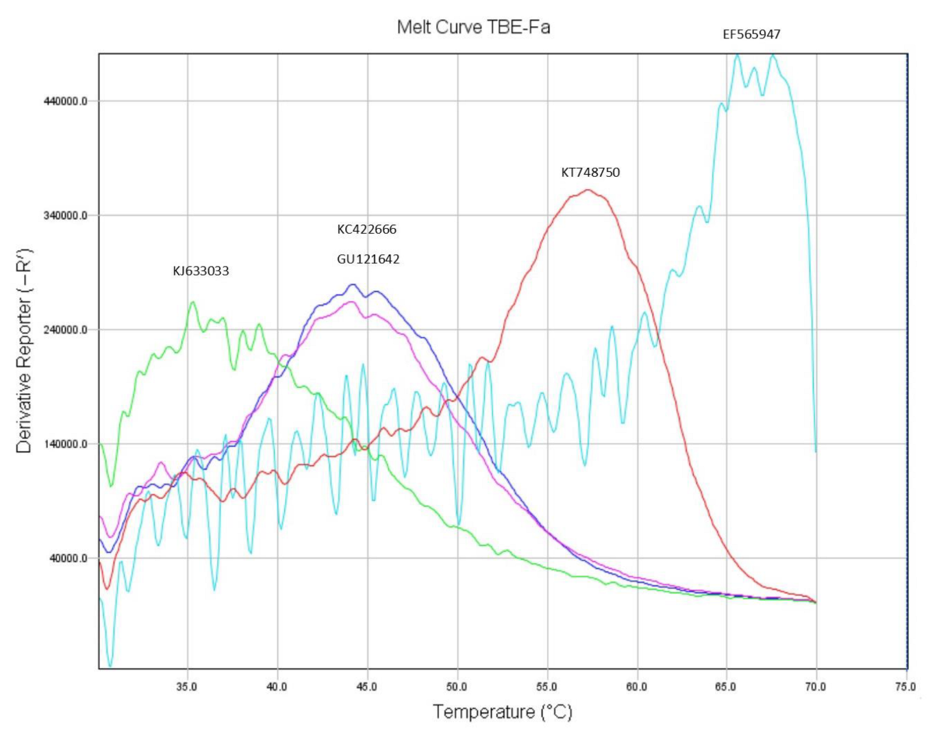
3.1. Primer Tm Analysis and Design Refinement

Primer Tm was determined for duplexes of primers and oligonucleotides representing ten different TBEV variants based on sequences in GenBank. Examples of first derivative melting curves are shown in Figure 1. Curves tended to be broad and jagged, but the Tm could be determined within ± 1.5 °C, which was sufficient for the purposes of this study. Results for the Tm testing of the prototype primer set, TBE-Fa, TBE-Fg and TBE-R, are shown in Table 3.
By comparing the Tm determinations with the amplification results, we were able to conclude that a Tm of 55 °C is sufficient to allow efficient amplification, despite being 5 °C below the annealing temperature. This conclusion was based on the fact that TBEV-Vasilchenko, which efficiently amplifies, has target sequences corresponding to JX315851 (Tm = 55–57.4 °C) and KT749573 (Tm = 56 °C) in the forward and reverse primers, respectively.
Regarding the forward primers TBE-Fa and TBE-Fg, we were able to draw the following inferences: (1) the 3′ nucleotide has little effect on the Tm of the primers, as TBE-Fa and TBE-Fg, with A and G respectively at this position, differed by only a fraction of a degree; (2) an A:C mismatch at position 8 reduces Tm by approximately 10 °C; (3) the in silico estimate of primer Tm was ca. 7 °C too low; the in silico Tm was 58.4 °C and 58.9 °C for primers TBE-Fa and TBEV-Fg, respectively, while the in vitro value was 66 °C.
These conclusions led to the design of a new pair of forward primers, TBE-Fa8T and TBE-Fg8T, where 8C is replaced with 8T, giving an A:T base pair in place of an A:C mismatch in Siberian and Far Eastern type target sequences, which is expected to increase Tm by ca. 10 °C, while a moderately stable G:T mismatch, in European-type sequences, is expected to reduce Tm by 4 °C to 62 °C.
The target sequences of the reverse primer TBE-R were more variable, and it was not possible to draw direct conclusions regarding the influence of individual mismatches on primer stability. However, we conjectured that replacing inosine with G at position 16 would replace I:C with G:C in most target sequences, increasing the Tm by ca. 4 °C, while for the remaining sequences, the substitution of G:T for I:T would produce little change. This change resulted in the primer TBE-R16G. We also surmised that replacing C at position 4 with T would replace a destabilising C:A with stable T:A in most sequences, while causing a minor (2–3 °C) reduction in stability in European-type sequences due to the replacement of C:G with T:G. The latter primer, TBE-R16G4T, was not fully tested as TBE-R16G proved adequate for the purposes of this project. We surmised that these modifications would allow for the sensitive detection of all TBEV strains of the Siberian subtype, with the possible exception of those represented by sequence JX315851 without serious loss of sensitivity for European strains, and that some Far Eastern subtype strains might also be detected.
Table 4 shows the results of primer Tm analysis for the refined primers. These results were in agreement with the predictions. The results for primer TBE-R16G4T can be found in Appendix A.

3.2. Detection of TBEV

The ability of both primer sets to detect ten virus strains was tested (Table 5).
The prototype primers detected all six TBEV-Eu and both TBEV-Sib, although the amplification of strains Hypr (TBEV-Eu) and Latvia-1-96 (TBEV-Sib) was poor. TBEV-FE and the louping ill virus were not amplified.
The refined primers amplified all nine TBEV strains, although the amplification of TBEV-FE and Hypr remained poor. DNA sequencing (Table 5) confirmed the identity of the strain level, with the exception of Hypr. A BLAST search of the obtained sequences confirmed the subtype and identity. Amplification of the louping ill virus was not observed.
Testing using the RotorGene real-time PCR system (QIAGEN GmbH, Hilden, Germany) gave similar results to those obtained with the StepOne PCR instrument, detecting the same virus strain and with cycle threshold (Ct) values differing by no more than +/− 5 cycles, except in the case of TBEV-FE strain Sofjin, which showed a much higher Ct value with the RotorGene PCR instrument. For the other flavivirus tested, an apparent cross-reaction with Toscana Virus lineage B was observed, but gave an anomalous amplification curve distinct from those observed for TBEV. An EQA sample containing TBEV Hypr was tested, but did not amplify.

3.3. Sensitivity and Efficiency of the Real-Time PCR

With the prototype primer set, the efficiencies of the real-time PCR for the synthetic plasmid (pTBE), 1993/783 and Vasilchenko were 89%, 77% and 73%, respectively. The efficiency for Latvia-1-96 with this primer set could not be determined because of poor amplification.
With the refined primer set, the efficiency of the real-time PCR for pTBE, 1993/783, Latvia-1-96 and Vasilchenko was 94%, 94%, 91% and 83%, respectively.
Sensitivity testing for the refined primer set using a tenfold serial dilution of plasmid pTBE showed that all reactions containing one hundred copies or more were positive, while one of three reactions containing ten virus copies was positive. The limit of detection was therefore between 10 and 100 DNA copies per reaction.
The reverse primer TBE-R16G4T, together with the refined forward primers TBE-Fa8T and TBE-Fg8T, was tested against a dilution series of pTBE. Efficiency (>90%) and sensitivity were similar to those for TBE-R16G. TBE-R16G4T was not tested further as its benefits were expected to be chiefly realised with TBEV-FE strains, of which only one was available for testing.

4. Discussion

TBEV may cause severe disease in humans. Further, the constantly increasing number of reported TBEV strains in I. ricinus in Europe and the spread of I. persulcatus and more virulent TBEV-Eu and TBEV-Sib strains westward is of concern [19]. This creates a need for improved diagnostic tools and methods for surveillance with better strain coverage and strain identification potential. In the present study, we developed a real-time PCR assay for the detection of TBEV-Eu and TBEV-Sib. The refined primers (TBE-Fa8T, TBE-Fg8T and TBE-R16G) detected all of the TBEV strains tested, although the detection of the Hypr strain was less sensitive than expected, possibly due to mismatches with the reverse primer. Although our aim was to detect TBEV-Eu and TBEV-Sib, we consider the additional detection of TBEV-FE an advantage rather than a drawback, as the subtypes can be distinguished by Sanger sequencing. Testing the refined primers on the RotorGene real-time PCR system TBEV showed minor cross-reactions with other flaviviruses. The cultivated viruses were analysed on both PCR systems and differences varied with +/− 5 Ct values. Taking into account different PCR systems and analytical variations, this difference in Ct values may be considered small.
In this study, we explored a new approach to primer design improvement based on the Tm determination of fully matched and partially matched duplexes using synthetic target sequences. This approach is inexpensive, as the cost of short oligonucleotides is low. At least for SYBR-green PCR, it provides a direct measurement of primer Tm under the conditions in which they will be used. It also provides a useful check on in silico determined Tm’s, which, as in the present case, may be quite inaccurate. The approach proved fruitful, allowing for the estimation of the effect of individual mismatches on primer stability and the design of improved primers that sensitively detected both TBEV-Eu and TBEV-Sib. Interestingly, efficient amplification could be achieved at annealing temperatures 5 °C above Tm, which is possibly a reflection of the broad melting peaks observed. Our results confirmed previous findings that the A:C mismatch is highly destabilising [28], apparently to a greater degree even than purine:purine and pyrimidine:pyrimidine mismatches.
The efficiency of the real-time PCR varied with the different subtypes of TBEV, though it was above 80% for both TBEV-Eu and TBEV-Sib, for the strains tested. Despite the high efficiency with the plasmid, pTBE (94%), the real-time PCR could only detect one of three reactions containing ten copies of the plasmid. Other studies have reported high- and single-molecule sensitivities for real-time PCR methods [9,29,30,31]. Most of these assays are either limited to a single subtype or group of strains [9,29,31], whereas Lindblom et al., in 2014, multiplexed the two methods by Schwaiger and Cassinotti, 2003, and Gäumann et al., 2010, and could therefore detect all TBEV-Eu, -Sib and -FE with high sensitivity. However, the PCR products are too short to be differentiated by Sanger sequencing and additional methods must be performed for confirmation of positive results [30,31,32]. In the current study, we were able to design primers amplifying multiple TBEV subtypes, not cross-reacting with the louping ill virus, the most closely related flavivirus, and providing an amplicon suitable for subtype and strain differentiation by Sanger sequencing. However, these desirable characteristics do seem to come at the cost of single-molecule sensitivity.

5. Conclusions

The development of sensitive and specific generic PCR for RNA viruses such as TBEV is a challenging task because of their high sequence variability. Using a new approach to primer design, which combines in silico and in vitro determinations of primer Tm to accommodate mismatches, we were able to meet this challenge. The developed real-time PCR should prove a useful tool in the surveillance of the spread of virulent strains of TBEV-Sib and TBEV-Eu in Europe. The new approach to primer design should be widely applicable to the challenge of designing generic PCR for variable targets.

Supplementary Materials

The following supporting information can be downloaded at: https://www.mdpi.com/article/10.3390/microbiolres14040106/s1, Supplementary file S1.

Author Contributions

Å.K.A. formulated the objective of the study and assembled sample material. B.N.P. and A.J. designed the study and wrote the manuscript. B.N.P. designed the prototype primers. A.J. conceived the Tm method for primer design and designed the refined primers. B.N.P., K.M.P. and C.B. did the laboratory work. All authors have read and agreed to the published version of the manuscript.

Funding

This research was partly funded by the ScandTick project (grant number 167226) supported by EU Interreg IV A program, the ScandTick Innovation project (grant number 20200422) supported by EU Interreg V program and Barentsregionprosjektet B1214 and B1710 supported by the Norwegian Ministry of Health and Care Services. The APC was funded by the University of South-Eastern Norway.

Institutional Review Board Statement

Not applicable.

Informed Consent Statement

Not applicable.

Data Availability Statement

Specific data can be made available upon request.

Conflicts of Interest

The authors declare no conflict of interest.

Appendix A

Table A1. Results of melting temperature (Tm) determination for duplexes between the refined reverse primer TBE-R16G4T and oligonucleotides representing the target sequences in fully matched and representative mismatched TBEV variants. Positions where the primer is modified relative to the prototype, TBE-R, are in underlined, bold type. Mismatches are highlighted according to the following system: Pale blue: G:T mismatch; green: pyrimidine-pyrimidine mismatch; grey: purine:purine mismatch; yellow: A:C mismatch.
SubtypeTBE-R16G4TTm
5′GACTCTGCACAACAAGGACA3′
EF565947Eu3′CTGGGACGTGTTGTTTCTGT5′57 °C
KT749573Sib3′CTGAGACGTGTTGTTCCTGT5′66 °C
KT748750Sib3′CTGAGACGTGTAGTTCCTGT5′60 °C
AB022296FE3′ATGAGACGTATTGTTCCTGT5′55 °C
KJ633033Bai3′CTGAGACGTGTTATCCCTGT5′51 °C
JX315851Sib3′TTGAGACGTATTGTTCCAGT5′50 °C
KU761569FE3′ATGAGACGTATTGTCCCTAT5′41 °C
KC422666FE3′ATGAGCCGTGTTATTCCTGT5′46 °C
GU121642FE3′ATGAGCTGTGTTGTTCCTGT5′54 °C
AF231807FE3′TTGAGACGTATCGTTCCTAT5′40 °C

References

  1. Pulkkinen, L.I.A.; Butcher, S.J.; Anastasina, M. Tick-Borne Encephalitis Virus: A Structural View. Viruses 2018, 10, 350. [Google Scholar] [CrossRef] [PubMed]
  2. Ecker, M.; Allison, S.L.; Meixner, T.; Heinz, F.X. Sequence analysis and genetic classification of tick-borne encephalitis viruses from Europe and Asia. J. Gen. Virol. 1999, 80 Pt 1, 179–185. [Google Scholar] [CrossRef]
  3. Adelshin, R.V.; Sidorova, E.A.; Bondaryuk, A.N.; Trukhina, A.G.; Sherbakov, D.Y.; White Iii, R.A.; Andaev, E.I.; Balakhonov, S.V. “886-84-like” tick-borne encephalitis virus strains: Intraspecific status elucidated by comparative genomics. Ticks Tick-Borne Dis. 2019, 10, 1168–1172. [Google Scholar] [CrossRef] [PubMed]
  4. Dai, X.; Shang, G.; Lu, S.; Yang, J.; Xu, J. A new subtype of eastern tick-borne encephalitis virus discovered in Qinghai-Tibet Plateau, China. Emerg. Microbes Infect. 2018, 7, 74. [Google Scholar] [CrossRef]
  5. Demina, T.V.; Dzhioev, Y.P.; Verkhozina, M.M.; Kozlova, I.V.; Tkachev, S.E.; Plyusnin, A.; Doroshchenko, E.K.; Lisak, O.V.; Zlobin, V.I. Genotyping and characterization of the geographical distribution of tick-borne encephalitis virus variants with a set of molecular probes. J. Med. Virol. 2010, 82, 965–976. [Google Scholar] [CrossRef] [PubMed]
  6. Dobler, G.; Gniel, D.; Petermann, R.; Pfeffer, M. Epidemiology and distribution of tick-borne encephalitis. Wien. Med. Wochenschr. 2012, 162, 230–238. [Google Scholar] [CrossRef]
  7. Jaaskelainen, A.E.; Tonteri, E.; Pieninkeroinen, I.; Sironen, T.; Voutilainen, L.; Kuusi, M.; Vaheri, A.; Vapalahti, O. Siberian subtype tick-borne encephalitis virus in Ixodes ricinus in a newly emerged focus, Finland. Ticks Tick-Borne Dis. 2016, 7, 216–223. [Google Scholar] [CrossRef]
  8. Bogovic, P.; Strle, F. Tick-borne encephalitis: A review of epidemiology, clinical characteristics, and management. World J. Clin. Cases 2015, 3, 430–441. [Google Scholar] [CrossRef]
  9. Andreassen, A.; Jore, S.; Cuber, P.; Dudman, S.; Tengs, T.; Isaksen, K.; Hygen, H.O.; Viljugrein, H.; Anestad, G.; Ottesen, P.; et al. Prevalence of tick borne encephalitis virus in tick nymphs in relation to climatic factors on the southern coast of Norway. Parasites Vectors 2012, 5, 177. [Google Scholar] [CrossRef]
  10. Vikse, R.; Paulsen, K.M.; Edgar, K.S.; Pettersson, J.H.O.; Ottesen, P.S.; Okbaldet, Y.B.; Kiran, N.; Lamsal, A.; Lindstedt, H.E.H.; Pedersen, B.N.; et al. Geographical distribution and prevalence of tick-borne encephalitis virus in questing Ixodes ricinus ticks and phylogeographic structure of the Ixodes ricinus vector in Norway. Zoonoses Public Health 2020, 67, 370–381. [Google Scholar] [CrossRef]
  11. Norwegian Surveillance System for Communicable Diseases (MSIS), Norwegian Institute of Public Health. 2022. Available online: https://msis.no/ (accessed on 10 August 2022).
  12. Mehl, R. The distribution and host relations of Norwegian ticks (Acari, Ixodides). Fauna Norwgica Ser. B 1983, 30, 46–51. [Google Scholar]
  13. Soleng, A.; Edgar, K.S.; Paulsen, K.M.; Pedersen, B.N.; Okbaldet, Y.B.; Skjetne, I.E.B.; Gurung, D.; Vikse, R.; Andreassen, A.K. Distribution of Ixodes ricinus ticks and prevalence of tick-borne encephalitis virus among questing ticks in the Arctic Circle region of northern Norway. Ticks Tick-Borne Dis. 2018, 9, 97–103. [Google Scholar] [CrossRef] [PubMed]
  14. Hvidsten, D.; Frafjord, K.; Gray, J.S.; Henningsson, A.J.; Jenkins, A.; Kristiansen, B.E.; Lager, M.; Rognerud, B.; Slåtsve, A.M.; Stordal, F.; et al. The distribution limit of the common tick, Ixodes ricinus, and some associated pathogens in north-western Europe. Ticks Tick-Borne Dis. 2020, 11, 101388. [Google Scholar] [CrossRef] [PubMed]
  15. Kjær, L.J.; Soleng, A.; Edgar, K.S.; Lindstedt, H.E.H.; Paulsen, K.M.; Andreassen, Å.K.; Korslund, L.; Kjelland, V.; Slettan, A.; Stuen, S.; et al. A large-scale screening for the taiga tick, Ixodes persulcatus, and the meadow tick, Dermacentor reticulatus, in southern Scandinavia, 2016. Parasites Vectors 2019, 12, 338. [Google Scholar] [CrossRef]
  16. Jaenson, T.G.T.; Wilhelmsson, P. First records of tick-borne pathogens in populations of the taiga tick Ixodes persulcatus in Sweden. Parasites Vectors 2019, 12, 559. [Google Scholar] [CrossRef]
  17. Laaksonen, M.; Sajanti, E.; Sormunen, J.J.; Penttinen, R.; Hanninen, J.; Ruohomaki, K.; Saaksjarvi, I.; Vesterinen, E.J.; Vuorinen, I.; Hytonen, J.; et al. Crowdsourcing-based nationwide tick collection reveals the distribution of Ixodes ricinus and I. persulcatus and associated pathogens in Finland. Emerg. Microbes Infect. 2017, 6, e31. [Google Scholar] [CrossRef]
  18. Jaaskelainen, A.E.; Tonteri, E.; Sironen, T.; Pakarinen, L.; Vaheri, A.; Vapalahti, O. European subtype tick-borne encephalitis virus in Ixodes persulcatus ticks. Emerg. Infect. Dis. 2011, 17, 323–325. [Google Scholar] [CrossRef]
  19. Kuivanen, S.; Smura, T.; Rantanen, K.; Kamppi, L.; Kantonen, J.; Kero, M.; Jaaskelainen, A.; Jaaskelainen, A.J.; Sane, J.; Myllykangas, L.; et al. Fatal Tick-Borne Encephalitis Virus Infections Caused by Siberian and European Subtypes, Finland, 2015. Emerg. Infect. Dis. 2018, 24, 946–948. [Google Scholar] [CrossRef]
  20. Hasle, G.; Bjune, G.; Edvardsen, E.; Jakobsen, C.; Linnehol, B.; Røer, J.E.; Mehl, R.; Røed, K.H.; Pedersen, J.; Leinaas, H.P. Transport of ticks by migratory passerine birds to Norway. J. Parasitol. 2009, 95, 1342–1351. [Google Scholar] [CrossRef]
  21. Klitgaard, K.; Chriél, M.; Isbrand, A.; Jensen, T.K.; Bødker, R. Identification of Dermacentor reticulatus Ticks Carrying Rickettsia raoultii on Migrating Jackal, Denmark. Emerg. Infect. Dis. 2017, 23, 2072–2074. [Google Scholar] [CrossRef]
  22. Untergasser, A.; Cutcutache, I.; Koressaar, T.; Ye, J.; Faircloth, B.C.; Remm, M.; Rozen, S.G. Primer—New capabilities and interfaces. Nucleic Acids Res. 2012, 40, e115. [Google Scholar] [CrossRef] [PubMed]
  23. Ye, J.; Coulouris, G.; Zaretskaya, I.; Cutcutache, I.; Rozen, S.; Madden, T.L. Primer-BLAST: A tool to design target-specific primers for polymerase chain reaction. BMC Bioinform. 2012, 13, 134. [Google Scholar] [CrossRef] [PubMed]
  24. Katoh, K.; Standley, D.M. MAFFT multiple sequence alignment software version 7: Improvements in performance and usability. Mol. Biol. Evol. 2013, 30, 772–780. [Google Scholar] [CrossRef]
  25. Waterhouse, A.M.; Procter, J.B.; Martin, D.M.; Clamp, M.; Barton, G.J. Jalview Version 2—A multiple sequence alignment editor and analysis workbench. Bioinformatics 2009, 25, 1189–1191. [Google Scholar] [CrossRef] [PubMed]
  26. Bustin, S.A.; Benes, V.; Garson, J.A.; Hellemans, J.; Huggett, J.; Kubista, M.; Mueller, R.; Nolan, T.; Pfaffl, M.W.; Shipley, G.L.; et al. The MIQE guidelines: Minimum information for publication of quantitative real-time PCR experiments. Clin Chem 2009, 55, 611–622. [Google Scholar] [CrossRef]
  27. Reusken, C.; Baronti, C.; Mögling, R.; Papa, A.; Leitmeyer, K.; Charrel, R.N. Toscana, West Nile, Usutu and tick-borne encephalitis viruses: External quality assessment for molecular detection of emerging neurotropic viruses in Europe, 2017. Euro Surveill. 2019, 24. [Google Scholar] [CrossRef]
  28. You, Y.; Moreira, B.G.; Behlke, M.A.; Owczarzy, R. Design of LNA probes that improve mismatch discrimination. Nucleic Acids Res. 2006, 34, e60. [Google Scholar] [CrossRef]
  29. Wang, Q.Y.; Li, F.; Shen, X.X.; Fu, S.H.; He, Y.; Lei, W.W.; Liang, G.D.; Wang, H.Y.; Ma, X.J. A Reverse-transcription Recombinase-aided Amplification Assay for the Rapid Detection of the Far-Eastern Subtype of Tick-borne Encephalitis Virus. Biomed. Environ. Sci. BES 2019, 32, 357–362. [Google Scholar] [CrossRef]
  30. Schwaiger, M.; Cassinotti, P. Development of a quantitative real-time RT-PCR assay with internal control for the laboratory detection of tick borne encephalitis virus (TBEV) RNA. J. Clin. Virol. Off. Publ. Pan Am. Soc. Clin. Virol. 2003, 27, 136–145. [Google Scholar] [CrossRef]
  31. Gäumann, R.; Mühlemann, K.; Strasser, M.; Beuret, C.M. High-throughput procedure for tick surveys of tick-borne encephalitis virus and its application in a national surveillance study in Switzerland. Appl. Env. Microbiol. 2010, 76, 4241–4249. [Google Scholar] [CrossRef]
  32. Lindblom, P.; Wilhelmsson, P.; Fryland, L.; Sjöwall, J.; Haglund, M.; Matussek, A.; Ernerudh, J.; Vene, S.; Nyman, D.; Andreassen, A.; et al. Tick-borne encephalitis virus in ticks detached from humans and follow-up of serological and clinical response. Ticks Tick-Borne Dis. 2014, 5, 21–28. [Google Scholar] [CrossRef] [PubMed]
Figure 1. Examples of the melting curves of duplexes between the primer TBE-Fa and oligonucleotides representing the target sequence in five TBEV variants. The x-axis shows the temperature, and the y-axis shows the derivative reporter signal. The temperature at the peak of the curve is the melting temperature. EF565947: Norway1 (light blue) represents TBEV-EU and is fully matched; KT748750: Est 228 (red) represents TBEV-Sib, with one mismatch; KC422666: Zabaikalye 50-03 (dark blue) and GU121642: Svetlogorie (magenta) represent TBEV-FE, with three mismatches; KJ633033: 886-84 (green) represents the Baikalian subtype, with four mismatches. See Table 3 for sequence information.
Microbiolres 14 00106 g001
Table 1. Primer sequences for the detection of tick-borne encephalitis virus. Highlights indicate bases modified to improve primer performance. I = inosine.
PrimerSequence 5′–3′
Prototype primer setTBE-FaATGTGTACGACGCCAACAAA
TBE-FgATGTGTACGACGCCAACAAG
TBE-RGACCCTGCACAACAAIGACA
Refined primer setTBE-Fa8TATGTGTATGACGCCAACAAA
TBE-Fg8TATGTGTATGACGCCAACAAG
TBE-R16GGACCCTGCACAACAAGGACA
TBE-R16G4T 1GACTCTGCACAACAAGGACA
1 This primer is a further refinement on R16G, which offers further potential improvements in the detection of TBEV-FE, but which was not experimentally tested.
Table 2. Virus strains included in the study.
Virus StrainVirus/Subtype MaterialAccession Number
1993/783TBEV-EuCulture supernatantMT311860
AbsettarovTBEV-EuSupplied as RNAKJ000002
HochosterwitzTBEV-EuCulture supernatantMT311861
HyprTBEV-EuSupplied as RNA/EQAU39292
Neudörfl 1TBEV-EuVaccineU27495
SokoupTBEV-EuSupplied as RNANA
Latvia 1-96TBEV-SibCulture supernatantGU183382
VasilchenkoTBEV-SibSupplied as RNAAF069066
SofjinTBEV-FESupplied as RNAAB062064
LI/NORLouping ill virusCulture supernatantD12936
-Zika EQA NA
-Dengue type 1EQA NA
-Dengue type 2EQA NA
-Dengue type 3EQA NA
-Dengue type 4EQA NA
-Yellow fever EQA NA
-Toscana lineage AEQA NA
-Toscana lineage BEQA NA
-West Nile lineage 1EQA NA
-West Nile lineage 2EQA NA
-Utsu virusEQA NA
1 TBEV vaccine, TicoVac, PfizerEU; European; Sib: Siberian; FE: Far-Eastern; EQA: Exterrnal quality assurance; NA: not applicable.
Table 3. Results of melting temperature (Tm) determination for duplexes between the prototype primers, TBE-Fa, TBE-Fg and TBE-R, and oligonucleotides representing the fully matched (EF565947: Norway1) and mismatched complementary target sequences in representative TBEV variants. Mismatches are highlighted according to the following system: Pale blue: G:T or I:N mismatch; green: pyrimidine-pyrimidine mismatch; grey: purine:purine mismatch; yellow: A:C mismatch. ΔTm is relative to the duplex with EF565947.
Accession No.Sub-
Type
TBE-FaTmΔTmTBE-FgTmΔTmTBE-RTmΔTm
5′ATGTGTACGACGCCAACAAA 3′ 5′ATGTGTACGACGCCAACAAG3′ 5′GACCCTGCACAACAAIGACA3′
EF565947EU3′TACACATGCTGCGGTTGTTT5′ 166 °C 3′TACACATGCTGCGGTTGTTT5′66 °C 3′CTGGGACGTGTTGTTTCTGT5′ 260 °C
KT749573Sib3′TACACATACTACGGTTGTCT5′43 °C23 °C3′TACACATACTACGGTTGTCT5′42 °C24 °C3′CTGAGACGTGTTGTTCCTGT5′ 3,456 °C4 °C
KT748750Sib3′TACACATACTGCGGTTGTTT5′56 °C10 °C3′TACACATACTGCGGTTGTTT5′56 °C10 °C3′CTGAGACGTGTAGTTCCTGT5′50 °C10 °C
AB022296FE3′TGCACATACTGCGTTTGTTT5′43 °C23 °C3′TGCACATACTGCGTTTGTTT5′42 °C24 °C3′ATGAGACGTATTGTTCCTGT5′44 °C16 °C
KJ633033Bai3′TACATATACTGCGTTTGTTC5′37 °C29 °C3′TACATATACTGCGTTTGTTC5′39 °C27 °C3′CTGAGACGTGTTATCCCTGT5′38 °C22 °C
JX315851Sib3′TACACATACTGCGGTTGTTC5′ 355 °C11 °C3′TACACATACTGCGGTTGTTC5′57 °C9 °C3′TTGAGACGTATTGTTCCAGT5′38 °C22 °C
KU761569FE3′TACACATACTGCGATTGTTT5′ 4,546 °C20 °C3′TACACATACTGCGATTGTTT5′45 °C21 °C3′ATGAGACGTATTGTCCCTAT5′<35 °C-
KC422666FE3′TACACATACTGCGATTGTTC5′43 °C23 °C3′TACACATACTGCGATTGTTC5′46 °C20 °C3′ATGAGCCGTGTTATTCCTGT5′ 536 °C24 °C
GU121642FE3′TGCACATACTACGGTTGTTT5′44 °C22 °C3′TGCACATACTACGGTTGTTT5′42 °C24 °C3′ATGAGCTGTGTTGTTCCTGT5′46 °C14 °C
AF231807Sib3′TACACATACTGCGGTTGTTC5′57 °C9 °C3′TACACATACTGCGGTTGTTC5′58 °C8 °C3′TTGAGACGTATCGTTCCTAT5′<35 °C-
1 100% identity to TBE-EU Absettarov, Hochosterwitz, HYPR and Neudörfl; 2 100% identity to TBE-EU 1993/783 Absettarov, Hochosterwitz and Neudörfl; 3 100% identity to TBE-Sib Vasilchenko; 4 100% identity to TBE-Sib Latvia 1-96; 5 100% identity to TBE-FE Sofijn. EU: European; Sib: Siberian; Bai: Baikalian; FE: Far Eastern.
Table 4. Results of melting temperature (Tm) determination for duplexes between the refined forward primer TBE-Fa8T, reverse primer TBE-R16G and oligonucleotides representing fully matched and representative mismatched TBE variants. The altered nucleotides in the primers are shown in bold, underlined text. Mismatches are highlighted according to the following system: Pale blue: G:T mismatch; green: pyrimidine-pyrimidine mismatch; grey: purine:purine mismatch; yellow: A:C mismatch. ΔTm is relative to the fully matched duplex. Because no sequence fully matched TBE-R16G, no ΔTm was calculated. Tms for refined forward primer TBE-Fg8T were not determined as testing of the prototype primers TBE-Fa/TBE-Fg showed that the substituting G for A at the 3′ terminal position had a minimal effect on Tm.
Accession no.SubtypeTBE-Fa8TTmΔTmTBE-R16GTm
5′ATGTGTATGACGCCAACAAA3′ 5′GACCCTGCACAACAAGGACA3′
EF565947EU3′TACACATGCTGCGGTTGTTT5′61 °C4 °C3′CTGGGACGTGTTGTTTCTGT5′61 °C
KT749573Sib3′TACACATACTACGGTTGTCT5′53 °C12 °C3′CTGAGACGTGTTGTTCCTGT5′61 °C
KT748750Sib3′TACACATACTGCGGTTGTTT5′65 °C0 °C3′CTGAGACGTGTAGTTCCTGT5′55 °C
AB022296FE3′TGCACATACTGCGTTTGTTT5′55 °C10 °C3′ATGAGACGTATTGTTCCTGT5′50 °C
KJ633033Bai3′TACATATACTGCGTTTGTTC5′47 °C18 °C3′CTGAGACGTGTTATCCCTGT5′43 °C
JX315851Sib3′TACACATACTGCGGTTGTTC5′65 °C0 °C3′TTGAGACGTATTGTTCCAGT5′42 °C
KU761569FE3′TACACATACTGCGATTGTTT5′56 °C9 °C3′ATGAGACGTATTGTCCCTAT5′<35 °C
KC422666FE3′TACACATACTGCGATTGTTC5′56 °C9 °C3′ATGAGCCGTGTTATTCCTGT5′42 °C
GU121642FE3′TGCACATACTACGGTTGTTT5′54 °C11 °C3′ATGAGCTGTGTTGTTCCTGT5′54 °C
AF231807FE3′TACACATACTGCGGTTGTTC5′64 °C1 °C3′TTGAGACGTATCGTTCCTAT5′<35 °C
Table 5. Detection of TBEV using the refined primers TBE-Fa8T, TBE-Fg8T and TBE-R16G. Sanger sequencing confirmed the TBEV subtypes.
Virus StrainReal-Time PCR ResultSequencing
1993/783PositiveConfirmed
AbsettarovPositiveConfirmed
HochosterwitzPositiveConfirmed
HyprPositiveNot confirmed
Neudörfl 1PositiveConfirmed
SokoupPositiveConfirmed (TBEV-Eu)
Latvia-1-96PositiveConfirmed
VasilchenkoPositiveConfirmed
SofijnPositiveConfirmed
LI/NORNegativeNot sequenced
1 TBEV vaccine, TicoVac, Pfizer.
Disclaimer/Publisher’s Note: The statements, opinions and data contained in all publications are solely those of the individual author(s) and contributor(s) and not of MDPI and/or the editor(s). MDPI and/or the editor(s) disclaim responsibility for any injury to people or property resulting from any ideas, methods, instructions or products referred to in the content.

