1. Introduction
Colorectal cancer (CRC) is the third leading cause of cancer-related deaths worldwide, with over 1.8 million new cases and nearly 900,000 deaths each year, and its incidence continues to rise in developing countries. CRC is more common in men than in women and is three to four times more prevalent in developed countries than in developing ones [
1].
Although significant progress has been made in understanding the biology and treatment of this disease, the burden of associated complications remains substantial. Between 15% and 40% of patients with colorectal cancer experience surgical emergencies, most commonly perforation or obstruction. The prevalence of perforation in colorectal cancer patients is 2–9% [
2], and it is associated with poor oncological outcomes and high postoperative morbidity and mortality.
Radical surgery is one of the primary treatment options for perforated colon cancer. This intervention is complex and carries significant risks, including postoperative complications such as infection, bleeding, and anastomotic leakage. Nevertheless, it can also offer favorable clinical and oncological outcomes for these patients.
Studies that specifically compare perforated and non-perforated colon cancer remain relatively few, and most published series pool perforation together with obstruction or hemorrhage under the broader label of complicated colorectal cancer. To our knowledge, no comparative analysis has been reported from a North African tertiary center, where patient presentation patterns, time to surgery, and access to laparoscopic equipment in emergency settings differ meaningfully from those of European and North American cohorts. The objective of the present study was therefore to investigate the clinical and oncological outcomes following radical surgery for perforated colon cancer and to compare them with those of surgery for uncomplicated colon cancer.
2. Materials and Methods
2.1. Patients
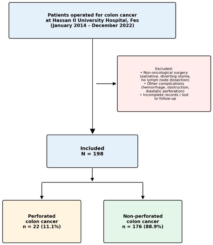
This was a descriptive and comparative retrospective study conducted at the Department of Visceral Surgery of Hassan II University Hospital in Fes, Morocco. The study covered an 8-year period from January 2014 to December 2022. Inclusion criteria comprised all patients admitted for the management of colonic cancer (including rectosigmoid junction cancer) who underwent oncological surgery. Exclusion criteria included patients who underwent non-oncological surgery for colonic cancer, such as palliative procedures (e.g., diverting stomas or resections without lymph node dissection); patients with other complications such as hemorrhage, obstruction, or diastatic perforation; and cases with incomplete records or patients lost to follow-up. The flow of patient inclusion is summarized in
Figure 1.
2.2. Groups
Eligible patients were divided into two groups according to the presence or absence of tumor perforation at presentation: a perforated colon cancer group and a non-perforated colon cancer group. Perforation was defined as a full-thickness disruption of the colonic wall at the tumor site, confirmed intraoperatively, and was further characterized as either generalized peritonitis or localized intra-abdominal abscess based on intraoperative findings.
2.3. Data Collection
Baseline and outcome data were collected retrospectively from medical records and operative reports. Variables of interest included age, sex, medical history and comorbidities, tumor location, surgical approach, creation and type of stoma, restoration of bowel continuity, length of postoperative hospital stay, length of intensive care unit (ICU) stay, postoperative complications graded according to the Clavien–Dindo classification, 30-day mortality, completeness of resection (R status), and 3-year recurrence.
2.4. Ethical Approval
The local institutional review board of the Department of Surgery at Hassan II University Hospital of Fes approved this retrospective study and granted a waiver of patient informed consent, with emphasis on patient confidentiality. All methods were carried out in accordance with relevant guidelines and regulations.
2.5. Surgical Management
All operations were performed by senior visceral surgeons of Hassan II University Hospital. In elective cases without perforation, a laparoscopic approach was preferred whenever feasible, with conversion to laparotomy in case of hemodynamic instability, locally advanced disease, or technical difficulty. Standard oncological resection was performed according to tumor location: right hemicolectomy for right-sided tumors, left hemicolectomy for left-sided tumors, and sigmoidectomy for sigmoid and rectosigmoid lesions, with central vascular ligation and adequate lymphadenectomy in all cases. In patients with perforated colon cancer, the surgical strategy was adapted to intraoperative findings. A Hartmann’s procedure was preferred in patients with generalized peritonitis or hemodynamic instability. Primary anastomosis, with or without a protective stoma, was reserved for selected patients with localized abscesses, limited peritoneal contamination, and stable hemodynamics. Peritoneal lavage with warm saline was systematically performed.
2.6. Statistical Analysis
Data were entered and coded in Microsoft Excel, and the statistical analysis was conducted in two steps. In the first step, a descriptive analysis was performed: continuous variables are presented as mean ± standard deviation (with range, where appropriate), and categorical variables are presented as frequencies and percentages. In the second step, comparisons between the perforated and non-perforated groups were performed using Student’s t-test for continuous variables and the Chi-square test for categorical variables. A two-sided p-value < 0.05 was considered statistically significant. The primary endpoints of this study were the necessity and type of stoma (Hartmann’s procedure, protective ileostomy, or double stoma), postoperative morbidity and 30-day mortality, and the length of postoperative and intensive care unit stays. Secondary endpoints were oncological outcomes, including resection margins (R status) and 3-year recurrence. Formal testing of normality (e.g., Kolmogorov–Smirnov or Shapiro–Wilk) was not performed; this is acknowledged in the Strengths and Limitations Section. No multivariate analysis was performed in view of the limited size of the perforated subgroup and the consequent risk of model overfitting.
3. Results
A total of 198 patients underwent surgery for colon cancer in the visceral surgery departments of Hassan II University Hospital in Fes between January 2014 and December 2022. Of these, 22 (11.1%) were managed for perforated colon cancer. The flow of patient inclusion is shown in
Figure 1.
The mean age was 65.5 years (range, 26–94 years). There were 111 male patients (56.1%), giving a male-to-female ratio of 1.27. The most common comorbidities were hypertension (25%) and diabetes (16%), as seen in
Table 1.
The colonic tumor was located in the right colon in 83 patients (41.9%), in the left colon in 30 patients (15.2%), and in the sigmoid colon or rectosigmoid junction in 85 patients (42.9%). Within the perforated group, 9 patients (40.9%) had right-sided cancer, 9 patients (40.9%) had sigmoid cancer, and 4 patients (18.2%) had left-sided cancer.
All patients admitted for perforated colon cancer underwent a CT scan as part of the diagnostic work-up. Of the 22 perforated patients, 11 (50%) presented with generalized peritonitis and 11 (50%) with intra-abdominal abscess; abscess size ranged from 3 to 10 cm (mean, 5 cm). The mean time between symptom onset and surgery was 5 days.
Among patients with perforated colon cancer, 4.5% underwent laparoscopic surgery, 9.1% underwent laparoscopy converted to laparotomy, and 86.4% underwent open laparotomy. In contrast, among patients without perforation, 56.2% underwent laparoscopy, 9.7% underwent laparoscopy with conversion, and 34% underwent laparotomy. This difference was statistically significant
Table 2.
Stoma creation was required in 72.7% of patients with perforated colon cancer compared with only 4% of patients with non-perforated colon cancer, a highly significant difference
Table 3. The decision to forgo stoma creation in the perforated group was based primarily on intraoperative findings: all patients in this group who received a primary anastomosis had a localized intra-abdominal abscess (mean size, 4 cm) rather than generalized peritonitis. The absence of diffuse peritoneal contamination and the relatively small abscess size were the key factors in this decision.
Among patients operated for perforated colon cancer, 9 underwent a Hartmann’s procedure (56.2%), 6 had a double stoma (37.5%), and 1 had a primary anastomosis with a protective stoma (6.3%). In contrast, among patients with non-perforated colon cancer, no patient underwent a Hartmann’s procedure: 3 patients (42.9%) had a primary anastomosis with a protective ileostomy, and 4 patients (57.1%) had a double stoma.
Stoma reversal was not achieved in 6 of the 16 patients with perforated colon cancer who underwent a stoma (37.5%). In contrast, all patients with non-perforated colon cancer who received a stoma underwent stoma reversal
Table 4.
The mean postoperative hospital stay was 11 days in the perforated group and 5 days in the non-perforated group, a statistically significant difference.
Postoperative complications were more frequent in the perforated group (36.4% vs. 13.6%,
p = 0.006). The distribution by Clavien–Dindo grade is shown in
Table 5.
In the perforated group, 7 patients had wound infections and 1 patient had postoperative hemorrhage that was managed by embolization. In the non-perforated group, 11 patients had wound infections, 4 had anastomotic fistulas managed conservatively, and 9 had postoperative peritonitis due to anastomotic leakage; of the latter, 2 required transfer to the ICU for closer monitoring and management.
Fifteen patients in the perforated group (68.2%) were admitted to the ICU postoperatively, compared with 84 patients (47.7%) in the non-perforated group; this difference was not statistically significant. The mean ICU stay was 3 days in the perforated group and 1 day in the non-perforated group; this difference was numerically higher but did not reach statistical significance.
The 30-day postoperative mortality was 4.5% in the perforated group and 1.1% in the non-perforated group; the difference was not statistically significant.
The R2 resection rate was significantly higher in the perforated group than in the non-perforated group, and the 3-year recurrence rate was likewise significantly higher in patients with perforation
Table 6.
4. Discussion
In this comparative retrospective study of 198 patients operated on for colon cancer over an 8-year period, perforation was associated with substantially worse surgical and oncological outcomes. Compared with non-perforated cases, patients with perforated colon cancer were significantly more likely to undergo open surgery and stoma creation (most often a Hartmann’s procedure), to experience postoperative complications, and to undergo R2 resection. They also had longer hospital and intensive care unit stays, a higher rate of permanent stoma, and a higher 3-year recurrence rate. Thirty-day mortality was numerically higher in the perforated group but did not reach statistical significance. The following sections place these findings in the context of the existing literature.
Colorectal cancer is one of the leading causes of cancer-related death worldwide. Approximately one-third of patients with this disease present with emergency symptoms such as obstruction or tumor perforation that require emergency surgical intervention. This scenario is often associated with a high postoperative mortality rate and poor survival outcomes [
3,
4,
5,
6,
7,
8,
9,
10,
11].
Several studies have reported a negative impact of complications in colon cancer on patient survival. However, most have grouped heterogeneous emergencies—obstruction, bleeding, and perforation—without distinguishing between them [
2,
3,
4,
6,
7,
8,
9]. Few retrospective studies have directly compared perforated and non-perforated colon cancer. Our study aimed to address this gap by examining the clinical and oncological outcomes of surgery for perforated colon cancer and comparing them with those of surgery for non-perforated colon cancer.
In our cohort, the most commonly used surgical approach in the perforated group was laparotomy, whereas laparoscopy was the preferred approach in the non-perforated group. The laparoscopic approach is associated with reduced postoperative complications and faster recovery, but it may be technically challenging in perforated colon cancer because of hemodynamic instability and the need for rapid emergency intervention. In our setting, the absence of dedicated laparoscopic equipment in the emergency operating rooms further contributed to the preference for open surgery in these urgent situations.
Stoma creation was required in 72.7% of patients with perforated colon cancer compared with only 4% of patients with non-perforated colon cancer. This marked difference reflects the risks of performing an anastomosis in a septic environment and the hemodynamic instability often present in patients with perforation. Alvarez and colleagues [
12] reported a similarly high stoma rate in perforated colon cancer (71%). However, stoma creation is not without drawbacks, particularly the risk of a permanent stoma.
We also observed significant differences in the type of stoma between groups. In the perforated group, 56.2% of patients underwent a Hartmann’s procedure, 37.5% had a double stoma, and only 6.3% had a primary anastomosis with a protective stoma. In contrast, no patient in the non-perforated group required a Hartmann’s procedure: 42.9% underwent a primary anastomosis with a protective ileostomy and 57.1% had a double stoma.
Compared with Biondo and colleagues [
4], who reported a Hartmann’s procedure rate of 23.3%, and Daniels and colleagues [
13], who reported a rate of 31%, our Hartmann’s rate was higher (56.2%). Several factors likely explain this difference. The mean delay between symptom onset and surgery in our perforated group was 5 days, which is longer than typically reported in European series and likely contributes to a higher degree of peritoneal contamination at operation. In addition, half of our perforated patients presented with generalized peritonitis rather than localized abscess, a distribution that favors the safer option of a terminal colostomy over primary anastomosis. Finally, the absence of dedicated laparoscopic equipment in our emergency operating rooms, combined with a surgical culture that prioritizes damage-control strategies in hemodynamically unstable patients, further drives the choice toward Hartmann’s procedure.
Patients with perforated colon cancer who underwent a stoma in our cohort also had a higher risk of a permanent stoma: 6 of 16 (37.5%) did not undergo stoma reversal, whereas all patients in the non-perforated group with a stoma had it reversed. These findings highlight the importance of considering the risks associated with stoma creation and of selecting the most appropriate surgical technique according to disease severity.
The mean postoperative hospital stay was 11 days in the perforated group and 5 days in the non-perforated group. This difference likely reflects the greater severity of the disease and the higher frequency of postoperative complications in patients with perforation.
Postoperative ICU admission was numerically more frequent in the perforated group (68.2% vs. 47.7%), although the difference was not statistically significant. Likewise, the mean ICU stay was longer in the perforated group (3 vs. 1 day) but did not reach statistical significance. Kwan [
7] reported a mean ICU stay of 4.8 days in perforated cases and 0.8 days in non-perforated cases. Differences in patient comorbidities and disease severity between cohorts may account for these variations.
The rate of postoperative complications was significantly higher in the perforated group (36.4% vs. 13.6%). Our complication rate in perforated cases was lower than that reported by Daniels [
13] (56%) and Alvarez [
12] (70%), but higher than that reported by Biondo [
4] (26%) and Kwan [
7] (44%).
The 30-day mortality was 4.5% in the perforated group and 1.1% in the non-perforated group. Although this difference was not statistically significant, mortality was numerically higher in patients with colonic perforation. Mortality rates in the literature vary considerably: Biondo [
4] reported 8.6%, Alvarez [
12] 29%, Carraro [
9] 22.9%, and Kiziltan [
5] 27.2%.
The R2 resection rate was 27.3% in the perforated group versus 0.6% in the non-perforated group. These findings are close to those reported by Daniels [
13], who reported an R2 rate of 23% in perforated colon cancer; in their non-perforated group, however, the rate was higher (15.9%) than in our cohort.
Recurrence is a feared complication in the management of colon cancer. In our study, the 3-year recurrence rate was 27.3% in the perforated group versus 4.5% in the non-perforated group, a highly statistically significant difference. Our rate is lower than those reported by Chen [
8] (56.5%) and Asano [
14] (41%), higher than that of Carraro [
9] (22.9%), and similar to that of Biondo [
4] (30.1%). Several factors can influence postoperative recurrence, including cancer stage, the type of initial treatment, and adherence to postoperative follow-up.
Strengths and Limitations
The main strength of this study is that it directly compares perforated and non-perforated colon cancer as two separate groups, whereas most published series pool perforation with obstruction or hemorrhage under a generic complicated colon cancer label. The 8-year single-center recruitment with a stable surgical team also limits inter-institutional variability, and both surgical and oncological endpoints were analyzed.
The main limitations are the retrospective single-center design and the relatively small perforated group (n = 22), which reduces statistical power for endpoints such as 30-day mortality and ICU stay. Tumor stage and adjuvant chemotherapy were not integrated into the comparative analysis, and long-term outcomes such as 5-year overall and disease-free survival were beyond the scope of this work and warrant further multicenter investigation.