1. Introduction
Autoimmune hepatitis (AIH) is a chronic immune-mediated inflammatory liver disease characterized by interface hepatitis on histology, elevated serum transaminases and immunoglobulin G levels, and circulating autoantibodies; it predominantly affects women [
1]. The fundamental diagnostic and conceptual framework of AIH was established by the International Autoimmune Hepatitis Group (IAIHG) [
2], and subsequent reviews have confirmed that the disease is driven mainly by T-cell-mediated immune responses in genetically predisposed individuals [
3,
4,
5,
6].
Clinically, AIH exhibits marked heterogeneity, ranging from asymptomatic or insidious disease to acute hepatitis or even acute liver failure [
1]. Large-scale epidemiological reviews have shown that AIH affects patients of all ages, with increasing recognition of late-onset disease in elderly populations [
1,
4], a feature also emphasized in clinical reviews from Western cohorts [
7].
AIH is frequently associated with other autoimmune disorders. Classic immunopathological reviews have reported that approximately 20–30% of patients with AIH have concomitant autoimmune diseases, including autoimmune thyroid disease, Sjögren’s syndrome, rheumatoid arthritis, and other systemic autoimmune conditions, reflecting a shared autoimmune diathesis [
6,
7,
8]. In Japan, nationwide surveys have shown that approximately one-quarter of patients have concurrent extrahepatic autoimmune diseases (EADs), most commonly, chronic thyroiditis and Sjögren’s syndrome, followed by rheumatoid arthritis [
9,
10].
Despite the high prevalence of EAD comorbidity, its clinical significance in AIH remains incompletely understood. Most previous studies have evaluated EADs as a single pooled category, without sufficient stratification by specific autoimmune disease subtypes [
6]. However, modern immunopathological reviews have shown that systemic autoimmune diseases are characterized by distinct dominant immune pathways, regulatory T-cell dysfunctions, and cytokine profiles [
4,
5,
6,
11]. These observations raise the plausible hypothesis that different autoimmune backgrounds may influence the clinical phenotype and disease behavior of AIH, a concept that has not yet been systematically examined.
Among the various EADs, systemic sclerosis (SSc) represents a particularly rare but clinically intriguing comorbidity in AIH. In a Japanese nationwide survey [
9], SSc was observed in only approximately 0.8% of patients with AIH, underscoring its rarity. Consequently, the clinicopathological characteristics, immunological features, and treatment response of SSc-associated AIH (SSc-AIH) remain largely unknown. Nevertheless, immunological reviews emphasize that the immune milieu in SSc differs substantially from that of other systemic autoimmune diseases [
4,
5,
6], suggesting that SSc-AIH may represent a distinct clinical subset rather than a coincidental overlap.
In addition, international response definitions and treatment endpoints in AIH have been recently refined, emphasizing the importance of standardized assessment of biochemical response and relapse in observational studies [
12]. However, real-world treatment strategies vary across regions; for example, ursodeoxycholic acid (UDCA) is frequently used in Japan as an adjunct or initial therapy in selected cases, which may differ from approaches emphasized in Western guidelines. These practice differences underscore the need to interpret single-center cohorts within their regional clinical context while maintaining compatibility with internationally accepted diagnostic frameworks.
At our institution, approximately 60% of patients with AIH present with EADs—substantially higher than the proportions reported in nationwide surveys— and we have observed an unusually high proportion of SSc-AIH. In recent years, we have also noted an increase in patients diagnosed with AIH in the absence of EADs (AIH without EAD; NEAD-AIH). Notably, some NEAD-AIH cases occurred shortly after immune-modifying exposures, such as COVID-19 vaccination or prior immune checkpoint inhibitor (ICI) therapy, raising the hypothesis that such triggers may precipitate AIH in susceptible individuals. However, these observations remain preliminary and require cautious clinical interpretation.
Given these unique institutional trends, a more detailed evaluation of AIH phenotypes according to both the presence or absence and the specific type of EAD is warranted. Therefore, this study aimed to clarify the clinicopathological characteristics of AIH based on EAD status and explore phenotype differences associated with major EAD subtypes, including Sjögren’s syndrome, autoimmune thyroid disease, and SSc.
2. Materials and Methods
2.1. Study Design and Patient Selection
This single-center retrospective observational study included consecutive patients diagnosed with AIH at Gunma University Hospital, a tertiary referral center in Gunma Prefecture, Japan, between January 2014 and December 2023. Patients were identified through electronic medical records and were eligible if they met diagnostic criteria for AIH according to either the revised International Autoimmune Hepatitis Group (IAIHG) scoring system (definite or probable AIH) [
2] or the “typical/atypical” classification in the clinical practice guidelines of the Japan Society of Hepatology (JSH) [
13]. The index date was defined as the date of AIH diagnosis documented in the medical record. Extracted variables included age, sex, laboratory data at diagnosis (aspartate aminotransferase (AST), alanine aminotransferase (ALT), total bilirubin (T-Bil), albumin (Alb), prothrombin time activity (PT%), and prothrombin time–international normalized ratio (PT-INR)), immunological markers (serum immunoglobulin G (IgG) level and antinuclear antibody (ANA) titer), disease severity, liver biopsy status and histological findings, overlap with other liver diseases, treatment and medications, observation period, relapse, and outcomes. To enhance diagnostic robustness in this real-world cohort, all cases were reviewed for compatibility with both the revised IAIHG scoring system and the JSH guideline-based classification, which reflects routine Japanese diagnostic workflows. In patients categorized as “atypical” AIH under the JSH framework, the treating physicians’ diagnostic rationale was confirmed based on the overall clinical picture, including autoantibody profiles, IgG elevation, exclusion of competing etiologies (viral hepatitis, drug-induced liver injury, and metabolic liver disease), and, when available, histological findings and response to therapy. This dual-framework approach was intended to minimize misclassification and ensure that included cases represented clinically credible AIH across the spectrum of disease presentations encountered in tertiary care practice. Additional autoantibodies were assessed in selected patients according to clinical indication. Anti–liver–kidney microsomal type 1 (LKM-1) antibody testing was performed primarily in ANA–negative cases, consistent with Japanese clinical practice, while anti-smooth muscle antibody (SMA) testing was performed in diagnostically uncertain cases [
13]. Because these tests were not systematically performed in all patients, the results were considered exploratory and descriptive only.
2.2. Definitions: Acute-Onset AIH and Disease Severity
Acute-onset AIH was defined according to the JSH guidelines [
13] as newly developed hepatitis in individuals without a known history of chronic liver disease accompanied by an acute clinical presentation and AST or ALT ≥ 300 IU/L at diagnosis. Because definitions of acute-onset AIH vary across international cohorts, we adopted the JSH definition to ensure consistency with Japanese clinical practice and facilitate comparison with nationwide Japanese data. AIH severity at diagnosis was classified according to the JSH guidelines [
13], incorporating the degree of transaminase elevation, T-Bil, PT-INR, and the presence or absence of hepatic encephalopathy.
2.3. Definition and Classification of Extrahepatic Autoimmune Diseases
Patients were categorized as EAD-associated AIH (EAD-AIH) or AIH without EAD (NEAD-AIH). EAD diagnoses were made by the corresponding specialty departments and recorded in the medical records, including Sjögren’s syndrome (SS), SSc, rheumatoid arthritis (RA), autoimmune thyroid disease including chronic thyroiditis and Graves’ disease (AITD), and other autoimmune conditions. When available, we extracted SSc subtype (limited vs. diffuse cutaneous involvement) and SSc-related autoantibodies (anti-centromere antibody, anti-topoisomerase I (Scl-70) antibody, and anti–RNA polymerase III antibody), given their clinical relevance to organ involvement and renal risk.
Among patients with EAD-AIH, analyses were performed focusing on major EAD subtypes (SS-AIH, SSc-AIH, RA-AIH, and AITD-AIH). Because some patients had more than one autoimmune disease, direct comparisons between EAD subtypes were not performed. Instead, each EAD subtype was analyzed separately by comparing (i) patients with that subtype versus all other EAD-AIH patients and/or (ii) patients with that subtype versus the remaining AIH cohort. These analyses were intended to explore phenotype differences associated with each autoimmune background rather than establish mutually exclusive subgroup comparisons.
2.4. Histological Evaluation
Liver biopsy status at diagnosis was recorded. Histological findings were extracted from pathology reports and included interface hepatitis and other features consistent with AIH. Fibrosis stage was categorized based on pathology reports, and advanced fibrosis was defined as stage ≥ F3, consistent with prior clinical studies.
2.5. Overlap with Other Liver Diseases and Comorbidities
Overlap with other hepatic autoimmune diseases and comorbid liver conditions was assessed. AIH–primary biliary cholangitis (AIH–PBC) overlap was recorded when documented by the treating physicians and/or supported by clinical features. Other comorbidities (primary sclerosing cholangitis (PSC), metabolic dysfunction-associated steatotic liver disease (MASLD), and hepatitis B virus (HBV) carrier status) were also recorded.
2.6. Treatment and Outcome Assessment
Corticosteroids were considered the main treatment for AIH. Prednisolone (PSL) was generally selected for patients with moderate-to-severe biochemical activity, jaundice, or active disease requiring immunosuppression. However, in selected patients with diagnostic uncertainty at presentation, mild biochemical activity, low AIH scores, or concern for corticosteroid-related adverse effects, ursodeoxycholic acid (UDCA) was used as initial therapy at the discretion of the attending physician, reflecting Japanese clinical practice [
13,
14]. At our institution, UDCA was administered as initial therapy in most patients even before a definitive diagnosis was established. After diagnostic confirmation, UDCA monotherapy was continued when liver enzyme abnormalities improved or remained stable, whereas PSL was initiated when biochemical deterioration or insufficient response was observed. The decision to continue or discontinue UDCA in combination with PSL was left to the discretion of the treating physician. When relapse occurred during PSL tapering, treatment intensification (dose escalation and/or addition of azathioprine) was recorded.
Treatment response in steroid-treated cases was assessed according to the 2022 IAIHG response criteria [
12]. In this study, biochemical improvement after PSL initiation was captured as an indicator of PSL efficacy. Relapse was defined as the documented clinical need for treatment intensification due to biochemical worsening during PSL tapering or after dose reduction.
2.7. Time-Period Analysis of NEAD-AIH Frequency
To explore temporal changes in the proportion of NEAD-AIH, the study period was divided into two phases (2014–2018 and 2019–2023), consistent with the Results section. We also recorded whether selected recent NEAD-AIH cases occurred after potential immune-modifying exposures (e.g., COVID-19 vaccination or prior ICI therapy); these observations were descriptive and interpreted as hypothesis-generating.
2.8. Analysis of Steroid-Treated Subgroup and Renal Outcomes in SSc-AIH
Patients who received PSL therapy were analyzed as a clinically relevant subgroup to evaluate treatment response and relapse patterns. Baseline biochemical variables (ALT, AST, PT-INR, and IgG) and acute-onset presentation were compared between EAD-AIH and NEAD-AIH within PSL-treated patients.
To evaluate renal safety during corticosteroid therapy in SSc-AIH, estimated glomerular filtration rate (eGFR) was recorded at baseline and 6 months after PSL initiation. We compared SSc-AIH with non-SSc-AIH (NSSc-AIH) patients receiving PSL. Renal function deterioration was defined as a ≥25% decline in eGFR from baseline to 6 months.
2.9. Statistical Analysis
Continuous variables are presented as medians with interquartile ranges and compared using the Mann–Whitney U test. Categorical variables are presented as numbers (percentages) and were compared using Fisher’s exact test. Because the number of PSL-treated SSc-AIH patients was limited (n = 3), analyses of renal outcomes in SSc-AIH were considered exploratory. A two-sided p value < 0.05 was considered statistically significant. All analyses were performed using SPSS version 29 (SPSS Inc., Tokyo, Japan). Because multiple subgroup comparisons were performed with limited sample sizes, these analyses were considered exploratory and no formal adjustment for multiple comparisons was applied; therefore, subtype-specific findings should be interpreted as hypothesis-generating.
To explore the clinical relevance of ANA titers, patients were additionally stratified into low- and high-titer groups using commonly applied cutoffs (<1:160 vs. ≥1:160 and <1:320 vs. ≥1:320), and disease severity parameters were compared between groups.
3. Results
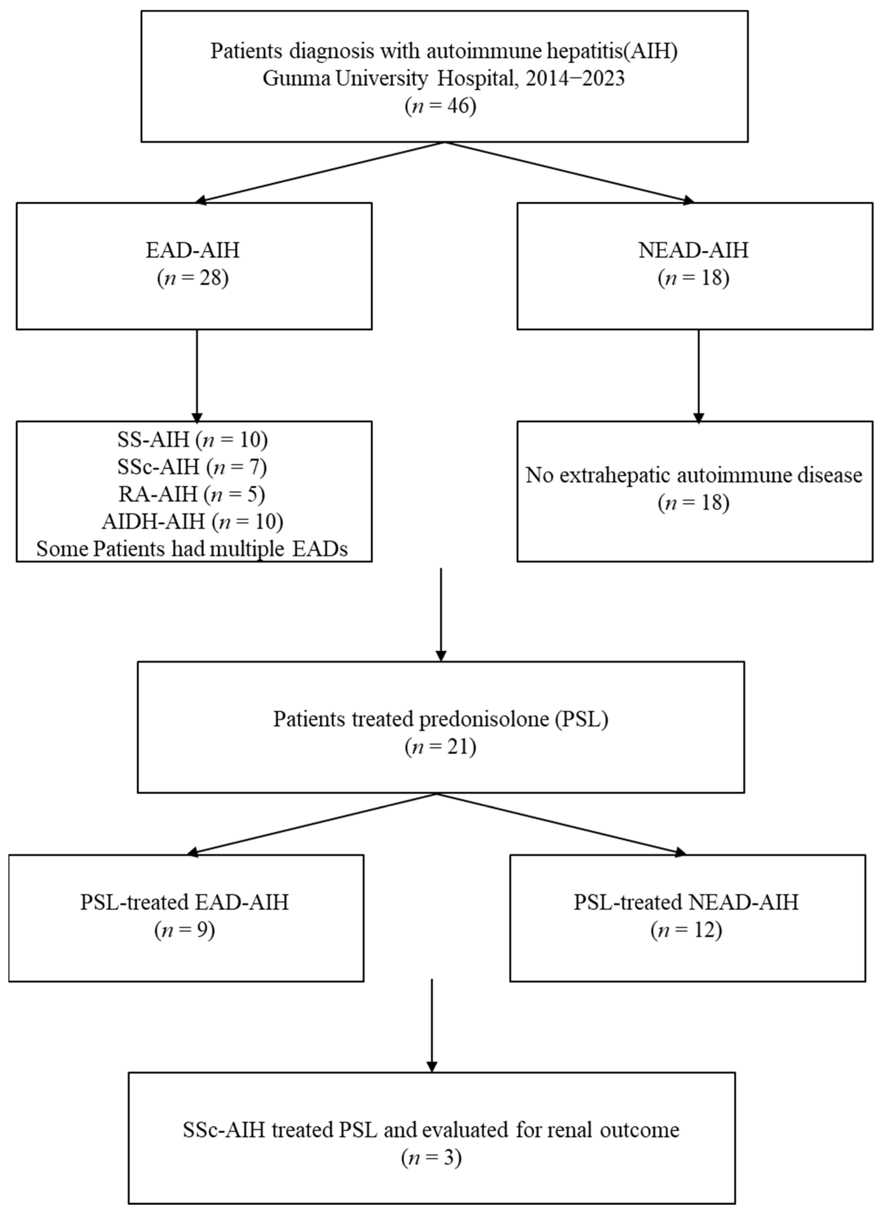
The study design and patient selection are summarized in
Figure 1.
Consecutive patients diagnosed with autoimmune hepatitis (AIH) at Gunma University Hospital between 2014 and 2023 were included (n = 46) and classified into EAD-associated AIH (EAD-AIH, n = 28) and AIH without EAD (NEAD-AIH, n = 18). Major EAD subtypes included Sjögren’s syndrome (SS), systemic sclerosis (SSc), rheumatoid arthritis (RA), and autoimmune thyroid disease (AITD); some patients had more than one EAD. Prednisolone (PSL) was administered to 21 patients. Among PSL-treated patients, those with SSc-AIH (n = 3) were additionally evaluated for renal outcomes.
3.1. Patient Characteristics at Diagnosis
A total of 46 patients were diagnosed with AIH during the study period. The median age at diagnosis was 63 years, and 41 patients (89%) were female. Twenty-eight patients (61%) had at least one EAD. Acute-onset hepatitis was observed in 11 patients (24%), and 15 patients (33%) were classified as having moderate or severe disease according to the Japanese clinical practice guidelines. Liver biopsy was performed in 36 patients (78%), and advanced fibrosis (≥F3) was identified in 11 cases (31%). The median revised IAIHG score was 17, and 28 patients (61%) fulfilled the criteria for definite AIH. Based on the Japanese classification system, 26 patients (57%) were categorized as having typical AIH.
Autoantibody profiles at diagnosis were broadly consistent with established AIH cohorts, with ANA positivity observed in the majority of patients and a wide distribution of titers. ANA titers showed a wide distribution at diagnosis. When patients were stratified into low- and high-titer groups using multiple cutoff values, no significant differences were observed in disease severity, biochemical parameters (AST, ALT, total bilirubin, PT%, PT-INR), or clinical outcomes. Thus, ANA titer itself was not associated with AIH severity in this cohort. In patients who underwent liver biopsy, pathology reports frequently described features compatible with AIH, including interface hepatitis and portal inflammatory infiltration with variable lymphoplasmacytic components. Although the original pathology reports were not uniformly structured for research purposes, the presence of advanced fibrosis (≥F3) in approximately one-third of biopsied cases indicates that a substantial subset of patients had already progressed to advanced-stage disease at diagnosis, despite the availability of serological screening and increasing awareness of AIH in routine clinical practice. Additional autoantibody testing was performed in a subset of patients. Anti-LKM-1 antibodies were measured in 11 patients and were positive in one case (9%), who was negative for hepatitis C virus infection and had no concomitant autoimmune thyroid disease. Anti-SMA antibodies were assessed in nine patients, of whom seven (78%) tested positive. Because testing was not performed systematically across the cohort, these findings were interpreted descriptively and were not included in comparative analyses. Detailed baseline characteristics of the study population are summarized in
Table 1.
3.2. Spectrum and Timing of Extrahepatic Autoimmune Diseases
Among the 28 patients with EAD-AIH, the most frequent EADs were SS (n = 10, 36%), AITD (n = 9, 32%), and SSc (n = 7, 25%). RA was present in five patients (18%). Less common EADs included systemic lupus erythematosus, dermatomyositis, Harada’s disease, psoriasis vulgaris, autoimmune hemolytic anemia, and IgG4-related disease, each observed in one patient (3.6%). Because several patients had more than one autoimmune condition, the total number of EADs exceeded the number of EAD-AIH patients.
Regarding the timing of EAD onset relative to AIH diagnosis, 18 patients (64%) developed EADs prior to the onset of AIH, five (18%) within one year of AIH diagnosis, three (11%) between one and three years after AIH onset, and two (7%) more than three years later (
Table 2). Thus, in the majority of cases, EADs preceded the diagnosis of AIH, suggesting that an autoimmune background often existed before the development of hepatic involvement.
We also compared AIH severity between patients whose EAD preceded AIH diagnosis and those who developed EAD after AIH onset. No significant differences were observed in biochemical severity, frequency of acute-onset presentation, or treatment requirements between the two groups.
3.3. Overlap with Other Liver Diseases and Hepatic Comorbidities
Among the entire cohort, six patients had autoimmune liver disease overlap: four had AIH–primary biliary cholangitis (AIH–PBC) overlap, all of whom also had EADs and were therefore classified in the EAD-AIH group. Two patients had primary sclerosing cholangitis (PSC) and neither had any documented EAD. In addition, four patients had other hepatic comorbidities, including metabolic dysfunction-associated steatotic liver disease (MASLD) in three patients and hepatitis B virus (HBV) carrier status in one patient. Notably, all four of these patients belonged to the NEAD-AIH group (
Table 2).
3.4. Treatment and Overall Clinical Outcomes
Prednisolone (PSL) therapy was administered to 21 patients (46%), whereas ursodeoxycholic acid (UDCA) was prescribed to 46 patients (100%). Among the 20 patients whose response to PSL could be evaluated at 6 months, all showed biochemical improvement after PSL initiation. During PSL tapering, seven patients experienced relapse; three were controlled by PSL dose escalation alone, while four required the addition of azathioprine. Two patients developed acute liver failure, including one fatal case; importantly, both occurred in the NEAD-AIH group (
Table 1). These findings indicate that although most patients responded biochemically to PSL, severe outcomes were confined to patients without concomitant EAD.
3.5. Comparison Between EAD-AIH and NEAD-AIH
When patients were stratified by EAD status, clear differences in clinical presentation and disease severity emerged. Compared with patients with EAD-AIH, those with NEAD-AIH were more likely to present with acute-onset hepatitis (44% vs. 18%) and moderate-to-severe disease. At diagnosis, the NEAD-AIH group exhibited significantly higher levels of AST, ALT, total bilirubin, and IgG, together with significantly lower levels of albumin and prothrombin activity. Steroid use was more frequent in the NEAD-AIH group (71%), and all cases of liver failure occurred exclusively in this group (
Table 3).
A notable temporal change was observed in the proportion of NEAD-AIH. The frequency of NEAD-AIH increased from 5 of 23 cases (21%) during 2014–2018 to 13 of 23 cases (54%) during 2019–2023. Among recent NEAD-AIH cases, one occurred shortly after COVID-19 vaccination and another following an immune checkpoint inhibitor (ICI)-induced hepatic immune-related adverse event (irAE). Each exposure was observed in a single case; therefore, these observations were descriptive and no causal relationship could be established (
Table 3).
3.6. Comparison by EAD Subtype
Among the 28 patients with EAD-AIH, subgroup analyses were conducted for SS-AIH (
n = 10), SSc-AIH (
n = 7), RA-AIH (
n = 5), and AITD-AIH (
n = 10). The principal findings are summarized in
Table 4.
Patients with SS-AIH had a significantly longer observation period (median 78.5 months) and higher prothrombin time (PT) compared with the overall AIH cohort. No cases of advanced fibrosis were observed in this subgroup. Overlap with PBC was present in 30% of SS-AIH cases.
Patients with SSc-AIH were characterized by notably low serum IgG levels (median 1365 mg/dL) and universal positivity for anti-centromere antibodies, indicating a distinct immunological profile.
Patients with RA-AIH tended to show lower PT and higher ANA titers, although these differences did not reach statistical significance. Importantly, all RA-AIH patients experienced relapse during follow-up.
Patients with AITD-AIH showed relatively lower ANA titers and relatively high IgG and ALT levels. Overlap with PBC was observed in 30% of AITD-AIH cases.
3.7. Analysis of Steroid-Treated Patients
Among the 21 patients who received PSL therapy, those with NEAD-AIH had significantly higher baseline ALT levels (median 787 vs. 192 IU/L) and PT-INR values (median 1.3 vs. 1.0) compared with those with EAD-AIH. Similar trends were observed for AST and prothrombin activity. Acute-onset hepatitis was also more frequent in the NEAD-AIH group (67% vs. 22%). In contrast, the observation period tended to be longer in the EAD-AIH group (
Table 5).
3.8. Renal Outcomes in Patients with SSc-AIH
Among PSL-treated patients, three had SSc-AIH. All three were classified as having limited cutaneous SSc and were positive for anti-centromere antibodies while being negative for anti–topoisomerase I and anti–RNA polymerase III antibodies. Baseline IgG levels were significantly lower in patients with SSc-AIH than in those with non-SSc-AIH (median 1153 vs. 2344 mg/dL, p = 0.003), suggesting lower baseline immunologic activity.
Renal function was assessed by changes in estimated glomerular filtration rate (eGFR) from baseline to 6 months after PSL initiation. A ≥25% decline in eGFR occurred in 33% of patients in both the SSc-AIH and non-SSc-AIH groups (
p = 1.00), indicating no excess renal deterioration in SSc-AIH associated with corticosteroid therapy. Relapse was observed in one-third of patients in each group; notably, relapse in the SSc-AIH group was successfully controlled by PSL dose re-escalation alone, without the need for azathioprine (
Table 6).
4. Discussion
In this single-center retrospective study, we demonstrated that the clinical phenotypes of autoimmune hepatitis (AIH) differ substantially according to both the presence and the type of extrahepatic autoimmune diseases (EADs). Approximately 60% of our cohort had concomitant EADs, which is higher than the approximately 25% reported in a nationwide Japanese survey [
9,
10] but comparable to frequencies reported in Western tertiary care cohorts [
8,
15,
16]. These findings indicate that EAD comorbidity is common in real-world clinical practice at referral centers and highlight the importance of considering autoimmune background when evaluating patients with AIH.
Previous studies have suggested that late-onset AIH may exhibit distinct immunological and genetic backgrounds, including higher ANA positivity and an increased prevalence of autoimmune thyroid disease [
17]. To explore this possibility in our cohort, we compared patients aged ≥65 years with those aged <65 years; however, we observed no significant differences in ANA positivity rates or the prevalence of autoimmune thyroid disease between the two groups. Although our sample size was limited, these findings suggest that age-related immunological differences may not fully account for the clustering of extrahepatic autoimmune diseases in our cohort.
Compared with patients with EAD-AIH, those with AIH without EAD (NEAD-AIH) in our cohort exhibited significantly more severe biochemical abnormalities at diagnosis, including higher AST, ALT, total bilirubin, and IgG levels, together with lower albumin and prothrombin activity. Acute-onset hepatitis and corticosteroid use were also significantly more frequent in the NEAD-AIH group, and, notably, all cases of liver failure occurred exclusively in this group. These findings are consistent with previous reports suggesting that the presence of extrahepatic autoimmune diseases may be associated with a milder hepatic disease activity at AIH onset [
18].
Several explanations may account for this difference. First, patients with established EADs are often under regular medical follow-up, which may facilitate earlier detection of liver enzyme abnormalities and lead to diagnosis of AIH at a relatively earlier and less advanced stage [
16,
18]. Second, shared but distinct immunogenetic backgrounds across autoimmune conditions may modulate the hepatic inflammatory response, resulting in a less aggressive biochemical phenotype in EAD-AIH, as suggested by current immunopathological models of autoimmune hepatitis [
3,
4,
5,
6,
11]. In contrast, NEAD-AIH may remain clinically silent until a more abrupt and severe inflammatory activation occurs, leading to acute-onset presentation and higher disease severity, a concept consistent with recent reports of increasing acute-onset AIH and trigger-dependent disease onset after immune-modifying exposures such as immune checkpoint inhibitors or COVID-19 vaccination [
5,
10,
19,
20,
21].
A notable observation in our study was the temporal increase in NEAD-AIH, from 21% during 2014–2018 to 54% during 2019–2023. Although the reasons for this shift remain unclear, a few recent NEAD-AIH cases in our cohort occurred shortly after COVID-19 vaccination or following immune checkpoint inhibitor (ICI) therapy. The recent literature has reported immune-mediated hepatic injury and AIH-like syndromes in both of these contexts [
19,
20,
21]. While our sample size is far too small to establish any causal relationship, these observations are consistent with the hypothesis that immune-modifying exposures may act as triggers that unmask latent autoimmune susceptibility and are temporally associated with AIH onset in susceptible individuals.
Several non-mutually exclusive mechanisms may contribute to the observed temporal shift toward a higher proportion of NEAD-AIH. Beyond potential immune-modifying exposures, changes in referral patterns to tertiary care centers, increased availability and routine use of autoantibody testing, and heightened clinical awareness of acute-onset AIH may have collectively lowered the diagnostic threshold for recognizing AIH in patients without established autoimmune comorbidity. Moreover, secular changes in competing liver diseases may influence the clinical context in which unexplained liver enzyme abnormalities are investigated (e.g., increasing prevalence of MASLD), potentially affecting case ascertainment. Although our single-center dataset cannot disentangle the relative contribution of these factors, the consistent direction of change observed over a decade supports the need for multicenter verification using harmonized case definitions and systematic documentation of pre-onset immune stimuli and comorbid conditions.
It is also conceivable that increasing awareness and improved diagnostic recognition of acute-onset AIH have contributed to the apparent rise in NEAD-AIH. Therefore, this trend should be interpreted cautiously and regarded as hypothesis-generating. Large-scale, multicenter studies with standardized diagnostic criteria and careful documentation of preceding immune stimuli will be required to determine whether NEAD-AIH is truly increasing and clarify the contribution of environmental or iatrogenic immune triggers.
Subgroup analyses of EAD-AIH in our cohort revealed distinct immunologic and clinical features linked to specific autoimmune backgrounds, supporting the concept that EAD-AIH is not a homogeneous entity.
Although ANA was positive in the majority of patients, additional autoantibody testing was performed only in selected cases. Anti-LKM-1 antibodies were rarely detected, while anti-SMA antibodies were frequently positive among tested patients. Because these assays were not systematically performed, we could not formally evaluate associations between specific autoantibody profiles and EAD subtypes or disease severity. Nevertheless, given previous reports suggesting links between certain autoantibodies and extrahepatic autoimmunity and emphasizing the immunological heterogeneity of AIH [
4,
5,
8], comprehensive serological profiling in larger prospective cohorts may further clarify the immunological diversity of AIH.
Among these, SSc-AIH demonstrated particularly low serum IgG levels, which is consistent with reports indicating that patients with limited cutaneous systemic sclerosis may exhibit relatively low immunoglobulin levels or hypogammaglobulinemia [
22].
Systemic sclerosis itself is known to encompass heterogeneous subsets characterized by distinct autoantibody profiles and immune phenotypes [
23,
24]. Our observation that all SSc-AIH patients were anti-centromere antibody-positive and had limited cutaneous disease further supports the notion that specific SSc subsets may be preferentially associated with a particular AIH phenotype.
An important clinical concern when treating SSc-AIH is the potential risk of scleroderma renal crisis, which is a well-recognized and life-threatening complication associated particularly with diffuse cutaneous involvement and anti–RNA polymerase III antibody positivity. In our cohort, no excess renal deterioration was observed in patients with SSc-AIH during corticosteroid therapy. This finding is in line with previous case series and literature reviews of AIH–SSc overlap, in which corticosteroids were generally well tolerated and rarely complicated by renal crisis [
25,
26,
27,
28].
The favorable renal outcomes in our study are most likely explained by the absence of high-risk features for scleroderma renal crisis, such as diffuse cutaneous involvement or anti–RNA polymerase III antibody positivity, which are well-established risk factors in both classical reviews and contemporary cohorts [
29,
30]. Taken together, these data suggest that, with careful patient selection and appropriate monitoring, corticosteroid therapy may be a reasonable and safe option for treating AIH in patients with limited cutaneous SSc.
Patients with SS-AIH in our cohort showed favorable biochemical profiles and low relapse rates, which is in line with previous studies suggesting a relatively benign hepatic course in SS-associated AIH [
31,
32]. In contrast, RA-AIH cases tended to show lower prothrombin activity and higher ANA titers, and relapse occurred in all patients in this subgroup, although the small sample size precludes definitive conclusions. These contrasting patterns further support the notion that EAD subtypes are not clinically interchangeable and that the specific autoimmune background should be taken into account when interpreting serological findings, estimating prognosis, and planning long-term management.
From a practical standpoint, our findings suggest that initial evaluation of patients with AIH should include careful assessment of autoimmune background, including the presence and type of EAD. Patients with NEAD-AIH appear more likely to present with acute and severe disease and may therefore require closer monitoring and earlier consideration of immunosuppressive therapy. Conversely, in EAD-AIH—particularly in SSc-AIH—attention should be paid not only to hepatic disease activity but also to organ-specific risks associated with the underlying systemic autoimmune disease, such as renal involvement in SSc.
The strengths of this study include the systematic, phenotype-oriented stratification of AIH according to major EAD subtypes, rather than treating EAD as a single pooled category, and the assessment of renal trajectories after corticosteroid initiation in patients with SSc-AIH. In addition, the decade-long observation period enabled exploration of temporal changes in NEAD-AIH frequency in a real-world tertiary care setting.
Nevertheless, several limitations must be acknowledged. First, the retrospective, single-center design and relatively small sample size—especially within EAD subgroups—limit the generalizability of our findings. Second, liver biopsy was not performed in all patients, and key histological features such as emperipolesis or central zonal injury were not systematically assessed. Third, AIH–PBC overlap cases were grouped under EAD-AIH; although limited in number, this may have influenced biochemical comparisons. Fourth, all patients received UDCA, reflecting Japanese clinical practice [
13,
14], which differs somewhat from international guidelines [
1]. Finally, liver function trajectories were analyzed only in steroid-treated patients, and long-term outcomes beyond biochemical response and relapse could not be fully evaluated. In addition, because EAD diagnoses were based on specialist documentation in medical records, subclinical or evolving autoimmune conditions may have been under-recognized at the time of AIH diagnosis, potentially leading to misclassification in borderline cases. Additionally, because our subgroup analyses were exploratory and involved multiple comparisons, the possibility of type I error cannot be excluded; therefore, subtype-specific findings should be interpreted as hypothesis-generating and warrant confirmation in larger datasets.



 ,
,