Article

Elevated Incidence and Risk of Emergent Cirrhosis Complications in Alcoholic Cirrhosis Compared with Other Etiologies

1
Internal Medicine Residency Program, Joan C. Edwards School of Medicine, Marshall University, Huntington, WV 25755, USA
2
Department of Gastroenterology, Joan C. Edwards School of Medicine, Marshall University, Huntington, WV 25755, USA
*
Author to whom correspondence should be addressed.
Gastroenterol. Insights 2023, 14(4), 671-681; https://doi.org/10.3390/gastroent14040045
Submission received: 14 November 2023 / Revised: 7 December 2023 / Accepted: 11 December 2023 / Published: 15 December 2023
(This article belongs to the Section Liver)

Abstract

:
Gastrointestinal bleeding (GIB) is a common cause of urgent hospitalization in patients with cirrhosis. However, limited studies have examined the prevalence and risk of these complications based on etiology. This study aims to compare the occurrence and risk of cirrhosis complications on inpatient mortality between alcoholic cirrhosis (ALC) and other etiology-induced cirrhosis (NALC). This retrospective analysis included 7,159,694 patients. ALC was diagnosed based on ICD-10, while NALC included primary and secondary biliary cirrhosis, nonalcoholic steatohepatitis (NASH), and unspecified cirrhosis of the liver. GIB included bleeding from esophageal and gastric varices. Bivariate analyses using appropriate statistical tests were performed to compare the two groups. ALC patients had a significantly higher incidence of GIB compared with NALC patients (10.8% vs. 6.4%, p < 0.01), with an associated 60% higher risk of GIB than NALC patients (p < 0.01). ALC was associated with a higher prevalence of ascites (45.6% vs. 27.9%, p < 0.01) and hepatic encephalopathy (HE) (45.5% vs. 27.2%, p < 0.01) compared with NALC patients. The risk of ascites and HE was 2.2 times and 2.3 times higher, respectively, in ALC patients compared with NALC patients (p < 0.01). Furthermore, ALC patients had higher hospital mortality rates compared with NALC patients, with a 47% higher risk of hospital mortality after adjustment (p < 0.01). ALC patients also had prolonged hospital stays, higher charges, more emergency room (ER) visits, and more frequent esophagogastroduodenoscopy (EGD) requirements compared with those of NALC patients (p < 0.01). ALC patients have a significantly higher risk of developing GIB, ascites, and HE compared with NALC patients, leading to increased mortality and greater medical burden on hospitals.

1. Introduction

Cirrhosis results from different liver injury mechanisms that lead to liver parenchyma necrosis and fibrosis. Commonly, cirrhosis is characterized by diffuse nodular regeneration surrounded by fibrotic tissue—leading to compression and collapse of liver structures, causing permanent distortion of the hepatic vascular structure [1]. These pathologic changes lead to increased resistance in the portal vein and result in portal hypertension and hepatic synthetic dysfunction [2]. The prevalence of cirrhosis in the US was estimated at 0.27%, equal to more than 600,000 individuals, based on a survey conducted between 1999–2010 [3]. Based on a surveillance report published in November 2019 by NIH, the crude death rate from any etiology of cirrhosis was 13.7 per 100,000 in 2017, accounting for a total of 44,478 deaths [4]. The leading causes of cirrhosis in developed countries are hepatitis C virus infection, alcohol abuse, and metabolic-syndrome-induced nonalcoholic liver disease [2].
Cirrhosis is frequently asymptomatic and unsuspected until complications of liver disease are present. However, initial clinical presentation of patients with decompensated cirrhosis is common. Usually presenting with dramatic and life-threatening complications, including variceal bleeding, ascites, spontaneous bacterial peritonitis, and HE, acute GIB remains the most common medical emergency and a lethal complication in cirrhosis patients, with variceal bleeding representing ~60% of hemorrhage episodes in patients with cirrhosis [5]. Mortality during the first episode of variceal bleeding is estimated to be 15–20%, but it is even higher in severe patients (Child–Pugh C)—around 30% [6,7]. Variceal bleeding is due to intrahepatic vascular resistance caused by an architectural distortion of the liver, secondary to cirrhosis. In response to the increased portal pressure, collateral circulation develops by opening pre-existing vessels and the neoformation of vessels. As one of the essential collateral vessels, esophageal varices tend to increase in size when portal hypertension is present and easily rupture when vessel wall tension exceeds a specific limit [8,9].
Various etiologies for cirrhosis have been established with alcohol liver disease (ALD), hepatitis B, hepatitis C, and NASH being the most common clinical findings. A recent study has shown that the primary etiology of cirrhosis in the US shifted from viral hepatitis to nonalcoholic fatty liver disease, and it is secondary to metabolic syndrome and alcohol-associated liver disease [10]. Current research has shown that ALD is one of the most prevalent etiologies for cirrhosis in the United States, with a study conducted in 2017 finding that 27.3% of cirrhosis-related fatalities globally were caused by underlying ALD [11,12]. Deaths related to ALD are second only to hepatitis B, the most common cause of NALC, at 31.5% globally.
Metabolic syndrome, which can lead to NASH, is exceedingly prevalent, with rates of 19.5% in those aged 20 to 39 and 48.6% in those older than 60 [13]. There is no difference in the prevalence of metabolic syndrome between men and women; however, there has been an increase of 64.0% in those identifying as “other” in race or ethnicity and of 57.3% in Hispanics over 60. Of those diagnosed with NASH by ultrasound, 37.6% had progressive fibrosis with a 5-year development rate of HCC and 5-year survival of 11.3% and 75.2%, respectively [14]. NASH was indicated as the causative ailment for cirrhosis-related deaths in 7.7% of all mortalities globally [11,15]. This highlights the danger of cirrhosis, and the importance of diagnosing, understanding, and treating all aspects of the disease state. Additionally, it shows the importance of recognizing preventable complications of a disease and working with patients to mitigate those risks. Weight loss and exercise remain the first line of treatment in treating NASH.
One of the most significant causes of death in patients with cirrhosis is GIB. Despite advanced treatment options and the optimization of general medical care, mortality is still significantly higher than in other disease states. Adopting a multidisciplinary approach is crucial to further improving the odds of patient survival. Therefore, determining instances where early prevention and intervention can be established is critical for patients with cirrhosis; as of now, no study has been conducted to examine the association between the differing etiologies of cirrhosis and GIB in patients with cirrhosis.
Currently, there are few useful markers to identify potential candidates for variceal screening. Only platelet count and advanced Child–Pugh class have been shown to be independent risk factors of variceal formation [16]. Given that the cost-effectiveness of the endoscopic screening of patients with cirrhosis is debated, as opposed to just treating patients with nonselective beta-blockers, it is critical to identify more variables that can improve screening criteria [17]. To predict variceal formation more effectively, additional information on the etiology of cirrhosis is required to determine its impact on the risk of variceal development. Work has already been completed to determine the risk factors for mortality in patients presenting with esophageal varices, but identifying patients with the highest risk of possible development could help prevent prehospital deaths—improving patient outcomes based on etiological risk [18]. Individuals at high risk can be screened and treated before symptomatic presentation, and patients with associated causes can receive more prompt, intensive therapy.
The aim of this study is to determine the prevalence and risk of GIB among patients with ALC versus NALC. In addition, we also aim to assess other cirrhosis complications, including ascites and HE, between these two groups of cirrhosis patients.

2. Materials and Methods
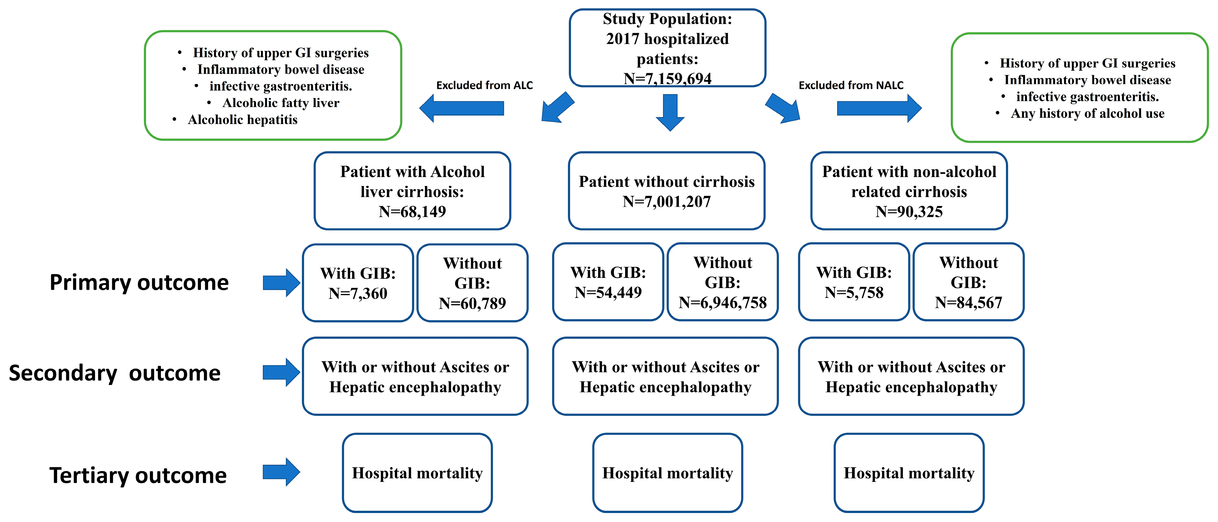
2.1. Database

A retrospective analysis was performed using the national inpatient sample (NIS) database, which was developed by the healthcare cost and utilization project (HCUP). The NIS is the largest publicly available all-payer inpatient healthcare database designed to estimate inpatient utilization, access, cost, quality, and outcomes—containing unweighted data from around 7 million hospital stays each year. The NIS approximates a 20% stratified sample of all discharges from US community hospitals, excluding rehabilitation and long-term acute care hospitals.

2.2. Data Collection and Outcomes

A total of 7,159,694 adult patients admitted to hospitals in 2017 were included in this study. Patients diagnosed with GIB (ICD-10-CM I85.01, I85.11, and K92.0-2) with ALC (ICD-10-CM K70.2, K70.30, K70.31, K70.40, K70.41, K70.9) and NALC (ICD-10-CM K75.81, K74.1-5, K74.60, K74.69) were compared with those without GIB. In selecting subjects with GIB, we excluded esophageal varices without bleeding, history of upper GI surgeries, IBD, and infective gastroenteritis. Given that this study relies on the ICD-10 code, the GIB group was delineated using the following parameters: patients were identified with diagnoses of esophageal varices with bleeding, hematemesis, and gastrointestinal hemorrhage, unspecified. To ensure the exclusion of patients with GIB secondary to peptic ulcer or gastritis, we specifically isolated cases of peptic ulcer with bleeding and gastritis with bleeding, excluding them from the initial groups. We also excluded subjects with alcoholic fatty liver and alcoholic hepatitis from the ALC groups. Focus was placed on ICD-10 codes, such as “Alcoholic fibrosis and sclerosis of the liver”, “Alcoholic cirrhosis of the liver”, and “Alcoholic hepatic failure”. Subsequently, we identified patients with diagnoses of HCV, NAFLD, NASH, HBV, PBS, or other etiology of cirrhosis using ICD-10 codes and excluded the cases from the group of patients with ALC. For the identification of NALC groups, a history of alcohol use was excluded.
More specifically, patients diagnosed with “NASH”, “hepatic fibrosis”, “hepatic sclerosis”, “hepatic fibrosis with hepatic sclerosis”, “primary biliary cirrhosis”, “secondary biliary cirrhosis”, “biliary cirrhosis, unspecified”, and “other and unspecified cirrhosis of the liver” were identified using ICD-10 codes from the database. Of note, the ICD-10 code for HCV, HBV, and drug-induced cirrhosis was the same as “unspecified cirrhosis of liver”. According to the ICD-10 code system, patients with a diagnosis of ALC have already been excluded from the above diagnostic code. The coexistence of both ALC and other etiologies of cirrhosis cannot be entirely ruled out. Consequently, we further excluded cases in the NALC groups that also presented with a diagnosis of ALC. Demographic data were collected, including age, race, and gender. Length of hospital stay (LOS), total hospital charge (TC), emergency department visit (ER visit) before hospitalization, and EGD requirement during hospitalization (EGD/total number of patients) were analyzed between ALC and NALC. All diagnoses included or excluded from this study were selected by the ICD-10-CM code.

2.3. Statistical Analysis

All demographic and hospital-related data in the study collected from NIS were categorical and thus were presented as several cases and percentages. Chi-squared analysis was used to analyze the association between ALC and NALC in a patient with GIB. Additionally, death during hospitalization between ALC and NALC was analyzed by Chi-squared. Multivariate logistic regression analysis was used to assess risk in the form of odds ratios for GIB and mortality with ALC and NALC. We adjusted for age, gender, and race as covariates to mitigate the effect of confounding factors. A 2-sample test for equal proportions was used, and a p-value < 0.05 was considered significant. IBM SPSS 28.0.1.1 was used for statistical analysis.

3. Results

3.1. Demographic and Medical Profiles across Various Groups

A total of 7,159,694 hospitalized patients during 2017 were included in this study, in which 158,478 subjects diagnosed with GIB with ALC or NALC were identified (68,149 with ALC and 90,329 with NALC) and compared with those without GIB (Figure 1). Overall, most NALC patients with GIB were older than the ALC with GIB patients (62.8 ± 0.1 vs. 55.8 ± 0.1 p < 0.01). (Table 1) There were significantly more male than female patients diagnosed with GIB and ALC (70.6% vs. 29.4%, p < 0.01), but there was no gender difference in the GIB and NALC group. Additionally, there were significantly more males with GIB in the ALC group compared with GIB in the NALC group (70.6% vs. 50.5%, p < 0.01). (Table 1) LOS was significantly longer in those with GIB and ALC versus NALC (6.28 ± 0.03 vs. 5.89 ± 0.03, p < 0.05). ALC patients with GIB also had higher TC ($68,743 ± 427 vs. $67,380 ± 385, p < 0.01), had a higher percentage of ER visits before hospitalization (79.6% vs. 74.7%, p < 0.01), and required more EGD during hospitalization (9.2% vs. 5.4% p < 0.01) when compared with NALC with GIB. (Table 1).

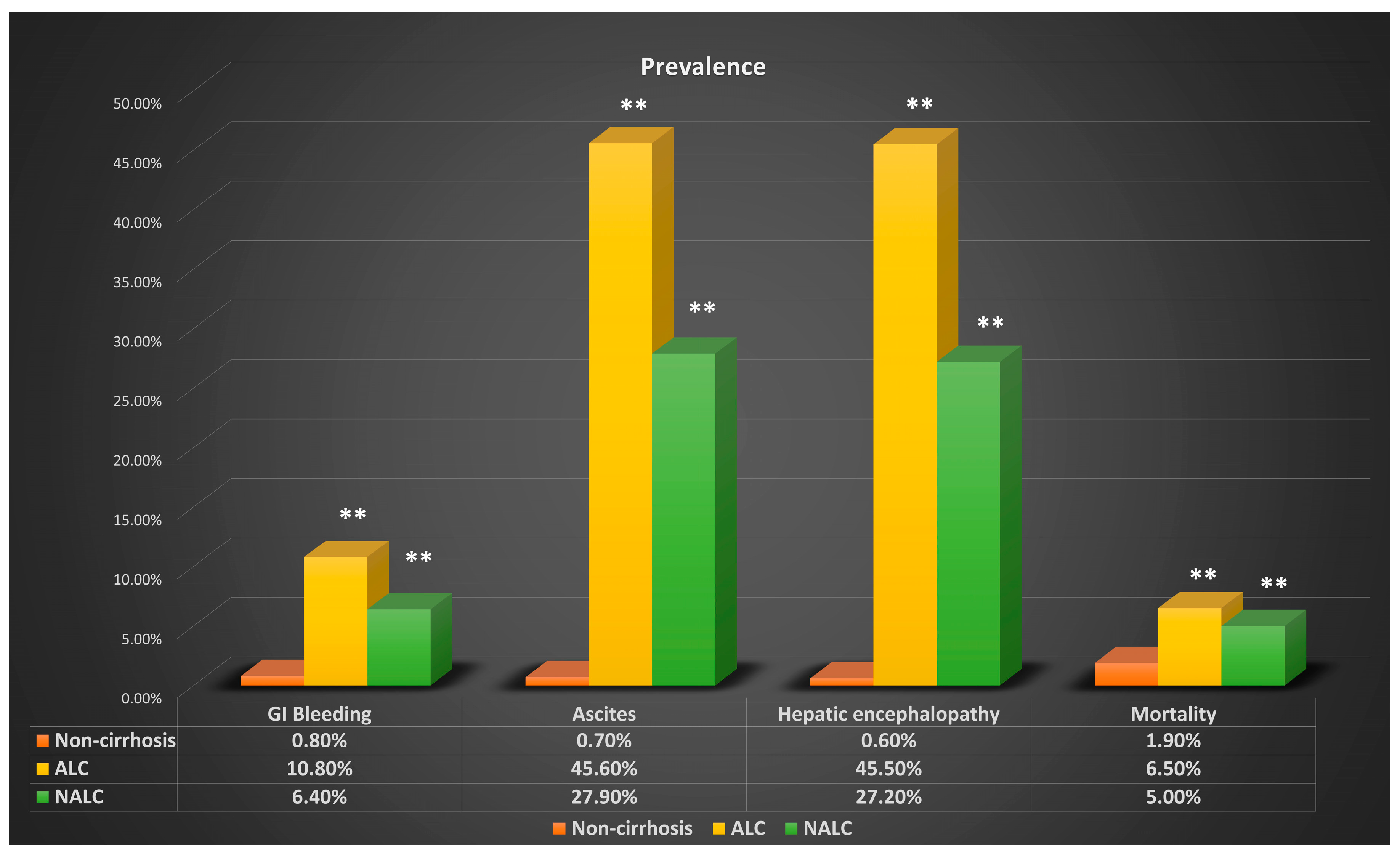
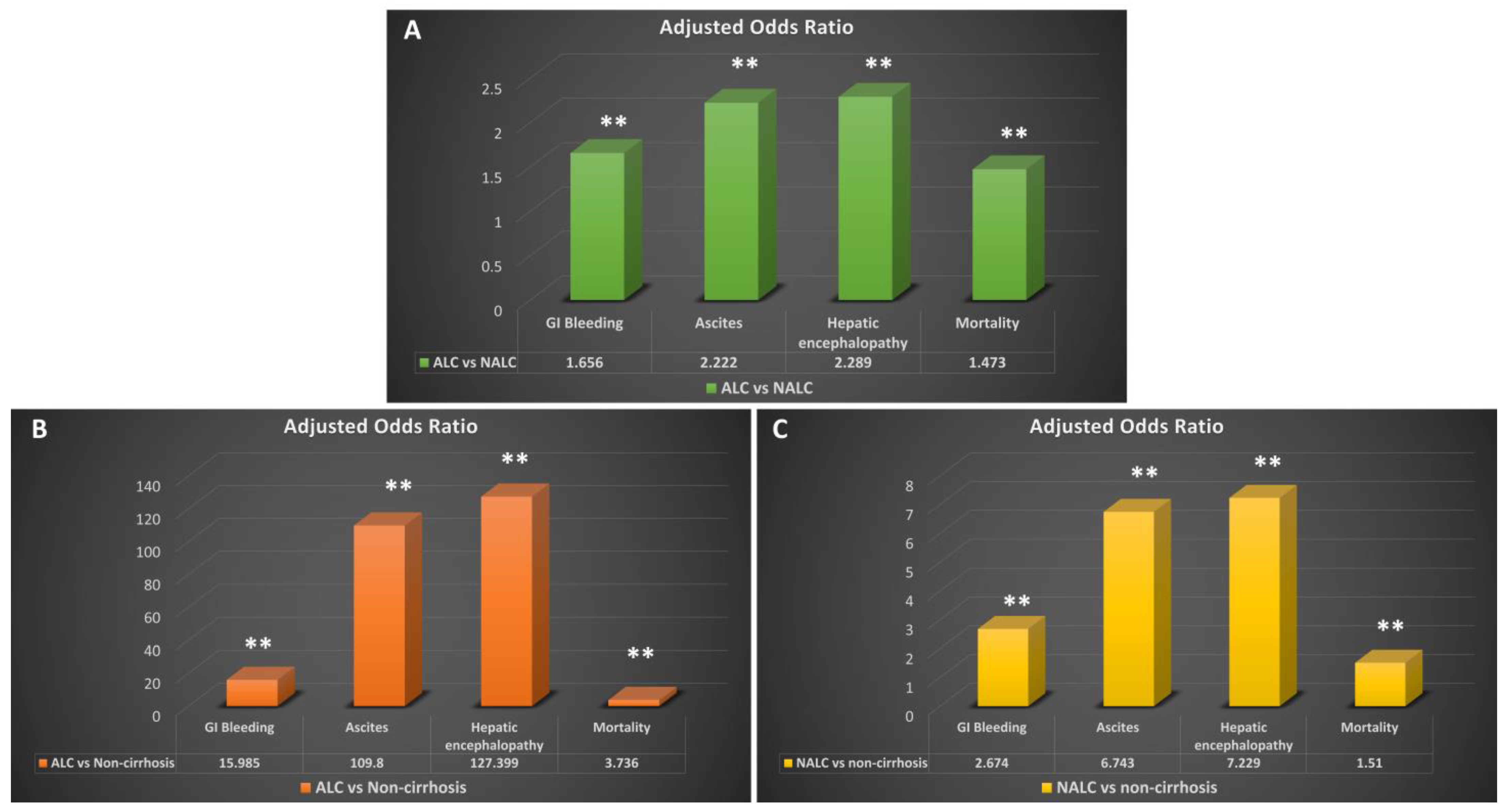
3.2. Complications of Cirrhosis across Various Groups

Subjects with ALC were significantly more likely to have GIB than those without cirrhosis (OR: 15.985, 95% CI: 15.560–16.421, p < 0.01) (Table 2). The odds ratio of GIB in patients with NALC was 2.674 (95% CI: 2.636–2.713, p < 0.01) compared with those without cirrhosis (Table 2). The incidence of GIB in those with ALC and NALC was 10.8% and 6.4%, respectively, and the incidence in those without cirrhosis was 0.8% (p < 0.001) (Table 2 and Figure 2). More importantly, when comparing the risk of GIB between patients with ALC versus NALC, subjects with ALC were significantly more likely to have GIB than those with NALC (OR: 1.656, 95% CI: 1.595–1.721, p < 0.01) (Table 2 and Figure 3A).
A subgroup analysis focusing on patients with NALC showed the incidence of GIB in patients with NASH was 6.3%, in patients with biliary cirrhosis it was 5.2%, and in patients with other cirrhosis etiologies (such as hepatitis B, hepatitis C, or drug-induced cirrhosis) it was 6.8%. Furthermore, the risk of GIB in individuals with NASH, biliary cirrhosis, or other cirrhosis etiologies was significantly lower compared with patients with ALC. (Table 3) The adjusted odds ratios for GIB were 0.55 (95% CI: 0.516–0.586) for NASH, 0.44 (95% CI: 0.375–0.518) for biliary cirrhosis, and 0.599 (95% CI: 0.577–0.621) for other cirrhosis etiologies. These results consistently indicate that patients with ALC face a higher risk of GIB compared with those with NALC. (Table 3)
Secondary outcome analysis showed that subjects with ALC or NALC were significantly more likely to have HE than those without cirrhosis (ALC: OR 127.4, 95% CI 125.0–129.8, p < 0.001) (NALC: OR 7.229, 95% CI: 7.165–7.294 p < 0.001) (Figure 3B,C). The incidences of HE in those with ALC and NALC were 45.5% and 27.2%, respectively, while the incidence in those without cirrhosis was 0.6% (p < 0.001) (Figure 2). More interestingly, the risk of having HE in patients with ALC was significantly higher than NALC, with an odds ratio of 2.289 (95% CI: 2.237–2.342, p < 0.001) (Figure 3A). The risk of ascites was found to be significantly higher in subjects with ALC (OR: 109.8, 95% CI: 107.8–111.6, p < 0.001) and NALC (OR: 6.743, 95% CI: 6.684–6.801, p < 0.001) (Figure 3). The incidence of ascites in patients with ALC was 45.6%, and in patients with NALC, it was 27.9%. The incidence in those without ascites was 0.7% (p < 0.001) (Figure 2).
The third outcome analysis showed that subjects with ALC or NALC had a significantly higher risk of inpatient mortality than those without cirrhosis. The OR of inpatient mortality in ALC and NALC was 3.74 (95% CI: 2.52–4.87, p < 0.01) and 1.51 (95% CI: 1.39–1.71, p < 0.01) compared with patients without cirrhosis, respectively (Table 2 and Figure 3B,C). The incidences of inpatient mortality in ALC and NALC were 6.5% and 5.0%, compared with 1.9% inpatient mortality in those without cirrhosis (Figure 2). Moreover, patients with ALC were also found to have a significantly higher risk of inpatient mortality than NALC patients (OR: 1.473, 95% CI: 1.37–1.52, p < 0.01) (Figure 3A).

4. Discussion

The most important findings in this study are the significantly increased risk of GIB in those with ALC compared with those with NALC. The foundation of this project was based on observation in a regional hospital in West Virginia that found that cirrhotic patients who presented with acute GIB more frequently had an alcohol abuse history instead of other etiologies of cirrhosis. This is the first study to specifically use extensive inpatient patient data to compare the risk of GIB with the etiology of liver cirrhosis and demonstrate a clear association. Our finding is even more significant as we have adjusted several important possible confounding factors, such as age, gender, and race.
One of the most severe but common complications in patients with cirrhosis is bleeding from gastro-esophageal varices. Despite the progression of modern advanced therapeutic interventions, the mortality rate is nearly 20% [19]. The most common cause of gastrointestinal varices is portal hypertension, with the most common etiology of portal hypertension being liver cirrhosis. It has been suggested that at least 75% of patients with cirrhosis develop varices during their lifetime [20], with variceal bleeding as a complication of portal hypertension occurring in about 40% of patients [21]. Although primary prophylaxis of variceal bleeding with variceal band ligation is highly effective and recommended for preventing extensive GIB, the goal of optimizing endoscopy screening is to balance the benefits, potential complications, and medical burden.
Current noninvasive methods to estimate the risk for the development of variceal bleeding include Child–Turcotte–Pugh class, albumin level, bilirubin level, portal vein diameter, platelet counts, and INR [17]. Among these, only platelet count and Child–Pugh class have been demonstrated as independent variables in predicting the risk of variceal formation [16]. Portal venous pressure and splenic size have shown some utility in multivariate analyses in predicting the presence and size of esophageal varices, respectively. A recent study has expanded the factors that may be good clinical indicators of esophageal variceal formation, but more work is needed to validate and refine these data [22]. Correspondingly, the efficacy of directing endoscopic screening toward particular patients is limited, resulting in a large burden of time and money. The cost-effectiveness of screening all cirrhosis patients endoscopically, compared with treatment with nonselective beta-blockers, is debated—it is necessary to determine additional risk factors to decrease this expensive and time-consuming burden [17]. Another study indicated that Child–Pugh, platelet count, and spleen size may serve better as indicators to start treatment until endoscopy can be performed [23]. Child–Pugh and CAGIB scores have been shown to be the best scores for predicting the prognosis of cirrhosis and acute variceal bleeding that are currently available [24]. With this wide array of tests, scores, and mixed results, it is critical for clinicians to have access to a wide array of prognostic reasoning before deciding the best path for patient care.
Due to the relatively low prevalence of clinically significant varices in cirrhosis patients who never had variceal bleeding, it is not cost-effective to perform screening endoscopy in every patient diagnosed with cirrhosis. However, none of these parameters or markers have a confident sensitivity or specificity to predict the development of acute variceal bleeding. Therefore, other predictors and risk factors should be considered in determining when to start screening endoscopy in patients with a high risk of variceal bleeding. In a prospective study involving only hepatitis C patients by Sanyal et al., it was found that decreased platelet counts, elevated bilirubin, and INR could be good indicators for varices with a high risk of bleeding [25]. They suggested that hepatitis C-induced cirrhosis with elevated bilirubin and low platelet counts should have indefinitely had screening endoscopy. Concerning our findings in patients with alcohol-abuse-related cirrhosis, screening endoscopy and bleeding prophylaxis should be performed due to the elevated risk of GIB.
Several potential mechanisms may contribute to this finding, as it has been known since the 1940s that alcohol abuse can cause mucosal inflammation and destroy mucosal integrity [26]. However, the exact mechanism of how alcohol impairs gastric mucosa has yet to be fully elucidated. It has been suggested that alcohol could disrupt the mucosal barrier and increase the mucosa’s permeability through damage to the epithelial cell membranes via oxidative stress [27]. Even a small amount of alcohol consumption can impair esophageal motility and weaken the lower esophageal sphincter [28], resulting in short periods of acid reflux. Both factors may lead to vulnerable varices rupturing and bleeding. Another potential mechanism associated with alcohol consumption is endothelial function. Oda et al. demonstrated that heavy alcohol consumption is associated with endothelial dysfunction and dilation capacity—another group also reported similar findings in Japan [29]. It has been suggested that heavy, chronic alcohol consumption decreases the endothelium modulation of alpha-adrenoceptor contraction in vascular smooth muscle and reduces the maximum relaxation capacity [30]. Heavy alcohol consumption could also reduce the endothelial synthesis of NO and accelerate vessel wall degradation, resulting in decreased vessel dilatory capacity [31]. All these mechanisms lead to increased fragility and stiffness in varices, which tend to rupture even under minor trauma.
Ascites is one of the most common symptoms in patients with decompensated liver cirrhosis. The pathophysiology of ascites in patients with cirrhosis is multifactorial, mainly including low albumin, relative volume overload, and hormone abnormality in the setting of impaired liver function and portal hypertension [32,33,34]. An intact intestinal barrier is formed mainly by epithelium and is critical for normal intestinal function. These epithelial cells are connected by tight junctions, which are also essential for intestinal integrity. Several studies have demonstrated that alcohol consumption increases intestinal permeability by damaging epithelial cells or tight junctions [35,36]. Alcohol metabolite acetaldehyde was found to destabilize epithelial cell tight junctions by downregulating claudin-1 and ZO-1, which are essential proteins for the formation of tight junctions [37]. When considered together, in patients with alcohol-induced cirrhosis, further epithelial damage and intestinal leakage secondary to a history of alcohol toxication would contribute to the elevated risk of ascites when compared with non-alcohol-related liver cirrhosis.
HE is another common complication in patients with decompensated liver cirrhosis, which usually presents with nonspecific symptoms such as altered mental status, personality change, or coma. Although the exact pathophysiology of HE is still not well understood, the most common mechanisms include neurotoxin accumulation, impaired neurotransmission secondary to a metabolic change in liver decompensation, and microbiota dysfunction [38,39]. Ammonia, which is mainly produced in the gastrointestinal tract, is the best-known neurotoxin linked to HE. It has been suggested that alcohol could change the microbiota in the gastrointestinal tract, which may contribute to the increased permeability of intestinal luminal to bacteria and cause the overproduction of ammonia [40]. Moreover, subjects with alcohol abuse were found to have abnormal microbiota in sigmoid biopsy [41], which is associated with a high level of endotoxin in the circulation [42].
Our study has several notable limitations. The NIS database gathered all relevant diagnostic codes; however, it did not encompass outpatient information—a significant aspect of clinical practice. The diagnosis of ALC and NALC in this study relied on ICD-10-CM codes, input across diverse hospital systems, and electronic medical records. Similarly, the diagnosis of each cirrhosis complication was code-based and presumed to involve imaging, endoscopy, laboratory results, and clinical presentations—although specific diagnostic methods were not explicitly stated. Furthermore, this database did not include laboratory, imaging, or procedural results, precluding the measurement of crucial prognostic indicators (e.g., albumin level, INR, or platelet counts) and risk analysis scores (e.g., Child–Pugh classification, MELD score, Lille Model, and Maddrey’s discriminant function) for cirrhosis. Additionally, no timeline information regarding the outcomes of cirrhosis was identified.
To address these limitations and further support our findings, we are currently conducting a 10-year retrospective study involving patients from our regional hospitals. We have identified over 10,000 cirrhosis patients and are meticulously gathering detailed medical information for each hospital visit and clinic visit, including laboratory results, procedures, imaging outcomes, hospital charges, and patient outcomes. Several important prognostic scores or indicators will be calculated and adjusted in the risk assessment analysis. These data have been collected with a focus on establishing a timeline for cause–effect association analysis.
Our study highlights the importance of liver cirrhosis etiology in assessing future complications regarding GIB, ascites, and HE. The data show an association between alcohol-induced cirrhosis with increased rates of the above complications and, as such, emphasize the importance of aggressive interventional examination of patients with ALC. Preventative therapy and screening can lead to early detection and treatment for these cirrhotic secondary ailments, especially GIB, leading to a further decrease in mortality rates. Additional studies can be conducted to provide a more precise understanding of the mechanism of alcohol-related complications in cirrhosis patients.

Author Contributions

X.W., conceptualization, investigation, data curation, data analysis, visualization, and drafting the manuscript; D.C., editing the manuscript, investigation; A.D., editing the manuscript, investigation; Z.W., editing the manuscript; J.W., editing the manuscript; W.M.F., conceptualization, project supervision, and editing the manuscript. All authors have read and agreed to the published version of the manuscript.

Funding

This research received no external funding.

Institutional Review Board Statement

The Marshall University School of Medicine Institutional Review Board has deemed studies using the NIS database as exempt from requiring IRB approval due to the de-identified and aggregated nature of the data in the database. The Case Western Reserve University/Metrohealth Medical Center Institutional Review Board has deemed studies using the NIS database as exempt from requiring IRB approval due to the de-identified and aggregated nature of the data in the database at the standard defined in Section 164.514(a) of the HIPAA Privacy Rule.

Informed Consent Statement

Patient consent was waived due to no patient’s identity being collected in this database.

Data Availability Statement

The data presented in this study are available on request from the corresponding author. The data are not publicly available due to patient and hospital information privacy and the requirement of H.CUP.

Conflicts of Interest

The authors declare no conflict of interest.

References

  1. Schuppan, D.; Afdhal, N.H. Liver Cirrhosis. Lancet 2008, 371, 838–851. [Google Scholar] [CrossRef]
  2. Tsochatzis, E.A.; Bosch, J.; Burroughs, A.K. Liver Cirrhosis. Lancet 2014, 383, 1749–1761. [Google Scholar] [CrossRef] [PubMed]
  3. Scaglione, S.; Kliethermes, S.; Cao, G.; Shoham, D.; Durazo, R.; Luke, A.; Volk, M.L. The Epidemiology of Cirrhosis in the United States: A Population-Based Study. J. Clin. Gastroenterol. 2015, 49, 690–696. [Google Scholar] [CrossRef]
  4. Termeie, O.; Fiedler, L.; Martinez, L.; Foster, J.; Perumareddi, P.; Levine, R.S.; Hennekens, C.H. Alarming Trends: Mortality from Alcoholic Cirrhosis in the United States. Am. J. Med. 2022, 135, 1263–1266. [Google Scholar] [CrossRef] [PubMed]
  5. Garcia-Tsao, G.; Sanyal, A.J.; Grace, N.D.; Carey, W. Prevention and Management of Gastroesophageal Varices and Variceal Hemorrhage in Cirrhosis. Hepatology 2007, 46, 922–938. [Google Scholar] [CrossRef]
  6. Moledina, S.M.; Komba, E. Risk Factors for Mortality among Patients Admitted with Upper Gastrointestinal Bleeding at a Tertiary Hospital: A Prospective Cohort Study. BMC Gastroenterol. 2017, 17, 165. [Google Scholar] [CrossRef]
  7. Carbonell, N.; Pauwels, A.; Serfaty, L.; Fourdan, O.; Lévy, V.G.; Poupon, R. Improved Survival after Variceal Bleeding in Patients with Cirrhosis over the Past Two Decades. Hepatology 2004, 40, 652–659. [Google Scholar] [CrossRef] [PubMed]
  8. Hilzenrat, N.; Sherker, A.H. Esophageal Varices: Pathophysiology, Approach, and Clinical Dilemmas. Int. J. Hepatol. 2012, 2012, 795063. [Google Scholar] [CrossRef]
  9. Jakab, S.S.; Garcia-Tsao, G. Screening and Surveillance of Varices in Patients with Cirrhosis. Clin. Gastroenterol. Hepatol. 2019, 17, 26–29. [Google Scholar] [CrossRef]
  10. Huang, D.Q.; Terrault, N.A.; Tacke, F.; Gluud, L.L.; Arrese, M.; Bugianesi, E.; Loomba, R. Global Epidemiology of Cirrhosis—Aetiology, Trends and Predictions. Nat. Rev. Gastroenterol. Hepatol. 2023, 20, 388–398. [Google Scholar] [CrossRef]
  11. Seitz, H.K.; Bataller, R.; Cortez-Pinto, H.; Gao, B.; Gual, A.; Lackner, C.; Mathurin, P.; Mueller, S.; Szabo, G.; Tsukamoto, H. Alcoholic Liver Disease. Nat. Rev. Dis. Prim. 2018, 4, 16. [Google Scholar] [CrossRef]
  12. Sepanlou, S.G.; Safiri, S.; Bisignano, S.; Ikuta, K.S.; Merat, S.; Saberifiroozi, M.; Poustchi, H.; Tsoi, D.; Colombara, D.V.; Abdoli, A.; et al. The Global, Regional, and National Burden of Cirrhosis by Cause in 195 Countries and Territories, 1990–2017: A Systematic Analysis for the Global Burden of Disease Study 2017. Lancet Gastroenterol. Hepatol. 2020, 5, 245–266. [Google Scholar] [CrossRef]
  13. Hirode, G.; Wong, R.J. Trends in the Prevalence of Metabolic Syndrome in the United States, 2011–2016. JAMA 2020, 323, 2526–2528. [Google Scholar] [CrossRef]
  14. Hashimoto, E.; Tokushige, K. Prevalence, Gender, Ethnic Variations, and Prognosis of Nash. J. Gastroenterol. 2011, 46 (Suppl. 1), 63–69. [Google Scholar] [CrossRef] [PubMed]
  15. Angulo, P. Gi Epidemiology: Nonalcoholic Fatty Liver Disease. Aliment. Pharmacol. Ther. 2007, 25, 883–889. [Google Scholar] [CrossRef]
  16. Zaman, A.; Becker, T.; Lapidus, J.; Benner, K. Risk Factors for the Presence of Varices in Cirrhotic Patients without a History of Variceal Hemorrhage. Arch. Intern. Med. 2001, 161, 2564–2570. [Google Scholar] [CrossRef]
  17. D’Amico, G.; Morabito, A. Noninvasive Markers of Esophageal Varices: Another Round, Not the Last. Hepatology 2004, 39, 30–34. [Google Scholar] [CrossRef]
  18. Cerqueira, R.M.; Andrade, L.; Correia, M.R.; Fernandes, C.D.; Manso, M.C. Risk Factors for in-Hospital Mortality in Cirrhotic Patients with Oesophageal Variceal Bleeding. Eur. J. Gastroenterol. Hepatol. 2012, 24, 551–557. [Google Scholar] [CrossRef] [PubMed]
  19. D’Amico, G.; De Franchis, R. Upper Digestive Bleeding in Cirrhosis. Post-Therapeutic Outcome and Prognostic Indicators. Hepatology 2003, 38, 599–612. [Google Scholar] [CrossRef]
  20. Garceau, A.J.; Chalmers, T.C. The Natural History of Cirrhosis. N. Engl. J. Med. 1963, 268, 469–473. [Google Scholar] [CrossRef] [PubMed]
  21. Conn, H.O.; Lindenmuth, W.W.; May, C.J.; Ramsby, G.R. Prophylactic Portacaval Anastomosis. Medicine 1972, 51, 27–40. [Google Scholar] [CrossRef] [PubMed]
  22. Elatty, E.A.A.; Elshayeb, E.I.; Badr, M.H.; Mousa, W.A.E.; El Mansory, M.F. Noninvasive Parameters for Assessment of Esophageal Varices. Egypt. J. Intern. Med. 2019, 31, 536–543. [Google Scholar] [CrossRef]
  23. Cherian, J.V.; Deepak, N.; Ponnusamy, R.P.; Somasundaram, A.; Jayanthi, V. Non-Invasive Predictors of Esophageal Varices. Saudi J. Gastroenterol. 2011, 17, 64–68. [Google Scholar]
  24. Zhao, Y.; Ren, M.; Lu, G.; Lu, X.; Yin, Y.; Zhang, D.; Wang, X.; Ma, W.; Li, Y.; Cai, G.; et al. The Prognosis Analysis of Liver Cirrhosis with Acute Variceal Bleeding and Validation of Current Prognostic Models: A Large Scale Retrospective Cohort Study. BioMed Res. Int. 2020, 2020, 7372868. [Google Scholar] [CrossRef] [PubMed]
  25. Sanyal, A.J.; Fontana, R.J.; Di Bisceglie, A.M.; Everhart, J.E.; Doherty, M.C.; Everson, G.T.; Donovan, J.A.; Malet, P.F.; Mehta, S.; Sheikh, M.Y.; et al. The Prevalence and Risk Factors Associated with Esophageal Varices in Subjects with Hepatitis C and Advanced Fibrosis. Gastrointest. Endosc. 2006, 64, 855–864. [Google Scholar] [CrossRef]
  26. Beazell, J.M.; Ivy, A.C. The Influence of Alcohol on the Digestive Tract; a Review. Q. J. Stud. Alcohol 1940, 1, 45–73. [Google Scholar] [CrossRef]
  27. Bishehsari, F.; Magno, E.; Swanson, G.; Desai, V.; Voigt, R.M.; Forsyth, C.B.; Keshavarzian, A. Alcohol and Gut-Derived Inflammation. Alcohol Res. 2017, 38, 163–171. [Google Scholar]
  28. Pan, J.; Li, C.; Chen, W.; Yu, C.; Li, Y.; Shen, Z. Alcohol Consumption and the Risk of Gastroesophageal Reflux Disease: A Systematic Review and Meta-Analysis. Alcohol Alcohol. 2018, 54, 62–69. [Google Scholar] [CrossRef]
  29. Tanaka, A.; Cui, R.; Kitamura, A.; Liu, K.; Imano, H.; Yamagishi, K.; Kiyama, M.; Okada, T.; Iso, H. Heavy Alcohol Consumption Is Associated with Impaired Endothelial Function. J. Atheroscler. Thromb. 2016, 23, 1047–1054. [Google Scholar] [CrossRef]
  30. Brizzolara, A.L.; Morris, D.G.; Burnstock, G. Ethanol Affects Sympathetic Cotransmission and Endothelium-Dependent Relaxation in the Rat. Eur. J. Pharmacol. 1994, 254, 175–181. [Google Scholar] [CrossRef]
  31. Kelm, M.; Preik, M.; Hafner, D.J.; Strauer, B.E. Evidence for a Multifactorial Process Involved in the Impaired Flow Response to Nitric Oxide in Hypertensive Patients with Endothelial Dysfunction. Hypertension 1996, 27, 346–353. [Google Scholar] [CrossRef]
  32. Moore, C.M.; Van Thiel, D.H. Cirrhotic Ascites Review: Pathophysiology, Diagnosis and Management. World J. Hepatol. 2013, 5, 251–263. [Google Scholar] [CrossRef] [PubMed]
  33. Arroyo, V. Pathophysiology, Diagnosis and Treatment of Ascites in Cirrhosis. Ann. Hepatol. 2002, 1, 72–79. [Google Scholar] [CrossRef] [PubMed]
  34. Arroyo, V.; Moreau, R.; Kamath, P.S.; Jalan, R.; Ginès, P.; Nevens, F.; Fernández, J.; To, U.; García-Tsao, G.; Schnabl, B. Acute-on-Chronic Liver Failure in Cirrhosis. Nat. Rev. Dis. Prim. 2016, 2, 16041. [Google Scholar] [CrossRef] [PubMed]
  35. Bode, C.; Bode, J.C. Alcohol’s Role in Gastrointestinal Tract Disorders. Alcohol Health Res. World 1997, 21, 76–83. [Google Scholar] [PubMed]
  36. Purohit, V.; Bode, J.C.; Bode, C.; Brenner, D.A.; Choudhry, M.A.; Hamilton, F.; Kang, Y.J.; Keshavarzian, A.; Rao, R.; Sartor, R.B.; et al. Alcohol, Intestinal Bacterial Growth, Intestinal Permeability to Endotoxin, and Medical Consequences: Summary of a Symposium. Alcohol 2008, 42, 349–361. [Google Scholar] [CrossRef] [PubMed]
  37. Wang, Y.; Tong, J.; Chang, B.; Wang, B.; Zhang, D.; Wang, B. Effects of Alcohol on Intestinal Epithelial Barrier Permeability and Expression of Tight Junction-Associated Proteins. Mol. Med. Rep. 2014, 9, 2352–2356. [Google Scholar] [CrossRef] [PubMed]
  38. Elwir, S.; Rahimi, R.S. Hepatic Encephalopathy: An Update on the Pathophysiology and Therapeutic Options. J. Clin. Transl. Hepatol. 2017, 5, 142–151. [Google Scholar] [CrossRef]
  39. Ferenci, P. Hepatic Encephalopathy. Gastroenterol. Rep. 2017, 5, 138–147. [Google Scholar] [CrossRef]
  40. Engen, P.A.; Green, S.J.; Voigt, R.M.; Forsyth, C.B.; Keshavarzian, A. The Gastrointestinal Microbiome: Alcohol Effects on the Composition of Intestinal Microbiota. Alcohol Res. 2015, 37, 223–236. [Google Scholar]
  41. Mutlu, E.A.; Gillevet, P.M.; Rangwala, H.; Sikaroodi, M.; Naqvi, A.; Engen, P.A.; Kwasny, M.; Lau, C.K.; Keshavarzian, A. Colonic Microbiome Is Altered in Alcoholism. Am. J. Physiol. Gastrointest. Liver Physiol. 2012, 302, G966–G978. [Google Scholar] [CrossRef] [PubMed]
  42. Mutlu, E.; Keshavarzian, A.; Engen, P.; Forsyth, C.B.; Sikaroodi, M.; Gillevet, P. Intestinal Dysbiosis: A Possible Mechanism of Alcohol-Induced Endotoxemia and Alcoholic Steatohepatitis in Rats. Alcohol Clin. Exp. Res. 2009, 33, 1836–1846. [Google Scholar] [CrossRef] [PubMed]
Figure 1. Sample selection and study design flowchart.
Gastroent 14 00045 g001
Figure 2. Bar graph of prevalence for noncirrhosis, ALC, and NALC patients with different cirrhotic complications. ALC, alcohol liver cirrhosis; NALC, nonalcohol liver cirrhosis. Adjusted for age, sex, and race. ** p < 0.01 compared with ALC.
Gastroent 14 00045 g002
Figure 3. Adjusted odds ratio of GI bleeding, ascites, and hepatic encephalopathy: (A) Adjusted odds ratio of GIB, ascites, and HE in ALC compared with NALC. (B) Adjusted odds ratio of GIB, ascites, and HE in ALC compared with noncirrhosis patients. (C) Adjusted odds ratio of GIB, ascites, and HE in NALC compared with noncirrhosis patients. ALC, alcohol liver cirrhosis; NALC, nonalcohol liver cirrhosis. Adjusted for age, sex, race, obesity, hiatal hernia, and history of smoking. ** p < 0.01.
Gastroent 14 00045 g003
Table 1. Demographic characterization and hospital outcome in Alcohol liver cirrhosis and Nonalcohol liver cirrhosis patients.
Alcohol Liver CirrhosisNonalcohol Liver
Cirrhosis
p-Value
Age55.8 ± 0.0462.7 ± 0.08<0.05
Sex
 Female20,045 (29.4%)44,746 (49.5%)<0.01
 Male48,104 (70.6%)45,583 (50.5%)<0.01
Race
 White44,039 (66.6%)58,611 (66.6%)>0.05
 Black6410 (9.7%)9669 (11.0%)<0.05
 Hispanic11,415 (17.3%)13,922 (15.8%)<0.05
 Asian816 (1.2%)2239 (2.5%)<0.05
Hospital event
 LOS (days)6.28 ± 0.035.89 ± 0.03<0.05
 TC (USD)USD 68,743 ± 42767,380 ± 385<0.05
 ER visit (%)79.6%74.7%<0.05
 EGD/CASE9.2%5.4%<0.01
LOS, length of hospital stay; TC, total hospital charge; ER visit (%), emergency department percentage for each hospitalization; EGD/CASE, esophagogastroduodenoscopy requirement for each patient during hospitalization.
Table 2. Prevalence and odds ratio for ALC and/or NALC patients with cirrhosis complications.
Gastrointestinal Bleeding
CasePrevalenceORp-ValueAdjusted ORp-Value
ALC736010.80%15.446<0.0115.985<0.01
NALC57586.40%2.947<0.012.674<0.01
NC54,4490.80%
ALC vs. NALC 1.779<0.011.656<0.01
Hepatic Encephalopathy
CasePrevalenceORp-ValueAdjusted ORp-Value
ALC31,04045.50%135.817<0.01127.399<0.01
NALC24,56927.20%7.789<0.017.229<0.01
NC42,8510.60%
ALC vs. NALC 2.237<0.012.289<0.01
Ascites
CasePrevalenceORp-ValueAdjusted ORp-Value
ALC31,08945.60%114.7<0.01109.8<0.01
NALC25,20027.90%7.247<0.016.743<0.01
NC50,8280.70%
ALC vs. NALC 2.169<0.012.222<0.01
Mortality
CasePrevalenceORp-ValueAdjusted ORp-Value
ALC44006.50%3.626<0.013.736<0.01
NALC45245.00%1.664<0.011.51<0.01
NC130,8141.90%
ALC vs. NALC 1.308<0.011.473<0.01
The prevalence of each cirrhosis complication in different groups is determined by dividing the number of each complication in patients diagnosed with ALC, NALC, or in the normal control group by the total number of patients in the respective ALC, NALC, or normal control group. HE, ascites and mortality. NC, noncirrhosis; ALC, alcohol liver cirrhosis; NALC, nonalcohol liver cirrhosis; OR, odds ratio. Adjusted for age, race, and gender.
Table 3. Prevalence and odds ratio for subgroups of NALC patients with GIB compared with ALC.
Gastrointestinal Bleeding
CasePrevalenceORp-ValueAdjusted ORp-Value
NASH12166.3%2.813<0.012.663<0.01
BILI C.1645.2%2.449<0.012.275<0.01
OTHER C.54516.8%3.031<0.012.727<0.01
ALC vs. NASH 1.818<0.011.589<0.01
ALC vs. BILI C. 2.267<0.011.821<0.01
ALC vs. OTHER C. 1.669<0.011.538<0.01
ALC, alcohol liver cirrhosis; NASH, nonalcoholic steatohepatitis; BILI C., biliary cirrhosis; OTHER C., other etiology of cirrhosis; GIB, GI bleeding; OR, odds ratio. Adjusted for age, race, and gender.
Disclaimer/Publisher’s Note: The statements, opinions and data contained in all publications are solely those of the individual author(s) and contributor(s) and not of MDPI and/or the editor(s). MDPI and/or the editor(s) disclaim responsibility for any injury to people or property resulting from any ideas, methods, instructions or products referred to in the content.

