Review

Diagnostic Approach and Pathophysiological Mechanisms of Anemia in Chronic Liver Disease—An Overview

by
Cristina Maria Marginean
1,†,
Denisa Pirscoveanu
2,†,
Mihaela Popescu
3,†,
Anca Oana Docea
4,*,
Antonia Radu
5,
Alin Iulian Silviu Popescu
6,
Corina Maria Vasile
7,
Radu Mitrut
8,†,
Iulia Cristina Marginean
9,
George Alexandru Iacob
9,
Dan Mihai Firu
10 and
Paul Mitrut
1
1
Department of Internal Medicine, University of Medicine and Pharmacy of Craiova, 200349 Craiova, Romania
2
Department of Neurology, University of Medicine and Pharmacy of Craiova, 200349 Craiova, Romania
3
Department of Endocrinology, University of Medicine and Pharmacy of Craiova, 200349 Craiova, Romania
4
Department of Toxicology, University of Medicine and Pharmacy of Craiova, 200349 Craiova, Romania
5
Department of Pharmaceutical Botany, University of Medicine and Pharmacy of Craiova, 200349 Craiova, Romania
6
Department of Pneumology, University of Medicine and Pharmacy of Craiova, 200349 Craiova, Romania
7
Department of Pediatric and Adult Congenital Cardiology, Bordeaux University Hospital, 33600 Pessac, France
8
Department of Cardiology, University and Emergency Hospital, 050098 Bucharest, Romania
9
Faculty of Medicine, University of Medicine and Pharmacy of Craiova, 200349 Craiova, Romania
10
Department of Medical Semiology, University of Medicine and Pharmacy of Craiova, 200349 Craiova, Romania
*
Author to whom correspondence should be addressed.
These authors contributed equally to this work.
Gastroenterol. Insights 2023, 14(3), 327-341; https://doi.org/10.3390/gastroent14030024
Submission received: 8 June 2023 / Revised: 17 July 2023 / Accepted: 8 August 2023 / Published: 14 August 2023
(This article belongs to the Section Liver)

Abstract

:
Hematological abnormalities are frequently linked to chronic liver disease of any etiology. About 75% of patients with advanced chronic liver disease experience anemia. The causes of anemia are complex and multifactorial, particularly in cirrhotic patients. Acute and long-term blood loss from the upper gastrointestinal tract, malnutrition, an enlarged spleen brought on by portal hypertension, hemolysis, and coagulation issues are the main causes of anemia. Alcohol, a common cause of chronic liver disease, determines anemia through direct toxicity on the bone marrow, with the suppression of hematopoiesis, through vitamin B6, B12, and folate deficiency due to low intake and malabsorption. In patients with chronic hepatitis C virus infection, antiviral drugs such as pegylated interferon and ribavirin can also cause significant anemia. The use of interferon has been linked to bone marrow toxicity, and hemolytic anemia brought on by ribavirin is a well-known dose-dependent side effect. Within six months of the infection with hepatitis B, hepatitis C, and Epstein–Barr viruses, aplastic anemia associated with hepatitis is seen. This anemia is characterized by pancytopenia brought on by hypocellular bone marrow. Esophageal varices, portal hypertensive gastropathy, and gastric antral vascular ectasia can all cause acute and chronic blood loss. These conditions can progress to iron deficiency anemia, microcytic anemia, and hypochromic anemia. Another common hematologic abnormality in liver cirrhosis is macrocytosis, with multifactorial causes. Vitamin B12 and folate deficiency are frequent in liver cirrhosis, especially of alcoholic etiology, due to increased intestinal permeability, dysbiosis, and malnutrition. Many chronic liver diseases, like viral and autoimmune hepatitis, have a chronic inflammatory substrate. Proinflammatory cytokines, including tumor necrosis factor and interleukin 1, 6, and 10, are the main factors that diminish iron availability in progenitor erythrocytes and subsequent erythropoiesis, leading to the development of chronic inflammatory, normochromic, normocytic anemia.

1. Introduction

Anemias represent a category of diseases defined by a decrease in the blood concentration of hemoglobin (Hb) below 120 g/L in females and 130 g/L in males.
Transporting CO2 from the tissues to the lungs and oxygen from the lungs to the tissues is the job of erythrocytes. This is done by Hb (a tetrameric protein) consisting of heme and globin. In anemias, the ability to transport O2 by erythrocytes is altered, and the compensation mechanisms aim to improve the transport of O2 in the blood.
Following the scheme of the evolution of the red series, we can establish the pathogenesis of each type of anemia: damage to the pluripotent stem cells and erythrocyte precursors (“erythroid burst colony-forming unit”) will lead to hypoproliferative anemias (aplastic-hypoplastic), a disorder in DNA synthesis that is responsible for the constitution of megaloblastic anemias.
A disturbance in Hb synthesis leads to hypochromic anemias; acute and chronic hemorrhages characterize posthemorrhagic anemias; premature destruction of erythrocytes is characteristic of hemolytic anemias.
The pathogenetic classification of anemias is the most important and valuable in practice, secondary to erythrocyte morphology.
Between 50% and 87% of patients with advanced chronic liver disease (ACLD) have anemia, according to a number of observational studies, with patients with liver encephalopathy having the highest prevalence [1,2,3,4].
Several etiological factors of anemia in patients with ACLD have been described (Figure 1): acute or chronic blood loss due to gastroesophageal varices bleeding [5], portal hypertensive gastropathy, gastric antral vascular ectasia (GAVE) [6], or peptic ulcer [7,8], which aggravates anemia in ACLD. Defects of the lipid membrane of erythrocytes (functional and structural) can lead to the formation of acanthocytes, which have a short life, due to an increased susceptibility to degradation in the spleen [9,10].
In ACLD, portal hypertension-induced hypersplenism with splenomegaly can cause pancytopenia and influence anemia [11].
Malnutrition and malabsorption, usually present in patients with alcoholic liver disease, accompanied consecutively by vitamin B12 and folic acid deficiencies, lead to macrocytic anemia. Iron deficiency is recognized in extensive studies as a pathogenic mechanism of anemia in patients with ACLD, as it is known that the liver secretes hepcidin, the primary regulator of iron homeostasis [12].
In rarer cases, it was highlighted that aplastic anemia appears after six months from acute hepatitis and after orthotopic liver transplantation [13].
An increased risk of liver encephalopathy and a poor prognosis mark the clinical evolution of patients with ACLD and anemia [4,14], associated with an increased Child–Pugh score [15]. In these patients, anemia was also correlated with hepatorenal syndrome occurrence [16].

2. Materials and Methods

The goal of this review is to take a comprehensive and integrated approach to the pathogenic mechanisms of the most prevalent causes of anemia linked to chronic liver diseases, as well as to describe a few particularities of anemia in rare liver disease cases, researching the most exhaustive publications in the current literature in Google Scholar, PubMed, and Scopus. The keywords checked were hypertension portal, anemia, liver cirrhosis, chronic, and liver diseases.

3. Role of Iron Deficiency in Chronic Liver Diseases

Iron homeostasis is significantly influenced by the liver (Figure 2). Hepcidin, a hormone that regulates iron and is expressed in situations of excess iron and inflammation, is primarily produced by the liver. The contribution of hepcidin in the pathogenesis of liver diseases is still being investigated. Hepcidin represents one of the factors contributing to anemia in chronic liver diseases because it blocks iron absorption from enterocytes [17].
Low hepcidin levels were found in chronic liver disease (CLD), especially in patients with severe liver injury. In addition, CLDs (alcoholic and non-alcoholic liver disease and chronic viral hepatitis C) are linked to varying degrees of iron overload, which, in addition to inflammation, is a complex, but not fully understood, prerequisite for the production and control of hepcidin [17].
Genetic disorders of iron overload and iron deficiency, such as hereditary hemochromatosis and iron-refractory anemia, are characterized by abnormalities in the regulation of hepcidin production. In ineffective erythropoiesis, excessive suppression of hepcidin causes iron overload anemia (e.g., β-thalassemia), and hepcidin excess contributes to iron deficiency anemia and anemia associated with chronic inflammatory diseases (ACD) [18]. Tsochatzis et al. reported a close relationship between low hepcidin levels and the degree of liver inflammation and biochemical and histological inflammation markers, hepatocellular injury, and fibrosis, especially in patients with chronic viral hepatitis C [19].
According to a recent study, patients with autoimmune hepatitis, primary biliary cholangitis, and primary sclerosing cholangitis had lower serum hepcidin levels and a lower hepcidin/ferritin ratio than patients with chronic viral hepatitis B and C [20].
In contrast, Wang et al. noted a higher mean hepcidin level in patients with chronic hepatitis B without cirrhosis as well as in patients with hepatocellular carcinoma compared to the healthy population, but not in the cirrhotic patients, in an observational study on 46 patients with chronic hepatitis B whether or not diagnosed with liver cirrhosis. This information implied that, in addition to liver inflammation, iron loading and viral infection may also control hepcidin in the non-cirrhotic group [21].
Decreased hepcidin levels in cirrhotic patients cause hepatic iron overload, with the secondary progression of liver fibrosis [22].
Extensive and comprehensive studies of pathogenetic mechanisms of hepcidin regulation postulated that hepcidin plays an essential role in iron hemostasis [23], future research being necessary to elucidate the existence of other factors that modulate its expression in liver diseases of various etiologies.

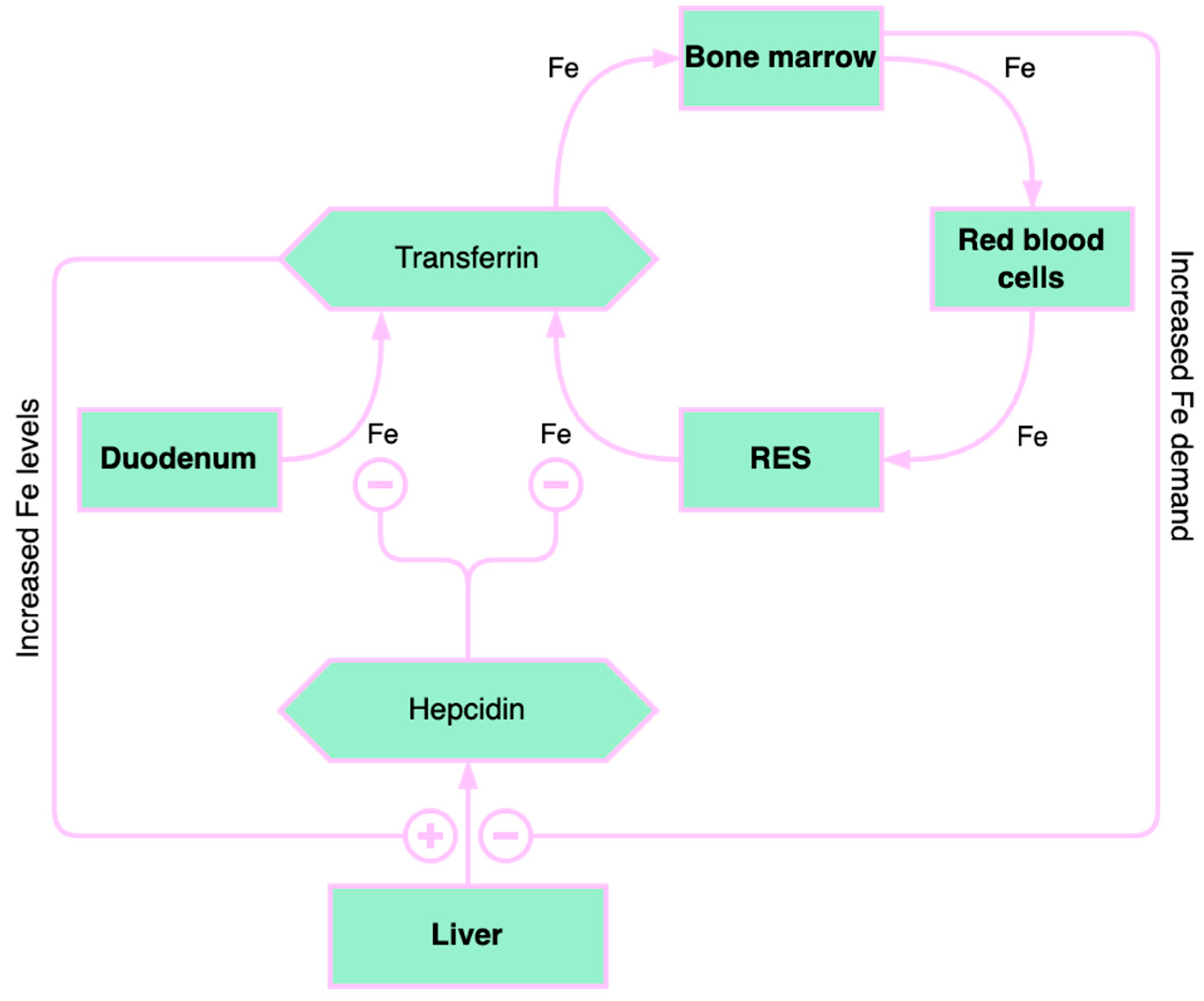
3.1. Liver Iron Storage and Chronic Liver Disease

The most important indicator of iron homeostasis is ferritin. The hepatocyte is the leading site for the synthesis of ferritin, as well as for the synthesis of transferrin, which is the main iron-binding protein. In CLD, an imbalance between iron deficiency and iron overload has been reported. Most of the iron amount is stored in ferritin, but hemosiderin is also used in case of severe iron overload [24]. Hepatic iron overload is associated with a worse outcome and with an increased risk of hepatocarcinoma [25].
In patients with severe CLD, there were described reduced levels of serum transferrin and iron and increased serum ferritin levels correlated with higher values of serum liver enzymes, which suggested the alleged role of inflammation in the pathogenesis of anemia in these patients [26].
In chronic inflammatory conditions, including chronic viral hepatitis, autoimmune hepatitis, chronic kidney and heart failure, organ rejection, inflammatory bowel diseases, endocrine diseases, systemic autoimmune diseases, and neoplasms, the impairment of erythropoiesis is secondary to an altered iron hemostasis, a cytokine-mediated process [27]. Proinflammatory cytokines, such as tumor necrosis factor (TNF-α) and interleukin-1, 6, and 10, are the main factors limiting iron availability in erythrocyte precursor cells, with a consequent alteration of erythropoiesis both in nonalcoholic steatohepatitis (NASH) and in chronic viral hepatitis [28].
In NASH, an increased expression of proinflammatory cytokines and hepcidin was described by Handa et al., who associated obesity-induced inflammation with increased serum hepcidin, which induces ACD [29].
ACD, also referred to as inflammatory anemia, is initially characterized by mild to moderate normochromic normocytic anemia and features of iron deficiency anemia (IDA), such as decreased reticulocytes and transferrin saturation. Anemia in CLD is primarily determined by IDA and ACD [30].

3.2. The Causes of Iron Deficiency in Chronic Liver Diseases

Patients with liver cirrhosis present a higher risk of gastrointestinal bleeding due to the decreased production of coagulation factors and thrombocytopenia, which, correlated with the degree of portal hypertension, defines the risk of bleeding in the gastrointestinal tract [31].
The most common cause of ID in advanced CLD is chronic gastrointestinal bleeding. ID causes IDA, a common pathology in this population [32,33]. Acute bleeding usually occurs through the rupture of esophageal varices or a hemorrhagic peptic ulcer [34]. Chronic blood loss is correlated with portal hypertensive gastropathy (PHG) and gastric antral vascular ectasia (GAVE), two primary gastric mucosa lesions caused by vascular ectasia. Histologically, it is defined by vascular dilatation in the mucosa and submucosa of the gastric body and fundus, without significant inflammation. The severity of PHG correlates to the stage of portal hypertension and the blood flow of the gastric mucosa or other local factors, which may determine its pathogenesis [35]. GAVE (“watermelon stomach”) is a rare condition that causes about 4% of non-variceal gastrointestinal bleeding [36]. Endoscopically, it is characterized by vascular ectasia with a sinuous appearance at the level of the longitudinal folds of the gastric antrum. The formation of GAVE is influenced by the presence of liver failure, which contributes to its pathogenesis, GAVE being more common in severe liver diseases. Other diseases besides liver failure can also cause the development of a “watermelon stomach”, such as chronic kidney injury, connective tissue diseases, or bone marrow transplantation. The pathogenesis is unclear; vasodilator agents such as prostaglandin E2 are not metabolized by the liver. Mechanical stress and hypomobility of the stomach could be other factors associated with GAVE [37].
The most common etiologies of lower gastrointestinal bleeding in liver cirrhosis are hemorrhoidal disease and portal hypertensive colopathy. Portal hypertensive enteropathy may be an occult source of chronic gastrointestinal bleeding in liver cirrhosis [38].

4. Pathophysiological Mechanisms of Anemia in Alcoholic Liver Disease

Alcohol consumption contributes to anemia through multiple mechanisms (Figure 3). Alcohol directly affects the hematopoietic cells of the bone marrow, manifesting as pancytopenia, reversible after stopping alcohol consumption. Folate deficiency, found in these patients, leads to macrocytic anemia. Alcohol decreases serum folate levels through various mechanisms, such as increased urinary folate excretion and decreased jejunal absorption [39]. Through its effects on methionine metabolism, folate deficiency contributes to the progression of liver disease by influencing DNA synthesis and stability as well as the epigenetic regulation of gene expression that is implicated in causing liver damage [40].
Patients with severe liver injury exhibit deficiencies of folic acid and vitamin B12, causing macrocytic anemia. Vitamin B12 and folate are necessary for the synthesis of thymidylate and purines. Therefore, their deficiencies lead to defects in DNA synthesis with subsequent macrocytic anemia [41]. Additionally, increased cholesterol deposition on the erythrocyte membrane, which in turn raises the erythrocyte surface, may contribute to macrocytic anemia in liver diseases [41,42].
Severe liver failure is also correlated with hemolytic anemias, where excessive destruction of erythrocytes and increased reticulocytes are described [43]. Immature erythrocytes are about 20% larger than mature erythrocytes, which explains macrocytosis.
Acetaldehyde, the product resulting from alcohol oxidation in the liver, increases the degradation of pyridoxal phosphate, which is a cofactor of aminolevulinic acid synthetase, an enzyme with a catalytic role in heme synthesis. Consequently, alcohol causes the inadequate production of heme and consequently causes the accumulation of iron in the mitochondria, determining the formation of sideroblasts [44]. Sideroblastic anemia is encountered in approximately 25–30% of alcoholic patients, reversible when consumption is stopped. Sideroblastic anemia occurs in alcoholic patients with concomitant folic acid deficiency [45].
The formation of acanthocytes (spur cells) is another mechanism of anemia in alcoholic liver disease [46]. The abnormal lipid metabolism leads to defects in lipid membrane composition and consequent formation of acanthocytes. Spur cell anemia (SCA) is present in up to 31% of patients with advanced liver cirrhosis, an independent negative predictive factor associated with an increased mortality risk [10,47]. SCA prognosis is extremely poor, with liver transplantation being the only effective treatment [48,49].

5. Portal Hypertension

An increase in vascular resistance to flow and an increase in portal flow are the pathophysiologic drivers of portal hypertension. Angiogenesis in liver diseases, which changes the diameters of the branches of the portal vein, achieves dramatic increases in vascular resistance. In liver cirrhosis, through architectural remodeling, a fixed, mechanical compression occurs, secondary to fibrosis, with changes in microcirculation (sinusoidal portal hypertension) and sinusoid capillarization, as well as dynamic changes due to a functional contraction of myofibroblasts, stellate cells, and smooth muscles from the venous wall. Vasodilatation with increased venous flow manifests mainly in the splanchnic territory and is caused by the local production of NO and PGI2. In the advanced stages of portal hypertension, vasodilatation is so marked that a decrease in adequate blood volume and systemic arterial hypotension are reported. As a result, vasoconstrictor and anti-natriuretic factors are activated, with consequent retention of water and salt, including ascitic-edematous syndrome. The pressure in the intestinal capillaries is altered, ascites and edema are formed, and kidney changes appear with the progression of the disease, especially vasoconstriction and decrease in free water excretion, inducing dilutional hyponatremia and hepatorenal syndrome [50].
The most important and severe portal hypertension complication is acute gastrointestinal bleeding, a common and possibly severe evolution of portal hypertension [51,52]. This is usually the result of esophageal varices rupture, the cause of approximately 70% of upper gastrointestinal bleeding [53]. Acute hemorrhage can result in iron deficiency anemia and severe hypovolemia.

6. Hematological Hypersplenism

Hypersplenism, secondary to splenomegaly, is associated with peripheral cytopenia, any of the cell lines being affected by hypersplenism, most frequently platelet, and the least sensitive, erythrocytes [54]. Multiple theories have been postulated as the pathophysiology of cytopenia in hypersplenism. The most acknowledged theory associates it with an increased volume of blood passing through the spleen and, consequently, increased cellular destruction [54]. As reported by Shah et al., the phagocytic activity of the spleen increases with its size, causing the destruction of erythrocytes and the appearance of anemia [55]. Due to hypersplenism, in liver cirrhosis patients the hemoglobin value does not increase after blood transfusions as in patients without liver cirrhosis, a finding documented by Tan CH et al. [56].
Pancytopenia secondary to hypersplenism is characterized by hemolytic anemia, due to intrasplenic destruction of erythrocytes, thrombocytopenia, and leukopenia [57].

7. Abnormalities of Hemostasis in Liver Diseases

The main hemostasis anomalies observed in CLD are presented in Table 1.

7.1. Primary Hemostasis—Platelet Count and Function

The primary hematological abnormality seen in patients with liver cirrhosis is thrombocytopenia. Up to 77.9% of patients experience it, making it a prognostic severity factor [58]. Reduced hepatic thrombopoietin (TPO) production and bone marrow suppression are linked to decreased platelet production. The liver is the main producer of TPO, the hormone that regulates platelet synthesis. Due to inadequate production and increased splenic degradation, the serum level of TPO is typically decreased in chronic liver diseases [59,60,61]. Platelet anisocytosis is also common [62].
The mechanisms of thrombocytopenia are different, including immunological destruction, increased consumption of platelets, bacterial translocation, and infection, requiring a careful differential diagnosis [63,64]. Immunologic destruction is more commonly found in primary biliary cholangitis, but can also be found in other etiologies of CLD. Increased consumption of platelets has been described in association with disseminated vascular coagulation [65]. Heparin-induced thrombocytopenia should also be considered in patients treated with heparin due to overlapping thrombotic complications [66]. Platelet function is affected in patients with liver cirrhosis, but the literature data are discordant. Some studies suggest a decrease in platelet function, and others hyperactivity [62].

7.2. Secondary Hemostasis—Procoagulant and Anticoagulant Factors

Normal coagulation mechanisms consist of interaction between pro- and anticoagulation factors. CLD is associated with an inefficient or inadequate synthesis of both types of factors, these patients being able to develop both hemorrhagic and increased thromboembolic complications [62,67,68]. Except for the VIII factor, all clotting factors can be deficient in liver disease [69].
Reduced production of fibrinogen or increased consumption as a result of disseminated intravascular coagulation cause fibrinogen deficiency in advanced CLD, such as cirrhosis [69]. Dysfibrinogenemia may occur due to sialic acid residues affecting fibrin polymerization in addition to decreased levels [70].
Vitamin K-dependent factors, such as prothrombin (II factors), VII, IX, X, protein C, and protein S, can become even more deficient as a result of inadequate intake or malabsorption of vitamin K. As a result, a lack of vitamin K-dependent proteins can lead to an increased risk of thrombosis and bleeding [71].
The liver also produces endogenous coagulation inhibitors, in addition to synthesizing coagulation factors. As a result, CLD has been linked to decreased levels of anticoagulant factors such as protein C, protein S, and antithrombin [71].

7.3. The Fibrinolytic System

Fibrinolysis is altered in patients with liver cirrhosis and may determine changes in pro- and antifibrinolytic factors [72]. The mechanisms of fibrinolysis impairment are multiple. A decrease in the circulation of coagulation factors and fibrinolysis products and an impairment of production have been described. Increased levels of tPA (tissue plasminogen activator), decreased levels of XIII factors, TAFI (thrombin-activatable fibrinolysis inhibitor), and increased fibrin degradation products are the results of the aforementioned mechanisms [73].
An amount of 30% of cirrhotic patients experience hyperfibrinolysis [62]. This complicates the outcomes of patients with liver cirrhosis by impeding primary hemostasis through the appearance of upper digestive hemorrhage through variceal bleeding [74]. Hyperfibrinolysis is overlapped with accelerated intravascular coagulation, an entity similar to disseminated intravascular coagulation, that can occur in ACLD [75].

8. Hemodilution Anemia

Hydrosaline retention causes a low hematocrit secondary to hemodilution. Such a phenomenon appears in hypervolemic states, described in pregnancy, heart failure, administration of excess fluids, and liver cirrhosis [76]. Therefore, the cause of anemia is an increase in plasma volume, rather than an absolute reduction in erythrocyte mass. Inhibition of the basal relaxation factor derived from the endothelium is reduced, as a response to chronic anemia, which leads to vasodilation. A compensatory mechanism, by increasing antidiuretic hormone, sodium, and water retention, appears to increase cardiac output [77]. Anemia-induced hypervolemia amplifies the hyperdynamic state brought on by pulmonary hypertension.

9. Aplastic Anemia in Chronic Liver Diseases

Aplastic anemia can occur during hepatitis. It represents a well-known and rare phenomenon, described as pancytopenia and hypocellular marrow, described in the evolution of acute hepatitis, two to three months after the acute infection. All infections with hepatotropic viruses, specifically viruses A, B, C, D, E, and G, have been associated with the condition [78]. Pancytopenia secondary to aplastic anemias can be severe and even lethal [79]. Other viruses, such as B19 parvovirus and Epstein–Barr virus, can cause aplastic anemia. B19 parvovirus can cause severe acute hepatitis associated with acute liver failure and aplastic anemia. Cytotoxic T cells (CD8+), which see hematopoietic cells and liver antigens as similar targets and kill bone marrow cells as a result, are linked to the pathophysiology of aplastic anemia in acute hepatitis. Gamma interferon mediates this process [80]. By mediating interferon γ or cytokine cascade, the incriminating viruses induce lymphocyte activation and apoptosis of hematopoietic cells in the bone marrow [81].

10. Treatment-Associated Anemia in Chronic Viral Hepatitis

During combined therapy for HCV infection, there were several photogenic mechanisms of anemia, ribavirin and interferon being the drugs incriminated. Anemia occurred mainly because of ribavirin-induced hemolysis [82]. Although ribavirin-induced anemia may be reversible by reducing the dose or complete treatment interruption, this pathological association aggravated the prognosis by seriously decreasing the rate of sustained virologic response. Another possible mechanism could be ATP depletion during ribavirin phosphorylation, leading to oxidative stress in erythrocytes and subsequent hemolysis [83]. The antiviral efficacy of ribavirin was improved using viramidine, a ribavirin prodrug selectively absorbed by the liver, implicitly reducing the risk of hemolytic anemia [84].
The administration of interferon contributed to the anemia mechanism by inducing bone marrow suppression and by decreasing compensatory reticulocytosis, as well as ribavirin-induced hemolytic anemia [84].
Direct-acting antivirals (DAAs) are new antiviral agents with high efficacy, regardless of genotype, which have revolutionized HCV treatment, having excellent safety, better tolerance, and lower side effects, including anemia.

11. Anemia in Rare Liver Diseases

11.1. Zieve Syndrome

Hemolytic anemia, cholestatic jaundice, and transient hyperlipidemia are the three symptoms of the rarely recognized condition known as Zieve syndrome, which is brought on by excessive alcohol consumption. Hemolytic anemia in these patients is due to hyperlipidemia and the presence of an abnormal lipid, possibly lysolecithin [85]. Additionally, alcohol-induced vitamin E deficiency can result in enzyme instability and erythrocyte hemolysis because it lowers polyunsaturated fatty acid levels and results in reduced erythrocyte glutathione oxidation [86]. A negative Coombs test, an essential feature in these patients, indicates a low probability that the anemia is autoimmune hemolytic anemia and thus insensitive to glucocorticoid therapy [85].

11.2. Wilson Disease

Wilson disease, an autosomal recessive disorder of copper transport known as hepatolenticular degeneration, is brought on by mutations in the ATP7B gene, which is found on the long arm of chromosome 13 (13q14.1). Although excessive copper is deposited in the liver, eyes, nervous system, and other tissues in Wilson disease, the primary clinical symptoms are typically caused by liver (42%) and neurological (34%) injury [87]. When the diagnosis of fulminant liver failure due to Wilson disease is assumed, Coombs-negative hemolytic anemia with features of acute intravascular hemolysis should be suspected [88].

11.3. Hereditary Hemochromatosis

Hereditary hemochromatosis is a genetic disease of iron metabolism, with autosomal recessive transmission. It is characterized by increased iron absorption in the duodenum deposited in the liver, heart, pancreas, thyroid, gonads, hypothalamus, joints, and skin. The excessive iron amount in the target organs is clinically manifested by hepatomegaly (95%), splenomegaly (50%), portal hypertension (ascites, esophageal varices, hypertensive gastropathy, and encephalopathy), skin hyperpigmentation (over 90%), glucose intolerance and diabetes (65%), and arthropathy (25–50%). The disease manifests with diverse clinical pathologies and syndromes, such as diabetes mellitus, chondrocalcinosis, erectile dysfunction, porphyria cutanea tarda, heart failure, liver cirrhosis, and hepatocellular carcinoma [89]. The association of anemia with hemochromatosis still has to be elucidated entirely in comprehensive studies describing cases of aplastic and megaloblastic anemia [90].

11.4. Primary Biliary Cholangitis

Primary biliary cholangitis is a liver condition that progresses over time and has an autoimmune cause. Possible factors associated with the onset and perpetuation of biliary duct injury are genetic, environmental, and immunologic. The condition is characterized by lesions of the interlobular and septal biliary ducts [91], which lead to the accumulation of bile and toxins (cholestasis) and in time to the destruction of liver tissue, fibrosis, and cirrhosis. Most patients present antimitochondrial antibodies (over 90%) directed against an enzymatic autoantigen (mitochondrial pyruvate dehydrogenase complex PDC-E2). Recent studies describe a genetic predisposition involving IL12A and IL12RB2 gene polymorphisms, the gene that codes for CD 101, and other genomic regions involved in cytokine regulation [92]. The etiological role of a gram-negative bacterium, Novosphingobium aromaticivorans, through molecular mimicry, by cross-reaction between bacterial and mitochondrial self-proteins, was also revealed. Environmental factors possibly incriminated are smoking and estrogens from oral contraceptives [92].
Being an autoimmune disease, primary biliary cirrhosis is associated with many other conditions due to similar immune mechanisms, such as Sjogren’s or Sicca syndrome, scleroderma, lupus, polymyositis, rheumatoid arthritis, osteoporosis, autoimmune thyroiditis, kidney injury, celiac disease, and rectocolitis. Biermer’s or hemolytic anemia is encountered in the evolution of primary biliary cholangitis. Because mild anemia is usually less symptomatic and easily overlooked, pernicious anemia should be suspected when primary biliary cholangitis is associated with macrocytic anemia [93].
There have been few reports of a connection between primary biliary cholangitis and warm autoimmune hemolytic anemia (wAIHA). Regardless of the cause, hemolysis is known to be present in more than 50% of CLD patients. Because primary biliary cholangitis is frequently associated with many autoimmune diseases, wAIHA may be an autoimmune association in patients with primary biliary cholangitis [94].

11.5. Alpha 1 Antitrypsin Deficiency

Lung and liver damage can result from the genetic disorder known as alpha 1 antitrypsin deficiency (AATD). The serpin family’s acute-phase protein alpha 1 antitrypsin (AAT) is connected to several inflammatory cascades primarily through protein interaction [95]. It is primarily synthesized in the liver, released into the bloodstream, and transported to specific tissues where it is concentrated, especially the bronchial epithelium [96,97].
Liver disease has two types: the child-specific form, hepatitis and cholestasis, occurs in 10% of children with AATD (usually a self-limiting phase), and the adult form, liver cirrhosis, which occurs in 30–50% of cases, especially in men [98]. The histopathological examination identifies positive PAS cell inclusions in the hepatocytic endoplasmic reticulum. Rarely, liver injury can result in malignant tumors, most often (but not exclusively) in the cirrhotic liver [99].
Acute phase protein AAT inhibits transferrin receptor binding and transferrin internalization in reticulocytes in a dose-dependent and competitive mechanism. In addition to this effect, AAT reduces the growth and proliferation of early erythroid progenitor cells (BFU-E). Related findings suggest that AAT could mediate the characteristic changes of ACD [100].
Types of anemia encountered in rare liver diseases are summarized in Table 2.

12. Conclusions

Anemia is a common finding in liver disease. The underlying mechanisms are multiple, requiring specialized investigations and treatments. The most frequent causes of anemia in CLD are IDA, ACD, hypersplenism, and gastrointestinal bleeding. Iron homeostasis results from a complex mechanism and can be notably perturbed in patients with CLD. The concomitant presence of inflammation greatly influences biological markers, often challenging diagnosis. Future research is required to substantiate the role of iron homeostasis proteins in the pathophysiology of CLD, which is still under debate. Other mechanisms of anemia are represented by aplastic anemia secondary to viral hepatitis. Anemia in patients with alcoholic liver disease may result from a variety of alcohol-related effects, including malabsorption, malnutrition, and direct toxic effects. Each etiology of anemia has a unique pathogenesis that necessitates a complex diagnostic process while also necessitating a thorough therapeutic strategy.

Author Contributions

Conceptualization, C.M.M., D.P., M.P., A.O.D. and P.M.; methodology, C.M.M., A.O.D., R.M. and D.M.F.; software, A.R., A.I.S.P., C.M.V., I.C.M. and G.A.I.; validation, D.P., A.I.S.P., M.P., A.R. and C.M.M.; formal analysis, D.P., C.M.V., R.M., A.I.S.P., I.C.M., G.A.I., D.M.F. and A.R.; investigation, M.P., A.O.D., A.R., A.I.S.P., C.M.V., R.M., I.C.M., D.M.F. and P.M.; resources, C.M.M.; data curation, I.C.M., A.R. and D.P.; writing—original draft preparation, C.M.M., D.P., A.R., A.I.S.P., C.M.V., R.M., I.C.M., G.A.I. and D.M.F.; writing—review and editing, M.P., A.O.D. and P.M.; visualization, A.I.S.P.; supervision, M.P.; project administration, C.M.M.; funding acquisition, C.M.M. All authors have read and agreed to the published version of the manuscript.

Funding

This study was supported by internal grant no. 26/510/2 May 2023 “Study of the effect of COVID-19 pandemic on quality of life in patients with chronic liver disease” of the University of Medicine and Pharmacy of Craiova, Romania.

Institutional Review Board Statement

Not applicable.

Informed Consent Statement

Not applicable.

Data Availability Statement

Not applicable.

Conflicts of Interest

The authors declare no conflict of interest.

References

  1. Maruyama, S.; Hirayama, C.; Yamamoto, S.; Koda, M.; Udagawa, A.; Kadowaki, Y.; Inoue, M.; Sagayama, A.; Umeki, K. Red blood cell status in alcoholic and non-alcoholic liver disease. J. Lab. Clin. Med. 2001, 138, 332–337. [Google Scholar] [CrossRef]
  2. Mathurin, S.A.; Agüero, A.P.; Dascani, N.A.; Prestera, J.A.; Gianserra, C.; Londero, E.; Chiorra, C. Anemia in hospitalized patients with cirrhosis: Prevalence, clinical relevance, and predictive factors. Acta Gastroenterol. Latinoam. 2009, 39, 103–111. [Google Scholar]
  3. Qamar, A.A.; Grace, N.D.; Groszmann, R.J.; Garcia–Tsao, G.; Bosch, J.; Burroughs, A.K.; Ripoll, C.; Maurer, R.; Planas, R.; Escorsell, A.; et al. Incidence, prevalence, and clinical significance of abnormal hematologic indices in compensated cirrhosis. Clin. Gastroenterol. Hepatol. 2009, 7, 689–695. [Google Scholar] [CrossRef] [Green Version]
  4. Kalaitzakis, E.; Josefsson, A.; Castedal, M.; Henfridsson, P.; Bengtsson, M.; Andersson, B.; Björnsson, E. Hepatic encephalopathy is related to anemia and fat-free mass depletion in liver transplant candidates with cirrhosis. Scand. J. Gastroenterol. 2013, 48, 577–584. [Google Scholar] [CrossRef]
  5. Reiberger, T.; Püspök, A.; Schoder, M.; Baumann-Durchschein, F.; Bucsics, T.; Datz, C.; Dolak, W.; Ferlitsch, A.; Finkenstedt, A.; Graziadei, I.; et al. Austrian consensus guidelines on the management and treatment of portal hypertension (Billroth III). Wien. Klin. Wochenschr. 2017, 129 (Suppl. 3), 135–158. [Google Scholar] [CrossRef] [Green Version]
  6. Ripoll, C.; Garcia-Tsao, G. The management of portal hypertensive gastropathy and gastric antral vascular ectasia. Dig. Liver Dis. 2011, 43, 345–351. [Google Scholar] [CrossRef]
  7. Luo, J.-C.; Leu, H.-B.; Hou, M.-C.; Huang, C.-C.; Lin, H.-C.; Lee, F.-Y.; Chang, F.-Y.; Chan, W.-L.; Lin, S.-J.; Chen, J.-W. Cirrhotic patients at increased risk of peptic ulcer bleeding: A nationwide population-based cohort study. Aliment. Pharmacol. Ther. 2012, 36, 542–550. [Google Scholar] [CrossRef] [Green Version]
  8. Gado, A.; Ebeid, B.; Axon, A. Prevalence and outcome of peptic ulcer bleeding in patients with liver cirrhosis. Alex. J. Med. 2014, 50, 143–148. [Google Scholar] [CrossRef]
  9. Alexopoulou, A.; Vasilieva, L.; Kanellopoulou, T.; Pouriki, S.; Soultati, A.; Dourakis, S.P. Presence of spur cells as a highly predictive factor of mortality in patients with cirrhosis. J. Gastroenterol. Hepatol. 2014, 29, 830–834. [Google Scholar] [CrossRef]
  10. Vassiliadis, T.; Mpoumponaris, A.; Vakalopoulou, S.; Giouleme, O.; Gkissakis, D.; Grammatikos, N.; Soufleris, K.; Kakafika, A.; Tziomalos, K.; Patsiaoura, K.; et al. Spur cells and spur cell anemia in hospitalized patients with advanced liver disease: Incidence and correlation with disease severity and survival. Hepatol. Res. 2010, 40, 161–170. [Google Scholar] [CrossRef]
  11. Gonzalez-Casas, R.; Jones, E.A.; Moreno-Otero, R. Spectrum of anemia associated with chronic liver disease. World J. Gastroenterol. 2009, 15, 4653–4658. [Google Scholar] [CrossRef]
  12. Gkamprela, E.; Deutsch, M.; Pectasides, D. Iron deficiency anemia in chronic liver disease: Etiopathogenesis, diagnosis and treatment. Ann. Gastroenterol. 2017, 30, 405–413. [Google Scholar] [CrossRef] [PubMed]
  13. Gonzalez-Casas, R.; Garcia-Buey, L.; Jones, E.A.; Gisbert, J.P.; Moreno-Otero, R. Systematic review: Hepatitis-associated aplastic anaemia–a syndrome associated with abnormal immunological function. Aliment. Pharmacol. Ther. 2009, 30, 436–443. [Google Scholar] [CrossRef] [PubMed]
  14. Wang, S.Q.; Zhan, Y.Q.; Hu, X.; Zhuang, Y.P.; Liu, H.Q.; Hong, M.F.; Zhong, H.J. Anemia Is Associated with Disease Severity, Hepatic Complications, and Progression of Wilson Disease: A Retrospective Cohort Study. Dig Dis. 2023, 41, 632–640. [Google Scholar] [CrossRef]
  15. Les, I.; Doval, E.; Flavià, M.; Jacas, C.; Cárdenas, G.; Esteban, R.; Guardia, J.; Córdoba, J. Quality of life in cirrhosis is related to potentially treatable factors. Eur. J. Gastroenterol. Hepatol. 2010, 22, 221–227. [Google Scholar] [CrossRef]
  16. Güngör, G.; Akyıldız, M.; Keskin, M.; Solak, Y.; Gaipov, A.; Bıyık, M.; Çifçi, S.; Ataseven, H.; Polat, H.; Demir, A. Is there any potential or additive effect of anemia on hepatorenal syndrome? Turk. J. Gastroenterol. 2016, 27, 273–278. [Google Scholar] [CrossRef] [PubMed]
  17. Milward, E.; Johnstone, D.; Trinder, D.; Ramm, G.; Olynyk, J. The nexus of iron and inflammation in hepcidin regulation: SMADs, STATs, and ECSIT. Hepatology 2007, 45, 253–256. [Google Scholar] [CrossRef] [Green Version]
  18. Wang, C.-Y.; Babitt, J.L. Liver iron sensing and body iron homeostasis. Blood 2019, 133, 18–29. [Google Scholar] [CrossRef] [Green Version]
  19. Tsochatzis, E.; Papatheodoridis, G.V.; Koliaraki, V.; Hadziyannis, E.; Kafiri, G.; Manesis, E.K.; Mamalaki, A.; Archimandritis, A.J. Serum hepcidin levels are related to the severity of liver histological lesions in chronic hepatitis C. J. Viral Hepat. 2010, 17, 800–806. [Google Scholar] [CrossRef]
  20. Lyberopoulou, A.; Chachami, G.; Gatselis, N.K.; Kyratzopoulou, E.; Saitis, A.; Gabeta, S.; Eliades, P.; Paraskeva, E.; Zachou, K.; Koukoulis, G.K.; et al. Low serum hepcidin in patients with autoimmune liver diseases. PLoS ONE 2015, 10, e0135486. [Google Scholar] [CrossRef]
  21. Wang, X.H.; Cheng, P.P.; Jiang, F.; Jiao, X.Y. The effect of hepatitis B virus infection on hepcidin expression in hepatitis B patients. Ann. Clin. Lab. Sci. 2013, 43, 126–134. [Google Scholar]
  22. Lunova, M.; Trautwein, C.; Strnad, P.; Nahon, P. Reply to: “Hepatic hepcidin expression is decreased in cirrhosis and HCC”. J. Hepatol. 2015, 62, 979–980. [Google Scholar] [CrossRef] [PubMed] [Green Version]
  23. Ganz, T. Hepcidin. Rinsho Ketsueki 2016, 57, 1913–1917. [Google Scholar] [PubMed]
  24. Peyrin-Biroulet, L.; Williet, N.; Cacoub, P. Guidelines on the diagnosis and treatment of iron deficiency across indications: A systematic review. Am. J. Clin. Nutr. 2015, 102, 1585–1594. [Google Scholar] [CrossRef] [PubMed] [Green Version]
  25. Nahon, P.; Sutton, A.; Rufat, P.; Ziol, M.; Thabut, G.; Schischmanoff, P.; Vidaud, D.; Charnaux, N.; Couvert, P.; Ganne–Carrie, N.; et al. Liver iron, HFE gene mutations, and hepatocellular carcinoma occurrence in patients with cirrhosis. Gastroenterology 2008, 134, 102–110. [Google Scholar] [CrossRef] [PubMed] [Green Version]
  26. Drakesmith, H.; Prentice, A.M. Hepcidin and the iron-infection axis. Science 2012, 338, 768–772. [Google Scholar] [CrossRef] [Green Version]
  27. Biciuşcă, V.; Popescu, M.; Petrescu, I.O.; Stan, I.S.; Durand, P.; Petrescu, M.; Velea, R.; Traşcă, D.M.; Popescu, I.A.S.; Udriştoiu, I.; et al. Hepatic pathological features in naïve patients with chronic hepatitis C who have developed thyroid disorder. Rom. J. Morphol. Embryol. 2021, 61, 1085–1097. [Google Scholar] [CrossRef]
  28. Stan, I.S.; Biciuşcă, V.; Durand, P.; Petrescu, A.-M.; Oancea, D.M.; Ciuciulete, A.R.; Petrescu, M.; Udriştoiu, I.; Camen, G.-C.; Bălteanu, M.A.; et al. Diagnostic and prognostic significance of hepatic steatosis in patients with chronic hepatitis C. Rom. J. Morphol. Embryol. 2021, 62, 765–775. [Google Scholar] [CrossRef]
  29. Handa, P.; Maliken, B.D.; Nelson, J.E.; Hennessey, K.A.; Vemulakonda, L.A.; Morgan-Stevenson, V.; Dhillon, B.K.; Gupta, R.; Yeh, M.M.; Kowdley, K.V. Differencesin hepatic expression of iron, inflammation and stress related genes in patients with nonalcoholic steatohepatitis. Ann. Hepatol. 2017, 16, 77–85. [Google Scholar] [CrossRef]
  30. Scheiner, B.; Semmler, G.; Maurer, F.; Schwabl, P.; Bucsics, T.A.; Paternostro, R.; Bauer, D.; Simbrunner, B.; Trauner, M.; Mandorfer, M.; et al. Prevalence of and risk factors for anaemia in patients with advanced chronic liver disease. Liver Int. 2020, 40, 194–204. [Google Scholar] [CrossRef] [Green Version]
  31. Mallet, M.; Rudler, M.; Thabut, D. Variceal bleeding in cirrhotic patients. Gastroenterol. Rep. 2017, 5, 185–192. [Google Scholar] [CrossRef] [PubMed] [Green Version]
  32. Kate, V.; Sureshkumar, S.; Gurushankari, B.; Kalayarasan, R. Acute Upper Non-variceal and Lower Gastrointestinal Bleeding. J. Gastrointest. Surg. 2022, 26, 932–949. [Google Scholar] [CrossRef] [PubMed]
  33. Stein, J.; Connor, S.; Virgin, G.; Ong, D.E.; Pereyra, L. Anemia and iron deficiency in gastrointestinal and liver conditions. World J. Gastroenterol. 2016, 22, 7908–7925. [Google Scholar] [CrossRef] [PubMed]
  34. Kalafateli, M.; Triantos, C.K.; Nikolopoulou, V.; Burroughs, A. Non-variceal Gastrointestinal Bleeding in Patients with Liver Cirrhosis: A Review. Dig. Dis. Sci. 2012, 57, 2743–2754. [Google Scholar] [CrossRef]
  35. Kim, M.Y.; Choi, H.; Baik, S.K.; Yea, C.J.; Won, C.S.; Byun, J.W.; Park, S.Y.; Kwon, Y.H.; Kim, J.W.; Kim, H.S.; et al. Portal hypertensive gastropathy: Correlation with portal hypertension and prognosis in cirrhosis. Dig. Dis. Sci. 2010, 55, 3561–3567. [Google Scholar] [CrossRef]
  36. Selinger, C.P.; Ang, Y.S. Gastric antral vascular ectasia (GAVE): An update on clinical presentation, pathophysiology and treatment. Digestion 2008, 77, 131–137. [Google Scholar] [CrossRef]
  37. Spahr, L.; Villeneuve, J.-P.; Dufresne, M.-P.; Tassé, D.; Bui, B.; Willems, B.; Fenyves, D.; Pomier-Layrargues, G. Gastric antral vascular ectasia in cirrhotic patients: Absence of relation with portal hypertension. Gut 1999, 44, 739–742. [Google Scholar] [CrossRef] [Green Version]
  38. Canlas, K.R.; Dobozi, B.M.; Lin, S.; Smith, A.D.; Rockey, D.C.; Muir, A.J.; Agrawal, N.M.; Poleski, M.H.; Patel, K.; McHutchison, J.G. Using Capsule Endoscopy toIdentify GI Tract Lesions in Cirrhotic Patients With Portal Hypertension and Chronic Anemia. J. Clin. Gastroenterol. 2008, 42, 844–848. [Google Scholar] [CrossRef]
  39. Halsted, C.H.; Villanueva, J.A.; Devlin, A.M.; Chandler, C.J. Metabolic interactions of alcohol and folate. J. Nutr. 2002, 132, 2367S–2372S. [Google Scholar] [CrossRef] [Green Version]
  40. Medici, V.; Halsted, C.H. Folate, alcohol, and liver disease. Mol. Nutr. Food Res. 2012, 57, 596–606. [Google Scholar] [CrossRef] [Green Version]
  41. Green, R.; Dwyre, D.M. Evaluation of macrocytic anemias. Semin. Hematol. 2015, 52, 279–286. [Google Scholar] [CrossRef] [PubMed]
  42. Grattagliano, I.; Calamita, G.; Cocco, T.; Wang, D.Q.; Portincasa, P. Pathogenic role of oxidative and nitrosative stress in primary biliary cirrhosis. World J. Gastroenterol. 2014, 20, 5746–5759. [Google Scholar] [CrossRef] [PubMed]
  43. Marginean, C.M.; Pirscoveanu, D.; Popescu, M.; Vasile, C.M.; Docea, A.O.; Mitruț, R.; Mărginean, I.C.; Iacob, G.A.; Firu, D.M.; Mitruț, P. Challenges in Diagnosis and Therapeutic Approach of Acute on Chronic Liver Failure—A Review of Current Evidence. Biomedicines 2023, 11, 1840. [Google Scholar] [CrossRef] [PubMed]
  44. Wiley, J.S.; Moore, M.R. Heme Biosynthesis and Its Disorders: Porphyrias and Sideroblastic Anemias; Churchill Livingstone: London, UK, 2000. [Google Scholar]
  45. Holloway, R. Sideroblastic anaemia secondary to chronic alcoholism: A case study and review. N. Z. J. Med. Lab. Sci. 2007, 61, 69. [Google Scholar]
  46. Privitera, G.; Meli, G. An unusual cause of anemia in cirrhosis: Spur cell anemia, a case report withreview of literature. Gastroenterol. Hepatol. Bed Bench 2016, 9, 335. [Google Scholar]
  47. Roy, A.; Rodge, G.; Goenka, M.K. Spur Cell Anaemia in Cirrhosis: A Narrative Review. J. Clin. Exp. Hepatol. 2023, 13, 500–508. [Google Scholar] [CrossRef]
  48. Gerber, B.; Stussi, G. Reversibility of spur cell anemia. Blood 2011, 118, 4304. [Google Scholar] [CrossRef] [Green Version]
  49. Nakatsuka, H.; Sato, Y.; Oya, H.; Yamamoto, S.; Kurosaki, I.; Shirai, Y.; Hatakeyama, K. A case of spur cell anemia resolved by living donor liver transplantation. Kanzo 2007, 48, 181–186. [Google Scholar] [CrossRef]
  50. Iwakiri, Y. Pathophysiology of portal hypertension. Clin. Liver Dis. 2014, 18, 281–291. [Google Scholar] [CrossRef] [Green Version]
  51. Garcia-Pagan, J.C.; De Gottardi, A.; Bosch, J. Review article: The modern management of portal hypertension--primary and secondary prophylaxis of variceal bleeding in cirrhotic patients. Aliment. Pharmacol. Ther. 2008, 28, 178–186. [Google Scholar] [CrossRef]
  52. Albillos, A. Preventing first variceal hemorrhage in cirrhosis. J. Clin. Gastroenterol. 2007, 41 (Suppl. 3), S305–S311. [Google Scholar] [CrossRef]
  53. Abraldes, J.G.; Bosch, J. The treatment of acute variceal bleeding. J. Clin. Gastroenterol. 2007, 41 (Suppl. 3), S312–S317. [Google Scholar] [CrossRef] [PubMed]
  54. Lv, Y.; Gong, X.; Xie, X.; Wang, B.; Yang, Y.; Li, Y. Clinical Study on the Relationship between Hematocytopenia and Splenomegaly Caused by Cirrhotic Portal Hypertension. Cell Biochem. Biophys. 2014, 70, 355–360. [Google Scholar] [CrossRef]
  55. Shah, S.H.; Hayes, P.C.; Allan, P.L.; Nicoll, J.; Finlayson, N.D. Measurement of spleen size and its relation to hypersplenism and portal hemodynamics in portal hypertension due to hepatic cirrhosis. Am J Gastroenterol. 1996, 91, 2580–2583. [Google Scholar]
  56. Tan, C.H.; Hall, J.A.; Hammonds, K.; Dodlapati, J.; Linz, W.J.; Henderson, S.M. Relationship betweensplenomegaly and transfusion requirements in patients with cirrhosis. In Baylor University Medical Center Proceedings; Taylor & Francis: London, UK, 2020; Volume 34, pp. 44–48. [Google Scholar]
  57. Laffi, G.; Marra, F.; Tarquini, R.; Abbate, R. Coagulation defects in cirrhosis—Old dogmas not yet ready for burial. J. Thromb. Haemost. 2006, 4, 2068–2069. [Google Scholar] [CrossRef]
  58. Greer, J.P.; Rodgers, G.M.; Glader, B.; Arber, D.A.; Appelbaum, F.R.; Dispenzieri, A.; Fehniger, T.A.; Michaelis, L.; Leonard, J.P. Wintrobe’s Clinical Hematology, 14th ed.; Wolters Kluwer: Philadelphia, PA, USA, 2019; pp. 1212–1214. [Google Scholar]
  59. Giannini, E.G.; Peck-Radosavljevic, M. Platelet Dysfunction: Status of Thrombopoietin in Thrombocytopenia Associated with Chronic Liver Failure. Semin. Thromb. Hemost. 2015, 41, 455–461. [Google Scholar] [PubMed]
  60. Martin, T.G.; Somberg, K.A.; Meng, Y.G.; Cohen, R.L.; Heid, C.A.; de Sauvage, F.J.; Shuman, M.A. Thrombopoietin levels in patients with cirrhosis before and after orthotopic liver transplantation. Ann. Intern. Med. 1997, 127, 285–288. [Google Scholar] [CrossRef]
  61. Eissa, L.A.; Gad, L.S.; Rabie, A.M.; El-Gayar, A.M. Thrombopoietin level in patients with chronic liver disease. Ann. Hepatol. 2008, 7, 235–244. [Google Scholar] [CrossRef]
  62. European Association for the Study of the Liver. EASL Clinical Practice Guidelines for the management of patients with decompensated cirrhosis. J. Hepatol. 2018, 69, 406–460, Erratum in J. Hepatol. 2018, 69, 1207. [Google Scholar] [CrossRef] [Green Version]
  63. Kim, K.; Ong, F.; Varadi, G.; Gupta, S.; Jorge, V.M. A Systematic Review and Meta-Analysis of Safety and Efficacy for Pre-Procedural Use of Thrombopoietin Receptor Agonists in Hepatic Cirrhosis Patients. Blood 2019, 134, 1094. [Google Scholar] [CrossRef]
  64. Mitchell, O.; Feldman, D.M.; Diakow, M.; Sigal, S.H. The pathophysiology of thrombocytopenia in chronic liver disease. Hepatic Med. Évid. Res. 2016, 8, 39–50. [Google Scholar] [CrossRef] [Green Version]
  65. Christodoulou, D.; Katsanos, K.; Zervou, E.; Theopistos, V.; Papathanasopoulos, A.; Christou, L.; Tsianos, E.V. Platelet IgG antibodies are significantly increased in chronic liver disease. Ann. Gastroenterol. 2011, 24, 47–52. [Google Scholar]
  66. Ikura, Y.; Ohsawa, M.; Okada, M.; Iwai, Y.; Wakasa, K. The significance of platelet consumption in the development of thrombocytopenia in patients with cirrhosis. Am. J. Med. Sci. 2013, 346, 199–203. [Google Scholar] [CrossRef] [PubMed]
  67. Voican, I.; Onisâi, M.; Nicolescu, A.; Vlădăreanu, A.M.; Vlădăreanu, R. Heparin induced thrombocytopenia: A review. Farmacia 2012, 60, 773–784. [Google Scholar]
  68. Koganov, E.S.; Carmichael, S.L.; Forde, E.E.; Freliger, A.L.; Michelson, A.D. Platelet Function in Thrombocytopenic Patients with Chronic Liver Disease. Blood 2017, 130, 2314. [Google Scholar] [CrossRef]
  69. Tischendorf, M.; Miesbach, W.; Chattah, U.; Chattah, Z.; Maier, S.; Welsch, C.; Zeuzem, S.; Lange, C.M. Differential Kinetics of Coagulation Factors and Natural Anticoagulants in Patients with Liver Cirrhosis: Potential Clinical Implications. PLoS ONE 2016, 11, e0155337. [Google Scholar] [CrossRef] [PubMed]
  70. Lisman, T.; Leebeek, F.W.G.; de Groot, P.G. Haemostatic abnormalities in patients with liver disease. J. Hepatol. 2002, 37, 280–287. [Google Scholar] [CrossRef] [PubMed]
  71. Girolami, A.; Ferrari, S.; Cosi, E.; Santarossa, C.; Randi, M.L. Vitamin K-dependent coagulation factors that may be responsible for both bleeding and thrombosis (FII, FVII, and FIX). Clin. Appl. Thromb. 2018, 24, 42S–47S. [Google Scholar] [CrossRef] [Green Version]
  72. Singhal, A.; Karachristos, A.; Bromberg, M.; Daly, E.; Maloo, M.; Jain, A.K. Hypercoagulability in end-stage liver disease: Prevalence and its correlation with severity of liver disease and portal vein thrombosis. Clin. Appl. Thromb. 2012, 18, 594–598. [Google Scholar] [CrossRef] [Green Version]
  73. Fisher, C.; Patel, V.C.; Stoy, S.H.; Singanayagam, A.; Adelmeijer, J.; Wendon, J.; Shawcross, D.; Lisman, T.; Bernal, W. Balanced haemostasis with both hypo- and hyper-coagulable features in critically ill patients with acute-on-chronic-liver failure. J. Crit. Care 2018, 43, 54–60. [Google Scholar] [CrossRef]
  74. Hartman, M.; Szalai, C.; Saner, F.H. Hemostasis in liver transplantation: Pathophysiology, monitoring, and treatment. World J. Gastroenterol. 2016, 22, 1541–1550. [Google Scholar] [CrossRef] [PubMed]
  75. Ferro, D.; Celestini, A.; Violi, F. Hyperfibrinogenolysis in liver disease. Clin. Liver Dis. 2009, 13, 21–31. [Google Scholar] [CrossRef]
  76. Drevon, L.; Maslah, N.; Soret-Dulphy, J.; Dosquet, C.; Ravdan, O.; Vercellino, L.; Belkhodja, C.; Parquet, N.; Brignier, A.C.; Schlageter, M.H.; et al. Anemia andhemodilution: Analysis of a single center cohort based on 2858 red cell mass measurements. Haematologica 2021, 106, 1167–1171. [Google Scholar] [CrossRef] [PubMed]
  77. Singer, C.E.; Vasile, C.M.; Popescu, M.; Popescu, A.I.S.; Marginean, I.C.; Iacob, G.A.; Popescu, M.D.; Marginean, C.M. Role of Iron Deficiency in Heart Failure—Clinical and Treatment Approach: An Overview. Diagnostics 2023, 13, 304. [Google Scholar] [CrossRef]
  78. Alshaibani, A.; Dufour, C.; Risitano, A.; de Latour, R.; Aljurf, M. Hepatitis-associated aplastic anemia. Hematol./Oncol. Stem Cell Ther. 2020, in press. [Google Scholar] [CrossRef] [PubMed]
  79. Rauff, B.; Idrees, M.; Shah, S.A.R.; Butt, S.; Butt, A.M.; Ali, L.; Hussain, A.; Ali, M. Hepatitis Associated Aplastic Anemia: A review. Virol. J. 2011, 8, 87. [Google Scholar] [CrossRef] [Green Version]
  80. Ikawa, Y.; Nishimura, R.; Kuroda, R.; Mase, S.; Araki, R.; Maeba, H.; Wada, T.; Toma, T.; Koizumi, S.; Yachie, A. Expansion of a liver-infiltratingcytotoxic T-lymphocyte clone in concert with the development of hepatitis-associated aplastic anaemia. Br. J. Haematol. 2013, 161, 599–602. [Google Scholar] [CrossRef] [PubMed]
  81. Young, N.S.; Calado, R.T.; Scheinberg, P. Current concepts in the pathophysiology and treatment of aplastic anemia. Blood 2006, 108, 2509–2519. [Google Scholar] [CrossRef]
  82. Cariani, E.; Pelizzari, A.M.; Rodella, A.; Gargiulo, F.; Imberti, L.; Manca, N.; Rossi, G. Immune-mediated hepatitis-associated aplastic anemia caused by the emergence of a mutant hepatitis B virus undetectable by standard assays. J. Hepatol. 2007, 46, 743–747. [Google Scholar] [CrossRef]
  83. De Franceschi, L.; Fattovich, G.; Turrini, F.; Ayi, K.; Brugnara, C.; Manzato, F.; Noventa, F.; Stanzial, A.M.; Solero, P.; Corrocher, R. Hemolytic anemia induced by ribavirin therapy in patients with chronic hepatitis C virus infection: Role of membrane oxidativedamage. Hepatology 2000, 31, 997–1004. [Google Scholar] [CrossRef]
  84. Van Vlierbergh, H.; Delanghe, J.R.; De Vos, M.; Leroux-Roel, G. Factors influencing ribavirin-induced hemolysis. J. Hepatol. 2001, 34, 911–916. [Google Scholar] [CrossRef] [PubMed]
  85. Lakshmipathy, A.; Krishnamani, R.; Felix, J. Unusual lipid and metabolic abnormalities secondary to alcohol abuse. Hosp. Physician 2004, 40, 43–46. [Google Scholar]
  86. Ampuero, J.; Romero-Gómez, M. Pharmacogenetics of ribavirin-induced anemia in hepatitis C. Pharmacogenomics 2016, 17, 1587–1594. [Google Scholar] [CrossRef] [PubMed]
  87. Senatore, F.J.; McDonald, K. Pitfalls of treating alcoholic hepatitis: Recognizing hemolytic anemia in Zieve’s syndrome. Am. J. Gastroenterol. 2016, 111, 577–579. [Google Scholar] [CrossRef]
  88. Grudeva-Popova, J.G.; Spasova, M.I.; Chepileva, K.G.; Zaprianov, Z.H. Acute hemolytic anemia as an initial clinical manifesta-tion of Wilson’s disease. Folia Med. 2000, 42, 42–46. [Google Scholar]
  89. Brake, S. Hemolytic Anemia Associated with Fulminant Hepatitis in Wilson’s Disease: Diagnosis and Management. Blood 2009, 114, 5088. [Google Scholar] [CrossRef]
  90. Sood, R.; Bakashi, R.; Hegade, V.S.; Kelly, S.M. Diagnosis and management of hereditary haemochromatosis. Br. J. Gen. Pr. 2013, 63, 331–332. [Google Scholar] [CrossRef] [Green Version]
  91. Khan, A.A.; Hadi, Y.; Hassan, A.; Kupec, J. Polycythemia and anemia in hereditary hemochromatosis. Cureus 2020, 12, e7607. [Google Scholar] [CrossRef] [Green Version]
  92. Carey, E.J.; Ali, A.H.; Lindor, K.D. Primary biliary cirrhosis. Lancet 2015, 386, 1565–1575, Erratum in Lancet 2015, 386, 1536. [Google Scholar] [CrossRef]
  93. Hirschfield, G.M.; Gershwin, M.E. The immunobiology and pathophysiology of primary biliary cirrhosis. Annu. Rev. Pathol. Mech. Dis. 2013, 8, 303–330. [Google Scholar] [CrossRef] [Green Version]
  94. Chung, C.S.; Hsu, Y.C.; Huang, S.Y.; Jeng, Y.M.; Chen, C.H. Primary biliary cirrhosis associated with pernicious anemia. Can. Fam. Physician 2010, 56, 889–891. [Google Scholar] [PubMed]
  95. Gonzalez-Moreno, E.I.; Martinez-Cabriales, S.A.; Cruz-Moreno, M.A.; Borjas-Almaguer, O.D.; Cortez-Hernandez, C.A.; Bosques-Padilla, F.J.; Garza, A.A.; Gonzalez-Gonzalez, J.A.; Garcia-Compean, D.; Ocampo-Candiani, J.; et al. Primary biliary cholangitis associated with warm autoimmune hemolytic anemia. J. Dig. Dis. 2016, 17, 128–131. [Google Scholar] [CrossRef]
  96. Ekeowa, U.I.; Marciniak, S.J.; Lomas, D.A. A(1)-Antitrypsin Deficiency and Inflammation. Expert Rev. Clin. Immunol. 2011, 7, 243–252. [Google Scholar] [CrossRef] [PubMed]
  97. Carroll, T.P.; O’Connor, C.A.; Floyd, O.; McPartlin, J.; Kelleher, D.P.; O’Brien, G.; Dimitrov, B.D.; Morris, V.B.; Taggart, C.C.; McElvaney, N.G. The prevalence of alpha-1 antitrypsin deficiency in Ireland. Respir. Res. 2011, 12, 91. [Google Scholar] [CrossRef] [PubMed]
  98. Blanco, I.; Lara, B.; de Serres, F. Efficacy of alpha1-antitrypsin augmentation therapy in conditions other than pulmonary emphysema. Orphanet J. Rare Dis. 2011, 6, 14. [Google Scholar] [CrossRef] [Green Version]
  99. Bals, R. Alpha-1-antitrypsin deficiency. Best Pract. Res. Clin. Gastroenterol. 2010, 24, 629–633. [Google Scholar] [CrossRef] [PubMed]
  100. Graziadei, I.; Gaggl, S.; Kaserbacher, R.; Braunsteiner, H.; Vogel, W. The acute-phase protein alpha 1-antitrypsin inhibits growth and proliferation of human early erythroid progenitor cells (burst-forming units-erythroid) and of human erythroleukemic cells (K562) in vitro by interfering with transferrin iron uptake. Blood 1994, 83, 260–268. [Google Scholar] [CrossRef]
Figure 1. Leading causes of anemia in chronic liver disease patients.
Gastroent 14 00024 g001
Figure 2. Overview of iron homeostasis. RES: reticuloendothelial system. “+” means “increase” and “–” means “decrease”.
Gastroent 14 00024 g002
Figure 3. Pathogenetic mechanisms of alcohol-induced anemia.
Gastroent 14 00024 g003
Table 1. Hemostasis abnormalities in chronic liver disease.
AnomaliesMechanisms
Primary hemostasisThrombocytopeniaimmunological destruction; increased consumption of platelets; bacterial translocation; infection
Secondary hemostasisAbnormalities in both pro- and anticoagulation factorsinefficient or inadequate synthesis; increased consumption (fibrinogen); malabsorption (vitamin K)
Fibrinolytic systemImpaired fibrinolysisincreased levels of tissue plasminogen activator; decreased levels of alpha-2 antiplasmin, XIII factors, and thrombin-activatable fibrinolysis inhibitor; increased fibrin degradation products
Table 2. Types of anemia in rare chronic liver diseases.
Liver DiseaseType of Anemia
Zieve syndromeCoombs-negative hemolytic anemia
Wilson diseaseCoombs-negative hemolytic anemia
Hereditary hemochromatosisaplastic/megaloblastic anemia
Primary biliary cholangitismegaloblastic/hemolytic/warm autoimmune hemolytic anemia
Alpha 1 antitrypsin deficiencyanemia of chronic disease
Disclaimer/Publisher’s Note: The statements, opinions and data contained in all publications are solely those of the individual author(s) and contributor(s) and not of MDPI and/or the editor(s). MDPI and/or the editor(s) disclaim responsibility for any injury to people or property resulting from any ideas, methods, instructions or products referred to in the content.

