Review

The Outcomes of Nutritional Support Techniques in Patients with Gastrointestinal Cancers

by
Vlad-Alexandru Ionescu
1,2,
Gina Gheorghe
1,2,*,
Ruxandra Oprita
1,2,
Madalina Stan-Ilie
1,2,
Raluca-Ioana Dascalu
1,
Ondin Zaharia
2,3,
Viorel Jinga
2,4,5,
Camelia Cristina Diaconu
2,5,6,* and
Gabriel Constantinescu
1,2
1
Department of Gastroenterology, Clinical Emergency Hospital of Bucharest, 105402 Bucharest, Romania
2
“Prof. Dr. Theodor Burghele” Clinical Hospital, University of Medicine and Pharmacy Carol Davila, 050474 Bucharest, Romania
3
Department of Internal Medicine, “Prof. Dr. Theodor Burghele” Hospital, 050653 Bucharest, Romania
4
Department of Urology, “Prof. Dr. Theodor Burghele” Hospital, 050653 Bucharest, Romania
5
Academy of Romanian Scientists, 050045 Bucharest, Romania
6
Department of Internal Medicine, Clinical Emergency Hospital of Bucharest, 105402 Bucharest, Romania
*
Authors to whom correspondence should be addressed.
Gastroenterol. Insights 2022, 13(3), 245-257; https://doi.org/10.3390/gastroent13030025
Submission received: 29 June 2022 / Revised: 26 July 2022 / Accepted: 3 August 2022 / Published: 5 August 2022
(This article belongs to the Collection Advances in Gastrointestinal Cancer)

Abstract

:
Gastrointestinal cancers represent a major cause of morbidity and mortality worldwide. A significant issue regarding the therapeutic management of these patients consists of metabolic disturbances and malnutrition. Nutritional deficiencies have a negative impact on both the death rates of these patients and the results of surgical or oncological treatments. Thus, current guidelines recommend the inclusion of a nutritional profile in the therapeutic management of patients with gastrointestinal cancers. The development of digestive endoscopy techniques has led to the possibility of ensuring the enteral nutrition of cancer patients without oral feeding through minimally invasive techniques and the avoidance of surgeries, which involve more risks. The enteral nutrition modalities consist of endoscopy-guided nasoenteric tube (ENET), percutaneous endoscopic gastrostomy (PEG), percutaneous endoscopic gastrostomy with jejunal tube extension (PEG-J), direct percutaneous endoscopic jejunostomy (DPEJ) or endoscopic ultrasound (EUS)-guided gastroenterostomy.

1. Introduction

Digestive cancers are a major public health problem worldwide and one of the most significant causes of morbidity and mortality [1]. Two important challenges of the therapeutic management of patients with digestive cancer are metabolic impairment and malnutrition. Among the cancers that can lead to the patient’s inability to ingest oral food are esophageal cancer, gastric cancer, and cancers causing gastric outlet obstruction (GOO)—pancreatic cancer, hepatobiliary neoplasia, or duodenal tumors. According to GLOBOCAN statistics from 2020, gastric cancer was the fifth most common malignancy in both sexes, with 1,089,103 cases (5.6% of all malignancy cases). Similarly, esophageal cancer ranked eighth, amounting to 604,100 cases (3.1% of all malignancy cases) [2]. As far as mortality is concerned, the same source reported gastric cancer as being the fourth leading cause of death, with 768,793 deaths reported (7.7% of all malignancy deaths), while esophageal cancer represented the sixth leading cause of death, with 544,076 deaths (5.5% of all malignancy deaths) [2]. In Romania, 3970 new cases of gastric cancer and 824 new cases of esophageal cancer were reported in both sexes in 2020, ranking seventh and 23rd, respectively [3]. Furthermore, 3246 deaths from gastric cancer and 716 from esophageal cancer were reported in Romania in 2020 [3]. The latest global statistics show that pancreatic cancer ranked 11th in the ranking of newly diagnosed cases of cancer in 2020 (495,773 new cases in 2020) and seventh in the ranking of cancer deaths (466,003 in 2020) [2]. In the same rankings, liver cancers ranked sixth (905,667 new cases in 2020) and third (830,180 deaths in 2020), respectively [2]. For gastric and esophageal cancers, Romania is in the category of countries with moderate risk (the incidence rate for gastric cancer is 9.4 cases/100,000 inhabitants; the incidence rate for esophageal cancer is 2.3 cases/100,000 inhabitants), and for pancreatic cancer it is in the category of countries with higher risk (the incidence rate for pancreatic cancer is 7.7 cases/100,000 inhabitants) [2]. One possible explanation may be related to the increased prevalence of some risk factors for these malignancies, such as alcohol consumption and smoking, but also the reduced level of physical activity, with implications on body weight [4]. Lower incidence rates have been reported for biliary cancers and small bowel cancers [2].
Both the tumoral growth and the therapies applied for these malignancies usually determine metabolic impairments or malnutrition, with a great impact on morbidity and mortality [5]. The European Society for Clinical Nutrition and Metabolism (ESPEN) defines malnutrition as the result of reduced nutrient intake or absorption, with consequences on body composition. Currently, the following criteria are needed to establish the diagnosis of malnutrition:
  • A body mass index (BMI) < 18.5 kg/m2,
  • Unintentional weight loss, associated either with low BMI or with a low fat free mass index [6].
The main underlying causes of malnutrition among cancer patients include the following:
  • Obstruction of the digestive tract, with dysphagia and recurrent vomiting,
  • Side effects of medical and surgical treatments that hinder adequate nutritional intake (nausea, anorexia, swallowing dysfunction, mucositis, etc.),
  • Metabolic changes secondary to the systemic inflammatory response associated with neoplastic disorders [7,8,9].
Upper gastrointestinal obstruction, usually secondary to digestive cancers, represents one of the major causes of malnutrition [5]. Malignancies that induce intolerance to oral ingestion include head and neck cancer, esophageal cancer, gastric cancer, and all cancers causing gastric outlet obstruction (GOO)—pancreatic cancer, hepatobiliary neoplasia, or duodenal tumors [10,11,12,13]. Hebuterne et al. evaluated the nutritional status of 1903 cancer patients in different stages of evolution (local cancer in 25% of patients, regional cancer in 31% and metastatic cancer in 44% of patients) [14]. In this study group, the prevalence of malnutrition showed variations based on the location of the tumor: pancreas, 66.7%; esophagus and stomach, 60.2%; head and neck, 48.9% [14].
Weight loss is found in almost 40–80% of cancer patients [15]. It is currently estimated that 10–20% of cancer deaths are determined by malnutrition [16]. Besides, malnutrition has been proven to have a negative impact on the results of surgical treatments, tolerance to chemotherapy, and risk of infection [7,8,9]. Hence, there is a bidirectional relationship: on one hand, cancer increases the risk of malnutrition, and on the other hand malnutrition increases the risk of treatment side effects, with unfavorable consequences in terms of morbidity and mortality [16,17].
Strong evidence supports that a nutritional profile should be included in the diagnostic and therapeutic management of cancer patients from the moment the neoplastic disease is identified [18]. Thus, to detect nutritional disorders at an early stage, ESPEN recommends:
  • A regular assessment of the nutritional intake, BMI and changes that occurred in body weight since the diagnosis of cancer [18].
  • In patients who present abnormalities in the screening evaluation, it is recommended to proceed with the evaluation of some parameters such as a quantitative evaluation of nutritional intake, an objective evaluation of muscle mass, physical performance, and symptoms secondary to nutritional disorders, and the degree of systemic inflammation [18].
  • The energy intake required for cancer patients should be similar to that of healthy subjects (25–30 kcal/kg/day) [18,19].
  • Protein intake required for cancer patients: 1–1.5 g/kg/day [18].
  • The intake of vitamins and minerals in cancer patients should be similar to that of healthy subjects. ESPEN does not encourage the use of high doses of micronutrients in the absence of specific deficiencies [18,20,21].
  • In cancer patients who present weight loss with insulin resistance, not only it is recommended to increase the energy intake, but also to reduce the glycemic load of the diet, by increasing the ratio between energy resulting from fats/energy resulting from carbohydrates [18,22].
  • In cancer patients who suffer from malnutrition without any swallowing disorder, nutritional counselling, treatment of symptoms that prevent oral intake, and oral nutritional supplements are recommended [18,23].
  • It is recommended to maintain or even increase the level of physical activity to support muscle mass and metabolic pattern [18,24].
  • In cancer patients, ESPEN recommends corticosteroids for 1–3 weeks, or progestins as pharmaceutical agents that may lead to an increased appetite [18,25,26].
  • In cancer patients whose nutritional status does not improve despite oral nutritional interventions, enteral nutrition is recommended. If enteral nutrition is insufficient or not feasible, parenteral nutrition (PN) is recommended [18].
Nevertheless, the specialized literature revealed heterogeneous data regarding the impact of oral nutritional interventions on cancer morbidity and mortality. In a meta-analysis that included 1414 cancer patients, Baldwin et al. highlighted the effectiveness of oral nutritional interventions on improving the nutritional status and quality of life [27]. However, the nutritional interventions investigated by these authors did not influence the mortality risk [27]. A limitation of this meta-analysis is represented by the heterogeneity of the evaluated studies, with a negative impact on the magnitude of the results [27]. Another study published in 2021 showed an improvement of in-hospital mortality among cancer patients who received nutritional support, in contrast with cancer patients who did not receive nutritional support (14.4% vs. 16.9%) [28]. Meanwhile, the same study showed evidence for a lack of improvement of the re-hospitalization rate in the study group that received nutritional support [28]. Among the limitations of this study were the possible misclassification due to the use of the International Classification of Diseases (ICD10) codes and the underreporting of forms of cancer, as well as cases of malnutrition [28]. In addition, the types of enteral nutrition used were not well described [28].
To prevent the complications associated with malnutrition, it is necessary to provide nutritional support through enteral or parenteral nutrition to patients who cannot have an adequate oral intake [29]. By parenteral nutrition, the supply of nutrients is provided intravenously, while enteral nutrition allows the supply of nutrients directly into the digestive tract [29]. Comparing the two forms of nutrition (enteral/parenteral), the literature supports the greater benefits of enteral nutrition due to a lower risk of infection and lower costs [30,31]. In addition, enteral nutrition could decrease intestinal bacterial translocation and could promote intestinal defense mechanisms [32,33]. Amano et al. tracked the survival rates in three groups of patients: patients with cancer and enteral nutrition, patients with cancer and parenteral nutrition, and patients with cancer without artificial nutrition [34]. This study demonstrated the benefits of both enteral and parenteral nutrition, reflected by the median survival period [34]. Thus, patients with enteral nutrition had an average survival period of 43 days, patients with parenteral nutrition, 33 days, and patients without artificial nutrition, 15 days [34]. Hence, nutritional therapy with enteral nutrients is preferred in patients with an accessible gastrointestinal system [29]. When short-term enteral nutrient is needed, nasal/oral enteral tube feeding is recommended [29]. In patients with long-term or permanent enteral nutrition, however, feeding is preferred by means of a percutaneous enterostomy performed endoscopically, by radiological access or surgery [35].
Nevertheless, the development of digestive endoscopy has allowed the emergence of several enteral access techniques (see Table 1) [29]. In comparison with surgery, endoscopic enteral access techniques are less invasive, allow faster recovery of patients, and involve lower costs [36].
Another endoscopic method that ensures adequate nutritional intake in cancer patients is represented by endoscopic stenting [36,37]. The placement of esophageal stents in patients with advanced local cancer leads to improved dysphagia during neoadjuvant therapy [37]. Furthermore, esophageal, or enteral stents could be used as palliative therapy. Complications that might occur include stent migration [37]. It should be mentioned, however, that this migration might also indicate the tumor response to neoadjuvant therapy [37].

2. Nutritional Support Techniques for Digestive Cancer Patients

Nutritional support techniques for cancer patients without oral feeding can be divided into three categories:
  • Tube feeding.
  • Endoscopic stents.
  • Gastrojejunostomy [15].

2.1. Nasogastric and Nasojejunal Tubes

Nasogastric and nasojejunal tubes allow the passage of the food bowl at the post-pyloric gastric level through flexible tubes inserted through the nostrils up to the level of the stomach or small intestine [18]. Nasogastric tubes (NGT) can be made of different materials (polyurethane, polyvinyl chloride, silicone) and may have different lumen diameters. In terms of the material, NGTs made of silicone polyurethane are usually preferred since they are more flexible and, consequently, less traumatic. Smaller diameter tubes are more comfortable for patients, but larger tube sizes enable easier administration of food formula and medications [18]. Nasojejunal tubes (NJT) are more flexible, smaller in diameter, and variable in length [18].
In most cases, the introduction of an NGT can be carried out safely using a blind technique. In patients with partial obstructive lesions or large hiatal hernias, NGT/NJT is recommended under endoscopic or fluoroscopic guidance [18]. While routine radiological confirmation of enteral tube position is commonly recommended for NJT, in the case of an NGT, radiological confirmation of the tube position might be considered only if insertion has been difficult or there is a doubt about the intragastric position of the tube [38,39].
An NGT is recommended for short periods of time (three to six weeks) because of the associated discomfort and potential complications, such as clogging, dislocation, irritation, ulceration and bleeding, and pulmonary aspiration [40,41]. ESPEN suggests the possibility of using NGTs with a smaller diameter and for longer periods of time, especially when the options of percutaneous endoscopic gastrostomy or radiologically inserted gastrostomy are not feasible [41]. If obstructive tumors are located proximal to the heart, an NGT could be considered as a first-line technique to avoid a delay in nutritional support [40]. One study that included 1866 patients, identified esophageal and cricopharyngeal cancer (81%) among the most common indications for NGT placement [42]. Immediate complications occurred in 3% of patients, three of them presenting with perforation (one peritoneal, one pericardial, one pleural perforation) and one of them died later [42].
An NJT should be used in patients with obstructive tumors located distal to the stomach, in patients who have had a gastrectomy, or in those who do not tolerate gastric feeding because of recurrent aspiration, severe gastroesophageal reflux, gastroparesis, or gastric outlet obstruction (GOO) [43]. Shastri et al. identified gastric cancer (59%), followed by pancreatic cancer, as the most common indications of NJT placement. An NJT was the safest procedure with low complication rates, because, in every single case, an NJT was introduced under fluoroscopic guidance [42]. Contraindications for an NJT, as well as for NGT placement, include basal skull fracture and facial fracture [18].

2.2. Percutaneous Endoscopic Gastrostomy (PEG)

A PEG can be utilized as part of the palliative treatment in patients with gastrointestinal neoplasia, as they ensure nutritive support and the decompression of distal obstructions [44,45]. A PEG is a safe and efficient technique that allows the long-term delivery of enteral nutrition. This form of enteral nutrition is recommended in cases in which dysphagia is anticipated to persist for more than three to four weeks [39,46,47]. Gastrostomy tubes are also used occasionally for decompression in patients with intestinal dysmotility, prolonged ileus, or inoperable bowel obstruction. These patients usually also benefit from a separate jejunostomy tube that allows enteral nutrition, or central venous access for parenteral nutrition.
Endoscopic placement of the PEG tube has a rate of success of up to 99.5% [47]. The reasons that can lead to the failure of this procedure include nasopharyngeal or complete esophageal obstruction, gastric resections, or inadequate transillumination [47]. The most frequent indication for the replacement of the PEG tube is the degradation of the tube itself [47].
Studies that evaluated patients having had undergone PEG, reported a short-term (30 days) rate of mortality that varies between 10% and 43% [48,49]. Furthermore, the long-term survival rates in the same patients proved to be relatively low [50,51]. For example, Meisel et al. found that the survival rate at the two-year mark in a cohort of 174 patients having had a PEG tube inserted did not exceed 34% [50]. However, it should be noted that the risk of both short-term and long-term mortality correlates with the preexisting comorbidities of the patients rather than the complications that may appear because of the mounting process of the gastrostomy tube [49,51]. One study of 181 patients with PEG tubes identified the following factors that increased the risk of mortality: old age, metastatic cancer, congestive heart failure, chronic pulmonary disease, pulmonary circulations disorders, renal failure, liver disease, and coagulopathy [51].
The rates of secondary complications following the insertion of a PEG tube vary between 16% and 70% [52,53]. On the one hand this variable rate reflects the endoscopic skills of doctors, but on the other hand it is also a result of the performance status of the patient. Most data suggest a higher rate of complications in older patients with multiple comorbidities, especially with a history of infections or aspiration [54].
A summary of the complications that may appear in the short and long term is presented in Table 2. Blomberg et al. carried out a study of 484 patients with PEG tubes, resulting in the following complications that were most frequent in the first two weeks after the procedure: leakage (10%), peristomal infection (11%), diarrhea (11%), and abdominal pain (13%). For a period longer than two weeks, the complications reported most frequently were peristomal infection (6%), leakage (8%), and diarrhea (10%) [52].
Taking into consideration that most patients with a PEG feeding tube develop only minor complications, this procedure is currently considered as a feasible and safe method for ensuring enteral nutrition without oral feeding in patients with cancer [55]. Laranjo et al. highlighted that the BMI and serum albumin and transferrin levels on hospital admission correlate well with the prognosis of the patient [55]. Under these circumstances, the authors recommend evaluating, in the early stages, whether the PEG tube is necessary to prevent the onset of malnutrition—a condition with a negative impact on survival chances [55]. In addition, a careful follow-up of these patients allows the identification of those with a high risk of complications after PEG placement [52,53]. For example, the combination of a low serum albumin level and an elevated serum C-reactive protein level is associated with a high risk of early death after PEG fitting [52,53].
In conclusion, avoiding complications depends mainly on the following measures: a correct establishment of the PEG placement indications, the experience of the medical team, compliance with aseptic techniques, and careful clinical and paraclinical follow-up of the patient. Regarding infectious complications, the latest data suggest the benefits of using antibiotic therapy prior to PEG placement [56].

2.3. Percutaneous Endoscopic Gastrostomy with Jejunal Tube Extension (PEG-J) and Direct Percutaneous Endoscopic Jejunostomy (DPEJ)

PEG-J and DPEJ are recommended as alternatives to an NJT in cases when it is anticipated that the patient will require post-pyloric enteral feeding for more than three to four weeks [43]. A few examples of patients who belong to the above category are patients with a high risk of aspiration (gastroparesis, gastric resection, severe gastroesophageal reflux disease, GOO), patients with multiple episodes of aspiration, or patients who cannot tolerate gastric nutrition. However, a PEG-J has not yet been proven to prevent aspiration [60,61,62].
PEG-J is based on the insertion of a jejunal tube through an adaptor specially mounted on the gastrostomy tube. The maneuver to insert the jejunal tube can be carried out at the same time as the PEG placement/insertion, or as a separate procedure by using a PEG tube that has previously been inserted. The migration of the jejunal tube towards the stomach can be avoided by placing the tube beyond the ligament of Treitz [43].
DPEJ is a method of enteral nutrition that requires the positioning of the feeding tube directly at the level of the jejunum. For this procedure, an enteroscope or a pediatric colonoscope is necessary, except for those patients who have undergone a prior gastrectomy. Higher rates of failure in inserting the tube and more frequent complications have been reported for DPEJ (15–20%) relative to PEG [63]. Thus, DPEJ must be taken into consideration for those patients with recurrent failure of the PEG-J procedure.
Enteral formulations may be nutritionally complete, including carbohydrates (40–60%), fats (15–30%), proteins (14–20%), and fibers [64]. Choosing the optimal enteral formulation depends on the tube feeding position, at the gastric or post-pyloric level [64]. In the case of a tube placed at the gastric level, a hyperosmolar solutions can be used; in the case of a tube placed at the post-pyloric level, the formulations should not exceed 550 mOsm [64].

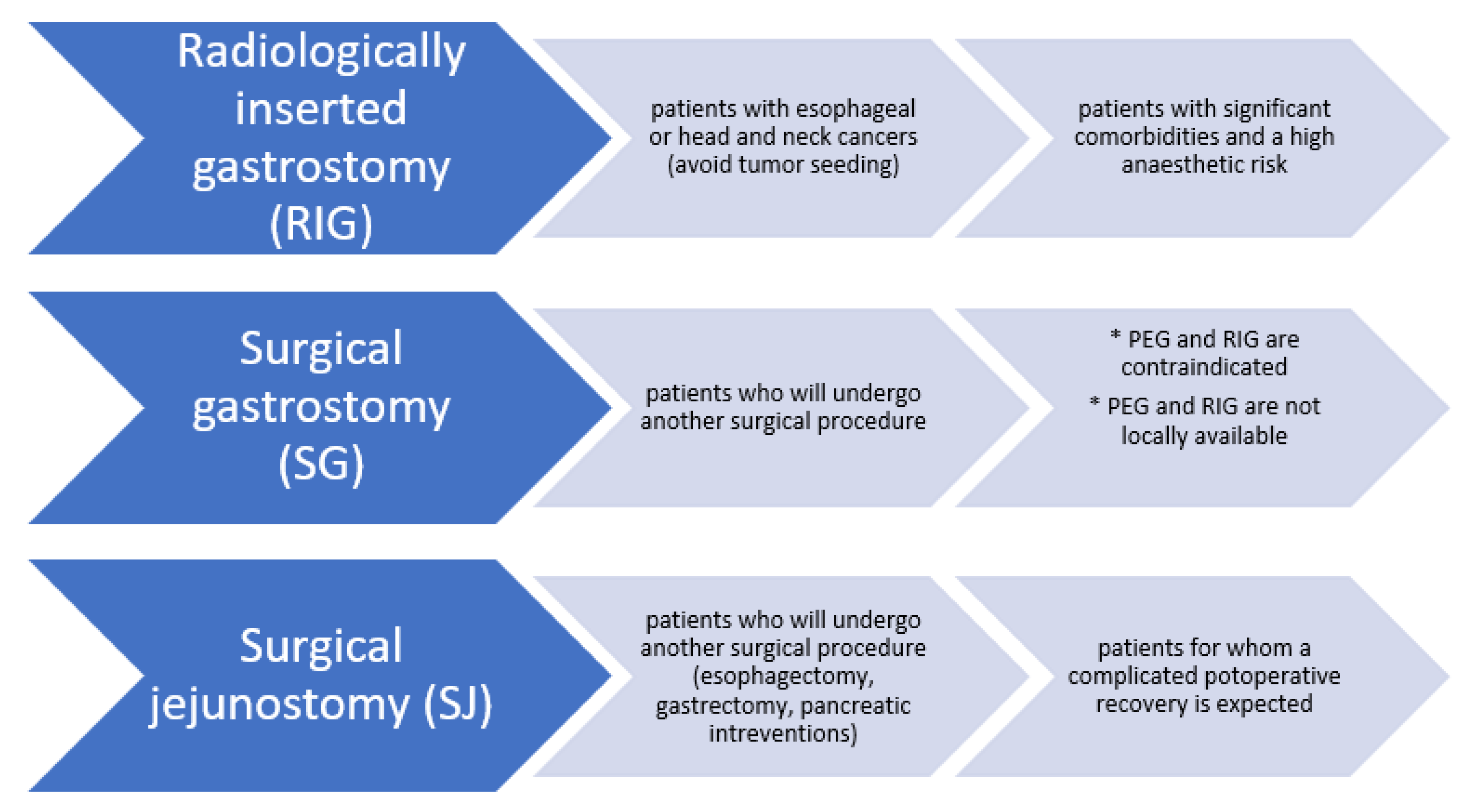
2.4. Non-Endoscopic Methods for Gastrostomy/Jejunostomy

Non-endoscopic methods for gastrostomy/jejunostomy and their indications are presented in Figure 1 [65,66,67,68,69].

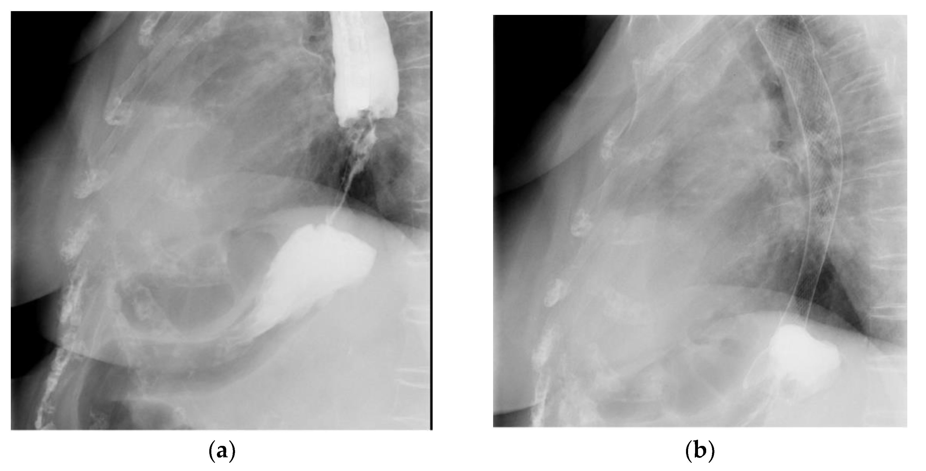
2.5. Esophageal Stents
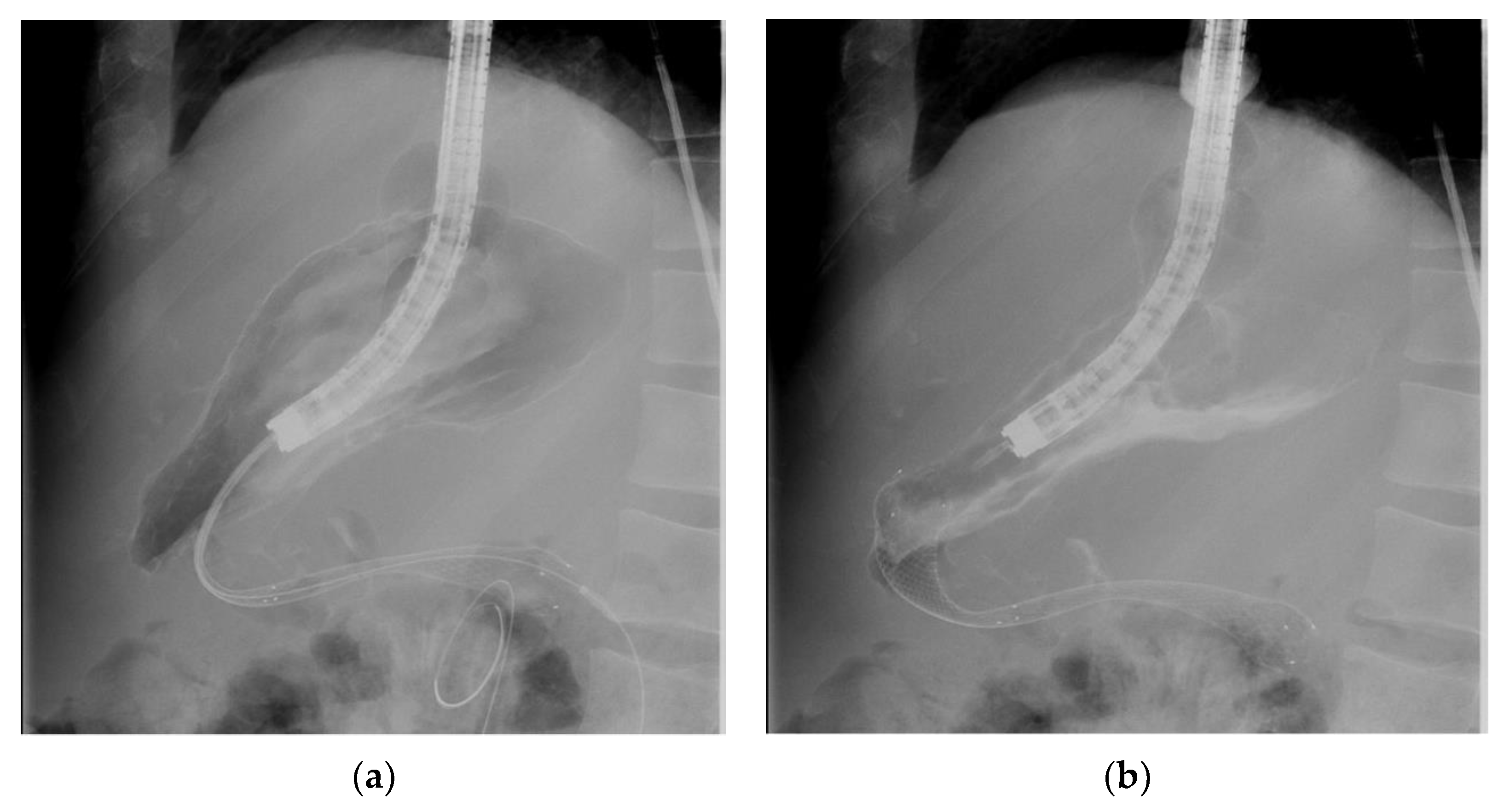
The endoscopic placement of esophageal self-expanding metallic stents (SEMS) allows nutrition in patients with malignant esophageal stenosis (esophageal cancer or extrinsic compression of the esophageal lumen by another malignancy), and it improves the quality of life of the patients [70,71]. The European Society of Gastrointestinal Endoscopy (ESGE) guidelines also recommend the placement of esophageal SEMS in patients with tracheoesophageal or bronchoesophageal malignancies [72]. Regarding the use of esophageal stents as part of neoadjuvant therapy, the ESGE guidelines do not recommend these prostheses as a bridge to surgery or prior to initiating neoadjuvant chemotherapy, because of the recurrence rate of adverse events [72].
By analyzing the results of esophageal SEMS on the morbidity and mortality of patients with malignant dysphagia against other therapies (photodynamic therapy, laser therapy, esophageal bypass surgery), different studies have shown comparable benefits [73,74]. However, the choice of therapeutic method is made according to the prognosis of each individual patient. Hence, at the present time, esophageal SEMS is indicated in patients with severe dysphagia and a short-term survival expectancy, while brachytherapy is recommended in patients with mild dysphagia and a relatively long-life expectancy [75,76]. Esophageal stents allow the quick and efficient relief of symptoms (within one to two days after the placement of the stent) [75]. Considering the most suitable type of esophageal stent, the ESGE guidelines recommend fully covered or partially covered self-expandable metal stents (fcSEMS/pcSEMS) for malignant dysphagia [77]. Uncovered SEMS and covered self-expandable plastic stents (SEPS) have been shown to have a higher risk of occlusion due to tumor growth and migration, respectively [78,79]. No significant differences were found between using fcSEMS and pcSEMS concerning the rates of recurrent dysphagia or other complications [80]. Even so, the major disadvantage of pcSEMS is the difficulty in endoscopic removal, for example, in patients with severe retrosternal pain secondary to stent placement, which seems not to improve with analgesic treatment [80]. Doosti-Irani et al. evaluated the complications that may occur after the placement of an esophageal stent and grouped them into two categories: major and minor complications (Table 3) [81].
The technique for the placement of esophageal stents involves the introduction of a guide wire to bypass the stenosis, the tracking of the guide wire by a cannula, the injection of a contrast substance to characterize the local morphology, and then the installation of the expandable metal prosthesis. This entire procedure is performed under a fluoroscopic screen (Figure 2a,b).

2.6. Enteral Stents

Enteral stents are recommended today as a first-line therapy for patients with malignant GOO, which is supported by the increased rates of technical success and short-term clinical success, the low rates of morbidity, and the shorter duration of hospitalization compared to surgical gastrojejunostomy [82,83,84,85]. A particular category of patients is represented by those with a longer life expectancy. In this situation, the progressive growth of the tumor can lead to the obstruction of the stent and recurrent GOO [86]. Orr et al. assessed the morbidity and mortality rates in a cohort of 43 patients with enteral stents [86]. The etiology of GOO was pancreatic adenocarcinoma (PDAC) in 62.8% of the cases and other malignant etiology was reported in the rest of the cases [86]. The 90-day mortality rate was 70.7% in patients with PDAC and 56.3% in patients with other malignant etiology. Moreover, seven patients required reintervention for recurrent GOO. Nevertheless, the authors pointed out differences in the recurrence of GOO after the installation of enteral stents compared to the etiology of GOO. Thus, among the patients who needed intervention for GOO, only one had PDAC (14.3%) and the others had other malignant etiology of GOO (85.7%) [86].
From the technical point of view, the installation of enteral stents involves the introduction of a guide wire to bypass the stenosis, tracking of the guide wire by a cannula, injection of a contrast substance to characterize the local morphology, and then the installation of the expandable metal prosthesis. The procedure is performed under a fluoroscopic screen (Figure 3a,b).

2.7. Surgical Gastrojejunostomy

Another therapeutic option for patients with malignant GOO is surgical gastrojejunostomy (GJS). Jeurnink et al. compared 1046 patients with duodenal stents and 297 patients with GJS. They reported a higher initial clinical success rate in the group of patients with duodenal stents compared to those with GJJ (89% vs. 72%) [87]. No significant differences were identified between the two groups of patients regarding the technical success rate, as well as the frequency of major early and late complications [87].

2.8. Endoscopic Ultrasound-Guided (EUS-Guided) Gastroenterostomy

Endoscopic ultrasound-guided gastroenterostomy is another alternative method that bypasses GOO by creating an anastomosis between the gastric lumen and the enteral lumen using a fully covered lumen-apposing metal stent (LAMS) [88]. Compared to surgical gastroenterostomy, EUS-guided gastroenterostomy is less invasive [88]. This echoendoscopic procedure has also proved to be a more durable therapeutic option for patients with malignant GOO, unlike enteral stents [89,90]. The literature reported a technical and clinical success rate of EUS-guided gastroenterostomy ranging from 85% to 92% [88].

3. Conclusions

The methods of enteral nutrition may ameliorate or maintain the quality of life of patients with cancer by providing access to proper nutrition and the administration of drugs. Before any endoscopic or surgical intervention, it is necessary to discuss in depth with the patient to define a set of realistic objectives and expectations. As a result, the patient must be informed of any risks associated with the procedure, including those that come along with anesthesia. Furthermore, any endoscopic procedure for enteral nutrition should be performed only in centers where there is a capacity to intervene through surgery or interventional radiology if complications occur [91]. By comparison with surgical methods, endoscopic methods that provide enteral nutrition to patients with digestive cancer without oral feeding are less invasive and have a lower risk of complications.
In conclusion, enteral feeding is a feasible method with evident clinical and nutritional benefits. Therefore, the medical team must consider it early, not for recovery, but to prevent malnutrition.

Author Contributions

Conceptualization, V.-A.I. and G.G.; methodology, C.C.D.; software, R.-I.D.; validation, C.C.D., V.J. and R.O.; formal analysis, R.-I.D.; investigation, G.G.; resources, M.S.-I. and O.Z.; data curation, V.-A.I.; writing—original draft preparation, V.-A.I.; writing—review and editing, C.C.D.; visualization, G.C. and R.O.; supervision, C.C.D.; project administration, V.-A.I.; funding acquisition, G.G. All authors have read and agreed to the published version of the manuscript.

Funding

This research received no external funding.

Institutional Review Board Statement

Not applicable.

Informed Consent Statement

Not applicable.

Conflicts of Interest

The authors declare no conflict of interest.

References

  1. Auth Nagai, H.; Kim, Y.H. Cancer prevention from the perspective of global cancer burden patterns. J. Thorac. Dis. 2017, 9, 448–451. [Google Scholar] [CrossRef]
  2. Global Cancer Observatory. Available online: https://gco.iarc.fr/today/data/factsheets/cancers/7-Stomach-fact-sheet.pdf (accessed on 31 May 2022).
  3. International Agency for Research on Cancer. Available online: https://gco.iarc.fr/today/data/factsheets/populations/642-romania-fact-sheets.pdf (accessed on 31 May 2022).
  4. Badicu, G.; Sani, S.H.Z.; Fathirezaie, Z. Predicting tobacco and alcohol consumption based on physical activity level and demographic characteristics in Romania students. Children 2020, 7, 71. [Google Scholar] [CrossRef] [PubMed]
  5. Arends, J.; Bachmann, P.; Baracos, V.; Barthelemy, N.; Bertz, H.; Bozzetti, F.; Fearon, K.; Hütterer, E.; Isenring, E.; Kaasa, S.; et al. ESPEN guidelines on nutrition in cancer patients. Clin. Nutr. 2017, 36, 11–48. [Google Scholar] [CrossRef] [PubMed] [Green Version]
  6. Cederholm, T.; Bosaeus, I.; Barazzoni, R.; Bauer, J.; Van Gossum, A.; Klek, S.; Muscaritoli, M.; Nyulasi, I.; Ockenga, J.; Schneider, S.M.; et al. Diagnostic criteria for malnutrition—An ESPEN Consensus Statement. Clin. Nutr. 2015, 34, 335–340. [Google Scholar] [CrossRef]
  7. Nitenberg, G.; Raynard, B. Nutritional support of the cancer patient: Issues and dilemmas. Crit. Rev. Oncol. Hematol. 2000, 34, 137–168. [Google Scholar] [CrossRef]
  8. Lees, J. Incidence of weight loss in head and neck cancer patients on commencing radiotherapy treatment at a regional oncology centre. Eur. J. Cancer Care 1999, 8, 133–136. [Google Scholar] [CrossRef]
  9. Arends, J.; Baracos, V.; Bertz, H.; Bozzetti, F.; Calder, P.C.; Deutz, N.E.; Erickson, N.; Laviano, A.; Lisanti, M.P.; Lobo, D.N.; et al. ESPEN expert group recommendations for action against cancer-related malnutrition. Clin. Nutr. 2017, 36, 1187–1196. [Google Scholar] [CrossRef] [Green Version]
  10. Nugent, B.; Lewis, S.; O’Sullivan, J.M. Enteral feeding methods for nutritional management in patients with head and neck cancers being treated with radiotherapy and/or chemotherapy. Cochrane Database Syst Rev. 2013, 1, CD007904. [Google Scholar] [CrossRef]
  11. Chen, M.J.; Wu, I.C.; Chen, Y.J.; Wang, T.E.; Chang, Y.F.; Yang, C.L.; Huang, W.C.; Chang, W.K.; Sheu, B.S.; Wu, M.S.; et al. Nutrition therapy in esophageal cancer-Consensus statement of the Gastroenterological Society of Taiwan. Dis. Esophagus 2018, 31, doy016. [Google Scholar] [CrossRef]
  12. Tendler, D.A. Malignant gastric outlet obstruction: Bridging another divide. Am. J. Gastroenterol. 2002, 97, 4–6. [Google Scholar] [CrossRef]
  13. Adler, D.G.; Baron, T.H. Endoscopic palliation of malignant gastric outlet obstruction using self-expanding metal stents: Experience in 36 patients. Am. J. Gastroenterol. 2002, 97, 72–78. [Google Scholar] [CrossRef]
  14. Hebuterne, X.; Lemarie, E.; Michallet, M.; Beauvillain de Montreuil, C.; Schneider, S.M.; Goldwasser, F. Prevalence of malnutrition and current use od nutrition support in patients with cancer. JPEN J. Parenter. Enteral. Nutr. 2014, 38, 196–204. [Google Scholar] [CrossRef]
  15. Nunes, G.; Fonseca, J.; Barata, A.T.; Dinis-Ribeiro, M.; Pimentel-Nunes, P. Nutritional Suport of Cancer Patients without Oral Feeding: Hoe to Select the Most Effective Technique? GE Port. J. Gastroenterol. 2020, 27, 172–184. [Google Scholar] [CrossRef]
  16. Rinninella, E.; Cintoni, M.; Raoul, P.; Pozzo, C.; Strippoli, A.; Bria, E.; Tortora, G.; Gasbarrini, A.; Mele, M.C. Effects of nutritional interventions on nutritional status in patients with gastric cancer: A systematic review and meta-analysis of randomized controlled trials. Clin. Nutr. ESPEN 2020, 38, 28–42. [Google Scholar] [CrossRef]
  17. Dijksterhuis, W.; Latenstein, A.; van Kleef, J.J.; Verhoeven, R.; de Vries, J.; Slingerland, M.; Steenhagen, E.; Heisterkamp, J.; Timmermans, L.M.; de van der Schueren, M.; et al. Cachexia and dietetic inter-ventions in patients with esophagogastric cancer: A multicenter cohort study. J. Natl. Compr. Cancer Netw. 2021, 19, 144–152. [Google Scholar] [CrossRef]
  18. Muscaritoli, M.; Arends, J.; Bachmann, P.; Baracos, V.; Barthelemy, N.; Berts, H.; Bozzetti, F.; Hutterer, E. ESPEN practical guideline: Clinical Nutrition in cancer. Clin. Nutr. 2021, 40, 2898–2913. [Google Scholar] [CrossRef]
  19. Cao, D.X.; Wu, G.H.; Zhang, B.; Quan, Y.J.; Wei, J.; Jin, H.; Jiang, Y.; Yang, Z.A. Resting energy expenditure and body composition in patients with newly detected cancer. Clin. Nutr. 2010, 29, 72–77. [Google Scholar] [CrossRef]
  20. Wang, L.; Sesso, H.D.; Glynn, R.J.; Christen, W.G.; Bubes, V.; Manson, J.E.; Buring, J.E.; Gaziano, J.M. Vitamin E and C supplementation and risk of cancer in men: Posttrial follow-up in the Physicians’ Health Study II randomized trial. Am. J. Clin. Nutr. 2014, 100, 915–923. [Google Scholar] [CrossRef] [Green Version]
  21. Klein, E.A.; Thompson, I.M., Jr.; Tangen, C.M.; Crowley, J.J.; Lucia, M.S.; Goodman, P.J.; Minasian, L.M.; Ford, L.G.; Parnes, H.L.; Gaziano, J.M.; et al. Vitamin E and the risk of prostate cancer: The selenium and vitamin E cancer prevention trial (SELECT). JAMA 2011, 306, 1549–1556. [Google Scholar] [CrossRef]
  22. Korber, J.; Pricelius, S.; Heidrich, M.; Müller, M.J. Increased lipid utilization in weight losing and weight stable cancer patients with normal body weight. Eur. J. Clin. Nutr. 1999, 53, 740–745. [Google Scholar] [CrossRef] [Green Version]
  23. Bourdel-Marchasson, I.; Blanc-Bisson, C.; Doussau, A.; Germain, C.; Blanc, J.F.; Dauba, J.; Lahmar, C.; Terrebonne, E.; Lecaille, C.; Ceccaldi, J.; et al. Nutritional advice in older patients at risk of malnutrition during treatment for chemotherapy: A two-year randomized controlled trial. PLoS ONE 2014, 9, e108687. [Google Scholar] [CrossRef] [PubMed]
  24. Fong, D.Y.; Ho, J.W.; Hui, B.P.; Lee, A.M.; Macfarlane, D.J.; Leung, S.S.; Lam, S.H.; Taylor, A.J. Physical activity for cancer survivors: Meta-analysis of randomised controlled trials. Br. Med. J. Int. Ed. 2012, 344, e70. [Google Scholar] [CrossRef] [PubMed] [Green Version]
  25. Moertel, C.G.; Schutt, A.J.; Reitemeier, R.J.; Hahn, R.G. Corticosteroid therapy of preterminal gastrointestinal cancer. Cancer 1974, 33, 1607–1609. [Google Scholar] [CrossRef]
  26. Ruiz Garcia, V.; Lopez-Briz, E.; Carbonell Sanchis, R.; Gonzalvez Perales, J.L.; Bort-Marti, S. Megestrol acetate for treatment of anorexia-cachexia syndrome. Cochrane Database Syst. Rev. 2013, 2013, CD004310. [Google Scholar] [CrossRef]
  27. Baldwin, C.; Spiro, A.; Ahern, R.; Emery, P.W. Oral nutritional interventions in malnourished patients with cancer: A systematic review and meta-analysis. J. Natl. Cancer Inst. 2012, 104, 371–385. [Google Scholar] [CrossRef] [Green Version]
  28. Kaegi-Braun, N.; Schuetz, P.; Mueller, B.; Kutz, A. Association of Nutritional Support with Clinical Outcomes in Malnourished Cancer Patients: A Population-Based Matched Cohort Study. Front. Nutr. 2021, 10, 603370. [Google Scholar] [CrossRef]
  29. Yolsuriyanwong, K.; Chand, B. Update on endoscopic enteral access. Tech. Gastrointest. Endosc. 2018, 20, 172–181. [Google Scholar] [CrossRef]
  30. Braunschweig, C.L.; Levy, P.; Sheean, P.M.; Wang, X. Enteral compared with parenteral nutrition: A meta-analysis. Am. J. Clin. Nutr. 2001, 74, 534–542. [Google Scholar] [CrossRef] [Green Version]
  31. Pritchard, C.; Duffy, S.; Edington, J.; Pang, F. Enteral nutrition and oral nutrition supplements: A review of the economics literature. JPEN J. Parenter. Enteral. Nutr. 2006, 30, 52–59. [Google Scholar] [CrossRef]
  32. MacFie, J. Bacterial Translocation, Gut Barrier Function and Nutritional Support. Surgery 2002, 20, 1–2. [Google Scholar] [CrossRef]
  33. Szefel, J.; Kruszewski, W.J.; Buczek, T. Enteral feeding and its impact on the gut immune system and intestinal mucosal barrier. Prz. Gastroenterol. 2015, 10, 71–77. [Google Scholar] [CrossRef] [Green Version]
  34. Amano, K.; Maeda, I.; Ishiki, H.; Miura, T.; Hatano, Y.; Tsukuura, H.; Taniyama, T.; Matsumoto, Y.; Matsuda, Y.; Kohara, H.; et al. Effects of enteral nutrition and parenteral nutrition on survival in patients with advanced cancer cachexia: Analysus of a multicenter prospective cohort study. Clin. Nutr. 2021, 40, 1168–1175. [Google Scholar] [CrossRef] [PubMed]
  35. Itkin, M.; DeLegge, M.H.; Fang, J.C.; McClave, S.A.; Kundu, S.; d’Othee, B.J.; Martinez–Salazar, G.M.; Sacks, D.; Swan, T.L.; Towbin, R.B.; et al. Multidisciplinary practical guidelines for gastrointestinal access for enteral nutrition and decompression from the Society of Interventional Radiology and American Gastroenterological Association (AGA) Institute, with endorsement by Canadian Interventional Radiological Association (CIRA) and Cardiovascular and Interventional Radiological Society of Europe (CIRSE). Gastroenterology 2011, 141, 742–765. [Google Scholar]
  36. Ho, C.S.; Yee, A.C.; McPherson, R. Complications of surgical and percutaneous non endoscopic gastrostomy: Review of 233 patients. Gastroenterology 1988, 95, 1206–1210. [Google Scholar] [CrossRef]
  37. Nagaraja, V.; Cox, M.R.; Eslick, G.D. Safety and efficacy of esophageal stents preceding or during neoadjuvant chemotherapy for esophageal cancer: A systematic review and meta-analysis. J. Gastrointest. Oncol. 2014, 5, 119–126. [Google Scholar]
  38. Best, C. How to set up and administer an enteral feed via a nasogastric tube. Nurs. Stand. 2017, 31, 42–47. [Google Scholar] [CrossRef]
  39. Kwon, R.S.; Banerjee, S.; Desilets, D.; Diehl, D.L.; Farraye, F.A.; Kaul, V.; Mamula, P.; Pedrosa, M.C.; Rodriguez, S.A.; Varadarajulu, S.; et al. ASGE Technology Committee Enteral nutrition access devices. Gastrointest. Endosc. 2010, 72, 236–248. [Google Scholar] [CrossRef]
  40. Löser, C.; Aschl, G.; Hébuterne, X.; Mathus-Vliegen, E.M.; Muscaritoli, M.; Niv, Y.; Rollins, H.; Singer, P.; Skelly, R.H. ESPEN guidelines on artificial enteral nutrition—Percutaneous endoscopic gastrostomy (PEG). Clin. Nutr. 2005, 24, 848–861. [Google Scholar]
  41. Bischoff, S.C.; Austin, P.; Boeykens, K.; Chourdakis, M.; Cuerda, C.; Jonkers-Schutema, C.; Lichota, M.; Nyulasi, I.; Schneider, S.M.; Stanga, Z.; et al. ESPEN guideline on home enteral nutrition. Clin. Nutr. 2020, 39, 5–22. [Google Scholar] [CrossRef] [Green Version]
  42. Shastri, Y.M.; Shirodkar, M.; Mallath, M.K. Endoscopic feeding tube placement in patients with cancer: A prospective clinical audit of 2055 procedures in 1866 patients. Aliment. Pharmacol. Ther. 2008, 27, 649–658. [Google Scholar] [CrossRef]
  43. Westaby, D.; Young, A.; O’Toole, P.; Smith, G.; Sanders, D.S. The provision of a percutaneously placed enteral tube feeding service. Gut 2010, 59, 1592–1605. [Google Scholar] [CrossRef] [Green Version]
  44. Mobily, M.; Patel, J.A. Palliative percutaneous endoscopic gastrostomy placement for gastrointestinal cancer: Roles, goals, and complications. World J. Gastrointest. Endosc. 2015, 7, 364–369. [Google Scholar] [CrossRef]
  45. McClave, S.A.; Ritchie, C.S. The role of endoscopically placed feeding or decompression tubes. Gastroenterol. Clin. North Am. 2006, 35, 83–100. [Google Scholar] [CrossRef]
  46. Villalba, C.M.; Rodriguez, J.A.V.; Sanchez, F.G. Percutaneous endoscopic gastrostomy. Indication, care and complications. Med. Clin. 2019, 152, 229–236. [Google Scholar] [CrossRef]
  47. Lochs, H.; Valentini, L.; Schutz, T.; Allison, S.P.; Howard, P.; Pichard, C. ESPEN Guidelines on adult enteral nutrition. Clin. Nutr. 2006, 25, 177–360. [Google Scholar] [CrossRef]
  48. Leeds, J.S.; McAlindon, M.E.; Grant, J.; Robson, H.E.; Lee, F.K.; Sanders, D.S. Survival analysis after gastrostomy: A single-centre, observational study comparing radiological and endoscopic insertion. Eur. J. Gastroenterol. Hepatol. 2010, 22, 591. [Google Scholar] [CrossRef]
  49. Johnston, S.D.; Tham, T.C.; Mason, M. Death after PEG: Results of the National Confidential Enquiry into Patient Outcome and Death. Gastrointest. Endosc. 2008, 68, 223. [Google Scholar] [CrossRef]
  50. Meisel, K.; Arnold, R.M.; Stijacic, C.I.; Boscardin, J.; Smith, A.K. Survival, Functional Status, and Eating Ability After Percutaneous Endoscopic Gastrostomy Tube Placement for Acute Stroke. J. Am. Geriatr. Soc. 2017, 65, 1848. [Google Scholar] [CrossRef]
  51. Arora, G.; Rockey, D.; Gupta, S. High in-hospital mortality after percutaneous endoscopic gastrostomy: Results of a nationwide population-based study. Clin. Gastroenterol. Hepatol. 2013, 11, 1437–1444.e3. [Google Scholar] [CrossRef]
  52. Blomberg, J.; Lagergren, J.; Martin, L.; Mattsson, F.; Lagergren, P. Complications after percutaneous endoscopic gastrostomy in a prospective study. Scand. J. Gastroenterol. 2012, 47, 737–742. [Google Scholar] [CrossRef]
  53. Keung, E.Z.; Liu, X.; Nuzhad, A.; Rabinowits, G.; Patel, V. In-hospital and long-term outcomes after percutaneous endoscopic gastrostomy in patients with malignancy. J. Am. Coll. Surg. 2012, 215, 777–786. [Google Scholar] [CrossRef]
  54. Raha, S.K.; Woodhouse, K. The use of percutaneous endoscopic gastrostomy (PEG) in 161 consecutive elderly patients. Age Ageing 1994, 23, 162. [Google Scholar] [CrossRef]
  55. Laranjo, A.; Brito, M.; Nunes, G.; Santos, C.A.; Fonseca, J. Feasibility, safety and outcome of endoscopic gastrostomy in patients with esophageal cancer. Nutr. Hosp. 2020, 37, 660–666. [Google Scholar] [CrossRef]
  56. Amrendra, M.; Varun, K.; Zorisadday, G.; Neel, R.; Rajan, K.; Praveen, K. Prophylactic antibiotics for PEG tube placement: An update meta-analysis of randomized controlled trials. Am. J. Gastroenterol. 2019, 14, S326. [Google Scholar]
  57. Schneider, A.S.; Schettler, A.; Markowski, A.; Luettig, B.; Kaufmann, B.; Klamt, S.; Lenzen, H.; Momma, M.; Seipt, C.; Lankisch, T.; et al. Complication and mortality rate after percutaneous endoscopic gastrostomy are low and indication-dependent. Scand. J. Gastroenterol. 2014, 49, 891–898. [Google Scholar] [CrossRef]
  58. Hucl, T.; Spicak, J. Complications of percutaneous endoscopic gastrostomy. Best Pract. Res. Clin. Gastroenterol. 2016, 30, 769. [Google Scholar] [CrossRef]
  59. Macedo, C.; Almeida, N.; Alves, A.R.; Ferreira, A.M.; Figueiredo, P. Persistent Peristomal Leakage from Percutaneous Endoscopic Gastrostomy Successfully Treated with Argon Plasma Coagulation. GE Port. J. Gastroenterol. 2021, 28, 210. [Google Scholar] [CrossRef]
  60. Enestvedt, B.K.; Jorgensen, J.; Sedlack, R.E.; Coyle, W.J.; Obstein, K.L.; Al-Haddad, M.A.; Christie, J.A.; Davila, R.E.; Mullady, D.K.; Kubiliun, N.; et al. ASGE Training Committee 2013–2014. Endoscopic approaches to enteral feeding and nutrition core curriculum. Gastrointest. Endosc. 2014, 80, 34–41. [Google Scholar] [CrossRef]
  61. Jain, R.; Maple, J.T.; Anderson, M.A.; Appalaneni, V.; Ben-Menachem, T.; Decker, G.A.; Fanelli, R.D.; Fisher, L.; Fukami, N.; Ikenberry, S.O.; et al. ASGE Standards of Practice Committee. The role of endoscopy in enteral feeding. Gastrointest. Endosc. 2011, 74, 7–12. [Google Scholar] [CrossRef]
  62. Finucane, T.E.; Bynum, J.P. Use of tube feeding to prevent aspiration pneumonia. Lancet 1996, 348, 1421–1424. [Google Scholar] [CrossRef]
  63. Allum, W.H.; Blazeby, J.M.; Griffin, S.M.; Cunningham, D.; Jankowski, J.A.; Wong, R. Association of Upper Gastrointestinal Surgeons of Great Britain and Ireland, the British Society of Gastroenterology and the British Association of Surgical Oncology Guidelines for the management of oesophageal and gastric cancer. Gut 2011, 60, 1449–1472. [Google Scholar] [CrossRef] [Green Version]
  64. Waitzberg, D.L.; Torrinhas, R.S. Enteral feeding. Encycl. Food Health 2016, 2, 258–263. [Google Scholar]
  65. Sofue, K.; Takeuchi, Y.; Tsurusaki, M.; Shibamoto, K.; Sakamoto, N.; Kitajima, K.; Sone, M.; Sugimura, K.; Arai, Y. Value of Percutaneous Radiologic Gastrostomy for Patients with Advanced Esophageal Cancer. Ann. Surg. Oncol. 2016, 23, 3623–3631. [Google Scholar] [CrossRef]
  66. Lim, J.H.; Choi, S.H.; Lee, C.; Seo, J.Y.; Kang, H.Y.; Yang, J.I.; Chung, S.J.; Kim, J.S. Thirty-day mortality after percutaneous gastrostomy by endoscopic versus radiologic placement: A systematic review and meta-analysis. Intest. Res. 2016, 14, 333–342. [Google Scholar] [CrossRef] [Green Version]
  67. Oliveira, G.P.; Santos, C.A.; Fonseca, J. The role of surgical gastrostomy in the age of endoscopic gastrostomy: A 13 years and 543 patients retrospective study. Rev. Esp. Enferm. Dig. 2016, 108, 776–779. [Google Scholar] [CrossRef] [Green Version]
  68. Shin, J.H.; Park, A.W. Updates on percutaneous radiologic gastrostomy/gastrojejunostomy and jejunostomy. Gut Liver 2010, 4 (Suppl. 1), S25–S31. [Google Scholar] [CrossRef]
  69. Yuan, Y.; Zhao, Y.; Xie, T.; Hu, Y. Percutaneous endoscopic gastrostomy versus percutaneous radiological gastrostomy for swallowing distrubances. Cochrane Database Syst Rev 2016, 2, CD009198. [Google Scholar]
  70. Spaander, M.C.; Baron, T.H.; Siersema, P.D.; Fuccio, L.; Schumacher, B.; Escorsell, A.; Garcia-Pagán, J.C.; Dumonceau, J.M.; Conio, M.; De Ceglie, A.; et al. Esophageal stenting for benign and malignant disease: European Society of Gastrointestinal Endoscopy (ESGE) Clinical Guideline. Endoscopy 2016, 48, 939–948. [Google Scholar] [CrossRef] [Green Version]
  71. Spaander, M.C.W.; van der Bogt, R.D.; Baron, T.H.; Albers, D.; Blero, D.; De Ceglie, A.; Conio, M.; Czakó, L.; Everett, S.; Garcia-Pagán, J.C.; et al. Esophageal stenting for benign and malignant disease:European Society of Gastrointestinal Endoscopy (ESGE) Guideline–Update 2021. Endoscopy 2021, 53, 751–762. [Google Scholar]
  72. Sharma, B.; Raina, S.; Sharma, R.; Bodh, V.; Raina, S.K.; Sharma, N. Self-Expanding Metallic Stents (SEMS) in Inoperable Esophageal Cancer: A Prospective Analysis of Morbidity and Survival Outcomes. Indian J. Palliat. Care 2019, 25, 398–402. [Google Scholar]
  73. Aoki, T.; Osaka, Y.; Takagi, Y.; Okada, R.; Shinohara, M.; Tsuchida, A.; Sato, S.; Koyanagi, Y. Comparative study of self-expandable metallic stent and bypass surgery for inoperable esophageal cancer. Dis. Esophagus 2001, 14, 208–211. [Google Scholar] [CrossRef]
  74. Dallal, H.J.; Smith, G.D.; Grieve, D.C.; Ghosh, S.; Penman, I.D.; Palmer, K.R. A randomized trial of thermal ablative therapy versus expandable metal stents in the palliative treatment of patients with esophageal carcinoma. Gastrointest. Endosc. 2001, 54, 549–557. [Google Scholar] [CrossRef] [Green Version]
  75. Dai, Y.; Li, C.; Xie, Y.; Liu, X.; Zhang, J.; Zhou, J.; Pan, X.; Yang, S. Interventions for dysphagia in oesophageal cancer. Cochrane Database Syst. Rev. 2014, 2014, CD005048. [Google Scholar]
  76. Homs, M.Y.V.; Steyerberg, E.W.; Eijkenboom, W.M.H.; Tilanus, H.W.; Stalpers, L.J.A.; Bartelsman, J.F.; van Lanschot, J.J.; Wijrdeman, H.K.; Mulder, C.J.; Reinders, J.G.; et al. Single-dose brachytherapy versus metal stent placement for the palliation of dysphagia from oesophageal cancer: Multicentre randomised trial. Lancet 2004, 364, 1497–1504. [Google Scholar] [CrossRef]
  77. Tinusz, B.; Soos, A.; Hegyi, P.; Sarlos, P.; Szapary, L.; Eros, A.; Feczak, D.; Szakacs, Z.; Marta, K.; Venglovecz, V.; et al. Efficacy and safety of stenting and additional oncological treatment versus stenting alone in unresectable esophageal cancer: A meta-analysis and systematic review. Radiother. Oncol. 2020, 147, 169–177. [Google Scholar] [CrossRef]
  78. Vakil, N.; Morris, A.I.; Marcon, N.; Segalin, A.; Peracchia, A.; Bethge, N.; Zuccaro, G.; Bosco, J.J.; Jones, W.F. A prospective, randomized, controlled trial of covered expandable metal stents in the palliation of malignant esophageal obstruction at the gastroesophageal junction. Am. J. Gastroenterol. 2001, 96, 1791–1796. [Google Scholar] [CrossRef]
  79. Conio, M.; Repici, A.; Battaglia, G.; de Pretis, G.; Ghezzo, L.; Bittinger, M.; Messmann, H.; Demarquay, J.F.; Blanchi, S.; Togni, M.; et al. A randomized prospective comparison of self-expandable plastic stents and partially covered self-expandable metal stents in the palliation of malignant esophageal dysphagia. Am. J. Gastroenterol. 2007, 102, 2667–2677. [Google Scholar] [CrossRef]
  80. Vermeulen, B.D.; Siersema, P.D. Esophageal stenting in clinical practice: An Overview. Curr. Treat. Options Gastroenterol. 2018, 16, 260–273. [Google Scholar] [CrossRef] [Green Version]
  81. Doosti-Irani, A.; Mansournia, M.A.; Rahimi-Foroushani, A.; Haddad, P.; Holakouie-Naieni, K. Complications of stent placement in patients with esophageal cancer: A systematic review and network meta-analysis. PLoS ONE 2017, 12, e0184784. [Google Scholar] [CrossRef] [Green Version]
  82. Espinel, J.; Sanz, O.; Vivas, S.; Jorquera, F.; Munoz, F.; Olcoz, J.L.; Pinedo, E. Malignant gastrointestinal obstruction: Endoscopic stenting versus surgical palliation. Surg. Endosc. 2006, 20, 1083–1087. [Google Scholar] [CrossRef]
  83. Chen, Y.I.; Khashab, M.A. Endoscopic approach to gastrointestinal bypass in malignant gastric outlet obstruction. Curr. Opin. Gastroenterol. 2016, 32, 365–373. [Google Scholar] [CrossRef] [PubMed]
  84. Khashab, M.; Alawad, A.S.; Shin, E.J.; Kim, K.; Bourdel, N.; Sing, V.K.; Lennon, A.M.; Hutfless, S.; Sharaiha, R.Z.; Amateau, S.; et al. Enteral stenting versus gastrojejunostomy for palliation of malignant gastric outlet obstruction. Surg. Endosc. 2013, 27, 2068–2075. [Google Scholar] [CrossRef]
  85. Balaceanu, A.; Diaconu, C.; Mateescu, D.; Stănica, A. Hepatocellular carcinoma with hepatic and pulmonary metastasis, inferior vena cava and left pulmonary artery thrombosis in a patient with asymptomatic hepatitis C. Case report. Med. Ultrason. 2010, 12, 345–348. [Google Scholar] [PubMed]
  86. Orr, J.; Lockwood, R.; Gamboa, A.; Slaughter, J.C.; Obstein, K.L.; Yachimski, P. Enteral Stents for Malignant Gastric Outlet Obstruction: Low Reintervention Rates for Obstruction due to Pancreatic Adenocarcinoma Versus Other Etiologies. J. Gastrointest. Surg. 2021, 25, 720–727. [Google Scholar] [CrossRef] [PubMed]
  87. Jeurnink, S.M.; van Eijck, C.H.J.; Steyerberg, E.W.; Kuipers, E.J.; Siersema, P.D. Stent versus gastrojejunostomy for the palliation of gastric outlet obstruction: A systematic review. BMC Gastroenterol. 2007, 7, 18. [Google Scholar] [CrossRef] [Green Version]
  88. Dawod, Q.; Issa, D.; Shah, S.L.; Daod, S.; Sharaiha, R.Z. EUS-gided stent placement for afferent limb and gastrojejunal obstruction in patient with pancreatic cancer. VideoGIE 2021, 6, 257–259. [Google Scholar] [CrossRef]
  89. Rimbas, M.; Larghi, A.; Costamagna, G. Endoscopic ultrasound-guided gastroenterostomy: Are we ready for prime time? Endosc. Ultrasound 2017, 6, 235–240. [Google Scholar]
  90. Nageswaran, H.; Belgaumkar, A.; Kumar, R. Acute afferent loop syndrome in the early postoperative period following pancreaticoduodenectomy. Ann. R. Coll. Surg. Engl. 2015, 97, 349–353. [Google Scholar] [CrossRef] [PubMed] [Green Version]
  91. Hitawala, A.A.; Mousa, O.Y. Percutaneous Gastrostomy and Jenunostomy. Stat Pearls. 2022. Available online: https://www.ncbi.nlm.nih.gov/books/NBK559215/ (accessed on 15 May 2022).
Figure 1. Non-endoscopic methods for gastrostomy/jejunostomy and their indications.
Gastroent 13 00025 g001
Figure 2. (a) Esophageal malignant obstruction (from the collection of Dr. Gabriel Constantinescu, Clinical Emergency Hospital of Bucharest, Romania). (b) SEMS placed under endoscopic and fluoroscopic control in a patient with malignant esophageal stenosis (from the collection of Dr. Gabriel Constantinescu, Clinical Emergency Hospital of Bucharest, Romania).
Gastroent 13 00025 g002
Figure 3. The placement of a duodenal stent (a,b) in malignant duodenal stenosis under fluoroscopic screen (from the collection of Dr. Gabriel Constantinescu, Clinical Emergency Hospital of Bucharest, Romania).
Gastroent 13 00025 g003
Table 1. Endoscopic techniques for enteral access.
Endoscopic Techniques for Enteral Access
  • Endoscopy-guided nasoenteric tube (ENET)
  • Percutaneous endoscopic gastrostomy (PEG)
  • Percutaneous endoscopic gastrostomy with jejunal tube extension (PEG-J)
  • Direct percutaneous endoscopic jejunostomy (DPEJ)
  • Endoscopic ultrasound (EUS)-guided gastroenterostomy
Table 2. Complications of PEG placement [52,53,54,55,56,57,58,59].
Complications That May Occur at Any TimeEarly Complications of PEG PlacementLate Complications of PEG Placement
Tube dysfunction
Infection
Wound infection
Necrotizing fasciitis (rare complication)
Bleeding
Peristomal leakage
Ulcerations
Gastric outlet obstruction
Inadvertent gastrostomy tube removal
Pneumoperitoneum
Ileus
Esophageal and gastric perforation
Other early complications: small bowel wall hematoma with small bowel obstruction; sigmoid volvulus; transhepatic placement of a gastrostomy tube; damage of other abdominal organs
Deterioration of the gastrostomy site
Buried bumper syndrome
Colocutaneous fistula
Persistent gastric fistula following gastrostomy tube removal
PEG tract tumor seeding
Other late complications: gastric herniation; persistent abdominal wall pain.
Table 3. Complications that may occur after the placement of esophageal stents [81].
Major ComplicationsMinor Complications
  • Esophageal perforations (2%)
  • Pressure necrosis (2%)
  • Fistula formation (3%)
  • Pneumonia because of aspirations (5%)
  • Recurrent dysphagia (31%); stent migration (11%); tumor in- or overgrowth (14%); food obstruction (7%)
  • Gastroesophageal reflux (7%)
  • Post-procedural pain (30%)
Publisher’s Note: MDPI stays neutral with regard to jurisdictional claims in published maps and institutional affiliations.

