YAP1 Upregulates Cytoskeleton Regulator ARHGEF1 and Tissue Regeneration Factor NEDD9 in a Multiplex Proteomic Study
 , ,
, ,  ,
,  and
and
Abstract
1. Introduction
2. Materials and Methods
2.1. Chemicals and Antibodies
2.2. Generation of SH-SY5Y Stable Cells Expressing YAP1-GFP and Cell Lysate Preparation
2.3. Immunocytochemical Staining of Cell Lines and Human Neurons
2.4. Antibody-Based Multiplex Proteomic Screening
2.5. Bioinformatics Analysis and Heatmap Generation
2.6. Quantification of Proteins by Western Blotting
2.7. Statistical Analysis
3. Results
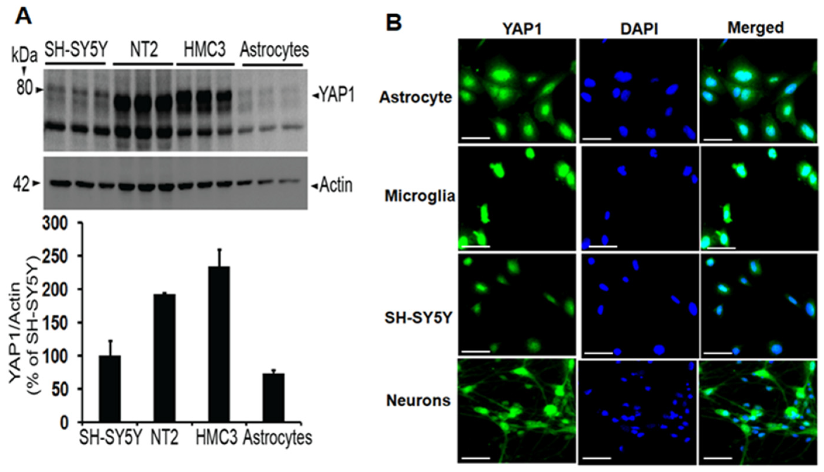
3.1. Neurons and Other Brain Cell Types Express YAP1 Protein
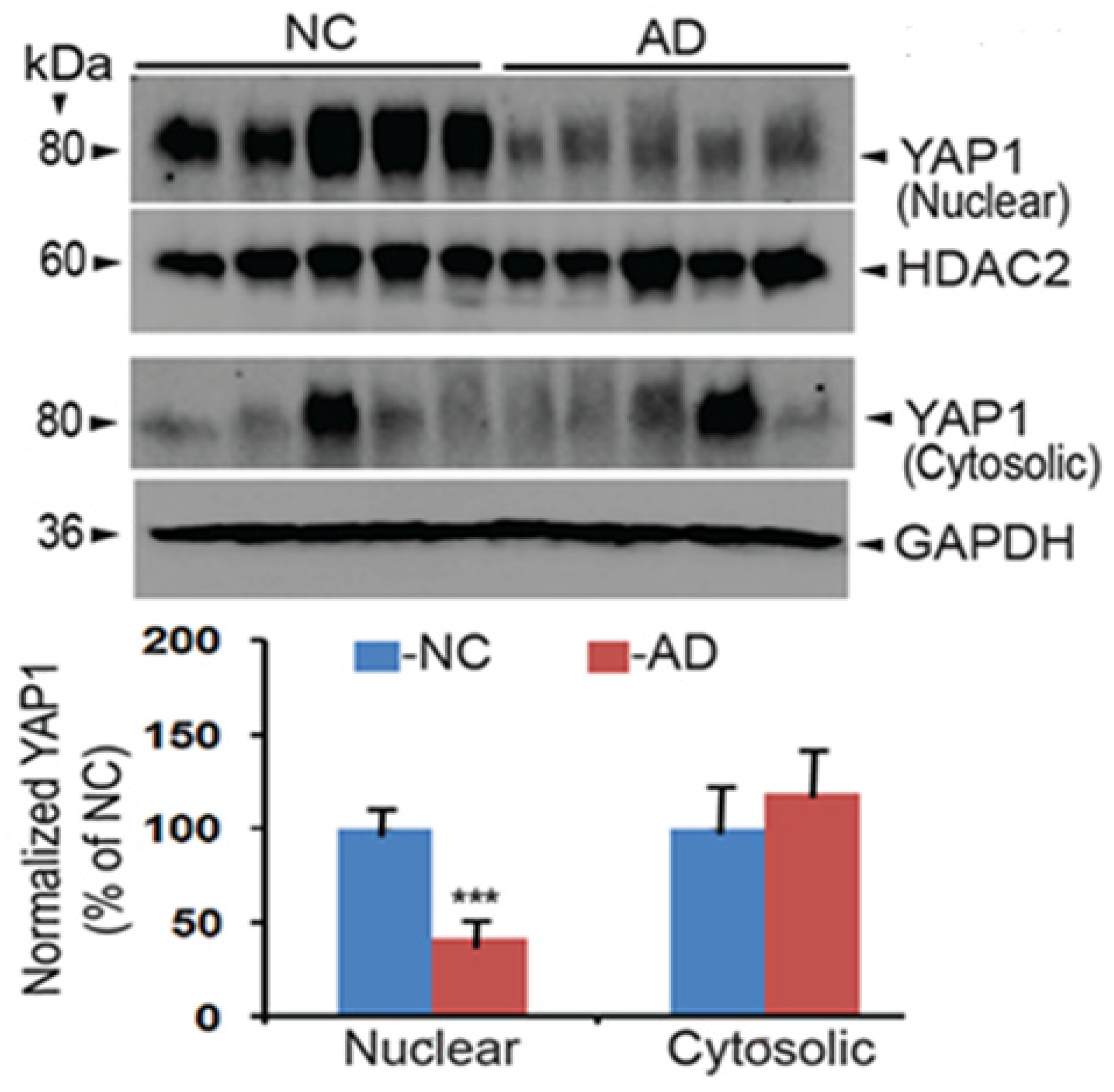
3.2. YAP1 Protein Expression Is Downregulated in the Nuclear Fractions of Alzheimer’s Brains
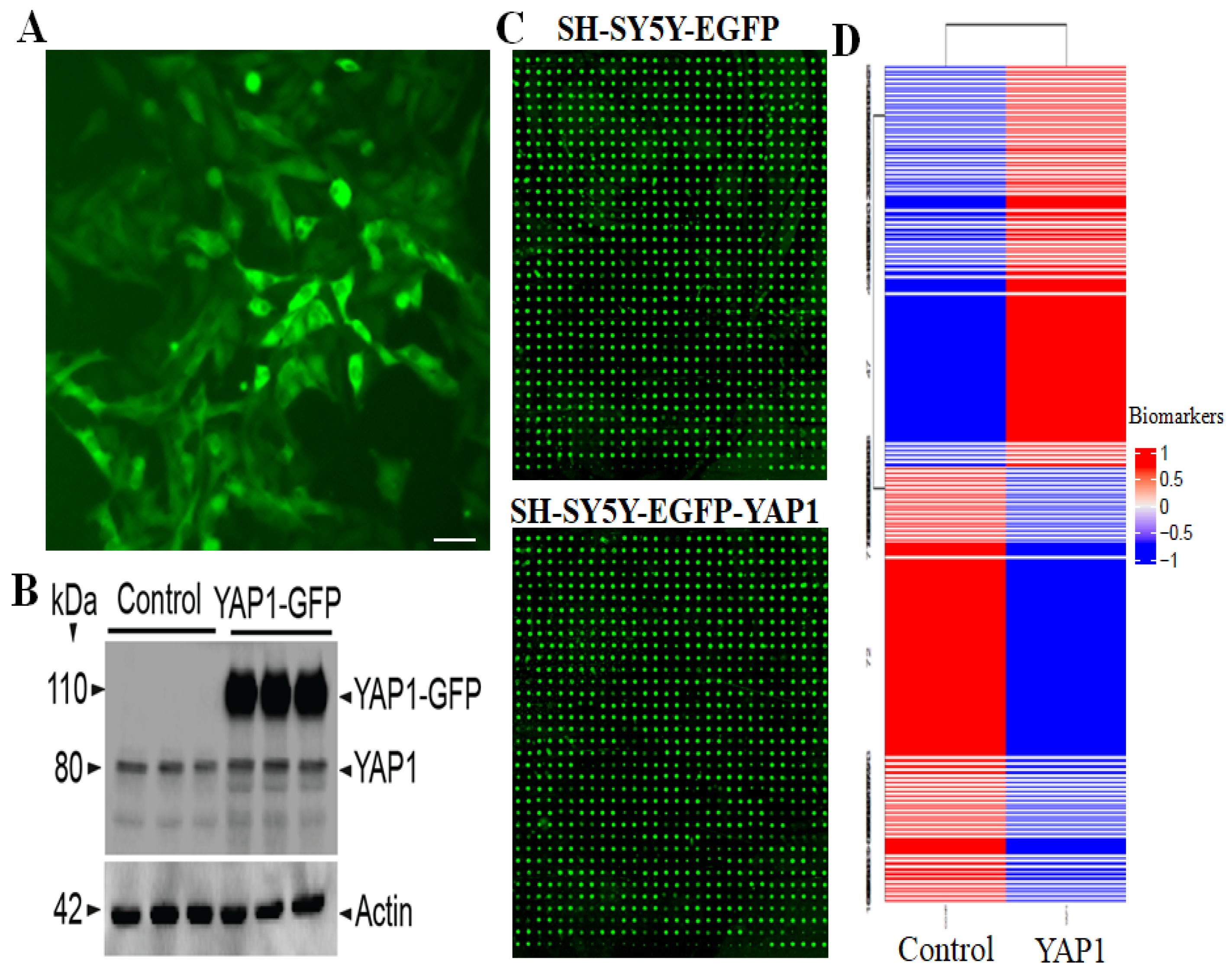
3.3. Multiplex Proteomic Screening Revealed Alterations in Specific Proteins and Pathways
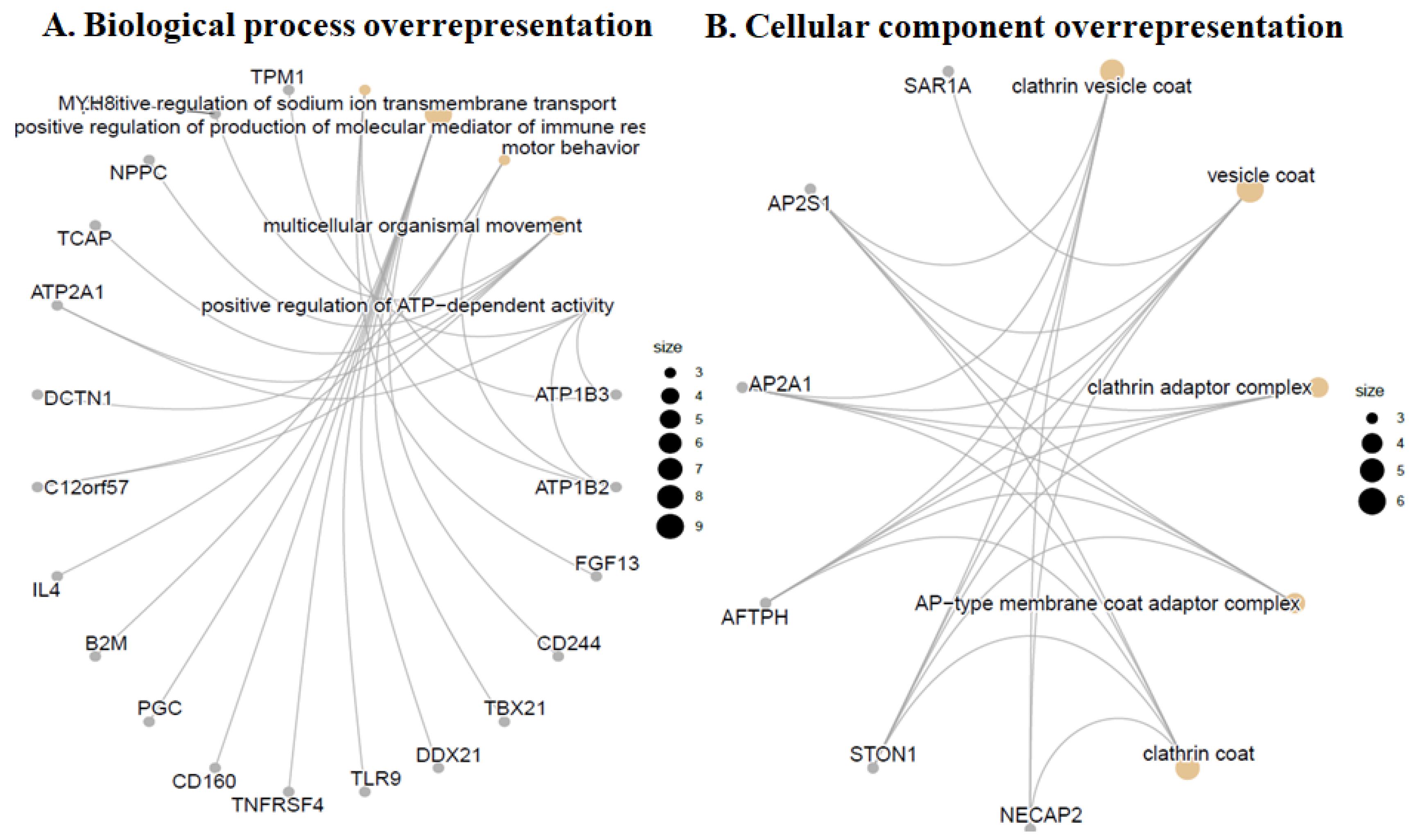
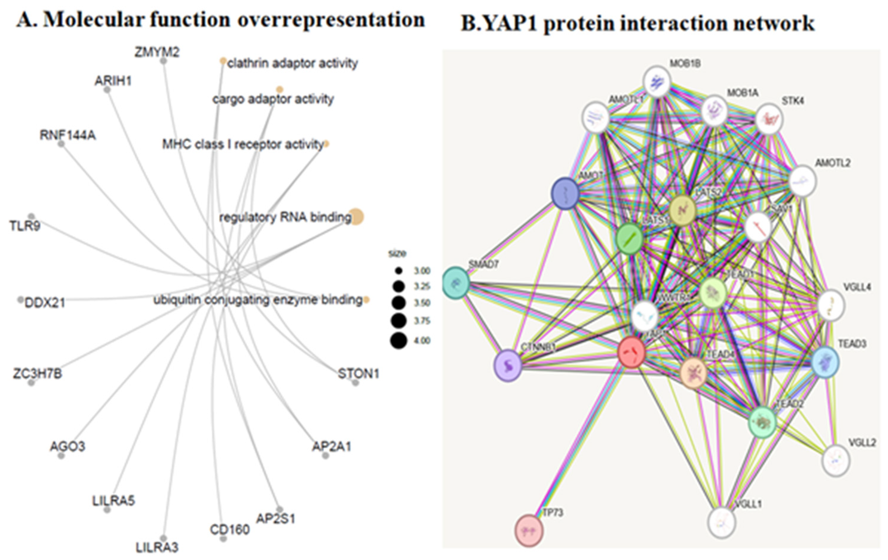
3.4. Gene Ontology (GO) Over-Representation Analysis
3.5. Ward Hierarchical Cluster Analysis Revealed an Increase in Specific Markers of Endocytosis and Actin Cytoskeleton
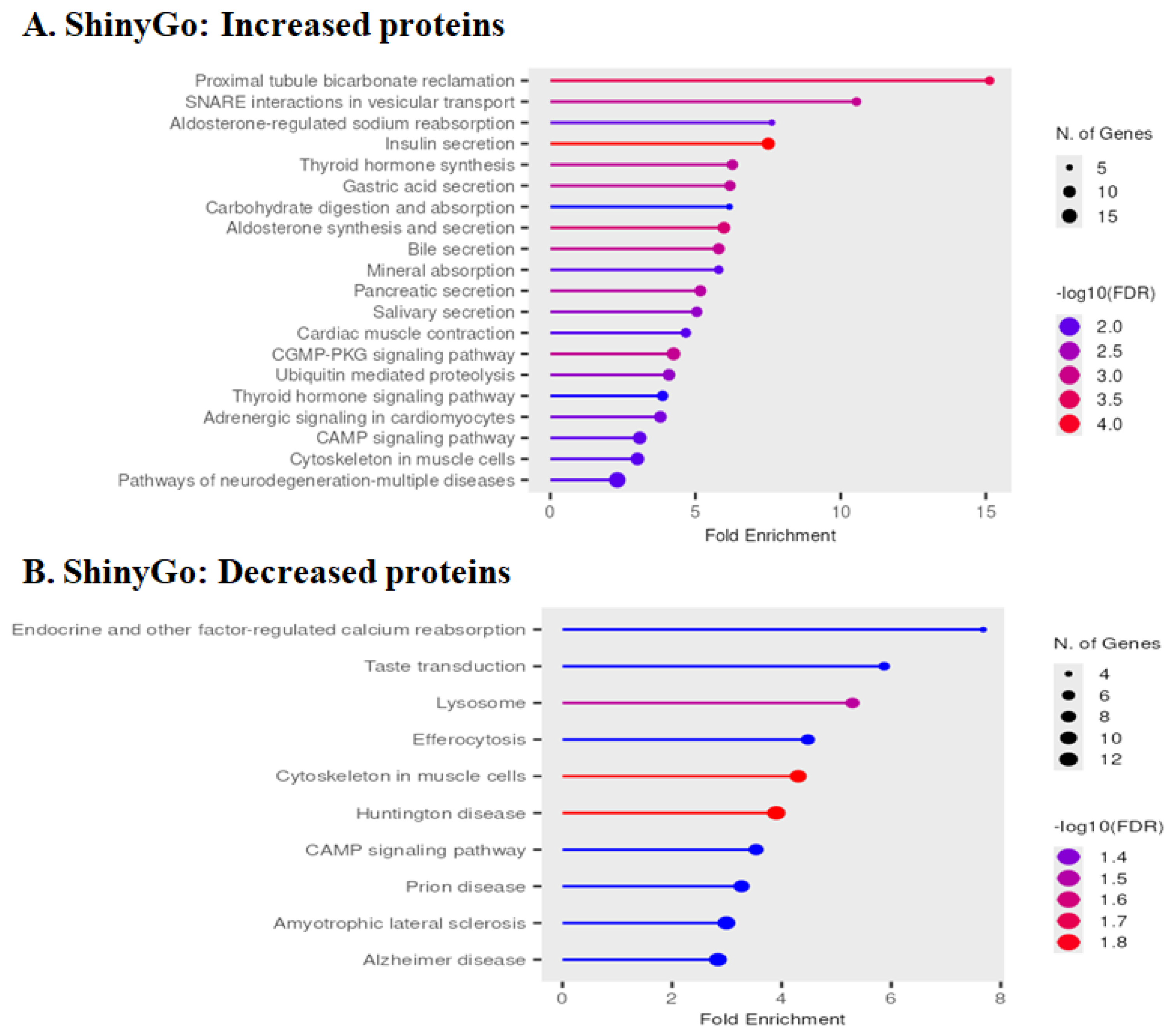
3.6. Pathway Enrichment Analysis by ShinyGo
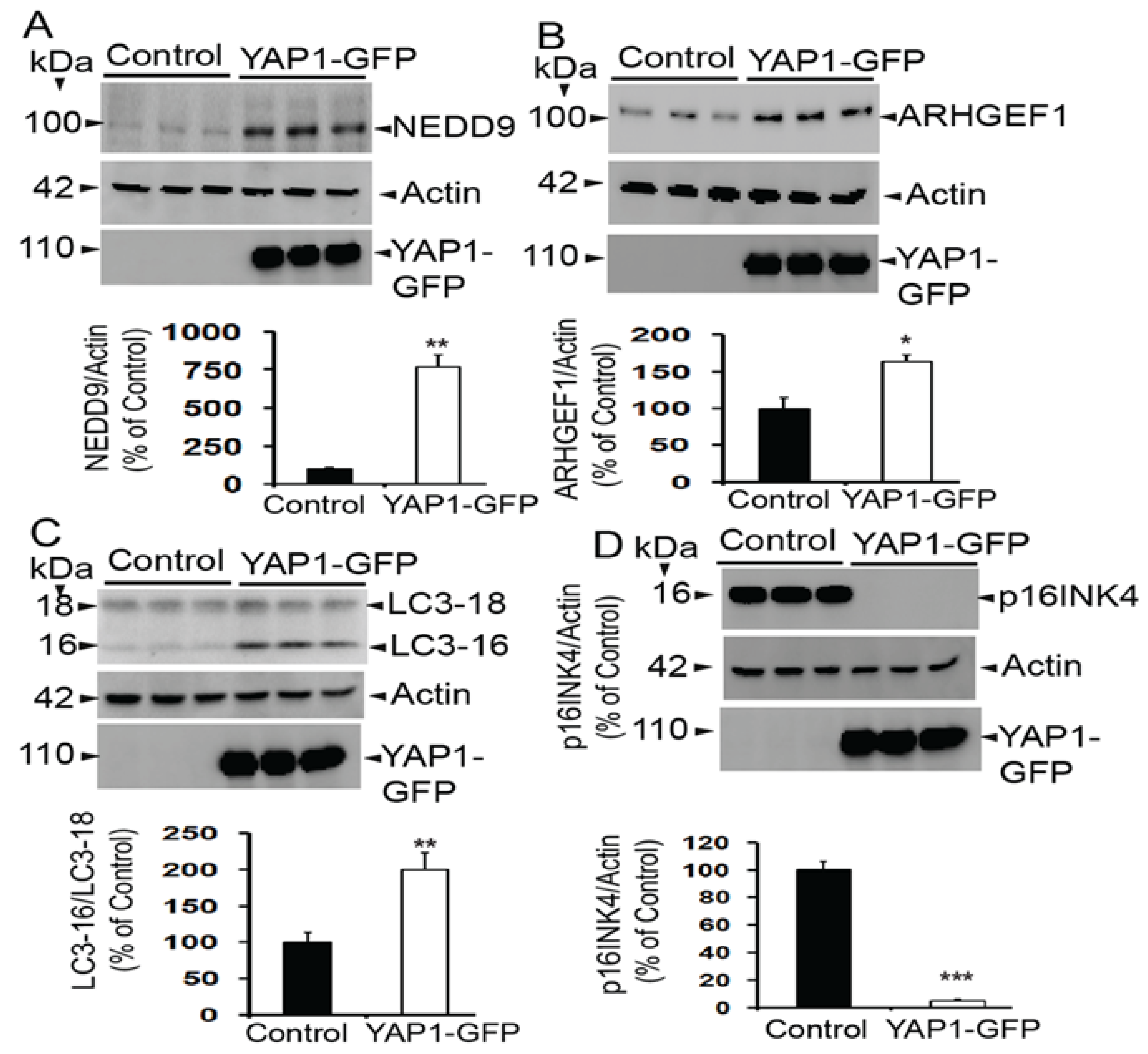
3.7. Validation of Multiplex Array Results by Immunoblots
3.8. YAP1 Also Changes Expression of LC3 and p16INK4a
4. Discussion
Limitations and Future Directions
5. Conclusions
Author Contributions
Funding
Institutional Review Board Statement
Informed Consent Statement
Data Availability Statement
Conflicts of Interest
References
- Levy, D.; Adamovich, Y.; Reuven, N.; Shaul, Y. Yap1 phosphorylation by c-Abl is a critical step in selective activation of proapoptotic genes in response to DNA damage. Mol. Cell 2008, 29, 350–361. [Google Scholar] [CrossRef] [Scilit] [PubMed]
- Zhao, B.; Ye, X.; Yu, J.; Li, L.; Li, W.; Li, S.; Yu, J.; Lin, J.D.; Wang, C.Y.; Chinnaiyan, A.M.; et al. TEAD mediates YAP-dependent gene induction and growth control. Genes. Dev. 2008, 22, 1962–1971. [Google Scholar] [CrossRef] [Scilit]
- Low, B.C.; Pan, C.Q.; Shivashankar, G.V.; Bershadsky, A.; Sudol, M.; Sheetz, M. YAP/TAZ as mechanosensors and mechanotransducers in regulating organ size and tumor growth. FEBS Lett. 2014, 588, 2663–2670. [Google Scholar] [CrossRef] [Scilit]
- Ye, B.; Yue, M.; Chen, H.; Sun, C.; Shao, Y.; Jin, Q.; Zhang, C.; Yu, G. YAP/TAZ as master regulators in liver regeneration and disease: Insights into mechanisms and therapeutic targets. Mol. Biol. Rep. 2024, 52, 78. [Google Scholar] [CrossRef] [Scilit]
- Panciera, T.; Azzolin, L.; Cordenonsi, M.; Piccolo, S. Mechanobiology of YAP and TAZ in physiology and disease. Nat. Rev. Mol. Cell Biol. 2017, 18, 758–770. [Google Scholar] [CrossRef] [Scilit]
- Totaro, A.; Panciera, T.; Piccolo, S. YAP/TAZ upstream signals and downstream responses. Nat. Cell Biol. 2018, 20, 888–899. [Google Scholar] [CrossRef] [Scilit]
- Piccolo, S.; Panciera, T.; Contessotto, P.; Cordenonsi, M. YAP/TAZ as master regulators in cancer: Modulation, function and therapeutic approaches. Nat. Cancer 2023, 4, 9–26. [Google Scholar] [CrossRef] [Scilit]
- Hansen, C.G.; Moroishi, T.; Guan, K.L. YAP and TAZ: A nexus for Hippo signaling and beyond. Trends Cell Biol. 2015, 25, 499–513. [Google Scholar] [CrossRef] [Scilit]
- Lin, K.C.; Park, H.W.; Guan, K.L. Regulation of the Hippo Pathway Transcription Factor TEAD. Trends Biochem. Sci. 2017, 42, 862–872. [Google Scholar] [CrossRef] [Scilit]
- Franklin, J.M.; Wu, Z.; Guan, K.L. Insights into recent findings and clinical application of YAP and TAZ in cancer. Nat. Rev. Cancer 2023, 23, 512–525. [Google Scholar] [CrossRef] [Scilit]
- Lian, I.; Kim, J.; Okazawa, H.; Zhao, J.; Zhao, B.; Yu, J.; Chinnaiyan, A.; Israel, M.A.; Goldstein, L.S.; Abujarour, R.; et al. The role of YAP transcription coactivator in regulating stem cell self-renewal and differentiation. Genes. Dev. 2010, 24, 1106–1118. [Google Scholar] [CrossRef] [Scilit]
- Bao, X.; Xu, X.; Wu, Q.; Zhang, J.; Feng, W.; Yang, D.; Li, F.; Lu, S.; Liu, H.; Shen, X.; et al. Sphingosine 1-phosphate promotes the proliferation of olfactory ensheathing cells through YAP signaling and participates in the formation of olfactory nerve layer. Glia 2020, 68, 1757–1774. [Google Scholar] [CrossRef] [Scilit]
- Shen, X.; Xu, X.; Xie, C.; Liu, H.; Yang, D.; Zhang, J.; Wu, Q.; Feng, W.; Wang, L.; Du, L.; et al. YAP promotes the proliferation of neuroblastoma cells through decreasing the nuclear location of p27(Kip1) mediated by Akt. Cell Prolif. 2020, 53, e12734. [Google Scholar] [CrossRef] [Scilit]
- Xie, C.; Shen, X.; Xu, X.; Liu, H.; Li, F.; Lu, S.; Gao, Z.; Zhang, J.; Wu, Q.; Yang, D.; et al. Astrocytic YAP Promotes the Formation of Glia Scars and Neural Regeneration after Spinal Cord Injury. J. Neurosci. 2020, 40, 2644–2662. [Google Scholar] [CrossRef] [Scilit]
- Xin, M.; Kim, Y.; Sutherland, L.B.; Murakami, M.; Qi, X.; McAnally, J.; Porrello, E.R.; Mahmoud, A.I.; Tan, W.; Shelton, J.M.; et al. Hippo pathway effector Yap promotes cardiac regeneration. Proc. Natl. Acad. Sci. USA 2013, 110, 13839–13844. [Google Scholar] [CrossRef] [Scilit]
- Loforese, G.; Malinka, T.; Keogh, A.; Baier, F.; Simillion, C.; Montani, M.; Halazonetis, T.D.; Candinas, D.; Stroka, D. Impaired liver regeneration in aged mice can be rescued by silencing Hippo core kinases MST1 and MST2. EMBO Mol. Med. 2017, 9, 46–60. [Google Scholar] [CrossRef] [Scilit]
- Fan, R.; Kim, N.G.; Gumbiner, B.M. Regulation of Hippo pathway by mitogenic growth factors via phosphoinositide 3-kinase and phosphoinositide-dependent kinase-1. Proc. Natl. Acad. Sci. USA 2013, 110, 2569–2574. [Google Scholar] [CrossRef] [Scilit]
- Fu, L.; Hu, Y.; Song, M.; Liu, Z.; Zhang, W.; Yu, F.X.; Wu, J.; Wang, S.; Izpisua Belmonte, J.C.; Chan, P.; et al. Up-regulation of FOXD1 by YAP alleviates senescence and osteoarthritis. PLoS Biol. 2019, 17, e3000201. [Google Scholar] [CrossRef] [Scilit]
- Santinon, G.; Brian, I.; Pocaterra, A.; Romani, P.; Franzolin, E.; Rampazzo, C.; Bicciato, S.; Dupont, S. dNTP metabolism links mechanical cues and YAP/TAZ to cell growth and oncogene-induced senescence. EMBO J. 2018, 37, e97780. [Google Scholar] [CrossRef] [Scilit]
- Xu, X.; Shen, X.; Feng, W.; Yang, D.; Jin, L.; Wang, J.; Wang, M.; Ting, Z.; Xue, F.; Zhang, J.; et al. D-galactose induces senescence of glioblastoma cells through YAP-CDK6 pathway. Aging 2020, 12, 18501–18521, Erratum in Aging 2020, 17, 1369–1370. https://doi.org/10.18632/aging.206264. [Google Scholar] [CrossRef] [Scilit]
- Jin, H.; Lian, N.; Zhang, F.; Bian, M.; Chen, X.; Zhang, C.; Jia, Y.; Lu, C.; Hao, M.; Yao, S.; et al. Inhibition of YAP signaling contributes to senescence of hepatic stellate cells induced by tetramethylpyrazine. Eur. J. Pharm. Sci. 2017, 96, 323–333. [Google Scholar] [CrossRef] [Scilit]
- Xu, X.; Shen, X.; Wang, J.; Feng, W.; Wang, M.; Miao, X.; Wu, Q.; Wu, L.; Wang, X.; Ma, Y.; et al. YAP prevents premature senescence of astrocytes and cognitive decline of Alzheimer’s disease through regulating CDK6 signaling. Aging Cell 2021, 20, e13465. [Google Scholar] [CrossRef] [Scilit]
- Joung, J.; Heo, Y.; Kim, Y.; Kim, J.; Choi, H.; Jeon, T.; Jang, Y.; Kim, E.J.; Lee, S.H.; Suh, J.M.; et al. Cell enlargement modulated by GATA4 and YAP instructs the senescence-associated secretory phenotype. Nat. Commun. 2025, 16, 1696. [Google Scholar] [CrossRef] [Scilit]
- Casella, G.; Munk, R.; Kim, K.M.; Piao, Y.; De, S.; Abdelmohsen, K.; Gorospe, M. Transcriptome signature of cellular senescence. Nucleic Acids Res. 2019, 47, 7294–7305, Erratum in Nucleic Acids Res. 2019, 47, 11476. https://doi.org/10.1093/nar/gkz879. [Google Scholar] [CrossRef] [Scilit]
- Sladitschek-Martens, H.L.; Guarnieri, A.; Brumana, G.; Zanconato, F.; Battilana, G.; Xiccato, R.L.; Panciera, T.; Forcato, M.; Bicciato, S.; Guzzardo, V.; et al. YAP/TAZ activity in stromal cells prevents ageing by controlling cGAS-STING. Nature 2022, 607, 790–798. [Google Scholar] [CrossRef] [Scilit]
- Belin, S.; Zuloaga, K.L.; Poitelon, Y. Influence of Mechanical Stimuli on Schwann Cell Biology. Front. Cell Neurosci. 2017, 11, 347. [Google Scholar] [CrossRef] [Scilit] [PubMed]
- Wang, J.; Chen, H.; Hou, W.; Han, Q.; Wang, Z. Hippo Pathway in Schwann Cells and Regeneration of Peripheral Nervous System. Dev. Neurosci. 2023, 45, 276–289. [Google Scholar] [CrossRef] [Scilit] [PubMed]
- Zhang, J.; Hu, J.; Liu, R.; Zhou, T.; Luo, X.; Liang, P.; Xie, Z.; Zhao, Q.; Chen, Y.; Du, D.; et al. YAP maintains the dynamics of TDP-43 condensates and antagonizes TDP-43 pathological aggregates. Nat. Cell Biol. 2025, 27, 1148–1160. [Google Scholar] [CrossRef] [Scilit]
- Lei, L.; Cheng, Y.; Yin, A.; Han, J.M.; Wu, G.; Yang, F.; Wang, Q.; Wang, J.Z.; Liu, R.; Li, H.L.; et al. Aging-dependent YAP1 reduction contributes to AD pathology by upregulating the Nr4a1-AKT/GSK-3beta axis. Transl. Neurodegener. 2025, 14, 29. [Google Scholar] [CrossRef] [Scilit]
- Yang, X.; Duan, H.; Li, S.; Zhang, J.; Dong, L.; Ding, J.; Li, X. Yap1 alleviates sepsis associated encephalopathy by inhibiting hippocampus ferroptosis via maintaining mitochondrial dynamic homeostasis. J. Cell Mol. Med. 2024, 28, e70156. [Google Scholar] [CrossRef] [Scilit]
- Srivastava, T.; Nguyen, H.; Haden, G.; Diba, P.; Sowa, S.; LaNguyen, N.; Reed-Dustin, W.; Zhu, W.; Gong, X.; Harris, E.N.; et al. TSG-6-Mediated Extracellular Matrix Modifications Regulate Hypoxic-Ischemic Brain Injury. J. Neurosci. 2024, 44. [Google Scholar] [CrossRef] [Scilit]
- Gong, P.; Zhang, Z.; Zou, C.; Tian, Q.; Chen, X.; Hong, M.; Liu, X.; Chen, Q.; Xu, Z.; Li, M.; et al. Hippo/YAP signaling pathway mitigates blood-brain barrier disruption after cerebral ischemia/reperfusion injury. Behav. Brain Res. 2019, 356, 8–17, Erratum in Behav. Brain Res. 2019, 416, 113531. [Google Scholar] [CrossRef] [Scilit]
- Li, D.; Ji, J.X.; Xu, Y.T.; Ni, H.B.; Rui, Q.; Liu, H.X.; Jiang, F.; Gao, R.; Chen, G. Inhibition of Lats1/p-YAP1 pathway mitigates neuronal apoptosis and neurological deficits in a rat model of traumatic brain injury. CNS Neurosci. Ther. 2018, 24, 906–916. [Google Scholar] [CrossRef] [Scilit]
- Xu, M.; Zhang, D.F.; Luo, R.; Wu, Y.; Zhou, H.; Kong, L.L.; Bi, R.; Yao, Y.G. A systematic integrated analysis of brain expression profiles reveals YAP1 and other prioritized hub genes as important upstream regulators in Alzheimer’s disease. Alzheimers Dement. 2018, 14, 215–229. [Google Scholar] [CrossRef] [Scilit]
- Fan, W.; Jurado-Arjona, J.; Alanis-Lobato, G.; Peron, S.; Berger, C.; Andrade-Navarro, M.A.; Falk, S.; Berninger, B. The transcriptional co-activator Yap1 promotes adult hippocampal neural stem cell activation. EMBO J. 2023, 42, e110384. [Google Scholar] [CrossRef] [Scilit]
- Noordin, R.; Othman, N. Proteomics technology—A powerful tool for the biomedical scientists. Malays. J. Med. Sci. 2013, 20, 1–2. [Google Scholar]
- Aslam, B.; Basit, M.; Nisar, M.A.; Khurshid, M.; Rasool, M.H. Proteomics: Technologies and Their Applications. J. Chromatogr. Sci. 2017, 55, 182–196. [Google Scholar] [CrossRef] [Scilit]
- R Core Team. R: A Language and Environment for Statistical Computing; R Foundation for Statistical Computing: Vienna, Austria, 2017; Available online: https://www.R-project.org/ (accessed on 13 May 2026).
- Yu, G.; Wang, L.G.; Han, Y.; He, Q.Y. clusterProfiler: An R package for comparing biological themes among gene clusters. OMICS 2012, 16, 284–287. [Google Scholar] [CrossRef] [Scilit]
- Lakshmana, M.K.; Yoon, I.S.; Chen, E.; Bianchi, E.; Koo, E.H.; Kang, D.E. Novel role of RanBP9 in BACE1 processing of amyloid precursor protein and amyloid beta peptide generation. J. Biol. Chem. 2009, 284, 11863–11872. [Google Scholar] [CrossRef] [Scilit]
- Wang, H.; Dey, D.; Carrera, I.; Minond, D.; Bianchi, E.; Xu, S.; Lakshmana, M.K. COPS5 (Jab1) protein increases beta site processing of amyloid precursor protein and amyloid beta peptide generation by stabilizing RanBP9 protein levels. J. Biol. Chem. 2013, 288, 26668–26677. [Google Scholar] [CrossRef] [Scilit]
- Wang, H.; Devadoss, D.; Nair, M.; Chand, H.S.; Lakshmana, M.K. Novel Alzheimer risk factor IQ motif containing protein K is abundantly expressed in the brain and is markedly increased in patients with Alzheimer’s disease. Front. Cell Neurosci. 2022, 16, 954071. [Google Scholar] [CrossRef] [Scilit]
- Tanaka, H.; Homma, H.; Fujita, K.; Kondo, K.; Yamada, S.; Jin, X.; Waragai, M.; Ohtomo, G.; Iwata, A.; Tagawa, K.; et al. YAP-dependent necrosis occurs in early stages of Alzheimer’s disease and regulates mouse model pathology. Nat. Commun. 2020, 11, 507. [Google Scholar] [CrossRef] [Scilit]
- Szklarczyk, D.; Kirsch, R.; Koutrouli, M.; Nastou, K.; Mehryary, F.; Hachilif, R.; Gable, A.L.; Fang, T.; Doncheva, N.T.; Pyysalo, S.; et al. The STRING database in 2023: Protein–protein association networks and functional enrichment analyses for any sequenced genome of interest. Nucleic Acids Res. 2022, 51, D638–D646. [Google Scholar] [CrossRef] [Scilit]
- Ge, S.X.; Jung, D.; Yao, R. ShinyGO: A graphical gene-set enrichment tool for animals and plants. Bioinformatics 2020, 36, 2628–2629. [Google Scholar] [CrossRef] [Scilit]
- Tanida, I.; Ueno, T.; Kominami, E. LC3 and Autophagy. Methods Mol. Biol. 2008, 445, 77–88. [Google Scholar]
- Safwan-Zaiter, H.; Wagner, N.; Wagner, K.D. P16INK4A-More Than a Senescence Marker. Life 2022, 12, 1332. [Google Scholar] [CrossRef] [Scilit]
- Liu, J.Y.; Souroullas, G.P.; Diekman, B.O.; Krishnamurthy, J.; Hall, B.M.; Sorrentino, J.A.; Parker, J.S.; Sessions, G.A.; Gudkov, A.V.; Sharpless, N.E. Cells exhibiting strong p16(INK4a) promoter activation in vivo display features of senescence. Proc. Natl. Acad. Sci. USA 2019, 116, 2603–2611. [Google Scholar] [CrossRef] [Scilit]
- Yang, J.H.; Miner, A.E.; Fair, A.; Kinkel, R.; Graves, J.S. Senescence marker p16INK4a expression in patients with multiple sclerosis. Mult. Scler. Relat. Disord. 2024, 84, 105498. [Google Scholar] [CrossRef] [Scilit]
- Huang, L.; Li, S.; Dai, Q.; Zhang, A.; Yu, Q.; Du, W.; Zhao, P.; Mo, Y.; Xu, K.; Chen, S.; et al. Astrocytic Yes-associated protein attenuates cerebral ischemia-induced brain injury by regulating signal transducer and activator of transcription 3 signaling. Exp. Neurol. 2020, 333, 113431. [Google Scholar] [CrossRef] [Scilit]
- Yu, H.; Cao, X.; Li, W.; Liu, P.; Zhao, Y.; Song, L.; Chen, J.; Chen, B.; Yu, W.; Xu, Y. Targeting connexin 43 provides anti-inflammatory effects after intracerebral hemorrhage injury by regulating YAP signaling. J. Neuroinflamm. 2020, 17, 322. [Google Scholar] [CrossRef] [Scilit]
- Huang, Z.; Wang, Y.; Hu, G.; Zhou, J.; Mei, L.; Xiong, W.C. YAP Is a Critical Inducer of SOCS3, Preventing Reactive Astrogliosis. Cereb. Cortex 2016, 26, 2299–2310. [Google Scholar] [CrossRef] [Scilit]
- Morimoto, N.; Nagai, M.; Miyazaki, K.; Kurata, T.; Takehisa, Y.; Ikeda, Y.; Kamiya, T.; Okazawa, H.; Abe, K. Progressive decrease in the level of YAPdeltaCs, prosurvival isoforms of YAP, in the spinal cord of transgenic mouse carrying a mutant SOD1 gene. J. Neurosci. Res. 2009, 87, 928–936. [Google Scholar] [CrossRef] [Scilit]
- Hoshino, M.; Qi, M.L.; Yoshimura, N.; Miyashita, T.; Tagawa, K.; Wada, Y.; Enokido, Y.; Marubuchi, S.; Harjes, P.; Arai, N.; et al. Transcriptional repression induces a slowly progressive atypical neuronal death associated with changes of YAP isoforms and p73. J. Cell Biol. 2006, 172, 589–604. [Google Scholar] [CrossRef] [Scilit]
- Chamberland, J.P.; Antonow, L.T.; Dias Santos, M.; Ritter, B. NECAP2 controls clathrin coat recruitment to early endosomes for fast endocytic recycling. J. Cell Sci. 2016, 129, 2625–2637. [Google Scholar] [CrossRef] [Scilit]
- Rajendrakumar, A.L.; Arbeev, K.G.; Bagley, O.; Yashin, A.I.; Ukraintseva, S. The association between rs6859 in NECTIN2 gene and Alzheimer’s disease is partly mediated by pTau. medRxiv 2024, 16, 1388363. [Google Scholar] [CrossRef] [Scilit]
- Kimura, I.; Nakayama, Y.; Zhao, Y.; Konishi, M.; Itoh, N. Neurotrophic effects of neudesin in the central nervous system. Front. Neurosci. 2013, 7, 111. [Google Scholar] [CrossRef] [Scilit]
- Shimizu, R.; Shikaura, R.; Hasegawa, H.; Kondo, M.; Masuda, Y.; Nakayama, Y.; Konishi, M. Neudesin attenuates ischemic brain injury by regulating Nos2 expression in microglia. Biochem. Biophys. Res. Commun. 2025, 784, 152647. [Google Scholar] [CrossRef] [Scilit]
- Woolfrey, K.M.; Srivastava, D.P. Control of Dendritic Spine Morphological and Functional Plasticity by Small GTPases. Neural Plast. 2016, 2016, 3025948. [Google Scholar] [CrossRef] [Scilit]
- Knutson, D.C.; Clagett-Dame, M. atRA Regulation of NEDD9, a gene involved in neurite outgrowth and cell adhesion. Arch. Biochem. Biophys. 2008, 477, 163–174. [Google Scholar] [CrossRef] [Scilit]
- Xing, Y.Y.; Yu, J.T.; Yan, W.J.; Chen, W.; Zhong, X.L.; Jiang, H.; Wang, P.; Tan, L. NEDD9 is genetically associated with Alzheimer’s disease in a Han Chinese population. Brain Res. 2011, 1369, 230–234. [Google Scholar] [CrossRef] [Scilit]
- Fu, Y.; He, F.; Tang, N.L.; Tam, C.W.; Lui, V.W.; Chiu, H.F.; Lam, L.C. NEDD9 gene polymorphism influences the risk of Alzheimer disease and cognitive function in Chinese older persons. Alzheimer Dis. Assoc. Disord. 2012, 26, 88–90. [Google Scholar] [CrossRef] [Scilit]
- Li, Y.; Grupe, A.; Rowland, C.; Holmans, P.; Segurado, R.; Abraham, R.; Jones, L.; Catanese, J.; Ross, D.; Mayo, K.; et al. Evidence that common variation in NEDD9 is associated with susceptibility to late-onset Alzheimer’s and Parkinson’s disease. Hum. Mol. Genet. 2008, 17, 759–767. [Google Scholar] [CrossRef] [Scilit] [PubMed]
- Knutson, D.C.; Mitzey, A.M.; Talton, L.E.; Clagett-Dame, M. Mice null for NEDD9 (HEF1alpha) display extensive hippocampal dendritic spine loss and cognitive impairment. Brain Res. 2016, 1632, 141–155. [Google Scholar] [CrossRef] [Scilit] [PubMed]
- Sharoar, M.G.; Zhou, J.; Benoit, M.; He, W.; Yan, R. Dynactin 6 deficiency enhances aging-associated dystrophic neurite formation in mouse brains. Neurobiol. Aging 2021, 107, 21–29. [Google Scholar] [CrossRef] [Scilit]
- Campo-Trapero, J.; Cano-Sanchez, J.; Palacios-Sanchez, B.; Llamas-Martinez, S.; Lo Muzio, L.; Bascones-Martinez, A. Cellular senescence in oral cancer and precancer and treatment implications: A review. Acta Oncol. 2008, 47, 1464–1474. [Google Scholar] [CrossRef] [Scilit]
- Niu, G.; Bak, A.; Nusselt, M.; Zhang, Y.; Pausch, H.; Flisikowska, T.; Schnieke, A.E.; Flisikowski, K. Allelic Expression Imbalance Analysis Identified YAP1 Amplification in p53- Dependent Osteosarcoma. Cancers 2021, 13, 1364. [Google Scholar] [CrossRef] [Scilit]
- Raj, N.; Bam, R. Reciprocal Crosstalk Between YAP1/Hippo Pathway and the p53 Family Proteins: Mechanisms and Outcomes in Cancer. Front. Cell Dev. Biol. 2019, 7, 159. [Google Scholar] [CrossRef] [Scilit] [PubMed]
- Anerillas, C.; Mazan-Mamczarz, K.; Herman, A.B.; Munk, R.; Lam, K.G.; Calvo-Rubio, M.; Garrido, A.; Tsitsipatis, D.; Martindale, J.L.; Altes, G.; et al. The YAP-TEAD complex promotes senescent cell survival by lowering endoplasmic reticulum stress. Nat. Aging 2023, 3, 1237–1250. [Google Scholar] [CrossRef] [Scilit]





| Numbers | Gender (M/F) | Age (Y) | Avg. Age (Y) | PMI (h) | Avg. PMI (h) |
|---|---|---|---|---|---|
| NC-1 | F | 58 | 78.2 ± 11.88 | 26.6 | 25.48 ± 4.23 |
| NC-2 | F | 77 | 28 | ||
| NC-3 | F | 84 | 18.37 | ||
| NC-4 | F | 86 | 29.18 | ||
| NC-5 | M | 86 | 25.28 | ||
| AD-1: Braak-3 | F | 85 | 89.6 ± 5.98 | 17.67 | 16.46 ± 6.15 |
| AD-2: Braak-3 | F | 97 | 20.66 | ||
| AD-3: Braak-3 | F | 87 | 22.32 | ||
| AD-4: Braak-6 | M | 95 | 15 | ||
| AD-5: Braak-6 | M | 84 | 6.66 |
| Biomarker | Control | YAP1 | Ratio | Z-Score |
|---|---|---|---|---|
| NECAP2 | 2842.2 | 17,595.85625 | 6.1909282 | 24.24106 |
| HRASLS2 | 3963.93 | 17,883.00124 | 4.5114321 | 16.40454 |
| C1QTNF6 | 7172.005 | 30,784.1465 | 4.2922651 | 15.3819 |
| MLANA | 7971.755 | 31,577.68934 | 3.9611967 | 13.83714 |
| NENF | 8540.74 | 26,220.81979 | 3.0700876 | 9.679228 |
| AAMDC | 11,441.935 | 34,569.90192 | 3.0213335 | 9.451742 |
| PSMG3 | 3612.53 | 10,530.97212 | 2.9151238 | 8.956168 |
| CCDC124 | 11,260.29 | 32,188.0279 | 2.8585434 | 8.692164 |
| CCDC40 | 11,606.205 | 32,901.31207 | 2.8348036 | 8.581395 |
| ATP1B4 | 8582.515 | 24,197.27492 | 2.8193688 | 8.509376 |
| SGCB | 3575.35 | 9637.037696 | 2.695411 | 7.930989 |
| AFAP1L2 | 11,417.26 | 29,729.79165 | 2.603934 | 7.504158 |
| TBC1D10A | 4897.34 | 12,600.11439 | 2.5728486 | 7.359114 |
| ARHGEF1 | 11,990.095 | 30,405.01054 | 2.535844 | 7.18645 |
| CEACAM21 | 12,934.665 | 32,600.91571 | 2.5204298 | 7.114528 |
| NEDD9 | 7149.23 | 17,649.76542 | 2.4687645 | 6.873458 |
| CCNA1 | 9850.31 | 23,938.24687 | 2.4302024 | 6.693527 |
| C5orf22 | 12,640.265 | 30,524.52924 | 2.4148647 | 6.621961 |
| ARHGEF6 | 9403.465 | 22,519.34664 | 2.3947924 | 6.528304 |
| ATP1B3 | 13,777.05 | 32,787.23206 | 2.3798442 | 6.458556 |
| ADRM1 | 12,350.02 | 28,773.95093 | 2.3298708 | 6.22538 |
| OVGP1 | 6283.445 | 14,499.13167 | 2.3075131 | 6.121059 |
| ARHGAP12 | 13,198.675 | 30,350.94912 | 2.2995452 | 6.083881 |
| AFF4 | 11,142.845 | 25,560.02972 | 2.2938513 | 6.057314 |
| INHA | 2289 | 5226.75186 | 2.2834215 | 6.008648 |
| Biomarker | Control | YAP1 | Ratio | Z Score |
|---|---|---|---|---|
| BMP8A | 3519.205 | 1530.569321 | 0.434919057 | −2.616455578 |
| SOX17 | 256,997.68 | 111,422.7754 | 0.433555569 | −2.622817607 |
| DNMT3A | 5654.15 | 2402.735552 | 0.424950798 | −2.662967431 |
| AP2S1 | 27,067.66 | 11,168.97539 | 0.412631731 | −2.720448138 |
| CANT1 | 6108.575 | 2507.032767 | 0.410412046 | −2.730805174 |
| GRM3 | 13,185 | 5372.991016 | 0.407507851 | −2.744356136 |
| BTF3L4 | 111,380.13 | 44,613.8194 | 0.400554564 | −2.776800141 |
| CD99 | 5207.55 | 2081.721646 | 0.399750679 | −2.780551066 |
| Cytochrome C (d) | 83,073.77 | 32,204.89888 | 0.387666274 | −2.836936841 |
| Nestin | 127,685.16 | 49,437.34844 | 0.38718163 | −2.839198185 |
| BTN3A1 | 5298.35 | 1982.393163 | 0.374152927 | −2.899990052 |
| ANTXR2 | 7864.435 | 2864.704529 | 0.364260691 | −2.94614718 |
| ACADS | 28,985.865 | 10,499.07826 | 0.362213729 | −2.955698293 |
| TPM1 | 104,427.55 | 36,056.11547 | 0.345273977 | −3.034739099 |
| CADM4 | 6209.25 | 2137.062241 | 0.344173973 | −3.039871712 |
| ZFYVE19 | 10,713.51 | 3505.75766 | 0.32722774 | −3.118942756 |
| BBS2 | 5735.075 | 1631.819504 | 0.284533246 | −3.318155063 |
| CASP1 | 10,737.58 | 3001.144668 | 0.279499167 | −3.341644049 |
| CEP152 | 27,038.09 | 7491.05731 | 0.277055713 | −3.353045193 |
| ADAMTS13 | 111,005.525 | 30,612.66509 | 0.275776049 | −3.359016101 |
| A2M | 138,485.355 | 36,955.85697 | 0.26685751 | −3.400629963 |
| MYH11 | 27,475.04 | 7280.489189 | 0.264985572 | −3.409364416 |
| MRPS26 | 15,175.005 | 3970.236258 | 0.26162998 | −3.425021591 |
| RABGAP1 | 14,088.765 | 3513.474486 | 0.249381297 | −3.48217389 |
| ADAM-9 | 239,757.37 | 35,329.43299 | 0.14735494 | −3.958228385 |
Disclaimer/Publisher’s Note: The statements, opinions and data contained in all publications are solely those of the individual author(s) and contributor(s) and not of MDPI and/or the editor(s). MDPI and/or the editor(s) disclaim responsibility for any injury to people or property resulting from any ideas, methods, instructions or products referred to in the content. |
© 2026 by the authors. Licensee MDPI, Basel, Switzerland. This article is an open access article distributed under the terms and conditions of the Creative Commons Attribution (CC BY) license.
Share and Cite
Devadoss, D.; Akkaoui, J.; Vashist, A.; Arias, A.Y.; Nefzi, A.; Lakshmana, M.K. YAP1 Upregulates Cytoskeleton Regulator ARHGEF1 and Tissue Regeneration Factor NEDD9 in a Multiplex Proteomic Study. Neurol. Int. 2026, 18, 96. https://doi.org/10.3390/neurolint18050096
Devadoss D, Akkaoui J, Vashist A, Arias AY, Nefzi A, Lakshmana MK. YAP1 Upregulates Cytoskeleton Regulator ARHGEF1 and Tissue Regeneration Factor NEDD9 in a Multiplex Proteomic Study. Neurology International. 2026; 18(5):96. https://doi.org/10.3390/neurolint18050096
Chicago/Turabian StyleDevadoss, Dinesh, Juliet Akkaoui, Arti Vashist, Adriana Yndart Arias, Adel Nefzi, and Madepalli K. Lakshmana. 2026. "YAP1 Upregulates Cytoskeleton Regulator ARHGEF1 and Tissue Regeneration Factor NEDD9 in a Multiplex Proteomic Study" Neurology International 18, no. 5: 96. https://doi.org/10.3390/neurolint18050096
APA StyleDevadoss, D., Akkaoui, J., Vashist, A., Arias, A. Y., Nefzi, A., & Lakshmana, M. K. (2026). YAP1 Upregulates Cytoskeleton Regulator ARHGEF1 and Tissue Regeneration Factor NEDD9 in a Multiplex Proteomic Study. Neurology International, 18(5), 96. https://doi.org/10.3390/neurolint18050096