Vagus Nerve Stimulation for Neuromodulation: Evolution from Bench to Bedside


Abstract
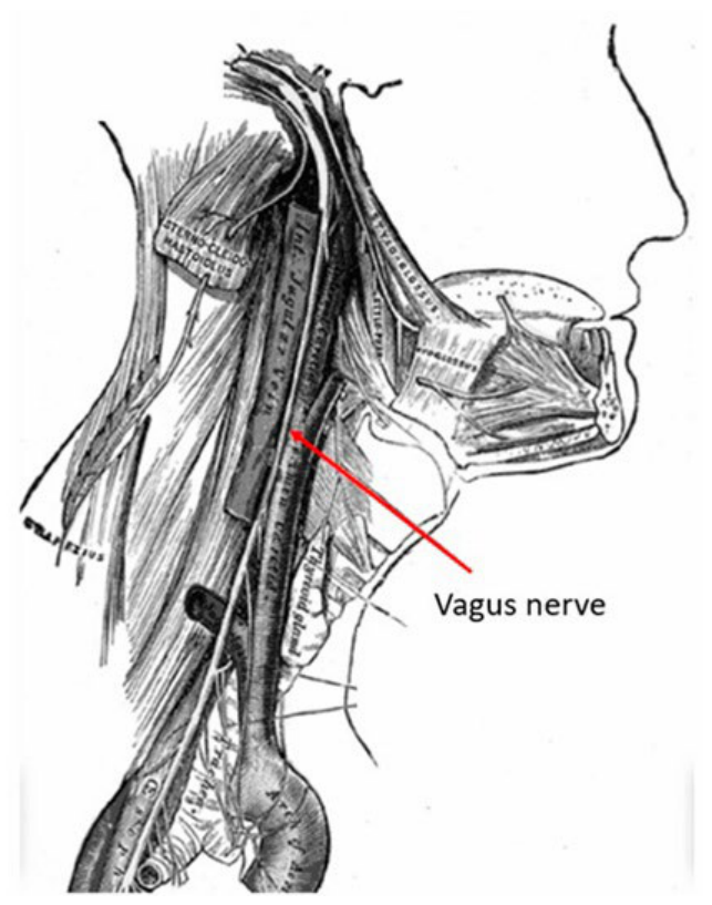
1. Anatomy of the Vagus Nerve and Neuromodulatory Projections
2. Material and Methods
3. Preliminary Laboratory Evidence
Central Effects of VNS in Animal Models
4. VNS for Epilepsy
4.1. Epilepsy Animal Models
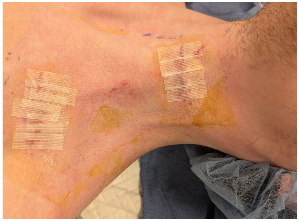
4.2. Surgical Procedure of Implantation of Vagus Nerve Stimulator
4.3. VNS for Epilepsy in Clinical Studies
5. VNS for Refractory Depression
6. The Substrate for Neural Plasticity with VNS in Motor Function Recovery
7. VNS-Paired Plasticity in Preclinical Studies
8. Clinical Application of Paired VNS for Stroke Rehabilitation in Adult Human Beings
9. Benefits of VNS in Therapy of Non-Motor or Non-Stroke Neurological Deficits
10. Role of Non-Invasive VNS in Neural Plasticity
11. Discussion
11.1. Epilepsy
11.2. VNS for Major Refractory Depression
11.3. VNS in the Management of Stroke
11.4. The Essence of Timing in Paired VNS
11.5. Neural Network Mechanisms of Paired VNS for the Treatment of Motor Deficits
11.6. Non-Invasive Paired VNS
11.7. VNS and Spinal Cord Injury Management
12. Conclusions
Future Directions
Author Contributions
Funding
Institutional Review Board Statement
Informed Consent Statement
Data Availability Statement
Acknowledgments
Conflicts of Interest
Abbreviations
| Ach | Acetylcholine |
| EEG | Electroencephalography |
| FDA | Food and Drug Administration |
| FMA-UE | Fugl–Meyer Assessment Upper Extremity |
| MAAVNS | Motor-Activated Auricular Vagus Nerve Stimulation |
| NTS | Nucleus Tractus Solitarius |
| RAVANS | Respiratory-gated Auricular Vagal Afferent Nerve Stimulation |
| RCT | Randomized Controlled Trial |
| taVNS | Transcutaneous Auricular Vagal Nerve Stimulation |
| tcVNS | Transcutaneous Cervical Vagal Nerve Stimulation |
| VNS | Vagus Nerve Stimulation |
References
- Foley, J.O.; Dubois, F.S. Quantitative studies of the vagus nerve in the cat: I. The ratio of sensory to motor fibers. J. Comp. Neurol. 1937, 67, 49–67. [Google Scholar] [CrossRef]
- Bailey, P.; Bremer, F. A sensory cortical representation of the vagus nerve: With a note on the effects of low blood pressure on the cortical electrogram. J. Neurophysiol. 1938, 1, 405–412. [Google Scholar] [CrossRef]
- Beckstead, R.M.; Norgren, R. An autoradiographic examination of the central distribution of the trigeminal, facial, glossopharyngeal, and vagal nerves in the monkey. J. Comp. Neurol. 1979, 184, 455–472. [Google Scholar] [CrossRef]
- Zhukova, G.P. The afferent pathway to the locus coeruleus from the nucleus of the solitary tract. Neurosci. Behav. Physiol. 1980, 10, 27–32. [Google Scholar] [CrossRef]
- Herbert, H. Evidence for projections from medullary nuclei onto serotonergic and dopaminergic neurons in the midbrain dorsal raphe nucleus of the rat. Cell Tissue Res. 1992, 270, 149–156. [Google Scholar] [CrossRef]
- Chandler, D.J.; Gao, W.J.; Waterhouse, B.D. Heterogeneous organization of the locus coeruleus projections to prefrontal and motor cortices. Proc. Natl. Acad. Sci. USA 2014, 111, 6816–6821. [Google Scholar] [CrossRef]
- Wilson, M.A.; Molliver, M.E. The organization of serotonergic projections to cerebral cortex in primates: Regional distribution of axon terminals. Neuroscience 1991, 44, 537–553. [Google Scholar] [CrossRef]
- Mesulam, M.M.; Geula, C. Nucleus basalis (Ch4) and cortical cholinergic innervation in the human brain: Observations based on the distribution of acetyl cholinesterase and choline acetyltransferase. J. Comp. Neurol. 1988, 275, 216–240. [Google Scholar] [CrossRef] [PubMed]
- Westlund, K.N.; Dan Coulter, J. Descending projections of the locus coeruleus and subcoeruleus/medial parabrachial nuclei in monkey: Axonal transport studies and dopamine-beta-hydroxylase immunocytochemistry. Brain Res. 1980, 2, 235–264. [Google Scholar] [CrossRef] [PubMed]
- Hulsey, D.R.; Riley, J.R.; Loerwald, K.W.; Rennaker, R.L.; Kilgard, M.P.; Hays, S.A. Parametric characterization of neural activity in the locus coeruleus in response to vagus nerve stimulation. Exp. Neurol. 2017, 289, 21–30. [Google Scholar] [CrossRef]
- Zanchetti, A.; Wang, S.C.; Moruzzi, G. The effect of vagal afferent stimulation on the EEG pattern of the cat. Electroencephalogr. Clin. Neurophysiol. 1952, 4, 357–361. [Google Scholar] [CrossRef]
- Chase, M.H.; Nakamura, Y. Cortical and subcortical EEG patterns of response to afferent abdominal vagal stimulation: Neurographic correlates. Physiol. Behav. 1968, 3, 605–610. [Google Scholar] [CrossRef]
- Zabara, J. Inhibition of experimental seizures in canines by repetitive vagal stimulation. Epilepsia 1992, 33, 1005–1012. [Google Scholar] [CrossRef] [PubMed]
- Krahl, S.E.; Senanayake, S.S.; Handforth, A. Destruction of peripheral C-fibers does not alter subsequent vagus nerve stimulation-induced seizure suppression in rats. Epilepsia 2001, 42, 586–589. [Google Scholar] [CrossRef]
- Woodbury, D.M.; Woodbury, J.W. Effects of vagal stimulation on experimentally induced seizures in rats. Epilepsia 1990, 31, S7–S19. [Google Scholar] [CrossRef]
- Naritoku, D.K.; Terry, W.J.; Helfert, R.H. Regional induction of fos immunoreactivity in the brain by anticonvulsant stimulation of the vagus nerve. Epilepsy Res. 1995, 22, 53–62. [Google Scholar] [CrossRef]
- Krahl, S.E.; Clark, K.B.; Smith, D.C.; Browning, R.A. Locus coeruleus lesions suppress the seizure-attenuating effects of vagus nerve stimulation. Epilepsia 1998, 39, 709–714. [Google Scholar] [CrossRef] [PubMed]
- Manta, S.; Dong, J.; Debonnel, G.; Blier, P. Enhancement of the function of rat serotonin and norepinephrine neurons by sustained vagus nerve stimulation. J. Psychiatry Neurosci. 2009, 34, 272–280. [Google Scholar] [CrossRef]
- Grimonprez, A.; Raedt, R.; Portelli, J.; Dauwe, I.; Larsen, L.E.; Bouckaert, C.; Delbeke, J.; Carrette, E.; Meurs, A.; Herdt, V.D.; et al. The antidepressant-like effect of vagus nerve stimulation is mediated through the locus coeruleus. J. Psychiatr. Res. 2015, 68, 1–7. [Google Scholar] [CrossRef]
- Hulsey, D.R.; Shedd, C.M.; Sarker, S.F.; Kilgard, M.P.; Hays, S.A. Norepinephrine and serotonin are required for vagus nerve stimulation directed cortical plasticity. Exp. Neurol. 2019, 320, 112975. [Google Scholar] [CrossRef]
- Meyers, E.C.; Kasliwal, N.; Solorzano, B.R.; Lai, E.; Bendale, G.; Berry, A.; Ganger, P.D.; Romero-Ortega, M.; Rennaker, R.L.; Kilgard, M.P.; et al. Enhancing plasticity in central networks improves motor and sensory recovery after nerve damage. Nat. Commun. 2019, 10, 5782. [Google Scholar] [CrossRef]
- Roosevelt, R.W.; Smith, D.C.; Clough, R.W.; Jensen, R.A.; Browning, R.A. Increased extracellular concentrations of norepinephrine in cortex and hippocampus following vagus nerve stimulation in the rat. Brain Res. 2006, 1119, 124–132. [Google Scholar] [CrossRef]
- Borland, M.S.; Vrana, W.A.; Moreno, N.A.; Fogarty, E.A.; Buell, E.P.; Sharma, P.; Engineer, C.T.; Kilgard, M.P. Cortical map plasticity as a function of vagus nerve stimulation intensity. Brain Stimul. 2016, 9, 117–123. [Google Scholar] [CrossRef] [PubMed]
- Vannemreddy, S.S.V.P.; Stone, J.L.; Slavin, K.V. Frederic Gibbs and his contributions to Epilepsy surgery and electroencephalography. Neurosurgery 2012, 70, 774–782. [Google Scholar] [CrossRef]
- Zabara, J. Peripheral control of hypersynchronous discharge in epilepsy. Electroencephalogr. Clin. Neurophysiol. 1985, 61, S162. [Google Scholar] [CrossRef]
- Lockard, J.S.; Congdon, W.C.; DuCharme, L.L. Feasibility and safety of vagal stimulation in monkey model. Epilepsia 1990, 31, S20–S26. [Google Scholar] [CrossRef]
- Revesz, D.; Rydenhag, B.; Ben-Menachem, E. Complications and safety of vagus nerve stimulation: 25 years of experience at a single center. J. Neurosurg. Pediatr. 2016, 18, 97–104. [Google Scholar] [CrossRef]
- Penry, J.K.; Dean, J.C. Prevention of intractable partial seizures by intermittent vagal stimulation in humans: Preliminary results. Epilepsia 1990, 31, S40–S43. [Google Scholar] [CrossRef] [PubMed]
- Uthman, B.M.; Wilder, B.J.; Penry, J.K.; Dean, C.; Ramsay, R.E.; Reid, S.A.; Hammond, E.J.; Tarver, W.B.; Wernicke, J.F. Treatment of epilepsy by stimulation of the vagus nerve. Neurology 1993, 43, 1338–1345. [Google Scholar] [CrossRef]
- Wilder, B.J.; Uthman, B.M.; Hammond, E.J. Vagal stimulation for control of complex partial seizures in medically refractory epileptic patients. Pacing Clin. Electrophysiol. 1991, 14, 108–115. [Google Scholar] [CrossRef]
- Ben-Menachem, E.; Mañon-Espaillat, R.; Ristanovic, R.; Wilder, B.J.; Stefan, H.; Mirza, W.; Tarver, W.B.; Wernicke, J.F. First International Vagus Nerve Stimulation Study Group. Vagus nerve stimulation for treatment of partial seizures: A controlled study of effect on seizures. Epilepsia 1994, 35, 616–626. [Google Scholar] [CrossRef] [PubMed]
- Ben-Menachem, E.; Hellstrom, K.; Waldton, C.; Augustinsson, L.E. Evaluation of refractory epilepsy treated with vagus nerve stimulation for up to 5 years. Neurology 1999, 52, 1265–1267. [Google Scholar] [CrossRef] [PubMed]
- The Vagus Nerve Stimulation Study Group. A randomized controlled trial of chronic vagus nerve stimulation for treatment of medically intractable seizures. Neurology 1995, 45, 224–230. [CrossRef]
- Handforth, A.; DeGiorgio, C.M.; Schachter, S.C.; Uthman, B.M.; Naritoku, D.K.; Tecoma, E.S.; Henry, T.R.; Collins, S.D.; Vaughn, B.V.; Gilmartin, R.C.; et al. Vagus nerve stimulation therapy for partial-onset seizures: A randomized active-control trial. Neurology 1998, 51, 48–55. [Google Scholar] [CrossRef]
- Ghani, S.; Vilensky, J.; Turner, B.; Tubbs, R.S.; Loukas, M. Meta analysis of vagus nerve stimulation treatment for epilepsy: Correlation between device setting parameters and acute response. Childs Nerv. Syst. 2015, 31, 2291–2304. [Google Scholar] [CrossRef]
- DeGiorgio, C.; Heck, C.; Bunch, S.; Britton, J.; Green, P.; Lancman, M.; Murphy, P.; Olejniczak, P.; Shih, J.; Arrambide, S.; et al. Vagus nerve stimulation for epilepsy: Randomized comparison of three stimulation paradigms. Neurology 2005, 65, 317–319. [Google Scholar] [CrossRef]
- DeGiorgio, C.M.; Thompson, J.; Lewis, P.; Arrambide, S.; Naritoku, D.; Handforth, A.; Labar, D.; Mullin, P.; Heck, C.; the VNS U.S. Study Group. Vagus nerve stimulation: Analysis of device parameters in 154 patients during the long-term XE5 study. Epilepsia 2001, 42, 1017–1020. [Google Scholar] [CrossRef]
- Elger, G.; Hoppe, C.; Falkai, P.; Rush, A.J.; Elger, C.E. Vagus nerve stimulation is associated with mood improvements in epilepsy patients. Epilepsy Res. 2000, 42, 203–210. [Google Scholar] [CrossRef]
- Rush, A.J.; George, M.S.; Sackeim, H.A.; Marangell, L.B.; Husain, M.M.; Miller, C.; Nahas, Z.; Haines, S.; Simpson, R., Jr.; Goodman, R. Vagus nerve stimulation (VNS) for treatment-resistant depressions: A multicenter study. Biol. Psychiatry 2000, 47, 276–286. [Google Scholar] [CrossRef]
- Bohning, D.E.; Lomarev, M.P.; Denslow, S.; Nahas, Z.; Shastri, A.; George, M.C. Feasibility of vagus nerve stimulation-synchronized blood oxygenation level-dependent functional MRI. Investig. Radiol. 2001, 36, 470–479. [Google Scholar] [CrossRef] [PubMed]
- Mu, Q.; Bohning, D.E.; Nahas, Z.; Walker, J.; Anderson, B.; Johnson, K.A.; Denslow, S.; Lomarev, M.; Moghadam, P.; Chae, J.-H.; et al. Acute vagus nerve stimulation using different pulse widths produces varying brain effects. Biol. Psychiatry 2004, 55, 816–825. [Google Scholar] [CrossRef] [PubMed]
- Perini, G.I.; Toffanin, T.; Pigato, G.; Ferri, G.; Follador, H.; Zonta, F.; Carlo, P.; Giulia, P.; Luca, D.; Giuseppe, R.; et al. Hippocampal gray volumes increase in treatment-resistant depression responding to vagus nerve stimulation. J. ECT 2017, 33, 160–166. [Google Scholar] [CrossRef]
- Tu, Y.; Fang, J.; Cao, J.; Wang, Z.; Park, J.; Jorgenson, K.; Lang, C.; Liu, J.; Zhang, G.; Zhao, Y.; et al. A distinct biomarker of continuous transcutaneous vagus nerve stimulation treatment in major depressive disorder. Brain Stimul. 2018, 11, 501–508. [Google Scholar] [CrossRef]
- George, M.S.; Rush, J.; Marangelli, L.B.; Sackeim, H.A.; Brannan, S.K.; Davis, S.M.; Howland, R.; Kling, M.A.; Moreno, F.; Rittberg, B.; et al. A one-year comparison of vagus nerve stimulation with treatment as usual for treatment-resistant depression. Biol. Psychiatry 2005, 58, 364–373. [Google Scholar] [CrossRef]
- Elliott, P.; Wallis, D.I. Serotonin and L-norepinephrine as mediators of altered excitability in neonatal rat motoneurons studied in vitro. Neuroscience 1992, 47, 533–544. [Google Scholar] [CrossRef] [PubMed]
- Gu, Q. Neuromodulatory transmitter systems in the cortex and their role in cortical plasticity. Neuroscience 2002, 111, 815–835. [Google Scholar] [CrossRef] [PubMed]
- Engineer, N.D.; Riley, J.R.; Seale, J.D.; Vrana, W.A.; Shetake, J.A.; Sudanagunta, S.P.; Borland, M.S.; Kilgard, M.P. Reversing pathological neural activity using targeted plasticity. Nature 2011, 470, 101–104. [Google Scholar] [CrossRef]
- Shetake, J.A.; Engineer, N.D.; Vrana, W.A.; Wolf, J.T.; Kilgard, M.P. Pairing tone trains with vagus nerve stimulation induces temporal plasticity in auditory cortex. Exp. Neurol. 2012, 233, 342–349. [Google Scholar] [CrossRef]
- Engineer, C.T.; Engineer, N.D.; Riley, J.R.; Seale, J.D.; Kilgard, M.P. Pairing speech sounds with vagus nerve stimulation drives stimulus-specific cortical plasticity. Brain Stimul. 2015, 8, 637–644. [Google Scholar] [CrossRef]
- Loerwald, K.W.; Borland, M.S.; Rennaker, R.L.; Hays, S.A.; Kilgard, M.P. The interaction of pulse width and current intensity on the extent of cortical plasticity evoked by vagus nerve stimulation. Brain Stimul. 2018, 11, 271–277. [Google Scholar] [CrossRef]
- Bowles, S.; Hickman, J.; Peng, X.; Williamson, W.R.; Huang, R.; Washington, K.; Donegan, D.; Welle, C.G. Vagus nerve stimulation drives selective circuit modulation through cholinergic reinforcement. Neuron 2022, 110, 2867–2885. [Google Scholar] [CrossRef]
- Tseng, C.T.; Gaulding, S.J.; Dancel, C.L.E.; Thorn, C.A. Local activation of α2 adrenergic receptors is required for vagus nerve stimulation induced motor cortical plasticity. Sci. Rep. 2021, 11, 21645. [Google Scholar] [CrossRef]
- Smith, D.C.; Modglin, A.A.; Roosevelt, R.W.; Neese, S.L.; Jensen, R.A.; Browning, R.A.; Clough, R.W. Electrical stimulation of the vagus nerve enhances cognitive and motor recovery following moderate fluid percussion injury in the rat. J. Neurotrauma 2005, 22, 1485–1502. [Google Scholar] [CrossRef] [PubMed]
- Porter, B.A.; Khodaparast, N.; Fayyaz, T.; Cheung, R.J.; Ahmed, S.S.; Vrana, W.A.; Rennaker, R.L.; Kilgard, M.P. Repeatedly pairing vagus nerve stimulation with a movement reorganizes primary motor cortex. Cereb. Cortex 2012, 22, 2365–2374. [Google Scholar] [CrossRef]
- Meyers, E.C.; Solorzano, B.R.; James, J.; Ganzer, P.D.; Lai, E.S.; Rennaker, R.L.; Kilgard, M.P.; Hays, S.A. Vagus nerve stimulation enhances stable plasticity and generalization of stroke recovery. Stroke 2018, 49, 710–717. [Google Scholar] [CrossRef]
- Darrow, M.J.; Mian, T.M.; Torres, M.; Haider, Z.; Danaphongse, T.; Rennaker, R.L., Jr.; Kilgard, M.P.; Hays, S.A. Restoration of somatosensory function by pairing vagus nerve stimulation with tactile rehabilitation. Ann. Neurol. 2020, 87, 194–205. [Google Scholar] [CrossRef]
- Dawson, J.; Pierce, D.; Dixit, A.; Kimberley, T.J.; Robertson, M.; Tarver, B.; Hilmi, O.; Mclean, J.; Forbes, K.; Kilgard, M.P.; et al. Safety, feasibility, and efficacy of vagus nerve stimulation paired with upper-limb rehabilitation after ischemic stroke. Stroke 2016, 47, 143–150. [Google Scholar] [CrossRef]
- Kilgard, M.P.; Rennaker, R.L.; Alexander, J.; Dawson, J. Vagus nerve stimulation paired with tactile training improved sensory function in a chronic stroke patient. NeuroRehabilitation 2018, 42, 159–165. [Google Scholar] [CrossRef] [PubMed]
- Kimberley, T.J.; Pierce, D.; Prudente, C.N.; Francisco, G.E.; Yozbatiran, N.; Smith, P.; Tarver, B.; Engineer, N.D.; Alexander, D.; Kline, D.K.; et al. Vagus nerve stimulation paired with upper limb rehabilitation after chronic stroke. Stroke 2018, 49, 2789–2792. [Google Scholar] [CrossRef]
- Dawson, J.; Liu, C.Y.; Francisco, G.E.; Cramer, S.C.; Wolf, S.L.; Dixit, A.; Alexander, J.; Ali, R.; Brown, B.L.; Feng, W.; et al. Vagus nerve stimulation paired with rehabilitation for upper limb motor function after ischaemic stroke (VNS-REHAB): A randomized, blinded, pivotal, device trial. Lancet 2021, 397, 1545–1553. [Google Scholar] [CrossRef] [PubMed]
- Ganzer, P.D.; Darrow, M.J.; Meyers, E.C.; Solorzano, B.R.; Ruiz, A.D.; Robertson, N.M.; Adcock, K.S.; James, J.T.; Jeong, H.S.; Becker, A.M.; et al. Closed-loop neuromodulation restores network connectivity and motor control after spinal cord injury. eLife 2018, 7, e32058. [Google Scholar] [CrossRef]
- Morrison, R.A.; Hays, S.A.; Kilgard, M.P. Vagus nerve stimulation as a potential adjuvant to rehabilitation for post-stroke motor speech disorders. Front. Neurosci. 2021, 15, 715928. [Google Scholar] [CrossRef]
- Vannemreddy, P.S.; Cummings, M.; Bahrii, R.V.; Slavin, K.V. Vagus Nerve Stimulation in Stroke Management: Brief Review of Evolution and Present Applications Paired with Rehabilitation. Brain Sci. 2025, 15, 346. [Google Scholar] [CrossRef] [PubMed]
- Burger, A.M.; D’agostini, M.; Verkuil, B.; Van Diest, I. Moving beyond belief: A narrative review of potential biomarkers for transcutaneous vagus nerve stimulation. Psychophysiology 2020, 57, e13571. [Google Scholar] [CrossRef]
- Bucksot, J.E.; Morales, C.K.; Skipton, S.K.; Hays, S.A. Parametric characterization of the rat Hering-Breuer reflex evoked with implanted and non-invasive vagus nerve stimulation. Exp. Neurol. 2020, 327, 113220. [Google Scholar] [CrossRef]
- Hays, S.A.; Rennaker, R.L.; Kilgard, M.P. How to fail with paired VNS therapy. Brain Stimul. 2023, 16, 1252–1258. [Google Scholar] [CrossRef]
- Burger, A.M.; Verkuil, B. Transcutaneous nerve stimulation via the tragus: Are we really stimulating the vagus nerve? Brain Stimul. 2018, 11, 945–946. [Google Scholar] [CrossRef]
- Hammer, N.; Löffler, S.; Cakmak, Y.O.; Ondruschka, B.; Planitzer, U.; Schultz, M.; Winkler, D.; Weise, D. Cervical vagus nerve morphometry and vascularity in the context of nerve stimulation– A cadaveric study. Sci. Rep. 2018, 8, 7997. [Google Scholar] [CrossRef] [PubMed]
- Chang, J.L.; Coggins, A.N.; Saul, M.; Paget-Blanc, A.; Straka, M.; Wright, J.; Datta-Chaudhuri, T.; Zanos, S.; Volpe, B.T. Transcutaneous auricular vagus nerve stimulation (tAVNS) delivered during upper limb interactive robotic training demonstrates novel antagonist control for reaching movements following stroke. Front. Neurosci. 2021, 15, 767302. [Google Scholar] [CrossRef] [PubMed]
- Li, J.N.; Xie, C.C.; Li, C.Q.; Zhang, C.F.; Tang, H.; Jin, C.N.; Ma, J.X.; Wen, L.; Zhang, K.M.; Niu, L.C. Efficacy and safety of transcutaneous auricular vagus nerve stimulation combined with conventional rehabilitation training in acute stroke patients: A randomized controlled trial conducted for 1 year involving 60 patients. Neural Regen. Res. 2022, 17, 1809–1813. [Google Scholar] [CrossRef]
- Bailey, P.; Gibbs, F.A. The surgical treatment of psychomotor epilepsy. J. Am. Med. Assoc. 1951, 10, 365–370. [Google Scholar] [CrossRef] [PubMed]
- Lanska, D.J. J.L. Corning and vagal nerve stimulation for seizures in the 1880s. Neurology 2022, 58, 452–459. [Google Scholar] [CrossRef]
- Afra, P.; Adamolekun, B.; Aydemir, S.; Watson, G. Evolution of the vagus nerve stimulation (VNS) therapy system technology for drug-resistant epilepsy. Front. Med. Technol. 2021, 3, 696543. [Google Scholar] [CrossRef]
- Dibué, M.; Greco, T.; Spoor, J.K.H.; Tahir, Z.; Specchio, N.; Hanggi, D.; Steiger, H.-J.; Kamp, M.A. Vagus nerve stimulation in patients with Lennox-Gastaut syndrome: A meta-analysis. Acta Neurol. Scand. 2021, 143, 497–508. [Google Scholar] [CrossRef]
- Clancy, J.A.; Mary, D.A.; Wittle, K.K.; Greenwood, J.P.; Deuchars, S.A.; Deuchars, J. Non-invasive vagus nerve stimulation in healthy humans reduces sympathetic nerve activity. Brain Stimul. 2014, 7, 871–877. [Google Scholar] [CrossRef]
- Voineskos, D.; Daskalakis, Z.J.; Blumberger, D.M. Management of treatment-resistant depression: Challenges and strategies. Neuropsychiatr. Dis. Treat. 2020, 16, 221–234. [Google Scholar] [CrossRef]
- George, M.S.; Nahas, Z.; Bohning, D.E.; Lomarev, M.; Denslow, S.; Osenbach, O.; Ballenger, J.C. Vagus nerve stimulation: A new form of therapeutic brain stimulation. CNS Spectr. 2000, 5, 43–52. [Google Scholar] [CrossRef] [PubMed]
- Sackeim, H.A.; Rush, J.; George, M.S.; Marangell, L.B.; Husain, M.M.; Nahas, Z.; Johnson, C.R.; Seidman, S.; Giller, C.; Haines, S.; et al. Vagus nerve stimulation (VNS™) for treatment-resistant depression: Efficacy, side effects, and predictors of outcome. Neuropsychopharmacology 2000, 25, 713–728. [Google Scholar] [CrossRef] [PubMed]
- Nahas, Z.; Marangell, L.B.; Husain, M.M.; Rush, J.; Sackeim, H.A.; Lisanby, S.H.; Martinez, J.M.; George, M.S. Two-year outcome of vagus nerve stimulation (VNS) for treatment of major depressive episodes. J. Clin. Psychiatry 2005, 66, 1097–1104. [Google Scholar] [CrossRef]
- Rush, A.J.; Marangell, L.B.; Sackeim, H.A.; George, M.S.; Brannan, S.K.; Davis, S.M.; Howland, R.; Kling, M.A.; Rittberg, B.R.; Burke, W.J.; et al. Vagus nerve stimulation for treatment resistant depression: A randomized, controlled acute phase trial. Biol. Psychiatry 2005, 58, 347–354. [Google Scholar] [CrossRef]
- Scherrmann, J.; Hoppe, C.; Kral, T.; Schramm, J.; Elger, C.E. Vagus nerve stimulation: Clinical experience in a large patient series. J. Clin. Neurophysiol. 2001, 18, 408–414. [Google Scholar] [CrossRef]
- Englot, D.J.; Hassnain, K.H.; Rolston, J.D.; Harward, S.C.; Sinha, S.R.; Haglund, M.M. Quality-of-life metrics with vagus nerve stimulation for epilepsy from provider survey data. Epilepsy Behav. 2017, 66, 4–9. [Google Scholar] [CrossRef]
- Khodaparast, N.; Kilgard, M.P.; Casavant, R.; Ruiz, A.; Qureshi, I.; Ganzer, P.D.; Rennaker, R.L.; Hays, S.A. Vagus nerve stimulation during rehabilitative training improves forelimb recovery after chronic ischemic stroke in rats. Neurorehab Neural Repair. 2016, 30, 676–684. [Google Scholar] [CrossRef]
- Engineer, N.D.; Kimberley, T.J.; Prudente, C.N.; Dawson, J.; Tarver, W.B.; Hays, S.A. Targeted vagus nerve stimulation for rehabilitation after stroke. Front. Neurosci. 2019, 13, 280. [Google Scholar] [CrossRef]
- Vora, I.; Gochyyev, P.; Engineer, N.; Wolf, S.L.; Kimberley, T.J. Distal versus proximal arm improvement after paired vagus nerve stimulation therapy after chronic stroke. Arch. Phys. Med. Rehabil. 2024, 105, 1709–1717. [Google Scholar] [CrossRef] [PubMed]
- Gerstner, W.; Lehmann, M.; Liakoni, V.; Corneil, D.; Brea, J. Eligibility traces and plasticity on behavioral time scales: Experimental support of neo Hebbian three-factor learning rules. Front. Neural Circuits 2018, 12, 53. [Google Scholar] [CrossRef]
- Yagishita, S.; Hayashi-Takagi, A.; Ellis-Davies, G.C.R.; Urakubo, H.; Ishii, S.; Kasai, H. A critical time window for dopamine actions on the structural plasticity of dendritic spines. Science 2014, 345, 1616–1620. [Google Scholar] [CrossRef] [PubMed]
- He, K.; Huertas, M.; Hong, S.U.Z.; Tie, X.; Hell, J.W.; Shouval, H.; Kirkwood, A. Distinct eligibility traces for LTP and LTD in cortical synapses. Neuron 2015, 88, 528–538. [Google Scholar] [CrossRef]
- Hays, S.A.; Khodaparast, N.; Ruiz, A.; Sloan, A.M.; Hulsey, D.R.; Rennaker, R.L.; Kilgard, M.P. The timing and amount of vagus nerve stimulation during rehabilitative training affect poststroke recovery of forelimb strength. Neuroreport 2014, 25, 676–682. [Google Scholar] [CrossRef]
- Ruiz, A.D.; Malley, K.M.; Danaphongse, T.T.; Ahmad, F.N.; Beltran, C.M.; White, M.L.; Baghdadi, S.; Pruitt, D.T.; Rennaker, R.L.; Kilgard, M.P.; et al. Vagus nerve stimulation must occur during tactile rehabilitation to enhance somatosensory recovery. Neuroscience 2023, 532, 79–86. [Google Scholar] [CrossRef] [PubMed]
- Jelinek, M.; Lipkova, J.; Duris, K. Vagus nerve stimulation as immunomodulatory therapy for stroke: A comprehensive review. Exp. Neurol. 2024, 372, 114628. [Google Scholar] [CrossRef]
- Noble, L.J.; Chuah, A.; Callahan, K.K.; Souza, R.R.; Mcintyre, C.K. Peripheral effects of vagus nerve stimulation on anxiety and extinction of conditioned fear in rats. Learn. Mem. 2019, 26, 245–251. [Google Scholar] [CrossRef]
- Brougher, J.; Sanchez, C.A.; Aziz, U.S.; Gove, K.F.; Thorn, C.A. Vagus nerve stimulation induced motor map plasticity does not require cortical dopamine. Front. Neurosci. 2021, 15, 693140. [Google Scholar] [CrossRef] [PubMed]
- Lemon, R.N. Descending pathways in motor control. Ann. Rev. Neurosci. 2008, 31, 195–218. [Google Scholar] [CrossRef]
- Garcia, R.G.; Lin, R.L.; Lee, J.; Kim, J.; Barbieri, R.; Sclocco, R.; Wasan, A.D.; Edwards, R.R.; Rosen, B.R.; Hadjikhani, N.; et al. Modulation of brainstem activity and connectivity by respiratory-gated auricular vagal afferent nerve stimulation (RAVANS) in migraine patients. Pain 2017, 158, 1461. [Google Scholar] [CrossRef] [PubMed]
- Cook, D.N.; Thompson, S.; Stomberg-Firestein, S.; Bikson, M.; George, M.S.; Jenkins, D.D.; Badran, B.W. Design and validation of a closed-loop, motor-activated auricular vagus nerve stimulation (MAAVNS) system for neurorehabilitation. Brain Stimul. 2020, 13, 800–803. [Google Scholar] [CrossRef] [PubMed]
- Badran, B.W.; Jenkins, D.D.; Cook, D.; Thompson, M.; DeVres, W.H.; Mappin, G.; Summers, P.; Bikson, M.; George, M.S. Transcutaneous auricular vagus nerve stimulation paired rehabilitation for oromotor feeding problems in newborns: An open label pilot study. Front. Hum. Neurosci. 2020, 14, 77. [Google Scholar] [CrossRef]
- Redgrave, J.N.; Moore, L.; Oyekunle, T.; Ebrahim, M.; Falidas, K.; Snowdon, N.; Majid, A. Transcutaneous auricular vagus nerve stimulation with concurrent upper limb repetitive task practice for poststroke motor recovery: A pilot study. J. Stroke Cerebrovasc. Dis. 2018, 27, 1998–2005. [Google Scholar] [CrossRef]
- Baig, S.S.; Falidas, K.; Laud, P.J.; Snowdon, N.; Farooq, M.U.; Ali, A.; Majid, A.; Redgrave, J.N. Transcutaneous auricular vagus nerve stimulation with upper limb repetitive task practice may improve sensory recovery in chronic stroke. J. Stroke Cerebrovasc. Dis. 2019, 28, 104348. [Google Scholar] [CrossRef]
- Capone, F.; Miccinilli, S.; Pellegrino, G.; Zollo, L.; Simonett, I.D.; Bressi, F.; Lucia, F.; Ranieri, F.; Falato, E.; Santo, A.D.; et al. Transcutaneous Vagus nerve stimulation combined with robotic rehabilitation improves upper limb function after stroke. Neural. Plast. 2017, 2017, 7876507. [Google Scholar] [CrossRef]
- ClinicalTrials.gov. Available online: https://clinicaltrials.gov/study/NCT04129242?term=NCT04129242&rank=1 (accessed on 1 January 2026).
- Badran, B.W.; Peng, X.; Baker-Vogel, B.; Hutchinson, S.; Finetto, P.; Rishe, K.; Fortune, A.; Kitchens, E.; O’Leary, G.H.; Short, A.; et al. Motor Activated Auditory Vagus Nerve Stimulation as a potential neuromodulation approach for post-stroke motor rehabilitation: A pilot study. Neurorehabilit. Neural Repair 2023, 37, 374–383. [Google Scholar] [CrossRef]
- ClinicalTrials.gov. Available online: https://clinicaltrials.gov/study/NCT04288245?term=NCT04288245&rank=1 (accessed on 1 January 2026).
- ClinicalTrials.gov. Available online: https://clinicaltrials.gov/study/NCT05601661?term=NCT05601661&rank=1 (accessed on 1 January 2026).
- Yozbatiran, N.; Francisco, G.E.; Korupolu, R. Safety and feasibility of paired vagus nerve stimulation with rehabilitation for improving upper extremity function in people with cervical spinal cord injury: Study protocol. Front. Neurol. 2024, 15, 1465764. [Google Scholar] [CrossRef] [PubMed]
- Kalagara, R.; Chennareddy, S.; Reford, E.; Bhimani, A.; Cummins, D.D.; Downes, M.H.; Tosto, J.M.; Bederson, J.B.; Mocco, J.; Putrino, D.; et al. Complications of implanted vagus nerve stimulation: A systematic review and meta-analysis. Cerebrovasc. Dis. 2025, 54, 112–120. [Google Scholar] [CrossRef] [PubMed]
- Wolf, S.L.; Winstein, C.J.; Miller, J.P.; Taub, E.; Uswatte, G.; Morris, D.; Giuliani, C.; Light, K.E.; Nichols-Larsen, D.; for the EXCITE Investigators. Effect of constraint-induced movement therapy on upper extremity function 3 to 9 months after stroke: The EXCITE randomized clinical trial. J. Am. Med. Assoc. 2006, 296, 2095–2104. [Google Scholar] [CrossRef] [PubMed]
- Michaelsen, S.M.; Dannenbaum, R.; Levin, M.F. Task-specific training with trunk restraint on arm recovery in stroke: Randomized control trial. Stroke 2006, 37, 186–192. [Google Scholar] [CrossRef]
- Graupe, D.; Basu, I.; Tuninetti, D.; Vannemreddy, P.; Slavin, K.V. Adaptively controlling deep brain stimulation in essential tremor patient via surface electromyography. Neurol. Res. 2010, 32, 899–904. [Google Scholar] [CrossRef]


Disclaimer/Publisher’s Note: The statements, opinions and data contained in all publications are solely those of the individual author(s) and contributor(s) and not of MDPI and/or the editor(s). MDPI and/or the editor(s) disclaim responsibility for any injury to people or property resulting from any ideas, methods, instructions or products referred to in the content. |
© 2026 by the authors. Licensee MDPI, Basel, Switzerland. This article is an open access article distributed under the terms and conditions of the Creative Commons Attribution (CC BY) license.
Share and Cite
Vannemreddy, P.; Slavin, K.V. Vagus Nerve Stimulation for Neuromodulation: Evolution from Bench to Bedside. Neurol. Int. 2026, 18, 94. https://doi.org/10.3390/neurolint18050094
Vannemreddy P, Slavin KV. Vagus Nerve Stimulation for Neuromodulation: Evolution from Bench to Bedside. Neurology International. 2026; 18(5):94. https://doi.org/10.3390/neurolint18050094
Chicago/Turabian StyleVannemreddy, Prasad, and Konstantin V. Slavin. 2026. "Vagus Nerve Stimulation for Neuromodulation: Evolution from Bench to Bedside" Neurology International 18, no. 5: 94. https://doi.org/10.3390/neurolint18050094
APA StyleVannemreddy, P., & Slavin, K. V. (2026). Vagus Nerve Stimulation for Neuromodulation: Evolution from Bench to Bedside. Neurology International, 18(5), 94. https://doi.org/10.3390/neurolint18050094