Review

From Genetics to Phenotype: Understanding the Diverse Manifestations of Cardiovascular Genetic Diseases in Pediatric Populations

by
Jule Leonie Gutmann
1,†,
Alina Spister
1,† and
Lara Baticic
2,*
1
Faculty of Medicine, University of Rijeka, 51000 Rijeka, Croatia
2
Department of Medical Chemistry, Biochemistry and Clinical Chemistry, Faculty of Medicine, University of Rijeka, 51000 Rijeka, Croatia
*
Author to whom correspondence should be addressed.
These authors contributed equally to this work.
Cardiogenetics 2025, 15(4), 29; https://doi.org/10.3390/cardiogenetics15040029 (registering DOI)
Submission received: 29 July 2025 / Revised: 17 September 2025 / Accepted: 25 September 2025 / Published: 11 October 2025
(This article belongs to the Section Inherited Heart Disease-Children)

Abstract

Congenital genetic heart defects are major contributors to pediatric morbidity and mortality, underscoring the importance of early detection and individualized therapeutic strategies. This review aimed to summarize current knowledge on a spectrum of inherited cardiovascular disorders, with a focus on their genetic etiology, molecular pathogenesis, and phenotypic presentation in children. Conditions discussed include Marfan syndrome, Noonan syndrome, various cardiomyopathies, Duchenne muscular dystrophy, DiGeorge syndrome, and the tetralogy of Fallot. These six conditions were selected to represent the spectrum of pediatric cardiovascular genetic diseases, encompassing connective tissue disorders, multisystem syndromes, primary myocardial diseases, neuromuscular cardiac involvement, and structural congenital defects, thereby illustrating how distinct genotypes lead to diverse phenotypes. For each disorder, the underlying genetic mutations, associated molecular pathways, cardiovascular involvement, clinical features, and approaches to diagnosis and management are examined. Emphasis is placed on the role of timely diagnosis, genetic counseling, and personalized treatment in improving patient outcomes. The review concludes by highlighting emerging research directions and novel therapeutic interventions aimed at enhancing care for these complex pediatric conditions.

1. Introduction

Congenital heart defects (CHDs) are the most prevalent congenital malformations and represent a leading cause of neonatal and infant mortality worldwide. By 2021, over 4.18 million children under five were living with CHD worldwide, which shows a 3.4% rise since 1990. Over the same period, CHD-related mortality fell by more than half, indicating substantial improvements in survival and disease burden [1]. They comprise a heterogeneous group of structural and functional cardiovascular abnormalities that originate from mutations critical for cardiac development and function. These mutations often follow autosomal dominant, autosomal recessive, or X-linked inheritance patterns [2]. Depending on the genetic context, CHDs may present as isolated cardiac anomalies or as part of syndromic conditions involving multiple organ systems.
The clinical spectrum of CHDs encompasses a wide range of phenotypes, from subclinical structural abnormalities identifiable only through echocardiography or other advanced imaging modalities, to critical conditions manifesting with overt signs of congestive heart failure, severe cyanosis due to impaired oxygen delivery, or sudden cardiac death during infancy and early childhood. The pathophysiological basis of this heterogeneity lies in the diverse structural malformations, ranging from septal defects and valvular dysplasia to complex anomalies involving multiple cardiac chambers and great vessels which can profoundly disrupt normal hemodynamic function. Furthermore, the early onset of disease, coupled with marked genetic heterogeneity, incomplete penetrance, and variable expressivity, complicates both prognostication and therapeutic planning. These factors render CHDs a major clinical challenge, necessitating the integration of genetic, imaging, and physiological data to optimize diagnostic accuracy, risk stratification, and individualized management strategies in pediatric cardiology [1,2,3,4,5]. Timely diagnosis and targeted management are essential for improving clinical outcomes, as early intervention can prevent or attenuate disease progression and reduce the risk of life-threatening complications. The incorporation of molecular genetic testing into clinical practice has markedly enhanced the ability to detect causative variants, allowing for more precise prognosis and personalized treatment. In addition, early genetic diagnosis enables the initiation of genetic counseling and family screening, which are essential for risk assessment, reproductive planning, and preventive strategies.
The aim of this review is to provide a comprehensive overview of main congenital genetic heart disorders, including Marfan syndrome, Noonan syndrome, hypertrophic cardiomyopathy, dilated cardiomyopathy with a focus on Duchenne muscular dystrophy, 22q11.2 deletion syndrome, and the tetralogy of Fallot. By examining their genetic etiologies, molecular mechanisms, cardiovascular manifestations, and pediatric clinical presentation, this work highlights the clinical heterogeneity of these conditions. Emphasis is placed on the need for individualized treatment strategies and multidisciplinary management approaches to optimize outcomes in pediatric patients affected by genetic heart disease.

2. Congenital Genetic Heart Defects

2.1. Marfan Syndrome

Marfan syndrome is an autosomal dominant connective tissue disorder characterized by multisystemic involvement, with the most clinically significant manifestations affecting the cardiovascular, ocular, and musculoskeletal systems. The cardiovascular system is particularly vulnerable, with progressive aortic root dilation and predisposition to dissection representing major causes of morbidity and mortality in affected individuals. Ocular manifestations include ectopia lentis, myopia, and increased risk of retinal detachment, while musculoskeletal involvement often presents as disproportionate tall stature, arachnodactyly, scoliosis, pectus deformities, and joint hypermobility. Importantly, phenotypic expression is highly variable, even within families carrying the same mutation, complicating both diagnosis and risk stratification [3,4].
Early recognition in pediatric patients is crucial, as timely diagnosis enables proactive surveillance of aortic dimensions, implementation of pharmacological therapies such as β-blockers or angiotensin receptor blockers, and timely surgical intervention when indicated. Given its multisystemic nature, optimal care requires a multidisciplinary approach integrating cardiology, ophthalmology, orthopedics, and genetics, alongside psychosocial support for affected children and their families. This comprehensive strategy not only mitigates life-threatening complications but also improves long-term quality of life and functional outcomes.

2.1.1. Genetic Basis and Phenotypic Impact of Inherited Variants

Marfan syndrome (MFS) is predominantly inherited with an autosomal dominant pattern. The penetrance is variable, and it is estimated to affect between 1 in 3000 and 1 in 5000 individuals [3,4]. Up to 25 percent of MFS cases result from de novo mutations [4,5].
In the majority of cases, MFS is caused by a mutation in the fibrillin-1 gene (FBN1) on chromosome 15. Fibrillin-1 serves as the main structural element of extracellular microfibrils which are considered essential for the creation and preservation of elastic fibers. In case of a mutation, the microfibrils are unable to perform their role as a scaffold for elastin and, consequently, the tissue’s structural integrity is weakened [3,5]. Up to this point, more than 1800 different variants of the fibrillin-1 gene have been recorded. Some of these variants result in milder phenotypes but still share some characteristics with the classic Marfan phenotype [4]. In less than 10 percent of patients with the typical Marfan phenotype, no FBN1 variant is detected through current diagnostic methods [4,5]. The genetic cause and its pathophysiological consequences are summarized in Figure 1.

2.1.2. Impact on the Heart and Cardiovascular System

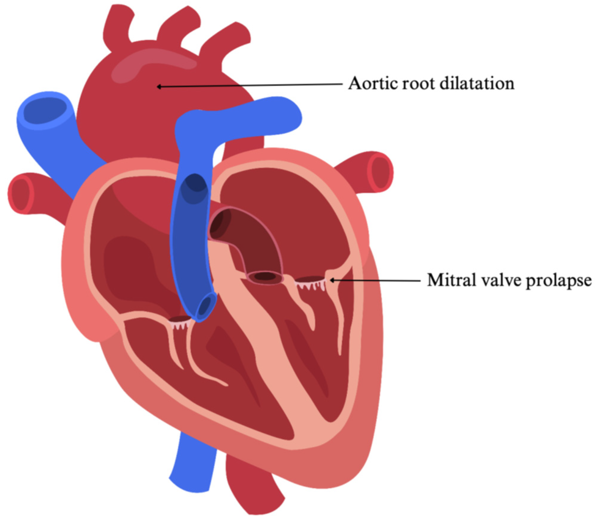
MFS exhibits a broad spectrum of clinical findings and can impact either a single organ system or multiple [4]. Cardiovascular involvement is frequently observed, although, during early childhood, the symptoms may be subtle and less pronounced. Commonly affected are the aorta and the heart valves [6]. The most observed cardiac manifestation in children is aortic root dilatation. Aortic root dissection is an unusual complication in children under the age of 10 years; it may, however, develop in adolescence [7,8]. Another common cardiac abnormality is mitral valve prolapses, which may be present with or without mitral regurgitation, tricuspid valve prolapses, and pulmonary artery dilation. Distal aortic dilation, primary cardiomyopathy or arrhythmias are less commonly seen in children [8]. The two most common anatomic anomalies of MFS are illustrated in Figure 2.

2.1.3. Symptoms and Clinical Presentation in Children

The clinical presentation of MFS is highly variable. Lens dislocation (ectopia lentis) is the most frequently observed ocular finding and bears the risk of superior and temporal lens subluxation. Additional ophthalmologic findings include retinal detachment, severe myopia, glaucoma, and early cataract formation [9,10,11]. The musculoskeletal system represents another commonly affected domain in individuals with Marfan syndrome. Patients often present with a tall stature, dolichostenomelia, and joint hypermobility. Further characteristic findings comprise pectus abnormalities, such as pectus excavatum or pectus carinatum, as well as scoliosis [9].

2.1.4. Diagnosis and Management

The diagnosis of MFS is usually clinical and relies on the Ghent nosology, revised in 2010. In the absence of any family history, the presence of aortic root dilatation together with ectopia lentis is sufficient for diagnosing MFS. The diagnosis can also be made if both aortic root dilatation and FBN1 mutation are present. If there is a positive family history of MFS the diagnosis can be established either by the presence of ectopia lentis or by the presence of aortic root dilatation [11,12,13,14].
Timely diagnosis and intervention are crucial to prevent progressive aortic root dilatation and its associated consequences. Pharmacological management involves the use of beta blockers to slow the progression of aortic root dilatation and to manage arterial hypertension. The latter is relatively rare in pediatric patients. Commonly used agents in this class are atenolol and propranolol. Angiotensin receptor blockers (ARBs), such as losartan, are utilized as an alternative or adjunct to beta blockers in attenuating aortic root enlargement. In children with MFS, combination therapy using both beta blockers and ARBs has shown greater effectiveness than either agent alone. In children with an aortic root diameter exceeding 50 mm, surgical intervention is required, as it remains the only definite measure to prevent aortic dissection. The main surgical techniques consist of aortic valve-sparing surgery, which involves replacing only the dilated aortic root, and aortic valve replacement surgery, where both the aortic root and valve are replaced. In children the first option is preferred [8,14,15,16].

2.2. Noonan Syndrome

Noonan Syndrome is a common genetic disorder characterized by significant phenotypic variability and multisystem involvement, most notably affecting the cardiovascular, craniofacial, and lymphatic systems [17,18]. This section outlines the genetic etiology, molecular mechanisms, and the diverse clinical spectrum of Noonan syndrome in pediatric patients, with particular emphasis on early recognition, cardiovascular manifestations, and the importance of a multidisciplinary approach to diagnosis and long-term management.

2.2.1. Genetic Basis and Phenotypic Impact of Inherited Variants

Noonan Syndrome is a heterogenous congenital disease predominantly with an autosomal dominant inheritance pattern, whereas autosomal recessive inheritance has been documented only in rare cases. The prevalence is estimated to be between 1 in 1000 and 1 in 2500 individuals [17,18].
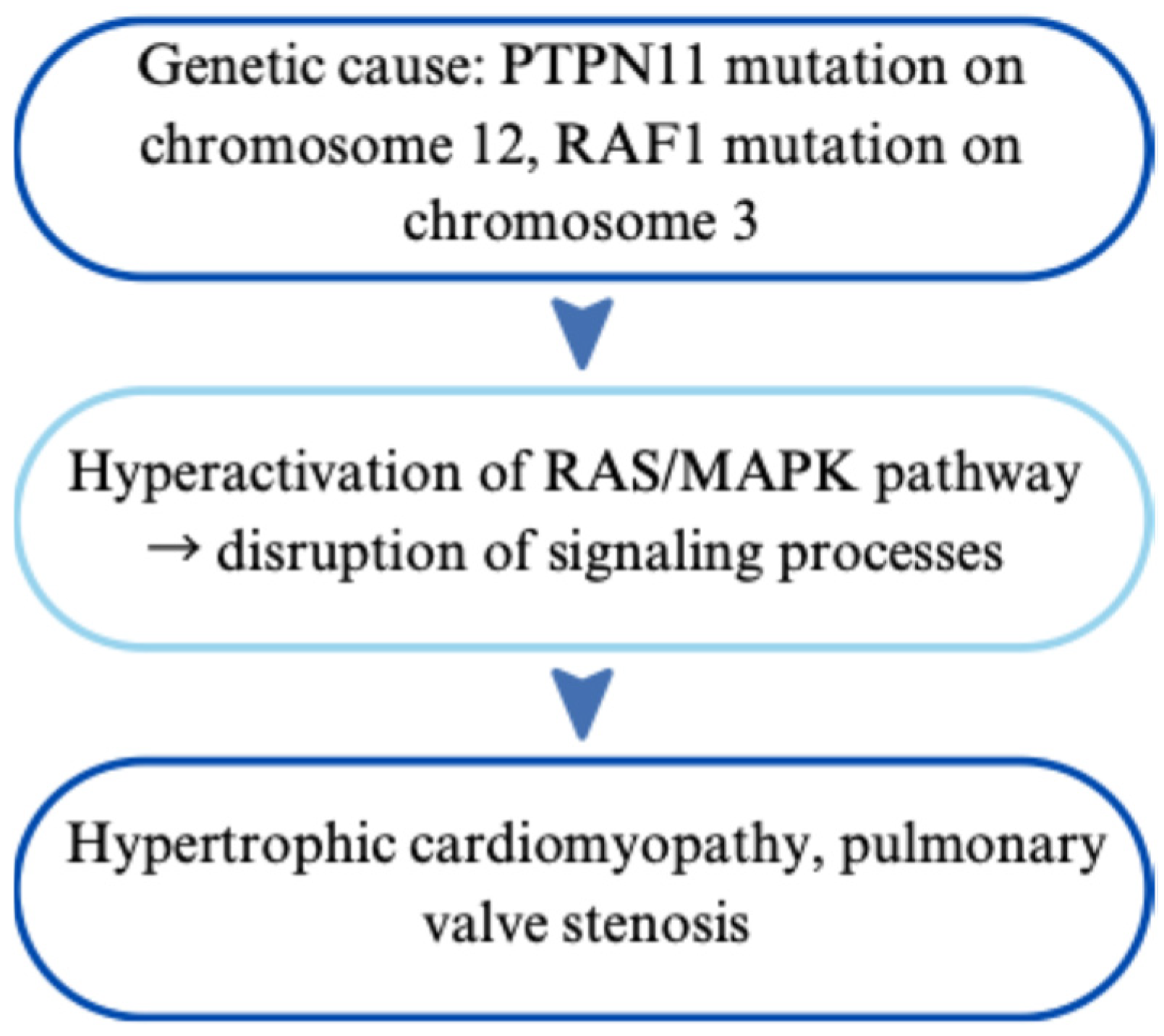
In the majority of cases, Noonan syndrome is caused by pathogenic variants in the PTPN11 gene located on chromosome 12 or the RAF1 gene on chromosome 3. Both genes are integral components of the RAS/mitogen-activated protein kinase (MAPK) signaling pathway. Consequently, Noonan syndrome is classified as a ‘RASopathy’ [17,18,19]. As a fundamental signaling cascade, the RAS/MAPK pathway plays a central role in regulating key cellular processes, including cell growth and proliferation, differentiation, survival, and apoptosis [18,19]. Collectively, the disease-causing mutations result in a hyperactivation of the RAS/MAPK pathway, disrupting the tightly regulated signaling processes essential for normal development [19]. An overview of the genetic basis and related pathophysiological mechanisms is provided in Figure 3.

2.2.2. Impact on the Heart and Cardiovascular System

The cardiovascular phenotype associated with Noonan syndrome and other RASopathies is multifaceted, primarily comprising two major categories: congenital heart disease (CHD) and hypertrophic cardiomyopathy (HCM) [18,20,21]. CHDs are present in approximately 80% of patients, with pulmonary valve stenosis being the most common, occurring in an estimated 40% of cases. Less common cardiac anomalies include atrial and ventricular septal defects, atrioventricular canal defects, and aortic coarctation. HCM is observed in approximately 20% of patients [21,22].

2.2.3. Symptoms and Clinical Presentation in Children

In addition to its cardiovascular manifestations, Noonan syndrome presents with a broad spectrum of non-cardiac clinical features involving multiple organ systems. The most frequently observed clinical characteristics include distinctive craniofacial features such as hypertelorism, down-slanting palpebral fissures, ptosis, low-set posteriorly rotated ears, and a webbed neck. These features are typically more pronounced during childhood and tend to become less apparent with advancing age [23,24]. Growth retardation is a common feature, typically becoming apparent after the first year of life. It often results in short stature relative to sex and family growth potential. Neurodevelopmental delay, particularly affecting motor and speech domains, is variably expressed and may range from mild intellectual disability that may improve with age to more severe manifestations, including in rare cases structural brain abnormalities. Characteristic musculoskeletal anomalies include anterior chest wall deformities, such as superior pectus carinatum and inferior pectus excavatum [23,24]. Lymphatic abnormalities are common in Noonan syndrome, demonstrating a cumulative incidence of approximately 20%. These include lymphedema, which is the most frequently observed manifestation, as well as chylothorax and ascites, which occur less often but are often present with greater clinical severity [23,25]. Cryptorchism represents a common genitourinary manifestation in males with Noonan syndrome, affecting an estimated 60–80% of cases [23].

2.2.4. Diagnosis and Management

The broad phenotypic variability associated with Noonan syndrome highlights the necessity of a multidisciplinary approach to both diagnosis and long-term management. Timely diagnosis and appropriate management are crucial to achieving optimal clinical outcomes. Nonetheless, individuals with milder phenotypic presentations often go undiagnosed or experience significant delays in diagnosis [26]. The diagnosis of Noonan syndrome is based on the presence of characteristic clinical features and is confirmed by identifying a pathogenic variant in one of the RAS/MAPK pathway genes. The clinical diagnosis is guided by the presence of major and minor diagnostic criteria. Major criteria include the characteristic craniofacial features of Noonan syndrome, congenital heart defects—particularly pulmonary valve stenosis and/or HCM -, short stature, anterior chest wall deformities, mild developmental delay, cryptorchidism in males, lymphatic dysplasia, and a positive family history of Noonan syndrome [26,27]. A definite clinical diagnosis can be established when the characteristic facial features are present in conjunction with at least one additional major diagnostic criterion [26].
Because of its heterogeneous clinical presentation and multisystemic involvement characteristic of the disorder, the management of Noonan syndrome is multidisciplinary and individualized. While there is currently no curative therapy, early diagnosis and coordinated care can significantly improve quality of life and long-term outcomes [26]. Cardiac involvement represents one of the most clinically relevant manifestations of Noonan syndrome and often requires early intervention. Management strategies may involve catheter-based procedures or surgical correction of valvular lesions, alongside pharmacological therapy for HCM. Pharmacologic agents typically include beta-blockers, diuretics, or calcium channel blockers, depending on the severity and clinical course. Regular cardiologic surveillance is recommended, even in individuals without an established cardiac diagnosis [27,28]. Short stature in individuals with Noonan syndrome may be considered for treatment with recombinant human growth hormone (rhGH), particularly in those with confirmed growth hormone deficiency or clinically significant growth impairment. Initiation of therapy is generally recommended around the age of four years to optimize growth potential and long-term outcomes [17,27,28,29]. Cryptorchidism is present in a high proportion of males with Noonan syndrome and typically requires surgical correction via orchiopexy in early childhood. Early intervention is recommended to reduce the risk of infertility and to lower the potential for malignant transformation later in life [27].

2.3. 22q11.2 Deletion Syndrome

22q11.2 deletion syndrome, previously referred to as DiGeorge syndrome, is a common microdeletion disorder with a broad spectrum of clinical manifestations affecting multiple organ systems. It is characterized by congenital heart defects, immune dysfunction, hypocalcemia, and distinctive craniofacial features, among others [30].

2.3.1. Genetic Basis and Phenotypic Impact of Inherited Variants

22q11.2 deletion syndrome results from a heterozygous microdeletion at chromosome 22 (22q11.2). In approximately 90% of cases, the deletion occurs de novo, while the remaining 10% are inherited in an autosomal dominant manner from a heterozygous parent. The syndrome exhibits variable expressivity and is estimated to occur in approximately 1 in 3000 to 1 in 4000 live births [30,31,32].
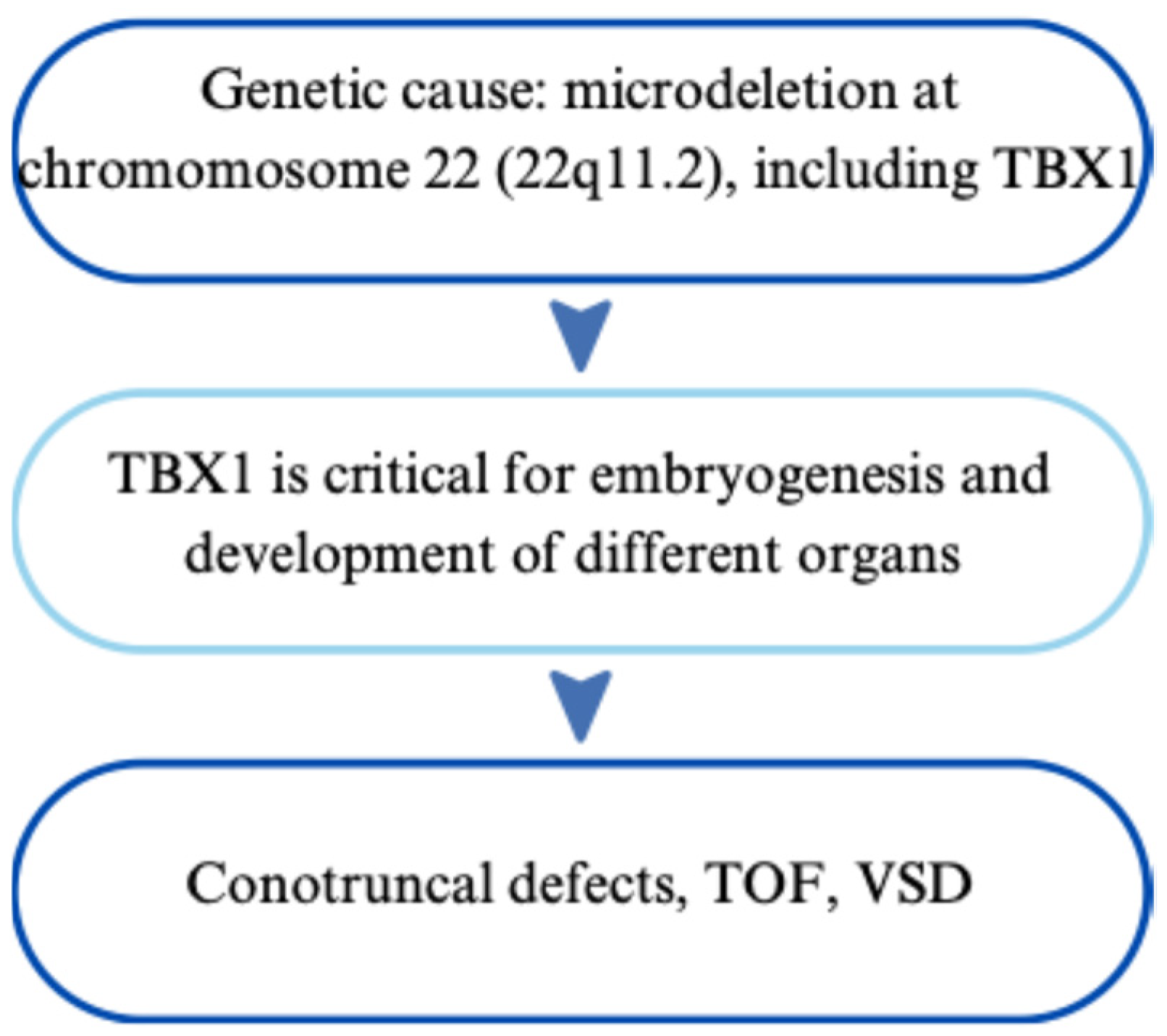
The microdeletion occurs in the long arm of chromosome 22 (22q11.2), most commonly encompassing a ~3 megabase region that includes approximately 40 genes. Among these, TBX1 is considered to be a key contributor to the core phenotypic features associated with the syndrome [30,32]. TBX1 is critical for embryogenesis and plays an essential role in the development of the heart, thymus, parathyroid glands, and craniofacial components [30,31,33]. The genetic cause and its clinical consequences are schematically presented in Figure 4.

2.3.2. Impact on the Heart and Cardiovascular System

Cardiovascular anomalies are among the most prevalent and clinically significant manifestations of 22q11.2 deletion syndrome, affecting approximately 70–80% of individuals with 22q11.2 deletion syndrome [34]. Congenital heart defects are the most frequently observed cardiovascular anomalies and exhibit a broad spectrum of severity [35]. The most characteristic cardiac malformations involve the conotruncal region, including the tetralogy of Fallot and persistent truncus arteriosus. In addition, individuals with 22q11.2 deletion syndrome may present with other congenital cardiac anomalies, including ventricular septal defects, interrupted aortic arch type B, aortic arch abnormalities, such as right-sided aortic arch or double aortic arch, pulmonary atresia, and hypoplastic right heart syndrome. Less commonly, patients may exhibit aortic root dilation, which may occur with or without the presence of congenital heart defects [32,34,35,36,37].

2.3.3. Symptoms and Clinical Presentation in Children

22q11.2 deletion syndrome presents with a wide spectrum of clinical manifestations in the pediatric population. The variability in phenotype contributes to delayed or missed diagnoses, particularly in cases with milder features. Facial dysmorphisms, although often subtle and evolving with age, are characteristic of 22q11.2 deletion syndrome and can aid in clinical recognition. Common features include a prominent nasal bridge, hypoplastic alae nasi, dysplastic ears, hypertelorism, a small mouth, micrognathia and/or retrognathia [38,39].
Thymus aplasia or hypoplasia is another frequently observed feature and may lead to variable degrees of T-cell immunodeficiency [39]. This immunologic impairment predisposes affected individuals to recurrent infections and poor vaccine responses. Additional extracardiac manifestations include cleft palate—ranging from overt clefting to submucosal clefts and velopharyngeal insufficiency—as well as hypocalcemia, which occurs frequently secondary to parathyroid hypoplasia [38]. The acronym CATCH has been commonly used to summarize the core clinical features of 22q11.2 deletion syndrome, encompassing Cardiac anomalies, Abnormal facies, Thymic hypoplasia or aplasia, Cleft palate, and Hypocalcemia. While this mnemonic captures several hallmark manifestations, it does not reflect the full phenotypic spectrum now recognized in affected individuals [39]. Several clinical manifestations are not captured by the acronym, including neurodevelopmental disorders, psychiatric conditions, renal anomalies, hearing loss, and skeletal abnormalities [32,38,39].

2.3.4. Diagnosis and Management

The diagnosis of 22q11.2 deletion syndrome is typically guided by clinical suspicion based on characteristic phenotypic features and is confirmed through molecular genetic testing. Clinical indicators that should prompt evaluation include congenital heart defects and the extracardiac manifestations mentioned above. The presence of multiple system involvement, especially when affecting the cardiac, immune, and endocrine systems concurrently, significantly increases the likelihood of an underlying 22q11.2 deletion. Molecular confirmation of 22q11.2 deletion syndrome typically involves one of three diagnostic approaches: fluorescence in situ hybridization (FISH), chromosomal microarray analysis (CMA), or multiplex ligation-dependent probe amplification (MLPA). These tests are usually performed on DNA from peripheral blood [40,41]. MLPA demonstrates a detection rate approaching 100% for 22q11.2 deletions, surpassing FISH, which identifies approximately 95% of cases [41].
The management of 22q11.2 deletion syndrome requires a comprehensive, multidisciplinary approach due to the condition’s broad phenotypic variability and multi-organ involvement. Cardiac anomalies often require early cardiologic intervention, as they remain the leading cause of mortality in children, accounting for approximately 87% of deaths [34]. Management typically involves catheter-based or surgical correction of structural defects, particularly conotruncal malformations. Long-term cardiologic follow-up is recommended, even in children without overt congenital heart disease [42]. Immunodeficiency, resulting from thymic hypoplasia or aplasia, necessitates immunologic assessment, including T-cell counts and function. Live vaccines should be given with caution, depending on the degree of T-cell immunodeficiency. Routine immunizations, including pneumococcal and influenza vaccines, are generally recommended and should be considered to reduce the risk of severe infections [41,42]. Palatal anomalies, such as cleft palate or velopharyngeal insufficiency, often require surgical intervention and coordinated care from speech-language pathologists and otolaryngologists. Serum calcium levels should be closely monitored, as hypocalcemia is a common finding. Calcium and vitamin D supplementation may be required intermittently or on a long-term basis, especially in cases of inadequate dietary intake or persistently low serum calcium concentrations [42]. Additional care may involve the management of hearing loss, renal anomalies, and neurodevelopmental and psychiatric care. Given the syndrome’s complexity, ongoing coordination between specialists in cardiology, immunology, endocrinology, neurology, psychiatry, and genetics is critical for optimal care.

2.4. Dilated Cardiomyopathy

Dilated cardiomyopathy (DCM) is a myocardial disease characterized by ventricular dilation and consequent enlargement. It predominantly affects one or both ventricles, most commonly the left ventricle, leading to impaired cardiac function. The diagnosis of DCM is established by the presence of left ventricular systolic dysfunction, typically defined as a left ventricular ejection fraction (LVEF) below 40%, in the absence of abnormal loading conditions such as hypertension or valvular disease. This section will discuss the clinical features of DCM with a particular focus on its manifestation in patients with Duchenne muscular dystrophy [43].

2.4.1. Genetic Basis and Phenotypic Impact of Inherited Variants

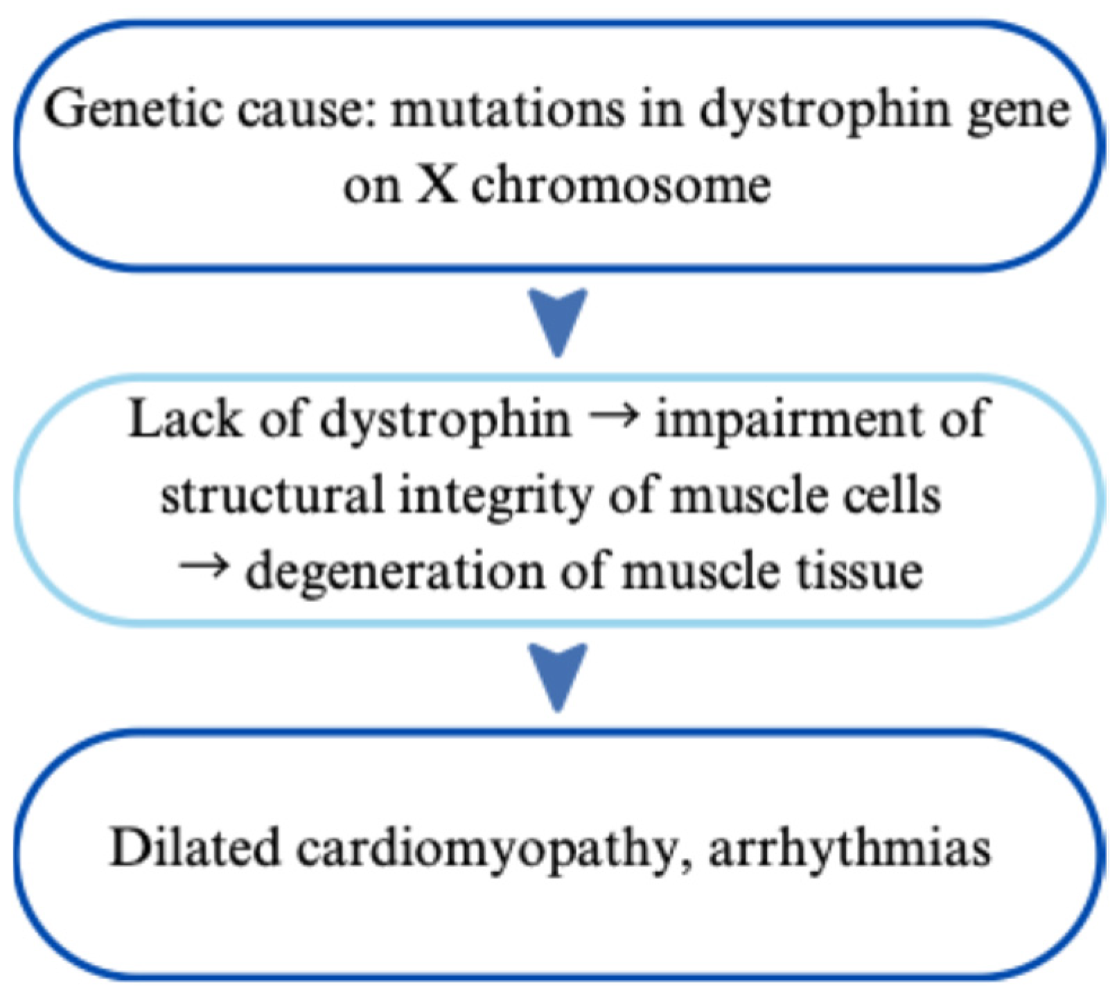
Duchenne muscular dystrophy (DMD) is a progressive neuromuscular disorder caused by X-linked recessive mutations in the dystrophin gene, resulting in the absence of functional dystrophin protein. The lack of dystrophin compromises the structural integrity of muscle cells, leading to progressive degeneration and weakness of both skeletal and cardiac muscle tissue [44]. The dystrophin gene mutation responsible for DMD is located on the Xp21 region of the X chromosome, which usually involves more than one exon. The majority of mutations (70–80%) are frameshift deletions or duplications, while nonsense (point) mutations account for approximately 20–30%. These genetic alterations often introduce premature stop codons, resulting in the lack of dystrophin [45,46,47]. In most cases, DMD is inherited in an X-linked recessive manner, typically transmitted from carrier mothers. Additionally, de novo mutations can occur. Female carriers are often asymptomatic but may exhibit varying degrees of cardiac involvement, attributed to insufficient X-chromosome inactivation or chromosomal abnormalities [48]. Consequently, affected individuals are usually male, inheriting the mutated X chromosome from their carrier mothers [49]. At the molecular level, the location and specific type of the mutation are significantly correlated with the severity and onset of cardiac manifestations in DMD. For instance, deletions involving exons 48–49 have been associated with earlier-onset DCM, whereas mutations affecting exons 51–52 may have a relative cardioprotective effect [46]. A case report has described a deletion of exon 55 linked to very early-onset DCM by age 3 [49]. Moreover, DMD variants involving dystrophin exons with percent spliced-in scores > 90, indicative of high expression in cardiac muscle frequency, are correlated with more severe cardiomyopathy [50]. Furthermore, polymorphisms in modifier genes such as LTBP4 and ACTN3 have been shown to increase susceptibility to DCM in affected individuals [51]. Figure 5 schematically outlines the genetic basis and its consequences for the cardiovascular system.

2.4.2. Impact on the Heart and Cardiovascular System

Cardiovascular involvement in DMD primarily manifests as DCM with variable severity. DMD presents with left ventricular dysfunction, myocardial fibrosis, arrhythmias, and the risk of heart failure [52]. Severe complications, including ventricular arrhythmias and sudden cardiac death, are particularly prevalent in individuals with rapid ventricular dilation or extensive myocardial fibrosis. Dystrophin is a cytoskeletal protein encoded by one of the largest human genes spanning approximately 2.4 Mb and comprising 79 exons [50,53]. It plays a critical role in stabilizing muscle cell membranes by connecting intracellular actin filaments to the extracellular matrix through the dystrophin-glycoprotein complex (dystroglycan). Due to its large size, the dystrophin gene is highly susceptible to mutations [45,54]. The lack of functional dystrophin disrupts membrane integrity and cellular signaling, leading to muscle fiber necrosis and subsequent replacement by adipose and connective tissue, which manifests clinically as muscle weakness and pseudohypertrophy [55].
In cardiomyocytes, dystrophin deficiency impairs dystrophin-associated protein complexes, resulting in sarcomere destabilization, calcium overload, myocardial fibrosis, and the progression of DCM [50,56].

2.4.3. Symptoms and Clinical Presentation in Children

Cardiac involvement in DMD is often not diagnosed during early childhood, as echocardiography and magnetic resonance imaging typically detect left ventricular dysfunction only after significant progression [57]. However, severe cases, such as those with exon 55 deletions may present with clinical manifestations as early as toddlerhood. The onset of DMD typically occurs between the ages of 2 and 5 years, with variability in clinical presentation. Children commonly develop symptoms such as fatigue, muscle weakness, and exercise intolerance around school age, corresponding with the heart’s diminishing ability to compensate, often reflected by a decline in left ventricular ejection fraction [49]. Patients typically develop signs of congestive heart failure and arrhythmias at a later stage, requiring careful monitoring and early detection to prevent fatal complications and improve quality of life [46,49,58].

2.4.4. Diagnosis and Management

The diagnosis of DMD typically involves biochemical blood tests revealing elevated creatine kinase and serum aldolase levels, alongside genetic testing methods such as MLPA, gene panels, and structural assays to identify mutations in the dystrophin gene and determine exon involvement. In cases where genetic testing is inconclusive, a muscle biopsy may be performed. The heart is usually examined by magnetic resonance imaging and echocardiography to detect and control cardiomyopathy [48].
The management of DMD primarily involves supportive care, including physical therapy, respiratory support, orthopedic interventions, and corticosteroid treatment [48]. Recent research has introduced exon-skipping antisense oligonucleotide therapies that aim to partially restore dystrophin function [52,59]. Cardiac treatment focuses on prophylactic use of angiotensin-converting enzyme (ACE) inhibitors and angiotensin receptor-neprilysin inhibitors (ARNi) to delay left ventricular dysfunction, alongside beta-blockers [52,60]. Additionally, arrhythmia management and implantable cardioverter-defibrillator (ICD) should be considered in high-risk patients presenting with severe rhythmic disturbances or myocardial fibrosis [52].

2.5. Hypertrophic Cardiomyopathy

Hypertrophic Cardiomyopathy (HCM) is a genetic heart disease which is characterized by an unexplained thickening of the myocardium, especially seen in the left ventricular wall and the interventricular septum that cannot be explained by coexisting disease. HCM symptoms can range from asymptomatic to severe heart failure and sudden cardiac death [61].

2.5.1. Genetic Basis and Phenotypic Impact of Inherited Variants

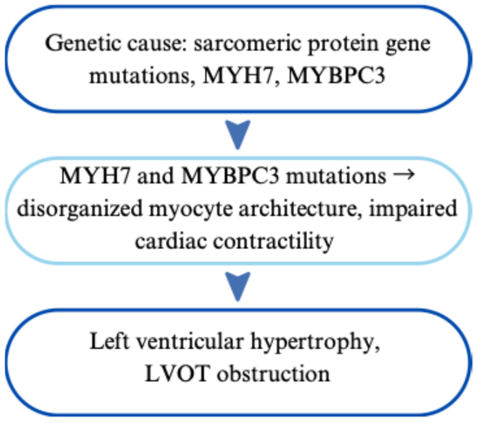
HCM is most commonly inherited in an autosomal dominant pattern. However, cases of autosomal recessive, X-linked and mitochondrial inheritance have also been reported [62]. Current genetic insights suggest that mutations in at least 29 different genes are associated with HCM, most commonly affecting sarcomeric protein genes such as MYH7 (encoding the β-myosin heavy chain) and MYBPC3 (encoding myosin-binding protein C) [63]. The affected gene is linked to the clinical picture of severity. Sequence variations that encode the MYBPC3 gene are usually present with a later onset and milder hypertrophy [64]. The MYBPC3 gene, located on chromosome 11, plays a crucial role in the regulation and structural integrity of thick filaments within cardiac sarcomeres [64,65]. In contrast, MYH7, located on chromosome 14 and encoding the β-myosin heavy chain, is frequently affected by missense mutations that are associated with earlier disease onset and a more severe HCM phenotype [65,66,67].
These mutations typically lead to disorganized myocyte architecture and impaired cardiac contractility [68]. Since the mutations are linked to variable penetrance and expressivity, the range of symptoms is wide. Furthermore, HCM is linked to zygosity. Homozygosity or compound heterozygosity in HCM-related genes is associated with earlier onset, more pronounced hypertrophy, and worse clinical outcomes compared to heterozygosity [69]. An overview of the genetic and clinical aspects of HCM is presented in Figure 6.

2.5.2. Impact on the Heart and Cardiovascular System

The clinical presentation of HCM ranges from asymptomatic to severe manifestations such as heart failure and sudden cardiac death. In advanced cases, it may lead to dynamic left ventricular outflow tract (LVOT) obstruction [70]. Particularly in patients with the non-obstructive form of HCM, symptoms are often absent at rest and may only become apparent or worsen under conditions of physical exertion, dehydration, or upon administration of certain medications such as diuretics, ACE inhibitors or angiotensin receptor blockers [71].

2.5.3. Symptoms and Clinical Presentation in Children

Clinical manifestations may include dyspnea, palpitations, dizziness up to syncope, angina pectoris, cardiac arrhythmias, and in rare cases, sudden cardiac death [72]. Additionally, mitral valve abnormalities and myocardial fibrosis are frequently observed in patients with HCM. On physical examination, a systolic ejection murmur is frequently detected, which intensifies during the Valsalva maneuver, standing, or with inotropic agents, and diminishes with passive leg elevation or the use of medications that reduce cardiac contractility [73,74].
All of these pathophysiological abnormalities significantly increase the risk of heart failure, stroke, and sudden cardiac death, particularly in young individuals [75,76,77,78].
The timing of diagnosis is often correlated with the severity of the disease. HCM diagnosed in childhood is more frequently associated with adverse outcomes, such as heart failure or sudden cardiac death. Early diagnosis of HCM in neonates has been consistently associated with worse survival outcomes [79]. Children with HCM may present with a broad spectrum of symptoms or may remain asymptomatic [80]. Children typically exhibit symptoms comparable to adults, but may also present with impaired growth [81,82]. As a compensation and a body’s natural way to increase systemic resistance some children adopt squatting behavior and thus lower their left-to-right pressure gradient [74,83].

2.5.4. Diagnosis and Management

HCM is often diagnosed insidiously because of its early asymptomatic course [84]. Diagnostic tools include echocardiography, which may reveal wall thickness exceeding 15 mm or the presence of left ventricular outflow tract obstruction (LVOTO). Additionally, electrocardiography (ECG) typically shows signs of left ventricular hypertrophy (LVH) and may also detect arrhythmias [85,86]. Cardiac magnetic resonance imaging is done to understand morphology and fibrosis [87]. Moreover, genetic testing and family screening of first-degree relatives are essential due to the hereditary nature of HCM [88].
The management of HCM primarily focuses on symptom relief, prevention of complications, and the implementation of lifestyle modifications [89]. Patients should avoid dehydration, alcohol, strenuous activity, and heat exposure [90]. An implantable cardioverter-defibrillator (ICD) is recommended, particularly for patients at high risk of sudden cardiac death (SCD), especially those with a history of symptomatic events such as arrhythmias or cardiac arrest [91,92]. Symptomatic patients are treated with the aim of reducing symptoms, primarily using beta-blockers; verapamil may be considered as a second-line option if beta-blockers are not well tolerated [89,93]. Other medications, such as digoxin, nitrates, ACE inhibitors and high-dose diuretics, should be avoided in obstructive HCM due to their potential to exacerbate symptoms and worsen clinical outcomes [77]. Invasive interventions, including septal myectomy or alcohol septal ablation, are typically considered when pharmacological therapy fails to achieve sufficient symptom control [94,95]. Dual-chamber pacemakers may be beneficial in certain cases, while heart transplantation is generally considered only in end-stage non-obstructive HCM, when other therapeutic options have been ineffective. Timely consideration of heart transplantation is crucial, as irreversible complications, such as severe pulmonary hypertension, may develop if the heart is unable to sufficiently compensate [94,96].

2.6. Tetralogy of Fallot

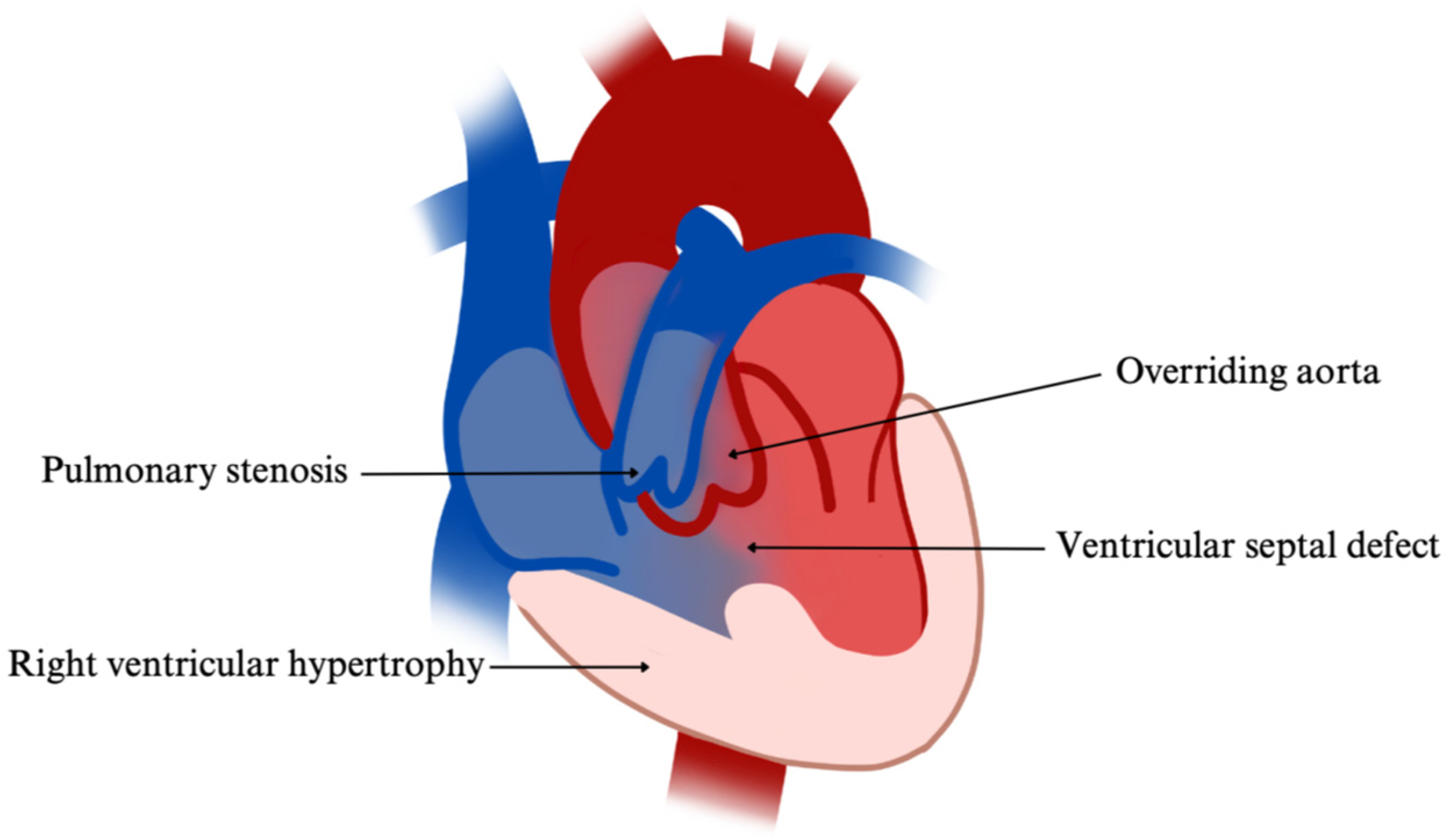
Tetralogy of Fallot (TOF) is the most common cyanotic congenital heart defect in children with an incidence of 3–5 per 10,000 live births in the United States [97]. TOF is defined by a combination of four congenital cardiac anomalies: a ventricular septal defect (VSD), right ventricular hypertrophy, an overriding aorta, and varying degrees of pulmonary stenosis. The extent of pulmonary outflow obstruction plays an important role in the development of a right-to-left shunt, thereby influencing the degree of systemic cyanosis [98,99]. Together, these defects impair effective oxygenation of blood within the systemic circulation.

2.6.1. Genetic Basis and Phenotypic Impact of Inherited Variants

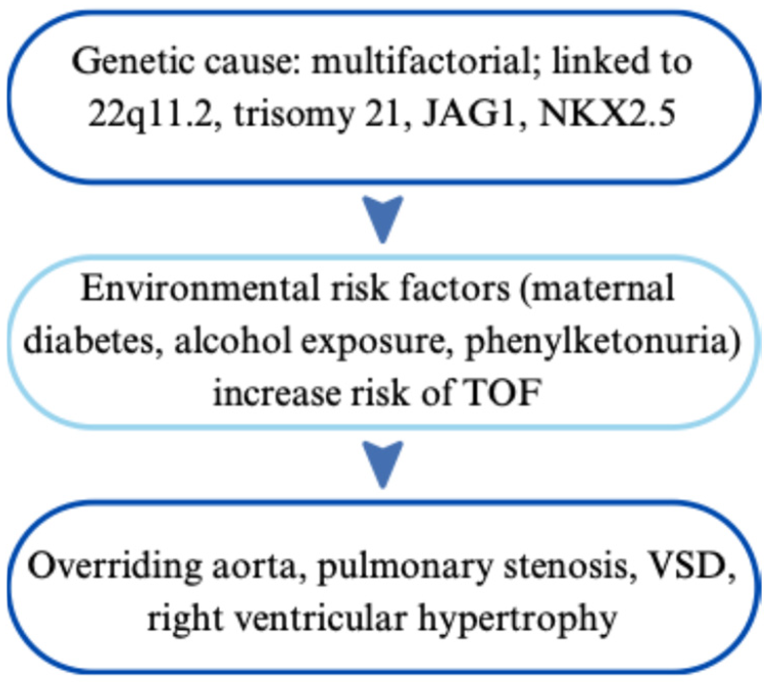
TOF mostly follows a multifactorial inheritance pattern, where both genetic and environmental factors contribute to its development [100,101]. In approximately 80% of cases, TOF occurs as an isolated, non-syndromic defect. The remaining cases are associated with genetic abnormalities [102]. The most common genetic abnormality associated with TOF is 22q11.2 deletion syndrome (DiGeorge syndrome), which affects the TBX1 gene and impairs cardiac neural crest cell migration, significant for conotruncal heart development [103]. Other genetic associations with the tetralogy of Fallot include trisomy 21; Alagille syndrome, which is linked to mutations in the JAG1 gene; and non-syndromic forms involving variants in NKX2.5 and ZFPM2 (FOG2), both of which regulate cardiac development through interactions with GATA transcription factors [102,104]. Mutations, such as polyalanine stretch elongations in the TBX1 gene, can reduce transcriptional activity by promoting protein aggregation [102,103]. Although the familial recurrence rate is low (2.5–3%), an increased incidence within certain families indicates a genetic predisposition within the population [98]. Environmental risk factors, such as maternal diabetes, alcohol exposure, and phenylketonuria, can further increase the risk of TOF [101]. Overall TOF results from a complex interplay of genetic mutations, epigenetic factors, and environmental influences. A schematic representation of the genetic and pathophysiological features of TOF can be found in Figure 7.

2.6.2. Impact on the Heart and Cardiovascular System

The clinical variability in TOF is associated with both genetic background and the degree of right ventricular outflow tract obstruction [99]. In TOF, infants with milder right ventricular outflow tract (RVOT) obstruction may initially have a left-to-right shunt through the ventricular septal defect, presenting with little or no cyanosis. As the obstruction worsens, right-to-left shunting occurs, leading to cyanosis and the development of Eisenmenger syndrome [105,106]. Moreover, specific associated gene variants can result in additional structural abnormalities, such as an atrial septal defect. In the context of TOF, the co-occurrence of an atrial septal defect frequently is observed in Down syndrome, termed pentalogy of Fallot [107,108]. The combination of the four anatomical defects in TOF results in significant alterations to cardiac physiology, leading to chronic insufficient oxygenation of the blood [106]. The VSD allows for the mixing of oxygenated and deoxygenated blood, while pulmonary stenosis obstructs blood flow to the lungs, leading to right ventricular hypertrophy due to the increased workload. The overriding aorta receives blood from both ventricles, which further exacerbates systemic hypoxia. These anatomical and physiological abnormalities can lead to complications such as arrhythmias, heart failure, and reduced exercise capacity, which if left untreated may become incompatible with life. The developmental background is based on the structural abnormalities resulting from an anterior and superior deviation of the infundibular septum during fetal phase, leading to misalignment of the ventricular septum and an overriding aortic root [98]. The four characteristic cardiac anomalies are illustrated in Figure 8.

2.6.3. Symptoms and Clinical Presentation in Children

Children with TOF often present with the first symptoms within the first few months of life. Infants may initially be asymptomatic, depending on the degree of malformation, but typically develop signs of heart failure by around six months of age. Cyanosis is often the earliest clinical manifestation in the tetralogy of Fallot, its severity correlating with the degree of right ventricular outflow tract obstruction. Hypercyanotic spells, commonly referred to as ‘Tet spells,’ are acute episodes of profound cyanosis precipitated by stressors such as crying, feeding, or agitation. These episodes are characterized by hypoxia, irritability, and represent one of the most critical complications associated with TOF [105]. These episodes lead to an increase in pulmonary vascular resistance and a simultaneous decrease in systemic vascular resistance, further exacerbating the right-to-left shunt and worsening systemic hypoxia [98]. Additional clinical manifestations may include feeding difficulties, dyspnea, failure to thrive, and developmental or growth delays [109,110]. Physical examination commonly reveals a harsh systolic murmur best heard at the left upper sternal border, corresponding to obstruction within the right ventricular outflow tract. Chronic hypoxia may result in digital clubbing and features suggestive of right ventricular hypertrophy may be present upon inspection or palpation [106,111]. Older children with TOF often adopt a squatting position or flex their knees as a compensatory mechanism. This posture increases systemic vascular resistance, which in turn diminishes right-to-left shunting and enhances pulmonary perfusion and arterial oxygenation. Furthermore, patients may present with hyperviscosity of the blood secondary to compensatory polycythemia and elevated hematocrit levels. This condition increases the risk of serious complications, including cerebral thrombosis, paradoxical embolism, heart failure, and cerebral abscesses [106,112].

2.6.4. Diagnosis and Management

The diagnosis of the tetralogy of Fallot typically involves transthoracic echocardiography, which allows for visualization of the characteristic anatomical defects and assessment of the pressure gradients across the right ventricular outflow tract. A chest X-ray may reveal a ‘boot-shaped’ heart due to right ventricular hypertrophy and an upturned cardiac apex. Electrocardiography (ECG) often demonstrates right axis deviation, right ventricular hypertrophy, and signs of right atrial enlargement. Additionally, a hyperoxia test may be performed to help distinguish cardiac from pulmonary causes of cyanosis. TOF can vary in its presentation and time of diagnosis depending on the severity of the malformation. The clinical presentation of TOF varies, ranging from asymptomatic cases in early infancy to overt cyanosis at birth; however, symptoms most commonly manifest within the first weeks to months of life [98]. TOF and other conotruncal abnormalities are often detected prenatally by fetal echocardiography, especially when routine screening raises concerns, comprehensive fetal cardiac assessment is done [113].
Management of TOF primarily a complete surgical repair with closure of the VSD and relief of the RVOT obstruction, typically performed within the first six months of life [114]. When the child’s condition and anatomy allow, early complete correction is preferred, as it offers the best long-term outcomes. In contrast, palliative procedures are reserved for critically ill neonates or those with very small pulmonary arteries. These include the modified Blalock-Taussig (BT) shunt, where a surgical graft connects a systemic artery to a pulmonary artery to increase blood flow to the lungs, and RVOT stenting, a catheter-based technique that keeps the natural outflow tract open to improve blood flow. These approaches provide vital short-term support, but do not replace the need for complete repair [115,116]. The procedure includes closure of the VSD and relief of the pulmonary stenosis. Before surgery, high doses of prostaglandins are given to maintain patency of the ductus arteriosus and keep an alternative route for blood flow, hence improving oxygenation [117]. Postoperative management of TOF requires lifelong follow-up, due to the risk of late complications such as arrhythmias, pulmonary regurgitation, and right ventricular dysfunction. Advances in surgical techniques have significantly improved long-term outcomes, resulting in enhanced life expectancy and quality of life for individuals with TOF [118].

3. Conclusions

Congenital genetic cardiac diseases in the pediatric population encompass a broad and diverse spectrum of phenotypic manifestations, shaped by complex interactions between genotype and environmental factors. Key pediatric cardiac diseases are summarized in Table 1. The six disorders discussed span from syndromic diseases like Marfan and Noonan to non-exclusively syndromic isolated cardiomyopathies such as hypertrophic and dilated forms. Collectively they underline the crucial impact of accurate and early diagnosis, clinical evaluation and the start of early management in pediatric cardiology.
Early detection is the key in reducing morbidity and mortality as well as ensuring the improvement of quality of life in all of the mentioned conditions. Routine prenatal screening for CHDs is typically performed using standard fetal ultrasound. In cases of high-risk pregnancies, such as those with a family history of CHD, abnormal ultrasound findings, suspected genetic syndromes, or maternal conditions known to affect cardiac development, targeted fetal echocardiography is recommended. When a genetic syndrome is suspected, prenatal genetic testing may be carried out additionally [119,120]. Recent advancements in molecular and genetic diagnosis, fetal imaging, and surgical interventions have significantly improved the targeted identification of pathological variants, helping direct prognosis, surveillance, and therapeutic strategies. However, a comprehensive understanding of the underlying genetic basis remains essential to further optimize diagnostic precision, develop targeted therapies and needs to be ongoing to develop further with time. Another critical component is the role of genetic counseling, in order to help families to understand and cope properly with the diagnosis, by giving them the opportunity to learn about inheritance pattern, recurrence risk, and their reproductive options. The management of these disorders requires an individual and multidisciplinary approach, involving not only cardiac but also extracardiac manifestations and psychosocial components.
Ongoing research into innovative new surgical interventions, new pharmacologic therapies, device implantation, and advanced gene-targeted treatments offer promising outcomes when applied at early stages and with a cohesive therapeutic plan.
Taken together, these advancements will deepen our understanding and enhance the management of cardiogenic diseases. Future research should prioritize the development of genotype-specific therapies, the potential application of gene-editing technologies, and long-term follow-up studies to optimize care and treatment from the prenatal period through adulthood, offering the potential to significantly alter the course of these diseases. The incorporation of precision medicine into clinical care of pediatric cardiology will continue to evolve, providing new opportunities for children born with genetic heart defects and their families.

Author Contributions

Conceptualization, J.L.G. and A.S.; writing—original draft preparation, J.L.G., A.S. and L.B.; writing—review and editing, J.L.G., A.S. and L.B.; visualization, J.L.G., A.S. and L.B.; supervision, L.B.; funding acquisition, L.B. All authors have read and agreed to the published version of the manuscript.

Funding

This research received no external funding.

Institutional Review Board Statement

This work was written up as part of the scientific project granted to Prof. Lara Baticic, PhD by the University of Rijeka, Croatia “Uniri-iz-25-224: Dynamic Changes in Endothelial Glycocalyx and Inflammatory Response in Patients with Acute Ischemic Stroke Treated with Mechanical Thrombectomy: Pathophysiological Aspects and Clinical Implications”.

Informed Consent Statement

Not applicable.

Data Availability Statement

No new data was created or analyzed in this study. Data sharing is not applicable to this article.

Conflicts of Interest

The authors declare no conflicts of interest.

References

  1. Xu, J.; Li, Q.; Deng, L.; Xiong, J.; Cheng, Z.; Ye, C. Global, regional, and national epidemiology of congenital heart disease in children from 1990 to 2021. Front. Cardiovasc. Med. 2025, 12, 1522644. Available online: https://pubmed.ncbi.nlm.nih.gov/40454242/ (accessed on 28 April 2025). [CrossRef]
  2. Diab, N.S.; Barish, S.; Dong, W.; Zhao, S.; Allington, G.; Yu, X.; Kahle, K.T.; Brueckner, M.; Jin, S.C. Molecular Genetics and Complex Inheritance of Congenital Heart Disease. Genes 2021, 12, 1020. Available online: https://pubmed.ncbi.nlm.nih.gov/34209044/ (accessed on 28 April 2025). [CrossRef]
  3. Coelho, S.G.; Almeida, A.G. Marfan syndrome revisited: From genetics to the clinic. Rev. Port. Cardiol. (Engl. Ed.) 2020, 39, 215–226. Available online: https://pubmed.ncbi.nlm.nih.gov/32439107/ (accessed on 30 April 2025). [CrossRef]
  4. Wright, M.J.; Connolly, H.M. Genetics, Clinical Features, and Diagnosis of Marfan Syndrome and Related Disorders. UpToDate. [updated 1 October 2024]. Available online: https://www.uptodate.com/contents/genetics-clinical-features-and-diagnosis-of-marfan-syndrome-and-related-disorders#topicContent (accessed on 6 June 2025).
  5. Milewicz, D.M.; Braverman, A.C.; De Backer, J.; Morris, S.A.; Boileau, C.; Maumenee, I.H.; Jondeau, G.; Evangelista, A.; Pyeritz, R.E. Marfan syndrome. Nat. Rev. Dis. Primers. 2021, 7, 64. Available online: https://pubmed.ncbi.nlm.nih.gov/34475413/ (accessed on 5 May 2025). [CrossRef] [PubMed]
  6. Lazea, C.; Bucerzan, S.; Crisan, M.; Al-Khzouz, C.; Miclea, D.; Şufană, C.; Cismaru, G.; Grigorescu-Sido, P. Cardiovascular manifestations in Marfan syndrome. Med. Pharm. Rep. 2021, 94, S25–S27. Available online: https://pmc.ncbi.nlm.nih.gov/articles/PMC8411824/ (accessed on 5 May 2025).
  7. Wozniak-Mielczarek, L.; Sabiniewicz, R.; Drezek-Nojowicz, M.; Nowak, R.; Gilis-Malinowska, N.; Mielczarek, M.; Łabuc, A.; Waldoch, A.; Wierzba, J. Differences in Cardiovascular Manifestation of Marfan Syndrome Between Children and Adults. Pediatr. Cardiol. 2019, 40, 393–403. Available online: https://pmc.ncbi.nlm.nih.gov/articles/PMC6399167/ (accessed on 6 May 2025). [CrossRef] [PubMed]
  8. Muiño-Mosquera, L.; Cervi, E.; De Groote, K.; Dewals, W.; Fejzic, Z.; Kazamia, K.; Mathur, S.; Milleron, O.; Mir, T.S.; Nielsen, D.G.; et al. Management of aortic disease in children with FBN1-related Marfan syndrome. Eur. Heart J. 2024, 45, 4156–4169. Available online: https://pmc.ncbi.nlm.nih.gov/articles/PMC11472455/ (accessed on 7 May 2025). [CrossRef] [PubMed]
  9. Kemezyte, A.; Gegieckiene, R.; Burnyte, B. Genotype-phenotype spectrum and prognosis of early-onset Marfan syndrome. BMC Pediatr. 2023, 23, 539. Available online: https://pmc.ncbi.nlm.nih.gov/articles/PMC10612290/ (accessed on 8 May 2025). [CrossRef]
  10. Akram, H.; Aragon-Martin, J.A.; Chandra, A. Marfan syndrome and the eye clinic: From diagnosis to management. Ther. Adv. Rare Dis. 2021, 2, 26330040211055738. Available online: https://pubmed.ncbi.nlm.nih.gov/37181104/ (accessed on 9 May 2025). [CrossRef]
  11. Esfandiari, H.; Ansari, S.; Mohammad-Rabei, H.; Mets, M.B. Management Strategies of Ocular Abnormalities in Patients with Marfan Syndrome: Current Perspective. J. Ophthalmic Vis. Res. 2019, 14, 71–77. Available online: https://pmc.ncbi.nlm.nih.gov/articles/PMC6388525/ (accessed on 10 May 2025). [CrossRef]
  12. Pollock, L.; Ridout, A.; The, J.; Nnadi, C.; Stavroulias, D.; Pitcher, A.; Blair, E.; Wordsworth, P.; Vincent, T.L. The Musculoskeletal Manifestations of Marfan Syndrome: Diagnosis, Impact, and Management. Curr. Rheumatol. Rep. 2021, 23, 81. Available online: https://pmc.ncbi.nlm.nih.gov/articles/PMC8626407/ (accessed on 10 May 2025). [CrossRef]
  13. Loeys, B.L.; Dietz, H.C.; Braverman, A.C.; Callewaert, B.L.; De Backer, J.; Devereux, R.B.; Hilforst-Hofstee, Y.; Jondeau, G.; Faivre, L.; Milewicz, D.; et al. The revised Ghent nosology for the Marfan syndrome. J. Med Genet. 2010, 47, 476–485. Available online: https://jmg.bmj.com/content/jmedgenet/47/7/476.full.pdf (accessed on 7 June 2025). [CrossRef]
  14. National Academies of Sciences, Engineering, and Medicine; Health and Medicine Division; Board on Health Care Services; Committee on Selected Heritable Disorders of Connective Tissue and Disability. Marfan Syndrome and Related Hereditary Aortopathis. In Selected Heritable Disorders of Connective Tissue and Disability; Volberding, P., Spicer, C.M., Cartaxo, T., Wedge, R., Eds.; National Academies Press: Washington, DC, USA, 2022; Chapter 3. Available online: https://www.ncbi.nlm.nih.gov/books/NBK584974/ (accessed on 28 April 2025).
  15. Marelli, S.; Micaglio, E.; Taurino, J.; Salvi, P.; Rurali, E.; Perrucci, G.L.; Dolci, C.; Udugampolage, N.S.; Caruso, R.; Gentilini, D.; et al. Marfan Syndrome: Enhanced Diagnostic Tools and Follow-up Management Strategies. Diagnostics 2023, 13, 2284. Available online: https://pubmed.ncbi.nlm.nih.gov/37443678/ (accessed on 28 April 2025). [CrossRef]
  16. Isselbacher, E.M.; Preventza, O.; Hamilton Black, J., 3rd; Augoustides, J.G.; Beck, A.W.; Bolen, M.A.; Braverman, A.C.; Bray, B.E.; Brown-Zimmerman, M.M.; Chen, E.P.; et al. 2022 ACC/AHA Guideline for the Diagnosis and Management of Aortic Disease: A Report of the American Heart Association/American College of Cardiology Joint Committee on Clinical Practice Guidelines. Circulation 2022, 146, e334–e482. Available online: https://pubmed.ncbi.nlm.nih.gov/36322642/ (accessed on 28 April 2025). [CrossRef] [PubMed]
  17. Dahlgren, J.; Noordam, C. Growth, Endocrine Features, and Growth Hormone Treatment in Noonan Syndrome. J. Clin. Med. 2022, 11, 2034. Available online: https://pmc.ncbi.nlm.nih.gov/articles/PMC8999676/ (accessed on 30 April 2025). [CrossRef] [PubMed]
  18. Sun, L.; Xie, Y.M.; Wang, S.S.; Zhang, Z.W. Cardiovascular Abnormalities and Gene Mutations in Children with Noonan Syndrome. Front. Genet. 2022, 13, 915129. Available online: https://pmc.ncbi.nlm.nih.gov/articles/PMC9234298/ (accessed on 1 April 2025). [CrossRef] [PubMed]
  19. Saint-Laurent, C.; Mazeyrie, L.; Yart, A.; Edouard, T. Novel therapeutic perspectives in Noonan syndrome and RASopathies. Eur. J. Pediatr. 2024, 183, 1011–1019. Available online: https://pmc.ncbi.nlm.nih.gov/articles/PMC10951041/ (accessed on 28 April 2025). [CrossRef]
  20. Zenker, M.; Wolf, C.M. Cardiovascular aspects of Noonan syndrome and related disorders. Med. Genet. 2025, 37, 113–124. Available online: https://pubmed.ncbi.nlm.nih.gov/40207038/ (accessed on 30 April 2025). [CrossRef]
  21. Linglart, L.; Gelb, B.D. Congenital heart defects in Noonan syndrome: Diagnosis, management, and treatment. Am. J. Med. Genet. C Semin. Med. Genet. 2020, 184, 73–80. Available online: https://pmc.ncbi.nlm.nih.gov/articles/PMC7682536/ (accessed on 28 April 2025). [CrossRef]
  22. Hilal, N.; Chen, Z.; Chen, M.H.; Choudhury, S. RASopathies and cardiac manifestations. Front. Cardiovasc. Med. 2023, 10, 1176828. Available online: https://pmc.ncbi.nlm.nih.gov/articles/PMC10387527/ (accessed on 30 April 2025). [CrossRef]
  23. Baldo, F.; Fachin, A.; Da Re, B.; Rubinato, E.; Bobbo, M.; Barbi, E. New insights on Noonan syndrome’s clinical phenotype: A single center retrospective study. BMC Pediatr. 2022, 22, 734. Available online: https://pmc.ncbi.nlm.nih.gov/articles/PMC9789552/ (accessed on 15 May 2025). [CrossRef]
  24. Roberts, A.E. Noonan Syndrome. In GeneReviews®; Adam, M.P., Feldman, J., Mirzaa, G.M., Pagon, R.A., Wallace, S.E., Amemiya, A., Eds.; University of Washington: Seattle, WA, USA, 1993. Available online: https://pubmed.ncbi.nlm.nih.gov/20301303/ (accessed on 20 June 2025). [PubMed]
  25. Sleutjes, J.; Kleimeier, L.; Leenders, E.; Klein, W.; Draaisma, J. Lymphatic Abnormalities in Noonan Syndrome Spectrum Disorders: A Systematic Review. Mol. Syndromol. 2022, 13, 1–11. Available online: https://pmc.ncbi.nlm.nih.gov/articles/PMC8832235/ (accessed on 15 May 2025). [CrossRef] [PubMed]
  26. Zenker, M.; Edouard, T.; Blair, J.C.; Cappa, M. Noonan syndrome: Improving recognition and diagnosis. Arch. Dis. Child. 2022, 107, 1073–1078. Available online: https://pmc.ncbi.nlm.nih.gov/articles/PMC9685729/ (accessed on 1 May 2025). [CrossRef] [PubMed]
  27. Allen, M.J.; Sharma, S. Noonan Syndrome. In StatPearls; StatPearls Publishing: Treasure Island, FL, USA, 2025. Available online: https://pubmed.ncbi.nlm.nih.gov/30335302/ (accessed on 19 June 2025). [PubMed]
  28. Wolf, C.M.; Zenker, M.; Burkitt-Wright, E.; Edouard, T.; García-Miñaúr, S.; Lebl, J.; Shaikh, G.; Tartaglia, M.; Verloes, A.; Östman-Smith, I. Management of cardiac aspects in children with Noonan syndrome—Results from a European clinical practice survey among paediatric cardiologists. Eur. J. Med. Genet. 2022, 65, 104372. Available online: https://pubmed.ncbi.nlm.nih.gov/34757052/ (accessed on 15 June 2025). [CrossRef] [PubMed]
  29. Kowalik, D.; Lewiński, A.; Stawerska, R. Management strategies for optimal linear growth in Noonan syndrome (NS) children: Minireview and case series of three patients with NS due to PTPN11 mutation and confirmed growth hormone deficiency. Endokrynol. Pol. 2024, 75, 592–603. Available online: https://pubmed.ncbi.nlm.nih.gov/40091322/ (accessed on 15 June 2025). [CrossRef]
  30. Szczawińska-Popłonyk, A.; Schwartzmann, E.; Chmara, Z.; Głukowska, A.; Krysa, T.; Majchrzycki, M.; Olejnicki, M.; Ostrowska, P.; Babik, J. Chromosome 22q11.2 Deletion Syndrome: A Comprehensive Review of Molecular Genetics in the Context of Multidisciplinary Clinical Approach. Int. J. Mol. Sci. 2023, 24, 8317. Available online: https://pmc.ncbi.nlm.nih.gov/articles/PMC10179617/ (accessed on 22 June 2025). [CrossRef]
  31. Biggs, S.E.; Gilchrist, B.; May, K.R. Chromosome 22q11.2 Deletion (DiGeorge Syndrome): Immunologic Features, Diagnosis, and Management. Curr. Allergy Asthma Rep. 2023, 23, 213–222. Available online: https://pmc.ncbi.nlm.nih.gov/articles/PMC9999075/ (accessed on 16 July 2025). [CrossRef]
  32. McDonald-McGinn, D.M.; Hain, H.S.; Emanuel, B.S.; Zackai, E.H. 22q11.2 Deletion Syndrome. In GeneReviews®; Adam, M.P., Feldman, J., Mirzaa, G.M., Pagon, R.A., Wallace, S.E., Amemiya, A., Eds.; University of Washington: Seattle, WA, USA, 1993. Available online: https://pubmed.ncbi.nlm.nih.gov/20301696/ (accessed on 26 June 2025). [PubMed]
  33. Zhao, Y.; Wang, Y.; Shi, L.; McDonald-McGinn, D.M.; Crowley, T.B.; McGinn, D.E.; Tran, O.T.; Miller, D.; Lin, J.R.; Zackai, E.; et al. Chromatin regulators in the TBX1 network confer risk for conotruncal heart defects in 22q11.2DS. NPJ Genom. Med. 2023, 8, 17. Available online: https://pmc.ncbi.nlm.nih.gov/articles/PMC10354062/ (accessed on 16 July 2025).
  34. Putotto, C.; Unolt, M.; Lambiase, C.; Marchetti, F.; Anaclerio, S.; Favoriti, A.; Tancredi, G.; Mastromoro, G.; Pugnaloni, F.; Liberati, N.; et al. Cardiac function in adolescents and young adults with 22q11.2 deletion syndrome without congenital heart disease. Eur. J. Med. Genet. 2023, 66, 104651. Available online: https://pubmed.ncbi.nlm.nih.gov/36404488/ (accessed on 16 July 2025).
  35. Diniz, B.L.; Deconte, D.; Gadelha, K.A.; Glaeser, A.B.; Guaraná, B.B.; de Moura, A.Á.; Rosa, R.F.M.; Zen, P.R.G. Congenital Heart Defects and 22q11.2 Deletion Syndrome: A 20-Year Update and New Insights to Aid Clinical Diagnosis. J. Pediatr. Genet. 2023, 12, 113–122. Available online: https://pmc.ncbi.nlm.nih.gov/articles/PMC10118709/ (accessed on 1 July 2025). [CrossRef]
  36. Putotto, C.; Pulvirenti, F.; Pugnaloni, F.; Isufi, I.; Unolt, M.; Anaclerio, S.; Caputo, V.; Bernardini, L.; Messina, E.; Moretti, C.; et al. Clinical Risk Factors for Aortic Root Dilation in Patients with 22q11.2 Deletion Syndrome: A Longitudinal Single-Center Study. Genes 2022, 13, 2334. Available online: https://pmc.ncbi.nlm.nih.gov/articles/PMC9778342/ (accessed on 16 July 2025). [CrossRef]
  37. Pavlicek, J.; Klaskova, E.; Kapralova, S.; Palatova, A.M.; Piegzova, A.; Spacek, R.; Gruszka, T. Major heart defects: The diagnostic evaluations of first-year-olds. BMC Pediatr. 2021, 21, 528. Available online: https://pmc.ncbi.nlm.nih.gov/articles/PMC8630885/ (accessed on 18 February 2025). [CrossRef] [PubMed]
  38. Funato, N. Craniofacial Phenotypes and Genetics of DiGeorge Syndrome. J. Dev. Biol. 2022, 10, 18. Available online: https://pubmed.ncbi.nlm.nih.gov/35645294/ (accessed on 18 February 2025). [CrossRef] [PubMed]
  39. Agarwal, M.; Kumar, V.; Dwivedi, A. Diagnosis of 22q11.2 deletion syndrome in children with congenital heart diseases and facial dysmorphisms. Med. J. Armed Forces India. 2023, 79, S196–S201. Available online: https://pmc.ncbi.nlm.nih.gov/articles/PMC10746800/ (accessed on 18 June 2025). [CrossRef] [PubMed]
  40. Yu, H.H.; Chien, Y.H.; Lu, M.Y.; Hu, Y.C.; Lee, J.H.; Wang, L.C.; Lin, Y.T.; Yang, Y.H.; Chiang, B.L. Clinical and Immunological Defects and Outcomes in Patients with Chromosome 22q11.2 Deletion Syndrome. J. Clin. Immunol. 2022, 42, 1721–1729. [Google Scholar] [CrossRef]
  41. Rojnueangit, K.; Khetkham, T.; Onsod, P.; Chareonsirisuthigul, T. Clinical Features to Predict 22q11.2 Deletion Syndrome Proven by Molecular Genetic Testing. J. Pediatr. Genet. 2020, 11, 22–27. Available online: https://pmc.ncbi.nlm.nih.gov/articles/PMC8847066/ (accessed on 1 April 2025). [CrossRef]
  42. Óskarsdóttir, S.; Boot, E.; Crowley, T.B.; Loo, J.C.Y.; Arganbright, J.M.; Armando, M.; Baylis, A.L.; Breetvelt, E.J.; Castelein, R.M.; Chadehumbe, M.; et al. Updated clinical practice recommendations for managing children with 22q11.2 deletion syndrome. Genet. Med. 2023, 25, 100338. Available online: https://pubmed.ncbi.nlm.nih.gov/36729053/ (accessed on 16 July 2025). [CrossRef]
  43. Mahmaljy, H.; Yelamanchili, V.S.; Singhal, M. Dilated Cardiomyopathy. In StatPearls; StatPearls Publishing: Treasure Island, FL, USA, 2025. Available online: https://pubmed.ncbi.nlm.nih.gov/28722940/ (accessed on 19 June 2025). [PubMed]
  44. Novelli, V.; Canonico, F.; Laborante, R.; Manzoni, M.; Arcudi, A.; Pompilio, G.; Mercuri, E.; Patti, G.; D’Amario, D. Unraveling the Genetic Heartbeat: Decoding Cardiac Involvement in Duchenne Muscular Dystrophy. Biomedicines. 2025, 13, 102. Available online: https://pubmed.ncbi.nlm.nih.gov/39857686/ (accessed on 18 February 2025). [CrossRef]
  45. Duan, D.; Goemans, N.; Takeda, S.; Mercuri, E.; Aartsma-Rus, A. Duchenne muscular dystrophy. Nat. Rev. Dis. Primers. 2021, 7, 13. Available online: https://pubmed.ncbi.nlm.nih.gov/33602943/ (accessed on 18 February 2025). [CrossRef]
  46. Schultz, T.I.; Raucci, F.J., Jr.; Salloum, F.N. Cardiovascular Disease in Duchenne Muscular Dystrophy: Overview and Insight Into Novel Therapeutic Targets. JACC Basic. Transl. Sci. 2022, 7, 608–625. Available online: https://pubmed.ncbi.nlm.nih.gov/35818510/ (accessed on 25 February 2025). [CrossRef]
  47. Bakhshandeh, M.; Behroozi, S. Next-generation sequencing approach to molecular diagnosis of Iranian patients with Duchenne/Becker muscular dystrophy: Several novel variants identified. eNeurologicalSci 2023, 30, 100446. Available online: https://pubmed.ncbi.nlm.nih.gov/36845278/ (accessed on 25 February 2025). [CrossRef]
  48. Venugopal, V.; Pavlakis, S. Duchenne Muscular Dystrophy. In StatPearls; StatPearls Publishing: Treasure Island, FL, USA, 2025. Available online: https://pubmed.ncbi.nlm.nih.gov/29493971/ (accessed on 19 June 2025). [PubMed]
  49. Lupu, M.; Pintilie, I.M.; Teleanu, R.I.; Marin, G.G.; Vladâcenco, O.A.; Severin, E.M. Early Cardiac Dysfunction in Duchenne Muscular Dystrophy: A Case Report and Literature Update. Int. J. Mol. Sci. 2025, 26, 1685. Available online: https://pubmed.ncbi.nlm.nih.gov/40004149/ (accessed on 18 February 2025). [CrossRef]
  50. Johnson, R.; Otway, R.; Chin, E.; Horvat, C.; Ohanian, M.; Wilcox, J.A.L.; Su, Z.; Prestes, P.; Smolnikov, A.; Soka, M.; et al. DMD-Associated Dilated Cardiomyopathy: Genotypes, Phenotypes, and Phenocopies. Circ. Genom. Precis. Med. 2023, 16, 421–430. Available online: https://pubmed.ncbi.nlm.nih.gov/37671549/ (accessed on 25 February 2025). [CrossRef] [PubMed]
  51. Landfeldt, E.; Alemán, A.; Abner, S.; Zhang, R.; Werner, C.; Tomazos, I.; Lochmüller, H.; Quinlivan, R.M.; Wahbi, K. Predictors of cardiac disease in duchenne muscular dystrophy: A systematic review and evidence grading. Orphanet J. Rare Dis. 2024, 19, 359. Available online: https://pubmed.ncbi.nlm.nih.gov/39342355/ (accessed on 18 February 2025). [CrossRef] [PubMed]
  52. Adorisio, R.; Mencarelli, E.; Cantarutti, N.; Calvieri, C.; Amato, L.; Cicenia, M.; Silvetti, M.; D’Amico, A.; Grandinetti, M.; Drago, F.; et al. Duchenne Dilated Cardiomyopathy: Cardiac Management from Prevention to Advanced Cardiovascular Therapies. J. Clin. Med. 2020, 9, 3186. Available online: https://pubmed.ncbi.nlm.nih.gov/33019553/ (accessed on 30 April 2025). [CrossRef] [PubMed]
  53. Sarker, S.; Eshaque, T.B.; Soorajkumar, A.; Nassir, N.; Zehra, B.; Kanta, S.I.; Rahaman, M.A.; Islam, A.; Akter, S.; Ali, M.K.; et al. Mutational spectrum and phenotypic variability of Duchenne muscular dystrophy and related disorders in a Bangladeshi population. Sci. Rep. 2023, 13, 21547. Available online: https://pubmed.ncbi.nlm.nih.gov/38057384/ (accessed on 30 April 2025). [CrossRef]
  54. Gatto, F.; Benemei, S.; Piluso, G.; Bello, L. The complex landscape of DMD mutations: Moving towards personalized medicine. Front. Genet. 2024, 15, 1360224. Available online: https://pubmed.ncbi.nlm.nih.gov/38596212/ (accessed on 30 April 2025). [CrossRef]
  55. Jama, A.; Alshudukhi, A.A.; Burke, S.; Dong, L.; Kamau, J.K.; Morris, B.; Alkhomsi, I.A.; Finck, B.N.; Voss, A.A.; Ren, H. Exploring lipin1 as a promising therapeutic target for the treatment of Duchenne muscular dystrophy. J. Transl. Med. 2024, 22, 664. Available online: https://pubmed.ncbi.nlm.nih.gov/39014470/ (accessed on 28 April 2025). [CrossRef]
  56. LaPelusa, A.; Asuncion, R.M.D.; Kentris, M. Muscular Dystrophy. In StatPearls; StatPearls Publishing: Treasure Island, FL, USA, 2025. Available online: https://pubmed.ncbi.nlm.nih.gov/32809417/ (accessed on 20 June 2025). [PubMed]
  57. Prakash, N.; Suthar, R.; Sihag, B.K.; Debi, U.; Kumar, R.M.; Sankhyan, N. Cardiac MRI and Echocardiography for Early Diagnosis of Cardiomyopathy Among Boys with Duchenne Muscular Dystrophy: A Cross-Sectional Study. Front. Pediatr. 2022, 10, 818608. Available online: https://pubmed.ncbi.nlm.nih.gov/35359887/ (accessed on 30 June 2025). [CrossRef]
  58. D’Amario, D.; Arcudi, A.; Narducci, M.L.; Novelli, V.; Canonico, F.; Parodi, A.; Dell’Era, G.; Di Francesco, M.; Laborante, R.; Borovac, J.A.; et al. Arrhythmic Risk Stratification and Sudden Cardiac Death Prevention in Duchenne Muscular Dystrophy: A Critical Appraisal. Rev. Cardiovasc. Med. 2025, 26, 27089. Available online: https://pubmed.ncbi.nlm.nih.gov/40160579/ (accessed on 30 June 2025). [CrossRef]
  59. Takeda, S.; Clemens, P.R.; Hoffman, E.P. Exon-Skipping in Duchenne Muscular Dystrophy. J. Neuromuscul. Dis. 2021, 8, S343–S358. Available online: https://pubmed.ncbi.nlm.nih.gov/34180420/ (accessed on 30 June 2025). [CrossRef]
  60. Arcudi, A.; Di Francesco, M.; Rodolico, D.; D’Amario, D. Angiotensin receptor-neprilysin inhibitor in symptomatic patients with Duchenne dilated cardiomyopathy: A primetime. ESC Heart Fail. 2022, 9, 3639–3642. Available online: https://pubmed.ncbi.nlm.nih.gov/35712811/ (accessed on 30 June 2025). [CrossRef]
  61. Zhao, M.; He, X.; Min, X.; Yang, H.; Wu, W.; Zhong, J.; Xu, H.; Chen, J. Recent Clinical Updates of Hypertrophic Cardiomyopathy and Future Therapeutic Strategies. Rev. Cardiovasc. Med. 2025, 26, 25132. Available online: https://pubmed.ncbi.nlm.nih.gov/40026515/ (accessed on 25 June 2025). [CrossRef] [PubMed]
  62. Yigit, G.; Kaulfuß, S.; Wollnik, B. Understanding inherited cardiomyopathies: Clinical aspects and genetic determinants. Med. Genet. 2025, 37, 103–111. Available online: https://pubmed.ncbi.nlm.nih.gov/40207042/ (accessed on 16 July 2025). [CrossRef] [PubMed]
  63. Lopes, L.R.; Ho, C.Y.; Elliott, P.M. Genetics of hypertrophic cardiomyopathy: Established and emerging implications for clinical practice. Eur. Heart J. 2024, 45, 2727–2734. Available online: https://pubmed.ncbi.nlm.nih.gov/38984491/ (accessed on 16 July 2025). [CrossRef]
  64. Tudurachi, B.S.; Zăvoi, A.; Leonte, A.; Țăpoi, L.; Ureche, C.; Bîrgoan, S.G.; Chiuariu, T.; Anghel, L.; Radu, R.; Sascău, R.A.; et al. An Update on MYBPC3 Gene Mutation in Hypertrophic Cardiomyopathy. Int. J. Mol. Sci. 2023, 24, 10510. Available online: https://pubmed.ncbi.nlm.nih.gov/37445689/ (accessed on 16 July 2025). [CrossRef] [PubMed]
  65. Velicki, L.; Jakovljevic, D.G.; Preveden, A.; Golubovic, M.; Bjelobrk, M.; Ilic, A.; Stojsic, S.; Barlocco, F.; Tafelmeier, M.; Okwose, N.; et al. Genetic determinants of clinical phenotype in hypertrophic cardiomyopathy. BMC Cardiovasc. Disord. 2020, 20, 516. Available online: https://pubmed.ncbi.nlm.nih.gov/33297970/ (accessed on 30 April 2025). [CrossRef]
  66. Kim, O.H.; Kim, J.; Kim, Y.; Lee, S.; Lee, B.H.; Kim, B.J.; Park, H.Y.; Park, M.H. Exploring novel MYH7 gene variants using in silico analyses in Korean patients with cardiomyopathy. BMC Med. Genomics 2024, 17, 225. Available online: https://pubmed.ncbi.nlm.nih.gov/39237976/ (accessed on 15 May 2025). [CrossRef]
  67. Bahout, M.; Severa, G.; Kamoun, E.; Bouhour, F.; Pegat, A.; Toutain, A.; Lagrange, E.; Duval, F.; Tard, C.; De la Cruz, E.; et al. MYH7-related myopathies: Clinical, myopathological and genotypic spectrum in a multicentre French cohort. J. Neurol. Neurosurg. Psychiatry 2025, 96, 453–461. Available online: https://pubmed.ncbi.nlm.nih.gov/39448255/ (accessed on 15 May 2025). [CrossRef]
  68. Hespe, S.; Waddell, A.; Asatryan, B.; Owens, E.; Thaxton, C.; Adduru, M.L.; Anderson, K.; Brown, E.E.; Hoffman-Andrews, L.; Jordan, E.; et al. Genes Associated with Hypertrophic Cardiomyopathy: A Reappraisal by the ClinGen Hereditary Cardiovascular Disease Gene Curation Expert Panel. J. Am. Coll. Cardiol. 2025, 85, 727–740. Available online: https://pubmed.ncbi.nlm.nih.gov/39971408/ (accessed on 10 May 2025). [CrossRef]
  69. Topriceanu, C.C.; Pereira, A.C.; Moon, J.C.; Captur, G.; Ho, C.Y. Meta-Analysis of Penetrance and Systematic Review on Transition to Disease in Genetic Hypertrophic Cardiomyopathy. Circulation 2024, 149, 107–123. Available online: https://pubmed.ncbi.nlm.nih.gov/37929589/ (accessed on 1 July 2025). [CrossRef] [PubMed]
  70. Gharibeh, L.; Smedira, N.G.; Grau, J.B. Comprehensive left ventricular outflow tract management beyond septal reduction to relieve obstruction. Asian Cardiovasc. Thorac. Ann. 2022, 30, 43–52. Available online: https://pmc.ncbi.nlm.nih.gov/articles/PMC8941720/ (accessed on 2 July 2025). [PubMed]
  71. Verheyen, N.; Auer, J.; Bonaros, N.; Buchacher, T.; Dalos, D.; Grimm, M.; Mayr, A.; Rab, A.; Reinstadler, S.; Scherr, D.; et al. Austrian consensus statement on the diagnosis and management of hypertrophic cardiomyopathy. Wien. Klin. Wochenschr. 2024, 136, 571–597. Available online: https://pubmed.ncbi.nlm.nih.gov/39352517/ (accessed on 5 July 2025). [CrossRef] [PubMed]
  72. Shen, H.; Dong, S.Y.; Ren, M.S.; Wang, R. Ventricular arrhythmia and sudden cardiac death in hypertrophic cardiomyopathy: From bench to bedside. Front. Cardiovasc. Med. 2022, 9, 949294. Available online: https://pmc.ncbi.nlm.nih.gov/articles/PMC9433716/ (accessed on 6 July 2025). [CrossRef]
  73. Basit, H.; Alahmadi, M.H.; Rout, P.; Sharma, S. Hypertrophic Cardiomyopathy. In StatPearls; StatPearls Publishing: Treasure Island, FL, USA, 2025. Available online: https://pubmed.ncbi.nlm.nih.gov/28613539/ (accessed on 14 June 2025). [PubMed]
  74. Thomas, S.L.; Heaton, J.; Makaryus, A.N. Physiology, Cardiovascular Murmurs. In StatPearls; StatPearls Publishing: Treasure Island, FL, USA, 2025. Available online: https://pubmed.ncbi.nlm.nih.gov/30247833/ (accessed on 12 June 2025). [PubMed]
  75. Hsu, J.C.; Huang, Y.T.; Lin, L.Y. Stroke risk in hypertrophic cardiomyopathy patients with atrial fibrillation: A nationwide database study. Aging 2020, 12, 24219–24227. Available online: https://pubmed.ncbi.nlm.nih.gov/33226371/ (accessed on 13 June 2025). [CrossRef]
  76. Braunwald, E. Hypertrophic Cardiomyopathy: A Brief Overview. Am. J. Cardiol. 2024, 212S, S1–S3. Available online: https://pubmed.ncbi.nlm.nih.gov/38368032/ (accessed on 14 June 2025). [CrossRef]
  77. Ommen, S.R.; Ho, C.Y.; Asif, I.M.; Balaji, S.; Burke, M.A.; Day, S.M.; Dearani, J.A.; Epps, K.C.; Evanovich, L.; Ferrari, V.A.; et al. 2024 AHA/ACC/AMSSM/HRS/PACES/SCMR Guideline for the Management of Hypertrophic Cardiomyopathy: A Report of the American Heart Association/American College of Cardiology Joint Committee on Clinical Practice Guidelines. Circulation 2024, 149, e1239–e1311. Available online: https://pubmed.ncbi.nlm.nih.gov/38718139/ (accessed on 18 July 2025). [CrossRef]
  78. Georgiopoulos, G.; Figliozzi, S.; Pateras, K.; Nicoli, F.; Bampatsias, D.; Beltrami, M.; Finocchiaro, G.; Chiribiri, A.; Masci, P.G.; Olivotto, I. Comparison of Demographic, Clinical, Biochemical, and Imaging Findings in Hypertrophic Cardiomyopathy Prognosis: A Network Meta-Analysis. JACC Heart Fail. 2023, 11, 30–41. Available online: https://pubmed.ncbi.nlm.nih.gov/36599547/ (accessed on 18 July 2025). [CrossRef]
  79. Li, X.; Hong, S.J.; Hong, H.; Zhang, Z.Q.; Li, J. Neonatal hypertrophic cardiomyopathy with dyspnoea as the first symptom: A case report. Front. Pediatr. 2023, 11, 1295539. Available online: https://pubmed.ncbi.nlm.nih.gov/38094187/ (accessed on 22 July 2025). [CrossRef]
  80. Shafqat, A.; Shaik, A.; Koritala, S.; Mushtaq, A.; Sabbah, B.N.; Nahid Elshaer, A.; Baqal, O. Contemporary review on pediatric hypertrophic cardiomyopathy: Insights into detection and management. Front. Cardiovasc. Med. 2024, 10, 1277041. Available online: https://pubmed.ncbi.nlm.nih.gov/38250029/ (accessed on 20 July 2025). [CrossRef]
  81. Norrish, G.; Field, E.; Kaski, J.P. Childhood Hypertrophic Cardiomyopathy: A Disease of the Cardiac Sarcomere. Front. Pediatr. 2021, 9, 708679. Available online: https://pubmed.ncbi.nlm.nih.gov/34277528/ (accessed on 18 July 2025). [CrossRef]
  82. Nawaz, A.; Sheng, Z.; Akram, M.J.; Li, J.; Liu, L.; Yuan, Y.; Tian, J. Clinical characteristics and mortality risk factors in pediatric hypertrophic, restrictive, and rapidly progressive hypertrophic cardiomyopathy: A retrospective cohort study with follow-up. Front. Cardiovasc. Med. 2025, 12, 1541651. Available online: https://pmc.ncbi.nlm.nih.gov/articles/PMC11994607/ (accessed on 18 July 2025). [CrossRef]
  83. Leggit, J.C.; Whitaker, D. Diagnosis and Management of Hypertrophic Cardiomyopathy: Updated Guidelines From the ACC/AHA. Am. Fam. Physician 2022, 105, 207–209. Available online: https://www.aafp.org/pubs/afp/issues/2022/0200/p207.html (accessed on 16 July 2025).
  84. Townsend, M.; Jeewa, A.; Khoury, M.; Cunningham, C.; George, K.; Conway, J. Unique Aspects of Hypertrophic Cardiomyopathy in Children. Can. J. Cardiol. 2024, 40, 907–920. Available online: https://pubmed.ncbi.nlm.nih.gov/38244986/ (accessed on 16 July 2025). [CrossRef]
  85. Maron, B.J.; Desai, M.Y.; Nishimura, R.A.; Spirito, P.; Rakowski, H.; Towbin, J.A.; Rowin, E.J.; Maron, M.S.; Sherrid, M.V. Diagnosis and Evaluation of Hypertrophic Cardiomyopathy: JACC State-of-the-Art Review. J. Am. Coll. Cardiol. 2022, 79, 372–389. Available online: https://pubmed.ncbi.nlm.nih.gov/35086660/ (accessed on 16 July 2025). [CrossRef] [PubMed]
  86. Trasca, L.; Popescu, M.R.; Popescu, A.C.; Balanescu, S.M. Echocardiography in the Diagnosis of Cardiomyopathies: Current Status and Future Directions. Rev. Cardiovasc. Med. 2022, 23, 280. Available online: https://pubmed.ncbi.nlm.nih.gov/39076629/ (accessed on 16 July 2025). [CrossRef] [PubMed]
  87. Nealy, Z.; Kramer, C. Imaging in Hypertrophic Cardiomyopathy: Beyond Risk Stratification. Heart Fail. Clin. 2023, 19, 419–428. Available online: https://pubmed.ncbi.nlm.nih.gov/37714584/ (accessed on 17 July 2025). [CrossRef] [PubMed]
  88. Ireland, C.G.; Ho, C.Y. Genetic Testing in Hypertrophic Cardiomyopathy. Am. J. Cardiol. 2024, 212S, S4–S13. Available online: https://pubmed.ncbi.nlm.nih.gov/38368035/ (accessed on 16 July 2025). [CrossRef]
  89. Ottaviani, A.; Mansour, D.; Molinari, L.V.; Galanti, K.; Mantini, C.; Khanji, M.Y.; Chahal, A.A.; Zimarino, M.; Renda, G.; Sciarra, L.; et al. Revisiting Diagnosis and Treatment of Hypertrophic Cardiomyopathy: Current Practice and Novel Perspectives. J. Clin. Med. 2023, 12, 5710. Available online: https://pubmed.ncbi.nlm.nih.gov/37685777/ (accessed on 15 July 2025). [CrossRef]
  90. Glavaški, M.; Velicki, L.; Vučinić, N. Hypertrophic Cardiomyopathy: Genetic Foundations, Outcomes, Interconnections, and Their Modifiers. Medicina 2023, 59, 1424. Available online: https://pubmed.ncbi.nlm.nih.gov/37629714/ (accessed on 13 July 2025). [CrossRef]
  91. Amano, M.; Kitaoka, H.; Yoshikawa, Y.; Sakata, Y.; Dohi, K.; Tokita, Y.; Kato, T.; Matsushima, S.; Kitai, T.; Okada, A.; et al. Validation of Guideline Recommendation on Sudden Cardiac Death Prevention in Hypertrophic Cardiomyopathy. JACC Heart Fail. 2025, 13, 1014–1026. Available online: https://pubmed.ncbi.nlm.nih.gov/40088231/ (accessed on 16 July 2025). [CrossRef]
  92. Aggarwal, K.; Boyapati, S.P.; Valecha, J.; Noor, A.; Kanwal, F.; Jain, R.; Kanagala, S.G. Arrhythmias and Hypertrophic Cardiomyopathy: Unravelling the Connection. Curr. Cardiol. Rev. 2024, 20, e240124226139. Available online: https://pubmed.ncbi.nlm.nih.gov/38279754/ (accessed on 12 July 2025). [CrossRef] [PubMed]
  93. Dicorato, M.M.; Citarelli, G.; Mangini, F.; Alemanni, R.; Albanese, M.; Cicco, S.; Greco, C.A.; Forleo, C.; Basile, P.; Carella, M.C.; et al. Integrative Approaches in the Management of Hypertrophic Cardiomyopathy: A Comprehensive Review of Current Therapeutic Modalities. Biomedicines 2025, 13, 1256. Available online: https://pubmed.ncbi.nlm.nih.gov/40427081/ (accessed on 10 July 2025). [CrossRef] [PubMed]
  94. Liu, C.; Zheng, F.; Gao, Y.; Wang, Z.; Zhang, X.; Tian, X. Case Report: Dual-chamber pacemaker for hypertrophic cardiomyopathy with bradyarrhythmia and idiopathic pericardial effusion: A report of two cases and literature review. Front. Cardiovasc. Med. 2025, 12, 1518000. Available online: https://pubmed.ncbi.nlm.nih.gov/40083822/ (accessed on 13 July 2025). [CrossRef] [PubMed]
  95. Gragnano, F.; Pelliccia, F.; Guarnaccia, N.; Niccoli, G.; De Rosa, S.; Piccolo, R.; Moscarella, E.; Fabris, E.; Montone, R.A.; Cesaro, A.; et al. Alcohol Septal Ablation in Patients with Hypertrophic Obstructive Cardiomyopathy: A Contemporary Perspective. J. Clin. Med. 2023, 12, 2810. Available online: https://pubmed.ncbi.nlm.nih.gov/37109147/ (accessed on 16 July 2025). [CrossRef]
  96. Torres, M.F.; Perez-Villa, F. Heart transplantation in patients with hypertrophic cardiomyopathy. Glob. Cardiol. Sci. Pract. 2018, 2018, 32. Available online: https://pubmed.ncbi.nlm.nih.gov/30393644/ (accessed on 10 July 2025). [CrossRef]
  97. Addepalli, A.; Guillen, M.; Dreyfuss, A.; Mantuani, D.; Nagdev, A.; Martin, D.A. Point-of-Care Ultrasound Diagnosis of Tetralogy of Fallot Causing Cyanosis: A Case Report. Clin. Pract. Cases Emerg. Med. 2022, 6, 280–283. Available online: https://pubmed.ncbi.nlm.nih.gov/36427029/ (accessed on 9 July 2025). [CrossRef]
  98. Horenstein, M.S.; Diaz-Frias, J.; Guillaume, M. Tetralogy of Fallot. In StatPearls; StatPearls Publishing: Treasure Island, FL, USA, 2025. Available online: https://pubmed.ncbi.nlm.nih.gov/30020660/ (accessed on 29 June 2025). [PubMed]
  99. Lindor, R.A.; Heller, K.; Hodgson, N.R.; Kishi, P.; Monas, J.; Rappaport, D.; Thomas, A.; Urumov, A.; Walker, L.E.; Majdalany, D.S. Adult Congenital Heart Disease in the Emergency Department. J. Pers. Med. 2024, 14, 66. Available online: https://pubmed.ncbi.nlm.nih.gov/38248767/ (accessed on 16 July 2025). [CrossRef]
  100. Yasuhara, J.; Garg, V. Genetics of congenital heart disease: A narrative review of recent advances and clinical implications. Transl. Pediatr. 2021, 10, 2366–2386. Available online: https://pubmed.ncbi.nlm.nih.gov/34733677/ (accessed on 16 July 2025). [CrossRef]
  101. Kalisch-Smith, J.I.; Ved, N.; Sparrow, D.B. Environmental Risk Factors for Congenital Heart Disease. Cold Spring Harb. Perspect. Biol. 2020, 12, a037234. Available online: https://pubmed.ncbi.nlm.nih.gov/31548181/ (accessed on 17 July 2025).
  102. Althali, N.J.; Hentges, K.E. Genetic insights into non-syndromic Tetralogy of Fallot. Front. Physiol. 2022, 13, 1012665. Available online: https://pubmed.ncbi.nlm.nih.gov/36277185/ (accessed on 19 July 2025). [CrossRef]
  103. Morton, S.U.; Quiat, D.; Seidman, J.G.; Seidman, C.E. Genomic frontiers in congenital heart disease. Nat. Rev. Cardiol. 2022, 19, 26–42. Available online: https://pubmed.ncbi.nlm.nih.gov/34272501/ (accessed on 20 July 2025).
  104. Diaz-Frias, J.; Kondamudi, N.P. Alagille Syndrome. In StatPearls; StatPearls Publishing: Treasure Island, FL, USA, 2025. Available online: https://pubmed.ncbi.nlm.nih.gov/29939604/ (accessed on 30 June 2025). [PubMed]
  105. Hayes-Lattin, M.; Salmi, D. Educational Case: Tetralogy of Fallot and a Review of the Most Common Forms of Congenital Heart Disease. Acad. Pathol. 2020, 7, 2374289520934094. Available online: https://pubmed.ncbi.nlm.nih.gov/32671199/ (accessed on 16 July 2025). [CrossRef] [PubMed]
  106. Wilson, R.; Ross, O.; Griksaitis, M.J. Tetralogy of Fallot. BJA Educ. 2019, 19, 362–369. Available online: https://pubmed.ncbi.nlm.nih.gov/33456859/ (accessed on 16 July 2025). [CrossRef] [PubMed]
  107. Zhong, Z.; Wen, T. Pentalogy of Fallot: A case report. J. Int. Med. Res. 2025, 53, 3000605251334463. Available online: https://pubmed.ncbi.nlm.nih.gov/40292549/ (accessed on 16 July 2025). [PubMed]
  108. Milczanowski, K.; Tyczyński, P.; Kukuła, K.; Michałowska, I.; Kowalik, E.; Kowalik, I.; Mazurkiewicz, Ł.; Biernacka, E.K.; Różański, J.; Demkow, M.; et al. Pentalogy of Fallot. Kardiol. Pol. 2024, 82, 330–332. Available online: https://pubmed.ncbi.nlm.nih.gov/38348616/ (accessed on 16 July 2025). [CrossRef]
  109. Robyn, S.; Veronica, N.; Stephen, B.; Joanne, P. Undernutrition in young children with congenital heart disease undergoing cardiac surgery in a low-income environment. BMC Pediatr. 2024, 24, 73. Available online: https://pubmed.ncbi.nlm.nih.gov/38262979/ (accessed on 17 July 2025). [CrossRef]
  110. Kordopati-Zilou, K.; Sergentanis, T.; Pervanidou, P.; Sofianou-Petraki, D.; Panoulis, K.; Vlahos, N.; Eleftheriades, M. Neurodevelopmental Outcomes in Tetralogy of Fallot: A Systematic Review. Children 2022, 9, 264. Available online: https://pubmed.ncbi.nlm.nih.gov/35204984/ (accessed on 17 July 2025). [CrossRef]
  111. Shahjehan, R.D.; Abraham, J. Intracardiac Shunts. In StatPearls; StatPearls Publishing: Treasure Island, FL, USA, 2025. Available online: https://pubmed.ncbi.nlm.nih.gov/32644395/ (accessed on 29 June 2025). [PubMed]
  112. Elizabeth Kaiser, A.; Husnain, M.A.; Fakhare Alam, L.; Kumar Murugan, S.; Kumar, R. Management of Fallot’s Uncorrected Tetralogy in Adulthood: A Narrative Review. Cureus 2024, 16, e67063. Available online: https://pubmed.ncbi.nlm.nih.gov/39286683/ (accessed on 18 February 2025).
  113. Vetten, Z.; Auld, B.; Han, D.Y.; Lee-Tannock, A.; Robertson, T.; Yim, D.; Brooks, P.; Hutchinson, D.; Alsweiler, J.; Gentles, T.L. Prenatal Predictors of Early Intervention in Simple Tetralogy of Fallot: A Retrospective Multi-Centre Study. Prenat. Diagn. 2025, 45, 743–751. [Google Scholar]
  114. Vanderlaan, R.D.; Barron, D.J. Optimal Surgical Management of Tetralogy of Fallot. CJC Pediatr. Congenit. Heart Dis. 2023, 2, 352–360. Available online: https://pubmed.ncbi.nlm.nih.gov/38161666/ (accessed on 16 July 2025). [CrossRef]
  115. Rasool, F.; Qureshi, A.Z.; Khan, A.; Kazmi, T.; Shah, S.A. Role of BT shunt in tetralogy of Fallot. Cardiol. Young 2024, 34, 2445–2448. Available online: https://pubmed.ncbi.nlm.nih.gov/39385596/ (accessed on 16 July 2025). [CrossRef] [PubMed]
  116. Luxford, J.C.; Adams, P.E.; Roberts, P.A.; Mervis, J. Right Ventricular Outflow Tract Stenting is a Safe and Effective Bridge to Definitive Repair in Symptomatic Infants with Tetralogy of Fallot1. Heart Lung Circ. 2023, 32, 638–644. Available online: https://pubmed.ncbi.nlm.nih.gov/36964005/ (accessed on 16 July 2025). [CrossRef] [PubMed]
  117. Akkinapally, S.; Hundalani, S.G.; Kulkarni, M.; Fernandes, C.J.; Cabrera, A.G.; Shivanna, B.; Pammi, M. Prostaglandin E1 for maintaining ductal patency in neonates with ductal-dependent cardiac lesions. Cochrane Database Syst. Rev. 2018, 2, CD011417. Available online: https://pubmed.ncbi.nlm.nih.gov/29486048/ (accessed on 16 July 2025). [CrossRef] [PubMed]
  118. Fan, Q.; Wang, Y.; An, Q.; Ling, Y. Right ventricular dysfunction following tetralogy of Fallot correction: Anatomical determinants and therapeutic strategies. Int. J. Surg. 2025, 111, 3979–3988. Available online: https://pubmed.ncbi.nlm.nih.gov/40171563/ (accessed on 16 July 2025). [CrossRef]
  119. Sun, H.Y. Prenatal diagnosis of congenital heart defects: Echocardiography. Transl. Pediatr. 2021, 10, 2210–2224. Available online: https://pubmed.ncbi.nlm.nih.gov/34584892/ (accessed on 16 July 2025). [CrossRef]
  120. Bucholz, E.M.; Morton, S.U.; Madriago, E.; Roberts, A.E.; Ronai, C. The Evolving Role of Genetic Evaluation in the Prenatal Diagnosis and Management of Congenital Heart Disease. J. Cardiovasc. Dev. Dis. 2024, 11, 170. Available online: https://pubmed.ncbi.nlm.nih.gov/38921669/ (accessed on 16 July 2025). [CrossRef]
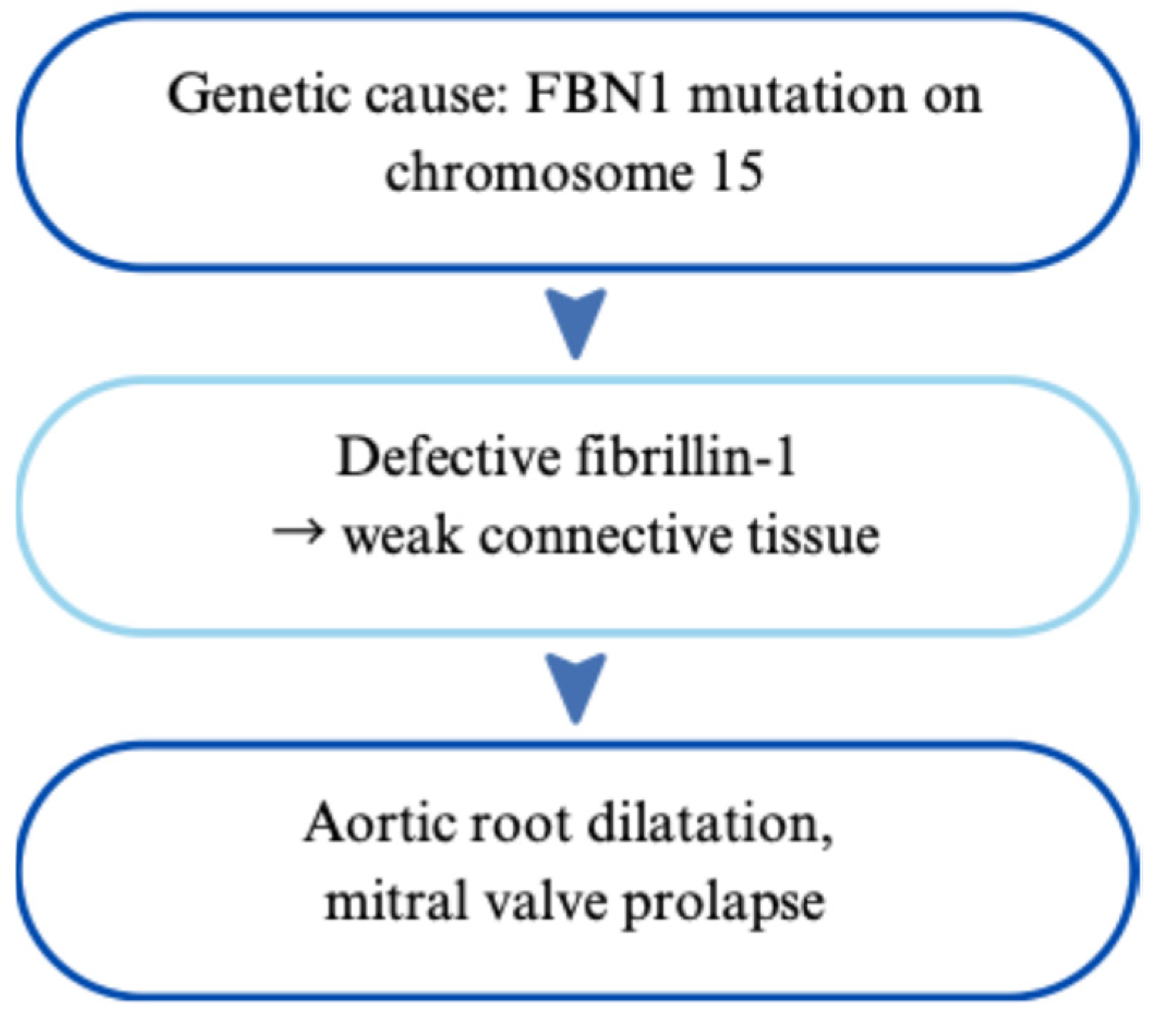
Figure 1. Genetic cause and pathophysiology of Marfan syndrome.
Cardiogenetics 15 00029 g001
Figure 2. Marfan syndrome. The diagram shows a heart with the two anatomic anomalies characteristic of Marfan syndrome.
Cardiogenetics 15 00029 g002
Figure 3. Genetic cause and pathophysiology of Noonan syndrome.
Cardiogenetics 15 00029 g003
Figure 4. Genetic cause and pathophysiology of 22q11.2 Deletion Syndrome.
Cardiogenetics 15 00029 g004
Figure 5. Genetic cause and pathophysiology of Duchenne Muscular Dystrophy.
Cardiogenetics 15 00029 g005
Figure 6. Genetic cause and pathophysiology of hypertrophic cardiomyopathy.
Cardiogenetics 15 00029 g006
Figure 7. Genetic cause and pathophysiology of tetralogy of Fallot.
Cardiogenetics 15 00029 g007
Figure 8. Tetralogy of Fallot. The diagram shows a heart with the 4 characteristic congenital cardiac anomalies.
Cardiogenetics 15 00029 g008
Table 1. Summary of Key pediatric genetic cardiac diseases.
DisorderGenes InvolvedMain Cardiac FeaturesDiagnostic ApproachManagement HighlightsReferences
Marfan
Syndrome
Autosomal dominant; FBN1 mutationsAortic root dilation, mitral valve prolapseGhent criteria; genetic testingβ-blockers, ARBs, surgical repair if >50 mm aortic dilation[3,4,5,6,7,8,9,10,11,12,13,14,15,16]
Noonan
Syndrome
Autosomal dominant; PTPN11, RAF1, RAS/MAPK genesPulmonary stenosis, hypertrophic cardiomyopathy (HCM)Clinical features + gene panelCardiac surgery, β-blockers, GH therapy in short stature[17,18,19,20,21,22,23,24,25,26,27,28,29]
22q11.2
Deletion
Syndrome
Microdeletion at 22q11.2; affects TBX1Conotruncal defects: TOF, truncus arteriosus, VSDFISH, MLPA, CMASurgical correction, immune and endocrine monitoring, calcium/Vitamin D replacement[30,31,32,33,34,35,36,37,38,39,40,41,42]
Duchenne Muscular DystrophyX-linked; DMD gene mutationsDilated cardiomyopathy (DCM), arrhythmiasGenetic testing, CMR, elevated CKSteroids, ACE inhibitors, ARNi, ICDs, exon-skipping therapy[43,44,45,46,47,48,49,50,51,52,53,54,55,56,57,58,59,60]
Hypertrophic CardiomyopathyMostly autosomal dominant; MYH7, MYBPC3Left ventricular hypertrophy, LVOT obstructionEchocardiography, MRI, genetic testingβ-blockers, ICD for SCD prevention, septal myectomy or ablation[61,62,63,64,65,66,67,68,69,70,71,72,73,74,75,76,77,78,79,80,81,82,83,84,85,86,87,88,89,90,91,92,93,94,95,96]
Tetralogy of Fallot (TOF)Multifactorial; linked to 22q11.2, JAG1, NKX2.5VSD, RVH, overriding aorta, pulmonary stenosisEcho, CXR, ECG; prenatal ultrasoundEarly surgical repair, prostaglandins pre-op, lifelong cardiology follow-up[97,98,99,100,101,102,103,104,105,106,107,108,109,110,111,112,113,114,115,116,117,118]
Disclaimer/Publisher’s Note: The statements, opinions and data contained in all publications are solely those of the individual author(s) and contributor(s) and not of MDPI and/or the editor(s). MDPI and/or the editor(s) disclaim responsibility for any injury to people or property resulting from any ideas, methods, instructions or products referred to in the content.