Case Report

TNNC1 Gene Mutation in Ebstein’s Anomaly and Left Ventricular Hypertrabeculation: A Case Report of a New Causative Mutation?

1
Pediatric Cardiology Department, “Vittore Buzzi” Children’s Hospital, 20154 Milan, Italy
2
Department of Pediatrics, Vittore Buzzi Children’s Hospital, University of Milan, 20122 Milan, Italy
3
Division of Neonatology, Department of Woman and Child Health and Public Health, University Hospital Fondazione Policlinico Gemelli Istituto di Ricovero e Cura a Carattere Scientifico (IRCCS), 00168 Rome, Italy
4
Fetal Therapy Unit “U.Nicolini”, V. Buzzi Children’s Hospital, ASST-FBF-Sacco, 20154 Milan, Italy
5
Clinical Genetics Unit, Department of Pediatrics, Buzzi Children’s Hospital, 20154 Milan, Italy
6
Pediatric and Adolescent Unit, Department of Internal Medicine, University of Pavia, 27100 Pavia, Italy
7
Department of Biomedical and Clinical Science “L. Sacco”, University of Milan, 20122 Milan, Italy
*
Author to whom correspondence should be addressed.
Cardiogenetics 2025, 15(3), 24; https://doi.org/10.3390/cardiogenetics15030024
Submission received: 8 July 2025 / Revised: 19 August 2025 / Accepted: 22 August 2025 / Published: 26 August 2025
(This article belongs to the Section Inherited Heart Disease-Children)

Abstract

Background: Ebstein’s anomaly (EA) is a rare congenital heart defect characterized by failure of tricuspid valve delamination during embryogenesis. Left ventricular (LV) hypertrabeculation results from incomplete myocardial compaction during fetal development. EA is associated with LV hypertrabeculation in 0.14% of cases, and EA is the most common congenital heart disease in LV hypertrabeculation (up to 29%), suggesting a shared embryogenetic pathway. Case Report: We describe a female patient prenatally diagnosed with EA and a large ventricular septal defect. Postnatal echocardiography confirmed EA with moderate regurgitation and revealed previously unnoticed left ventricular excessive trabeculations. Whole exome sequencing revealed a heterozygous never-described variant of unknown significance in the TNNC1 gene. Discussion: The genetic link between EA and LV hypertrabeculation remains unclear, though variants in sarcomeric or cytoskeletal genes like MYH7, TPM1, and NKX2.5—essential for cardiac development—have been implicated. A developmental hypothesis suggests that aberrant contraction during endocardial-to-mesenchymal and epicardial-to-mesenchymal transformation (5th–8th gestational weeks) may affect valve delamination and ventricular compaction via parallel signaling pathways. TNNC1 encodes troponin C1, a subunit of the troponin complex involved in muscle contraction. Its mutations are known to alter calcium sensitivity and impair cardiac contractility. Conclusions: EA and LV hypertrabeculation patients diagnosed in infancy have a greater risk of negative outcomes. Early, especially prenatal, diagnosis is crucial. Genetic analysis can provide fundamental insight into cardiac development. This new and rare variant of TNNC1 gene supports the hypothesis that early cardiomyocytes dysfunction disrupts both valve delamination and left ventricular compaction and that the two diseases share a common genetic pathway related to cardiomyocyte contraction.

1. Introduction

Ebstein’s anomaly (EA), first described by Dr William Ebstein in 1866, is a rare congenital heart defect that accounts for 0.3–0.5% of congenital heart defects. The prevalence at birth is approximately 1 in 200,000 live births [1,2,3,4]. The malformation consists of apical displacement of the tricuspid valve, resulting in regurgitation and enlargement of the right heart chambers. The specific EA features include attachment of the posterior and septal leaflets to the ventricular septum, apical displacement of the valve, redundant and fenestrated anterior leaflets, atrialization of the part of the right ventricle above the valve, and dilatation of the right ventricle and atrioventricular junction [2]. The pathogenesis is consistent with failure of delamination during embryonic development [2].
Left ventricular (LV) hypertrabeculation is morphologically characterized by prominent myocardial trabeculations and deep intertrabecular recesses, which give the left ventricle a spongy appearance. LV excessive trabeculations is thought to result from failure of the compaction process between the 5th and 8th weeks of embryonic development of the human myocardium [5]. EA can be associated with LV hypertrabeculation with an approximate prevalence of 0.14% [6], and EA is the most common congenital heart disease in LV hypertrabeculation [7]. These findings suggest that there are genetic factors that predispose patients to multiple cardiovascular manifestations [3].
The genetic correlation between EA and LV hypertrabeculation is still not fully understood. The first variant was described by Budde et al., who analyzed the genome of a German family (11 cases of LV hypertrabeculation previously called LV noncompaction, of whom 4 had EA). He described a mutation in the MYH7 gene (*160760, 14q11.2, AD) encoding the β-myosin heavy chain [8]. After his first description, other significant causative genes, such as NKX2.5 (*600584, 5q35.1, AD), GATA4 (*600576, 8p23.1, AD), TAZ (*300394, Xq28, XLR), and TBX20 (*606061, 7p14.2, inheritance not specified) were identified in several cases. Many of these genetic variants affect sarcomeric or cytoskeletal proteins that are crucial for cardiac development and function [9,10]. Other studies have reported mutations in nonsarcomeric proteins such as SCN5A (*600163, 3p22.2, AD), NONO (*300084, Xq13.1, XL), and KLHL26 (NM_001345981.1, 19p13.11, AD) in EA/LV hypertrabeculation [11,12,13]. The diversity of affected loci emphasizes the genetic complexity of tricuspid valve and ventricular development. Cardiogenesis begins with the formation of a heart tube composed of several layers: the outer epicardium, myocardial cells, cardiac jelly, and the endocardial layer. During development, the heart undergoes rightward looping, while the inner layer cells proliferate and differentiate into endocardial cushions, which will later form the cardiac valves. Meanwhile, the myocytes and endocardial cells in the ventricle proliferate to form deep trabeculations. These processes are regulated by common transcription factors (such as GATA4 and TBX20) and signaling pathways (including TGF-β/Smad, Wnt/β-catenin, and Notch), which govern both the formation of the tricuspid valve and the compaction of the LV [2,3].
We report a case of fetal diagnosis of Ebstein’s anomaly with a large muscular ventricular septal defect associated with LV hypertrabeculation, in which a new gene variant in a contractility protein was discovered.

2. Case Report

We describe a case of a firstborn baby conceived via intrauterine insemination with a male donor. At 18 + 3 gestational ages, fetal echocardiography revealed Ebstein’s anomaly with mild regurgitation associated with a large muscular ventricular septal defect. Both karyotype and array-CGH analyses were normal. The fetal heart remained compensated throughout the pregnancy.
At birth, the baby had normal adaptation (Apgar score of 9 at 1′ and 5′, weight of 3240 g, length of 52 cm, head circumference of 32.5 cm). The first cardiological evaluation confirmed the prenatal diagnosis but revealed increased trabeculation in the LV (Figure 1), a detail that was not clearly identified during pregnancy but was possibly recognizable on a second review of fetal ultrasound images. Abdominal ultrasound, brain ultrasound, and ophthalmological examination were performed, all of which yielded normal results. The baby was discharged on captopril and diuretics due to pulmonary overflow.
A neonatal cardiac MRI confirmed Ebstein’s anomaly with apically and anteriorly rotated septal and posterior leaflets, resulting in atrialization of the basal portions of the right ventricle, a 17 mm ventricular septal defect, and hypertrabeculation of the medium-apical LV, meeting the criteria for noncompaction.
At 1 month of age, a pulmonary banding procedure was needed.
During cardiological follow-up (approximately 17 months), the patient remained in good general condition, and growth was consistent but stable at the lower percentiles. Arrhythmias were never recorded.
Currently, periodic cardiological and cardiosurgical evaluations are ongoing to determine the next therapeutic steps.
Given the association between EA and LV hypertrabeculation, a whole exome analysis was performed. With next-generation sequencing, it is now possible to sequence large amounts of DNA, such as all the regions of an individual’s DNA that provide instructions for making proteins (whole exome analysis). These regions, called exons, are estimated to make up about 1 percent of a person’s genome.
Patient’s analysis revealed a variant of unknown significance, p.Glu66Lys, in the TNNC1 gene (*191040) that was heterozygous and of nonmaternal origin (Chr3 (GrCh38):g.524521128 (NM_003280.3: c.196G > A) in the exon of the gene TNNC1). It describes a substitution of glutamic acid for lysine at position 66 in cTnC. This variant was classified as of unknown significance according to ACMG guideline. The mutation is rare (gnomAD v4.0 MAF0) and it has never been described in the literature, but it is reported in the ClinVar database (ClinVar ID 1024458).

3. Discussion

This case highlights the complex interplay between EA and LV hypertrabeculation, presenting a unique challenge for diagnosis and management.
Patients born with both EA and LV hypertrabeculation diagnosed in infancy have a greater risk of negative outcomes such as heart failure and sudden cardiac death [14,15], along with anatomical variations that pose additional surgical challenges. As in our case, common comorbidities in EA/LV hypertrabeculation patients included septal defects (ASD or VSD) that could complicate the treatment and surgical approach [3,16].
The fetal diagnosis of EA plays a crucial role in predicting outcomes and guiding management, including close monitoring in high-risk pregnancies. This facilitates appropriate parental counseling and proactive planning for potential complications during the neonatal period. Prognostic factors identified in utero include the size of the fossa ovalis, the degree of right atrial dilation, and the presence of tricuspid regurgitation or right ventricular outflow tract obstruction [17,18].
However, the diagnosis of noncompaction cardiomyopathy in fetuses is challenging, and there is no universally accepted standard. According to a study by Stöllberger et al. [19], the echocardiographic criteria for diagnosing fetal ventricular noncompaction include at least four trabeculations protruding apically to the papillary muscle of the LV visible in one imaging plane in end-diastole, a two-layered structure with epicardial compacted (C) and endocardial noncompacted (NC) layers and an N/C ratio of ≥2, and perfusion of intraventricular blood into the intertrabecular spaces in color Doppler imaging. As highlighted in our case, fetal diagnosis of LV hypertrabeculation can be missed because of the unique characteristics of the fetal myocardium: the N/C ratio is naturally higher than that in children or adults and approaches 2. This calls for careful attention and close monitoring [20].
EA and LV hypertrabeculation are poorly understood in terms of genetic pathogenesis. The overlap of developmental pathways in the formation of the tricuspid valve and compact myocardium suggests a common morphogenetic origin for both conditions. Both defects are associated with dysfunctions in endocardial-to-mesenchymal transformation and epicardial-to-mesenchymal transition processes during embryogenesis, which are crucial for valve and myocardial structure formation [3,21,22]. In the case of EA, a defect in the delamination of the tricuspid valve causes apical displacement, whereas in LV hypertrabeculation, there is failure of myocardial compaction. Both defects are probably linked to dysfunctions in contraction mechanisms and the maintenance of cardiac structure. Although the clinical manifestations are different, the underlying mechanisms influencing heart morphogenesis and contractile function may overlap [3].
Recent genetic studies suggest that variants in sarcomeric and cytoskeletal genes may be at the root of the pathogenesis of both EA and LV hypertrabeculation. The MYH7 gene, which encodes the β-myosin heavy chain, is particularly associated with EA, LV hypertrabeculation, and their combination [23]. This variant likely disrupts salt bridge formation, destabilizes the myosin head and impairs myocardial function [5,8].
Our case revealed a new variant in the TNNC1 gene that was actually classified as being of unknown significance. The TNNC1 is a sarcomeric gene, it consists of six exons and five introns and encodes the protein troponin C1 (cTnC), one of the three essential subunits of the troponin complex involved in muscle contraction, which is predominantly expressed in cardiomyocytes and skeletal muscular cells [24]. The cTNC serves as a calcium (Ca2+) sensor within the myofilament and plays a key role in regulating cardiac contraction. Its tertiary structure consists of two globular domains, the C-domain and N-domain, connected by a flexible linker. The C-domain binds divalent ions (Ca2+ and/or Mg2+) at two helix-loop-helix motifs, which are critical for the structural and functional regulation of the sarcomere. The N-domain contains a nonfunctional site and a regulatory site that triggers contraction when calcium binds during systole [25].
Mutations in TNNC1 alter the conformational change of actin (the thin filament of the myocyte) and its transition from a blocked to a closed and then an open state, through a modification of the calcium sensitivity of the cardiac muscle, ultimately impairing the ability of the heart to contract efficiently. However, the precise mechanism by which the many variants influence the calcium binding is still unknown.
Missense mutations in cTnC have been linked to both dilated cardiomyopathy [26] with decrease sensibility to Ca2+, and hypertrophic cardiomyopathies with increased affinity to Ca2+, with or without LV hypertrabeculation [27]. Although rare compared to other sarcomeric mutations in cardiomyopathy, TNNC1-associated cardiomyopathies have shown a severe course with earlier onset of the disease and poorer prognosis [28].
Thin contractile filaments, such as cTnC, show a high degree of sequence conservation and have a limited number of described pathogenic mutations, likely related to severe outcomes due to genetic variants not being transmitted to subsequent generations.
However, more than 100 variants of TNNC1 have been described in the literature, although not all have been studied with functional analysis. In two separate papers, Tadros et al. and Retinoso et al. presented many pathogenic variants supporting the identification of TNNC1 as a gene associated with cardiomyopathy. Thanks to the use of cryo-electron microscopy they were able to localize each of the functionally studied variants. They found that most of the cardiomyopathy-related variants are located in the alpha-helical regions and in the linkers between NH2 and COOH-terminal regions Among the functionally studied variants that significantly affect the Ca2+ binding affinity, we highlight D62N, M81I, E94A, and R102C which are related to LV hypertrabeculation [25,28].
Furthermore, Wu et al. provided evidence of a regulatory role of the cTnC in the nuclear activity, leading to the possibility that thin filaments could also affect nuclear Ca2+-dependent signaling [29].
The major limitation of the study is that our variant currently lacks definitive evidence of pathogenicity, and it requires further characterization and study. However, the main purpose of our article is to describe a novel and rare variant (not previously reported) in a gene known to be associated with cardiomyopathy. Finding a mutation in a contractile protein supports the compelling hypothesis proposed by Thareja et al. [3], which suggests that early cardiomyocyte dysfunction may disrupt both valve delamination and left ventricular compaction, and that these two conditions may share a common genetic pathway related to cardiomyocyte contraction.
Rare variants can contribute to a better understanding of the molecular and cellular mechanisms underlying heart development.

4. Conclusions

The management of patients with a combination of EA and LV hypertrabeculation is extremely complex due to the severity and interaction of these two rare congenital conditions. Early diagnosis, particularly during the fetal period, is crucial for proper patient management, as it allows for closer monitoring and targeted therapeutic planning.
The pathogenesis of both conditions is still under investigation, but it is currently hypothesized that they share a common morphogenesis and a potential shared genetic origin, mostly related to abnormalities in contractility proteins.
A comprehensive, multidisciplinary approach involving gynecologists, pediatric cardiologists, surgeons, and geneticists is crucial to optimize care and effectively address the genetic and structural complexities associated with these conditions.

Author Contributions

S.M. and A.L. analyzed and interpreted the patient data regarding the diagnosis and the clinical follow-up. G.P. and C.C. collected the patient data and images and contributed to the writing of the manuscript. E.C. took care of the genetic analysis and interpretation. V.C. and G.V.Z. contributed to the pediatric care of the patient and contributed to elaborate the manuscript. I.R. made a major contribution to the preparation of the manuscript. All authors have read and agreed to the published version of the manuscript.

Funding

This research received no external funding.

Institutional Review Board Statement

Not applicable.

Informed Consent Statement

Consent to publish the information was obtained from the patient’s parent.

Data Availability Statement

The original contributions presented in this study are included in the article. Further inquiries can be directed to the corresponding author.

Conflicts of Interest

The authors declare that they have no competing interests.

Abbreviations

The following abbreviations are used in this manuscript:
CEpicardial compacted layer
Ca2+Calcium
cTnCTroponin C1
EAEbstein’s anomaly
LVLeft ventricle
LVNCLeft ventricular noncompaction
NCEndocardial noncompacted layer

References

  1. Lupo, P.J.; Langlois, P.H.; Mitchell, L.E. Epidemiology of Ebstein anomaly: Prevalence and patterns in Texas, 1999-2005. Am. J. Med. Genet. A 2011, 155A, 1007–1014. [Google Scholar] [CrossRef]
  2. Attenhofer Jost, C.H.; Connolly, H.M.; Dearani, J.A.; Edwards, W.D.; Danielson, G.K. Ebstein’s anomaly. Circulation 2007, 115, 277–285. [Google Scholar] [CrossRef]
  3. Thareja, S.K.; Frommelt, M.A.; Lincoln, J.; Lough, J.W.; Mitchell, M.E.; Tomita-Mitchell, A. A Systematic Review of Ebstein’s Anomaly with Left Ventricular Noncompaction. J. Cardiovasc. Dev. Dis. 2022, 9, 115. [Google Scholar] [CrossRef]
  4. Krieger, E.V.; Valente, A.M. Diagnosis and management of ebstein anomaly of the tricuspid valve. Curr. Treat. Options Cardiovasc. Med. 2012, 14, 594–607. [Google Scholar] [CrossRef]
  5. Vermeer, A.M.; van Engelen, K.; Postma, A.V.; Baars, M.J.; Christiaans, I.; De Haij, S.; Klaassen, S.; Mulder, B.J.; Keavney, B. Ebstein anomaly associated with left ventricular noncompaction: An autosomal dominant condition that can be caused by mutations in MYH7. Am. J. Med. Genet. C Semin. Med. Genet. 2013, 163C, 178–184. [Google Scholar] [CrossRef]
  6. Aras, D.; Tufekcioglu, O.; Ergun, K.; Ozeke, O.; Yildiz, A.; Topaloglu, S.; Deveci, B.; Sahin, O.; Kisacik, H.L.; Korkmaz, S. Clinical features of isolated ventricular noncompaction in adults long-term clinical course, echocardiographic properties, and predictors of left ventricular failure. J. Card. Fail. 2006, 12, 726–733. [Google Scholar] [CrossRef]
  7. Stähli, B.E.; Gebhard, C.; Biaggi, P.; Klaassen, S.; Valsangiacomo Buechel, E.; Attenhofer Jost, C.H.; Jenni, R.; Tanner, F.C.; Greutmann, M. Left ventricular noncompaction: Prevalence in congenital heart disease. Int. J. Cardiol. 2013, 167, 2477–2481. [Google Scholar] [CrossRef]
  8. Budde, B.S.; Binner, P.; Waldmüller, S.; Höhne, W.; Blankenfeldt, W.; Hassfeld, S.; Brömsen, J.; Dermintzoglou, A.; Wieczorek, M.; May, E.; et al. Noncompaction of the ventricular myocardium is associated with a de novo mutation in the beta-myosin heavy chain gene. PLoS ONE 2007, 2, e1362. [Google Scholar] [CrossRef]
  9. Nijak, A.; Alaerts, M.; Kuiperi, C.; Corveleyn, A.; Suys, B.; Paelinck, B.; Saenen, J.; Van Craenenbroeck, E.; Van Laer, L.; Loeys, B.; et al. Left ventricular noncompaction with Ebstein anomaly attributed to a TPM1 mutation. Eur. J. Med. Genet. 2018, 61, 8–10. [Google Scholar] [CrossRef]
  10. Sicko, R.J.; Browne, M.L.; Rigler, S.L.; Druschel, C.M.; Liu, G.; Fan, R.; Romitti, P.A.; Caggana, M.; Kay, D.M.; Brody, L.C.; et al. Genetic Variants in Isolated Ebstein Anomaly Implicated in Myocardial Development Pathways. PLoS ONE 2016, 11, e0165174. [Google Scholar] [CrossRef]
  11. Neu, A.; Eiselt, M.; Paul, M.; Sauter, K.; Stallmeyer, B.; Isbrandt, D.; Schulze-Bahr, E. A homozygous SCN5A mutation in a severe, recessive type of cardiac conduction disease. Hum. Mutat. 2010, 31, E1609–E1621. [Google Scholar] [CrossRef]
  12. Carlston, C.M.; Bleyl, S.B.; Andrews, A.; Meyers, L.; Brown, S.; Bayrak-Toydemir, P.; Bale, J.F.; Botto, L.D. Expanding the genetic and clinical spectrum of the NONO-associated X-linked intellectual disability syndrome. Am. J. Med. Genet. 2019, 179, 792–796. [Google Scholar] [CrossRef]
  13. Samudrala, S.S.K.; North, L.M.; Stamm, K.D.; Earing, M.G.; Frommelt, M.A.; Willes, R.; Tripathi, S.; Dsouza, N.R.; Zimmermann, M.T.; Mahnke, D.K.; et al. Novel KLHL26 variant associated with a familial case of Ebstein’s anomaly and left ventricular noncompaction. Mol. Genet. Genom. Med. 2020, 8, e1152. [Google Scholar] [CrossRef]
  14. McGee, M.; Warner, L.; Collins, N. Ebstein’s Anomaly, Left Ventricular Noncompaction, and Sudden Cardiac Death. Case Rep. Cardiol. 2015, 2015, 854236. [Google Scholar] [CrossRef]
  15. Pignatelli, R.H.; Texter, K.M.; Denfield, S.W.; Grenier, M.A.; Altman, C.A.; Ayres, N.A.; Chandra-Bose Reddy, S. LV Noncompaction in Ebstein’s anomaly in infants and outcomes. JACC Cardiovasc. Imaging 2014, 7, 207. [Google Scholar] [CrossRef]
  16. Hirono, K.; Hata, Y.; Ibuki, K.; Yoshimura, N. Familial Ebstein’s anomaly, left ventricular noncompaction, and ventricular septal defect associated with an MYH7 mutation. J. Thorac. Cardiovasc. Surg. 2014, 148, e223–e226. [Google Scholar] [CrossRef]
  17. Pavlova, M.; Fouron, J.C.; Drblik, S.P.; van Doesburg, N.H.; Bigras, J.L.; Smallhorn, J.; Harder, J.; Robertson, M. Factors affecting the prognosis of Ebstein’s anomaly during fetal life. Am. Heart J. 1998, 135, 1081–1085. [Google Scholar] [CrossRef]
  18. Torigoe, F.; Ishida, H.; Ishii, Y.; Ishii, R.; Narita, J.; Kawazu, Y.; Kayatani, F.; Inamura, N. Fetal echocardiographic prediction score for perinatal mortality in tricuspid valve dysplasia and Ebstein’s anomaly. Ultrasound Obstet. Gynecol. 2020, 55, 226–232. [Google Scholar] [CrossRef]
  19. Stöllberger, C.; Wegner, C.; Finsterer, J. Fetal Ventricular Hypertrabeculation/Noncompaction: Clinical Presentation, Genetics, Associated Cardiac and Extracardiac Abnormalities and Outcome. Pediatr. Cardiol. 2015, 36, 1319–1326. [Google Scholar] [CrossRef]
  20. Zhang, W.; Dai, X.; Liu, H.; Li, L.; Zhou, S.; Zhu, Q.; Chen, J. Case report: Prenatal diagnosis of fetal noncompaction cardiomyopathy with bradycardia accompanied by de novo CALM2 mutation. Front. Pediatr. 2022, 10, 1012600. [Google Scholar] [CrossRef]
  21. Gong, H.; Lyu, X.; Wang, Q.; Hu, M.; Zhang, X. Endothelial to mesenchymal transition in the cardiovascular system. Life Sci. 2017, 184, 95–102. [Google Scholar] [CrossRef] [PubMed]
  22. Lincoln, J.; Yutzey, K.E. Molecular and developmental mechanisms of congenital heart valve disease. Birth Defects Res. A Clin. Mol. Teratol. 2011, 91, 526–534. [Google Scholar] [CrossRef] [PubMed]
  23. Postma, A.V.; van Engelen, K.; van de Meerakker, J.; Rahman, T.; Probst, S.; Baars, M.J.; Bauer, U.; Pickardt, T.; Sperling, S.R.; Berger, F.; et al. Mutations in the sarcomere gene MYH7 in Ebstein anomaly. Circ. Cardiovasc. Genet. 2011, 4, 43–50. [Google Scholar] [CrossRef] [PubMed]
  24. Li, M.X.; Hwang, P.M. Structure and function of cardiac troponin C (TNNC1): Implications for heart failure, cardiomyopathies, and troponin modulating drugs. Gene 2015, 571, 153–166. [Google Scholar] [CrossRef]
  25. Reinoso, T.R.; Landim-Vieira, M.; Shi, Y.; Johnston, J.R.; Chase, P.B.; Parvatiyar, M.S.; Landstrom, A.P.; Pinto, J.R.; Tadros, H.J. A comprehensive guide to genetic variants and post-translational modifications of cardiac troponin C. J. Muscle Res. Cell Motil. 2021, 42, 323–342. [Google Scholar] [CrossRef]
  26. Landim-Vieira, M.; Johnston, J.R.; Ji, W.; Mis, E.K.; Tijerino, J.; Spencer-Manzon, M.; Jeffries, L.; Hall, E.K.; Panisello-Manterola, D.; Khokha, M.K.; et al. Familial dilated cardiomyopathy associated with a novel combination of compound heterozygous TNNC1 variants. Front. Physiol. 2020, 10, 1612. [Google Scholar] [CrossRef]
  27. Ploski, R.; Rydzanicz, M.; Ksiazczyk, T.M.; Franaszczyk, M.; Pollak, A.; Kosinska, J.; Michalak, E.; Stawinski, P.; Ziolkowska, L.; Bilinska, Z.T.; et al. Evidence for troponin C (TNNC1) as a gene for autosomal recessive restrictive cardiomyopathy with fatal outcome in infancy. Am. J. Med. Genet. A 2016, 170, 3241–3248. [Google Scholar] [CrossRef]
  28. Tadros, H.J.; Life, C.S.; Garcia, G.; Pirozzi, E.; Jones, E.G.; Datta, S.; Parvatiyar, M.S.; Chase, P.B.; Allen, H.D.; Kim, J.J.; et al. Meta-analysis of cardiomyopathy-associated variants in troponin genes identifies loci and intragenic hot spots that are associated with worse clinical outcomes. J. Mol. Cell Cardiol. 2020, 142, 118–125. [Google Scholar] [CrossRef]
  29. Wu, H.; Lee, J.; Vincent, L.G.; Wang, Q.; Gu, M.; Lan, F.; Churko, J.M.; Sallam, K.I.; Matsa, E.; Sharma, A.; et al. Epigenetic Regulation of Phosphodiesterases 2A and 3A Underlies Compromised β-Adrenergic Signaling in an iPSC Model of Dilated Cardiomyopathy. Cell Stem cell. 2015, 17, 89–100. [Google Scholar] [CrossRef]
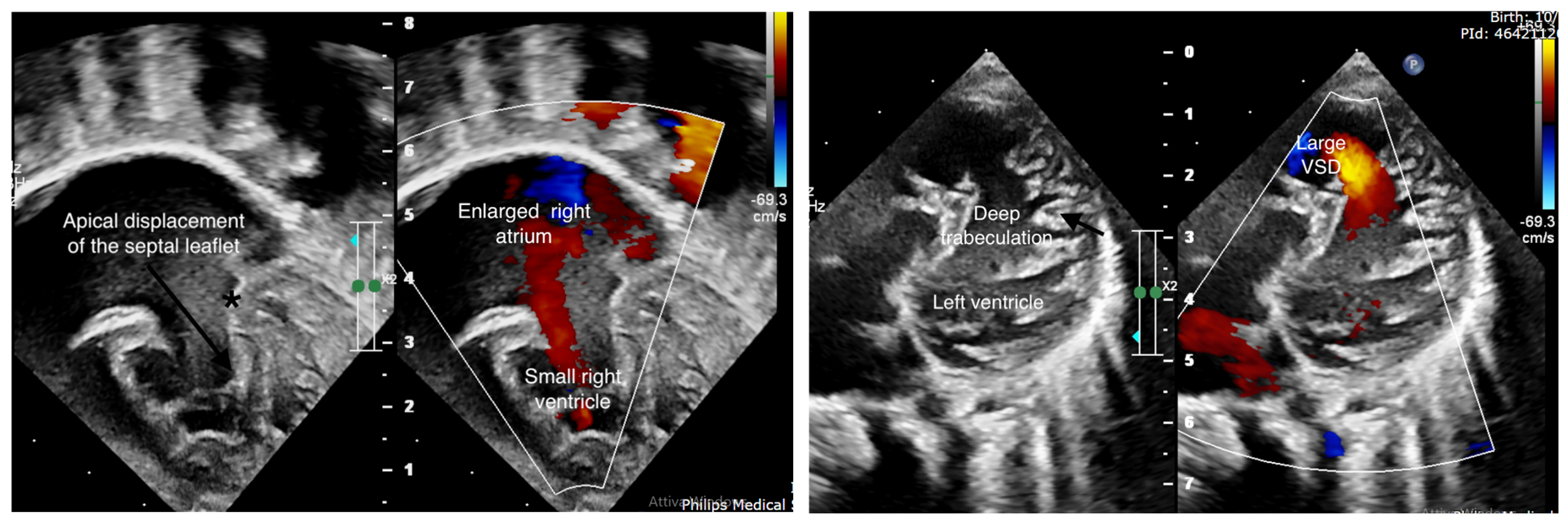
Figure 1. Ebstein’s anomaly and left ventricular hypertrabeculation. On the left side, Ebstein’s anomaly with displacement of the septal leaflet of the tricuspid valve (black arrow), and the asterisk shows the “normal” insertion of the septal leaflet. On the right side, the image shows the deep trabeculations of the left ventricle lateral wall. From this view, one can also see the large apical ventricular septal defect.
Cardiogenetics 15 00024 g001
Disclaimer/Publisher’s Note: The statements, opinions and data contained in all publications are solely those of the individual author(s) and contributor(s) and not of MDPI and/or the editor(s). MDPI and/or the editor(s) disclaim responsibility for any injury to people or property resulting from any ideas, methods, instructions or products referred to in the content.