Review

Dilated Cardiomyopathy and Sensorimotor Polyneuropathy Associated with a Homozygous ELAC2 Variant: A Case Report and Literature Review

by
Francesco Ravera
1,2,†,
Filippo Angelini
1,*,†,
Pier Paolo Bocchino
1,
Gianluca Marcelli
1,
Giulia Gobello
1,2,
Giuseppe Giannino
1,2,
Guglielmo Merlino
1,2,
Benedetta De Guidi
1,2,
Andrea Destefanis
1,2,
Giulia Margherita Brach Del Prever
2,3,
Carla Giustetto
1,2,
Guglielmo Gallone
1,2,
Stefano Pidello
1,
Antonella Barreca
4,
Silvia Deaglio
2,3,
Gaetano Maria De Ferrari
1,2,
Claudia Raineri
1 and
Veronica Dusi
1,2
1
Division of Cardiology, Cardiovascular and Thoracic Department, Città della Salute e della Scienza, 10126 Turin, Italy
2
Department of Medical Sciences, University of Turin, 10126 Turin, Italy
3
Immunogenetics and Transplant Biology Unit, Città della Salute e della Scienza, 10126 Turin, Italy
4
Division of Pathology, Città della Salute e della Scienza, 10126 Turin, Italy
*
Author to whom correspondence should be addressed.
These authors contributed equally to this work.
Cardiogenetics 2025, 15(3), 20; https://doi.org/10.3390/cardiogenetics15030020
Submission received: 2 June 2025 / Revised: 15 July 2025 / Accepted: 24 July 2025 / Published: 31 July 2025
(This article belongs to the Section Rare Disease-Genetic Syndromes)

Abstract

Variants in ELAC2, a gene encoding the mitochondrial RNase Z enzyme essential for mitochondrial tRNA processing, have been associated with severe pediatric-onset mitochondrial dysfunction, primarily presenting with developmental delay, hypertrophic cardiomyopathy (HCM), and lactic-acidosis. We hereby report the case of a 25-year-old young woman presenting with dilated cardiomyopathy (DCM) and peripheral sensorimotor polyneuropathy, harboring a homozygous variant in ELAC2. The same variant has been reported only once so far in a case of severe infantile-onset form of HCM and mitochondrial respiratory chain dysfunction, with in vitro data showing a moderate reduction in the RNase Z activity and supporting the current classification as C4 according to the American College of Medical Genetics (ACMG) criteria (PS3, PM2, PM3, PP4). Our extensive clinical, imaging, histological, and genetic investigations support a causal link between the identified variant and the patient’s phenotype, despite the fact that the latter might be considered atypical according to the current state of knowledge. A detailed review of the existing literature on ELAC2-related disease is also provided, highlighting the molecular mechanisms underlying tRNA maturation, mitochondrial dysfunction, and the variable phenotypic expression. Our case further expands the clinical spectrum of ELAC2-related cardiomyopathies to include a relatively late onset in young adulthood and underscores the importance of comprehensive genetic testing in unexplained cardiomyopathies with multisystem involvement.

1. Introduction

Mitochondrial dysfunction represents a key mechanism underlying several forms of inherited cardiomyopathies. Among the implicated genes, ELAC2 (elaC ribonuclease Z 2) encodes a long-form RNase Z enzyme involved in mitochondrial and nuclear tRNA 3′ end maturation, crucial for proper mitochondrial oxidative phosphorylation (OXPHOS) function. Variants in ELAC2 have been associated with pediatric-onset cardiomyopathy (mostly hypertrophic cardiomyopathy, HCM, rarely dilated cardiomyopathy, DCM), neuromuscular disorders, and metabolic abnormalities. Organs with high-energy requirements, such as the brain, heart, and skeletal muscle, rely heavily on mitochondrial function and are generally more severely affected. Here, we describe a case of isolated young adult-onset DCM with associated peripheral neuropathy in a patient carrying a homozygous ELAC2 variant and review the current literature on ELAC2-variant-related pathophysiology.

2. Clinical Case

A 25-year-old woman presented to our hospital complaining of dyspnea on minimal exertion and orthopnea that had developed over the past two weeks. Her past medical history was characterized by vasovagal syncopal episodes in childhood, with regular physical and psychomotor development. Her first-degree family history was negative for cardiovascular and renal disorders. The patient was not taking any chronic medications. Due to clinical suspicion of community-acquired pneumonia, antibiotic treatment had been started at home.
Two days later, the patient was admitted to the emergency department with an initial presentation of acute pulmonary distress without hypotension (blood pressure 130/80 mmHg). The electrocardiogram (ECG) showed sinus tachycardia at 120 bpm with short PR interval (110 ms), fragmented QRS with a right bundle branch block and a posterior left fascicular block (duration 120 ms), and diphasic negative/positive T waves in precordial leads (Figure 1a). Laboratory tests revealed markedly elevated NT-proBNP levels (14,753 pg/mL, reference range < 450 pg/mL), elevated high-sensitivity troponin I levels (192 ng/L, reference range: 0–16 ng/L), reduced glomerular filtration rate (44 mL/min/1.73 m2), and arterial lactate < 2 mmol/L.
Cardiac ultrasound showed a dilated left ventricle (end-diastolic volume index [EDVi] 100 mL/m2) with normal wall thickness (interventricular septum 9 mm, posterior wall 8 mm), a reduced left ventricular (LV) ejection fraction (LVEF 30%) due to global hypokinesia, severe mitral regurgitation, severe diastolic dysfunction (restrictive diastolic pattern), a non-dilated mildly hypertrophic (8 mm at the free wall) right ventricle (RV) with normal contractile function, moderate pulmonary hypertension (estimated systolic pulmonary arterial pressure, sPAP, 55 mmHg) and a mild circumferential pericardial effusion. The patient was admitted to our Cardiology Intensive Care Unit, where unloading therapy was initiated with intravenous diuretics and sodium nitroprusside, and non-invasive ventilation was started, leading to progressive improvement in both clinical and laboratory parameters.
A cardiac MRI performed two days after hospital admission confirmed significant dilation of the LV, measuring 154 mL/m2 (normal values: 62–98 mL/m2), with normal wall thickness and a severely impaired biventricular systolic function (LVEF 19%, RVEF 39%) due to diffuse hypokinesis. The tissue analysis highlighted diffuse mid-wall late gadolinium enhancement (LGE) with a ring-like pattern. The native T1 value in the LV myocardium was elevated (1186 ms, normal values: 950–1050 ms), as well as the extracellular volume (ECV 45%, normal values: 20–30%) (Figure 1b). The T2-weighted analysis showed mild hyperintensity with slightly increased values in the LV myocardium, measuring 60 ms (normal values: < 55 ms). Additionally, computed tomography (CT) coronary angiography excluded the presence of epicardial coronary disease. A panel of myocarditis-related virological and autoimmune tests turned out negative.
After achieving clinical stability, a right heart catheterization was performed during sodium nitroprusside continuous infusion at 1 µg/kg/min (systolic blood pressure 120/85 mmHg). The exam revealed a right atrial pressure of 2 mmHg, elevated pulmonary artery pressure of 35/23 mmHg (mean 28 mmHg), mildly elevated pulmonary capillary wedge pressure of 16 mmHg, reduced cardiac index of 2.17 L/min/m2, and a mild increase in pulmonary vascular resistance to 2.6 Woods Units. Notably, while NT-proBNP levels progressively normalized during hospitalization, high-sensitivity troponin I levels persisted above normal limits, with a peak of 350 ng/L on the fifth day after admission, and a value of 109 ng/L at discharge. Creatinine levels also remained elevated despite decongestion (1.39 at admission, 1.86 mg/dL at discharge).
Given the clinical presentation, an endomyocardial biopsy was also performed, revealing minimal edema and mild cellular polymorphism with slight cytoplasmic vacuolization and myofibrillar lysis. Trivial subendocardial and interstitial myocardial fibrosis were also noted. Very rare lymphocytes were observed (<7/mm2), and virological results on the biopsy were negative. Overall, Dallas criteria for myocarditis were not met. To further study the ultrastructural morphology of cardiomyocytes, electron microscopy analysis was performed, revealing degenerative aspects with marked mitochondrial hyperplasia and minimal atypia (presence of megamitochondria and concentric cristae), associated with subsarcolemmal glycogen accumulation (Figure 1c,d). Given the renal impairment not associated with a low-output hemodynamic state or venous congestion, and the histological finding of glycogen accumulation in the endomyocardial biopsy, the panel of tests was expanded to rule out the possible presence of metabolic and storage diseases: the measurement of alpha-glucosidase activity (Glycogen Storage Disease Type II/Pompe Disease) on leukocytes from peripheral blood and urinary oxalate (hyperoxaluria) were found to be within normal ranges. The cause of renal failure was attributed to the presence of multiple cysts affecting the renal parenchyma bilaterally. The patient was discharged on treatment with Sacubitril/Valsartan, Dapagliflozin, Carvedilol, and Spironolactone at the maximum tolerated doses.

2.1. Genetic Analysis

Genetic studies were initially based on the Illumina True Sight One panel (Illumina, San Diego, CA, USA), with analysis restricted to a panel of genes involved in hypertrophic, dilated, and arrhythmogenic cardiomyopathy; hyperoxaluria; polycystic kidney disease; nephrolithiasis; cystic kidney disease; glycogen storage disorders; and mitochondrial disorders (complete list of genes reported in the Supplementary Materials S1; for mitochondrial disorders only COX15, SCO2, SDHA, and SLC25A4 were included as part of the commercially available panel). This first round of genetic testing identified a heterozygous likely pathogenic variant (C4) in the NEK8 gene, a gene known for being associated with nephronophthisis, but with a recessive pattern of inheritance. Genetic testing was therefore expanded to include array comparative genomic hybridization (CGH) and clinical exome. By doing so, a homozygous missense single-nucleotide variant in exon 13 (c.1163A > G; p.Gln388Arg) of ELAC2 was identified. This variant has an extremely low frequency in gnomAD v4.1.0 population databases that is compatible with the Pathogenic Moderate PM2 criteria of ACMG. The variant has been reported once in ClinVar in a case of severe infantile-onset form of HCM and mitochondrial respiratory chain dysfunction, with in vitro data supporting pathogenicity (moderate reduction in the RNase Z activity) [1], adding a Pathogenic Strong PS3 criterion. The combination of PS3 and PM2 criteria led to the classification of the variant as likely pathogenic (C4). A further analysis of family history revealed that the patient’s parents, originating from a small village in Sicily, were fourth cousins, explaining the inheritance of the rare variant in homozygosity (PM3); in addition, a maternal aunt had married a second cousin, and two of her children had died in the first year of age. Both the mother and father, in their fifties and with normal ECG and cardiac ultrasound results, underwent Sanger sequencing for genetic testing, which confirmed that they each carry the same ELAC2 variant in a heterozygous state. The two older brothers, apparently healthy, have chosen to postpone genetic analysis so far. The family pedigree is reported as Supplementary Materials S2.

2.2. Follow-Up

Taking into account the rare ELAC2 variant identified in the genetic test and its previous association with peripheral sensorimotor disorders, an electromyography was performed, revealing a pattern consistent with a predominantly sensory axonal polyneuropathy, with no myopathic features.
After 3 months of optimal medical therapy, LVEF at cardiac ultrasound as well as tissue characterization on MRI remained unchanged, with persistent diffuse intramyocardial LGE and edema. High-sensitivity troponin I levels, albeit lower compared to the acute phase, were still elevated (95 ng/L at 2 months, 37 ng/L at 3 months), suggesting continuous heart damage. To assess a potential underlying inflammatory component contributing to the cardiomyopathy (a condition more commonly described during the ‘hot phases’ of arrhythmogenic cardiomyopathies) [2,3], a cardiac 18F-fludeoxyglucose positron emission tomography (FDG-PET) was performed, revealing a widespread hyper-uptake of the tracer in the left ventricular walls, consistent with the diffuse tissue abnormalities observed on cardiac MRI (Figure 1e). In light of these findings, a maintenance therapy with prednisone was initiated, and after three months, a follow-up FDG-PET was performed, demonstrating a significant reduction in myocardial tracer uptake (Figure 1f).
The persistent LV dysfunction led to the implantation of a transvenous single-lead implantable defibrillator (ICD) as primary prevention; during the subsequent follow-up, episodes of asymptomatic nonsustained monomorphic ventricular tachycardia were recorded, without device interventions.
Cardiopulmonary exercise testing at 6 months revealed a significantly reduced peak oxygen consumption of 15 mL/kg/min, representing only 42% of the predicted value. This, combined with the persistently reduced LVEF and the advanced NYHA functional class (III), led to the decision to place the patient on the active heart transplant waiting list.
At the 15-month follow-up, the patient remained stable, NYHA Class III, with an LVEF of 27% and had not experienced any hospitalizations for heart failure.

3. Discussion

3.1. Primary Mitochondrial Disorders and Mt-tRNA Maturation

Primary mitochondrial disorders (PMDs) are a collection of rare genetic conditions that result in compromised energy production. This impairment is typically caused by pathogenic variants in nuclear or mitochondrial genes that play crucial roles in oxidative phosphorylation. Indeed, the mitochondrion is the essential powerhouse for cellular growth and function. This is guaranteed by the production of ATP through oxidative phosphorylation by respiratory chain complexes (OXPHOS) that are localized in this cellular organelle. The OXPHOS system is composed of five enzymatic complexes (I-II-III-IV and V, also called ATP synthase), whose subunits are encoded by both nuclear and mitochondrial genes [4]. The mitochondrial genome consists of a circular double-stranded structure that, unlike nuclear DNA, is transcribed by RNA polymerase III as a single polycistronic transcript. This transcript contains 13 messenger RNAs (mt-mRNAs), 2 ribosomal RNAs (mt-rRNAs), and 22 transfer RNAs (mt-tRNAs) [5]. From the 13 mt-mRNAs, an equivalent number of peptides will be translated, which will become part of the OXPHOS system [6].
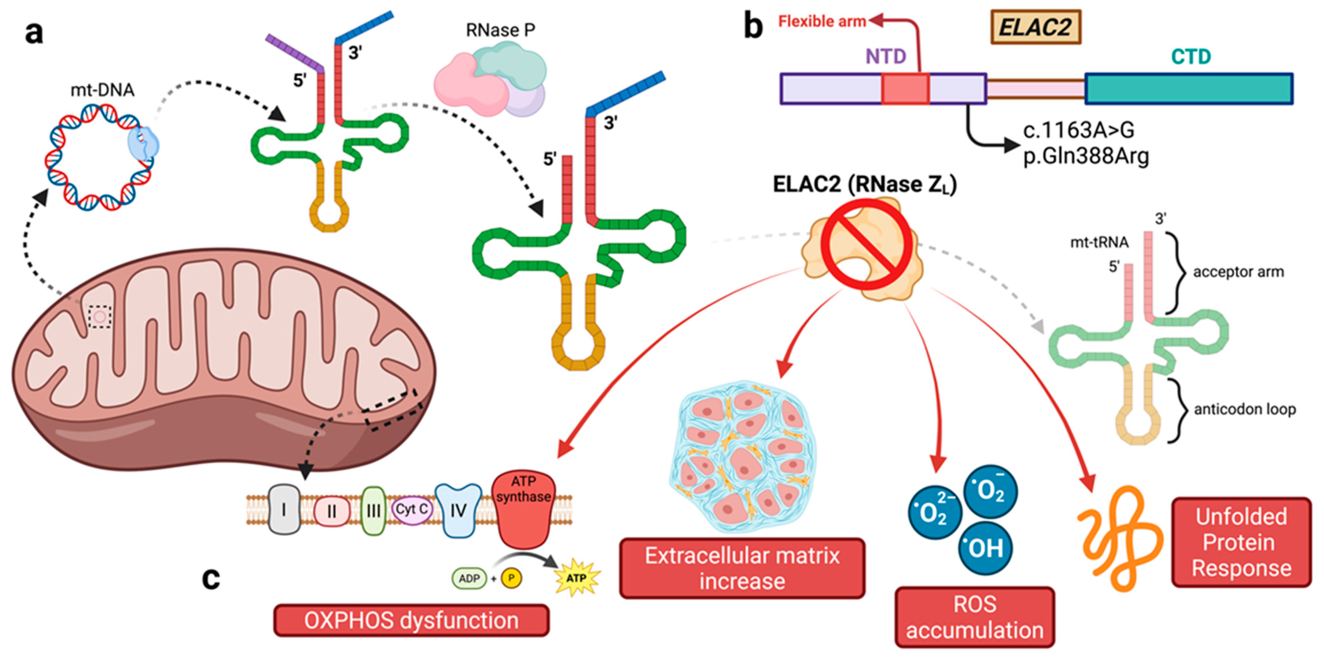
The tertiary structure of tRNA is formed by two fundamental parts that are opposite to each other: the acceptor arm, which is the site where the amino acid binds, and the anticodon loop, which contains the triplet of nucleotides responsible for recognizing the corresponding triplet in the mRNA sequence [7]. The acceptor arm contains the 5′ and 3′ extremities; in the immature precursor of mt-tRNA, these regions contain an excessive number of nucleotides that must be removed to allow the correct function of tRNA [8]. The processing of immature mt-tRNA starts with the 5′ cleavage, which is carried out by an endoribonuclease (RNase P). This protein complex is composed of a methyltransferase (MRPP1-TRMT10C), a dehydrogenase (MRPP2-SDR5C1), and the endoribonuclease subunit (MRPP3-PRORP) [9]. The 3′ end processing is catalyzed by an RNase Z enzyme, belonging to the β-lactamase family of metal-dependent (Zn2+ ions) endonucleases. In bacteria, RNase Z is present only in the short form (RNase Zs). However, in many eukaryotes, alongside the short form, a long form is also present (RNase ZL), resulting from gene duplication and containing two β-lactamase domains [10].
In humans, the short-form homolog is named ELAC1 and localizes in the cytosol and the nucleus. Conversely, the long-form is named ELAC2, and thanks to the presence of two alternative start codons, alternative translation initiation produces both a nuclear and a mitochondrially targeted form [11]. ELAC2 knockout disrupts 3′ end processing of both nuclear-encoded (nu-tRNAs) and mitochondrial-encoded tRNA precursors. For nu-tRNAs and canonical mt-tRNAs bearing a conserved elbow, ELAC2 alone recognizes and cleaves the 3′ trailers via direct ELAC2–RNA interaction. However, most human mt-tRNAs have structurally degenerate elbow regions, and efficient processing of these noncanonical substrates requires the TRMT10C–SDR5C1 subcomplex. In vitro reconstitution shows that TRMT10C–SDR5C1 stabilizes the tertiary fold of degenerate mt-tRNAs and mediates protein–protein contacts with ELAC2, forming a multisubunit mitochondrial RNase Z complex essential for proper mt-tRNA 3′ maturation [12].
From a clinical standpoint, cardiovascular involvement (CVI) has been identified in several patients with PMD. However, the age-related prevalence, clinical presentation, and prognostic significance remain inadequately understood. In particular, determining whether the outcomes of CVI are consistent across the various forms of PMD is challenging due to the infrequency and diversity of these subsets. In 2020 [13], Brambilla et al. reported a CVI in 36% of 86 children diagnosed with PMD, frequently appearing at a very young age (mean age of onset 6 years), including the pre- and neonatal phase in 16%, often representing the first sign of PMD. Hypertrophic, non-compaction, and dilated cardiomyopathies were the prevalent disorders, although pulmonary arterial hypertension was also found. CVI was linked to a poor prognosis. The ultimate outcome of PMD-related CVI was affected by the specific underlying etiology, indicating the necessity for personalized management of heart failure and strategies to prevent sudden death. Notably, the diagnosis was confirmed by molecular analysis in only 66% of the patients, with MELAS syndrome due to MTTL1 A3243G variant being the most frequent finding (9%), followed by TMEM70 (6%) and NARP and Barth syndrome (5% each). Only one patient was harboring an ELAC2 variant (details of the variant were not provided in the paper), underlying once more how rare these variants are.

3.2. The Role of ELAC2

ELAC2 consists of two β-lactamase domains: the N-terminal domain (NTD, Met1–Arg428) and the C-terminal domain (CTD, Pro481–Gln826) (Figure 2) [14]. ELAC2 is also involved in processing nuclear tRNAs and miRNAs, with mouse models indicating its role in prostate cancer when combined with additional risk factors [15,16] and through the interaction with the stress pathway mediated by NIK/nuclear factor kB (NF-kB) [17]. Moreover, in ELAC2 knockout models targeting megakaryocytes, the absence of this protein led to thrombocytopenia and bleeding disorders [18,19]. The clinical variant p.Gln388Arg is located in the NTD, which has no catalytic residues but retains a flexible arm crucial for pre-tRNA binding [20]. This function occurs in the nucleus without additional cofactors, while mitochondrial processing requires a complex with TRMT10C and SDR5C1 [12].
In 2011, Brzezniak et al. demonstrated that silencing ELAC2 in HeLa cells led to an accumulation of molecules containing unprocessed 3′ mt-tRNA while not affecting mature mt-tRNA levels, suggesting ELAC2′s crucial role in processing mt-tRNA [10]. They revealed that the maturation process of mt-tRNA is likely sequential, as silencing MRPP1, a subunit of the RNase P complex, disrupts both 5′ and 3′ mt-tRNA processing. This finding aligns with earlier research in yeast, indicating that RNase Z (to which ELAC2 belongs) has low affinity for mt-tRNA with extended 5′ ends [21].
Five years later, a murine model of heart- and skeletal muscle-specific ELAC2 knockout revealed a dramatic loss of mature mitochondrial mRNA, rRNA, and tRNA, impacting overall transcription and resulting in significant functional impairment of OXPHOS. This loss drastically reduced oxygen consumption by the respiratory chain complexes [15]. Interestingly, patients with a homozygous pathogenic variant of ELAC2 showed OXPHOS dysfunction in their fibroblasts without a corresponding significant reduction in mature mt-mRNA, suggesting a blockade in protein production specifically at the translation phase [22]. ELAC2′s localization to both the nucleus and mitochondria, attributable to its N-terminal sequence, underscores its involvement in processing not only tRNAs but also other non-coding RNAs, including microRNAs (miRNAs) [14].
Further insights into ELAC2′s pathogenic mechanisms arise from studies in Drosophila melanogaster, which lacks ELAC2 but possesses a homologous protein, RNaseZ [23]. Variants of these proteins analogous to those found in humans with HCM led to an increase in heart wall thickness and end-diastolic area of the heart lumen and a reduction in fractional shortening, attributed to enhanced ploidy and increased cardiomyocyte size alongside a notable rise in extracellular matrix quantities. These findings suggest that hypertrophic and dilated cardiomyopathy may represent different phases of the same disease [24]. Additionally, altered RNaseZ function correlated with decreased abundance of mt-mRNAs encoding respiratory chain protein subunits, leading to reduced OXPHOS activation and increased ROS [25], which can cause cellular and DNA damage [26].
The protective mitochondria response to membrane damage involves the Unfolded Protein Response (UPR), crucial for maintaining mitochondrial function [27]. Silencing the ELAC2 ortholog in Caenorhabditis elegans resulted in impaired UPR activation and subsequent cellular damage [28]. Evaluating the pathogenicity of variants identified in human subjects with various methods (Table 1) has demonstrated that patient-derived fibroblasts accumulate unprocessed mt-tRNA and mt-mRNA, leading to reduced OXPHOS proteins [22]. Lentiviral expression of the wild-type gene in these fibroblasts restored normal levels of mature mt-RNAs. In the largest case series on ELAC2 in the literature, Saoura et al. [1] provided strong evidence of the pathogenicity of multiple missense variants, including p.Gln388Arg, by analyzing RNase activity and finding significant reductions in enzyme function compared to the wild-type protein. Approximately half of the variants exhibited mild residual enzymatic activity, ranging from 20% to 80%, compared to the wild type (WT). Notably, four variants showed significantly lower activity, with Pro493Leu and Tyr729Cys exhibiting only 1% of the WT activity. In contrast, the variant found in our patient, p.Gln388Arg, demonstrated a residual activity of 38%.

3.3. ELAC2 Variants, Genotype–Phenotype Correlation, Cardiac Phenotype, and Disease Course

Including our patient, a total of 28 ELAC2 variants with minimum to severe cardiac involvement have been described so far all over the world (Table 1) in 10 case reports/series, leading to an overall number of 42 patients harboring ELAC2 variants; the single most represented variant is Phe154Leu, described in 20/42 (48%) patients, all Arab or Saudi Arabian. Only 10/42 were European (24%), including 6 Italian cases.
In 2013, Haack et al. identified, in a cohort of pediatric patients with muscle biopsy evidence of dysfunction in OXPHOS chain complex activity, five individuals from three different families carrying homozygous ELAC2 variants (three subjects: p.Phe154Leu and p.Leu423Phe) or double heterozygous variants (two subjects: p.Arg211*; Thr420Ile). All these patients presented with a severe form of hypertrophic cardiomyopathy (HCM), with onset in the first months of life (documented in all individuals before the age of six months) and, in three of them, early death. One of these patients showed rapid progression to a form of DCM at the age of four years. All patients exhibited intrauterine growth retardation or psychomotor retardation in the first months of life, and three of them presented with lactic acidosis [22].
The previously mentioned ELAC2 variant (p.Phe154Leu), reported in an Arab family, was found in homozygosity in a cohort of 16 patients from 15 different Saudi families with infantile-onset cardiomyopathy. All patients died before the age of 14 months and exhibited a phenotype of HCM, except for three cases, which presented with a DCM phenotype. Another notable manifestation was pericardial effusion, observed in seven patients [29].
In 2018, Soura et al. [1] investigated 12 infants from 12 different families, presenting with early-onset cardiomyopathy, with HCM being present in 10 subjects and DCM in 2. All subjects, except for one, also presented with lactic acidosis, raising suspicion of mitochondrial involvement. Isolated complex I deficiency was present in fibroblasts of seven subjects. Using whole-exome sequencing (WES) or panel-based next-generation sequencing, different ELAC2 variants were identified, predicted to be detrimental using PolyPhen-2, including three patients harboring the previously described homozygous p.Phe154Leu variant [22]. Ten novel missense variants were identified, including two frameshift variants and four splice site variants (one at the same splicing site as the variant described in subjects of Pakistani origin described by Akawi et al.) [1,30]. Ten out of thirteen patients died before the end of the study, with a median age at death of 5 months, illustrating the high mortality rate. Some patients exhibited significant neurological symptoms with minimal cardiac involvement, as shown in the Pakistani family with the splicing variant c.1423 + 2T > A affecting protein expression, leading to severe developmental delays and only borderline cardiac hypertrophy [30]. A similar cardiac phenotype was observed in a case of compound heterozygosity, with a predominant neurological phenotype (p.Gly132Arg and p.Ser347Phe; variant causing accumulation of unprocessed mitochondrial transcripts in the patient’s fibroblasts) [31].
A subsequent case series published in 2023 described five children from three unrelated families carrying a total of four previously unreported biallelic missense variants (three homozygous and two compound heterozygous). In three patients, a significant decrease in ELAC2 abundance was also demonstrated in fibroblast cell lines. Regarding the phenotypes, the common feature among all patients was neurological involvement (cerebellar ataxia, epilepsy, sensorimotor neuropathy, etc.), while the cardiac manifestations were more variable: one patient showed no evidence of altered cardiac structure, two patients had only mild left ventricular hypertrophy, and one patient developed a severe form of DCM (likely a rapid evolution from HCM with still increased wall thickness), leading to death due to cardiogenic shock at the age of one [32]. In three of the four patients with CVI, mean age at discovery was reported, and it ranged between 4 and 8 months. In the last one, a mild cardiomyopathy (not better characterized) was discovered by the age of 13.
Concerning the specific cardiac phenotype, most patients presented HCM at discovery, a minority DCM mostly attributed to HCM evolution. This might also be the case with our patient, as suggested by both ECG voltages and a non-dilated RV with normal contractile function and mild hypertrophy. Notably, young age at diagnosis, male gender, family history of HCM, and greater wall thickness have been reported as incremental risk factors for dilated-hypokinetic evolution of HCM, which carries an ominous prognosis [33].
Recent data from patients with HCM in the international, multicenter SHaRe (Sarcomeric Human Cardiomyopathy Registry - Boston Advanced Analytics, Boston, MA) show that at initial SHaRe site evaluation, 56 (5.5%) patients with childhood-diagnosed HCM had prevalent left ventricle systolic dysfunction (LVSD), and 92 (9.1%) developed incident LVSD during a median follow-up of 5.5 years. The overall LVSD prevalence was 14.7% in pediatric patients compared with 8.7% in adult-diagnosed HCM [34,35].
Overall, among the 42 patients described so far all over the world, the adult (>18 years) discovery of CVI has been described only in an Assyrian 69-year-old woman harboring p.Gly132Arg and p.Ser347Phe, and in our case. Concerning survival, the abovementioned Assyrian lady was also the oldest reported surviving patient with ELAC2 genetic variants, with our case being the second. The current literature does not allow for a definitive genotype–phenotype correlation due to the limited number of patients. Pathogenic variants in ELAC2 are spread throughout the gene, and their position or type does not correlate strongly with clinical phenotype [32]. The p.Gln388Arg variant was noted in a patient diagnosed with HCM at six months, presenting with lactic acidosis, psychomotor delays, and peripheral neuropathy, yet still alive at the age of 19. The same variant showed variable clinical expressions, with reported residual enzymatic activity at 38% compared to wild type [1]. Also, due to the small sample size, no definitive association could be drawn between overall residual enzymatic activity and the severity of the phenotype. Future research utilizing polygenic risk scores may help clarify genotype–phenotype relationships in cardiomyopathies [36].
Table 1. Patient summary. CES = clinical exome sequencing; CVI = cardiovascular involvement; DCM = dilated cardiomyopathy; HCM = hypertrophic cardiomyopathy; IUGR = intrauterine growth retardation; NGS = next-generation sequencing; OXPHOS = oxidative phosphorylation by respiratory chain complexes; TES = targeted-exome sequencing; WES = whole-exome sequencing.
StudyNumber of Patients
Divided by Nationality
ZygosityELAC2
Variant
Type of
Genetic Analysis
In Vitro
Validation of the Variants
PolyPhen-2 In-Silico ScoreAllele Frequencies (gnomAD v4.1.0)Extra-Cardiac FeaturesCardiac PhenotypeAge at
Discovery of CVI
Course
Haack et al. (2013) [22]German
(n = 2)
Compound heterozygousc.631C > T; p.Arg211*/
c.1559C > T; p.Thr520Ile
WESOXPHOS activities and accumulation of unprocessed mitochondrial RNAN/A/
1.000
0.0000074/
0.00000062
IUGR, psychomotor and growth retardation, muscular hypotonia, microcephaly, dysphagiaHCM3; 4 monthsDeath at 6 months; alive at 2.10 years
Arab (n = 1)Homozygousc.460T > C;
p.Phe154Leu
WESOXPHOS activities and accumulation of unprocessed mitochondrial RNA1.0000.00000062IUGR, muscular hypotoniaHCM2 monthsDeath at 11 months
Turkish
(n = 2)
Homozygousc.1267C > T;
p.Leu423Phe
WESOXPHOS activities and accumulation of unprocessed mitochondrial RNA1.0000.00000123IUGR, psychomotor retardation, muscular hypotoniaHCM; HCM and later DCM5 monthsAlive at 13 years; death at 4.9 years
Akawi et al. (2016) [30]Pakistani
(n = 5)
Homozygousc.1423 + 2 T > AWESProtein expressionN/AN/AIntellectual disability, muscle hypotonia, microcephalyMild septal hypertrophy in 2 subjects
(known only for 2 subjects)
2.5; 4 years (known only for 2 subjects)Alive at 2.5–19 years
Shinwari et al. (2017) [29]Arab (n = 16)Homozygousc.460T > C; p.Phe154LeuWESNo1.0000.00000062IUGR, developmental delay, seizures13 HCM; 3 DCM pericardial effusion (44%)2–7 monthsDeath at median age 4 months
Kim et al. (2017) [7]Korean (n = 1)Heterozygousc.95C > G; p.Pro32ArgTESNo0.1580.00027271Encephalopathy, IUGR, growth retardationTetralogy of Fallot2 daysDeath at 5 months
Paucar et al. (2018) [31]Assyrian
(n = 1)
Compound heterozygousc.394G > A; p.Gly132Arg/c.1040C > T; p.Ser347PheWESAccumulation of unprocessed mitochondrial transcripts, normal mitochondrial mRNAs, and tRNA steady-state levels1.000/
0.918
0.00012888/
0.00000248
Huntingtonian disorder, hearing loss, acanthocytosis, myopathy, polyneuropathyMild septal hypertrophyNot reportedAlive at 69 years
Saoura et al. (2019) [1]German
(n = 1)
Compound heterozygousc.202C > T; p.Arg68Trp/c.1478C > T; p.Pro493LeuWESImpairment of kinetic parameters in vitro with pre-mt-tRNA substrate1.000/
1.000
0.00000062/
0.00000929
Muscle weaknessHCMBirthDeath at 3 weeks
Irish
(n = 1)
Compound heterozygousc.297-2_297delinsTG/c.2342G > A; p.Arg781HisNGS panelImpairment of kinetic parameters in vitro with pre-mt-tRNA substrate (p.Arg781His variant)N/A/
1.000
N/A/
0.00089995
Developmental delay, IUGRHCM18 monthsAlive at 5 years
Caucasian
(n = 1)
Compound heterozygousc.2186A > G; p.Tyr729Cys/c.2342G > A; p.Arg781HisWESImpairment of kinetic parameters in vitro with pre-mt-tRNA substrate1.000/
1.000
0.00001301Rapidly progressive cardiac phenotypeHCM2 monthsDeath at 12 weeks
Arab
(n = 1)
[proband, 4 subjects in the family with both genotype and phenotype]
Homozygousc.460T > C; p.Phe154LeuWESMild impairment of kinetic parameters in vitro with pre-mt-tRNA substrate1.0000.00000062Rapidly progressive cardiac phenotypeHCMNeonatalDeath at 4 months
Italian
(n = 1)
Compound heterozygousc.798-1G > T/c.1690C > A; p.Arg564SerWESMild impairment of kinetic parameters in vitro with pre-mt-tRNA substrateN/A/
0.991
N/A/
N/A
Developmental delayDCM4 monthsDeath at 5 months
Italian
(n = 1)
Compound heterozygousc.1979A > T; p.Lys660Ile/c.2039C > T; p.Ala680ValWESImpairment of kinetic parameters in vitro with pre-mt-tRNA substrate (p.Lys660Ile variant)1.000/
1.000
0.00000062/
N/A
Not reportedHCM12 monthsHeart transplantation at 3.8 years
Italian
(n = 1)
Compound heterozygousc.245 + 2T > A/c.1264C > G; p.Leu422ValWESImpairment of kinetic parameters in vitro with pre-mt-tRNA substrate (p.Leu422Val variant)N/A/
1.000
0.00000682/
N/A
Rapidly progressive cardiac phenotypeDCM2 monthsDeath at 3 months
Italian
(n = 1)
Homozygousc.1163A > G; p.Gln388ArgWESImpairment of kinetic parameters in vitro with pre-mt-tRNA substrate1.0000.00000311Psychomotor retardation, fatigability, peripheral neuropathyHCM6 monthsAlive at 19 years
Polish
(n = 1)
Compound heterozygousc.457delA; p.Ile153Tyrfs*6/c.2342G > A; p.Arg781HisWESImpairment of kinetic parameters in vitro with pre-mt-tRNA substrate (p.Arg781His variant)N/A/
1.000
0.00000062Developmental delay, hypotonia, gastro-intestinal dysmotilityHCM8 monthsHeart transplantation at 10 months
Arab
(n = 1)
Homozygousc.460T > C; p.Phe154LeuWESMild impairment of kinetic parameters in vitro with pre-mt-tRNA substrate1.0000.00000062Mild muscular hypotoniaHCMBirthDeath at 2.5 months
African-American
(n = 1)
Compound heterozygousc.2245C > T;
p.His749Tyr/
c. 297-2_297-1delinsT
WESImpairment of kinetic parameters in vitro with pre-mt-tRNA substrate (p.His749Tyr variant)1.000/
N/A
0.00001363/
N/A
Global developmental delay, hypotoniaHCM4 monthsHeart transplantation at 10 months
Saudi-
Arabian
(n = 1)
Homozygousc.460T > C; p.Phe154LeuWESMild impairment of kinetic parameters in vitro with pre-mt-tRNA substrate1.0000.00000062Growth retardationHCM5 monthsDeath at 5 months
Brambilla et al. (2020) [13]Italian
(n = 1)
Not reportedNot reportedNot reportedNoN/AN/ANot reportedHCM and later DCMNot reportedDeath at 16 years
Mendes et al. (2022) [37]Brazilian
(n = 1)
Compound heterozygousc.225C > G; p.Tyr75*/
c.1924G > A; p.Val642Met
WESNoN/A/
1.000
0.00000062/0.00006939Pseudo-hypoaldosteronism, hypertension, thrombocytosisHCM6 monthsAlive at 6 years
Cafournet et al. (2023) [32]Pakistani
(n = 2)
Compound heterozygousc.591G > A; p.Trp197*/
c.1943C > T; p.Ala648Val
TESSorting Intolerant From Tolerant (SIFT) and PolyPhen-2 algorithmsN/A/
1.000
0.00000929Growth retardation, muscle hypotonia, cerebellar ataxia, sensorineural deafness, epilepsyMild hypertrophy1 monthAlive at 15 and 13 years
Malian
(n = 2)
Homozygousc.2249T > C; p.Met750ThrTESSorting Intolerant From Tolerant (SIFT) and PolyPhen-2 algorithms1.0000.00000558IUGR, growth retardationHCM with systolic dysfunction4 monthsDeath at 1 year; alive (age not reported)
Present caseItalian
(n = 1)
Homozygousc.1163A > G; p.Gln388ArgCESNo1.0000.00000311Sensory axonal polyneuropathyDCM25 yearsAlive at 26 years

3.4. The Importance of a Comprehensive Genetic Analysis

As shown in Table 1, most of the ELAC2 variants were identified using WES, while only a minority were detected through gene panels specifically designed for cardiomyopathies. This may also be due to the fact that one of the most commonly used gene panels did not include the ELAC2 gene. In this context, a complex phenotype characterized by an early onset and/or by a positive family history should always be considered for WES studies. This represents a significant issue already raised for many other genes, including SYNE1, which has already gained attention and is likely to become an increasingly important topic of discussion in the coming years, as it pertains to various genes with known implications in cardiomyopathies [38]. The presentation with atypical forms of HCM should also raise suspicion [34].
In 2018, a cohort of 91 pediatric patients with a cardiomyopathic phenotype (50% with DCM and 30% with HCM) was published. Approximately 40% of these individuals presented with a genetic variant classified as ≥ C3 following WES analysis. Notably, 60% of these variants would not have been identified using standard commercially available NGS gene panels [39]. In the same year, another study was published involving a series of 40 adult subjects with a diagnosis of either HCM or DCM. This study compared the diagnostic yield of four commercially available gene panels to that of WES. The diagnostic yield of the panels for the two phenotypes was 43% for HCM and 12% for DCM. Although WES did not demonstrate a significantly higher diagnostic yield, it revealed that the coverage of certain genes, such as TNNI3 and PLN, was inferior compared to the coverage provided by standard gene panels [40].
In the coming years, it will be essential to develop guidelines providing evidence-based recommendations on the clinical and familial criteria for determining which patients with a negative gene panel—or even those in the initial evaluation stage—should be considered for WES analysis.

4. Conclusions

Our extensive clinical and literature review significantly broadens the recognized clinical spectrum of ELAC2-related mitochondrial cardiomyopathy. Enhanced awareness and systematic investigation of ELAC2 variants are vital for precise genotype–phenotype characterization, improved diagnostic approaches, and tailored management strategies.
An additional critical aspect emerging from our case is the importance of a detailed family history investigation. Indeed, a comprehensive pedigree analysis is of the utmost importance, especially in cases of autosomal recessive conditions, to better understand inheritance mechanisms and to guide appropriate genetic counseling. Recognition of ELAC2 variants in cardiomyopathy has direct clinical implications for patient management, including genetic counseling, early surveillance for associated neurological and metabolic disorders, and consideration for early advanced heart failure therapies, including transplantation.

Supplementary Materials

The following supporting information can be downloaded at: https://www.mdpi.com/article/10.3390/cardiogenetics15030020/s1. Supplementary Materials S1: Genes included in the Illumina True Sight One panel and in the clinical exome panel; Supplementary Materials S2: Family tree.

Author Contributions

Conceptualization, F.R., F.A., C.R., and V.D.; Methodology, F.A. and V.D.; Investigation, F.R., F.A., and V.D.; Data Curation, F.R., F.A., and V.D.; Writing—Original Draft Preparation, F.R., F.A., and G.M. (Gianluca Marcelli); Writing—Review and Editing, F.A., P.P.B., G.G. (Giulia Gobello), G.G. (Giuseppe Giannino), G.M. (Guglielmo Merlino), B.D.G., A.D., G.M.B.D.P., C.G., G.G. (Guglielmo Gallone), S.P., and A.B.; Supervision, S.D., G.M.D.F., C.R., and V.D. All authors have read and agreed to the published version of the manuscript.

Funding

This research received no external funding.

Institutional Review Board Statement

The study was conducted in accordance with the Declaration of Helsinki and approved by the Ethics Committee of A.O.U. Città della Salute e della Scienza di Torino—A.O. Ordine Mauriziano—A.S.L. Città di Torino (protocol code 164/2023, 17 May 2023).

Informed Consent Statement

Written informed consent has been obtained from the patient to publish this paper.

Data Availability Statement

The data are available upon reasonable request from the corresponding author and may not become publicly available due to privacy and ethical reasons.

Conflicts of Interest

The authors declare no conflicts of interest.

References

  1. Saoura, M.; Powell, C.A.; Kopajtich, R.; Alahmad, A.; Al-Balool, H.H.; Albash, B.; Alfadhel, M.; Alston, C.L.; Bertini, E.; Bonnen, P.E.; et al. Mutations in ELAC2 associated with hypertrophic cardiomyopathy impair mitochondrial tRNA 3’-end processing. Hum. Mutat. 2019, 40, 1731–1748. [Google Scholar] [CrossRef]
  2. Protonotarios, A.; Wicks, E.; Ashworth, M.; Stephenson, E.; Guttmann, O.; Savvatis, K.; Sekhri, N.; Mohiddin, S.A.; Syrris, P.; Menezes, L.; et al. Prevalence of (18)F-fluorodeoxyglucose positron emission tomography abnormalities in patients with arrhythmogenic right ventricular cardiomyopathy. Int. J. Cardiol. 2018, 284, 99–104. [Google Scholar] [CrossRef] [PubMed]
  3. Angelini, F.; Ravera, F.; Gobello, G.; Manai, R.; Bocchino, P.P.; Barreca, A.; Deaglio, S.; Pidello, S.; Raineri, C.; De Ferrari, G.M.; et al. Mycophenolic Acid for Desmoplakin-Related Cardiomyopathy: A Possible New Arrow in the Quiver. Can. J. Cardiol. 2024, 40, 2589–2591. [Google Scholar] [CrossRef]
  4. Scarpulla, R.C. Transcriptional paradigms in mammalian mitochondrial biogenesis and function. Physiol. Rev. 2008, 88, 611–638. [Google Scholar] [CrossRef]
  5. Sanchez, M.I.G.L.; Mercer, T.R.; Davies, S.M.K.; Shearwood, A.-M.J.; Nygård, K.K.A.; Richman, T.R.; Mattick, J.S.; Rackham, O.; Filipovska, A. RNA processing in human mitochondria. Cell Cycle 2011, 10, 2904–2916. [Google Scholar] [CrossRef]
  6. Smeitink, J.; van den Heuvel, L.; DiMauro, S. The genetics and pathology of oxidative phosphorylation. Nat. Rev. Genet. 2001, 2, 342–352. [Google Scholar] [CrossRef]
  7. Kim, S.H.; Quigley, G.J.; Suddath, F.L.; McPherson, A.; Sneden, D.; Kim, J.J.; Weinzierl, J.; Rich, A. Three-dimensional structure of yeast phenylalanine transfer RNA: Folding of the polynucleotide chain. Science 1973, 179, 285–288. [Google Scholar] [CrossRef] [PubMed]
  8. Hartmann, R.K.; Gössringer, M.; Späth, B.; Fischer, S.; Marchfelder, A. The making of tRNAs and more—RNase P and tRNase Z. Prog. Mol. Biol. Transl. Sci. 2009, 85, 319–368. [Google Scholar] [CrossRef] [PubMed]
  9. Holzmann, J.; Frank, P.; Löffler, E.; Bennett, K.L.; Gerner, C.; Rossmanith, W. RNase P without RNA: Identification and functional reconstitution of the human mitochondrial tRNA processing enzyme. Cell 2008, 135, 462–474. [Google Scholar] [CrossRef]
  10. Brzezniak, L.K.; Bijata, M.; Szczesny, R.J.; Stepien, P.P. Involvement of human ELAC2 gene product in 3’ end processing of mitochondrial tRNAs. RNA Biol. 2011, 8, 616–626. [Google Scholar] [CrossRef]
  11. Rossmanith, W. Localization of human RNase Z isoforms: Dual nuclear/mitochondrial targeting of the ELAC2 gene product by alternative translation initiation. PLoS ONE 2011, 6, e19152. [Google Scholar] [CrossRef]
  12. Bhatta, A.; Kuhle, B.; Yu, R.D.; Spanaus, L.; Ditter, K.; Bohnsack, K.E.; Hillen, H.S. Molecular basis of human nuclear and mitochondrial tRNA 3’ processing. Nat. Struct. Mol. Biol. 2025, 32, 613–624. [Google Scholar] [CrossRef] [PubMed]
  13. Brambilla, A.; Olivotto, I.; Favilli, S.; Spaziani, G.; Passantino, S.; Procopio, E.; Morrone, A.; Donati, M.A. Impact of cardiovascular involvement on the clinical course of paediatric mitochondrial disorders. Orphanet J. Rare Dis. 2020, 15, 196. [Google Scholar] [CrossRef]
  14. Xue, C.; Tian, J.; Chen, Y.; Liu, Z. Structural insights into human ELAC2 as a tRNA 3’ processing enzyme. Nucleic Acids Res. 2024, 52, 13434–13446. [Google Scholar] [CrossRef]
  15. Siira, S.J.; Rossetti, G.; Richman, T.R.; Perks, K.; Ermer, J.A.; Kuznetsova, I.; Hughes, L.; Shearwood, A.-M.J.; Viola, H.M.; Hool, L.C.; et al. Concerted regulation of mitochondrial and nuclear non-coding RNAs by a dual-targeted RNase Z. EMBO Rep. 2018, 19, e46198. [Google Scholar] [CrossRef]
  16. Stentenbach, M.; Ermer, J.A.; Rudler, D.L.; Perks, K.L.; Raven, S.A.; Lee, R.G.; McCubbin, T.; Marcellin, E.; Siira, S.J.; Rackham, O.; et al. Multi-omic profiling reveals an RNA processing rheostat that predisposes to prostate cancer. EMBO Mol. Med. 2023, 15, e17463. [Google Scholar] [CrossRef] [PubMed]
  17. Blinka, S.; Mishra, R.; Hsieh, A.C. ELAC2 is a functional prostate cancer risk allele. Trends Mol. Med. 2023, 29, 586–588. [Google Scholar] [CrossRef]
  18. Melchinger, H.; Jain, K.; Tyagi, T.; Hwa, J. Role of Platelet Mitochondria: Life in a Nucleus-Free Zone. Front. Cardiovasc. Med. 2019, 6, 153. [Google Scholar] [CrossRef]
  19. Richman, T.R.; Ermer, J.A.; Baker, J.; Siira, S.J.; Kile, B.T.; Linden, M.D.; Rackham, O.; Filipovska, A. Mitochondrial gene expression is required for platelet function and blood clotting. Cell Rep. 2023, 42, 113312. [Google Scholar] [CrossRef]
  20. Valentín Gesé, G.; Hällberg, B.M. Structural basis of 3’-tRNA maturation by the human mitochondrial RNase Z complex. EMBO J. 2024, 43, 6573–6590. [Google Scholar] [CrossRef] [PubMed]
  21. Kufel, J.; Tollervey, D. 3’-processing of yeast tRNATrp precedes 5’-processing. RNA 2003, 9, 202–208. [Google Scholar] [CrossRef]
  22. Haack, T.B.; Kopajtich, R.; Freisinger, P.; Wieland, T.; Rorbach, J.; Nicholls, T.J.; Baruffini, E.; Walther, A.; Danhauser, K.; Zimmermann, F.A.; et al. ELAC2 mutations cause a mitochondrial RNA processing defect associated with hypertrophic cardiomyopathy. Am. J. Hum. Genet. 2013, 93, 211–223. [Google Scholar] [CrossRef]
  23. Dubrovsky, E.B.; Dubrovskaya, V.A.; Levinger, L.; Schiffer, S.; Marchfelder, A. Drosophila RNase Z processes mitochondrial and nuclear pre-tRNA 3’ ends in vivo. Nucleic Acids Res. 2004, 32, 255–262. [Google Scholar] [CrossRef] [PubMed]
  24. Migunova, E.; Theophilopoulos, J.; Mercadante, M.; Men, J.; Zhou, C.; Dubrovsky, E.B. ELAC2/RNaseZ-linked cardiac hypertrophy in Drosophila melanogaster. Dis. Model. Mech. 2021, 14, dmm048931. [Google Scholar] [CrossRef]
  25. Xie, X.; Dubrovsky, E.B. Knockout of Drosophila RNase ZL impairs mitochondrial transcript processing, respiration and cell cycle progression. Nucleic Acids Res. 2015, 43, 10364–10375. [Google Scholar] [CrossRef] [PubMed]
  26. Nandakumar, S.; Grushko, O.; Buttitta, L.A. Polyploidy in the adult Drosophila brain. eLife 2020, 9, e54385. [Google Scholar] [CrossRef] [PubMed]
  27. Zhao, Q.; Wang, J.; Levichkin, I.V.; Stasinopoulos, S.; Ryan, M.T.; Hoogenraad, N.J. A mitochondrial specific stress response in mammalian cells. EMBO J. 2002, 21, 4411–4419. [Google Scholar] [CrossRef]
  28. Held, J.P.; Feng, G.; Saunders, B.R.; Pereira, C.V.; Burkewitz, K.; Patel, M.R. A tRNA processing enzyme is a key regulator of the mitochondrial unfolded protein response. eLife 2022, 11, e71634. [Google Scholar] [CrossRef]
  29. Shinwari, Z.M.A.; Almesned, A.; Alakhfash, A.; Al-Rashdan, A.M.; Faqeih, E.; Al-Humaidi, Z.; Alomrani, A.; Alghamdi, M.; Colak, D.; Alwadai, A.; et al. The Phenotype and Outcome of Infantile Cardiomyopathy Caused by a Homozygous ELAC2 Mutation. Cardiology 2017, 137, 188–192. [Google Scholar] [CrossRef]
  30. Akawi, N.A.; Ben-Salem, S.; Hertecant, J.; John, A.; Pramathan, T.; Kizhakkedath, P.; Ali, B.R.; Al-Gazali, L. A homozygous splicing mutation in ELAC2 suggests phenotypic variability including intellectual disability with minimal cardiac involvement. Orphanet J. Rare Dis. 2016, 11, 139. [Google Scholar] [CrossRef]
  31. Paucar, M.; Pajak, A.; Freyer, C.; Bergendal, Å.; Döry, M.; Laffita-Mesa, J.M.; Stranneheim, H.; Lagerstedt-Robinson, K.; Savitcheva, I.; Walker, R.H.; et al. Chorea, psychosis, acanthocytosis, and prolonged survival associated with ELAC2 mutations. Neurology 2018, 91, 710–712. [Google Scholar] [CrossRef]
  32. Cafournet, C.; Zanin, S.; Guimier, A.; Hully, M.; Assouline, Z.; Barcia, G.; de Lonlay, P.; Steffann, J.; Munnich, A.; Bonnefont, J.-P.; et al. Novel ELAC2 Mutations in Individuals Presenting with Variably Severe Neurological Disease in the Presence or Absence of Cardiomyopathy. Life 2023, 13, 445. [Google Scholar] [CrossRef]
  33. Biagini, E.; Coccolo, F.; Ferlito, M.; Perugini, E.; Rocchi, G.; Bacchi-Reggiani, L.; Lofiego, C.; Boriani, G.; Prandstraller, D.; Picchio, F.M.; et al. Dilated-hypokinetic evolution of hypertrophic cardiomyopathy: Prevalence, incidence, risk factors, and prognostic implications in pediatric and adult patients. J. Am. Coll. Cardiol. 2005, 46, 1543–1550. [Google Scholar] [CrossRef]
  34. Angelini, F.; Bocchino, P.P.; Dusi, V.; Pidello, S.; De Ferrari, G.M.; Raineri, C. From thick walls to clear answers: Approaches to diagnosing hypertrophic cardiomyopathy and its mimics. Eur. Heart J. Suppl. 2025, 27, i40–i46. [Google Scholar] [CrossRef] [PubMed]
  35. Abou Alaiwi, S.; Roston, T.M.; Marstrand, P.; Claggett, B.L.; Parikh, V.N.; Helms, A.S.; Ingles, J.; Lampert, R.; Lakdawala, N.K.; Michels, M.; et al. Left Ventricular Systolic Dysfunction in Patients Diagnosed With Hypertrophic Cardiomyopathy During Childhood: Insights From the SHaRe Registry. Circulation 2023, 148, 394–404. [Google Scholar] [CrossRef]
  36. Thompson, J.-L.M.; Johnson, R.; Troup, M.; Rath, E.M.; Young, P.E.; Soka, M.J.; Ohanian, M.; Tarr, I.S.; Giannoulatou, E.; Fatkin, D. Polygenic Risk in Families With Dilated Cardiomyopathy. Circ. Genom. Precis. Med. 2024, 17, e004558. [Google Scholar] [CrossRef]
  37. Mendes, L.C.; de Oliveira Magalhães, R.; Pereira Dos Santos, R.K.; Araújo, R.S. Pseudohypoaldosteronism associated with hypertrophic cardiomyopathy, hypertension and thrombocytosis due to mutation in the ELAC2 gene: A case report. J. Pediatr. Endocrinol. Metab. 2022, 35, 1437–1442. [Google Scholar] [CrossRef]
  38. Ravera, F.; Dusi, V.; Bocchino, P.P.; Gobello, G.; Giannino, G.; Melis, D.; Brach Del Prever, G.M.; Angelini, F.; Saglietto, A.; Giustetto, C.; et al. Cardiovascular Involvement in SYNE Variants: A Case Series and Narrative Review. Cardiogenetics 2025, 15, 2. [Google Scholar] [CrossRef]
  39. Keisling, J.; Bedoukian, E.; Burstein, D.S.; Gaynor, J.W.; Gray, C.; Krantz, I.; Izumi, K.; Leonard, J.; Lin, K.Y.; Medne, L.; et al. Diagnostic Yield of Exome Sequencing in Pediatric Cardiomyopathy. J. Pediatr. 2024, 265, 113808. [Google Scholar] [CrossRef]
  40. Mak, T.S.H.; Lee, Y.-K.; Tang, C.S.; Hai, J.S.H.; Ran, X.; Sham, P.-C.; Tse, H.-F. Coverage and diagnostic yield of Whole Exome Sequencing for the Evaluation of Cases with Dilated and Hypertrophic Cardiomyopathy. Sci. Rep. 2018, 8, 10846. [Google Scholar] [CrossRef] [PubMed]
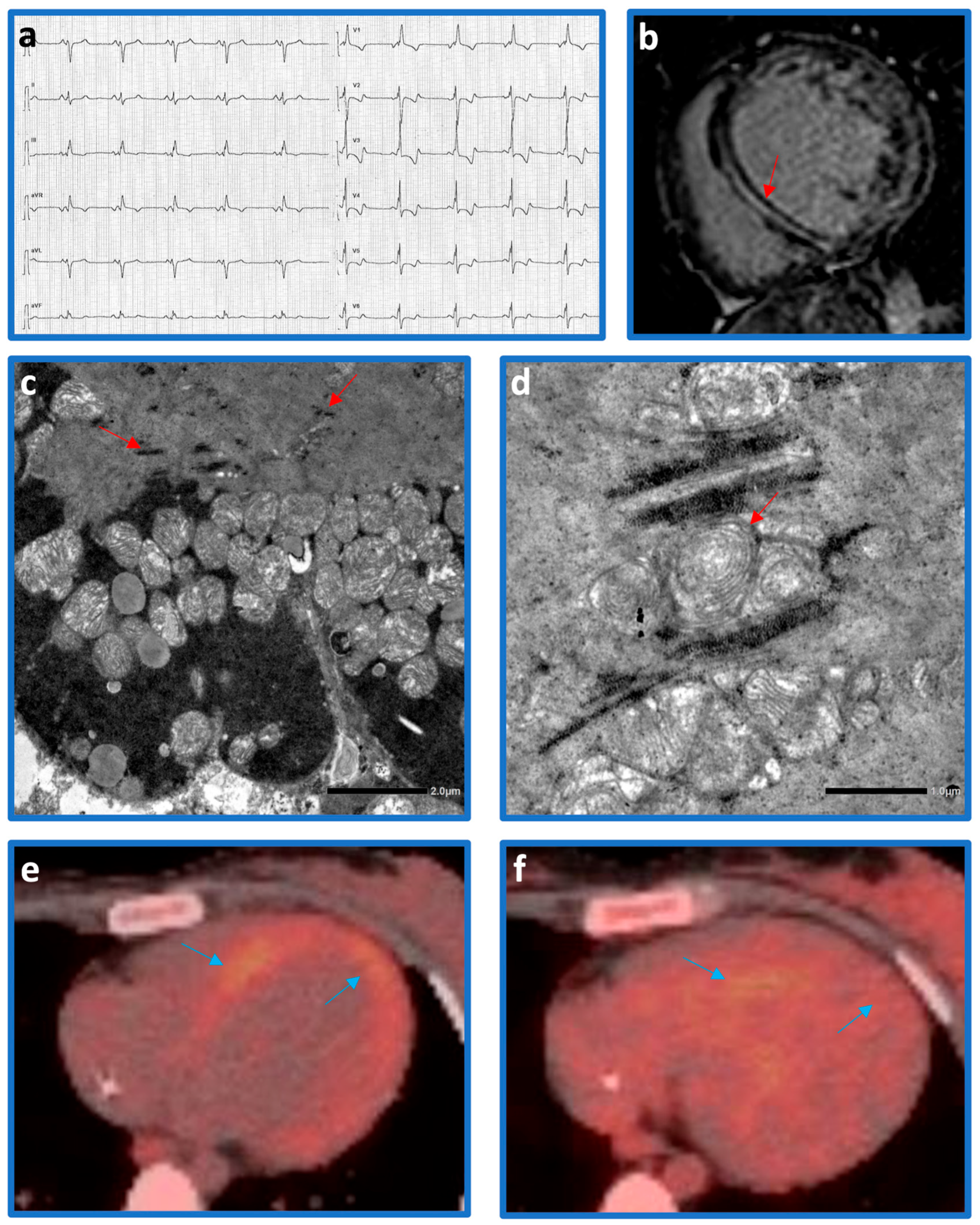
Figure 1. (a) ECG showing fragmented QRS with a right bundle branch block, a posterior left fascicular block, and diphasic negative/positive T waves in precordial leads. (b) Diffuse mid-wall LGE on cardiac MRI (red arrow). (c) Electron microscopy demonstrated increased glycogen granules (red arrows) mainly in subsarcolemmal regions (original magnification 4000×) and (d) rare mitochondria with concentric cristae (original magnification 8000×–red arrows). (e,f) A progressive reduction in the hyper-uptake of the tracer (blue arrows) in the left ventricular walls on FDG-PET has been observed following the initiation of corticosteroid therapy.
Cardiogenetics 15 00020 g001
Figure 2. (a) Schematic overview of mitochondrial tRNA maturation, from transcription to 5′ end cleavage by RNase P and subsequent 3′ end cleavage by ELAC2 (RNase ZL). (b) Gene structure of ELAC2 and patient’s variant localization. (c) The functional deficiency of ELAC2 leads to the accumulation of immature tRNAs lacking 3′ end cleavage. This results in oxidative chain dysfunction, accompanied by increased production of ROS and activation of physiological stress pathways, such as the unfolded protein response. These alterations promote enhanced extracellular matrix production, leading to myocardial fibrosis. CTD = C-terminal domain; mt-DNA = mitochondrial-DNA; mt-tRNA = mitochondrial-transfer RNA; NTD = N-terminal domain; OXPHOS = oxidative phosphorylation by respiratory chain complexes; ROS = Reactive Oxygen Species.
Cardiogenetics 15 00020 g002
Disclaimer/Publisher’s Note: The statements, opinions and data contained in all publications are solely those of the individual author(s) and contributor(s) and not of MDPI and/or the editor(s). MDPI and/or the editor(s) disclaim responsibility for any injury to people or property resulting from any ideas, methods, instructions or products referred to in the content.