Case Report

Sudden Cardiac Death in Biventricular Arrhythmogenic Cardiomyopathy: A New Undescribed Variant of the MYH6 Gene

1
Department of Cardiology, Hospital de Santa Marta, Centro Hospitalar Universitário de Lisboa Central, 1169-024 Lisbon, Portugal
2
Department of Genetics, Hospital de Santa Marta, Centro Hospitalar Universitário de Lisboa Central, 1169-024 Lisbon, Portugal
3
GenoMed—Diagnósticos de Medicina Molecular, S.A., 1649-028 Lisbon, Portugal
*
Author to whom correspondence should be addressed.
Cardiogenetics 2023, 13(4), 145-153; https://doi.org/10.3390/cardiogenetics13040014
Submission received: 2 July 2023 / Revised: 28 July 2023 / Accepted: 11 October 2023 / Published: 23 October 2023
(This article belongs to the Section Cardiovascular Genetics in Clinical Practice)

Abstract

:
Arrhythmogenic cardiomyopathy (ACM) may present with sudden cardiac arrest (SCA), and demonstration of a pathogenic variant in ACM-related genes is crucial for its definitive diagnosis. A 42-year-old female patient with family history of sudden cardiac death (SCD) was referred to the cardiomyopathy clinic after two episodes of aborted SCA. In the second episode, the patient was transported under cardiopulmonary resuscitation (downtime of 57 min) until extracorporeal membrane oxygenation was implanted. A thorough diagnostic work-up led to a diagnosis of biventricular ACM. Genetic testing revealed a previously undescribed variant in ACM patients in the MYH6 gene, c.3673G>T p.(Glu 1225*), which inserts a premature stop codon. This was considered a possible pathogenic variant originating a truncated protein, previously undescribed in ACM. The patient’s 23-year-old daughter was positive for the MYH6 variant and had ECG abnormalities suggestive of ACM. This case details the complex differential diagnosis of SCA and explores the current recommendations for the diagnosis of biventricular ACM. The identification of a MYH6 variant in a patient with ACM, recurrent SCA, and family history of SCD appears to support the hypothesis of the pathogenicity of MYH6 variants in ACM, in which the association of phenotype with sarcomere variants is still unclear.

1. Introduction

Arrhythmogenic cardiomyopathy (ACM) is a biventricular heart muscle disease with left ventricle (LV) involvement which may exceed the severity of the right ventricle (RV) disease (arrhythmogenic left ventricular cardiomyopathy (ALVC)). While functional evaluation of the LV is an important diagnostic tool, it is insufficient for the diagnosis of ALVC, which requires tissue characterization with contrast-enhanced cardiac magnetic resonance imaging (CMR) assessment of late gadolinium enhancement (LGE) [1].
In patients with suspected ALVC, demonstration of a pathogenic variant in ACM-related genes is crucial for a definitive diagnosis, according to the recommended diagnostic criteria [2]. Variants in over 25 different genes have been associated with ACM [3], mainly in genes encoding proteins of different junctional complexes, particularly the cardiac desmosomes [4,5]. Pathogenic missense variants in ILK [6], encoding the protein integrin-linked kinase, and truncating variants in FLNC [7], encoding filamin-C, a protein involved in coupling of the cytoskeleton with cell–cell junctions, have been identified in patients with ACM.
However, other genes unrelated to junctional complexes may be involved in ACM, albeit with a lesser degree of evidence regarding their pathogenicity in ACM. Pathogenic variants of the DES gene were associated with abnormal cytoplasmic aggregation of proteins, altering the regular structure of the sarcomeres [8], and missense variants in the DES gene have been linked with ALVC or biventricular ACM [9,10]. Other genes encoding Z-band proteins, such as LDB3 for cypher and ACTN2 for α-actinin-2, were also identified in patients with ACM [11,12]. Moreover, variants in genes commonly involved in channelopathies were also reported in ACM patients. These include SCN5A [13], described in patients with Brugada syndrome or long QT syndrome, RYR2 [14], encoding the ryanodine receptor 2 playing a crucial role in cardiomyocyte excitation–contraction coupling, and PLN [15], encoding phospholamban, which is the principal regulator of the Ca2+-ATPase of cardiac sarcoplasmic reticulum [16]. Other non-desmosomal genes encoding nuclear envelope proteins, including LMNA [17,18], encoding lamin A/C, TMEM43 [19,20], encoding the nuclear transmembrane envelope protein luma, and LEMD2 [21], encoding LEM domain containing protein-2, which is also involved in nuclear structural organization, have also been associated with ACM phenotypes.
In this clinical case, we detail a previously undescribed variant of the MYH6 gene in a patient with biventricular ACM: a possible pathogenic variant linked with ACM phenotype and explore its associations with different phenotypes. This case also illustrates the complex differential diagnosis of sudden cardiac arrest (SCA), including cardiomyopathies, channelopathies, acute coronary syndromes, and Kounis syndrome, reviewing the extensive diagnostic work-up, including 12-lead ECG, echocardiography, coronary angiography, CMR, and the importance of genetic testing.

2. Detailed Case Description

The authors present the case of a 42-year-old Caucasian female patient with a family history of sudden cardiac death (SCD), as her father died suddenly at the age of 51. The genogram is shown in Figure S1. The patient was referred to our Cardiomyopathy outpatient clinic after two episodes of SCA. The patient had a first episode of SCA 2 years before, during tubal ligation surgery. Follow-up ECG, transthoracic echocardiogram (TTE) and 24-Holter monitoring were reported as normal, and the patient was lost to follow-up. The reason why an implantable cardioverter-defibrillator (ICD) in secondary prevention was not considered at this time is not known. The timeline is illustrated in File S1.
The second episode of SCA occurred during anesthesia induction for elective termination of pregnancy, with ventricular fibrillation (VF) submitted to defibrillation (three DC shocks). The patient was under monitoring, and there were no precursors to VF, such as nonsustained episodes of supraventricular or ventricular arrhythmias. The patient was transported under cardiopulmonary resuscitation to the Intensive Care Unit, with a prolonged downtime (57 min), requiring venoarterial extracorporeal membrane oxygenation (VA-ECMO) for hemodynamic support.
The admission ECG (Figure 1a) showed a right bundle branch block with right axis deviation, and the chest X-ray showed bilateral lung edema (Figure S2). Admission bedside transthoracic echocardiography revealed a severely impaired LV function with apical and antero-mid segment hypokinesis, and preserved contractility of the basal LV segments. No pericardial effusion was noted. Invasive coronary angiography excluded coronary disease. Ventriculography was also performed, with no LV apical ballooning. Initial laboratory analyses revealed a normal complete blood count (without eosinophilia) and no liver, kidney, and thyroid dysfunction or C-reactive protein elevation (4 mg/L). There was also no electrolyte imbalance.
The patient presented a favorable evolution under ECMO, and the decannulation was performed after three days, without neurological sequelae. A subcutaneous implantable cardioverter-defibrillator was implanted in secondary prevention, and the patient was discharged with a referral to the Cardiomyopathy outpatient clinic and to the Immunology clinic.
A TTE was performed in the Cardiomyopathy outpatient clinic, revealing a non-dilated LV, no LV hypertrophy, subtle systolic dysfunction (LVEF 63%, GLS −13.6%), with mid-basal anterior and lateral wall hypokinesis (Figure 2, Videos S1 and S2). The E/e’ ratio was 6.6, suggestive of normal LV filling pressures. There was no significant valve disease or pulmonary hypertension. The left atrium was nondilated (24 mL/m2). The RV was non-dilated, with preserved RV systolic function (TAPSE 19 mm, DTI S’ 11 cm/s, RV global strain −25.3%), and there was no pericardial effusion.
Follow-up ambulatory ECG (Figure 1b) showed low voltage QRS complexes in the limb leads, and a fragmented QRS complex in lead III, without ventricular pre-excitation or repolarization abnormalities, namely, prolonged QT interval, Brugada pattern, or abnormal T wave inversion. 24 h-Holter monitoring (with bisoprolol therapy) documented 322 premature ventricular contractions but no periods of nonsustained ventricular tachycardia. Stress testing revealed a normal exercise capacity with no exercise-induced arrhythmias or repolarization abnormalities. A flecainide test did not show an ECG pattern suggestive of Brugada syndrome.
Cardiac magnetic resonance imaging (Videos S3–S6) was performed 10 days after admission, showing preserved LV function (ejection fraction 61%) with lateral wall hypokinesis and mild RV dilatation, with preserved RV function without wall motion abnormalities. Intramural and subepicardial LGE in the basal to mid segments of the inferolateral wall was noted (Figure 3). Native T1 mapping levels were slightly elevated in the myocardial region with LGE. T2 mapping levels were normal.
Allergy tests excluded the possibility of Kounis syndrome, as the patient showed tolerance to the anesthetics administered during both surgical procedures (propofol and alfentanil). Drug hypersensitivity myocarditis was also not likely, as T2 mapping levels were normal in CMR, suggesting absence of edema [22].
The findings of LV wall motion abnormalities, intramural and subepicardial LGE in the inferolateral wall, and the presence of low QRS voltages in the limb leads, in a patient with two episodes of SCA and family history of SCD, led to the suspicion of ALVC, in accordance with the 2020 international diagnostic criteria for ACM (the ‘Padua criteria’) [1]. However, the ECG repolarization and the right bundle branch block are not typical of ALVC and CMR detected the presence of RV dilatation. These findings are suggestive of ACM with biventricular involvement [1].
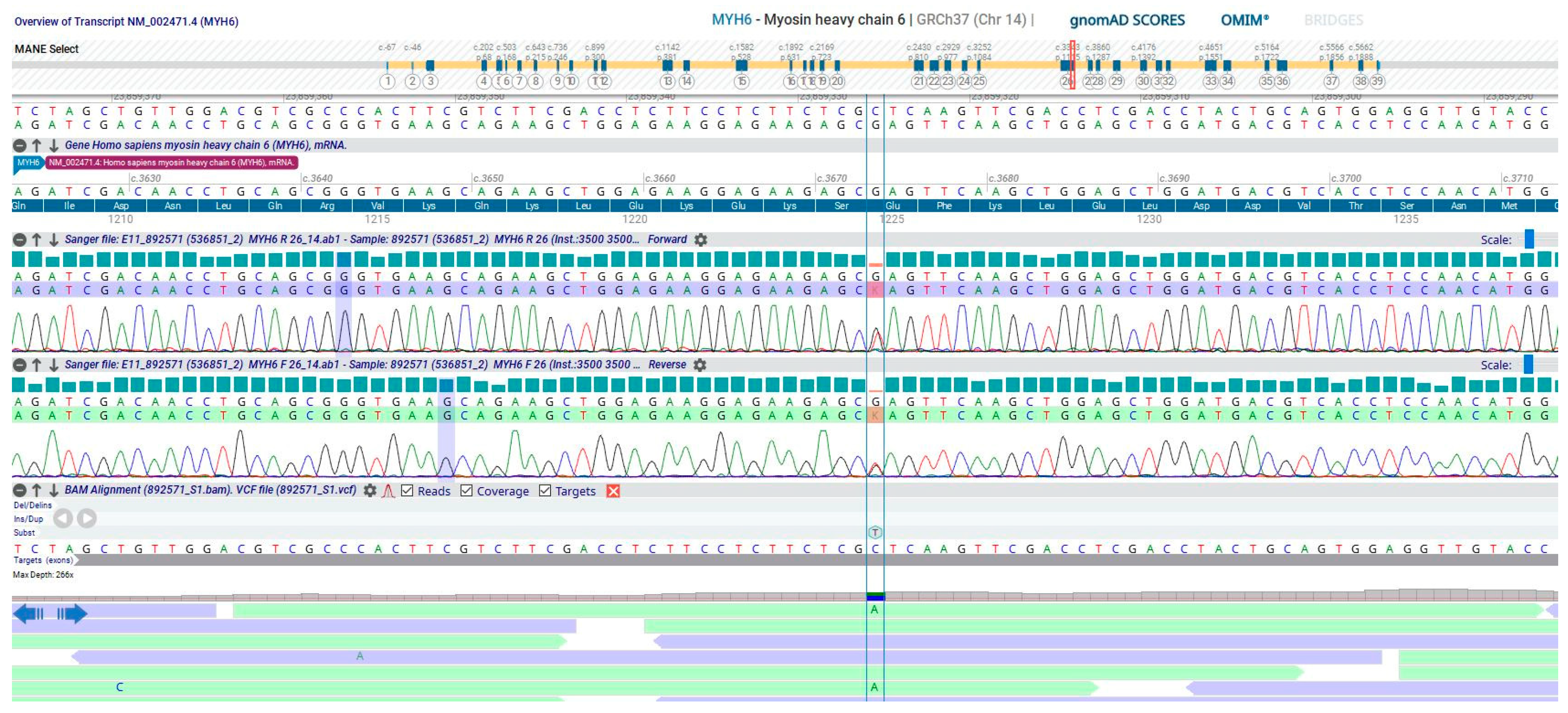
Genetic testing (extended cardiomyopathy 196 gene panel—Figure S3) revealed a previously undescribed variant in the MYH6 gene (NM_002471.3; chr. 14), (OMIM * 160710); c.3673G>T p.(Glu 1225*) (Figure 4)—a variant not present in the population databases (ACMG 2015 PM2) that introduces a premature stop codon. After multidisciplinary discussion with the Genetics department, the authors consider this is a possible pathogenic variant of the MYH6 gene, originating a truncated protein. Although PVS1 criteria were not met, according to ACMG 2015, this is a frameshift variant, predicted to undergo nonsense-mediated RNA decay, and the exon is present in biologically relevant transcripts. Genetic testing and clinical screening (including ECG, Holter monitoring, TTE and CMR) were also performed in the patient’s children. The patient’s 23-year-old daughter was positive for the aforementioned truncating MYH6 variant, with an ECG showing low QRS voltages in the limb leads, a terminal activation duration of QRS ≥ 55 ms in V1, and inverted T waves in leads III and aVF (Figure S4). However, her CMR did not show any structural LV or RV abnormalities or LGE. Despite our recommendation, the patient’s siblings declined undergoing clinical screening or genetic testing for the MYH6 variant.
In a three-year follow-up, there were no supraventricular or ventricular arrhythmias or ICD therapies.

3. Discussion

It is of paramount importance to be mindful of the differential diagnosis of SCA, as, while coronary heart disease is the most frequent etiology, cardiomyopathies, myocarditis, and channelopathies are possible causes, where the differential diagnosis may be challenging.
ACM is an inherited heart muscle disease characterized by substitution of the ventricular myocardium by fibrofatty tissue. While classically termed arrhythmogenic RV dysplasia/cardiomyopathy, new insights arising from postmortem investigations, genotype–phenotype correlation studies, and myocardial tissue characterization by contrast-CMR led to increased awareness that the LV is frequently involved. Notably, some phenotypic variants of ACM are characterized by a predominant involvement of the LV [2]. The differential diagnosis frequently poses a challenge, as the ALVC phenotype can overlap with that of other genetic cardiomyopathies, acquired dilated cardiomyopathy, myocarditis, or sarcoidosis.
The fibrofatty scar tissue is an arrhythmogenic myocardial substrate, associated with ventricular arrhythmias in ACM. ACM may present with life-threatening arrhythmias or SCA, despite the absence of family history or intense sports activity. In patients with SCA or sustained ventricular arrhythmias, an ICD in secondary prevention is recommended [2].
According to the ‘Padua criteria’, the demonstration of ACM-causing gene variants or familial ACM are major criteria for the definitive diagnosis of biventricular ACM, in association with consistent LV structural abnormalities [1]. The spectrum of ACM phenotypes is broader than previously believed and the 2010 Task Force criteria lacked specific criteria for diagnosis of left-sided phenotypes, thus resulting in underdiagnosis of ALVC [2]. Furthermore, the differential diagnosis between DCM and ALVC is challenging, possibly leading to a misdiagnosis of DCM in patients with ALVC [1]. Moreover, patients with a high arrhythmic burden or SCD due to ventricular arrhythmias may also have undiagnosed ALVC since the electrical abnormalities may precede structural changes in ALVC [2]. CMR plays a vital role in the differential diagnosis of ALVC, DCM, and SCD due to ventricular arrhythmias, allowing for detailed tissue characterization.
As our current knowledge on pathogenic and likely pathogenic variants in ACM is expanding, the identification of new variants linked with ACM is of utmost importance. The MYH6 gene, located on chromosome 14q11.2, encodes the alpha heavy chain subunit of cardiac myosin [23]. This protein is expressed in the heart from fetal life to adulthood and is actively involved in energy transduction and force development in the cardiac muscle [24] but also in cardiac morphogenesis [25,26]. Currently, there is evidence of the association of MYH6 variants with congenital heart disease (from septal defects in patients with premature stop codon mutations [27] to hypoplastic left heart syndrome [28]) and arrhythmias, namely, Wolff–Parkinson–White syndrome [29], atrial fibrillation [30], and sinus node dysfunction [31], as well as ventricular arrhythmias and SCA [31,32]. MYH6 variants have also been associated with different cardiomyopathies, including hypertrophic cardiomyopathy [33] and dilated cardiomyopathy, particularly characterized by late onset [32,34], peripartum cardiomyopathy [35], and LV non-compaction [36].
Sarcomeric variants have been reported in patients with a clinical diagnosis of ACM, and they are uncommon and predicted damaging in silico [37,38]. However, there is limited functional and segregational evidence that these variants are causative of ACM, and, as such, the clinical value of the identification of these variants in patients with ACM is still uncertain [38,39]. Basic science may provide further insight into the role of newly discovered variants in the pathogenesis of cardiomyopathies. While animal models have assessed the association between MYH6 variants and congenital heart disease or cardiomyopathies [26,40], studying the pathogenicity of MYH6 variants in the development of cardiomyopathies is significantly hindered due to the embryonic lethality in MYH6-knockout mice. Human MYH6-knockout embryonic stem cell lines using CRISPR/Cas9 systems may provide further clarification on the precise role of MYH6 in cardiomyopathies [41].
Among patients with ACM with non-desmosomal gene variants, several DES variants have been described, including DES-p.A120D [42], DES-p.E401D [10], or DES-p.L115I [9]. These variants have notably been associated with a striking frequency of arrhythmias and SCD. The DES-p.E401D and DES-p.L115I variants in particular have been associated with an ALVC phenotype and SCD, probably due to a disruption of the desmin filament network and its connection with membrane proteins within the intercalated disc. [9,10]. Other than the described MYH6 variant, the patient in this clinical case did not carry rare variants in DES or pathogenic variants in other non-desmosomal genes. Nevertheless, the patient’s phenotype is similar to the one described in the DES variants reported in ACM patients, including biventricular involvement, SCA, and high arrhythmic burden. Indeed, DES-p.L115I variant carriers presenting biventricular ACM also showed LV myocardial fibrosis with a subepicardial distribution on histology [9].
Unfortunately, an autopsy was not performed after her father’s SCD, which would have been helpful in confirming the presence of structural cardiac abnormalities or an MYH6 variant on molecular autopsy. Her 23-year-old daughter was positive for the truncating MYH6 variant, and her ECG showed abnormalities described in ALVC [1], such as low QRS voltages in the limb leads and a prolonged QRS terminal activation duration in V1. Although her CMR did not show any structural abnormalities suggestive of ACM, regular follow-up with CMR imaging and Holter monitoring is necessary to detect early signs of ACM phenotype, as electrical abnormalities may precede structural changes in ACM [2].

4. Conclusions

This case highlights the importance of genetic testing in patients with cardiomyopathies, and the identification of new pathogenic variants may provide valuable insight in genetic testing in patients with ACM and in familial screening. In the detailed clinical case, the identification of a MYH6 variant in a patient with recurrent SCA and family history of SCD appears to support the hypothesis of the pathogenicity of MYH6 variants in ACM, a clinical entity in which the association of phenotype with sarcomere variants is still unclear.

Supplementary Materials

The following supporting information can be downloaded at: https://www.mdpi.com/article/10.3390/cardiogenetics13040014/s1, File S1: Timeline; Figure S1: Patient’s genogram showing family members with the MYH6 gene variant and with sudden cardiac death; Figure S2: Admission chest X-ray demonstrating bilateral lung edema; Figure S3: Genetic testing—Next Generation Sequencing Cardiomyopathy 196 gene panel; Figure S4: Patient’s 23-year-old daughter’s ECG showing sinus rhythm at 67 beats per minute, low QRS voltages in the limb leads, a terminal activation duration of QRS ≥55 ms in lead V1 and inverted T waves in leads III and aVF. Normal PR (130 ms) and QTc (406 ms) intervals; Video S1: Transthoracic echocardiogram parasternal short axis view showing anterior and lateral wall hypokinesis. Video S2: Transthoracic echocardiogram apical four-chamber view showing reduced global systolic function (left ventricular ejection fraction (LVEF) 63%, global longitudinal strain −13.6%), with mid-basal lateral wall hypokinesis. Non-dilated left atrium. Non-dilated right ventricle, with preserved systolic function and no regional wall motion abnormalities; Video S3: Cardiac magnetic resonance imaging short axis view detailing preserved left ventricular function (LVEF 61%) with lateral wall hypokinesis and mild right ventricle (RV) dilatation, with preserved RV function without wall motion abnormalities; Video S4: Cardiac magnetic resonance imaging four-chamber view detailing preserved left ventricular function (LVEF 61%) with lateral wall hypokinesis and mild right ventricle (RV) dilatation, with preserved RV function without wall motion abnormalities; Video S5: Cardiac magnetic resonance imaging two-chamber view showing detailing preserved left ventricular function (LVEF 61%); Video S6: Cardiac magnetic resonance imaging three-chamber view showing preserved left ventricular function (LVEF 61%).

Author Contributions

Conceptualization, P.G.B. and S.A.R.; methodology, P.G.B.; software, P.G.B.; validation, P.G.B., D.A. and S.A.R.; data curation, P.G.B. and I.C.; writing—original draft preparation, P.G.B.; writing—review and editing, P.G.B., I.C., J.V., D.A. and S.A.R.; visualization, P.G.B. and I.C.; supervision, D.A. and S.A.R.; project administration, P.G.B. and S.A.R. All authors have read and agreed to the published version of the manuscript.

Funding

This research received no external funding.

Institutional Review Board Statement

Not applicable.

Informed Consent Statement

The patient has provided written informed consent for the submission of this manuscript, in accordance with COPE guidelines.

Data Availability Statement

The patient’s data and complementary exams are currently not publicly shared, as it would compromise patient privacy. These are accessible to the Hospital de Santa Marta’s Cardiomyopathy and Genetics teams and are available from the corresponding author upon reasonable request.

Acknowledgments

The authors would like to thank the Cardiomyopathies and the Genetics Department of the Santa Marta Hospital for their invaluable input in improving the manuscript.

Conflicts of Interest

The authors declare no conflict of interest.

References

  1. Corrado, D.; Perazzolo Marra, M.; Zorzi, A.; Beffagna, G.; Cipriani, A.; Lazzari, M.; Migliore, F.; Pilichou, K.; Rampazzo, A.; Rigato, I.; et al. Diagnosis of arrhythmogenic cardiomyopathy: The Padua criteria. Int. J. Cardiol. 2020, 319, 106–114. [Google Scholar] [CrossRef] [PubMed]
  2. Corrado, D.; Basso, C. Arrhythmogenic left ventricular cardiomyopathy. Heart 2022, 108, 733–743. [Google Scholar] [CrossRef] [PubMed]
  3. Gerull, B.; Brodehl, A. Insights Into Genetics and Pathophysiology of Arrhythmogenic Cardiomyopathy. Curr. Heart Fail. Rep. 2021, 18, 378–390. [Google Scholar] [CrossRef]
  4. Patel, D.M.; Green, K.J. Desmosomes in the Heart: A Review of Clinical and Mechanistic Analyses. Cell Commun. Adhes. 2014, 21, 109–128. [Google Scholar] [CrossRef]
  5. Gerull, B.; Heuser, A.; Wichter, T.; Paul, M.; Basson, C.T.; McDermott, D.A.; Lerman, B.B.; Markowitz, S.M.; Ellinor, P.T.; MacRae, C.A.; et al. Mutations in the desmosomal protein plakophilin-2 are common in arrhythmogenic right ventricular cardiomyopathy. Nat. Genet. 2004, 36, 1162–1164. [Google Scholar] [CrossRef] [PubMed]
  6. Brodehl, A.; Rezazadeh, S.; Williams, T.; Munsie, N.M.; Liedtke, D.; Oh, T.; Ferrier, R.; Shen, Y.; Jones, S.J.; Stiegler, A.L.; et al. Mutations in ILK, encoding integrin-linked kinase, are associated with arrhythmogenic cardiomyopathy. Transl. Res. 2019, 208, 15–29. [Google Scholar] [CrossRef] [PubMed]
  7. Ortiz-Genga, M.F.; Cuenca, S.; Ferro, M.D.; Zorio, E.; Salgado-Aranda, R.; Climent, V.; Padrón-Barthe, L.; Duro-Aguado, I.; Jiménez-Jáimez, J.; Hidalgo-Olivares, V.M.; et al. Truncating FLNC Mutations Are Associated With High-Risk Dilated and Arrhythmogenic Cardiomyopathies. J. Am. Coll. Cardiol. 2016, 68, 2440–2451. [Google Scholar] [CrossRef]
  8. Brodehl, A.; Hedde, P.N.; Dieding, M.; Fatima, A.; Walhorn, V.; Gayda, S.; Šarić, T.; Klauke, B.; Gummert, J.; Anselmetti, D.; et al. Dual Color Photoactivation Localization Microscopy of Cardiomyopathy-associated Desmin Mutants. J. Biol. Chem. 2012, 287, 16047–16057. [Google Scholar] [CrossRef]
  9. Protonotarios, A.; Brodehl, A.; Asimaki, A.; Jager, J.; Quinn, E.; Stanasiuk, C.; Ratnavadivel, S.; Futema, M.; Akhtar, M.M.; Gossios, T.D.; et al. The Novel Desmin Variant p.Leu115Ile Is Associated With a Unique Form of Biventricular Arrhythmogenic Cardiomyopathy. Can. J. Cardiol. 2021, 37, 857–866. [Google Scholar] [CrossRef]
  10. Bermúdez-Jiménez, F.J.; Carriel, V.; Brodehl, A.; Alaminos, M.; Campos, A.; Schirmer, I.; Milting, H.; Abril, B.; Álvarez, M.; López-Fernández, S.; et al. Novel Desmin Mutation p.Glu401Asp Impairs Filament Formation, Disrupts Cell Membrane Integrity, and Causes Severe Arrhythmogenic Left Ventricular Cardiomyopathy/Dysplasia. Circulation 2018, 137, 1595–1610. [Google Scholar] [CrossRef]
  11. Lopez-Ayala, J.M.; Ortiz-Genga, M.; Gomez-Milanes, I.; Lopez-Cuenca, D.; Ruiz-Espejo, F.; Sanchez-Munoz, J.J.; Oliva-Sandoval, M.J.; Monserrat, L.; Gimeno, J.R. A mutation in the Z-line Cypher/ZASP protein is associated with arrhythmogenic right ventricular cardiomyopathy. Clin. Genet. 2015, 88, 172–176. [Google Scholar] [CrossRef]
  12. Good, J.-M.; Fellmann, F.; Bhuiyan, Z.A.; Rotman, S.; Pruvot, E.; Schläpfer, J. ACTN2 variant associated with a cardiac phenotype suggestive of left-dominant arrhythmogenic cardiomyopathy. Hear. Case Rep. 2020, 6, 15–19. [Google Scholar] [CrossRef]
  13. Te Riele, A.S.J.; Agullo-Pascual, E.; James, C.A.; Leo-Macias, A.; Cerrone, M.; Zhang, M.; Lin, X.; Lin, B.; Rothenberg, E.; Sobreira, N.L.; et al. Multilevel analyses of SCN5A mutations in arrhythmogenic right ventricular dysplasia/cardiomyopathy suggest non-canonical mechanisms for disease pathogenesis. Cardiovasc. Res. 2017, 113, 102–111. [Google Scholar] [CrossRef]
  14. Tiso, N.; Stephan, D.A.; Nava, A.; Bagattin, A.; Devaney, J.M.; Stanchi, F.; Larderet, G.; Brahmbhatt, B.; Brown, K.; Bauce, B.; et al. Identification of mutations in the cardiac ryanodine receptor gene in families affected with arrhythmogenic right ventricular cardiomyopathy type 2 (ARVD2). Hum. Mol. Genet. 2001, 10, 189–194. [Google Scholar] [CrossRef]
  15. van der Zwaag, P.A.; van Rijsingen, I.A.; Asimaki, A.; Jongbloed, J.D.; van Veldhuisen, D.J.; Wiesfeld, A.C.; Cox, M.G.; van Lochem, L.T.; de Boer, R.A.; Hofstra, R.M.; et al. Phospholamban R14del mutation in patients diagnosed with dilated cardiomyopathy or arrhythmogenic right ventricular cardiomyopathy: Evidence supporting the concept of arrhythmogenic cardiomyopathy. Eur. J. Heart Fail. 2012, 14, 1199–1207. [Google Scholar] [CrossRef] [PubMed]
  16. Simmerman, H.; Jones, L. Phospholamban: Protein Structure, Mechanism of Action, and Role in Cardiac Function. Physiol. Rev. 1998, 78, 921–947. [Google Scholar] [CrossRef] [PubMed]
  17. Quarta, G.; Syrris, P.; Ashworth, M.; Jenkins, S.; Alapi, K.Z.; Morgan, J.; Muir, A.; Pantazis, A.; McKenna, W.J.; Elliott, P.M. Mutations in the Lamin A/C gene mimic arrhythmogenic right ventricular cardiomyopathy. Eur. Heart J. 2012, 33, 1128–1136. [Google Scholar] [CrossRef] [PubMed]
  18. Maggi, L.; Mavroidis, M.; Psarras, S.; Capetanaki, Y.; Lattanzi, G. Skeletal and Cardiac Muscle Disorders Caused by Mutations in Genes Encoding Intermediate Filament Proteins. Int. J. Mol. Sci. 2021, 22, 4256. [Google Scholar] [CrossRef] [PubMed]
  19. Merner, N.D.; Hodgkinson, K.A.; Haywood, A.F.; Connors, S.; French, V.M.; Drenckhahn, J.-D.; Kupprion, C.; Ramadanova, K.; Thierfelder, L.; McKenna, W.; et al. Arrhythmogenic Right Ventricular Cardiomyopathy Type 5 Is a Fully Penetrant, Lethal Arrhythmic Disorder Caused by a Missense Mutation in the TMEM43 Gene. Am. J. Hum. Genet. 2008, 82, 809–821. [Google Scholar] [CrossRef]
  20. Padrón-Barthe, L.; Villalba-Orero, M.; Gómez-Salinero, J.M.; Domínguez, F.; Román, M.; Larrasa-Alonso, J.; Ortiz-Sánchez, P.; Martínez, F.; López-Olañeta, M.; Bonzón-Kulichenko, E.; et al. Severe Cardiac Dysfunction and Death Caused by Arrhythmogenic Right Ventricular Cardiomyopathy Type 5 Are Improved by Inhibition of Glycogen Synthase Kinase-3β. Circulation 2019, 140, 1188–1204. [Google Scholar] [CrossRef]
  21. Abdelfatah, N.; Chen, R.; Duff, H.J.; Seifer, C.M.; Buffo, I.; Huculak, C.; Clarke, S.; Clegg, R.; Jassal, D.S.; Gordon, P.M.; et al. Characterization of a Unique Form of Arrhythmic Cardiomyopathy Caused by Recessive Mutation in LEMD2. JACC Basic Transl. Sci. 2019, 4, 204–221. [Google Scholar] [CrossRef]
  22. Ferreira, V.M.; Schulz-Menger, J.; Holmvang, G.; Kramer, C.M.; Carbone, I.; Sechtem, U.; Kindermann, I.; Gutberlet, M.; Cooper, L.T.; Liu, P.; et al. Cardiovascular Magnetic Resonance in Nonischemic Myocardial Inflammation. J. Am. Coll. Cardiol. 2018, 72, 3158–3176. [Google Scholar] [CrossRef]
  23. Miyata, S.; Minobe, W.; Bristow, M.R.; Leinwand, L.A. Myosin Heavy Chain Isoform Expression in the Failing and Nonfailing Human Heart. Circ. Res. 2000, 86, 386–390. [Google Scholar] [CrossRef]
  24. Mahdavi, V.; Chambers, A.P.; Nadal-Ginard, B. Cardiac alpha- and beta-myosin heavy chain genes are organized in tandem. Proc. Natl. Acad. Sci. USA 1984, 81, 2626–2630. [Google Scholar] [CrossRef]
  25. England, J.; Loughna, S. Heavy and light roles: Myosin in the morphogenesis of the heart. Cell Mol. Life Sci. 2013, 70, 1221–1239. [Google Scholar] [CrossRef]
  26. Abu-Daya, A.; Sater, A.K.; Wells, D.E.; Mohun, T.J.; Zimmerman, L.B. Absence of heartbeat in the Xenopus tropicalis mutation muzak is caused by a nonsense mutation in cardiac myosin myh6. Dev. Biol. 2009, 336, 20–29. [Google Scholar] [CrossRef]
  27. Razmara, E.; Garshasbi, M. Whole-exome sequencing identifies R1279X of MYH6 gene to be associated with congenital heart disease. BMC Cardiovasc. Disord. 2018, 18, 137. [Google Scholar] [CrossRef] [PubMed]
  28. Anfinson, M.; Fitts, R.H.; Lough, J.W.; James, J.M.; Simpson, P.M.; Handler, S.S.; Mitchell, M.E.; Tomita-Mitchell, A. Significance of α-Myosin Heavy Chain (MYH6) Variants in Hypoplastic Left Heart Syndrome and Related Cardiovascular Diseases. J. Cardiovasc. Dev. Dis. 2022, 9, 144. [Google Scholar] [CrossRef] [PubMed]
  29. Bowles, N.E.; Jou, C.J.; Arrington, C.B.; Kennedy, B.J.; Earl, A.; Matsunami, N.; Meyers, L.L.; Etheridge, S.P.; Saarel, E.V.; Bleyl, S.B.; et al. Exome analysis of a family with Wolff-Parkinson-White syndrome identifies a novel disease locus. Am. J. Med. Genet. A 2015, 167, 2975–2984. [Google Scholar] [CrossRef]
  30. Chalazan, B.; Mol, D.; Darbar, F.A.; Ornelas-Loredo, A.; Al-Azzam, B.; Chen, Y.; Tofovic, D.; Sridhar, A.; Alzahrani, Z.; Ellinor, P.; et al. Association of Rare Genetic Variants and Early-Onset Atrial Fibrillation in Ethnic Minority Individuals. JAMA Cardiol. 2021, 6, 811. [Google Scholar] [CrossRef] [PubMed]
  31. Lam, L.; Ingles, J.; Turner, C.; Kilborn, M.; Bagnall, R.D.; Semsarian, C. Exome sequencing identifies a novel mutation in the MYH6 gene in a family with early-onset sinus node dysfunction, ventricular arrhythmias, and cardiac arrest. Hear. Case Rep. 2015, 1, 141–145. [Google Scholar] [CrossRef]
  32. Zhao, T.; Ma, Y.; Zhang, Z.; Xian, J.; Geng, X.; Wang, F.; Huang, J.; Yang, Z.; Luo, Y.; Lin, Y. Young and early-onset dilated cardiomyopathy with malignant ventricular arrhythmia and sudden cardiac death induced by the heterozygous LDB3, MYH6, and SYNE1 missense mutations. Ann. Noninvasive Electrocardiol. 2021, 26, e12840. [Google Scholar] [CrossRef] [PubMed]
  33. Rubattu, S.; Bozzao, C.; Pennacchini, E.; Pagannone, E.; Musumeci, B.M.; Piane, M.; Germani, A.; Savio, C.; Francia, P.; Volpe, M.; et al. A Next-Generation Sequencing Approach to Identify Gene Mutations in Early- and Late-Onset Hypertrophic Cardiomyopathy Patients of an Italian Cohort. Int. J. Mol. Sci. 2016, 17, 1239. [Google Scholar] [CrossRef] [PubMed]
  34. Carniel, E.; Taylor, M.R.; Sinagra, G.; Di Lenarda, A.; Ku, L.; Fain, P.R.; Boucek, M.M.; Cavanaugh, J.; Miocic, S.; Slavov, D.; et al. α-Myosin Heavy Chain. Circulation 2005, 112, 54–59. [Google Scholar] [CrossRef]
  35. Morales, A.; Painter, T.; Li, R.; Siegfried, J.D.; Li, D.; Norton, N.; Hershberger, R.E. Rare Variant Mutations in Pregnancy-Associated or Peripartum Cardiomyopathy. Circulation 2010, 121, 2176–2182. [Google Scholar] [CrossRef] [PubMed]
  36. Vershinina, T.; Fomicheva, Y.; Muravyev, A.; Jorholt, J.; Kozyreva, A.; Kiselev, A.; Gordeev, M.; Vasichkina, E.; Segrushichev, A.; Pervunina, T.; et al. Genetic Spectrum of Left Ventricular Non-Compaction in Paediatric Patients. Cardiology 2020, 145, 746–756. [Google Scholar] [CrossRef]
  37. Murray, B.; Hoorntje, E.T.; Riele, A.S.J.M.T.; Tichnell, C.; van der Heijden, J.F.; Tandri, H.; Berg, M.P.v.D.; Jongbloed, J.D.H.; Wilde, A.A.M.; Hauer, R.N.W.; et al. Identification of sarcomeric variants in probands with a clinical diagnosis of arrhythmogenic right ventricular cardiomyopathy (ARVC). J. Cardiovasc. Electrophysiol. 2018, 29, 1004–1009. [Google Scholar] [CrossRef]
  38. Chen, K.; Rao, M.; Guo, G.; Chen, X.; Chen, L.; Song, J. Sarcomere variants in arrhythmogenic cardiomyopathy: Pathogenic factor or bystander? Gene 2019, 687, 82–89. [Google Scholar] [CrossRef]
  39. Medeiros-Domingo, A.; Saguner, A.M.; Magyar, I.; Bahr, A.; Akdis, D.; Brunckhorst, C.; Duru, F.; Berger, W. Arrhythmogenic right ventricular cardiomyopathy: Implications of next-generation sequencing in appropriate diagnosis. Europace 2017, 19, 1063–1069. [Google Scholar] [CrossRef]
  40. Huang, X.; Yan, L.; Kou, S.; Meng, J.; Lu, Z.; Lin, C.-P.; Liu, C.; Zhang, H. Generation and characterization of a Myh6-driven Cre knockin mouse line. Transgenic. Res. 2021, 30, 821–835. [Google Scholar] [CrossRef]
  41. Hao, E.; Zhang, G.; Mu, L.; Ma, N.; Wang, T. Establishment of a human MYH6 compound heterozygous knockout hESC line to model cardiomyopathy and congenital heart defects by CRISPR/Cas9 system. Stem. Cell Res. 2021, 50, 102128. [Google Scholar] [CrossRef] [PubMed]
  42. Brodehl, A.; Dieding, M.; Klauke, B.; Dec, E.; Madaan, S.; Huang, T.; Gargus, J.; Fatima, A.; Šaric, T.; Cakar, H.; et al. The Novel Desmin Mutant p.A120D Impairs Filament Formation, Prevents Intercalated Disk Localization, and Causes Sudden Cardiac Death. Circ. Cardiovasc. Genet. 2013, 6, 615–623. [Google Scholar] [CrossRef] [PubMed]
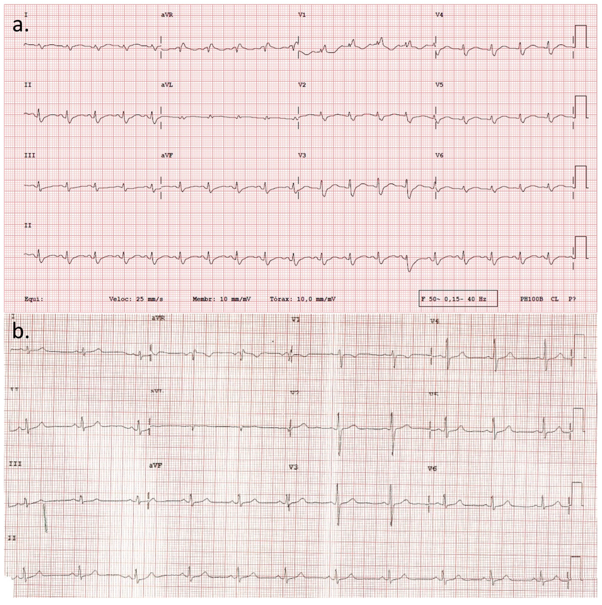
Figure 1. Admission 12-lead ECG showing sinus rhythm, heart rate of 110 bpm, right axis deviation, and right bundle branch block (a). Cardiomyopathy outpatient clinic 12-lead ECG revealing sinus rhythm at 65 bpm, low voltage QRS complexes in the limb leads, and a fragmented QRS complex in lead III (b).
Cardiogenetics 13 00014 g001
Figure 2. Transthoracic echocardiogram global longitudinal strain (GLS) bull’s eye plot. Impaired GLS (−13.6%), particularly in the mid and basal segments of the anterior and lateral walls.
Cardiogenetics 13 00014 g002
Figure 3. Contrast-enhanced magnetic resonance imaging revealing intramural and subepicardial late gadolinium enhancement in the basal to mid segments of inferolateral wall. Two-chamber view (a), three-chamber view (b), modified three-chamber view (c), and mid-ventricular short axis view (d).
Cardiogenetics 13 00014 g003
Figure 4. Genetic sequencing data—MYH6 variant c.3673G>T p.(Glu 1225*).
Cardiogenetics 13 00014 g004
Disclaimer/Publisher’s Note: The statements, opinions and data contained in all publications are solely those of the individual author(s) and contributor(s) and not of MDPI and/or the editor(s). MDPI and/or the editor(s) disclaim responsibility for any injury to people or property resulting from any ideas, methods, instructions or products referred to in the content.