Article

Transthyretin Gene Variants and Associated Phenotypes in Danish Patients with Amyloid Cardiomyopathy

1
Department of Cardiology, Aarhus University Hospital, DK-8200 Aarhus, Denmark
2
Department of Molecular Medicine, Aarhus University Hospital, DK-8200 Aarhus, Denmark
3
Department of Neurology, Aarhus University Hospital, DK-8200 Aarhus, Denmark
*
Author to whom correspondence should be addressed.
Cardiogenetics 2022, 12(1), 1-11; https://doi.org/10.3390/cardiogenetics12010001
Submission received: 1 November 2021 / Revised: 15 December 2021 / Accepted: 28 December 2021 / Published: 4 January 2022
(This article belongs to the Special Issue Cardiogenetics: Feature Papers 2021)

Abstract

:
Genotyping divides transthyretin cardiac amyloidosis (ATTR-CA) in hereditary (ATTRv) and wild type (ATTRwt) forms. This study investigated the prevalence and clinical presentation of ATTRv in a contemporary cohort of consecutive ATTR-CA patients diagnosed at a tertiary Danish amyloidosis center. Age at diagnosis, clinical- and echocardiographic data, and transthyretin (TTR) genotype were recorded. Relatives of ATTRv patients underwent clinical phenotyping and predictive gene testing. Genetic testing in 102 patients identified four TTR variant carriers: p.Pro63Ser, p.Ala65Ser (n = 2) and p.Val142Ile. The mean age of ATTRv index patients was significantly lower compared to ATTRwt patients: 70.2 ± 1.2 versus 80.0 ± 6.2, p-value: 0.005. Evaluation of ATTRv families identified seven TTR variant carriers with a median age of 65 years (range 48–76) and three were diagnosed with ATTR-CA by DPD-scintigraphy. Family members with ATTR-CA were all asymptomatic and had normal levels of cardiac biomarkers. In conclusion, the prevalence of ATTRv in a contemporary Danish ATTR-CA cohort is 4%. ATTRv index patients were significantly younger age at diagnosis than ATTRwt patients. Non-p.Leu131Met TTR variants have reduced penetrance at the age of 65 years in which approximately half of variant carriers have asymptomatic ATTR-CA with normal LV systolic function and cardiac biomarker analyses.

1. Introduction

Transthyretin amyloidosis (ATTR) is caused by abnormal deposition of misfolded transthyretin protein that aggregates as amyloid fibrils in various organs [1]. Transthyretin is a plasma transport protein produced primarily in the liver. ATTR is divided into two subtypes; an acquired wild-type variant (ATTRwt) and a hereditary form (ATTRvt), the latter caused by genetic missense variants in the TTR gene encoding tranthyretin. Accumulation of transthyretin in the heart causes amyloid cardiomyopathy (ATTR-CA) which leads to progressive biventricular heart failure with restrictive pathophysiology [2,3,4,5]. In ATTRv, the variant transthyretin may also accumulate in other organs than the heart. Among patients with ATTRv, the nervous system is often involved with amyloid deposition in the peripheral nerves causing a phenotype with familial polyneuropathy while other TTR missense variants lead to a mixed cardiac and polyneuropathy phenotype [1]. Frequent clinical precursors of the ATTR-CA phenotype are carpal tunnel syndrome (CTS), spinal stenosis, or tendon ruptures caused by amyloid fibril accumulation in these connective tissues. These clinical precursors often precede a subsequent diagnosis of ATTR by several years [6].
Data from the National Amyloidosis Center (NAC), United Kingdom (UK) using the NAC-score prognostic staging system has highlighted that the presence of a variant TTR genotype is associated with a higher disease burden and increased mortality than ATTRwt [7,8]. The different ATTRv genotypes also have prognostic impact in terms of the anticipated age-related penetrance and phenotypic disease expression, e.g., cardiac versus nervous system involvement [9]. Therefore, it is suggested that all patients with confirmed ATTR-CA undergo TTR genotyping to screen for ATTRv [1,10]. More than two decades ago the p.Leu131Met TTR founder variant associated with primary cardiac involvement was described in a large family of Danish ATTRv patients and has since been considered as the primary ATTRv variant in Denmark [9,11,12]. However, an increasing number of patients with ATTR are diagnosed due to an increased awareness of the disease and with the introduction of improved diagnostic methods that more easily raise the suspicion of ATTR-CA and subsequently confirm the diagnosis. In particular, the use of echocardiographic longitudinal strain analysis with identification of the relative apical sparring pattern in conditions with left ventricular (LV) hypertrophy (LVH) [13]. Furthermore, the increasing use of bone scintigraphy (99mTechnetium-3,3-diphosphono-1,2-propanodicarboxylic acid [99m-Tc-DPD]) has undoubtedly contributed to the increased number of patients diagnosed with ATTR-CA. This advancement in the screening process and the diagnostic algorithms used in routine clinical practice might change the conception of the prevalence and genetic spectrum of other TTR variants in Danish ATTR-CA patients [1,14]. The aims of this study were therefore to report the results of TTR genotyping in a contemporary cohort of consecutive ATTR-CA patients diagnosed at a tertiary referral amyloidosis center in Denmark; secondly, to describe the clinical characteristics among ATTRv versus ATTRwt patients at time of diagnosis; and, lastly, to report genotype-phenotype relations in identified ATTRv families.

2. Materials and Methods

Consecutive patients who were given a definitive ATTR-CA diagnosis at the Department of Cardiology, Aarhus University Hospital in the period from January 2008 to December 2020 were included in a retrospective analysis. The study was carried out in accordance with the Declaration of Helsinki. All patients were investigated by 12-lead electrocardiogram (ECG), transthoracic echocardiography (TTE), and cardiac biomarker analysis with baseline determinations of plasma N-terminal pro-brain natriuretic peptide (NT-pro-BNP) and estimated glomerular filtration rate (eGFR). Echocardiographic parameters included measurements of LV end-diastolic diameter, LV ejection fraction, interventricular wall thickness, LV global longitudinal strain, left atrial size, and graduation of diastolic dysfunction according to standard protocols [15]. Relative apical sparring of longitudinal strain was calculated by the formula: (average apical LS/(average basal LS + mid-LS) with a cut-off value of 1.0 being suggestive for ATTR-CA [13]. Cardiac light chain amyloidosis (AL) was excluded in all patients as patients who had an abnormal plasma kappa/lambda free light chain ratio and presence of plasma- or urine monoclonal proteins. In such patients, transthyretin amyloid subtyping was made by immunostaining and protein mass spectrometry of endomyocardial biopsies [1]. In the remaining patients without suspicion of plasma cell disease, ATTR-CA was diagnosed by a positive 99m-Tc-DPD-scintigraphy (Perugini grade > 1) according to standard diagnostic algorithms [14]. Age and National Amyloidosis Center (NAC) stage at the time of diagnosis were recorded [7]. TTR genotyping was performed using next-generation sequencing methods.
Next, relatives of ATTRv index patients were offered predictive genetic testing for the TTR variant by direct Sanger sequencing, and variant carriers underwent clinical phenotyping with ECG, TTE, and cardiac biomarker analysis as described previously [3]. A 99m-Tc-DPD scintigraphy was performed if ATTR-CA was suspected in variant carriers. If possible, the age and cause of death of deceased family members were recorded by interviews of family members, and if possible, DNA extracted from formalin-fixed paraffin-embedded non-cardiac tissue was tested for the identified TTR variant by direct Sanger sequencing. All alive ATTRv carriers gave written informed consent to publication of their genetic findings.

Statistics

Normal distributed continuous variables are expressed as mean with standard deviations (SD), and otherwise as medians with interquartile ranges. Differences between group means were compared with the Wilcoxon rank-sum test.

3. Results

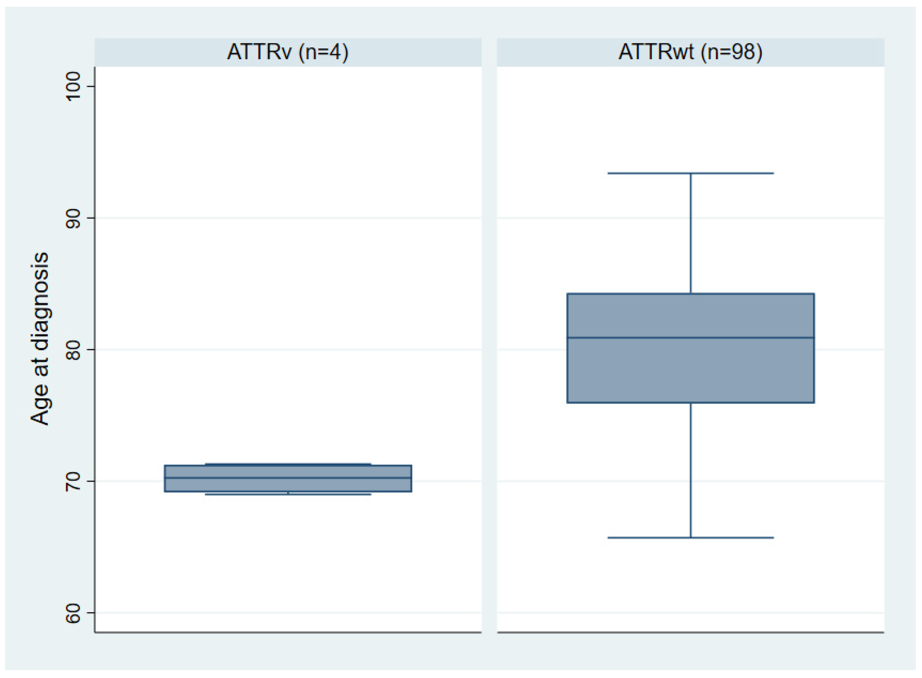
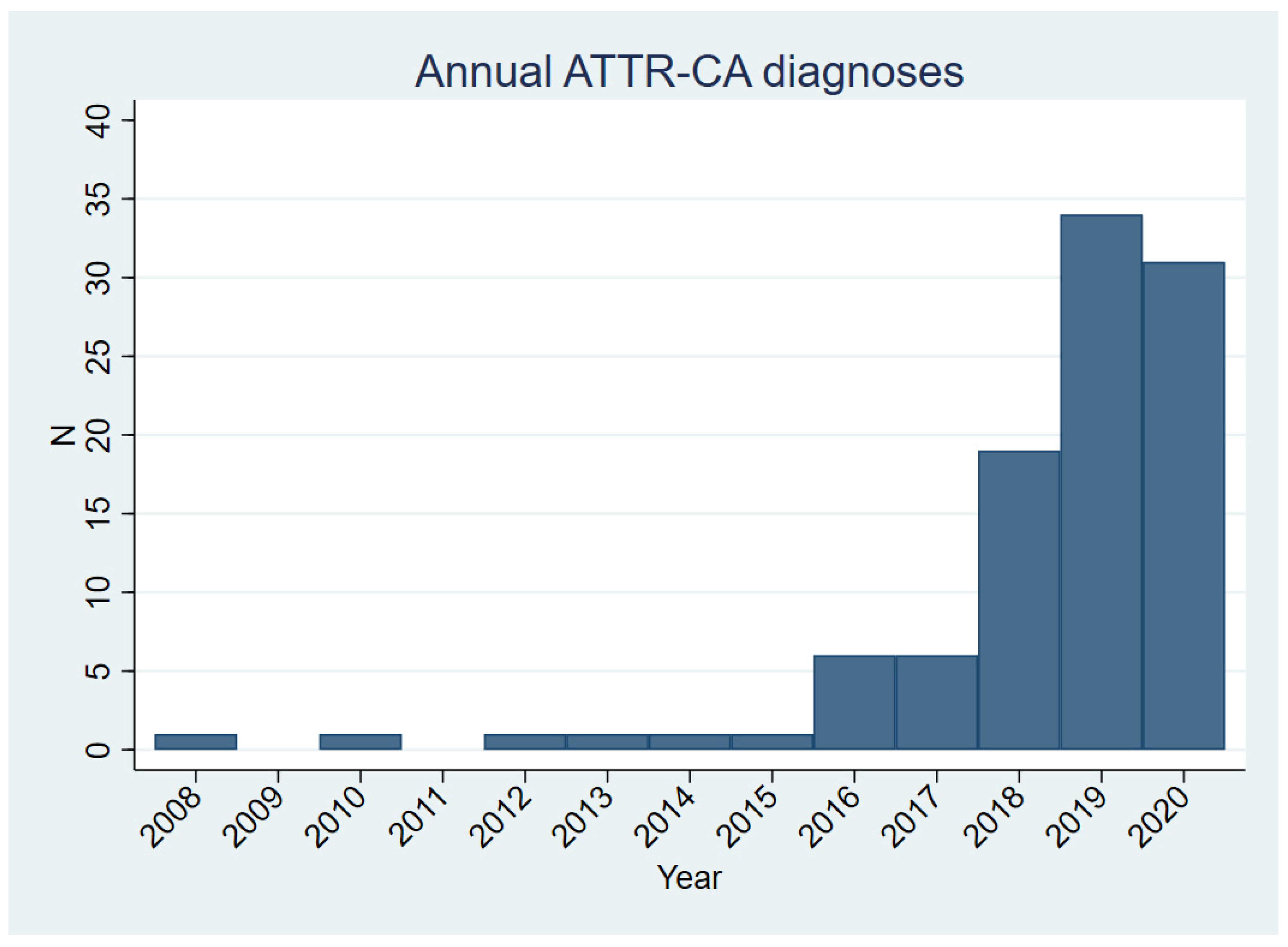
3.1. Clinical Characteristics of the ATTR-CA Cohort

During a 13-year period, 102 patients were diagnosed with ATTR-CA and underwent TTR genotyping (Table 1). A preliminary report from the entire ATTR-FAP and ATTR-CA cohort in the period from 2001 to 2019 has been published previously as a conference abstract [16]. In the last three years of the period (2018–2020), a 4- to 6-fold increase in ATTR-CA diagnostic activity was observed (Figure 1). All genotyped ATTR-CA patients except one were Caucasians and 93% were males. Half of the patients were diagnosed by 99m-Tc-DPD scintigraphy. The remaining patients had abnormal plasma kappa/lambda free light chain ratio or monoclonal plasma or urine protein. In these patients, transthyretin amyloid deposits were detected by use of immunohistochemistry or amyloid subtyping protein mass spectrometry of endomyocardial biopsies (Table 1). Two-thirds of patients had paroxysmal or persistent atrial fibrillation or -flutter; 25% had atrioventricular conduction disease treated with pacemaker device, and 28% had obstructive coronary artery disease with a >90% coronary artery stenosis or previous revascularization. Previous CTS surgery had been performed in 39% of the patients. None of the patients had obvious symptoms or signs of sensory-motor polyneuropathy or autonomic dysfunction.

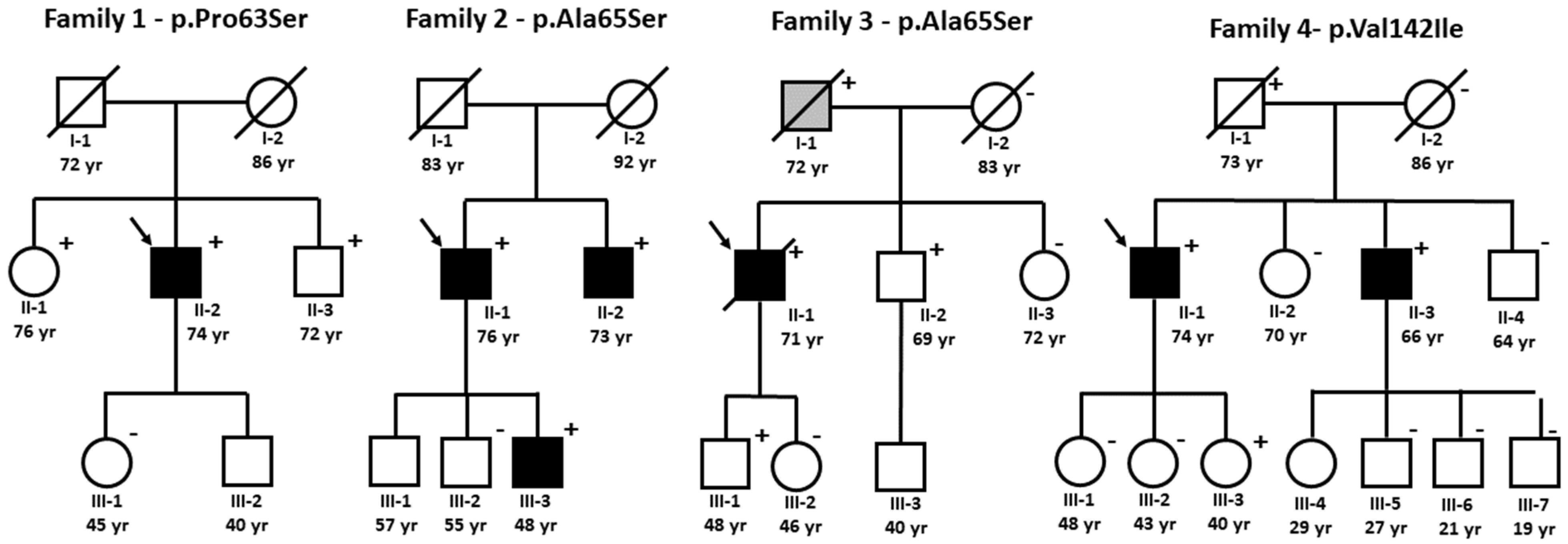
3.2. ATTRv Index Patients

Sequencing of the coding regions of the TTR gene identified four (4%) patients, all with Danish (Caucasian) ethnicity, who carried the TTR variants: c.187C>T/p.Pro63Ser; c.193G>T/p.Ala65Ser (n = 2); and c.424G>A/p.Val142Ile. ATTRv index patients were characterized by a significantly younger mean age at diagnosis (70.2 ± 1.2, [range 69.0–71.3] versus 80.0 ± 6.2 [range 65.7–93.4]; p-value 0.005), than ATTRwt patients (Figure 2). All other clinical characteristics of ATTRv patients were comparable to ATTRwt patients (Table 1).

3.3. Family Studies of ATTRv

3.3.1. Family 1—p.Pro63Ser

The index patient (II-2, age 71, Figure 3, Table 2) was diagnosed with ATTR-CA after twelve months of progressive exertional limitation and dyspnea (NYHA IIB-IIIA) with subsequent development of edemas and orthopnea. Two years previously, he was diagnosed with chronic atrial fibrillation suspected attributed to hypertensive heart disease. Examinations showed severe left ventricular hypertrophy (LVH) with severely reduced LV function (LVEF 19%; LV-GLS-5.5%) and a longitudinal strain plot showing a relative apical sparring pattern. The patient complained of sensory disturbances in the radial fingers suggestive of CTS. A DPD-scintigraphy confirmed ATTR-CA being in NAC-stage 2 (NT-proBNP 3091 ng/L; eGFR 75 mL/min). A cardiac resynchronization pacemaker with a cardioverter defibrillator (CRTD) was implanted due to slow atrioventricular conduction and non-sustained ventricular tachycardia in chronic atrial fibrillation. Three years later, at the age of 74, the patient was still alive but had progressed to NAC-stage 3. There was no family history of heart failure, and the parents died 72- and 86-years old, respectively. The identified p.Pro63Ser TTR variant was considered of unknown significance since it had not been reported before in ATTR-CA patients and was absent in control exomes (https://gnomad.broadinstitute.org/, accessed on 31 October 2021).
To assess the pathogenicity of the p.Pro63Ser variant family, screening was initiated. The index patient had two siblings both carrying the TTR variant. The younger brother (II-3), who was 72-years old had a pacemaker implanted due to total atrioventricular block but had no symptoms of heart failure. Thorough investigations with TTE, cardiac magnetic resonance imaging (CMR), and DPD-scintigraphy ruled out ATTR-CA, and a possible explanation for atrioventricular block and septal midwall late gadolinium enhancement on cardiac magnetic resonance imaging was a previous titer-verified Borrelia Burgdorferi infection (Table 2). The 76-year-old sister (II-1) had mild left ventricular hypertrophy on both TTE. A subsequent 99m-Tc-DPD scintigraphy showed no cardiac DPD uptake. Based on these findings, evidence for a pathogenic impact of the p.Pro63Ser variant in development of ATTR-CA could not be established.

3.3.2. Family 2 and 3—p.Ala65Ser

The p.Ala65Ser variant was identified in two non-related families. The index patient (II-1, age 69, Figure 3, Table 2) in family 2 was previously described [17]. In brief, the index patient was diagnosed with ATTR-CA in NAC-stage 1 (NT-proBNP 2191 ng/l; eGFR 46 mL/min) after admission for a syncopal episode. CTS surgery was performed seven years previously. A TTE showed severe LVH and endomyocardial biopsies stained positive for transthyretin. Continuous ECG monitoring revealed short runs of non-sustained ventricular tachycardia and episodes of atrioventricular block, and a dual-chamber implantable cardioverter was implanted. The patient died of septicemia after seven years of follow-up. The parents with unknown genetic status died of non-cardiac causes at the age of 83 and 92 years, respectively. The brother (II-2) and the youngest son (III-3) were variant carriers. Both were asymptomatic and had normal biomarker analyses. They were both diagnosed with ATTR-CA-based cardiac DPD uptake (Perugini grade 2–3).
In family 3, the index patient was diagnosed with advanced ATTR-CA NAC-stage 3 (NT-proBNP 4452 ng/L; eGFR 26 mL/min) at the age of 69. He had significant comorbidity with diabetes, arterial hypertension, obesity, prior mitral valve annuloplasty, and CTS. Coronary artery disease was ruled out, and despite treatment with CRTD, he died after 18 months of follow-up of biventricular heart failure. Predictive genetic testing of the brother (II-2, age 69), and the son (III-1, age 48 years) was positive. However, in contrast with the affected variant carriers in family 2, both had normal biomarker analyses and Perugini grade 0 on DPD scintigraphy. Post-mortem DNA analysis of the father (I-1) confirmed a carrier status of the p.Ala65Ser variant. The father died suddenly at the age of 72 years after in-hospital cardiac arrest in relation to colorectal surgery. Two years prior to this, the father had a coronary artery bypass surgery procedure performed after which there had been considerations about pacemaker implantation due to bradycardia. The family reported a history of congestive heart failure symptoms. No data were available about ECG or TTE findings or information about other relatives.

3.3.3. Family 4—p.Val142Ile

This 71-year-old index Caucasian patient (II-1, Figure 3, Table 2) presented with heart failure symptoms and a history of CTS. TTE, CMR, and biomarkers were suggestive of ATTR-CA, NAC-stage 1 (NT-proBNP 1732 ng/l; eGFR 87 mL/min), which was confirmed by a positive DPD scan and an endomyocardial biopsy with amyloid subtyping. Three years after the diagnosis, at the age of 74, the patient had progressed to stage 2 NAC. Relatives were invited for screening and the 64-year-old brother (II-3) also carried the p.Val142Ile variant and was diagnosed with an early asymptomatic stage of ATTR-CA with only mildly elevated NT-proBNP on 354 ng/l. Postmortem genetic analysis of their father (I-1) was positive for the variant. The father had died at 73 years old of prostatic cancer without a prior heart failure diagnosis. One younger carrier (III-3) was unaffected at the age of 40.

4. Discussion

Transthyretin cardiac amyloidosis is increasingly diagnosed worldwide. The increased disease awareness and availability of diagnostic DPD scintigraphy have identified the presence of ATTR-CA in approximately 13% of patients with heart failure with preserved ejection fraction and in 16% of patients with aortic valve stenosis [2,18,19,20]. Expert consensus recommends that all ATTR-CA patients should undergo TTR genotyping to rule out ATTRv [1]. In our cohort of genotyped Caucasian ATTR-CA patients with Danish ancestry, the overall frequency of identified TTR variants was less than 5%. Furthermore, we observed that the Danish ATTRv patients were amongst the youngest patients in our cohort having a mean age of 70 years, and all had a prior history of CTS. The previously reported and well-characterized Danish p.Leu131Met variant has a complete penetrance in the age span between 40 and 60 years [11]. Therefore, ATTRv patients in Denmark are typically characterized by being 10–30 years younger at the time of diagnosis compared to ATTRwt patients having a mean age of 80 years. Thus, our data indicate that genotyping in ATTR-CA patients with Danish ethnicity could be focused on those diagnosed before the age of 75 or those with a positive family history suspicious for ATTR-CA or CTS. As the ATTRv phenotype overlaps with hypertrophic cardiomyopathy, it is also recommendable to include the TTR gene in hypertrophic cardiomyopathy gene panels [21,22].
The observed prevalence of ATTRv in Denmark corresponds well with the DISCOVERY study including 1007 ATTR-CA patients (83% US patients) with an overall prevalence of ATTRv of 7.4% [23]. The vast majority of ATTRv patients were African Americans (89%) and 92% were carriers of the p.Val142Ile founder variant [23]. The National Amyloidosis Center, UK, reported a much higher prevalence of ATTRv of 46.7% in their cohort with the vast majority being the p.Val142Ile variant [24]. This discrepancy in ATTRv frequencies may reflect selection bias where ATTRv patients are overrepresented at tertiary amyloidosis centers. Furthermore, the UK NAC cohort has a more mixed ethnicity in contrast with the Danish cohort, which likely explains the difference in TTR variant frequencies [24]. Moreover, our study was limited by the relatively small sample size compared to the UK NAC and DISCOVERY cohorts.
The p.Val142Ile variant is very rare in Caucasians with a minor allele frequency of 0.003098% in control exomes compared to 1.62% in Africans/African American and 0.05926% in Latinos/Hispanics [25]. In UK NAC ATTRv patients, 42% had the p.Val142Ile variant and all were of African or Caribbean ethnicity [24]. In DISCOVERY, they reported four non-African Americans with the p.Val142Ile variant [23]. Surprisingly, we report a Danish family in which p.Val142Ile co-segregated with a late-onset ATTR-CA phenotype in two affected brothers with no known African ancestry. Likewise, the p.Val142Ile variant has been reported sporadically before in two English families and three Italian patients [26,27,28]. This could indicate a common worldwide founder p.Val142Ile allele originating in Africa, or that codon 142 is a mutational hotspot. A case-control study in African Americans showed that the p.Val142Ile allele is clearly associated with the risk of developing congestive heart failure, and the phenotype in the Danish p.Val142Ile carriers resemble the phenotype in African Americans and African Caribbean patients with respect to disease onset and lack of neurological involvement [29,30].
Studies of TTR variant pathogenicity in terms of age-related clinical penetrance and variable phenotypic disease expression are often limited by small family sizes. However, we sought to investigate the pathogenicity of identified variants by in-depth genetic analysis of ATTRv families and clinical phenotyping of seven asymptomatic variant carriers.
The ATTRv index patients with p.Ala65Ser and p.Pro63Ser variants all had CTS and developed ATTR-CA phenotypes comparable to p.Val142Ile carriers with a mean age of 70 years at diagnosis. With the inclusion of our data, the p.Ala65Ser variant has now been reported in four unrelated ATTR-CA patients [31,32]. Family studies of the variants were limited by small family sizes. Currently, our data from family 1 do not indicate any pathogenic impact of the p.Pro63Ser variant. Opposite to the p.Leu131Met variant that expresses complete age-related penetrance between the age of 40 to 50 years, the p.Ala65Ser and p.Val142Ile variant had reduced penetrance of the ATTR-CA phenotype as only three of five non-index carriers had signs of silent ATTR-CA without symptoms or elevated biomarkers [33]. Predictive genetic testing should be offered to relatives at risk of disease development to identify ATTRv patients as early as possible. Asymptomatic ATTRv patients could be future candidates for early disease-modifying treatment which is rapidly developing. Clinical trials have assessed the effect of different treatment strategies; transthyretin stabilizers that counteract tissue amyloid disposition, e.g., tafamidis, reduction in hepatic transthyretin secretion by antisense oligonucleotides (inotersen), and small interfering ribonucleic acids (patisiran/vutisiran) [34,35,36].

5. Limitations

The study has several limitations. The study included consecutive patients diagnosed with ATTR-CA a tertiary referral center over a 13-year period, but data was compiled retrospectively without a strict imaging protocol including cardiac magnetic resonance imaging. Furthermore, the study was a single-center study with a relatively small and unequal group sample size compared to other studies, which could introduce a lack of statistical power and risk of type 1 statistical errors.

6. Conclusions

The prevalence of ATTRv in a contemporary cohort of Danish ATTR-CA patients was 4%, and symptomatic ATTRv index patients were characterized by a cardiac ATTR phenotype preceded by CTS and an age of approximately 70 years at the time of diagnosis. The prevalence of ATTRv may exhibit geographical variations, however, in Danish patients, genetic screening for ATTRv could be reserved to ATTR-CA patients with an age below 75 to 80 years at the time of diagnosis. Predictive genetic testing of relatives and clinical screening of variant carriers with 99m-Tc-DPD scintigraphy identifies individuals at risk of disease development and diagnoses ATTRv in early and asymptomatic stages.

Author Contributions

The authors were involved in the following different phases of the study; the conception and design (T.B.R. and S.H.P.), data acquisition (T.B.R., B.T.L., R.H.S., T.S.C., H.V., A.J.T., H.M. and S.H.P.), or analysis and interpretation of the data (T.B.R., B.T.L., A.M.D. and S.H.P.); the drafting of the paper (T.B.R., B.T.L. and S.H.P.), revising it critically for intellectual content (all authors); and the final approval of the version to be published (all authors). All authors agree to be accountable for all aspects of the work. All authors have read and agreed to the published version of the manuscript.

Funding

This research received no external funding.

Institutional Review Board Statement

The study was carried out in accordance with Declaration of Helsinki. Ethical review and approval were waived for this study, due to the circumstance that genetic testing was carried out based on a clinical indication and follow clinical guidelines.

Informed Consent Statement

All participants gave written informed consent to database inclusion, TTR genotyping and if relevant subsequent publication of data.

Data Availability Statement

The data presented in this study are available on request from the corresponding author. The data are not publicly available due to their containing information that could compromise the privacy of research participants.

Conflicts of Interest

The authors declare no conflict of interest.

References

  1. Maurer, M.S.; Bokhari, S.; Damy, T.; Dorbala, S.; Drachman, B.M.; Fontana, M.; Grogan, M.; Kristen, A.V.; Lousada, I.; Nativi-Nicolau, J.; et al. Expert Consensus Recommendations for the Suspicion and Diagnosis of Transthyretin Cardiac Amyloidosis. Circ. Heart Fail. 2019, 12, e006075. [Google Scholar] [CrossRef]
  2. Gonzalez-Lopez, E.; Gallego-Delgado, M.; Guzzo-Merello, G.; Javier de Haro-del Moral, F.; Cobo-Marcos, M.; Robles, C.; Bornstein, B.; Salas, C.; Lara-Pezzi, E.; Alonso-Pulpon, L.; et al. Wild-type transthyretin amyloidosis as a cause of heart failure with preserved ejection fraction. Eur. Heart. J. 2015, 36, 2585–2594. [Google Scholar] [CrossRef] [Green Version]
  3. Clemmensen, T.S.; Molgaard, H.; Sorensen, J.; Eiskjaer, H.; Andersen, N.F.; Mellemkjaer, S.; Andersen, M.J.; Tolbod, L.P.; Harms, H.J.; Poulsen, S.H. Inotropic myocardial reserve deficiency is the predominant feature of exercise haemodynamics in cardiac amyloidosis. Eur. J. Heart Fail. 2017, 19, 1457–1465. [Google Scholar] [CrossRef] [Green Version]
  4. Clemmensen, T.S.; Soerensen, J.; Hansson, N.H.; Tolbod, L.P.; Harms, H.J.; Eiskjaer, H.; Mikkelsen, F.; Wiggers, H.; Andersen, N.F.; Poulsen, S.H. Myocardial Oxygen Consumption and Efficiency in Patients with Cardiac Amyloidosis. J. Am. Heart Assoc. 2018, 7, e009974. [Google Scholar] [CrossRef] [Green Version]
  5. Clemmensen, T.S.; Eiskjaer, H.; Molgaard, H.; Larsen, A.H.; Soerensen, J.; Andersen, N.F.; Tolbod, L.P.; Harms, H.J.; Poulsen, S.H. Abnormal Coronary Flow Velocity Reserve and Decreased Myocardial Contractile Reserve Are Main Factors in Relation to Physical Exercise Capacity in Cardiac Amyloidosis. J. Am. Soc. Echocardiogr. 2018, 31, 71–78. [Google Scholar] [CrossRef]
  6. Sekijima, Y.; Uchiyama, S.; Tojo, K.; Sano, K.; Shimizu, Y.; Imaeda, T.; Hoshii, Y.; Kato, H.; Ikeda, S. High prevalence of wild-type transthyretin deposition in patients with idiopathic carpal tunnel syndrome: A common cause of carpal tunnel syndrome in the elderly. Hum. Pathol. 2011, 42, 1785–1791. [Google Scholar] [CrossRef] [Green Version]
  7. Gillmore, J.D.; Damy, T.; Fontana, M.; Hutchinson, M.; Lachmann, H.J.; Martinez-Naharro, A.; Quarta, C.C.; Rezk, T.; Whelan, C.J.; Gonzalez-Lopez, E.; et al. A new staging system for cardiac transthyretin amyloidosis. Eur. Heart J. 2018, 39, 2799–2806. [Google Scholar] [CrossRef]
  8. Law, S.; Petrie, A.; Chacko, L.; Cohen, O.C.; Ravichandran, S.; Gilbertson, J.A.; Rowczenio, D.; Wechalekar, A.; Martinez-Naharro, A.; Lachmann, H.J.; et al. Disease progression in cardiac transthyretin amyloidosis is indicated by serial calculation of National Amyloidosis Centre transthyretin amyloidosis stage. ESC Heart Fail. 2020, 7, 3942–3949. [Google Scholar] [CrossRef]
  9. Damy, T.; Kristen, A.V.; Suhr, O.B.; Maurer, M.S.; Plante-Bordeneuve, V.; Yu, C.R.; Ong, M.L.; Coelho, T.; Rapezzi, C.; Thaos Investigators. Transthyretin cardiac amyloidosis in continental Western Europe: An insight through the Transthyretin Amyloidosis Outcomes Survey (THAOS). Eur. Heart J. 2019, ehz173. [Google Scholar] [CrossRef] [Green Version]
  10. Brown, E.E.; Lee, Y.Z.J.; Halushka, M.K.; Steenbergen, C.; Johnson, N.M.; Almansa, J.; Tedford, R.J.; Cingolani, O.; Russell, S.D.; Sharma, K.; et al. Genetic testing improves identification of transthyretin amyloid (ATTR) subtype in cardiac amyloidosis. Amyloid 2017, 24, 92–95. [Google Scholar] [CrossRef]
  11. Svendsen, I.H.; Steensgaard-Hansen, F.; Nordvag, B.Y. A clinical, echocardiographic and genetic characterization of a Danish kindred with familial amyloid transthyretin methionine 111 linked cardiomyopathy. Eur. Heart J. 1998, 19, 782–789. [Google Scholar] [CrossRef]
  12. Frederiksen, T.; Gotzsche, H.; Harboe, N.; Kiaer, W.; Mellemgaard, K. Familial primary amyloidosis with severe amyloid heart disease. Am. J. Med. 1962, 33, 328–348. [Google Scholar] [CrossRef]
  13. Phelan, D.; Collier, P.; Thavendiranathan, P.; Popovic, Z.B.; Hanna, M.; Plana, J.C.; Marwick, T.H.; Thomas, J.D. Relative apical sparing of longitudinal strain using two-dimensional speckle-tracking echocardiography is both sensitive and specific for the diagnosis of cardiac amyloidosis. Heart 2012, 98, 1442–1448. [Google Scholar] [CrossRef]
  14. Perugini, E.; Guidalotti, P.L.; Salvi, F.; Cooke, R.M.; Pettinato, C.; Riva, L.; Leone, O.; Farsad, M.; Ciliberti, P.; Bacchi-Reggiani, L.; et al. Noninvasive etiologic diagnosis of cardiac amyloidosis using 99mTc-3,3-diphosphono-1,2-propanodicarboxylic acid scintigraphy. J. Am. Coll. Cardiol. 2005, 46, 1076–1084. [Google Scholar] [CrossRef] [Green Version]
  15. Galderisi, M.; Cosyns, B.; Edvardsen, T.; Cardim, N.; Delgado, V.; Di Salvo, G.; Donal, E.; Sade, L.E.; Ernande, L.; Garbi, M.; et al. Standardization of adult transthoracic echocardiography reporting in agreement with recent chamber quantification, diastolic function, and heart valve disease recommendations: An expert consensus document of the European Association of Cardiovascular Imaging. Eur. Heart. J. Cardiovasc. Imaging 2017, 18, 1301–1310. [Google Scholar]
  16. Rasmussen, T.B.; Vase, H.; Ladefoged, B.; Dybro, A.M.; Clemmensen, T.; Terkelsen, A.J.; Molgaard, H.; Petersen, L.N.; Poulsen, S.H. Prevalence of hereditary transthyretin cardiac amyloidosis (mATTR) in Western Denmark. Eur. Heart J. 2020, 41 (Suppl. S2), ehaa946.2120. [Google Scholar] [CrossRef]
  17. Mullertz, K.M.; Baerentzen, S.; Jensen, H.K.; Molgaard, H. Phenotypic characterization of late onset cardiac amyloidosis caused by the transthyretin mutation TTRA45S, p.(Ala65Ser). Amyloid 2017, 24, 70–71. [Google Scholar] [CrossRef]
  18. Treibel, T.A.; Fontana, M.; Gilbertson, J.A.; Castelletti, S.; White, S.K.; Scully, P.R.; Roberts, N.; Hutt, D.F.; Rowczenio, D.M.; Whelan, C.J.; et al. Occult Transthyretin Cardiac Amyloid in Severe Calcific Aortic Stenosis: Prevalence and Prognosis in Patients Undergoing Surgical Aortic Valve Replacement. Circ. Cardiovasc. Imaging 2016, 9, e005066. [Google Scholar] [CrossRef] [Green Version]
  19. Castano, A.; Narotsky, D.L.; Hamid, N.; Khalique, O.K.; Morgenstern, R.; DeLuca, A.; Rubin, J.; Chiuzan, C.; Nazif, T.; Vahl, T.; et al. Unveiling transthyretin cardiac amyloidosis and its predictors among elderly patients with severe aortic stenosis undergoing transcatheter aortic valve replacement. Eur. Heart J. 2017, 38, 2879–2887. [Google Scholar] [CrossRef] [Green Version]
  20. Hahn, V.S.; Yanek, L.R.; Vaishnav, J.; Ying, W.; Vaidya, D.; Lee, Y.Z.J.; Riley, S.J.; Subramanya, V.; Brown, E.E.; Hopkins, C.D.; et al. Endomyocardial Biopsy Characterization of Heart Failure with Preserved Ejection Fraction and Prevalence of Cardiac Amyloidosis. JACC Heart Fail. 2020, 8, 712–724. [Google Scholar] [CrossRef]
  21. Maurizi, N.; Rella, V.; Fumagalli, C.; Salerno, S.; Castelletti, S.; Dagradi, F.; Torchio, M.; Marceca, A.; Meda, M.; Gasparini, M.; et al. Prevalence of cardiac amyloidosis among adult patients referred to tertiary centres with an initial diagnosis of hypertrophic cardiomyopathy. Int. J. Cardiol. 2020, 300, 191–195. [Google Scholar] [CrossRef]
  22. Lopes, L.R.; Futema, M.; Akhtar, M.M.; Lorenzini, M.; Pittman, A.; Syrris, P.; Elliott, P.M. Prevalence of TTR variants detected by whole-exome sequencing in hypertrophic cardiomyopathy. Amyloid 2019, 26, 243–247. [Google Scholar] [CrossRef]
  23. Akinboboye, O.; Shah, K.; Warner, A.L.; Damy, T.; Taylor, H.A.; Gollob, J.; Powell, C.; Karsten, V.; Vest, J.; Maurer, M.S. DISCOVERY: Prevalence of transthyretin (TTR) mutations in a US-centric patient population suspected of having cardiac amyloidosis. Amyloid 2020, 27, 223–230. [Google Scholar] [CrossRef]
  24. Rowczenio, D.; Quarta, C.C.; Fontana, M.; Whelan, C.J.; Martinez-Naharro, A.; Trojer, H.; Baginska, A.; Ferguson, S.M.; Gilbertson, J.; Rezk, T.; et al. Analysis of the TTR gene in the investigation of amyloidosis: A 25-year single UK center experience. Hum. Mutat. 2019, 40, 90–96. [Google Scholar] [CrossRef] [Green Version]
  25. Karczewski, K.J.; Francioli, L.C.; Tiao, G.; Cummings, B.B.; Alföldi, J.; Wang, Q.; Collins, R.L.; Laricchia, K.M.; Ganna, A.; Birnbaum, D.P.; et al. The mutational constraint spectrum quantified from variation in 141,456 humans. Nature 2020, 581, 434–443. [Google Scholar] [CrossRef]
  26. Gillmore, J.D.; Booth, D.R.; Pepys, M.B.; Hawkins, P.N. Hereditary cardiac amyloidosis associated with the transthyretin Ile122 mutation in a white man. Heart 1999, 82, e2. [Google Scholar] [CrossRef]
  27. Hamidi Asl, K.; Nakamura, M.; Yamashita, T.; Benson, M.D. Cardiac amyloidosis associated with the transthyretin Ile122 mutation in a Caucasian family. Amyloid 2001, 8, 263–269. [Google Scholar]
  28. Rapezzi, C.; Quarta, C.C.; Obici, L.; Perfetto, F.; Longhi, S.; Salvi, F.; Biagini, E.; Lorenzini, M.; Grigioni, F.; Leone, O.; et al. Disease profile and differential diagnosis of hereditary transthyretin-related amyloidosis with exclusively cardiac phenotype: An Italian perspective. Eur. Heart J. 2013, 34, 520–528. [Google Scholar] [CrossRef] [Green Version]
  29. Dungu, J.N.; Papadopoulou, S.A.; Wykes, K.; Mahmood, I.; Marshall, J.; Valencia, O.; Fontana, M.; Whelan, C.J.; Gillmore, J.D.; Hawkins, P.N.; et al. Afro-Caribbean Heart Failure in the United Kingdom: Cause, Outcomes, and ATTR V122I Cardiac Amyloidosis. Circ. Heart Fail. 2016, 9, e003352. [Google Scholar] [CrossRef]
  30. Quarta, C.C.; Buxbaum, J.N.; Shah, A.M.; Falk, R.H.; Claggett, B.; Kitzman, D.W.; Mosley, T.H.; Butler, K.R.; Boerwinkle, E.; Solomon, S.D. The amyloidogenic V122I transthyretin variant in elderly black Americans. N. Engl. J. Med. 2015, 372, 21–29. [Google Scholar] [CrossRef] [Green Version]
  31. Janunger, T.; Anan, I.; Holmgren, G.; Lovheim, O.; Ohlsson, P.I.; Suhr, O.B.; Tashima, K. Heart failure caused by a novel amyloidogenic mutation of the transthyretin gene: ATTR Ala45Ser. Amyloid 2000, 7, 137–140. [Google Scholar] [CrossRef]
  32. Suhr, O.B.; Andersen, O.; Aronsson, T.; Jonasson, J.; Kalimo, H.; Lundahl, C.; Lundgren, H.; Melberg, A.; Nyberg, J.; Olsson, M.; et al. Report of five rare or previously unknown amyloidogenic transthyretin mutations disclosed in Sweden. Amyloid 2009, 16, 208–214. [Google Scholar] [CrossRef]
  33. Jacobson, D.R.; Pastore, R.D.; Yaghoubian, R.; Kane, I.; Gallo, G.; Buck, F.S.; Buxbaum, J.N. Variant-sequence transthyretin (isoleucine 122) in late-onset cardiac amyloidosis in black Americans. N. Engl. J. Med. 1997, 336, 466–473. [Google Scholar] [CrossRef]
  34. Maurer, M.S.; Schwartz, J.H.; Gundapaneni, B.; Elliott, P.M.; Merlini, G.; Waddington-Cruz, M.; Kristen, A.V.; Grogan, M.; Witteles, R.; Damy, T.; et al. Tafamidis Treatment for Patients with Transthyretin Amyloid Cardiomyopathy. N. Engl. J. Med. 2018, 379, 1007–1016. [Google Scholar] [CrossRef]
  35. Benson, M.D.; Waddington-Cruz, M.; Berk, J.L.; Polydefkis, M.; Dyck, P.J.; Wang, A.K.; Plante-Bordeneuve, V.; Barroso, F.A.; Merlini, G.; Obici, L.; et al. Inotersen Treatment for Patients with Hereditary Transthyretin Amyloidosis. N. Engl. J. Med. 2018, 379, 22–31. [Google Scholar] [CrossRef]
  36. Adams, D.; Gonzalez-Duarte, A.; O’Riordan, W.D.; Yang, C.C.; Ueda, M.; Kristen, A.V.; Tournev, I.; Schmidt, H.H.; Coelho, T.; Berk, J.L.; et al. Patisiran, an RNAi Therapeutic, for Hereditary Transthyretin Amyloidosis. N. Engl. J. Med. 2018, 379, 11–21. [Google Scholar] [CrossRef]
Figure 1. Numbers of ATTR-CA patients diagnosed annually in the period from 2008 to 2020.
Cardiogenetics 12 00001 g001
Figure 2. Age distribution of ATTR-CA patients according to TTR genotype.
Cardiogenetics 12 00001 g002
Figure 3. Pedigrees of families with identified TTR variants. Squares indicate males and circles females. Black symbol: affected; non-filled symbol: unaffected; grey symbol: history suggestive of ATTR; crossed symbol: deceased individual. Age refers to age at death (crossed symbols), at diagnosis (black symbols), or at last evaluation (non-filled symbols). Arrows indicate index patients. Genotype-positive individuals are marked with (+) and genotype-negative individuals with (−).
Cardiogenetics 12 00001 g003
Table 1. Baseline characteristics of 102 patients diagnosed with ATTR-CA.
No TTR Variant (n = 98)TTR Variant (n = 4)p-Value
Mean age at diagnosis (years) [range]80.0 ± 6.2 [65.7–93.4]70.2 ± 1.2 [69–71.3]0.005
Male sex93%100%0.582
Diagnostic method
DPD-scintigraphy
Endomyocardial biopsy
Protein mass spectrometry
47%
33%
20%
50%
25%
25%
LV end-diastolic diameter (mm)47.5 ± 5.649.5 ± 6.20.524
LV ejection fraction (%)48.1 ± 10.739.8 ± 21.20.480
Interventricular septum (mm)17 ± 320 ± 80.437
LV global longitudinal strain (%)−12.4 ± 3.7−9.5 ± 4.10.204
NT-proBNP (ng/L) *1871 [889; 3706]2641 [1962; 3772]0.343
eGFR (mL/min)63.1 ± 19.7 58.5 ± 27.70.737
Mean NAC-stage1.5 ± 0.71.75 ± 1.00.534
NAC-stage 1
NAC stage 2
NAC-stage 3
63%
24%
13%
50%
25%
25%
Atrial fibrillation or -flutter63%100%0.126
Pacemaker24%75%0.026
Implantable cardioverter defibrillator3%75%0.001
Previous carpal tunnel surgery38%100%0.137
Obstructive coronary artery disease27%25%0.946
* Median with IQR; LV: left ventricular; NT-proBNP: N-terminal pro-brain natriuretic peptide; eGFR: estimated glomerular filtration rate; NAC-stage: National Amyloidosis Center stage [7]; TTR: transthyretin gene.
Table 2. Clinical phenotypes in TTR variant carriers.
IDAge *ECGTTEDPD (PG)NT-proBNP (ng/L)eGFR (mL/min)CTS
IVS (mm)LVEDD (mm)LVEF (%)GLS (%)RALSLAEDD
Family 1—p.Pro63Ser
II-176SR144760n/an/a+2044488-
II-271AF155830−5.71.93+23309175+
II-372SR, AVB104952-210.81-108190-
Family 2—p.Ala65Ser
II-169AF, AVB, nsVT284751−6.71.12+1n/a219146+
II-273SR114962−21.10.69-13<50>90-
III-349SR104869−21.30.69-12–3<50>90-
Family 3—p.Ala65Ser
II-169SR, AVB, nsVT185045−7.71.58+33445226+
II-269SR134863−22.20.67+10<5066-
III-148SR95057−18.70.54--0<50>90-
Family 4: p.Val142Ile
II-171SR154152−14.61.18+23173287+
II-364SR164951−18.91.29+2335488+
III-340SR84058−25.10.59---<50>90-
* At diagnosis or at last screening for ATTR-CA; ECG: 12-lead electrocardiogram; TTE: transthoracic echocardiography; DPD: 99mTechnetium labelled 3,3-diphosphono-1,2-propanodicarboxylic acid (99mTc-DPD) scintigraphy; NT-proBNP: N-terminal pro-brain natriuretic peptide; eGFR: estimated glomerular filtration ratio (CKD-EPI formula); CTS: carpal tunnel syndrome; IVS: interventricular septum; LVEDD: left ventricular end-diastolic diameter; LVEF: left ventricular ejection fraction; GLS: global longitudinal strain; RALS, relative apical longitudinal strain; LAE, left atrial enlargement (>34 mL/m2 body surface area); DD: diastolic dysfunction grade (1–4) [15]. Data from index patients are shown in bold letters.
Publisher’s Note: MDPI stays neutral with regard to jurisdictional claims in published maps and institutional affiliations.

