

Non-spherical Polymeric Nanocarriers for Therapeutics: The Effect of Shape on Biological Systems and Drug Delivery Properties

Abstract
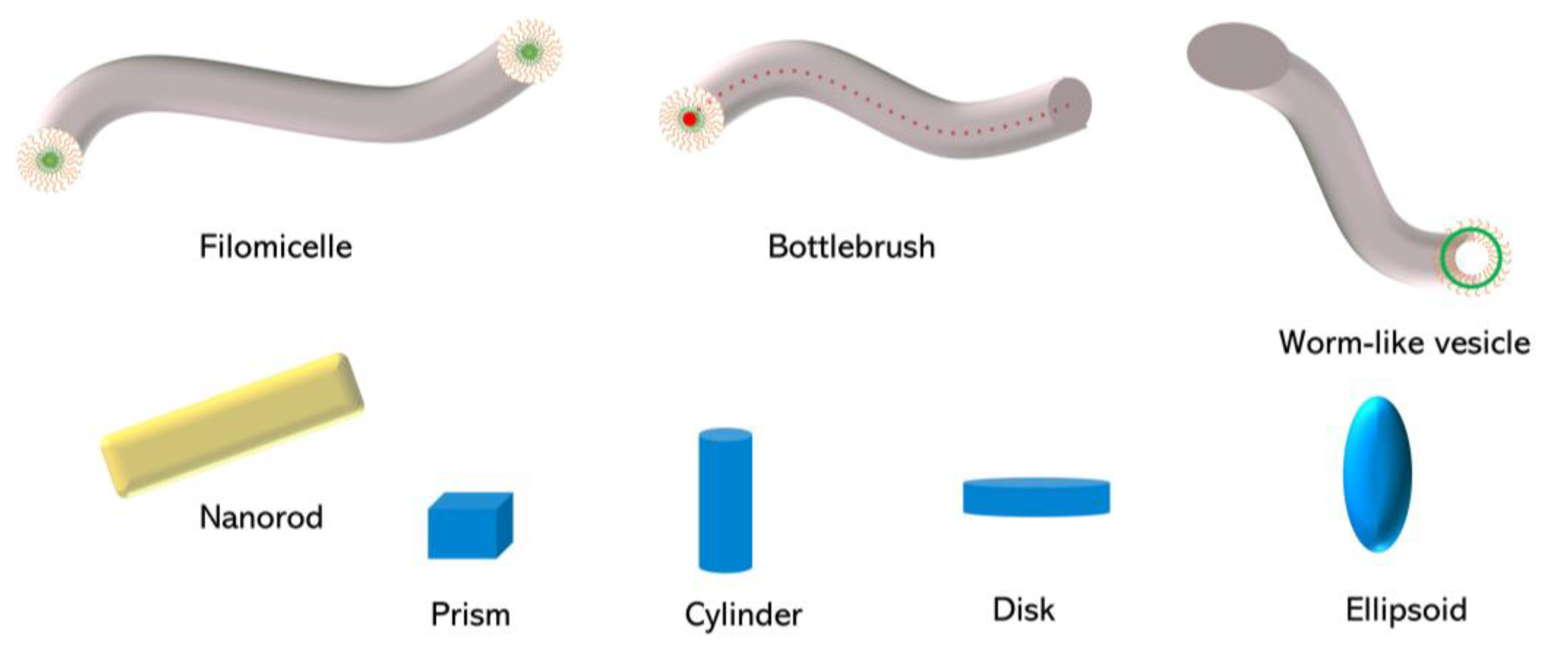
1. Introduction
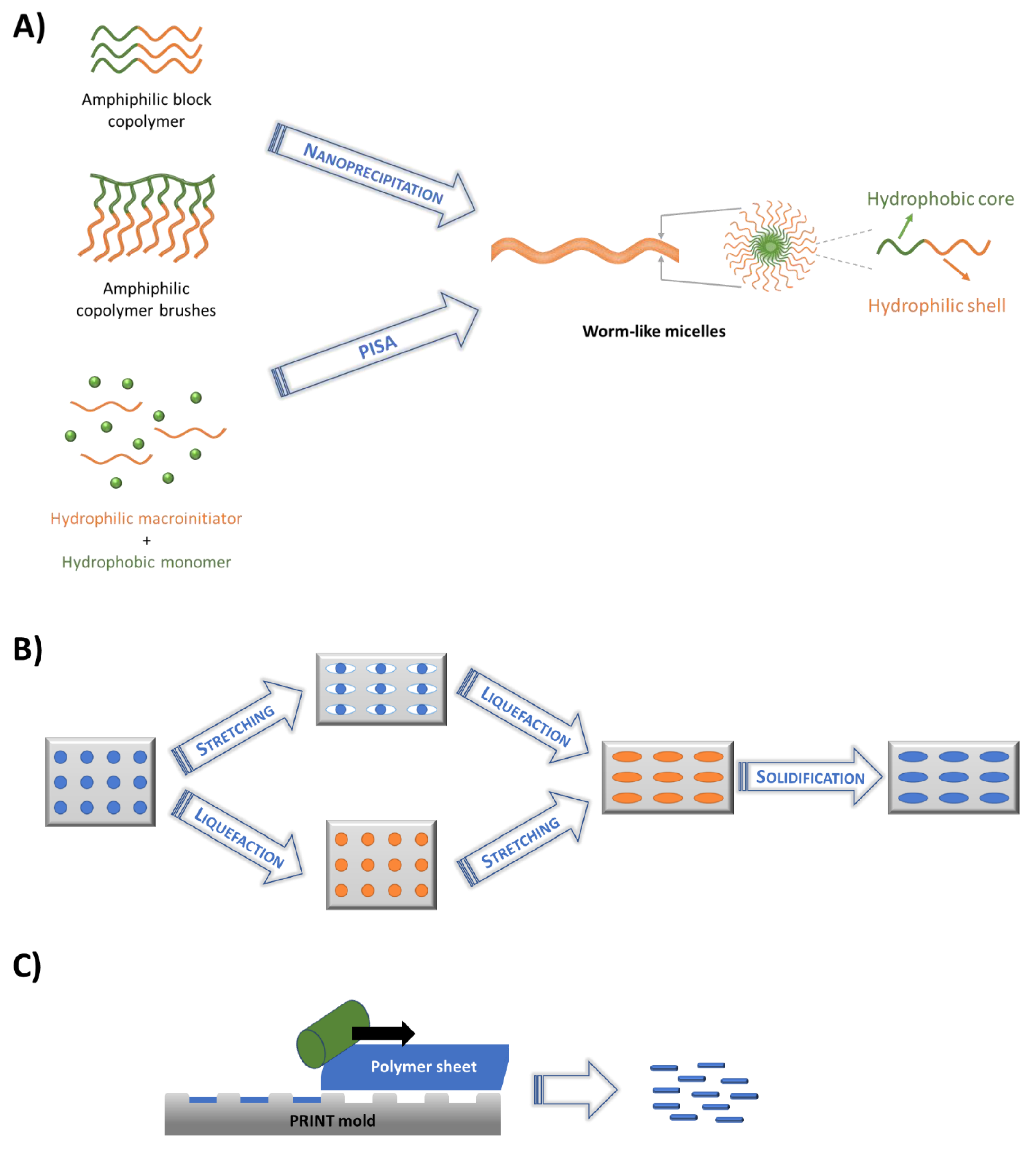
2. Fabrication of Non-spherical Polymeric Nanoparticles
2.1. Self-Assembly Techniques
2.2. Membrane Stretching Technique
2.3. Particle Replication in Nonwetting Template (PRINT)
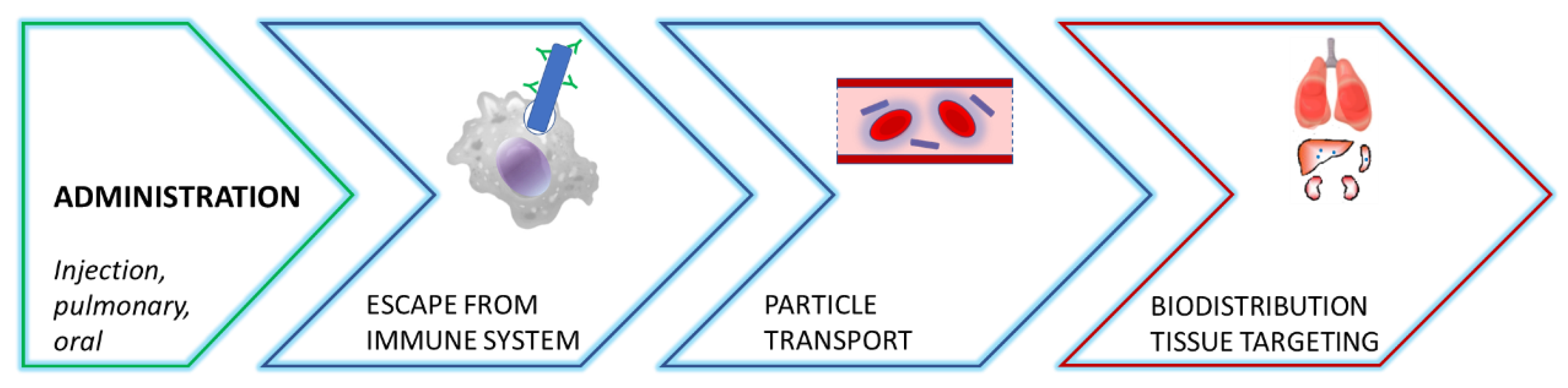
3. Effect of Particle Shape on Overcoming Biological Barriers
3.1. Interactions with Immune System
3.2. Particle Transport
3.3. Biodistribution and Targeted Delivery
4. Effect of Particle Shape on Drug Delivery Properties

| Fabrication technique | Shape | Material | Drug | Target | Ref. |
|---|---|---|---|---|---|
| Conventional self-assembly | Filomicelles | PEG-PEE, PEG-PCL | PTX | human-derived tumors in mice | [15] |
| Filomicelles | PEG-b-P(CPTKMA-co-PEMA) | Conjugated CPT | Tumor bearing mice | [16] | |
| Nanorods | PEG-PCL | DOX | HeLa, HepG2, OB cells; Balb/c mice bearing H22 tumor xenografts. | [17] | |
| Crosslinked wormlike vesicles | PEG-PLA-PEG | DOX | HeLa cells | [19] | |
| Filomicelles | PEG-PPS | Chloroquine | plasmacytoid dendritic cells | [22] | |
| Worm-like/rod-like vesicles | POEGMA-b-P(ST-co-VBA) | DOX | MCF-7 cells | [32] | |
| Filomicelles | PEG-PCL | PTX | A549 Tumor-bearing mice | [61] | |
| Tubular polymersomes /worm-like micelles | PEG, PTMC, PCL, and PDLLA block copolymers | DEX | retinal (ARPE-19) cells; ex vivo porcine eyes | [65] | |
| Nanorods | PEG-xCPT | CPT/DOX | MCF-7/ADR cancer cells | [75] | |
| Filomicelles | PEG-PCL, PEG-PBCL | PTX | A549 lung cancer cells, EC4 liver cancer cells | [100,101] | |
| Filomicelles | PEG-PLA | Betulin derivative | HeLa cells | [108] | |
| Filomicelles | PEG-PCL | PTX, retinoic acid | A549, HepG2, U2os, EC4 | [109] | |
| Filomicelles | PEG-PLGA | PTX, 17AAG, rapamycin | CaCo-2 human colorectal adenocarcinoma cells | [110] | |
| Filomicelles | P(MeOx-b-BuOx-b-MeOx) | ETO, C6CP, PTX | Small/ non-small cell lung cancer models | [111] | |
| Filomicelles, nanorods | poly(ether-anhydrides) | DOX | Murine breast cancer model | [113] | |
| pH-responsive wormlike micelles | PEG-PDPA | RGD-DM1 | Orthotopic brain tumor model | [117] | |
| pH-responsive wormlike micelles | mPEG-ser-[poly(Lys-DEAP)]2 | Chlorin e6 | KB cells and tumor-bearing mice | [118] | |
| pH-responsive wormlike micelles | PEG-PDPA | Succinobucol | Metastatic breast cancer Model | [119] | |
| Unimolecular polymer brushes | Nanorods | PNB-g-PGA | Conjugated CPT | HeLa, LS174T, and HEK cells | [31] |
| Nanoworms, lamellae, vesicles | PHPMA-b-(NBMA-co-CMA) | DOX | HeLa cells | [33] | |
| Cylindrical bottlebrushes | cellulose-g-(CPT-b-OEGMA) | Conjugated CPT | MCF-7 induced multicellular spheroids and tumor-bearing mice | [73] | |
| pH sensitive nanorods | PHF-g-(PCL-PEG) | DOX | A459 human lung cancer cells | [105] | |
| Nanorods, Nanoworms | PHEMA-g-(PtBA-b-PEG) | IR780 | photothermal therapy in MCF-7 tumor models | [114] | |
| Cylindrical brushes | HA-polybenzofulvene | DOX | HCT116, MCF-7, 16HBE cell lines | [115] | |
| Nanorods | PLGA | Docetaxel | Human ovarian carcinoma cells; Mice bearing tumor xenografts | [38,39] |
5. Conclusions
Funding
Institutional Review Board Statement
Informed Consent Statement
Data Availability Statement
Conflicts of Interest
References
- Mitchell, M.J.; Billingsley, M.M.; Haley, R.M.; Wechsler, M.E.; Peppas, N.A.; Langer, R. Engineering precision nanoparticles for drug delivery. Nat. Rev. Drug Discov. 2021, 20, 101–124. [Google Scholar] [CrossRef]
- Siemer, S.; Bauer, T.A.; Scholz, P.; Breder, C.; Fenaroli, F.; Harms, G.; Dietrich, D.; Dietrich, J.; Rosenauer, C.; Barz, M.; et al. Targeting cancer chemotherapy resistance by precision medicine-driven nanoparticle-formulated cisplatin. ACS Nano 2021, 15, 18541–18556. [Google Scholar] [CrossRef]
- Duan, X.; Li, Y. Physicochemical characteristics of nanoparticles affect circulation, biodistribution, cellular internalization, and trafficking. Small 2013, 9, 1521–1532. [Google Scholar] [CrossRef]
- Kapate, N.; Clegg, J.R.; Mitragotri, S. Non-spherical micro- and nanoparticles for drug delivery: Progress over 15 years. Adv. Drug Deliv. Rev. 2021, 177, 113807. [Google Scholar] [CrossRef]
- He, C.; Hu, Y.; Yin, L.; Tang, C.; Yin, C. Effects of particle size and surface charge on cellular uptake and biodistribution of polymeric nanoparticles. Biomaterials 2010, 31, 3657–3666. [Google Scholar] [CrossRef]
- Hickey, J.W.; Santos, J.L.; Williford, J.M.; Mao, H.Q. Control of polymeric nanoparticle size to improve therapeutic delivery. J. Control. Release 2015, 219, 536–547. [Google Scholar] [CrossRef]
- Kang, H.; Rho, S.; Stiles, W.R.; Hu, S.; Baek, Y.; Hwang, D.W.; Kashiwagi, S.; Kim, M.S.; Choi, H.S. Size-dependent epr effect of polymeric nanoparticles on tumor targeting. Adv. Healthc. Mater. 2020, 9, 1901223. [Google Scholar] [CrossRef]
- Sadat, S.M.A.; Jahan, S.T.; Haddadi, A. Effects of size and surface charge of polymeric nanoparticles on in vitro and in vivo applications. J. Biomater. Nanobiotechnology 2016, 7, 91–108. [Google Scholar] [CrossRef]
- Cabral, H.; Kataoka, K. Multifunctional nanoassemblies of block copolymers for future cancer therapy. Sci. Technol. Adv. Mater. 2010, 11, 014109. [Google Scholar] [CrossRef]
- Albanese, A.; Tang, P.S.; Chan, W.C.W. The effect of nanoparticle size, shape, and surface chemistry on biological systems. Annu. Rev. Biomed. Eng. 2012, 14, 1–16. [Google Scholar] [CrossRef]
- Champion, J.A.; Katare, Y.K.; Mitragotri, S. Particle shape: A new design parameter for micro- and nanoscale drug delivery carriers. J. Control. Release 2007, 121, 3–9. [Google Scholar] [CrossRef]
- Jelonek, K.; Li, S.; Kasperczyk, J.; Wu, X.; Orchel, A. Effect of polymer degradation on prolonged release of paclitaxel from filomicelles of polylactide/poly(ethylene glycol) block copolymers. Mater. Sci. Eng. C 2017, 75, 918–925. [Google Scholar] [CrossRef]
- Truong, N.P.; Whittaker, M.R.; Mak, C.W.; Davis, T.P. The importance of nanoparticle shape in cancer drug delivery. Expert Opin. Drug Deliv. 2015, 12, 129–142. [Google Scholar] [CrossRef]
- Choucair, A.; Eisenberg, A. Control of amphiphilic block copolymer morphologies using solution conditions. Eur. Phys. J. E 2003, 10, 37–44. [Google Scholar] [CrossRef]
- Geng, Y.; Dalhaimer, P.; Cai, S.; Tsai, R.; Tewari, M.; Minko, T.; Discher, D.E. Shape effects of filaments versus spherical particles in flow and drug delivery. Nat. Nanotech. 2007, 2, 249–255. [Google Scholar] [CrossRef]
- Ke, W.; Lu, N.; Japir, A.A.W.M.M.; Zhou, Q.; Xi, L.; Wang, Y.; Dutta, D.; Zhou, M.; Pan, Y.; Ge, Z. Length effect of stimuli-responsive block copolymer prodrug filomicelles on drug delivery efficiency. J. Control. Release 2020, 318, 67–77. [Google Scholar] [CrossRef]
- Li, D.; Tang, Z.; Gao, Y.; Sun, H.; Zhou, S. A bio-inspired rod-shaped nanoplatform for strongly infecting tumor cells and enhancing the delivery efficiency of anticancer drugs. Adv. Funct. Mater. 2016, 26, 66–79. [Google Scholar] [CrossRef]
- Ma, Q.; Remsen, E.E.; Clark, C.G.; Kowalewski, T.; Wooley, K.L. Chemically induced supramolecular reorganization of triblock copolymer assemblies: Trapping of intermediate states via a shell-crosslinking methodology. Proc. Natl. Acad. Sci. USA 2002, 99, 5058–5063. [Google Scholar] [CrossRef]
- Yang, X.; Grailer, J.J.; Rowland, I.J.; Javadi, A.; Hurley, S.A.; Steeber, D.A.; Gong, S. Multifunctional spio/dox-loaded wormlike polymer vesicles for cancer therapy and mr imaging. Biomaterials 2010, 31, 9065–9073. [Google Scholar] [CrossRef]
- Zhang, L.; Eisenberg, A. Multiple morphologies of “crew-cut” aggregates of polystyrene- b -poly(acrylic acid) block copolymers. Science 1995, 268, 1728–1731. [Google Scholar] [CrossRef]
- Zhang, L.; Yu, K.; Eisenberg, A. Ion-induced morphological changes in “crew-cut” aggregates of amphiphilic block copolymers. Science 1996, 272, 1777–1779. [Google Scholar] [CrossRef]
- Allen, M.E.; Golding, A.; Rus, V.; Karabin, N.B.; Li, S.; Lescott, C.J.; Bobbala, S.; Scott, E.A.; Szeto, G.L. Targeted delivery of chloroquine to antigen-presenting cells enhances inhibition of the type i interferon response. ACS Biomater. Sci. Eng. 2021, 7, 5666–5677. [Google Scholar] [CrossRef]
- Shen, X.; Liu, X.; Li, R.Y.; Yun, P.; Li, C.L.; Su, F.; Li, S.M. Biocompatibility of filomicelles prepared from poly(ethylene glycol)-polylactide diblock copolymers as potential drug carrier. J. Biomater. Sci.-Polym. Ed. 2017, 28, 1677–1694. [Google Scholar] [CrossRef]
- Rodriguez-Hernandez, J.; Checot, F.; Gnanou, Y.; Lecommandoux, S. Toward ‘smart’ nano-objects by self-assembly of block copolymers in solution. Prog. Polym. Sci. 2005, 30, 691–724. [Google Scholar] [CrossRef]
- Williford, J.M.; Archang, M.M.; Minn, I.; Ren, Y.; Wo, M.; Vandermark, J.; Fisher, P.B.; Pomper, M.G.; Mao, H.Q. Critical length of peg grafts on ipei/DNA nanoparticles for efficient in vivo delivery. ACS Biomater. Sci. Eng. 2016, 2, 567–578. [Google Scholar] [CrossRef]
- Williford, J.M.; Ren, Y.; Huang, K.V.; Pan, D.; Mao, H.Q. Shape transformation following reduction-sensitive peg cleavage of polymer/DNA nanoparticles. J. Mater. Chem. B 2014, 2, 8106–8109. [Google Scholar] [CrossRef]
- Li, Z.; Ma, J.; Lee, N.S.; Wooley, K.L. Dynamic cylindrical assembly of triblock copolymers by a hierarchical process of covalent and supramolecular interactions. J. Am. Chem. Soc. 2011, 133, 1228–1231. [Google Scholar] [CrossRef]
- Müllner, M.; Dodds, S.J.; Nguyen, T.H.; Senyschyn, D.; Porter, C.J.H.; Boyd, B.J.; Caruso, F. Size and rigidity of cylindrical polymer brushes dictate long circulating properties in vivo. ACS Nano 2015, 9, 1294–1304. [Google Scholar] [CrossRef]
- Zhang, Z.; Liu, C.; Li, C.; Wu, W.; Jiang, X. Shape effects of cylindrical versus spherical unimolecular polymer nanomaterials on in vitro and in vivo behaviors. Research 2019, 2019, 2391486. [Google Scholar] [CrossRef]
- Zhang, Z.; Zhang, L.; Zhao, J.; Li, C.; Wu, W.; Jiang, X. Length effects of cylindrical polymer brushes on their in vitro and in vivo properties. Biomater. Sci. 2019, 7, 5124–5131. [Google Scholar] [CrossRef]
- Baumgartner, R.; Kuai, D.; Cheng, J.J. Synthesis of controlled, high-molecular weight poly(l-glutamic acid) brush polymers. Biomater. Sci. 2017, 5, 1836–1844. [Google Scholar] [CrossRef]
- Karagoz, B.; Esser, L.; Duong, H.T.; Basuki, J.S.; Boyer, C.; Davis, T.P. Polymerization-induced self-assembly (pisa)–control over the morphology of nanoparticles for drug delivery applications. Polym. Chem. 2014, 5, 350–355. [Google Scholar] [CrossRef]
- Zhang, W.J.; Hong, C.Y.; Pan, C.Y. Efficient fabrication of photosensitive polymeric nano-objects via an ingenious formulation of raft dispersion polymerization and their application for drug delivery. Biomacromolecules 2017, 18, 1210–1217. [Google Scholar] [CrossRef]
- Finnegan, J.R.; Pilkington, E.H.; Alt, K.; Rahim, M.A.; Kent, S.J.; Davis, T.P.; Kempe, K. Stealth nanorods via the aqueous living crystallisation-driven self-assembly of poly(2-oxazoline)s. Chem. Sci. 2021, 12, 7350–7360. [Google Scholar] [CrossRef]
- Barua, S.; Yoo, J.W.; Kolhar, P.; Wakankar, A.; Gokarn, Y.R.; Mitragotri, S. Particle shape enhances specificity of antibody-displaying nanoparticles. Proc. Natl. Acad. Sci. USA 2013, 110, 3270–3275. [Google Scholar] [CrossRef]
- Kolhar, P.; Anselmo, A.C.; Gupta, V.; Pant, K.; Prabhakarpandian, B.; Ruoslahti, E.; Mitragotri, S. Using shape effects to target antibody-coated nanoparticles to lung and brain endothelium. Proc. Natl. Acad. Sci. USA 2013, 110, 10753–10758. [Google Scholar] [CrossRef]
- Yoo, J.W.; Doshi, N.; Mitragotri, S. Endocytosis and intracellular distribution of plga particles in endothelial cells: Effect of particle geometry. Macromol. Rapid Commun. 2010, 31, 142–148. [Google Scholar] [CrossRef]
- Chu, K.S.; Hasan, W.; Rawal, S.; Walsh, M.D.; Enlow, E.M.; Luft, J.C.; Bridges, A.S.; Kuijer, J.L.; Napier, M.E.; Zamboni, W.C. Plasma, tumor and tissue pharmacokinetics of docetaxel delivered via nanoparticles of different sizes and shapes in mice bearing skov-3 human ovarian carcinoma xenograft. Nanomed. Nanotechnol. Biol. Med. 2013, 9, 686–693. [Google Scholar] [CrossRef]
- Enlow, E.M.; Luft, J.C.; Napier, M.E.; DeSimone, J.M. Potent engineered plga nanoparticles by virtue of exceptionally high chemotherapeutic loadings. Nano Lett. 2011, 11, 808–813. [Google Scholar] [CrossRef]
- Rolland, J.P.; Maynor, B.W.; Euliss, L.E.; Exner, A.E.; Denison, G.M.; DeSimone, J.M. Direct fabrication and harvesting of monodisperse, shape-specific nanobiomaterials. J. Am. Chem. Soc. 2005, 127, 10096–10100. [Google Scholar] [CrossRef]
- Fu, X.; Cai, J.; Zhang, X.; Li, W.-D.; Ge, H.; Hu, Y. Top-down fabrication of shape-controlled, monodisperse nanoparticles for biomedical applications. Adv. Drug Deliv. Rev. 2018, 132, 169–187. [Google Scholar] [CrossRef] [PubMed]
- Chen, J.; Clay, N.E.; Park, N.-H.; Kong, H. Non-spherical particles for targeted drug delivery. Chem. Eng. Sci. 2015, 125, 20–24. [Google Scholar] [CrossRef] [PubMed]
- Elsabahy, M.; Wooley, K.L. Design of polymeric nanoparticles for biomedical delivery applications. Chem. Soc. Rev. 2012, 41, 2545. [Google Scholar] [CrossRef] [PubMed]
- Williford, J.M.; Santos, J.L.; Shyam, R.; Mao, H.Q. Shape control in engineering of polymeric nanoparticles for therapeutic delivery. Biomater. Sci. 2015, 3, 894–907. [Google Scholar] [CrossRef]
- Ku, K.H.; Shin, J.M.; Yun, H.; Yi, G.-R.; Jang, S.G.; Kim, B.J. Multidimensional design of anisotropic polymer particles from solvent-evaporative emulsion. Adv. Funct. Mater. 2018, 28, 1802961. [Google Scholar] [CrossRef]
- Jiang, X.; Qu, W.; Pan, D.; Ren, Y.; Williford, J.-M.; Cui, H.; Luijten, E.; Mao, H.-Q. Plasmid-templated shape control of condensed DNA–block copolymer nanoparticles. Adv. Mater. 2013, 25, 227–232. [Google Scholar] [CrossRef]
- Osawa, S.; Osada, K.; Hiki, S.; Dirisala, A.; Ishii, T.; Kataoka, K. Polyplex micelles with double-protective compartments of hydrophilic shell and thermoswitchable palisade of poly(oxazoline)-based block copolymers for promoted gene transfection. Biomacromolecules 2016, 17, 354–361. [Google Scholar] [CrossRef]
- Chen, Y. Shaped hairy polymer nanoobjects. Macromolecules 2012, 45, 2619–2631. [Google Scholar] [CrossRef]
- Zhou, Y.; Huang, W.; Liu, J.; Zhu, X.; Yan, D. Self-assembly of hyperbranched polymers and its biomedical applications. Adv. Mater. 2010, 22, 4567–4590. [Google Scholar] [CrossRef]
- Khor, S.Y.; Quinn, J.F.; Whittaker, M.R.; Truong, N.P.; Davis, T.P. Controlling nanomaterial size and shape for biomedical applications via polymerization-induced self-assembly. Macromol. Rapid Commun. 2019, 40, 1800438. [Google Scholar] [CrossRef]
- Phan, H.; Taresco, V.; Penelle, J.; Couturaud, B. Polymerisation-induced self-assembly (pisa) as a straightforward formulation strategy for stimuli-responsive drug delivery systems and biomaterials: Recent advances. Biomater. Sci. 2021, 9, 38–50. [Google Scholar] [CrossRef] [PubMed]
- Zhu, X.; Vo, C.; Taylor, M.; Smith, B.R. Non-spherical micro- and nanoparticles in nanomedicine. Mater. Horiz. 2019, 6, 1094–1121. [Google Scholar] [CrossRef]
- Barrio, J.; Sánchez-Somolinos, C. Light to shape the future: From photolithography to 4d printing. Adv. Opt. Mater. 2019, 7, 1900598. [Google Scholar] [CrossRef]
- Zhao, Z.; Ukidve, A.; Krishnan, V.; Mitragotri, S. Effect of physicochemical and surface properties on in vivo fate of drug nanocarriers. Adv. Drug Deliv. Rev. 2019, 143, 3–21. [Google Scholar] [CrossRef] [PubMed]
- Chenthamara, D.; Subramaniam, S.; Ramakrishnan, S.G.; Krishnaswamy, S.; Essa, M.M.; Lin, F.-H.; Qoronfleh, M.W. Therapeutic efficacy of nanoparticles and routes of administration. Biomater. Res. 2019, 23, 20. [Google Scholar] [CrossRef] [PubMed]
- Champion, J.A.; Mitragotri, S. Role of target geometry in phagocytosis. Proc. Natl. Acad. Sci. USA 2006, 103, 4930–4934. [Google Scholar] [CrossRef] [PubMed]
- Champion, J.A.; Mitragotri, S. Shape induced inhibition of phagocytosis of polymer particles. Pharm. Res. 2009, 26, 244–249. [Google Scholar] [CrossRef]
- Doshi, N.; Mitragotri, S. Macrophages recognize size and shape of their targets. PLoS ONE 2010, 5, 10051. [Google Scholar] [CrossRef]
- Geng, Y.; Discher, D.E. Visualization of degradable worm micelle breakdown in relation to drug release. Polymer 2006, 47, 2519–2525. [Google Scholar] [CrossRef]
- Mathaes, R.; Winter, G.; Besheer, A.; Engert, J. Influence of particle geometry and pegylation on phagocytosis of particulate carriers. Int. J. Pharm. 2014, 465, 159–164. [Google Scholar] [CrossRef]
- Christian, D.A.; Cai, S.; Garbuzenko, O.B.; Harada, T.; Zajac, A.L.; Minko, T.; Discher, D.E. Flexible filaments for in vivo imaging and delivery: Persistent circulation of filomicelles opens the dosage window for sustained tumor shrinkage. Mol. Pharm. 2009, 6, 1343–1352. [Google Scholar] [CrossRef] [PubMed]
- Larnaudie, S.C.; Sanchis, J.; Nguyen, T.-H.; Peltier, R.; Catrouillet, S.; Brendel, J.C.; Porter, C.J.H.; Jolliffe, K.A.; Perrier, S. Cyclic peptide-poly(hpma) nanotubes as drug delivery vectors: In vitro assessment, pharmacokinetics and biodistribution. Biomaterials 2018, 178, 570–582. [Google Scholar] [CrossRef] [PubMed]
- Mahmud, A.; Discher, D.E. Lung vascular targeting through inhalation delivery: Insight from filamentous viruses and other shapes. Iubmb Life 2011, 63, 607–612. [Google Scholar] [CrossRef] [PubMed]
- Peiris, P.M.; Bauer, L.; Toy, R.; Tran, E.; Pansky, J.; Doolittle, E.; Schmidt, E.; Hayden, E.; Mayer, A.; Keri, R.A. Enhanced delivery of chemotherapy to tumors using a multicomponent nanochain with radio-frequency-tunable drug release. ACS Nano 2012, 6, 4157–4168. [Google Scholar] [CrossRef] [PubMed]
- Ridolfo, R.; Tavakoli, S.; Junnuthula, V.; Williams, D.S.; Urtti, A.; van Hest, J.C.M. Exploring the impact of morphology on the properties of biodegradable nanoparticles and their diffusion in complex biological medium. Biomacromolecules 2021, 22, 126–133. [Google Scholar] [CrossRef]
- Venkataraman, S.; Hedrick, J.L.; Ong, Z.Y.; Yang, C.; Ee, P.L.R.; Hammond, P.T.; Yang, Y.Y. The effects of polymeric nanostructure shape on drug delivery. Adv. Drug Deliv. Rev. 2011, 63, 1228–1246. [Google Scholar] [CrossRef]
- Zhang, Y.J.; Zhang, Z.K.; Liu, C.R.; Chen, W.Z.; Li, C.; Wu, W.; Jiang, X.Q. Synthesis and biological properties of water-soluble polyphenylthiophene brushes with poly(ethylene glycol)/polyzwitterion side chains. Polym. Chem. 2017, 8, 1672–1679. [Google Scholar] [CrossRef]
- Cooley, M.; Sarode, A.; Hoore, M.; Fedosov, D.A.; Mitragotri, S.; Sen Gupta, A. Influence of particle size and shape on their margination and wall-adhesion: Implications in drug delivery vehicle design across nano-to-micro scale. Nanoscale 2018, 10, 15350–15364. [Google Scholar] [CrossRef]
- Jurney, P.; Agarwal, R.; Singh, V.; Choi, D.; Roy, K.; Sreenivasan, S.V.; Shi, L. Unique size and shape-dependent uptake behaviors of non-spherical nanoparticles by endothelial cells due to a shearing flow. J. Control. Release 2017, 245, 170–176. [Google Scholar] [CrossRef]
- Paul, D.; Achouri, S.; Yoon, Y.Z.; Herre, J.; Bryant, C.E.; Cicuta, P. Phagocytosis dynamics depends on target shape. Biophys. J. 2013, 105, 1143–1150. [Google Scholar] [CrossRef]
- Champion, J.A.; Walker, A.; Mitragotri, S. Role of particle size in phagocytosis of polymeric microspheres. Pharm. Res. 2008, 25, 1815–1821. [Google Scholar] [CrossRef] [PubMed]
- Li, S.; Bobbala, S.; Vincent, M.P.; Modak, M.; Liu, Y.G.; Scott, E.A. Pi-stacking enhances stability, scalability of formation, control over flexibility, and circulation time of polymeric filamentous nanocarriers. Adv. Nanobiomed Res. 2021, 1, 2100063. [Google Scholar] [CrossRef] [PubMed]
- Bai, S.; Jia, D.; Ma, X.B.; Liang, M.Y.; Xue, P.; Kang, Y.J.; Xu, Z.G. Cylindrical polymer brushes-anisotropic unimolecular micelle drug delivery system for enhancing the effectiveness of chemotherapy. Bioact. Mater. 2021, 6, 2894–2904. [Google Scholar] [CrossRef] [PubMed]
- Mullner, M.; Yang, K.; Kaur, A.; New, E.J. Aspect-ratio-dependent interaction of molecular polymer brushes and multicellular tumour spheroids. Polym. Chem. 2018, 9, 3461–3465. [Google Scholar] [CrossRef]
- Zhou, Z.X.; Ma, X.P.; Jin, E.L.; Tang, J.B.; Sui, M.H.; Shen, Y.Q.; Van Kirk, E.A.; Murdoch, W.J.; Radosz, M. Linear-dendritic drug conjugates forming long-circulating nanorods for cancer-drug delivery. Biomaterials 2013, 34, 5722–5735. [Google Scholar] [CrossRef]
- Kim, Y.; Dalhaimer, P.; Christian, D.A.; Discher, D.E. Polymeric worm micelles as nano-carriers for drug delivery. Nanotechnology 2005, 16, S484. [Google Scholar] [CrossRef]
- Yin, C.F.; Xiao, P.P.; Liang, M.K.; Li, J.; Sun, Y.; Jiang, X.Q.; Wu, W. Effects of irgd conjugation density on the in vitro and in vivo properties of cylindrical polymer brushes. Biomater. Sci. 2022, 10, 3236–3244. [Google Scholar] [CrossRef]
- Banerjee, A.; Qi, J.; Gogoi, R.; Wong, J.; Mitragotri, S. Role of nanoparticle size, shape and surface chemistry in oral drug delivery. J. Control. Release 2016, 238, 176–185. [Google Scholar] [CrossRef]
- Hu, X.; Hu, J.; Tian, J.; Ge, Z.; Zhang, G.; Luo, K.; Liu, S. Polyprodrug amphiphiles: Hierarchical assemblies for shape-regulated cellular internalization, trafficking, and drug delivery. J. Am. Chem. Soc. 2013, 135, 17617–17629. [Google Scholar] [CrossRef]
- Li, D.; Zhuang, J.; He, H.; Jiang, S.; Banerjee, A.; Lu, Y.; Wu, W.; Mitragotri, S.; Gan, L.; Qi, J. Influence of particle geometry on gastrointestinal transit and absorption following oral administration. ACS Appl. Mater. Interfaces 2017, 9, 42492–42502. [Google Scholar] [CrossRef]
- Muro, S.; Garnacho, C.; Champion, J.A.; Leferovich, J.; Gajewski, C.; Schuchman, E.H.; Mitragotri, S.; Muzykantov, V.R. Control of endothelial targeting and intracellular delivery of therapeutic enzymes by modulating the size and shape of icam-1-targeted carriers. Mol. Ther. 2008, 16, 1450–1458. [Google Scholar] [CrossRef] [PubMed]
- Shuvaev, V.V.; Ilies, M.A.; Simone, E.; Zaitsev, S.; Kim, Y.; Cai, S.S.; Mahmud, A.; Dziubla, T.; Muro, S.; Discher, D.E.; et al. Endothelial targeting of antibody-decorated polymeric filomicelles. ACS NANO 2011, 5, 6991–6999. [Google Scholar] [CrossRef] [PubMed]
- Fish, M.B.; Thompson, A.J.; Fromen, C.A.; Eniola-Adefeso, O. Emergence and utility of nonspherical particles in biomedicine. Ind. Eng. Chem. Res. 2015, 54, 4043–4059. [Google Scholar] [CrossRef] [PubMed]
- Sharma, G.; Valenta, D.T.; Altman, Y.; Harvey, S.; Xie, H.; Mitragotri, S.; Smith, J.W. Polymer particle shape independently influences binding and internalization by macrophages. J. Control. Release 2010, 147, 408–412. [Google Scholar] [CrossRef] [PubMed]
- Möller, J.; Luehmann, T.; Hall, H.; Vogel, V. The race to the pole: How high-aspect ratio shape and heterogeneous environments limit phagocytosis of filamentous escherichia coli bacteria by macrophages. Nano Lett. 2012, 12, 2901–2905. [Google Scholar] [CrossRef] [PubMed]
- Li, Z.; Sun, L.; Zhang, Y.; Dove, A.P.; O’Reilly, R.K.; Chen, G. Shape effect of glyco-nanoparticles on macrophage cellular uptake and immune response. ACS Macro Lett. 2016, 5, 1059–1064. [Google Scholar] [CrossRef] [PubMed]
- Anselmo, A.C.; Mitragotri, S. Impact of particle elasticity on particle-based drug delivery systems. Adv. Drug Deliv. Rev. 2017, 108, 51–67. [Google Scholar] [CrossRef] [PubMed]
- Shi, D.; Beasock, D.; Fessler, A.; Szebeni, J.; Ljubimova, J.Y.; Afonin, K.A.; Dobrovolskaia, M.A. To pegylate or not to pegylate: Immunological properties of nanomedicine’s most popular component, polyethylene glycol and its alternatives. Adv. Drug Deliv. Rev. 2022, 180, 114079. [Google Scholar] [CrossRef]
- Adriani, G.; Tullio, M.D.; Ferrari, M.; Hussain, F.; Pascazio, G.; Liu, X.; Decuzzi, P. The preferential targeting of the diseased microvasculature by disk-like particles. Biomaterials 2012, 33, 5504–5513. [Google Scholar] [CrossRef]
- Huang, X.; Li, L.; Liu, T.; Hao, N.; Liu, H.; Chen, D.; Tang, F. The shape effect of mesoporous silica nanoparticles on biodistribution, clearance, and biocompatibility in vivo. ACS Nano 2011, 5, 5390–5399. [Google Scholar] [CrossRef]
- Janát-Amsbury, M.M.; Ray, A.; Peterson, C.M.; Ghandehari, H. Geometry and surface characteristics of gold nanoparticles influence their biodistribution and uptake by macrophages. Eur. J. Pharm. Biopharm. 2011, 77, 417–423. [Google Scholar]
- Wang, G.; Inturi, S.; Serkova, N.J.; Merkulov, S.; McCrae, K.; Russek, S.E.; Banda, N.K.; Simberg, D. High-relaxivity superparamagnetic iron oxide nanoworms with decreased immune recognition and long-circulating properties. ACS Nano 2014, 8, 12437–12449. [Google Scholar] [CrossRef] [PubMed]
- Huda, P.; Binderup, T.; Pedersen, M.C.; Midtgaard, S.R.; Elema, D.R.; Kjær, A.; Jensen, M.; Arleth, L. PET/CT based in vivo evaluation of 64Cu labelled nanodiscs in tumor bearing mice. PLoS ONE 2015, 10, e0129310. [Google Scholar] [CrossRef] [PubMed]
- Geisbert, T.W.; Jahrling, P.B. Exotic emerging viral diseases: Progress and challenges. Nat. Med. 2004, 10, S110–S121. [Google Scholar] [CrossRef] [PubMed]
- Shukla, S.; Eber, F.J.; Nagarajan, A.S.; DiFranco, N.A.; Schmidt, N.; Wen, A.M.; Eiben, S.; Twyman, R.M.; Wege, C.; Steinmetz, N.F. The impact of aspect ratio on the biodistribution and tumor homing of rigid soft-matter nanorods. Adv. Healthc. Mater. 2015, 4, 874–882. [Google Scholar]
- Zhang, B.; Sai Lung, P.; Zhao, S.; Chu, Z.; Chrzanowski, W.; Li, Q. Shape dependent cytotoxicity of plga-peg nanoparticles on human cells. Sci. Rep. 2017, 7, 7315. [Google Scholar] [CrossRef] [PubMed]
- Oltra, N.S.; Swift, J.; Mahmud, A.; Rajagopal, K.; Loverde, S.M.; Discher, D.E. Filomicelles in nanomedicine–from flexible, fragmentable, and ligand-targetable drug carrier designs to combination therapy for brain tumors. J. Mater. Chem. B 2013, 1, 5177. [Google Scholar] [CrossRef] [PubMed]
- Oltra, N.S.; Nair, P.; Discher, D.E. From stealthy polymersomes and filomicelles to “self” peptide-nanoparticles for cancer therapy. Annu. Rev. Chem. Biomol. Eng. 2014, 5, 281–299. [Google Scholar] [CrossRef]
- Li, X.; Uppala, V.V.S.; Cooksey, T.J.; Robertson, M.L.; Madsen, L.A. Quantifying drug cargo partitioning in block copolymer micelle solutions. ACS Appl. Polym. Mater. 2020, 2, 3749–3755. [Google Scholar] [CrossRef]
- Cai, S.; Vijayan, K.; Cheng, D.; Lima, E.M.; Discher, D.E. Micelles of different morphologies—Advantages of worm-like filomicelles of peo-pcl in paclitaxel delivery. Pharm. Res. 2007, 24, 2099–2109. [Google Scholar] [CrossRef]
- Nair, P.R.; Karthick, S.; Spinler, K.R.; Vakili, M.R.; Lavasanifar, A.; Discher, D.E. Filomicelles from aromatic diblock copolymers increase paclitaxel-induced tumor cell death and aneuploidy compared with aliphatic copolymers. Nanomedicine 2016, 11, 1551–1569. [Google Scholar] [CrossRef]
- Ridolfo, R.; Arends, J.J.; Hest, J.C.M.; Williams, D.S. Wormlike nanovector with enhanced drug loading using blends of biodegradable block copolymers. Biomacromolecules 2020, 21, 2199–2207. [Google Scholar] [CrossRef] [PubMed]
- Sun, X.; Liu, X.; Li, C.; Wang, Y.; Liu, L.; Su, F.; Li, S. Self-assembled micelles prepared from poly(ε-caprolactone)-poly(ethylene glycol) and poly(ε-caprolactone/glycolide)-poly(ethylene glycol) block copolymers for sustained drug delivery. J. Appl. Polym. Sci. 2018, 135, 45732. [Google Scholar] [CrossRef]
- Naolou, T.; Meister, A.; Schöps, R.; Pietzsch, M.; Kressler, J. Synthesis and characterization of graft copolymers able to form polymersomes and worm-like aggregates. Soft Matter 2013, 9, 10364–10372. [Google Scholar] [CrossRef]
- Wu, Y.; Xiao, Y.; Huang, Y.; Xu, Y.; You, D.; Lu, W.; Yu, J. Rod-shaped micelles based on phf-g-(pcl-peg) with ph-triggered doxorubicin release and enhanced cellular uptake. Biomacromolecules 2019, 20, 1167–1177. [Google Scholar] [CrossRef] [PubMed]
- Crothers, M.; Zhou, Z.; Ricardo, N.M.P.S.; Yang, Z.; Taboada, P.; Chaibundit, C.; Attwood, D.; Booth, C. Solubilisation in aqueous micellar solutions of block copoly(oxyalkylene)s. Int. J. Pharm. 2005, 293, 91–100. [Google Scholar] [CrossRef] [PubMed]
- Yang, C.; Huang, S.; Wang, X.; Wang, M. Theranostic unimolecular micelles of highly fluorescent conjugated polymer bottlebrushes for far red/near infrared bioimaging and efficient anticancer drug delivery. Polym. Chem. 2016, 7, 7455–7468. [Google Scholar] [CrossRef]
- Jelonek, K.; Kasperczyk, J.; Li, S.; Nguyen, T.H.N.; Orchel, A.; Chodurek, E.; Paduszyński, P.; Jaworska-Kik, M.; Chrobak, E.; Bębenek, E. Bioresorbable filomicelles for targeted delivery of betulin derivative. Vitr. Study. Int. J. Pharm. 2019, 557, 43–52. [Google Scholar] [CrossRef]
- Nair, P.R.; Alvey, C.; Jin, X.; Irianto, J.; Ivanovska, I.; Discher, D.E. Filomicelles deliver a chemo-differentiation combination of paclitaxel and retinoic acid that durably represses carcinomas in liver to prolong survival. Bioconjugate Chem. 2018, 29, 914–927. [Google Scholar] [CrossRef]
- Jelonek, K.; Li, S.; Kaczmarczyk, B.; Marcinkowski, A.; Orchel, A.; Musiał-Kulik, M.; Kasperczyk, J. Multidrug pla-peg filomicelles for concurrent delivery of anticancer drugs—The influence of drug-drug and drug-polymer interactions on drug loading and release properties. Int. J. Pharm. 2016, 510, 365–374. [Google Scholar] [CrossRef]
- Wan, X.; Min, Y.; Bludau, H.; Keith, A.; Sheiko, S.S.; Jordan, R.; Wang, A.Z.; Sokolsky-Papkov, M.; Kabanov, A.V. Drug combination synergy in worm-like polymeric micelles improves treatment outcome for small cell and non-small cell lung cancer. ACS Nano 2018, 12, 2426–2439. [Google Scholar] [CrossRef]
- Jaksch, S.; Schulz, A.; Di, Z.; Luxenhofer, R.; Jordan, R.; Papadakis, C.M. Amphiphilic triblock copolymers from poly(2-oxazoline) with different hydrophobic blocks: Changes of the micellar structures upon addition of a strongly hydrophobic cancer drug. Macromol. Chem. Phys. 2016, 217, 1448–1456. [Google Scholar] [CrossRef]
- Chen, T.; Guo, X.; Liu, X.; Shi, S.; Wang, J.; Shi, C.; Qian, Z.; Zhou, S. A strategy in the design of micellar shape for cancer therapy. Adv. Healthc. Mater. 2012, 1, 214–224. [Google Scholar] [CrossRef] [PubMed]
- Li, H.A.; Liu, H.; Nie, T.Q.; Chen, Y.; Wang, Z.Y.; Huang, H.H.; Liu, L.X.; Chen, Y.M. Molecular bottlebrush as a unimolecular vehicle with tunable shape for photothermal cancer therapy. Biomaterials 2018, 178, 620–629. [Google Scholar] [CrossRef] [PubMed]
- Licciardi, M.; Scialabba, C.; Giammona, G.; Paolino, M.; Razzano, V.; Grisci, G.; Giuliani, G.; Makovec, F.; Cappelli, A. Design and development of hyaluronan-functionalized polybenzofulvene nanoparticles as cd44 receptor mediated drug delivery system. J. Nanoparticle Res. 2017, 19, 197. [Google Scholar] [CrossRef]
- Guo, J.; Hong, H.; Chen, G.; Shi, S.; Nayak, T.R.; Theuer, C.P.; Barnhart, T.E.; Cai, W.; Gong, S. Theranostic unimolecular micelles based on brush-shaped amphiphilic block copolymers for tumor-targeted drug delivery and positron emission tomography imaging. ACS Appl. Mater. Interfaces 2014, 6, 21769–21779. [Google Scholar] [CrossRef] [PubMed]
- Zeng, L.; Zou, L.; Yu, H.; He, X.; Cao, H.; Zhang, Z.; Yin, Q.; Zhang, P.; Gu, W.; Chen, L.; et al. Treatment of malignant brain tumor by tumor-triggered programmed wormlike micelles with precise targeting and deep penetration. Adv. Funct. Mater. 2016, 26, 4201–4212. [Google Scholar] [CrossRef]
- Lee, J.O.; Oh, K.T.; Kim, D.; Lee, E.S. Ph-sensitive short worm-like micelles targeting tumors based on the extracellular ph. J. Mater. Chem. B 2014, 2, 6363–6370. [Google Scholar] [CrossRef]
- He, X.Y.; Yu, H.J.; Bao, X.Y.; Cao, H.Q.; Yin, Q.; Zhang, Z.W.; Li, Y.P. Ph-responsive wormlike micelles with sequential metastasis targeting inhibit lung metastasis of breast cancer. Adv. Healthc. Mater. 2016, 5, 439–448. [Google Scholar] [CrossRef]


| Fabrication Technique | Non-spherical Shapes | Size Range | Materials | Ref. |
|---|---|---|---|---|
| Self-assembly | ||||
| Conventional | Filomicelles/worms Short and Long rods Vesicles | Ø = 20–60 nm L = 100–1800 nm | PS-PAA, PS-PEO, PEE-PEG, PCL-PEG, PCL-PEO, PEG-PPS, PEG-PLA PAA-PMA-PS, PEG-PLA-PEG | [14,15,16,17,18,19,20,21,22,23,24] |
| Nucleic acid complexation | Nanorods, nanoworms | Ø < 80 nm L > 140 nm | DNA/PEG-PPA, DNA/(lPEI)-PEG | [25,26] |
| Unimolecular–polymer brushes | Worms Cylindrical | Ø = 17–35 nm L = 35–1200 nm | PCL-(PEGMA-co-GMA), PNB-g-(PS-b-PMA-b-PAA), PGMA-g-PEG, PNB-g-PGA | [27,28,29,30,31] |
| PISA | Worms Rods Vesicles | Ø = 20–32 nm L = 90–635 nm | POEGMA-P(ST-co-VBA), PHPMA-(NBMA-co-CMA), PMeOx-b-PiPrOx | [32,33,34] |
| Membrane stretching | Disks Rods | Ø = 100–240 nm L = 360–500 nm | PS, PLGA | [35,36,37] |
| Trapezoid, cones Rods Cylinders | 80–600 nm | PEG, PLA, PLGA | [38,39,40,41] |
| Non-Spherical Nanocarriers | Effect on Biological Processes | Ref. |
|---|---|---|
| Long Filomicelles (>10 µm length) |
| [15,16,17,28,29,30,56,57,58,59,60,61,62,63,64,65,66,67] |
| Short Filomicelles, Nanorods Ellipsoids (<10 µm length) |
| [16,17,28,29,30,35,36,56,68,69,70,71,72,73,74,75,76,77] |
| Nanodisks |
| [35,56,68,69,71,78,79,80,81,82] |
Disclaimer/Publisher’s Note: The statements, opinions and data contained in all publications are solely those of the individual author(s) and contributor(s) and not of MDPI and/or the editor(s). MDPI and/or the editor(s) disclaim responsibility for any injury to people or property resulting from any ideas, methods, instructions or products referred to in the content. |
© 2022 by the authors. Licensee MDPI, Basel, Switzerland. This article is an open access article distributed under the terms and conditions of the Creative Commons Attribution (CC BY) license (https://creativecommons.org/licenses/by/4.0/).
Share and Cite
Lagarrigue, P.; Moncalvo, F.; Cellesi, F. Non-spherical Polymeric Nanocarriers for Therapeutics: The Effect of Shape on Biological Systems and Drug Delivery Properties. Pharmaceutics 2023, 15, 32. https://doi.org/10.3390/pharmaceutics15010032
Lagarrigue P, Moncalvo F, Cellesi F. Non-spherical Polymeric Nanocarriers for Therapeutics: The Effect of Shape on Biological Systems and Drug Delivery Properties. Pharmaceutics. 2023; 15(1):32. https://doi.org/10.3390/pharmaceutics15010032
Chicago/Turabian StyleLagarrigue, Prescillia, Filippo Moncalvo, and Francesco Cellesi. 2023. "Non-spherical Polymeric Nanocarriers for Therapeutics: The Effect of Shape on Biological Systems and Drug Delivery Properties" Pharmaceutics 15, no. 1: 32. https://doi.org/10.3390/pharmaceutics15010032
APA StyleLagarrigue, P., Moncalvo, F., & Cellesi, F. (2023). Non-spherical Polymeric Nanocarriers for Therapeutics: The Effect of Shape on Biological Systems and Drug Delivery Properties. Pharmaceutics, 15(1), 32. https://doi.org/10.3390/pharmaceutics15010032