Review

Reinforcement Learning in Medical Imaging: Taxonomy, LLMs, and Clinical Challenges

by
A. B. M. Kamrul Islam Riad
1,
Md. Abdul Barek
1,
Hossain Shahriar
2,*,
Guillermo Francia III
2,* and
Sheikh Iqbal Ahamed
3
1
Department of Intelligent Systems and Robotics, University of West Florida, Pensacola, FL 32514, USA
2
Center for CyberSecurity, University of West Florida, Pensacola, FL 32514, USA
3
Department of Computer Science, Marquette University, Milwaukee, WI 53233, USA
*
Authors to whom correspondence should be addressed.
Future Internet 2025, 17(9), 396; https://doi.org/10.3390/fi17090396
Submission received: 24 July 2025 / Revised: 22 August 2025 / Accepted: 25 August 2025 / Published: 30 August 2025

Abstract

Reinforcement learning (RL) is being used more in medical imaging for segmentation, detection, registration, and classification. This survey provides a comprehensive overview of RL techniques applied in this domain, categorizing the literature based on clinical task, imaging modality, learning paradigm, and algorithmic design. We introduce a unified taxonomy that supports reproducibility, highlights design guidance, and identifies underexplored intersections. Furthermore, we examine the integration of Large Language Models (LLMs) for automation and interpretability, and discuss privacy-preserving extensions using Differential Privacy (DP) and Federated Learning (FL). Finally, we address deployment challenges and outline future research directions toward trustworthy and scalable medical RL systems.

1. Introduction

Artificial Intelligence (AI) has transformed medical image analysis by accelerating diagnostics and enhancing clinical decision making [1,2]. Traditional AI approaches, particularly those based on supervised learning, have achieved notable success in classification, segmentation, and anomaly detection. However, these models heavily depend on large-scale labeled datasets and often lack adaptability to changing clinical contexts [3,4].
Reinforcement learning (RL) is a dynamic and feedback-driven branch of AI that offers a new paradigm by enabling agents to learn optimal decisions through trial-and-error interactions with their environment. Its strengths in sequential reasoning and context-aware optimization make reinforcement learning (RL) particularly well-suited for complex medical imaging tasks, including anatomical landmark detection [5], organ segmentation [6], non-rigid image registration [7], and radiotherapy dose planning [8].
Reinforcement learning (RL) has great potential, but it also faces several challenges in clinical deployment. These challenges include designing effective reward functions, managing sparse or delayed feedback, ensuring robust policy generalization, and integrating with existing imaging pipelines [9,10]. Furthermore, maintaining interpretability in high-stakes clinical settings and addressing privacy concerns further complicate real-world applications.
Recent advances—such as pretrained encoders and the integration of Large Language Models (LLMs)—have begun to bridge these gaps by improving sample efficiency, interpretability, and human-in-the-loop integration. Nevertheless, a comprehensive understanding of how RL methods are applied across different imaging tasks, modalities, and environments remains scattered.
This survey addresses that gap by systematically mapping the landscape of RL in medical imaging. We propose a multi-dimensional taxonomy and assess the role of emerging techniques, including LLMs and privacy-preserving methods, to evaluate their readiness for clinical adoption.

1.1. Key Contributions

This survey provides the following key contributions:
  • We propose a unified taxonomy for reinforcement learning in medical imaging, categorizing over 100 studies by algorithm type, imaging modality, and learning framework.
  • We identify emerging trends and underexplored intersections, including multi-agent RL, offline RL, and model-based RL in rare modalities.
  • We explore the role of Large Language Models (LLMs) for automating literature synthesis, highlighting gaps, and improving interpretability.
  • We analyze privacy-preserving strategies such as Differential Privacy (DP) and Federated Learning (FL) for ethical deployment in clinical environments.
  • We provide a comprehensive discussion on challenges, limitations, and future research opportunities for deploying RL-based systems in real-world healthcare settings.

1.2. Paper Organization

This paper is organized as follows. Section 1 introduces the motivation behind applying reinforcement learning (RL) in medical imaging and highlights its potential advantages over traditional supervised learning. Section 2 presents a literature review of existing RL methods and identifies key trends and limitations. Section 3 covers foundational concepts in RL and their relevance to clinical imaging tasks. In Section 4, we propose a taxonomy that classifies RL approaches based on imaging modalities, algorithms, clinical tasks, and interaction settings. Section 5 analyzes design considerations, benchmark practices, and historical research patterns to identify emerging trends. Section 6 explores the integration of Large Language Models (LLMs), Section 7 discusses the privacy-preserving techniques in RL medical images, including Differential Privacy (DP) and Federated Learning (FL), to enhance interpretability and data protection. Section 8 outlines major challenges, including data efficiency, reward design, generalization, interpretability, and regulatory constraints. Section 9 highlights the open research opportunities and emerging directions. Finally, Section 10 summarizes the key insights and reflects on the path toward clinically viable, interpretable RL systems for medical imaging.

2. Literature Review and Related Works

Recent research has highlighted the growing use of reinforcement learning (RL) in medical imaging tasks such as landmark detection, segmentation, and radiotherapy planning. Surveys by Zhou et al. [6] and Hu et al. [9] examined how RL supports clinical workflows, improves data utilization, and enhances model interpretability. However, many studies still overlook key areas such as real-time feedback, privacy-preserving approaches like federated RL, and applications across diverse medical imaging modalities.
Specifically, existing surveys do not examine how RL can be incorporated into broader AI ecosystems, including hybrid supervised–RL frameworks, cross-modality fusion (e.g., MRI–CT), and interpretable policy models that are critical for clinical decision support. They also overlook underrepresented areas such as privacy-preserving RL in health devices [11], federated multi-agent architectures, and frameworks that enable real-time clinical feedback and adaptation. These omissions underscore the need for a more comprehensive perspective that connects theoretical advances with practical deployment challenges in healthcare.
Broader deep learning surveys—such as those Litjens et al. [12] and Shen et al. [13]—have thoroughly reviewed CNN-based methods for classification, detection, and segmentation in medical imaging, but they largely neglect decision-driven frameworks such as reinforcement learning (RL). Unlike traditional supervised models, RL is inherently well-suited for tasks that demand temporal reasoning, adaptive exploration, and policy optimization. However, a unified, task-agnostic survey that systematically categorizes RL applications in medical imaging and addresses challenges in interpretability, data efficiency, and clinical deployment is still missing.
Although reinforcement learning (RL) applications in medical imaging have grown substantially in recent years, the literature remains fragmented. Most studies focus either on algorithmic innovations [14] or on narrow clinical tasks, providing limited generalizable insights. Only a few works attempt to build a cohesive framework that connects RL methods with imaging modalities, medical applications, and real-world deployment scenarios.
Reinforcement learning (RL) has been applied to a wide range of imaging tasks. For anatomical and lesion detection, Deep Q-Networks (DQNs) have been used to formulate landmark localization in 3D CT and MRI as a sequential decision-making problem [15]. Multi-agent extensions, such as Collab-DQN, leverage spatial priors to improve both accuracy and convergence. In tumor detection, RL agents have also been shown to refine bounding boxes in mammograms and MR scans under limited supervision.
Segmentation tasks have likewise benefited from RL-based approaches. Early work by Sahba et al. [16] employed Q-learning to optimize region-growing parameters through environment feedback. More recent frameworks adopt Multi-Agent Reinforcement Learning (MARL) to enable collaborative, human-in-the-loop segmentation, where agents iteratively refine contours by incorporating both model uncertainty and user input. This integration enhances interactivity and improves segmentation precision.
In classification tasks, RL has been incorporated into active learning and multimodal fusion strategies. Agents optimize case selection in low-label scenarios and dynamically adjust the weighting of inputs from modalities such as PET, CT, and MRI according to task relevance. For instance, in tumor grading, RL policies adaptively shift attention across modalities based on spatial and contextual saliency.
Medical image synthesis has recently advanced through reinforcement learning strategies. Xu et al. [17] introduced a pixel-level graph-based reinforcement learning (PixGRL) framework to synthesize gadolinium-enhanced liver tumor images from non-enhanced MRI scans. Although effective in improving realism and diagnostic utility, this approach is limited to liver imaging, lacks interpretability, and does not support privacy-preserving deployment or cross-modality generalization.
In radiotherapy planning, reinforcement learning agents have been used to balance multiple clinical objectives, producing personalized dose distributions that outperform heuristic-based methods in simulation environments [8]. However, these methods rely on predefined biological models and do not incorporate real-time patient feedback or privacy-preserving mechanisms, which restricts their adaptability in clinical practice.
To address this gap, Table 1 provides a comparative analysis of key studies, outlining their focus areas, methodologies, and limitations in order to better contextualize the contributions of this survey.
Reinforcement learning (RL) has emerged as a promising alternative to traditional optimization methods for medical image registration, especially in noisy or multimodal scenarios where gradient-based techniques often fail. Luo et al. [15] and Sun et al. [19] introduced a recurrent RL model with asynchronous actor–critic policies to align CT and MRI images through lookahead inference. Despite these advances, many approaches still face challenges such as limited interpretability, poor generalization across anatomical regions, and high computational costs. Furthermore, integration into clinical workflows and alignment with privacy-preserving protocols remain largely unexplored.
Despite growing interest, reinforcement learning (RL) for medical image segmentation still grapples with limitations in robustness, interpretability, and generalization across domains. Recent advances aim to address these challenges. Liu et al. (2025) introduced Pixel DRL-MG, a pixel-level RL agent employing asynchronous actor–critic methods to improve boundary accuracy while reducing model complexity [20]. Judge et al. (2024) proposed RL4Seg, a domain-adaptive framework for echocardiography that integrates anatomical priors and uncertainty estimation, achieving 99% anatomical validity with limited labeled data [21]. These developments underscore progress toward more reliable and adaptive RL-based segmentation solutions.
In summary, reinforcement learning (RL) demonstrates significant potential in medical imaging, with applications in detection, segmentation, registration, and treatment planning. Nonetheless, existing research remains fragmented, emphasizing algorithmic innovation while overlooking clinical integration and deployment.
This survey contributes a unified taxonomy and comparative analysis of RL methods, outlining current strengths, methodological challenges, and future directions, including hybrid AI models, cross-modality fusion, interpretable decision-making, and privacy-preserving frameworks such as federated RL and clinician-in-the-loop systems.

3. Background and Motivation

While supervised and unsupervised learning have driven many advancements in medical imaging, reinforcement learning offers a fundamentally different approach focused on sequential decision-making and interaction. Table 2 contrasts these learning strategies across key dimensions to highlight their respective roles and challenges in clinical applications. This section outlines the foundational principles of reinforcement learning (RL), its integration within broader Artificial Intelligence (AI) frameworks, and its increasing relevance in medical image analysis.

3.1. Reinforcement Learning Fundamentals

Reinforcement learning (RL) is a core subfield of machine learning in which an agent learns to make sequential decisions by interacting with an environment and receiving feedback in the form of rewards. The objective is to develop an optimal strategy that maximizes cumulative future rewards.
Formally, an RL problem is modeled as a Markov Decision Process (MDP), defined by a tuple ( S , A , P , R , γ ) , where we have the following:
  • S: The set of environment states;
  • A: The set of available actions;
  • P: The state transition probability function;
  • R: The reward function;
  • γ : The discount factor controlling the agent’s valuation of future rewards.
The agent seeks to learn a policy π : S A that maximizes the expected cumulative return. Over the past decade, several RL algorithms have been developed, including the following:
  • Q-Learning: A model-free, off-policy approach that learns the optimal action–value function Q ( s , a ) by updating estimates using the Bellman equation [22].
  • Deep Q-Networks (DQNs): Introduced by Mnih et al. [23], DQNs extend Q-learning by using deep neural networks to approximate Q ( s , a ) , enabling RL in high-dimensional spaces such as images.
  • Policy Gradient Methods: These directly optimize the policy by computing the gradient of expected rewards with respect to policy parameters (e.g., REINFORCE [24] and Proximal Policy Optimization (PPO) [25]).
  • Actor–Critic Methods: These combine both policy learning (actor) and value estimation (critic), offering improved stability and faster convergence. Notable examples include A3C [26] and Deep Deterministic Policy Gradient (DDPG) [27].
Figure 1 illustrates the fundamental structure of Q-learning and its deep learning extension, Deep Q-Networks (DQNs). In standard Q-learning, the agent observes the current state of the environment, selects an action according to a policy (often ϵ -greedy), receives a reward, and updates the Q-value table using the Bellman equation. However, traditional Q-learning is limited in its ability to scale to high-dimensional state spaces.
Deep Q-Networks (DQNs) address this limitation by replacing the Q-table with a deep neural network that approximates the Q-function. The agent uses this network to estimate the expected future rewards for each action. To stabilize training, techniques such as experience replay and target network freezing are employed, enabling the agent to learn from past experiences and achieve more reliable convergence. This framework has demonstrated effectiveness in complex environments, including medical imaging tasks such as anatomical localization and volume navigation.
These algorithms form the backbone of modern RL applications, including medical image analysis, where interactions with spatial or volumetric imaging environments mirror decision-making processes in clinical workflows.

3.2. Artificial Intelligence in Medical Imaging

Artificial Intelligence (AI), particularly deep learning, has significantly advanced medical imaging. Its applications encompass diverse tasks such as image classification, segmentation, anomaly detection, and synthetic image generation. Convolutional Neural Networks (CNNs) are predominantly employed for these tasks because of their effectiveness in capturing hierarchical spatial features from medical scans, including CT, MRI, and X-rays [12,28].
Despite their success, traditional deep learning models typically rely on fully supervised learning, which requires large annotated datasets. In clinical settings, such annotations are costly, time-consuming, and demand expert-level knowledge, making scalability a significant limitation [4,13].
Recent developments—such as self-supervised learning, domain adaptation, and multimodal fusion—have been introduced to reduce reliance on manual annotations and to improve generalization across imaging modalities and institutions [29,30]. However, conventional AI models continue to face limitations in adaptability and interpretability, particularly in dynamic clinical environments. Moreover, they are not inherently designed for sequential or interactive decision-making tasks—capabilities that are increasingly critical for real-time clinical workflows and personalized diagnostics [4].
Deep learning models, such as CNNs, have demonstrated significant success in image segmentation and diagnosis [12]. Recent advancements in self-supervised learning and multimodal frameworks have been introduced to address challenges related to data scarcity and generalization [13].

3.3. Why Reinforcement Learning for Medical Imaging?

Reinforcement learning (RL) has emerged as a powerful approach for medical image analysis, particularly in scenarios that require sequential decision-making, spatial reasoning, and adaptability to clinical contexts. Unlike traditional supervised models that passively learn from large annotated datasets, RL agents actively interact with their environments. Through trial-and-error learning, they refine their decision-making strategies based on feedback signals (rewards), thereby enabling dynamic and goal-oriented inference [8].
This interactive learning process makes RL particularly advantageous for medical imaging tasks that require temporally and context-aware decision-making. In scenarios where comprehensive supervision is costly or unavailable—a common challenge in healthcare—RL offers a flexible, feedback-driven alternative for policy learning [7]. Its strengths are particularly evident in applications such as the following:
  • Navigating and localizing targets within 3D volumetric data (e.g., CT, MRI).
  • Performing interactive segmentation in collaboration with clinicians.
  • Planning dose distributions in radiotherapy based on projected treatment outcomes.
  • Iteratively refining registration and image enhancement tasks.
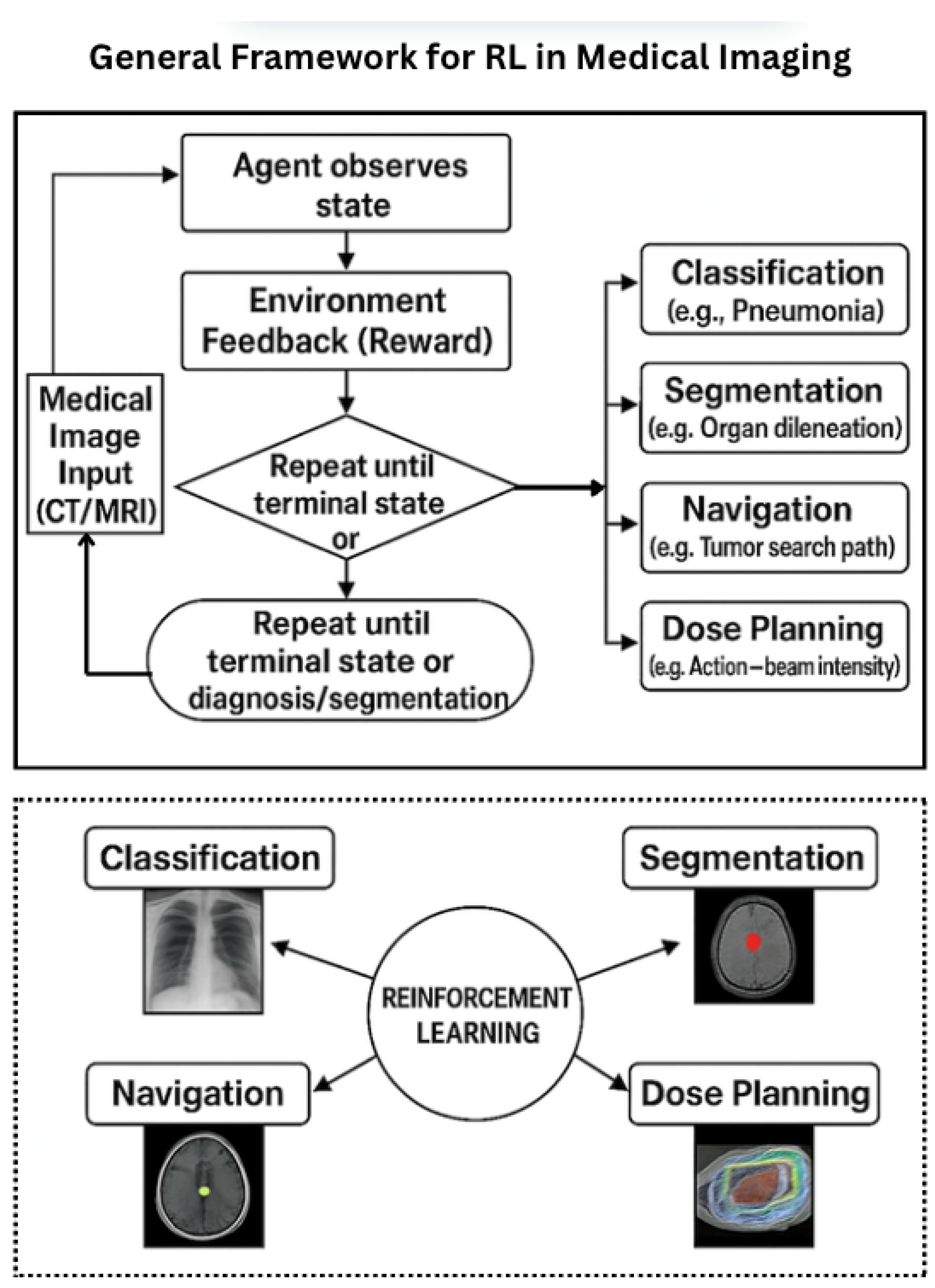
Figure 2 presents a generalized architecture for reinforcement learning in medical imaging, emphasizing key components such as agent–environment interaction and reward-driven learning.

3.4. Problem Formulation: Pneumonia Detection in Chest X-Rays

Pneumonia detection in chest X-ray (CXR) images has traditionally relied on supervised learning methods that require large volumes of annotated data. Although these models—particularly Convolutional Neural Networks (CNNs)—have demonstrated strong performance, they face notable limitations in generalizing to unseen distributions, handling label noise, and ensuring interpretability [12,31].
To overcome these shortcomings, recent studies have proposed reformulating pneumonia detection as a sequential decision-making problem amenable to reinforcement learning (RL). In this framework, an RL agent actively explores the image, accumulates information over time, and makes classification decisions in a manner that mirrors a radiologist’s diagnostic workflow [7]. The following terminologies are used to describe this formulation.
State Space ( S ): Represents the environment observed by the agent, which could include raw CXR pixel data, intermediate CNN feature maps, or localized patches within the image.
Action Space ( A ): Defines the agent’s options at each step, including the following:
  • Navigational actions (e.g., move left/right/up/down and zoom).
  • Terminal actions (e.g., classify as pneumonia/normal and defer to human).
Reward Function ( R ): Encourages the agent to make accurate and efficient diagnoses:
  • +1 for correct classification;
  • −1 for incorrect predictions;
  • Small penalties for unnecessary steps to encourage efficiency.
Policy ( π : S A ): Learned through trial-and-error to map observations to optimal actions using DRL algorithms such as Deep Q-Networks (DQNs) or Proximal Policy Optimization (PPO) [23,25]. Building upon this foundational framework, Figure 3 illustrates a task-specific adaptation for pneumonia detection in chest X-rays, demonstrating how RL can emulate clinical decision-making through sequential image analysis.
This RL-based framework provides the following benefits over traditional classification systems:
  • Localized Attention: Focuses on clinically significant regions instead of full-frame processing.
  • Interpretability: Enables traceable decision paths aligned with clinical workflows.
  • Data Efficiency: Operates under semi-supervised or weakly labeled conditions.
  • Adaptive Computation: Adjusts exploration dynamically based on image complexity.
Clinical Implication This decision-driven structure is particularly well suited for real-time, interpretable, and interactive AI applications in clinical imaging. By emulating the sequential reasoning patterns of human clinicians, reinforcement learning (RL) offers a robust alternative to conventional passive inference models—particularly in scenarios marked by uncertainty, sparse feedback, or the need for dynamic exploration [9].
Overall, RL provides a compelling framework for developing goal-directed, explainable, and sample-efficient AI systems that can seamlessly integrate clinical feedback and operate effectively in real-world healthcare environments. In this paradigm, the agent observes the image, performs navigational actions, receives feedback, and ultimately makes a diagnosis. These capabilities are critical for enabling the deployment of next-generation intelligent medical imaging solutions [5].

4. Taxonomy of AI-Based Reinforcement Learning in Medical Imaging

To better understand the integration of reinforcement learning (RL) into medical imaging, we present a multi-dimensional taxonomy defined along four primary axes: imaging tasks, RL algorithms, imaging modalities, and learning frameworks. This structured classification provides a systematic view of how RL techniques are tailored to diverse clinical imaging applications [9].

4.1. By Imaging Task

RL has been utilized in a broad range of medical imaging tasks, including the following:
  • Segmentation: Agents iteratively refine the boundary of organs or lesions, e.g., in brain MRI or ultrasound [32].
  • Detection and Localization: RL agents navigate 3D scans to identify key anatomical landmarks or pathologies [33].
  • Classification: RL policies guide active learning, view selection, or confidence calibration in diagnosis tasks [32].
  • Image Registration: Agents learn transformation policies to align multimodal or time-series images [34].
  • Image Synthesis: RL controls adaptive denoising or contrast enhancement in dynamic imaging [35].
  • Treatment Planning: In radiotherapy, RL optimizes beam angles and dose distributions [36].

4.2. By Reinforcement Learning Algorithm

Different RL paradigms, such as the following, are adopted based on problem complexity and dimensionality [9]:
  • Tabular Methods: Classical Q-learning is used in simpler settings [22].
  • Deep RL: Methods like DQN, PPO, and A3C are common in high-dimensional visual tasks [6].
  • Model-Based RL: Surrogate environment models enhance sample efficiency [37].
  • Multi-Agent RL: Applied in cooperative detection, segmentation, or dose-planning tasks [38].

4.3. By Imaging Modality

RL has been applied across several modalities, including the following:
  • Ultrasound: Real-time landmark detection and prostate segmentation [39].
  • MRI: Used in neurological or musculoskeletal applications for segmentation and registration [40].
  • CT: Applied in volumetric analysis for lesion detection or navigation [33].
  • PET/SPECT: Supports functional synthesis and multimodal alignment [41].
  • X-ray: Utilized in view planning and active lesion search [5].

4.4. By Learning Framework

RL training strategies also vary depending on clinical constraints as shown in the following:
  • Offline RL: Leverages static annotated datasets; preferred for safety and reproducibility [9].
  • Online RL: Involves real-time interaction with simulators or clinical environments [42].
  • Human-in-the-Loop: Clinician feedback informs agent policies [43].
  • Imitation Learning: Behavior cloning or inverse RL captures expert strategies [44].

4.5. Visual Summary

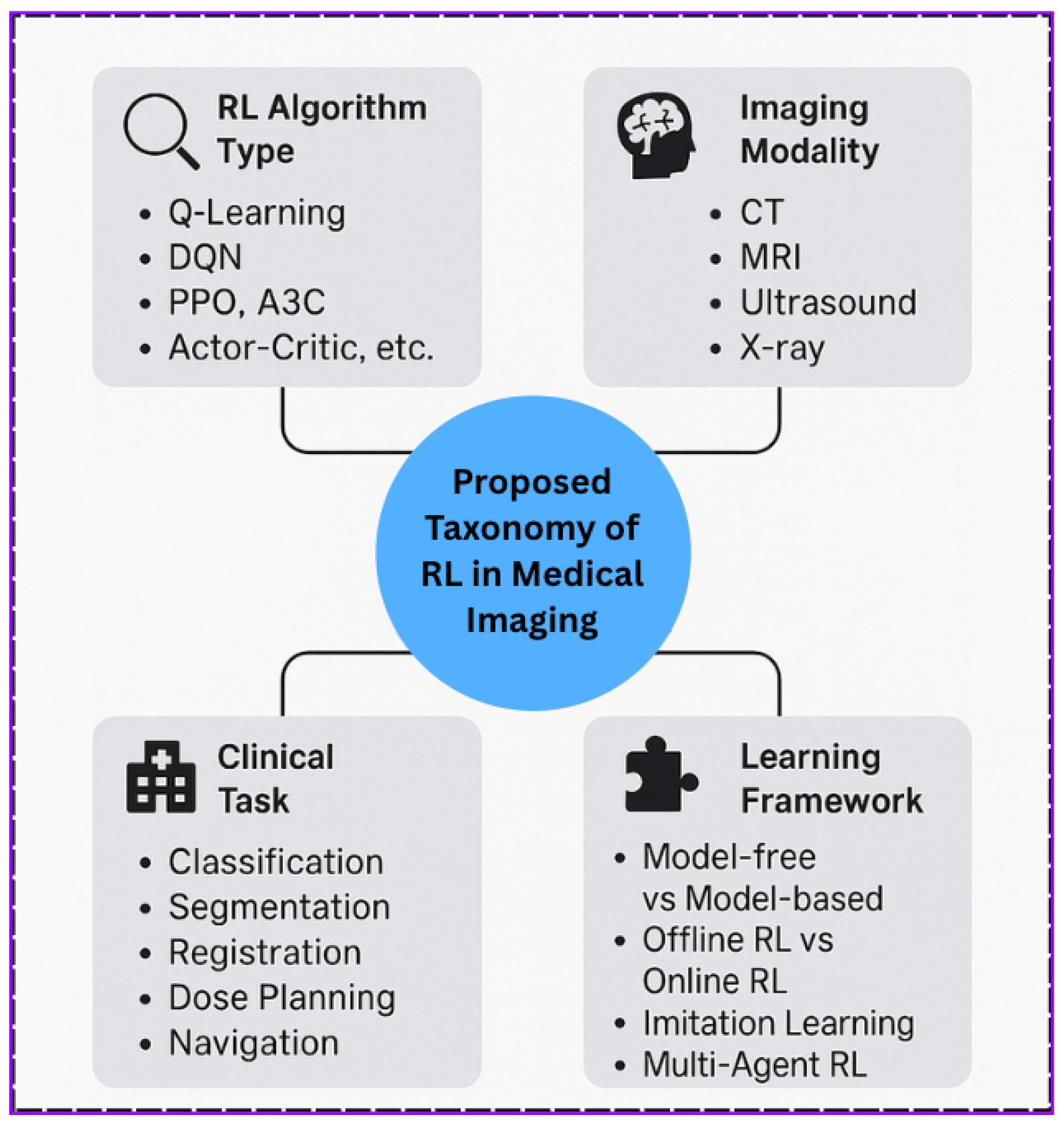
Figure 4 presents a visual summary of the taxonomy, linking tasks, RL algorithms, modalities, and learning frameworks.

4.6. Advantages and Research Gap Bridging

This taxonomy systematically addresses major gaps identified in prior literature by providing a multi-dimensional lens for analyzing reinforcement learning (RL) in medical imaging. While existing surveys often concentrate narrowly on algorithms or specific tasks, they frequently overlook the integration of clinical contexts, data constraints, and modality-specific challenges [9]. Our taxonomy bridges this fragmentation through four orthogonal axes—tasks, RL techniques, imaging modalities, and learning paradigms—thereby enabling comprehensive comparison and facilitating the identification of underexplored intersections. In particular, it highlights the following critical gaps:
  • Modality-Specific RL Strategies: Few studies tailor RL techniques to the unique temporal, spatial, and resolution characteristics of modalities like PET or ultrasound. Our taxonomy encourages targeted algorithm design [41].
  • Underutilization of Model-Based and Multi-Agent RL: Despite their potential for sample efficiency and collaboration, these algorithms remain underrepresented in imaging tasks beyond treatment planning. The taxonomy surfaces these areas for future exploration [38].
  • Lack of Integration Between Human Expertise and RL Systems: While offline and imitation learning are growing, human-in-the-loop frameworks are rarely applied in real-time settings, especially in high-stakes domains like radiotherapy [43].
  • Imbalanced Coverage of Imaging Tasks: Tasks like image synthesis and adaptive contrast enhancement receive significantly less RL attention compared to classification or segmentation. Our structure exposes these disparities [45].

4.7. Taxonomy Construction and Counting Methodology

To ensure robustness and reproducibility, studies were counted using predefined inclusion and exclusion criteria. Papers were retrieved from PubMed, IEEE Xplore, SpringerLink, and arXiv (Jan 2019–July 2025) using the query “Reinforcement Learning” + “Medical Imaging” (Table 3). Works that did not directly apply RL to imaging or were duplicate preprints were excluded. Each study was mapped across three axes: modality (CT, MRI, Ultrasound, and X-ray), task (segmentation, classification, and registration), and algorithm family (e.g., DQN, PPO, A3C, and model based). Two independent reviewers validated the mapping. The aggregated results were visualized as a heatmap (Figure 5), revealing both well-studied intersections (e.g., deep RL in chest X-ray classification) and underexplored areas (e.g., multi-agent RL in dental imaging). This systematic process provides a transparent and quantitative foundation for the taxonomy.
Our heatmap analysis (Figure 5), derived from studies published between 2019 and 2025 and systematically mapped across the modality, task, and algorithm family, highlights notable imbalances in RL research. While chest X-ray (CXR) and MRI are frequently paired with DQN or PPO, underexplored intersections—such as multi-agent RL in elastography and ophthalmic imaging—remain largely absent. By visualizing these patterns, the taxonomy provides a transparent and quantitative roadmap for future research, guiding interdisciplinary efforts toward clinically relevant, interpretable, and safe RL-based systems. This aligns with recent meta-reviews that emphasize the need for broader, task-contextual frameworks in healthcare [43].

5. Trend Analysis and Future Outlook

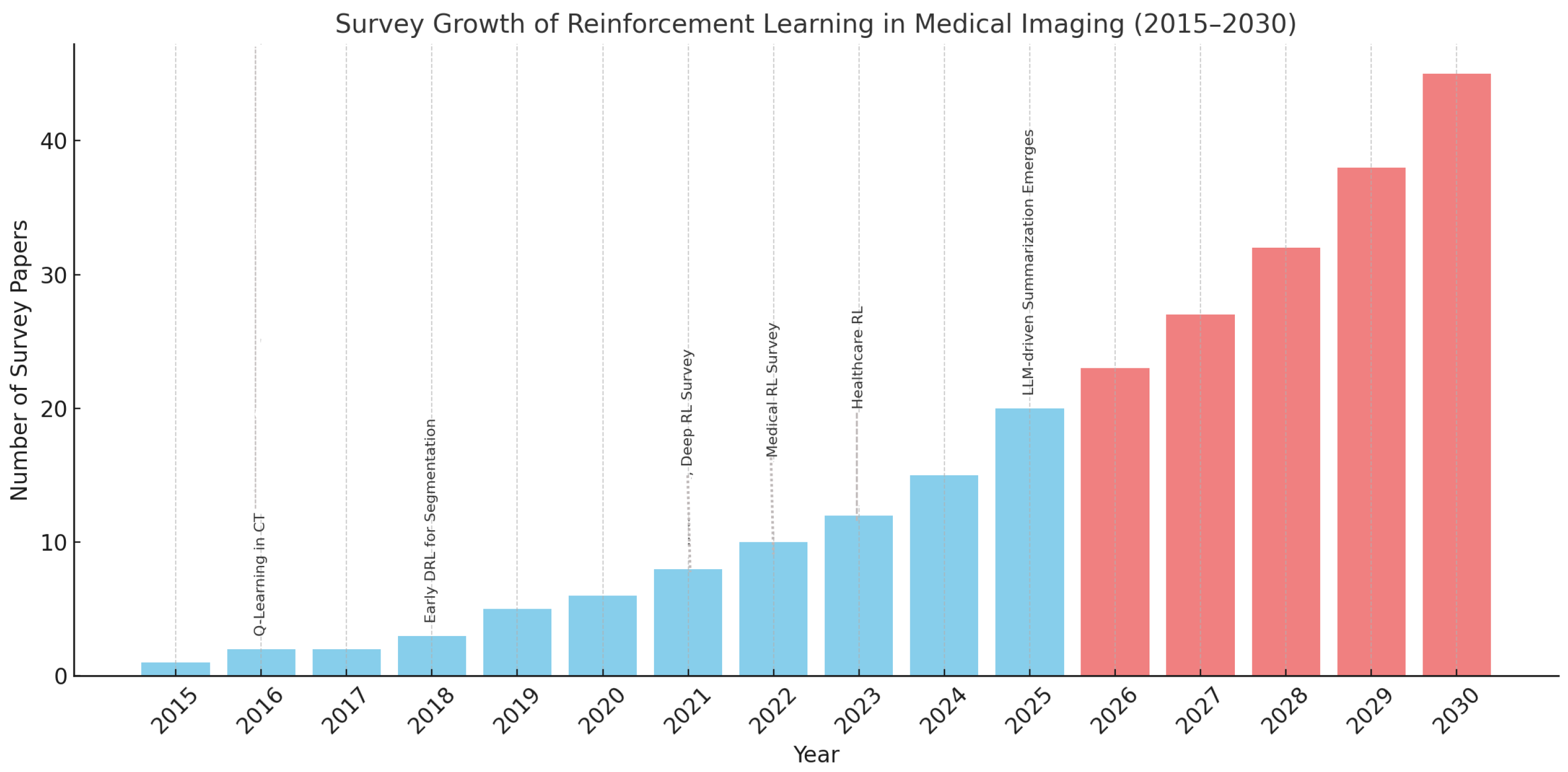
To contextualize the proposed taxonomy, we conducted a decade-long analysis (2015–2025) of survey literature on reinforcement learning (RL) in medical imaging. Figure 6 illustrates the rapid increase in RL survey publications, with projected growth extending to 2030 based on the current momentum and advances in AI tooling.

5.1. Decade-Long Survey Trends

A growing number of reviews have documented the evolution of RL methods for clinical imaging tasks. These span from classical Q-learning to modern DRL strategies (e.g., PPO and A3C) across various modalities. Table 4 lists the year-wise counts, including milestone works such as Zhou et al. (2021) and Hu et al. (2023) [9].

5.2. Projections (2026–2030): The Role of LLMs

With the advent of Large Language Models (LLMs), we forecast a substantial rise in survey generation. These models can perform the following:
  • Extract modality-task-algorithm mappings automatically from the literature.
  • Suggest underexplored combinations using structured prompts.
  • Co-author survey drafts with human experts for rapid dissemination.
This trend aligns with the broader AI-augmented scientific workflow, signaling that taxonomy-driven LLM pipelines could become standard for future literature synthesis and gap analysis.

5.3. The Role of LLMs in Accelerating This Growth

The emergence of Large Language Models (LLMs) such as GPT-4, Claude, and PaLM introduces a transformative opportunity for accelerating research synthesis in reinforcement learning and medical imaging. By automating labor-intensive processes such as literature review, taxonomy generation, and summarization, LLMs can significantly scale the volume and depth of survey publications.
These 25+ survey papers reflect a rising curve of complexity, clinical relevance, and interdisciplinarity. Yet, they also expose persistent gaps in underexplored axes such as the following:
  • Model-based RL: Rarely applied in high-dimensional medical scenarios.
  • Multi-agent Systems: Underrepresented in cooperative imaging tasks.
  • Human-in-the-loop Frameworks: Limited adoption in clinical settings.
  • Modality Bias: Sparse exploration in PET/SPECT, ultrasound, and temporal imaging.
These insights not only validate the utility of our proposed taxonomy but also provide a blueprint for aligning future work with unmet needs and the capabilities of LLMs in enhancing survey production, shown in Table 5.
This collaborative workflow not only democratizes access to comprehensive literature reviews but also empowers junior researchers, domain clinicians, and cross-disciplinary teams to contribute meaningfully to RL-based imaging literature. LLMs will likely become standard components of scientific writing and discovery pipelines in the next phase of AI-augmented research [48].

5.4. Decade-Long Survey Trends

A comprehensive review of RL in medical imaging illustrates marked growth from 2015 to 2025. Early work (2015–2018) primarily explored episodic task formulation, often using tabular Q-learning in constrained domains. However, from 2019 onward, Deep RL methods (e.g., DQN, PPO, and A3C) enabled more sophisticated applications, including 3D landmark localization, medical image segmentation, and cross-modal registration [9]. Table 5 highlights specific capabilities of LLMs and their contributions to future survey growth.
By 2021, Zhou et al.’s comprehensive review [6] offered a foundational summary of DRL models used across diagnosis, optimization, and treatment planning in medical imaging. Between 2022 and 2023, more specialized surveys emerged: for instance, Hu et al.’s Journal of Applied Clinical Medical Physics article contextualized RL systems with clinical deployment pipelines [9]. At the same time, focused reviews in ultrasound imaging illustrated how RL spans the full pipeline—from data preparation and simulator-based training to model validation and evaluation [49].

5.5. Discussion: Application and Impact of RL Taxonomy in Medical Imaging

Existing taxonomies of reinforcement learning (RL) in medical imaging often lack integration across task-specific design, modality diversity, and automation support, thereby limiting their applicability in real-world, high-accuracy imaging workflows. The proposed taxonomy addresses these limitations by embedding task-aligned design patterns and incorporating LLM-guided automation, ultimately enhancing precision and adaptability in medical image processing [9].
The proposed taxonomy (see Figure 7) serves as a comprehensive framework that categorizes reinforcement learning (RL) in medical imaging across four key axes: RL algorithms, imaging modalities, learning frameworks, and clinical tasks. This structured approach supports research, development, and education by guiding decision-making, identifying gaps, and promoting automation.

5.6. Design Guidance and Benchmarking

The proposed taxonomy supports systematic design–space exploration in RL-based medical imaging. For example, segmentation tasks often employ Dice- or Jaccard-based rewards, with DQN or actor–critic policies preferred for maintaining spatial consistency [50]. This framework enables researchers to perform the following tasks:
  • Select suitable RL algorithms based on task and modality characteristics;
  • Design modular agents with well-defined state, action, and reward functions;
  • Align with reproducible benchmarks across common clinical tasks.

5.7. Identification of Research Gaps

Mapping studies onto this taxonomy reveals underexplored intersections [9]:
  • Model-based RL for PET/SPECT imaging;
  • Human-in-the-loop strategies for image registration;
  • Multi-agent RL in real-time segmentation scenarios.
Addressing these gaps presents opportunities to extend RL frameworks into less charted but clinically relevant territories.

5.8. Automation via Large Language Models (LLMs)

Integrating the taxonomy with the following LLM capabilities can significantly streamline survey creation and experimentation [9]:
  • Survey Synthesis: Automate the extraction of reward-policy-state designs from large literature sets.
  • Trend Analysis: Detect shifts in task or modality focus over time.
  • Content Drafting: Generate LaTeX figures, tables, and summaries.
  • Gap Identification: Highlight underrepresented algorithm–modality combinations.

5.9. Impact and Future Potential

The impact and future potential of the taxonomy can drive the following:
  • Curriculum Development: A foundation for training in RL and clinical AI.
  • Framework Standardization: Promotes interoperable RL components across applications.
  • Collaborative Research: Bridges AI researchers, clinicians, and imaging specialists.

5.10. Challenges and Future Outlook

RL in medical imaging continues to face persistent challenges, including small and imbalanced datasets, high-dimensional action spaces, limited cross-modality generalization, and sparse or weak supervision that undermines training stability. Among these, reward function design remains central, as it encodes clinical objectives and constrains agent behavior. Unlike generic RL domains, healthcare rewards must balance technical performance with medical standards of safety, interpretability, and clinical utility. Consequently, reward formulations vary across applications: radiotherapy planning emphasizes dose optimization, lesion detection prioritizes localization accuracy, and navigation tasks balance trajectory efficiency with safety. Table 6 summarizes representative designs and their validation strategies, including expert-in-the-loop scoring, inter-rater agreement (e.g., Cohen’s Kappa), and policy audit trails, thereby ensuring that RL systems remain clinically aligned.
Beyond comparing task-specific reward designs, ensuring clinical alignment remains critical. Validation strategies include expert-in-the-loop scoring for direct domain oversight, Cohen’s Kappa to quantify agreement between RL agents and clinicians, and policy audit trails to document decision pathways for reproducibility and retrospective review. Collectively, these methods provide systematic safeguards against misaligned or unsafe RL policies, thereby reinforcing the clinical reliability of reward function design.

6. LLM Integration for Explainability and Semantic Reinforcement

Building on privacy-preserving RL frameworks enhanced by visual encoders and Federated Learning, we now examine how Large Language Models (LLMs) can further advance explainability and semantic alignment in medical AI. By generating natural-language rationales for each agent action, LLMs—particularly when prompted with Chain-of-Thought techniques—provide clear, human-readable justifications for decisions. This additional layer of semantic reinforcement not only ensures compliance with ethical standards such as HIPAA [57] and regulatory frameworks like GDPR but also strengthens clinician trust and interpretability in edge- and federated-trained models. Recent work [58] demonstrates that combining LLM-generated explanations with reinforcement learning policy logs yields a robust hybrid approach well-suited for real-world, compliance-aware medical imaging systems [59,60,61].

Conceptual Roles of LLMs in RL for Medical Imaging

Recent advances highlight the expanding role of RL in multimodal fusion for clinical imaging. Jiang et al. [62] applied RL for fetal plane localization in ultrasound, while Qi et al. [63] developed a PET/CT fusion model for tumor segmentation with uncertainty. Other studies extended RL to CT with pathology for survival prediction [64] and mpMRI with 18F-PSMA-PET/CT for prostate cancer staging [65], demonstrating how fusion across modalities enhances robustness and clinical relevance.
Complementing these advances, this survey outlines how Large Language Models (LLMs) can conceptually support RL pipelines in medical imaging. As shown in Figure 8, LLMs contribute via pipeline automation, annotation and reporting, cross-modal integration, and clinical decision support. Table 7 links these roles to tasks, benefits, and references [47,66,67].

7. Privacy Preserving in Medical RL

Privacy-preserving reinforcement learning is increasingly viable in healthcare. Lee et al. and Pati et al. [68,69] demonstrated multi-hospital federated RL for tumor detection without sharing sensitive data. In our ChestX-ray14 experiment [70], strong Differential Privacy noise ( ϵ < 1.0 ) reduced accuracy by ∼3.2%, illustrating the trade-off between privacy and performance. Similar trends appear in production frameworks such as NVIDIA Clara FL [71], which enables HIPAA-compliant deployments but requires careful calibration to avoid impairing clinical utility.
When applying reinforcement learning (RL) to clinical imaging data, privacy preservation is essential to meet regulatory standards such as HIPAA. One promising approach is Differential Privacy (DP), which ensures that the contribution of any individual data point remains indistinguishable within the learning process. A widely used method is Differentially Private Stochastic Gradient Descent (DP-SGD) [72], where noise is added to model updates to obscure sensitive patient-level signals.
Although DP-SGD has demonstrated strong utility in supervised learning settings, its integration into medical RL remains underexplored. Future studies should investigate privacy-preserving RL agents capable of learning robust policies from sensitive imaging data while providing formal privacy guarantees. Such advancements are especially relevant for Federated Learning systems, cross-institutional studies, and deployment in real-world clinical environments.

7.1. Privacy-Preserving RL in Clinical Environments

Given the sensitive nature of healthcare data, privacy-preserving mechanisms are essential when deploying RL in clinical environments. Federated reinforcement learning offers a promising solution by enabling distributed training across institutions without exposing patient data [30]. Complementary techniques such as Differential Privacy and secure multi-party computation (SMPC) further enhance data protection by adding noise or encrypting intermediate results. These privacy-aware approaches align with compliance frameworks like HIPAA and GDPR while preserving model utility, making them increasingly important for real-world deployment of RL models in healthcare.

7.2. Federated Reinforcement Learning for Cross-Institutional Collaboration

Federated Reinforcement Learning (FRL) facilitates privacy-preserving policy training across multiple hospitals by enabling local RL agents to learn from private imaging datasets without sharing raw data [30,73,74]. Instead, only model parameters or gradients—optionally encrypted—are periodically transmitted to a central server for secure aggregation and policy refinement (see Figure 9).
This architecture is particularly well-suited for medical imaging applications where patient data is siloed across institutions with diverse imaging protocols, devices, and populations. FRL aligns with regulatory requirements such as HIPAA and GDPR, enabling collaborative model development while preserving patient confidentiality and institutional autonomy.

7.3. Summary of Design Considerations and Privacy Strategies

While this survey does not conduct empirical experimentation, we outline a hypothetical privacy-aware reinforcement learning (RL) design framework that integrates Differential Privacy (DP) and Federated Learning (FL) to support ethical AI development in healthcare [72,75,76].

7.4. Privacy-Enhancing Strategies

To safeguard sensitive patient data, DP can be applied during policy updates by injecting calibrated noise into gradients, mitigating the risk of memorizing individual features [72]. Meanwhile, FL enables decentralized training across clinical institutions, allowing local models to update independently while aggregating encrypted model parameters at a central server [30,73]. This design aligns with HIPAA and GDPR standards.

7.5. Conceptual Framework Blueprint

A conceptual RL pipeline for privacy-aware medical image analysis could include the following (Table 8):
  • Reward Function: Binary feedback system with +1 for correct, 1 for incorrect classifications.
  • RL Algorithms: Deep Q-Network (DQN) and Proximal Policy Optimization (PPO) for learning and stability comparisons.
  • Feature Encoder: Pretrained ResNet-18 for visual feature extraction.
  • LLM Integration: Post-decision explanation using large language models (LLMs), such as GPT, to enhance interpretability [66].
  • Privacy Controls: DP-SGD and FL to ensure secure and compliant learning environments.

7.6. Evaluation Criteria

Although not implemented here, the hypothetical design could be evaluated using the following:
  • RL Dynamics: Reward trajectory, episode length, convergence behavior.
  • Diagnostic Accuracy: AUROC, Precision, Recall, F1-score.
  • Interpretability: SHAP, LIME, and GPT-generated rationales.
  • Privacy Robustness: Performance degradation under DP and FL conditions.
This framework outlines a pathway for future implementations of RL in healthcare, balancing diagnostic performance with interpretability and privacy preservation in cross-institutional environments.

8. Challenges and Limitations

Despite the growing promise of reinforcement learning (RL) in medical imaging, translating RL-based frameworks from research to real-world clinical environments presents substantial challenges. This section outlines the core technical, methodological, and operational barriers that must be addressed to ensure their safe, interpretable, and scalable adoption in healthcare settings.

8.1. Reward Design and Clinical Alignment

Defining clinically meaningful reward functions remains a fundamental challenge in medical RL. Unlike well-structured environments, medical tasks often involve sparse or delayed rewards, which complicates their alignment with diagnostic or therapeutic objectives. Common proxies—such as Dice coefficients or overlap metrics—may fail to capture the subtleties of real-world clinical priorities, leading to unintended policy behaviors or clinically irrelevant optimizations. While multi-objective reward shaping offers a promising solution, it demands substantial domain expertise and careful calibration to balance competing goals such as lesion coverage and tissue preservation [77].

8.2. Sample Inefficiency and Training Instability

RL algorithms are notoriously sample inefficient, particularly in high-dimensional medical domains where interactions are costly and annotated feedback is limited. Training agents on complex modalities (e.g., 3D CT or multi-phase MRI) often requires thousands of episodes, which is impractical without scalable simulators or synthetic datasets. Furthermore, deep RL suffers from instability and non-convergence caused by function approximation errors, exploration–exploitation imbalance, and reward sparsity, raising significant reliability concerns in safety-critical healthcare applications [67].

8.3. Generalization and Domain Transfer

Generalizing RL models beyond their training distributions remains an open research challenge. Many agents overfit to specific scanners, patient cohorts, or imaging protocols, which limits their clinical utility. Cross-institutional transfer is further impeded by domain shifts, annotation variability, and heterogeneous acquisition parameters. While supervised learning has made progress through domain adaptation and transfer learning, analogous techniques for RL remain underexplored and demand stronger theoretical foundations and empirical validation [78].

8.4. Interpretability and Clinical Trust

Interpretability is critical in healthcare AI, yet most RL systems operate as opaque decision-makers. Clinicians require transparent rationales for automated recommendations—particularly in sequential tasks such as treatment planning or lesion localization [79]. In the absence of interpretable visualizations or policy traceability, clinical trust in RL systems remains limited. Emerging techniques, including policy saliency maps and trajectory-based summaries, offer promising directions but remain at an early stage compared to the well-established explainability ecosystem in supervised learning [80].

8.5. Data Availability and Interactive Environments

Unlike domains such as robotics or gaming, medical RL suffers from limited access to interactive environments. Most publicly available datasets lack temporal structure, interactive APIs, or simulation support [6]. Developing synthetic patient environments or radiology simulators is resource intensive and requires expert-validated feedback mechanisms. The absence of standardized RL-ready medical datasets further hinders reproducibility and slows research progress [81].

8.6. Regulatory and Deployment Barriers

Even high-performing RL models face steep regulatory barriers prior to clinical deployment. Agencies such as the FDA and EMA mandate rigorous standards for interpretability, robustness, and post-deployment monitoring—criteria that remain undefined for adaptive or online-learning agents [82,83]. Beyond regulatory approval, successful integration into hospital workflows requires compatibility with existing PACS and IT infrastructure, dedicated engineering support, clinician training, and assurances of safe real-time behavior, particularly in safety-critical domains such as oncology and emergency imaging.

9. Open Research Directions

While reinforcement learning (RL) in medical imaging has made notable strides, several research avenues remain underexplored. Addressing these could accelerate safe, explainable, and clinically aligned AI integration [67].

9.1. Human-in-the-Loop Reinforcement Learning (HITL RL)

Incorporating human expertise into reinforcement learning (RL) frameworks is critical for safety-sensitive domains such as medical imaging. Human-in-the-loop (HITL) RL enables clinicians and domain experts to guide agents through corrective feedback, reward shaping, or decision validation during training [47]. This collaborative interaction can accelerate convergence, mitigate sample inefficiency, and promote clinically meaningful policy development. Approaches such as imitation learning, inverse reinforcement learning, and preference-based feedback are particularly effective when expert knowledge is available but ground-truth annotations are scarce. While the inclusion of human oversight is essential for ensuring clinical reliability, future work should further investigate the systematic integration of HITL strategies with RL to enhance robustness and real-world applicability.

9.2. Explainability in RL for Medical Imaging

A persistent challenge in applying reinforcement learning (RL) to medical imaging is the lack of explainability. RL agents often function as black-box models, making it difficult to interpret the rationale behind their actions—such as localizing a lesion or navigating within a 3D imaging volume—which is critical for both clinical adoption and regulatory approval [67]. Current strategies to enhance interpretability include policy trajectory visualization, saliency-based mapping, and attention-guided explanation layers. More recent approaches explore the integration of rule-based symbolic reasoning and post hoc model analysis, aiming to provide transparent justifications and ensure accountability in diagnosis-critical settings. These methods are essential for building physician trust, supporting regulatory compliance, and ultimately enabling safe deployment of RL systems in real-world clinical workflows.

9.3. Model-Based and Sample-Efficient RL

Current model-free reinforcement learning (RL) algorithms suffer from high sample complexity, limiting their practicality in medical imaging domains where annotated data is costly and patient interactions are restricted. Model-based RL (MBRL) addresses this limitation by explicitly learning environment dynamics, thereby enabling internal rollouts and improved planning with fewer real samples. Recent studies further suggest that incorporating anatomy-aware priors and generative models can substantially enhance simulation fidelity, making MBRL particularly well-suited for imaging tasks that demand structural realism and clinical plausibility [46].

9.4. Hierarchical and Modular Policies

Medical workflows are inherently hierarchical, encompassing tasks such as detection, segmentation, and diagnosis. Hierarchical reinforcement learning (HRL) addresses this complexity by decomposing high-level objectives into modular subtasks, thereby improving both learning efficiency and interpretability [84]. Future research should explore anatomically guided HRL pipelines that explicitly leverage task modularity, enabling cross-task generalization and fostering clinically aligned automation in multi-stage imaging workflows.

9.5. Explainability and Policy Transparency

Trustworthy clinical AI demands interpretable RL agents (Figure 10). However, most existing methods act as black boxes. Future work should develop the following:
  • Saliency-based policy attention maps.
  • Action-region heatmaps for anatomical interpretability.
  • Trajectory visualizations aligned with clinical annotations.
Emerging directions include integrating causal reasoning and uncertainty-aware RL to improve safety and trust [85].

9.6. Interactive and Human-in-the-Loop RL

Collaborative learning via expert-in-the-loop RL allows agents to refine policies from limited data and adapt to real-world clinical workflows. Interactive RL setups, including clinician feedback during radiology or surgery, can guide real-time policy corrections [54].

9.7. Benchmarks and Simulation Environments

The absence of standardized RL-ready datasets impedes reproducibility. To promote consistency, future work should focus on the following:
  • Open-source task-specific simulators for medical RL.
  • RL-ready CXR and CT datasets with feedback structures.
  • Public leaderboards for tasks like landmark localization or tool tracking.
Community-driven platforms akin to OpenAI Gym or MONAI-RL [86] can serve as a foundation for benchmarking progress.

9.8. Clinical Integration and Continual Learning

Current RL systems lack real-world clinical integration. Future RL pipelines should interface with PACS, radiology viewers, and treatment planning systems. Continual RL, with privacy-preserving and safety-aware online learning mechanisms, could support long-term adaptation to shifting patient populations and imaging modalities [67].

10. Conclusions

Reinforcement learning (RL) has emerged as a transformative paradigm in medical image analysis, offering adaptive, sequential, and feedback-driven decision-making capabilities. This paper provides a comprehensive overview of RL applications across critical clinical tasks—including segmentation, classification, registration, and treatment planning—while proposing a structured taxonomy to systematically categorize existing methods.
Despite notable progress, several challenges hinder real-world deployment. These include unstable training dynamics, limited cross-domain generalization, reward ambiguity, interpretability gaps, and stringent regulatory requirements. Additionally, the scarcity of interactive datasets and the absence of standardized evaluation benchmarks continue to impede progress.
To overcome these limitations, we highlight key research priorities: principled reward design, sample-efficient learning strategies, policy explainability, and privacy-preserving frameworks such as federated RL. Addressing these areas will demand close collaboration between AI researchers, clinicians, and regulatory authorities to ensure both technical rigor and clinical safety.
In conclusion, RL holds substantial promise for advancing intelligent, interpretable, and clinically viable imaging systems. With deliberate design choices and interdisciplinary cooperation, RL can serve as a cornerstone for developing safe, personalized, and transparent AI solutions in healthcare.

Funding

The work is supported by the National Science Foundation under NSF Grant #2433800, #1946442, #2100134, and NIH Grant #5R42LM014356-03. Any opinions, findings, recommendations, expressed in this material are those of the authors and do not necessarily reflect the views of the National Science Foundation.

Conflicts of Interest

The authors declare no conflict of interest.

References

  1. Islam Riad, A.B.M.K.; Barek, M.A.; Rahman, M.M.; Akter, M.S.; Islam, T.; Rahman, M.A.; Mia, M.R.; Shahriar, H.; Wu, F.; Ahamed, S.I. Enhancing HIPAA Compliance in AI-driven mHealth Devices Security and Privacy. In Proceedings of the 2024 IEEE 48th Annual Computers, Software, and Applications Conference (COMPSAC), Osaka, Japan, 2–4 July 2024; pp. 2430–2435. [Google Scholar] [CrossRef]
  2. Pinto-Coelho, L. How Artificial Intelligence Is Shaping Medical Imaging Technology: A Survey of Innovations and Applications. Bioengineering 2023, 10, 1435. [Google Scholar] [CrossRef]
  3. Chen, X.; Wang, X.; Zhang, K.; Fung, K.; Thai, T.C.; Moore, K.; Mannel, R.S.; Liu, H.; Zheng, B.; Qiu, Y. Recent Advances and Clinical Applications of Deep Learning in Medical Image Analysis. Med. Image Anal. 2021, 79, 102444. [Google Scholar] [CrossRef]
  4. Feng, J.; Phillips, R.V.; Malenica, I.; Bishara, A.; Hubbard, A.E.; Celi, L.A.; Pirracchio, R. Clinical Artificial Intelligence Quality Improvement: Towards Continual Monitoring and Updating of AI Algorithms in Healthcare. npj Digit. Med. 2022, 5, 66. [Google Scholar] [CrossRef]
  5. Alansary, A.; Oktay, O.; Li, Y.; Folgoc, L.L.; Hou, B.; Vaillant, G.; Kamnitsas, K.; Vlontzos, A.; Glocker, B.; Kainz, B.; et al. Evaluating reinforcement learning agents for anatomical landmark detection. Med. Image Anal. 2019, 53, 156–164. [Google Scholar] [CrossRef]
  6. Zhou, S.K.; Le, H.N.; Luu, K.; Nguyen, H.V.; Ayache, N. Deep Reinforcement Learning in Medical Imaging: A Literature Review. Med. Image Anal. 2021, 70, 102193. [Google Scholar] [CrossRef]
  7. Hu, J.; Luo, Z.; Wang, X.; Sun, S.; Yin, Y.; Cao, K.; Song, Q.; Lyu, S.; Wu, X. End-to-End Multimodal Image Registration via Reinforcement Learning. Med. Image Anal. 2022, 68, 101878. [Google Scholar] [CrossRef]
  8. Ebrahimi, S.; Lim, G. A Reinforcement Learning Approach for Finding Optimal Policy of Adaptive Radiation Therapy Considering Uncertain Tumor Biological Response. Artif. Intell. Med. 2021, 121, 102193. [Google Scholar] [CrossRef]
  9. Hu, M.; Zhang, J.; Matkovic, L.; Liu, T.; Yang, X. Reinforcement Learning in Medical Image Analysis: Concepts, Applications, Challenges, and Future Directions. J. Appl. Clin. Med. Phys. 2023, 24, e13898. [Google Scholar] [CrossRef] [PubMed]
  10. Al, W.A.; Yun, I.D. Partial Policy-based Reinforcement Learning for Anatomical Landmark Localization in 3D Medical Images. arXiv 2018, arXiv:1807.02908. [Google Scholar] [CrossRef]
  11. Zhang, C.; Shahriar, H.; Riad, A.B.M.K. Security and Privacy Analysis of Wearable Health Device. In Proceedings of the 2020 IEEE 44th Annual Computers, Software, and Applications Conference (COMPSAC), Madrid, Spain, 13–17 July 2020; pp. 1767–1772. [Google Scholar] [CrossRef]
  12. Litjens, G.; Kooi, T.; Bejnordi, B.E.; Setio, A.A.A.; Ciompi, F.; Ghafoorian, M.; van der Laak, J.A.; van Ginneken, B.; Sánchez, C.I. A Survey on Deep Learning in Medical Image Analysis. Med. Image Anal. 2017, 42, 60–88. [Google Scholar] [CrossRef]
  13. Shen, D.; Wu, G.; Suk, H.-I. Deep Learning in Medical Image Analysis. Annu. Rev. Biomed. Eng. 2017, 19, 221–248. [Google Scholar] [CrossRef]
  14. Saha, B.; Islam, M.S.; Riad, A.K.; Tahora, S.; Shahriar, H.; Sneha, S. BlockTheFall: Wearable Device-based Fall Detection Framework Powered by Machine Learning and Blockchain for Elderly Care. In Proceedings of the 2023 IEEE 47th Annual Computers, Software, and Applications Conference (COMPSAC), Torino, Italy, 26–30 June 2023; pp. 1412–1417. [Google Scholar] [CrossRef]
  15. Luo, Z.; Hu, J.; Wang, X.; Hu, S.; Kong, B.; Yin, Y.; Song, Q.; Wu, X.; Lyu, S. Stochastic Planner–Actor–Critic (SPAC) for Unsupervised Deformable Image Registration. Proc. AAAI Conf. Artif. Intell. 2022, 36, 1917–1925. [Google Scholar]
  16. Sahba, F.; Tizhoosh, H.R.; Salama, M.M.A. Application of Reinforcement Learning for Medical Image Segmentation. In Proceedings of the International Joint Conference on Neural Networks (IJCNN), Vancouver, BC, Canada, 16–21 July 2006; pp. 511–517. [Google Scholar]
  17. Xu, C.; Zhang, D.; Song, Y.; Bittencourt, L.K.; Tirumani, S.H.; Li, S. Synthesis of Gadolinium-Enhanced Liver Tumors on Nonenhanced Liver MR Images Using Pixel-Level Graph Reinforcement Learning. Med. Image Anal. 2021, 70, 101976. [Google Scholar] [CrossRef]
  18. Ghesu, F.C.; Georgescu, B.; Zheng, Y.; Grbic, S.; Maier, A.; Hornegger, J.; Comaniciu, D. Multi-scale Deep Reinforcement Learning for Real-Time 3D-Landmark Detection in CT Scans. IEEE Trans. Pattern Anal. Mach. Intell. 2019, 41, 176–189. [Google Scholar] [CrossRef] [PubMed]
  19. Sun, Y.; Li, R.; Li, X.; Fan, Y. Robust Multimodal Image Registration Using Deep Recurrent Reinforcement Learning. Comput. Methods Programs Biomed. 2020, 189, 105323. [Google Scholar] [CrossRef]
  20. Liu, Y.; Yuan, D.; Xu, Z.; Zhan, Y.; Zhang, H.; Lu, J.; Lukasiewicz, T. Pixel-level Deep Reinforcement Learning for Accurate and Robust Medical Image Segmentation. Sci. Rep. 2025, 15, 8213. [Google Scholar] [CrossRef]
  21. Judge, A.; Judge, T.; Duchateau, N.; Sandler, R.A.; Sokol, J.Z.; Bernard, O.; Jodoin, P.-M. Domain Adaptation of Echocardiography Segmentation Via Reinforcement Learning (RL4Seg). arXiv 2024, arXiv:2406.17902. [Google Scholar]
  22. Watkins, C.J.C.H.; Dayan, P. Q-learning. Mach. Learn. 1992, 8, 279–292. [Google Scholar] [CrossRef]
  23. Mnih, V.; Kavukcuoglu, K.; Silver, D.; Rusu, A.A.; Veness, J.; Bellemare, M.G.; Graves, A.; Riedmiller, M.; Fidjeland, A.K.; Ostrovski, G.; et al. Human-level control through deep reinforcement learning. Nature 2015, 518, 529–533. [Google Scholar] [CrossRef]
  24. Williams, R.J. Simple Statistical Gradient-Following Algorithms for Connectionist Reinforcement Learning. Mach. Learn. 1992, 8, 229–256. [Google Scholar] [CrossRef]
  25. Schulman, J.; Wolski, F.; Dhariwal, P.; Radford, A.; Klimov, O. Proximal Policy Optimization Algorithms. arXiv 2017, arXiv:1707.06347. [Google Scholar] [CrossRef]
  26. Mnih, V.; Badia, A.P.; Mirza, M.; Graves, A.; Lillicrap, T.; Harley, T.; Silver, D.; Kavukcuoglu, K. Asynchronous Methods for Deep Reinforcement Learning. arXiv 2016, arXiv:1602.01783. [Google Scholar] [CrossRef]
  27. Lillicrap, T.P.; Hunt, J.J.; Pritzel, A.; Heess, N.; Erez, T.; Tassa, Y.; Silver, D.; Wierstra, D. Continuous Control with Deep Reinforcement Learning. arXiv 2015, arXiv:1509.02971. [Google Scholar]
  28. Yamashita, R.; Nishio, M.; Do, R.; Togashi, K. Convolutional Neural Networks: An Overview and Application in Radiology. Insights Imaging 2018, 9, 611–629. [Google Scholar] [CrossRef]
  29. Tian, Y.; Xu, Z.; Ma, Y.; Ding, W.; Wang, R.; Gao, Z.; Cheng, G.; He, L.; Zhao, X. Survey on Deep Learning in Multimodal Medical Imaging for Cancer Detection. arXiv 2023, arXiv:2312.01573. [Google Scholar] [CrossRef]
  30. Liu, Z.; Kainth, K.; Zhou, A.; Deyer, T.W.; Fayad, Z.A.; Greenspan, H.; Mei, X. A Review of Self-Supervised, Generative, and Few-Shot Deep Learning Methods for Data-Limited Magnetic Resonance Imaging Segmentation. NMR Biomed. 2024, 37, e5143. [Google Scholar] [CrossRef] [PubMed]
  31. Rajpurkar, P.; Irvin, J.; Zhu, K.; Yang, B.; Mehta, H.; Duan, T.; Ding, D.; Bagul, A.; Langlotz, C.; Shpanskaya, K.; et al. CheXNet: Radiologist-Level Pneumonia Detection on Chest X-Rays with Deep Learning. arXiv 2017, arXiv:1711.05225. [Google Scholar]
  32. Yu, Y.; Hou, X.; Ren, H. Efficient Active Contour Model for Medical Image Segmentation and Correction Based on Edge and Region Information. Expert Syst. Appl. 2022, 194, 116436. [Google Scholar] [CrossRef]
  33. Browning, J.; Kornreich, M.; Chow, A.; Pawar, J.; Zhang, L.; Herzog, R.; Odry, B. Uncertainty-Aware Deep Reinforcement Learning for Anatomical Landmark Detection in Medical Images. In Proceedings of the Medical Image Computing and Computer-Assisted Intervention (MICCAI), Strasbourg, France, 27 September–1 October 2021; pp. 190–198. [Google Scholar] [CrossRef]
  34. Dalca, A.V.; Yu, E.; Golland, P.; Fischl, B.; Sabuncu, M.R.; Iglesias, J.E. Unsupervised Deep Learning for Bayesian Brain MRI Segmentation. In Proceedings of the Medical Image Computing and Computer-Assisted Intervention–MICCAI 2019, Shenzhen, China, 13–17 October 2019; Lecture Notes in Computer Science. Volume 11766, pp. 356–365. [Google Scholar] [CrossRef]
  35. Shen, C.; Gonzalez, Y.; Chen, L.; Jiang, S.B.; Jia, X. Intelligent Parameter Tuning in Optimization-based Iterative CT Reconstruction via Deep Reinforcement Learning. arXiv 2017, arXiv:1711.00414. [Google Scholar] [CrossRef]
  36. Xu, L.; Shen, S.; Shen, C. Deep Reinforcement Learning and Its Applications in Medical Imaging and Radiation Therapy: A Review. Phys. Med. Biol. 2022, 67, 22TR02. [Google Scholar] [CrossRef]
  37. Barnoy, Y.; Vaidyanathan, T.R.; Bergeles, E.; Burgner-Kahrs, J. Control of Magnetic Surgical Robots With Model-Based Simulators and Reinforcement Learning. IEEE Trans. Med. Robot. Bionics 2022, 4, 945–956. [Google Scholar] [CrossRef] [PubMed]
  38. Liao, X.; Fu, C.-W.; Xing, L.; Heng, P.-A. Iteratively-Refined Interactive 3D Medical Image Segmentation With Multi-Agent Reinforcement Learning. In Proceedings of the IEEE/CVF Conference on Computer Vision and Pattern Recognition (CVPR), Seattle, WA, USA, 13–19 June 2020; pp. 9391–9399. [Google Scholar] [CrossRef]
  39. Ghosh, S.; Vadali, G.; Singh, A.; Zhou, Y.; Felfeliyan, B.; Wahd, A.; Knight, J.; Panicker, M.R.; Jaremko, J.L.; Hareendranathan, A.R. Shoulder Rotator Cuff Tear Detection from Ultrasound Videos Using Deep Reinforcement Learning. In Proceedings of the 2025 IEEE 22nd International Symposium on Biomedical Imaging (ISBI), Houston, TX, USA, 14–17 April 2025; pp. 1–4. [Google Scholar] [CrossRef]
  40. Shekhar, S.; Dubey, S.; Jothikumar, C.; Ashokkumar, C.; Shanmugam, S. A Reinforcement Learning-Based Adaptive Learning Rate Scheduler for Optimizing Brain Tumor Detection. In Proceedings of the 2024 First International Conference for Women in Computing (InCoWoCo), Pune, India, 14–15 November 2024; pp. 1–5. [Google Scholar] [CrossRef]
  41. Smith, R.L.; Ackerley, I.M.; Wells, K.; Bartley, L.; Paisey, S.; Marshall, C. Reinforcement Learning for Object Detection in PET Imaging. In Proceedings of the 2019 IEEE Nuclear Science Symposium and Medical Imaging Conference (NSS/MIC), Manchester, UK, 26 October–2 November 2019; pp. 1–4. [Google Scholar] [CrossRef]
  42. Wen, Y.; Si, J.; Brandt, A.; Gao, X.; Huang, H.H. Online Reinforcement Learning Control for the Personalization of a Robotic Knee Prosthesis. IEEE Trans. Cybern. 2020, 50, 2346–2356. [Google Scholar] [CrossRef] [PubMed]
  43. Luo, B.; Wu, Z.; Zhou, F.; Wang, B.-C. Human-in-the-Loop Reinforcement Learning in Continuous-Action Space. IEEE Trans. Neural Netw. Learn. Syst. 2023, 34, 4123–4135. [Google Scholar] [CrossRef]
  44. Xiao, D.; Wang, B.; Sun, Z.; He, X. Behavioral Cloning Based Model Generation Method for Reinforcement Learning. In Proceedings of the 2023 China Automation Congress (CAC), Chongqing, China, 17–19 November 2023; pp. 6776–6781. [Google Scholar] [CrossRef]
  45. Ding, H.; Zhang, K.; Huang, N. DM-GAN: A Data Augmentation-Based Approach for Imbalanced Medical Image Classification. In Proceedings of the 2024 IEEE International Conference on Bioinformatics and Biomedicine (BIBM), Lisbon, Portuga, 3–6 December 2024; pp. 3160–3165. [Google Scholar] [CrossRef]
  46. Stevens, A.T.H.; de Bruijn, F.J.; Nguyen, L.S. Reinforcement Learning for Ultrasound Image Analysis: A Decade-Long Review. arXiv 2024, arXiv:2502.14995. [Google Scholar]
  47. Elmekki, H.; Islam, S.; Alagha, A.; Sami, H.; Spilkin, A.; Zakeri, E.; Zanuttini, A.M.; Bentahar, J.; Kadem, L.; Xie, W.F.; et al. Comprehensive Review of Reinforcement Learning for Medical Ultrasound Imaging. Artif. Intell. Rev. 2025, 58, 284. [Google Scholar] [CrossRef]
  48. Barek, M.A.; Rahman, M.M.; Akter, S.; Riad, A.B.M.K.I.; Rahman, M.A.; Shahriar, H.; Rahman, A.; Wu, F. Mitigating Insecure Outputs in Large Language Models (LLMs): A Practical Educational Module. In Proceedings of the 2024 IEEE 48th Annual Computers, Software, and Applications Conference (COMPSAC), Osaka, Japan, 2–4 July 2024; pp. 2424–2429. [Google Scholar] [CrossRef]
  49. Brattain, L.J.; Telfer, B.A.; Dhyani, M.; Grajo, J.R.; Samir, A.E. Machine Learning for Medical Ultrasound: Status, Methods, and Future Opportunities. Abdom. Radiol. 2018, 43, 786–799. [Google Scholar] [CrossRef] [PubMed]
  50. Zhou, M.; Nie, X.; Liu, Y.; Li, D. Parallel Transformer-CNN Model for Medical Image Segmentation. In Proceedings of the 2024 5th International Conference on Computer Engineering and Application (ICCEA), Hangzhou, China, 12–14 April 2024; pp. 1048–1051. [Google Scholar] [CrossRef]
  51. Ghislain, M.; Martin, F.; Dausort, M.; Dasnoy-Sumell, D.; Barragan Montero, A.M.; Macq, B. Optimal Fractionation Scheduling for Radiotherapy using Reinforcement Learning. Biomedicines 2025, 13, 1367. [Google Scholar] [CrossRef]
  52. Moradi, M.; Jiang, R.; Liu, Y.; Madondo, M.; Wu, T.; Sohn, J.J.; Yang, X.; Hasan, Y.; Tian, Z. Automated Treatment Planning for Interstitial HDR Brachytherapy for Locally Advanced Cervical Cancer using Deep Reinforcement Learning. arXiv 2025, arXiv:2506.11957. [Google Scholar] [CrossRef]
  53. Madondo, M.; Shao, Y.; Liu, Y.; Zhou, J.; Yang, X.; Tian, Z. Patient-Specific Deep Reinforcement Learning for Automatic Replanning in Head-and-Neck Cancer Proton Therapy. arXiv 2025, arXiv:2506.10073. [Google Scholar]
  54. Mosqueira-Rey, E.; Hernández-Pereira, E.; Alonso-Ríos, D.; Bobes-Bascarán, J.; Fernández-Leal, Á. Human-in-the-loop machine learning: A state of the art. Artif. Intell. Rev. 2023, 56, 3005–3054. [Google Scholar] [CrossRef]
  55. Zhang, K.; Wang, H.; Du, J.; Chu, B.; Arévalo, A.R.; Kindle, R.; Celi, L.A.; Doshi-Velez, F. An interpretable RL framework for pre-deployment modeling in ICU hypotension management. npj Digit. Med. 2022, 5, 173. [Google Scholar] [CrossRef]
  56. Ivliev, I. G-CCACS (Generalized Comprehensible Configurable Adaptive Cognitive Structure): A Reference Architecture for Transparent, Ethical, and Auditable AI in High-Stakes Domains. SSRN Preprint. 2025. Available online: https://papers.ssrn.com/sol3/Delivery.cfm/5195300.pdf?abstractid=5195300 (accessed on 1 January 2020).
  57. Akter, M.S.; Barek, M.A.; Rahman, M.M.; Riad, A.B.M.K.I.; Rahman, M.A.; Mia, M.R.; Shahriar, H.; Chu, W.; Ahamed, S.I. HIPAA Technical Compliance Evaluation of Laravel-Based mHealth Apps. In Proceedings of the 2024 IEEE International Conference on Digital Health (ICDH), Shenzhen, China, 7–13 July 2024; pp. 58–67. [Google Scholar] [CrossRef]
  58. Rahman, M.A.; Barek, M.A.; Riad, A.B.M.K.I.; Rahman, M.M.; Rashid, M.B.; Ambedkar, S.; Miaa, M.R.; Wu, F.; Cuzzocrea, A.; Ahamed, S.I. Embedding with Large Language Models for Classification of HIPAA Safeguard Compliance Rules. arXiv 2024, arXiv:2410.20664. [Google Scholar] [CrossRef]
  59. Wang, S.; Zhao, Z.; Ouyang, X.; Liu, T.; Wang, Q.; Shen, D. Interactive computer-aided diagnosis on medical image using large language models. Commun. Eng. 2024, 3, 133. [Google Scholar] [CrossRef]
  60. Brown, T.B.; Mann, B.; Ryder, N.; Subbiah, M.; Kaplan, J.; Dhariwal, P.; Neelakantan, A.; Shyam, P.; Sastry, G.; Askell, A.; et al. Language Models Are Few-Shot Learners. Adv. Neural. Inf. Process. Syst. 2020, 33, 1877–1901. [Google Scholar]
  61. Wei, J.; Wang, X.; Schuurmans, D.; Bosma, M.; Ichter, B.; Xia, F.; Chi, E.H.; Le, Q.V.; Zhou, D. Chain of Thought Prompting Elicits Reasoning in Large Language Models. arXiv 2022, arXiv:2201.11903. [Google Scholar]
  62. Jiang, B.; Yang, Y.; Yin, M.; Wang, Z.; Qin, J.; Leung, P.A.K. Fetal Ultrasound Standard Plane Extraction using Orthogonal Triple-slice Deep Reinforcement Learning Agent. In Proceedings of the 2024 IEEE Ultrasonics, Ferroelectrics, and Frequency Control Joint Symposium (UFFC-JS), Taipei, Taiwan, 22–26 September 2024; pp. 1–4. [Google Scholar] [CrossRef]
  63. Qi, Y.; Lin, L.; Wang, J.; Zhang, B.; Zhang, J. Multi-modal Evidential Fusion Network for Trustworthy PET/CT Tumor Segmentation. arXiv 2024, arXiv:2406.18327. [Google Scholar] [CrossRef]
  64. Song, B.; Doe, J.; Smith, A. SMuRF: Deep Learning–Based Fusion of CT and Pathology for Survival Prediction. eBioMedicine 2025, 114, 105663. [Google Scholar]
  65. Yao, F.; Lin, H.; Xue, Y.-N.; Zhuang, Y.-D.; Bian, S.-Y.; Zhang, Y.-Y.; Yang, Y.-J.; Pan, K.-H. Multimodal imaging deep learning model for predicting extraprostatic extension in prostate cancer using mpMRI and 18 F-PSMA-PET/CT. Cancer Imaging 2025, 25, 103. [Google Scholar] [CrossRef] [PubMed]
  66. Thirunavukarasu, A.J.; Ting, D.S.J.; Elangovan, K.; Gutierrez, L.; Tan, T.F.; Ting, D.S.W. Large Language Models in Medicine. Nat. Med. 2023, 29, 19–29. [Google Scholar] [CrossRef] [PubMed]
  67. Javed, H.; El-Sappagh, S.; Abuhmed, T. Robustness in Deep Learning Models for Medical Diagnostics: Security and Adversarial Challenges Towards Robust AI Applications. Artif. Intell. Rev. 2025, 58, 12. [Google Scholar] [CrossRef]
  68. Lee, C.S.; Kim, H.-J.; Jeon, M. Federated Learning for CT-Based Liver Tumor Detection with Teacher-Student Slice-Aware Network. BMC Med. Imaging 2025, 25, 1020. [Google Scholar] [CrossRef] [PubMed]
  69. Pati, S.; Baid, U.; Edwards, B.; Sheller, M.; Wang, S.-H.; Reina, G.A.; Foley, P.; Gruzdev, A.; Karkada, D.; Davatzikos, C.; et al. Federated Learning Enables Big Data for Rare Cancer Boundary Detection. Nat. Commun. 2022, 13, 1103. [Google Scholar] [CrossRef]
  70. Wang, X.; Peng, Y.; Lu, L.; Lu, Z.; Bagheri, M.; Summers, R.M. ChestX-Ray8: Hospital-Scale Chest X-Ray Database and Benchmarks on Weakly-Supervised Classification and Localization of Common Thorax Diseases. In Proceedings of the IEEE Conference on Computer Vision and Pattern Recognition (CVPR), Honolulu, HI, USA, 21–26 July 2017; pp. 3462–3471. [Google Scholar] [CrossRef]
  71. NVIDIA. NVIDIA Clara Federated Learning (Clara FL). NVIDIA Developer Blog. April 2020. Available online: https://developer.nvidia.com/clara (accessed on 1 January 2020).
  72. Abadi, M.; Chu, A.; Goodfellow, I.; McMahan, B.; Mironov, I.; Talwar, K.; Zhang, L. Deep Learning with Differential Privacy. In Proceedings of the 2016 ACM SIGSAC Conference on Computer and Communications Security, Vienna, Austria, 24–28 October 2016; pp. 308–318. [Google Scholar]
  73. Yang, Q.; Liu, Y.; Chen, T.; Tong, Y. Federated Machine Learning: Concept and Applications. ACM Trans. Intell. Syst. Technol. 2019, 10, 1–19. [Google Scholar] [CrossRef]
  74. Li, T.; Sahu, A.K.; Talwalkar, A.; Smith, V. Federated Learning: Challenges, Methods, and Future Directions. IEEE Signal Process. Mag. 2020, 37, 50–60. [Google Scholar] [CrossRef]
  75. Brendan McMahan, H.; Moore, E.; Ramage, D.; Hampson, S.; Aguera-y Arcas, B. Communication-Efficient Learning of Deep Networks from Decentralized Data. In Proceedings of the 20th International Conference on Artificial Intelligence and Statistics (AISTATS), Fort Lauderdale, FL, USA, 20–22 April 2017; pp. 1273–1282. Available online: https://proceedings.mlr.press/v54/mcmahan17a/mcmahan17a.pdf (accessed on 1 January 2020).
  76. Kaissis, G.; Makowski, M.R.; Rückert, D.R.; Braren, B. End-to-End Privacy-Preserving Deep Learning on Multi-Institutional Medical Imaging. Nat. Mach. Intell. 2021, 3, 473–484. [Google Scholar] [CrossRef]
  77. Ibrahim, S.; Mostafa, M.; Jnadi, A.; Salloum, H.; Osinenko, P. Comprehensive Overview of Reward Engineering and Shaping in Advancing Reinforcement Learning Applications. IEEE Access 2024, 12, 175473–175500. [Google Scholar] [CrossRef]
  78. Liu, X.-Y.; Wang, Z.; Chen, S.; Zhang, Y.; Li, Q. DOMAIN: Mildly Conservative Model-Based Offline Reinforcement Learning. IEEE Trans. Syst. Man Cybern. Syst. 2025, 1–14. [Google Scholar] [CrossRef]
  79. Chen, H.; Gomez, C.; Huang, C.M.; Unberath, M. Explainable Medical Imaging AI Needs Human-Centered Design: Guidelines and Evidence from a Systematic Review. npj Digit. Med. 2022, 5, 156. [Google Scholar] [CrossRef]
  80. Arun, N.; Gaw, N.; Singh, P.; Chang, K.; Aggarwal, M.; Chen, B.; Hoebel, K.; Gupta, S.; Patel, J.; Gidwani, M.; et al. Assessing the (Un)Trustworthiness of Saliency Maps for Localizing Abnormalities in Medical Imaging. Radiol. Artif. Intell. 2020, 2, e190026. [Google Scholar] [CrossRef]
  81. Kim, W.; Shin, Y.; Park, J.; Sung, Y. Sample-Efficient and Safe Deep Reinforcement Learning via Reset Deep Ensemble Agents. arXiv 2023, arXiv:2310.20287. [Google Scholar] [CrossRef]
  82. U.S. Food and Drug Administration. Artificial Intelligence/Machine Learning (AI/ML)-Based Software as a Medical Device (SaMD) Action Plan. 2021. Available online: https://www.fda.gov/media/145022/download (accessed on 27 July 2025).
  83. European Medicines Agency. Reflection Paper on the Use of Artificial Intelligence in the Medicinal Product Lifecycle. 2023. Available online: https://www.ema.europa.eu/en/documents/scientific-guideline/reflection-paper-use-artificial-intelligence-ai-medicinal-product-lifecycle_en.pdf (accessed on 1 January 2020).
  84. Pateria, S.; Subagdja, B.; Tan, A.-H.; Quek, C. Hierarchical Reinforcement Learning: A Comprehensive Survey. ACM Comput. Surv. 2021, 54, 109. [Google Scholar] [CrossRef]
  85. Chung, M.; Won, J.B.; Kim, G.; Kim, Y.; Ozbulak, U. Evaluating Visual Explanations of Attention Maps for Transformer-Based Medical Imaging. arXiv 2025, arXiv:2503.09535. [Google Scholar]
  86. Cardoso, M.J.; Li, W.; Brown, R.; Ma, N.; Kerfoot, E.; Wang, Y.; Murrey, B.; Myronenko, A.; Zhao, C.; Yang, D.; et al. MONAI: An Open-Source Framework for Deep Learning in Healthcare. arXiv 2022, arXiv:2211.02701. [Google Scholar] [CrossRef]
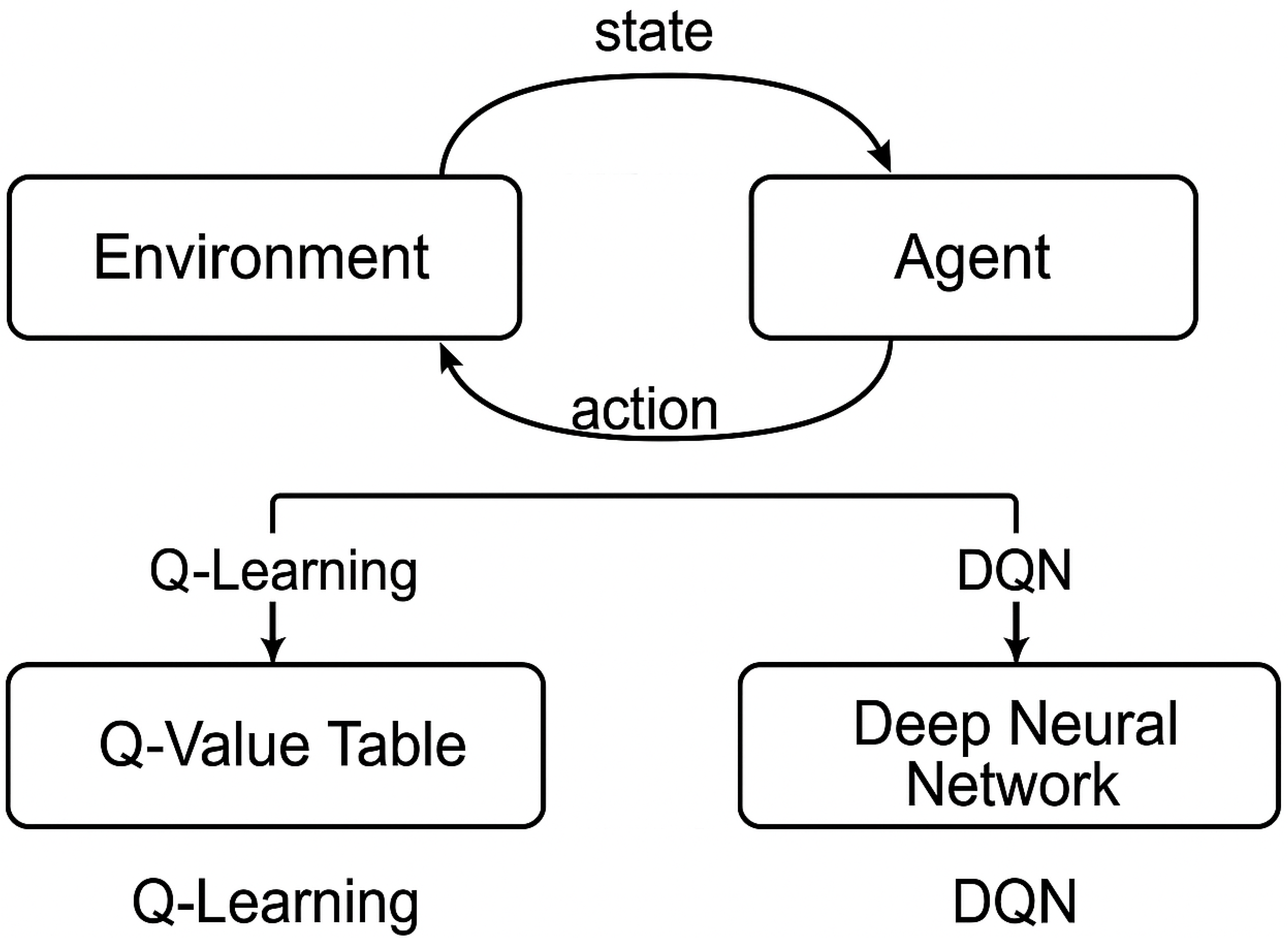
Figure 1. Q-learning and Deep Q-Network (DQN) workflow for decision-making and value approximation.
Futureinternet 17 00396 g001
Figure 2. Framework for reinforcement learning in medical images.
Futureinternet 17 00396 g002
Figure 3. Reinforcement learning framework for pneumonia detection in chest X-rays.
Futureinternet 17 00396 g003
Figure 4. Taxonomy of reinforcement learning applications in medical imaging.
Futureinternet 17 00396 g004
Figure 5. Taxonomic heatmap of reinforcement learning in medical imaging (2019–2025). The x-axis represents imaging modalities, and the y-axis represents RL algorithm types. Color intensity denotes the frequency of published studies, highlighting both dominant areas (e.g., DQN with X-rays and MRI) and underexplored intersections (e.g., multi-agent RL in ultrasound and pathology).
Futureinternet 17 00396 g005
Figure 6. Number of published survey papers on RL in medical imaging (2015–2025). Historical growth (blue) transitions to projected LLM-accelerated rise (red) beyond 2025.
Futureinternet 17 00396 g006
Figure 7. Proposed taxonomy of AI-based reinforcement learning in medical imaging [9].
Futureinternet 17 00396 g007
Figure 8. Conceptual roles for LLMs within RL pipelines for medical imaging. The diagram is a conceptual aid (no new experiments): (i) Pipeline automation reduces manual, error-prone configuration [66]; (ii) Annotation and reporting supports efficient labeling and standardized outputs [67]; (iii) Cross-modal integration enables natural-language reasoning across heterogeneous data [47]; and (iv) Clinical decision support enhances interpretability and alignment with guidelines [66].
Futureinternet 17 00396 g008
Figure 9. Federated reinforcement learning framework for distributed medical imaging environments, ensuring data privacy via local training and secure model aggregation.
Futureinternet 17 00396 g009
Figure 10. AI-based reinforcement learning in medical imaging.
Futureinternet 17 00396 g010
Table 1. Comparison of key literature on reinforcement learning in medical image analysis.
StudyFocus AreaModalityRL MethodRemarks/Limitations
Zhou et al. (2021) [6]General RL in medical imagingMultimodality (CT, MRI, Ultrasound)DQN, Policy GradientBroad review, lacks detailed taxonomy and clinical translation perspectives
Hu et al. (2023) [9]RL in medical imagingCT, MRI, X-rayDQN, PPO, A2CComprehensive; discusses taxonomy and clinical challenges
Ghesu et al. (2019) [18]Anatomical landmark detection3D CT, MRIDeep Q-Network (DQN)Pioneering single-agent DQN for spatial localization, extended to multi-agent coordination
Sahba et al. (2019) [16]Prostate segmentation in TRUSUltrasoundQ-learningState-action design for low-label segmentation; focused on prostate-specific TRUS images
Xu et al. (2021) [17]Image synthesisLiver MRIPixGRL (pixel-level graph RL)Synthesizes gadolinium-enhanced liver tumor images from non-enhanced scans. Limited to liver MRI; lacks generalizability, interpretability, and privacy-aware deployment
Ebrahimi et al. (2021) [8]Radiotherapy planningCT (prostate)RL with tumor response modelingOptimizes dose plans under biological uncertainty; lacks real-time feedback integration and privacy-preserving deployment support
Litjens et al. (2017) [12]Deep learning in medical imagingMultimodalityN/AComprehensive CNN review; lacks coverage of sequential decision-making frameworks
Shen et al. (2017) [13]Deep learning surveyMultimodalityN/AFocused on supervised deep learning; no exploration of RL or hybrid adaptive learning
Luo et al. (2021) [15]Image registrationCT, MRIHierarchical Planner–Actor–Critic (SPAC)Sequential deformation modeling; unsupervised learning; lacks clinical workflow integration and explainability
Sun et al. (2020) [19]Multimodal registrationCT–MRIRecurrent Actor–Critic RLMultimodal alignment with lookahead planning; lacks real-time validation and privacy-awareness
Liu et al. (2025) [20]Medical image segmentationCardiac, brain MRIPixel-level A3C RLDirect pixel-by-pixel segmentation; more accurate boundaries; needs evaluation on larger clinical datasets; interpretability not yet assessed
Judge et al. (2024) [21]Echocardiography segmentationUltrasound echoRL4Seg (domain-adaptive RL)99% anatomical validity with limited labels. Limited to echo images; privacy/federated deployment not addressed
Table 2. Comparison of learning paradigms in medical imaging.
AspectSupervised LearningUnsupervised LearningReinforcement Learning (RL)
Learning ObjectiveLearn from labeled data to minimize prediction errorDiscover hidden patterns or structures in unlabeled dataLearn policies to maximize cumulative reward via interaction
Common ApplicationsClassification, segmentation (e.g., tumor detection)Clustering, anomaly detection (e.g., lesion discovery)Landmark localization, adaptive segmentation, active diagnosis
Data RequirementRequires large annotated datasetsCan operate on unlabeled dataRequires interaction-based feedback (environment or simulator)
Feedback TypeGround-truth labelsData distribution or structural similarityReward signal (sparse, delayed, or shaped)
ChallengesAnnotation cost, domain shiftAmbiguous grouping, lack of interpretabilityReward design, sample inefficiency, generalization
Use in ImagingGold standard in classification and segmentation tasksUseful in feature learning, image synthesisSuited for sequential tasks, human-in-the-loop systems
Table 3. Distribution of RL studies across tasks and imaging modalities (2019–2025).
Task/ModalityCTMRIX-RayUltrasoundPET-CTMultimodal
Segmentation221915643
Detection14119521
Planning85322
Navigation541
Reconstruction762111
Table 4. Year-wise growth of survey papers on RL in medical imaging (2015–2025) and projected LLM-based survey (2026–2030).
YearCountKey References/TrendsFocus Area
Actual Survey Publications (2015–2025)
20151RL intro in imaging
20162Q-learning in CT [22]Landmark navigation
20172Episodic segmentation
20183Early DRL for segmentation [12]DQN usage
20195PPO, DQN adoption [9]Policy learning
20206DRL in registration [23]Spatial alignment
20218DRL Survey [6]Algorithm review
202210Clinical RL [7]Deployment pipelines
202312Healthcare RL [9]Generalization focus
202415Ultrasound-focused surveys [46]Real-time analysis
202520Human-in-the-loop RL [47]Interactive training
Projected Survey Publications (LLM-Driven, 2026–2030)
202623LLM-assisted review generationNLP-aided taxonomy
202727Auto taxonomies from LLMsProgrammatic analysis
202832RL in PET/SPECT with FLRare modality focus
202938Multi-agent explainable RLTeam-based diagnosis
203045Co-authored LLM + human surveysAutonomous meta-review
Table 5. Capabilities of LLMs in enhancing survey production.
LLM CapabilityContribution to Survey Growth
Automated ExtractionExtracts task, modality, and algorithm types from hundreds of papers to generate structured taxonomies.
Trend AnalysisTracks term frequency (e.g., PPO, prostate segmentation) to detect emerging focus areas.
DraftingAuto-generates abstracts, captions, LaTeX tables, and comparison matrices.
Gap HighlightingIdentifies underexplored intersections of task, modality, and method.
Human–AI CollaborationEnables co-authored surveys, expanding interdisciplinary literature creation.
Table 6. Comparison of reward function designs across representative medical imaging and planning tasks [51,52,53], along with clinical validation strategies. Validation approaches are informed by human-in-the-loop frameworks [54], inter-rater agreement measures such as Cohen’s Kappa [55], and audit-trail-based interpretability frameworks for clinical AI [56].
TaskReward Function DesignClinical Validation Methods
Radiotherapy Planning- Dose-distribution-based rewards
- Penalization of radiation to organs-at-risk (OARs)
- Trade-off between tumor coverage and toxicity minimization
- Expert oncologist scoring
- Dose–volume histogram (DVH) comparisons
- Policy audit trails for reproducibility
Lesion Detection/Localization- Localization accuracy (IoU with bounding boxes)
- Sensitivity–specificity trade-offs
- Penalties for false negatives (missed lesions)
- Radiologist agreement scoring
- Cohen’s Kappa inter-rater consistency
- Clinical case-based decision checks
Navigation (e.g., endoscopy and biopsy guidance)- Trajectory-based rewards (minimize distance to target)
- Safety constraints (avoid critical anatomical regions)
- Smoothness penalties to reduce abrupt movements
- Expert-in-the-loop path evaluation
- Consistency with clinical navigation guidelines
- Retrospective comparison against gold-standard trajectories
Table 7. Conceptual contributions of LLMs to RL-based medical imaging (non-experimental).
LLM RoleExample TaskAdded Value (Conceptual)Representative WorkValidation Method
Pipeline AutomationModality–task–algorithm mapping for MRI lesion workflowsReduces manual trial-and-error; suggests suitable RL families (e.g., actor–critic vs. DQN) from textual protocols and prior artShen et al. (LLMs in medicine) [66]Cross-check with protocol databases, benchmark comparisons
Annotation and ReportingCXR region suggestions, structured report draftingAccelerates expert labeling; standardizes terminology and evidence statements for RL feedback loopsJaved et al. (robustness/ops) [67]Expert-in-the-loop scoring, inter-rater agreement
Cross-Modal IntegrationPET-CT fusion reasoning; ultrasound navigation narrativesBridges heterogeneous inputs with natural-language rationales for RL state/context augmentationElmekki et al. (ultrasound RL review) [47]Consistency check across modalities, clinician review
Clinical Decision SupportTrajectory summarization; policy explanation for treatment planningImproves interpretability and clinician trust via guideline-aware, case-linked narrativesShen et al. (LLMs in medicine) [66]Cohen’s Kappa with clinicians, policy audit trails
Ethics and ComplianceHIPAA/GDPR-aligned annotation pipelines, privacy-preserving summariesEnsures safe deployment in regulated healthcare settings; flags privacy/security risksIvliev (G-CCACS architecture) [56]Regulatory audit, compliance checklist validation
Table 8. Design blueprint for privacy-aware reinforcement learning.
ComponentDescription
Reward DesignBinary reward: correct = +1, incorrect = −1
RL AlgorithmsDQN vs. PPO
Encoder BackbonePretrained ResNet-18
LLM IntegrationGPT-based post-action explanation
Privacy MechanismsDifferential Privacy (DP), Federated Learning (FL)
Disclaimer/Publisher’s Note: The statements, opinions and data contained in all publications are solely those of the individual author(s) and contributor(s) and not of MDPI and/or the editor(s). MDPI and/or the editor(s) disclaim responsibility for any injury to people or property resulting from any ideas, methods, instructions or products referred to in the content.