1. Introduction
Semaglutide is a long-acting glucagon-like peptide-1 receptor agonist (GLP-1 RA) that has demonstrated exceptional efficacy in managing type 2 diabetes and obesity, providing proven benefits in glycemic control, weight loss, and cardiovascular risk reduction [
1,
2,
3,
4]. These benefits led to the success of injectable semaglutide therapies (e.g., Ozempic and Wegovy) and established semaglutide as a cornerstone treatment in its class [
3]. However, like most peptide drugs, semaglutide faces formidable barriers to oral delivery. It is highly susceptible to enzymatic degradation in the gastrointestinal (GI) tract and exhibits very low transepithelial permeability, resulting in an oral bioavailability typically below 1% [
5,
6,
7,
8]. Achieving clinically meaningful plasma levels via the oral route thus requires specialized formulation strategies to overcome proteolytic and absorption hurdles.
A major advancement in this arena was the introduction of oral semaglutide (Rybelsus), which co-formulates semaglutide with the absorption enhancer sodium
N-(8-[2-hydroxybenzoyl]amino) caprylate (SNAC) [
2]. SNAC is a synthetic derivative of caprylic acid that facilitates the uptake of semaglutide in the stomach through a unique mechanism: after tablet disintegration, a high local concentration of SNAC raises the gastric pH and increases the fluidity of the mucosal cell membranes without opening tight junctions, thereby enabling transcellular transport of the peptide across the gastric epithelium [
2]. This SNAC-based technology enabled the first oral GLP-1 RA therapy and demonstrated the feasibility of oral delivery of a large peptide in humans [
9].
Nevertheless, important limitations remain. SNAC-mediated absorption is inherently low in efficiency and highly variable [
10]. Achieving even this level of exposure necessitates high doses of excipients and strict dosing requirements (e.g., prolonged fasting and minimal co-ingestion of liquids) to create the appropriate gastric environment [
10]. Furthermore, the enhancement effect of SNAC is mechanism-specific and localized to the stomach, which may not readily translate to other peptides or less controlled GI conditions [
11]. These formulation and performance constraints highlight the need to explore alternative permeation enhancers that could offer different or potentially complementary absorption-enhancing characteristics while maintaining safety [
12].
Medium-chain fatty acids (MCFAs) have long been investigated as permeation enhancers, and sodium caprate (C10) is a well-established representative of this class [
13]. In addition to its established role as a permeation enhancer, C10 is of translational interest because it is not a completely novel synthetic excipient from a regulatory perspective. Standard reference data indicate that C10 has an LD
50 of 3.7 g/kg following oral gavage in rats [
10,
13]. Furthermore, C10 has been approved by the FDA as a direct food additive and, within the food sector, has been evaluated without a specified daily intake limit [
14,
15]. Mechanistically, C10 promotes macromolecular absorption by reversibly opening tight junctions and modulating cell membrane structure in the intestinal epithelium [
10]. This differs from SNAC’s primarily transcellular, pH-dependent mode of action. C10 has been utilized in various oral delivery platforms and has demonstrated transient, reversible enhancement of peptide absorption in both preclinical models and clinical trials [
10,
11,
14,
16,
17,
18]. Notably, SNAC and C10 are two of the most advanced oral absorption enhancers tested in humans, each with a substantial track record of development. In vitro and in vivo comparisons suggest that neither agent holds a decisive advantage in efficacy: both typically yield only single-digit percentage increases in peptide bioavailability accompanied by considerable inter-subject variability [
2]. Despite this, no systematic, formulation-integrated comparison of SNAC and C10 has been reported in the context of oral semaglutide. In other words, it remains unclear whether a C10-based immediate-release(IR) semaglutide tablet can achieve systemic exposure comparable to that of the SNAC-based reference formulation under equivalent conditions.
In addition to its established permeability-enhancing effects, we explored whether C10 could effectively function within the gastric environment of an oral semaglutide formulation. Although C10 is a weaker alkalizing agent than SNAC, its potential effectiveness is supported by the fact that the buffering capacity of the fasted human stomach is reported to be relatively low [
19,
20]. This inherent buffering capacity is expected to be similarly limited in beagle dogs, which exhibit lower basal acid secretion rates than humans [
21,
22]. Under such low-buffer conditions, the alkalizing potential of C10 may be sufficient to modulate the local microenvironmental pH, thereby attenuating pepsin activity and ensuring peptide stability. This rationale led us to investigate an IR tablet designed to achieve rapid co-localization of semaglutide and C10 in the stomach.
To address this knowledge gap, the present study employed a formulation-centric, stepwise development approach to evaluate C10 as a permeation enhancer for oral semaglutide [
23]. Initial mechanistic studies, including Caco-2 permeation assays and rat pharmacokinetic evaluations, were conducted to establish the concentration-dependent absorption-enhancing potential of C10 relative to SNAC. Guided by these findings, IR tablet prototypes incorporating semaglutide and C10 were developed to promote rapid disintegration and to create local microenvironmental conditions favorable for enhancer action, particularly through pH modulation and efficient dispersion under gastric-relevant conditions. The resulting formulation designs were assessed via biorelevant dissolution testing, and the lead prototype was further evaluated in beagle dogs to characterize systemic exposure and translational pharmacokinetic performance. This integrated strategy enabled a side-by-side evaluation of C10-based and SNAC-based semaglutide formulations across preformulation, in vitro, and in vivo stages. Overall, the study aimed to determine whether a rationally designed C10-containing IR tablet could achieve systemic semaglutide exposure comparable to that of the clinically established SNAC-based reference, while providing an objective assessment of the feasibility and limitations of C10 as an alternative enhancer for oral peptide delivery.
2. Materials and Methods
2.1. Materials
Semaglutide (peptide purity > 98%) was obtained from Fujian Genohope Biotech Ltd. (Putian, China) and Sinopep-Allsino Biopharmaceutical Co., Ltd. (Hangzhou, China). Sodium N-[8-(2-hydroxybenzoyl)amino]caprylate (SNAC, purity > 99%) was purchased from GLPBIO (Montclair, CA, USA). Sodium caprate was purchased from Tokyo Chemical Industry Co., Ltd. (Tokyo, Japan). The reference drug product (Rybelsus® 14 mg tablets) was manufactured by Novo Nordisk (Bagsværd, Denmark). Lactose monohydrate (Pharmatose 200 M) was obtained from DFE Pharma GmbH & Co. KG (Goch, Germany). Povidone K90 was obtained from Ashland Inc. (Wilmington, DE, USA). Microcrystalline cellulose (MCC, Heweten 102) was obtained from JRS Pharma (Rosenberg, Germany). Crospovidone (Kollidon CL) and copovidone (Kollidon VA64) were obtained from BASF (Ludwigshafen, Germany). Sodium starch glycolate was obtained from Roquette (Lestrem, France). Croscarmellose sodium (Ac-Di-Sol) was obtained from IFF (Wilmington, DE, USA). Sodium bicarbonate was obtained from Hebei Huachen Pharmaceutical Co., Ltd. (Cangzhou, China). Magnesium stearate was obtained from Nitika Pharmaceuticals Ltd. (Maharashtra, India). Hank’s balanced salt solution (HBSS) and 4-(2-hydroxyethyl)-1-piperazineethanesulfonic acid (HEPES) buffer solution were obtained from Welgene Inc. (Gyeongsan, Republic of Korea). Hydrochloric acid and Tween 80 were purchased from Daejung Chemicals & Metals Co., Ltd. (Siheung, Republic of Korea). MTT reagent, 3-(4,5-dimethylthiazol-2-yl)-2,5-diphenyltetrazolium bromide, was purchased from Sigma-Aldrich (St. Louis, MO, USA). The LDH cytotoxicity assay was performed using a Cytotoxicity LDH Assay Kit-WST purchased from Dojindo Molecular Technologies, Inc. (Kumamoto, Japan). High-performance liquid chromatography (HPLC)-grade acetonitrile and methanol were obtained from Duksan Pure Chemicals Co., Ltd. (Ansan, Republic of Korea). All other chemicals and solvents were of analytical grade and were used as received. Ultrapure water (18.2 MΩ·cm) was prepared using an in-house purification system.
2.2. Preformulation Studies
The preformulation strategy for the present study was established to support the rational design of oral semaglutide tablets. While semaglutide solubility and excipient compatibility were previously characterized in our quality-by-design (QbD)-based study [
24], the current work focused on further characterizing the morphology of semaglutide raw materials and the acid-neutralizing capacity and dispersion behavior of C10. These assessments were critical to refining the selection of C10-containing IR tablet architectures.
2.2.1. Morphology
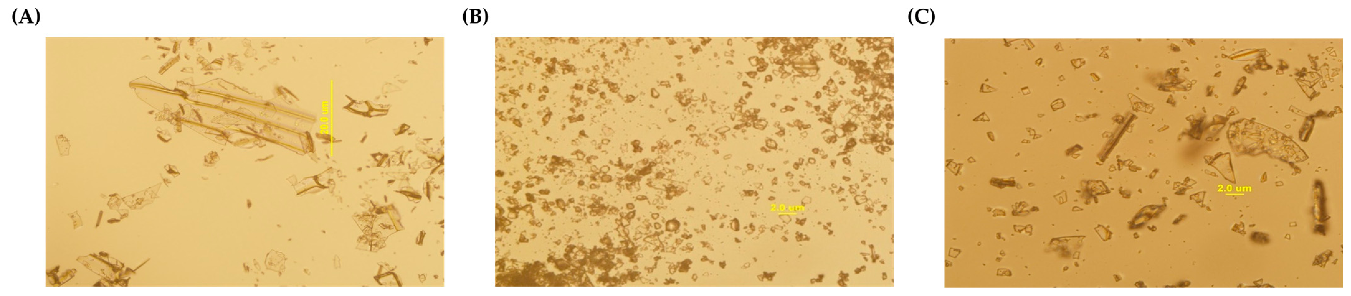
The morphology of semaglutide was examined using an optical microscope (IX2-ILL100; Olympus, Tokyo, Japan) to evaluate three distinct material profiles: two different batches from one supplier (Genohope 1 and 2) and one from an alternative supplier (Sinopep, Hangzhou, China). Samples were dispersed on glass slides, and representative images were captured at 100× and 400× magnification in bright-field mode. The particle size distribution, degree of agglomeration, and overall physical uniformity were qualitatively assessed to evaluate lot-to-lot and supplier-dependent variability, as well as their potential impact on blend uniformity and downstream formulation performance.
2.2.2. Preliminary Assessment of Acid-Neutralization and Dispersion Behavior of C10 Under Gastric-Relevant Conditions
To evaluate the acid-neutralizing and dispersion behavior of C10, conditions reflecting fasted gastric fluid volumes in beagle dogs and humans were established. The canine-mimetic condition consisted of 25 mL of 0.01 N HCl supplemented with 10 mL of water, while the human-mimetic condition consisted of 50 mL of 0.01 N HCl supplemented with 120 mL of water [
25,
26,
27,
28,
29]. Each medium was prepared in a 200 mL beaker, and C10 was added at 100, 300, or 500 mg at room temperature. The mixtures were stirred with a magnetic stir bar at 150 rpm for 15 min. The pH was measured immediately after the 15 min stirring period and again after an additional 30 min using a pH meter (Starter 3100; OHAUS Corporation, Parsippany, NJ, USA).
Dispersion properties were evaluated using a dynamic light scattering (DLS) particle size analyzer (ELSZ-2000, Otsuka Electronics Co., Ltd., Osaka, Japan). Approximately 3.0 mL of each prepared sample was transferred into a disposable polymethyl methacrylate (PMMA) cuvette, and measurements were performed at 25 °C. The Z-average diameter, particle size distribution, and polydispersity index (PDI) were determined. Each DLS measurement was performed with 50 accumulations and repeated six times for each sample. The resulting data were analyzed using the instrument software to characterize the particle size properties of the C10 dispersion system.
2.3. Formulation of Semaglutide Oral Tablets
A dry granulation-based manufacturing process was established to ensure the physicochemical stability of semaglutide, which is susceptible to moisture and heat, while simultaneously achieving excellent flowability and compressibility. The development of these IR oral tablets was further guided by preformulation findings, excipient compatibility results, and the functional role of C10 as a medium-chain fatty acid (MCFA)-derived permeation enhancer. Three tablet architectures—single-layer (SSL-T), bilayer (SBL-T), and dry compression-coated (SCC-T) tablets—were engineered to systematically evaluate the influence of enhancer placement, microenvironmental pH, and compression configuration on semaglutide release and absorption [
30,
31].
2.3.1. Preparation of Single-Layer Tablets (SSL-Ts)
For the preparation of SSL-T, semaglutide (14.0 mg), sodium caprate (300.0 mg), lactose monohydrate (300.0 mg), croscarmellose sodium (17.0 mg), and copovidone (13.0 mg) were accurately weighed and manually blended for approximately 100 cycles to ensure a uniform distribution of the drug and the enhancer (
Table 1). The blended mixture was then subjected to dry granulation via slugging. Slugging was performed using a rotary tablet press (PR-LM 08; PTK Co., Ltd., Gimpo-si, Republic of Korea) equipped with 15.0 mm round punches and dies under a compression force of 20–25 kN. The resulting slugs were milled using a hammer mill (Polymix PX-MFC 90 D; Kinematica AG, Malters, Switzerland) and passed through a 3.0 mm sieve to obtain free-flowing granules. Subsequently, magnesium stearate (8.0 mg) was incorporated as a lubricant with 50 mixing cycles. Finally, the granules were compressed into tablets using the same rotary tablet press equipped with 16.7 × 8.3 mm oblong punches. The pre-compression force was set at ≤1.0 kN, and the main compression force was maintained at ≥6.6 kN to ensure the mechanical strength and rapid disintegration required for IR tablets.
2.3.2. Preparation of Bilayer Tablets (SBL-Ts)
SBL-T was designed to separate semaglutide from C10 during initial dissolution (
Table 2). The tablet comprised two distinct layers: (i) an upper layer containing semaglutide (14.0 mg), lactose monohydrate (70.0 mg), microcrystalline cellulose (100.0 mg), and povidone K90 (5.0 mg); and (ii) a lower layer composed of sodium caprate (300.0 mg), lactose monohydrate (200.0 mg), crospovidone (30.4 mg), and copovidone VA64 (12.6 mg).
Each layer was granulated separately via the same slugging and milling procedure described for SSL-T, sieved through a 3.0 mm mesh, and lubricated with magnesium stearate (2.0 mg for the upper layer and 5.5 mg for the lower layer) with 50 mixing cycles. The bilayer tablets were manufactured using a bilayer rotary compression machine (PR-3000 Series; PTK Co., Ltd., Gimpo-si, Republic of Korea) equipped with 15.6 × 7.8 mm oblong punches.
The manufacturing process involved a sequential layering technique: the lower-layer granules (C10 layer) were first filled into the die and lightly pre-tamped, followed by the addition of the upper-layer granules (drug layer). The pre-compression force was set at ≤1.0 kN, and the final tablets were compressed under a main compression force of 13–14 kN. This compression profile was maintained to ensure adequate interlayer adhesion and to prevent physical defects such as capping or layer separation.
2.3.3. Preparation of Dry Compression-Coated Tablets (SCC-Ts)
SCC-T was designed as a core–shell system to control the timing of semaglutide exposure to C10 (
Table 3). The tablet comprised (i) an inner core containing semaglutide (14.0 mg), microcrystalline cellulose (77.0 mg), crospovidone (5.0 mg), and povidone K90 (2.0 mg); and (ii) an outer layer composed of C10 (300.0 mg), lactose monohydrate (330.0 mg), crospovidone (30.4 mg), and copovidone VA64 (12.6 mg).
Each component for the core and outer layers was dry-granulated separately via the same slugging and milling procedure described for SSL-T, sieved through a 3.0 mm mesh, and subsequently lubricated with magnesium stearate (2.0 mg for the core and 7.0 mg for the outer layer) with 50 mixing cycles.
The lubricated core granules were compressed into core tablets using a rotary tablet press equipped with 6.0 mm round punches under a compression force of ≥0.5 kN. For compression coating, the outer layer granules were first filled into a die, and the core tablet was centrally positioned. The final compression-coated tablets were then produced using a rotary compression machine equipped with 12.5 mm round punches under a compression force of 5–6 kN.
This core–shell architecture enabled evaluation of how delayed exposure of semaglutide to C10 influences dissolution behavior and in vivo absorption [
32].
2.4. In Vitro Study
In vitro experiments were performed to characterize semaglutide dissolution from test formulations and to evaluate the permeability-enhancing effects of C10 using Caco-2 monolayers. Dissolution testing was performed under physiologically relevant pH conditions to simulate gastric and intestinal environments. Caco-2 transport studies assess apical-to-basolateral semaglutide transport and epithelial tolerance during enhancer exposure. All analyses were performed using validated analytical methods.
2.4.1. In Vitro Dissolution Test
The dissolution behavior of semaglutide was assessed using the reference drug product and three test formulations (SSL-T, SBL-T, and SCC-T). Dissolution testing was performed using a dissolution tester (708-DS, Agilent Technologies, Santa Clara, CA, USA) in accordance with USP <711> using Apparatus II (paddle method). Three dissolution media (900 mL each) were used to reflect physiologically relevant gastrointestinal pH conditions: pH 1.2 simulated gastric fluid containing 0.75% Brij 35, pH 4.0 acetate buffer containing 0.75% Brij 35, and pH 6.8 phosphate buffer. To improve semaglutide solubility under acidic conditions and maintain sink conditions, Brij 35 was included in the pH 1.2 and pH 4.0 media. Dissolution testing was conducted at 37.0 ± 0.5 °C with a paddle rotation speed of 50 rpm.
For each test condition, six tablets (n = 6) were evaluated, and 4 mL samples were withdrawn at 5, 10, 15, 20, 30, 45, and 60 min. The collected samples were immediately filtered through 0.45 µm PTFE syringe filters and analyzed by HPLC using an Agilent 1260 Infinity II system (Agilent Technologies, Santa Clara, CA, USA) equipped with an Agilent C18 column (4.6 × 150 mm, 5 µm). The flow rate was 1.0 mL/min, the injection volume was 40 µL, and detection was performed at 220 nm. Dissolution profile similarity between the test and reference products was assessed using the similarity factor (f2). For the f2 calculation, different time points were selected depending on the dissolution behavior in each medium. Specifically, under the pH 1.2 condition, five time points (10, 15, 30, 45, and 60 min) were included because the reference product exceeded 85% dissolution at 60 min, whereas under the pH 4.0 and pH 6.8 conditions, three time points were used for each medium (pH 4.0: 10, 20, and 30 min; pH 6.8: 10, 15, and 30 min). An f2 value of 50 or greater was considered to indicate similarity between the two dissolution profiles.
2.4.2. Caco-2 Cell Assay for In Vitro Permeability
Differentiated Caco-2 monolayers grown on 12 mm Transwell
® inserts with a 0.4 µm PET membrane (Corning, NY, USA) were used to evaluate the in vitro permeability of semaglutide. The apical transport buffer consisted of Hank’s balanced salt solution (HBSS) supplemented with 25 mM HEPES (pH 7.4), whereas the basolateral transport buffer consisted of HBSS supplemented with 25 mM HEPES (pH 7.4) and 0.02% (
w/
v) Tween 80 to minimize nonspecific adsorption of the peptide [
33,
34]. Prior to the transport experiment, monolayer integrity was confirmed by measuring transepithelial electrical resistance (TEER), and only inserts meeting the predefined acceptance criteria were used. The monolayers were washed twice with pre-warmed transport buffer in both the apical and basolateral chambers and equilibrated at 37 °C for 15 min.
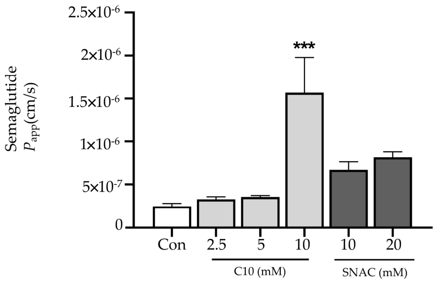
Semaglutide was prepared at a final concentration of 200 µM and added to the apical chamber either alone or in combination with permeation enhancers. Based on the experimental design, C10 was tested at 2.5, 5, and 10 mM, whereas SNAC was tested at 10 and 20 mM. The apical and basolateral chambers received 0.5 mL of sample solution and 1.5 mL of transport buffer, respectively. The plates were then incubated at 37 °C in a 5% CO
2 atmosphere for 1 h, after which samples were collected from both apical and basolateral compartments for quantitative analysis. Apparent permeability coefficients (
Papp) were calculated according to the following equation:
where dQ/
dt is the steady-state flux (µM/s) of semaglutide across the monolayer,
A is the membrane surface area (cm
2), and
C0 is the initial semaglutide concentration in the apical chamber (µM).
Collected transport samples were quantified by RP-HPLC using a Gemini C18 column (150 × 4.6 mm, 5 µm; Phenomenex, Torrance, CA, USA). Chromatographic analysis was performed at 30 °C with a flow rate of 1.0 mL/min. The mobile phase consisted of (A) 0.1% trifluoroacetic acid (TFA) in DW and (B) 0.1% TFA in acetonitrile (ACN), with the following gradient program: 40% B at 0–0.5 min, 70% B at 10.0–11.0 min, and 40% B at 11.5–15.0 min. Semaglutide was detected by UV absorbance at 280 nm and fluorescence detection at λex 280 nm and λem 340 nm. In parallel, 3-(4,5-dimethylthiazol-2-yl)-2,5-diphenyltetrazolium bromide (MTT) and Lactate dehydrogenase (LDH) assays were performed to assess potential cytotoxicity and barrier disruption associated with enhancer exposure.
2.5. In Vivo Oral Permeability Study
In vivo studies were conducted in Sprague–Dawley (SD) rats and beagle dogs to characterize the pharmacokinetic (PK) profiles of semaglutide following oral administration of formulations containing the MCFA-derived permeation enhancer C10 or SNAC, and the reference drug product. All animal experiments were conducted in accordance with institutional and national guidelines for the care and use of laboratory animals and received approval from the relevant Institutional Animal Care and Use Committees (IACUCs).
2.5.1. Pharmacokinetic Evaluation of Oral Semaglutide with C10 in Sprague–Dawley Rats
Male SD rats (7 weeks old, 230–270 g) were obtained from Young Bio (Seongnam, Gyeonggi, Republic of Korea). Animals were acclimatized for a minimum of one week under controlled environmental conditions (temperature 21 ± 2 °C, relative humidity 35–65%, 12 h light/dark cycle, and 10–15 air changes per hour) with free access to standard chow and water. The study protocol received approval from the IACUC of Dongduk Women’s University (approval no. 202407-01, on 18 July 2024).
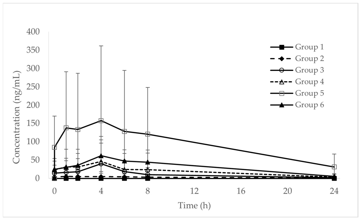
Prior to dosing, the rats were fasted for approximately 16 h with free access to water. Animals were randomly allocated to six treatment groups (n = 5 per group) and administered the following oral treatments: Group 1, distilled water (vehicle control); Group 2, semaglutide (3 mg/kg); Group 3, semaglutide (3 mg/kg) with C10 (100 mg/kg); Group 4, semaglutide (3 mg/kg) with C10 (200 mg/kg); Group 5, semaglutide (3 mg/kg) with C10 (300 mg/kg); and Group 6, semaglutide (3 mg/kg) with SNAC (200 mg/kg). Semaglutide and the enhancers were suspended in an appropriate aqueous vehicle immediately before administration and administered by oral gavage at a dose volume adjusted according to body weight.
Blood samples (approximately 350 µL per time point) were collected from the jugular vein into dipotassium ethylenediaminetetraacetate (K2-EDTA) vacuatainer tubes at predetermined intervals (pre-dose and 0.5, 1, 2, 4, 6, 8, and 24 h post-dose). Samples were centrifuged at 12,000 rpm for 5 min at 4 °C to obtain plasma according to the rate study-specific plasma preparation procedure. The supernatant was transferred into labeled tubes and stored at −70 °C until quantitative analysis of semaglutide was performed using a validated liquid chromatography–tandem mass spectrometry (LC–MS/MS, QTRAP® 6500, AB Sciex LLC, Framingham, MA, USA) method.
2.5.2. Pharmacokinetic Evaluation of Semaglutide After Oral Administration in Beagle Dogs
PK studies were conducted in beagle dogs to compare the systemic exposure to semaglutide following oral administration of the reference drug product and the optimized semaglutide tablet formulation. Male beagle dogs (2–3 years old, 10–12 kg) were obtained from Raonbio Inc. (Yongin, Gyeonggi, Republic of Korea). Animals were individually housed in stainless-steel cages under controlled environmental conditions (temperature 21 ± 2 °C, relative humidity 35–65%, 12 h light/dark cycle, and 10–15 air changes per hour). During the acclimatization period, animals were provided with free access to water and a standard canine diet. All experimental procedures were conducted in accordance with institutional animal welfare policies and national regulations governing the care and use of laboratory animals. The study protocol was reviewed and approved by the IACUC of the NDIC (approval no. NDIC-IACUC P243036, approved on 16 August 2024).
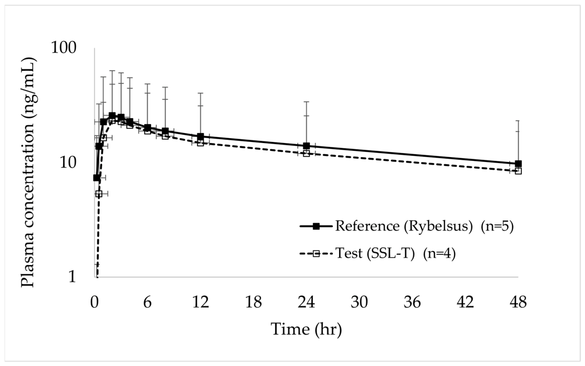
Before administration, the dogs were fasted for approximately 16 h, with water provided ad libitum. Animals were randomly assigned to two treatment groups and were administered a single oral dose. The reference group received a commercial semaglutide tablet (Rybelsus 14 mg, n = 5), and the test group received the SSL-T (14 mg, n = 4). Each tablet was administered with approximately 20 mL of water to ensure complete swallowing, and food was reintroduced 4 h post-dosing.
Blood samples (approximately 3 mL per time point) were collected from the jugular vein into K2-EDTA vacutainer tubes at pre-dose (0 h) and at 0.25 h, 0.5 h, 1 h, 2 h, 3 h, 4 h, 6 h, 8 h, 12 h, 24 h, and 48 h following administration. Immediately after collection, the samples were gently inverted to ensure anticoagulation and subsequently centrifuged at 4000 rpm for 10 min at 4 °C according to the beagle dog study-specific plasma preparation procedure. Plasma was separated subsequently and stored at −70 °C until semaglutide concentrations were quantified using LC–MS/MS.
2.6. Liquid Chromatography–Tandem Mass Spectrometry Quantitative Analysis
Plasma concentrations of semaglutide in rat and beagle dog studies were quantified using a validated LC–MS/MS (QTRAP® 6500, AB Sciex LLC, Framingham, MA, USA) method. Sample preparation was conducted using protein precipitation. Briefly, 50 µL of plasma was aliquoted into 1.5 mL microtubes and mixed with 50 µL of an internal standard solution (liraglutide, 100–300 ng/mL in acetonitrile), followed by the addition of 200 µL of methanol. The mixture was vortexed for 5 min and subsequently centrifuged at 17,600× g for 5 min at 4 °C. The supernatant (150 µL) was transferred into autosampler vials, and 10 µL was injected into the LC–MS/MS system for analysis.
Chromatographic separation was performed using an Agilent 1290 Infinity II system equipped with a Waters ACQUITY UPLC® CSHTM C18 column (2.1 × 50 mm, 1.7 µm). The mobile phases comprised (A) water with 0.1% formic acid and (B) acetonitrile with 0.1% formic acid. A linear gradient was employed as follows: 35% B at 0.0–0.25 min, 75% B at 3.0 min, 90% B at 3.10–3.20 min, and 35% B at 3.40–4.00 min. The flow rate was 0.3 mL/min, increased to 0.4 mL/min after 3.10 min, and was maintained until 4.00 min. The column temperature was maintained at 40–50 °C, and the autosampler was kept at 10 °C.
Mass spectrometric detection was performed using a SCIEX QTRAP 6500 system equipped with an electrospray ionization source operating in positive ion mode. Quantification was conducted using multiple-reaction monitoring (MRM). The optimized MRM transitions were m/z 1029.167 → 1238.000 for semaglutide and m/z 938.608 → 1064.000 for the internal standard (liraglutide). Instrument parameters were set as follows: the curtain gas was 30 psi, ion source gases 1 and 2 were set at 50 psi, ion spray voltage was 5500 V, and source temperature was 400 °C.
Calibration standards were prepared through spiking blank plasma to yield final semaglutide concentrations of 1, 5, 10, 50, 100, 500, and 1000 ng/mL. Linearity was assessed using weighted (1/x2) linear regression, and correlation coefficients (r ≥ 0.99) were considered acceptable. The lower limit of quantification (LLOQ) was 1 ng/mL, which was defined by a signal-to-noise ratio of ≥ 5 and accuracy and precision within ±20%. Quality-control samples at low, medium, and high concentrations were included to verify the accuracy and precision based on FDA/EMA bioanalytical guidelines.
Chromatograms were processed using Analyst 1.6.3 software, and semaglutide concentrations were determined based on the ratios of analyte-to-internal-standard peak-area ratios. Concentrations below the LLOQ were reported as below the quantification limit (BQL). All validated analytical results were subsequently employed for PK calculations (
Section 2.7).
2.7. Pharmacokinetic Analysis
PK parameters were calculated using standard non-compartmental analysis (NCA) with Phoenix® WinNonlin® software version 8.3.2 (Pharsight, Mountain View, CA, USA). The area under the plasma concentration–time curve from time zero to the last quantifiable concentration (AUClast) was determined using the linear-up/log-down trapezoidal method. The area under the curve extrapolated to infinity (AUCinf) was determined as AUClast + Clast/kel, where kel was the terminal elimination rate constant obtained from the log-linear regression of the terminal phase.
The terminal half-life (
t½) was calculated as 0.693/
kel. The maximum plasma concentration (
Cmax) and the time to reach this concentration (
tmax) were determined directly from the observed individual concentration–time profiles. All PK parameters were calculated using actual sampling times, and values BQL were treated as zero prior to the first quantifiable time point and as missing thereafter [
35].
2.8. Statistical Analysis
Data were expressed as means ± SD. Treatment means were compared by one-way ANOVA, and pairwise comparisons were calculated using the least significant difference (LSD) test. Differences were considered statistically significant when p ≤ 0.05. Minitab® version 21.0 (Minitab Inc., University Park, PA, USA) was used for all statistical analyses.
4. Discussion
In this study, C10, an MCFA-derived permeation modulator, was evaluated as an alternative permeation enhancer for oral semaglutide delivery through an integrated assessment encompassing formulation design, dissolution behavior, epithelial permeability, and in vivo pharmacokinetics in both rats and beagle dogs. The overall findings support the feasibility of C10-based IR semaglutide tablet formulations as an alternative to SNAC-based technology and provide mechanistic and translationally relevant insights for the oral delivery of peptide therapeutics for obesity and diabetes.
4.1. Mechanistic Interpretation of Oral Absorption Enhancer-Mediated Permeability
Semaglutide exhibits intrinsically low epithelial permeability due to its unique physicochemical properties, such as high molecular weight and hydrophilicity, necessitating the use of permeation enhancers to achieve measurable systemic exposure following oral administration. In Caco-2 monolayers, C10 increased transepithelial transport of semaglutide in a concentration-dependent manner, resulting in a pronounced increase in apparent permeability compared with SNAC under the tested in vitro conditions. This observation is consistent with established MCFA-mediated mechanisms involving reversible modulation of membrane organization and transient loosening of tight junctions, which differ from the primarily transcellular and pH-dependent mechanism reported for SNAC.
4.2. Impact of Gastric Microenvironment Modulation on Semaglutide Delivery
While C10 has traditionally been regarded as having a weaker alkalizing potential compared to SNAC, the results of this study challenge the assumption that this difference limits its clinical utility. Given that the buffering capacity of the fasted human and canine stomach is relatively low, we hypothesized that C10 could provide sufficient neutralization to create a favorable absorption window. The preformulation data (
Table 4) validated this hypothesis, demonstrating that C10 at 300 mg can effectively elevate the local pH to 6.22, a critical threshold for semaglutide delivery.
This pH modulation serves a dual purpose that is essential for overcoming the formidable barriers to oral peptide absorption. First, it addresses the solubility hurdle of semaglutide, which is notoriously poorly soluble in the acidic range of pH 2.0 to 5.0. By shifting the environment above pH 6.0, C10 ensures that the peptide is fully solubilized and available for uptake. Simultaneously, this alkaline shift is vital for the enhancer’s own functionality. With a pKa of approximately 4.8, C10 must remain in its ionized (salt) form to act as a permeation modulator. At the established pH of 6.22, C10 is predominantly ionized, preventing its precipitation into insoluble capric acid and maintaining a high effective concentration at the site of action.
Furthermore, the present study highlights that the physical state of the C10 dispersion is as decisive as its chemical ionization. Effective permeation enhancement by MCFAs is known to be driven by free monomers and small, dynamic micellar structures that can readily partition into the epithelial membrane [
17,
36,
37]. Our DLS analysis (
Table 5) revealed that the 300 mg dose resides in an optimal physicochemical range, maintaining a fine sub-micron dispersion (approximately 273 nm). Interestingly, while increasing the dose to 500 mg provided a higher pH, it triggered massive aggregation (approximately 17,642 nm). This excessive aggregation, likely due to high ionic strength, sequesters the active species into inactive clusters and depletes the concentration of free monomers available for membrane interaction [
37].
These integrated insights explain why 300 mg was identified as the optimal dose for our SSL-T formulation. By simultaneously satisfying the chemical requirement for ionization and the physical requirement for fine dispersion, this rationally designed microenvironment enabled the C10-based system to achieve systemic exposure in beagle dogs comparable to the clinically established SNAC-based reference product.
4.3. In Vitro–In Vivo Translation of C10-Mediated Absorption Enhancement
The in vivo pharmacokinetic results further support the functional relevance of C10 as a permeation enhancer under physiological conditions. In Sprague–Dawley rats, C10 increased systemic exposure to semaglutide in a clear dose-dependent manner, indicating that MCFA-mediated enhancement is maintained after oral administration. The pronounced increase in exposure at higher C10 loadings suggests a threshold-like effect, likely reflecting the need to achieve a sufficient local enhancer concentration at the epithelial interface. This interpretation is consistent with the non-linear enhancer–membrane interactions reported for fatty acid-based permeation enhancers. At equivalent enhancer doses, C10 produced systemic exposure comparable to that achieved with SNAC. The overall agreement between the in vitro permeability trends and the in vivo rat pharmacokinetic data supports the view that the observed enhancement is mechanistically meaningful rather than a model-specific artifact [
38]. Collectively, these findings suggest that the in vitro observations capture key features relevant to in vivo absorption behavior.
4.4. Influence of Formulation Architecture on Dissolution and Absorption
The three IR tablet architectures evaluated in this study—single-layer (SSL-T), bilayer (SBL-T), and compression-coated (SCC-T)—enabled a systematic assessment of how formulation structure influences dissolution behavior and enhancer deployment. All formulations achieved rapid drug release across pH 1.2, 4.0, and 6.8, consistent with the requirements for IR oral semaglutide delivery. SSL-T and SBL-T exhibited dissolution profiles comparable to those of the reference drug product (Rybelsus), as supported by f2 similarity factors greater than 50. These results suggest reference-like early-stage release and rapid availability of semaglutide in the dissolution medium. In contrast, SCC-T showed a short initial lag phase, most likely due to hydration and disintegration of the outer compression-coated layer before exposure of the drug-containing core.
Although SCC-T eventually achieved a comparable extent of dissolution, this early delay affected the early-stage release profile and resulted in a non-similar f2 value compared with the reference product. These findings indicate that, for the present oral semaglutide formulation, rapid co-release of semaglutide and C10 is more favorable than delayed or staged release, because prompt local availability of both the peptide and enhancer is required to establish the intended absorption-enhancing microenvironment.
From a formulation perspective, the overall rapid dissolution observed across the different architectures highlights the flexibility of C10-based systems and their adaptability to various manufacturing strategies, although early-stage release characteristics can be influenced by the spatial arrangement of the drug-containing and enhancer-containing layers [
39,
40].
4.5. Cross-Species Pharmacokinetic Performance and Clinical Implications
The beagle dog pharmacokinetic results further support the translational potential of the optimized C10-based tablet formulation. In this larger animal model, the plasma concentration–time profile of semaglutide following administration of the optimized formulation was broadly comparable to that of the SNAC-based reference product, with similar Cmax, AUClast, and elimination half-life values. These findings indicate that the C10-based IR system was able to reproduce systemic exposure at a level comparable to the reference product under physiologically more demanding conditions.
This observation is particularly relevant because oral absorption of peptide therapeutics is generally more challenging and variable in larger species than in rodents. In this context, the beagle dog data provide important support for the translational robustness of the formulation strategy identified through the preceding preformulation, in vitro, and rat in vivo studies. The comparable exposure achieved in dogs suggests that the C10-based system can maintain its functional performance beyond proof-of-concept conditions and within a formulation format more relevant to eventual clinical application. At the same time, the present findings should be interpreted within the known variability of oral peptide delivery systems employing permeation enhancers. Considerable inter-animal variability was observed in both treatment groups, which is consistent with the pharmacokinetic behavior commonly reported for this class of formulations.
Nevertheless, the overall overlap in exposure and disposition characteristics between the optimized C10-based tablet and the reference product supports the feasibility of C10 as an alternative enhancer platform for oral semaglutide delivery.
4.6. Study Limitations and Future Directions
Several limitations of the present study should be acknowledged. First, the Caco-2 model used for permeability assessment does not fully capture the complexity of the gastrointestinal environment in vivo, including mucus, luminal dilution, regional differences in absorption, and dynamic fluid movement. Second, although the rat and beagle dog studies provided useful pharmacokinetic information, species-specific differences in gastrointestinal physiology may limit direct extrapolation of these findings to humans. Third, while no abnormal clinical signs were observed following single-dose administration in the animal studies, the long-term safety and tolerability of repeated exposure to high levels of C10 were not evaluated. Another important consideration for MCFA-based permeation enhancement is the potential role of bile acids and bile salts in the intestinal lumen. Under physiological conditions, fatty acid-based enhancers such as C10 may interact with endogenous bile salts and form mixed colloidal or micellar structures, which can influence the apparent solubility, free monomer concentration, membrane partitioning, and consequently the permeation-enhancing activity of the enhancer. Therefore, the enhancer performance observed under simplified in vitro or gastric-relevant conditions may not fully represent the dynamic intestinal environment where bile salts are present. Although the present study focused primarily on the gastric microenvironment and the systemic performance of C10-based immediate-release tablets, the interaction between C10, bile salts, and semaglutide should be further investigated using biorelevant intestinal media or bile salt-containing in vitro models. Such studies would help clarify how endogenous bile components influence the availability and absorption-enhancing function of C10 in vivo. Future studies should therefore focus on strengthening both the mechanistic and translational understanding of this formulation system. Additional approaches, such as in situ perfusion studies and physiologically based pharmacokinetic modeling, may help to better define the relationship between local gastrointestinal conditions and semaglutide absorption. In parallel, dedicated repeat-dose safety studies, including histopathological evaluation, will be needed to further characterize the tolerability of C10-containing formulations under conditions more relevant to chronic clinical use. Despite these limitations, the present findings provide a coherent basis for continued development of C10-based oral semaglutide formulations. The combined preformulation, in vitro, and in vivo results support the view that C10 can serve as a viable alternative enhancer platform when incorporated into an appropriately designed IR system.
5. Conclusions
This study demonstrates that C10, an MCFA-derived permeation enhancer, is a mechanistically and pharmaceutically relevant approach for improving the oral absorption of semaglutide. The preformulation results showed that C10 can establish a favorable gastric microenvironment for semaglutide delivery by modulating local pH while maintaining a physicochemically suitable dispersed state, supporting its dual role in both drug solubilization and absorption enhancement. In vitro permeability studies and rat pharmacokinetic data further showed that C10-mediated enhancement was concentration-dependent and remained functionally relevant after oral administration. Among the tablet architectures evaluated, the IR single-layer design provided the most appropriate balance of reference-like dissolution behavior, mechanistic simplicity, and practical suitability for further development. The optimized C10-based tablet achieved systemic exposure comparable to that of the SNAC-based reference product in beagle dogs under the tested conditions, supporting the translational potential of this formulation strategy. Taken together, these findings support the feasibility of C10 as an alternative enhancer platform for oral semaglutide delivery and provide a basis for further formulation optimization and preclinical development of C10-based oral peptide systems. Building on these results, our ongoing research is further investigating how different alkalizing agents influence the local gastric microenvironment and oral absorption of semaglutide, with the aim of expanding the applicability of MCFA-based enhancer systems for oral peptide delivery.





 ,
,