Cellulose and Its Derivatives-Based Skin Dressings: Design, Smart Advances and Applications
Abstract
1. Introduction
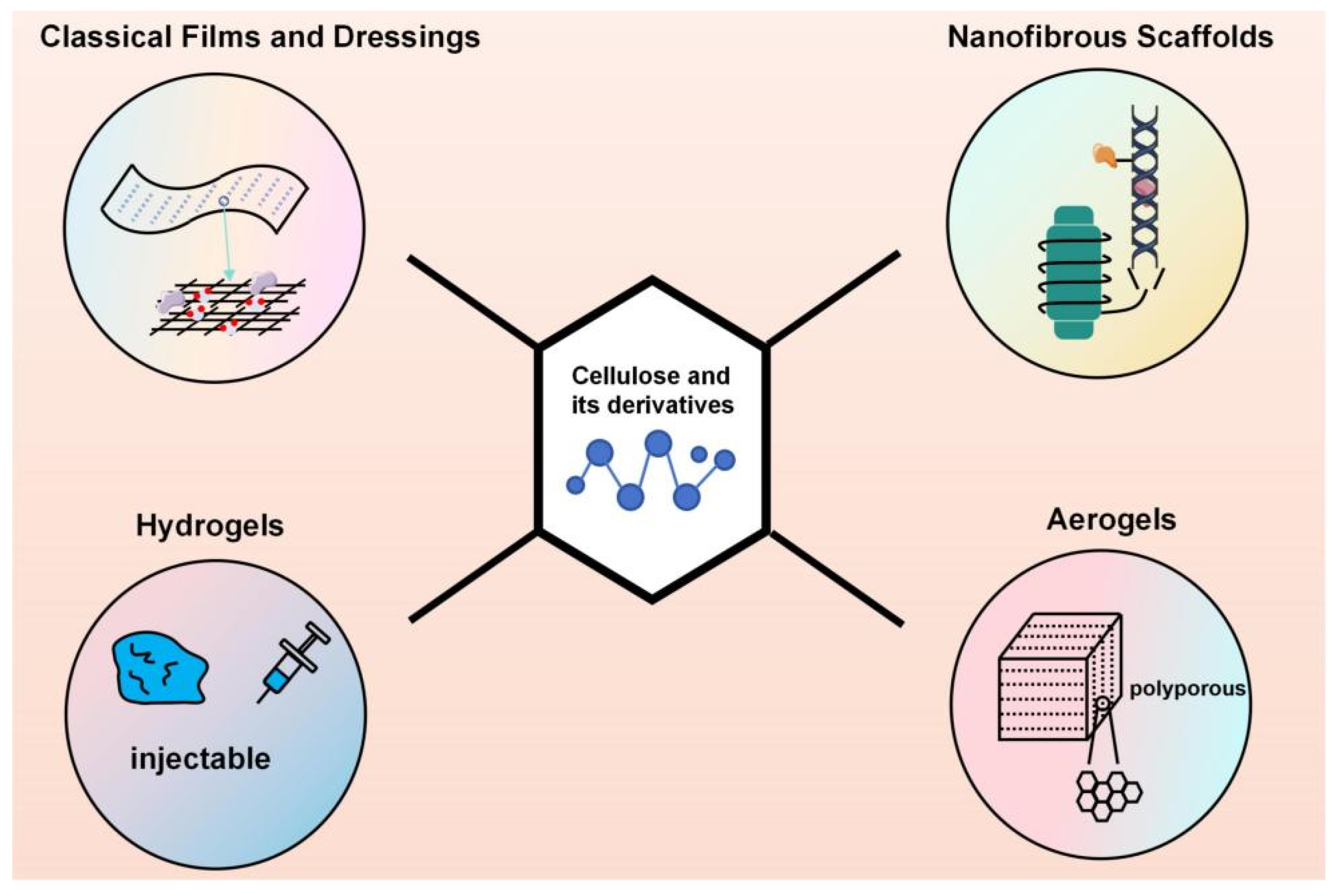
2. Construction Forms of Cellulose and Its Derivatives-Based Dressings
2.1. Classical Films and Dressings
2.2. Nanofibrous Scaffolds
2.3. Hydrogels
2.4. Aerogels
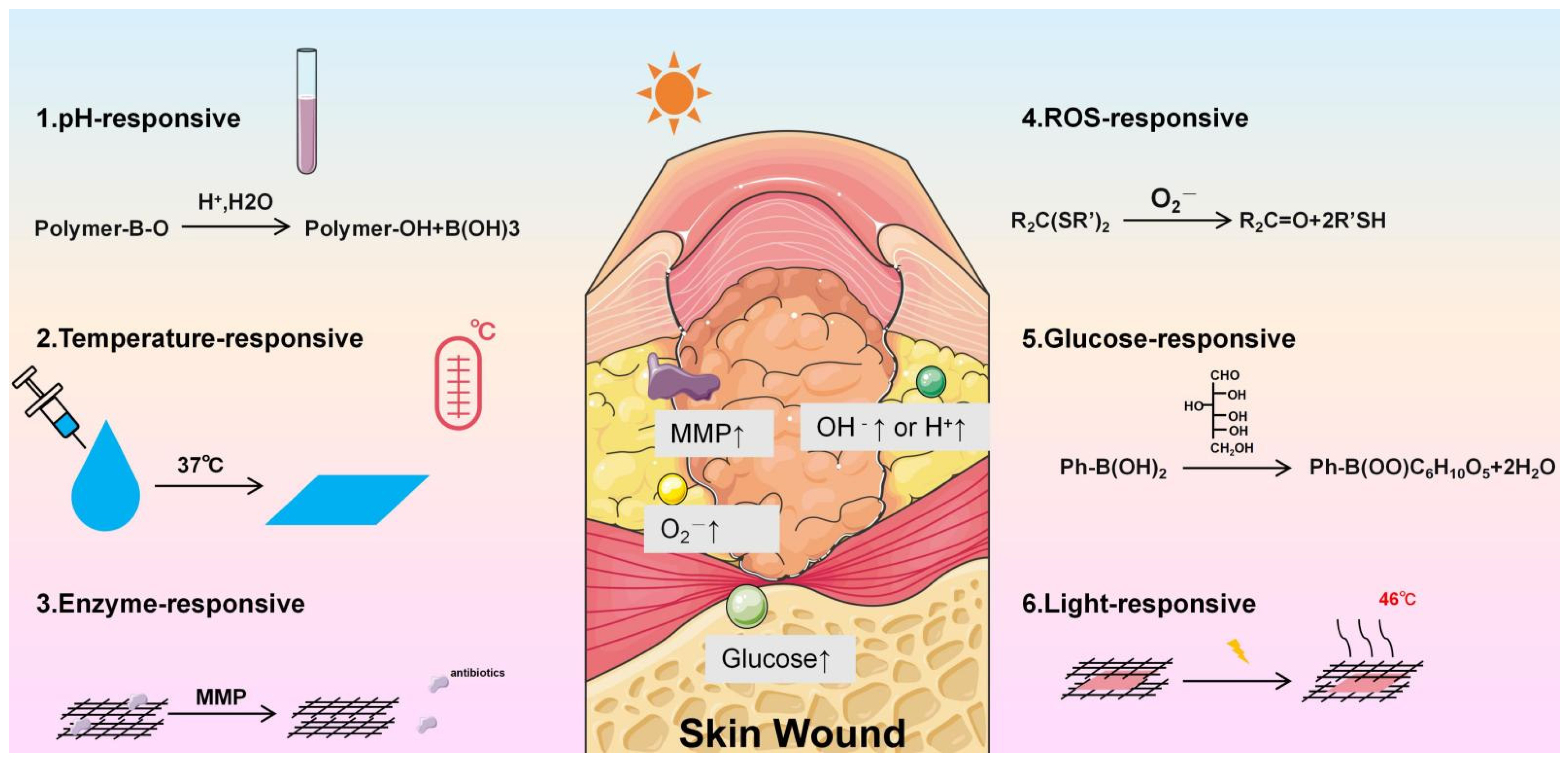
2.5. Smart Responsive Systems
2.5.1. pH-Responsive Systems
2.5.2. Temperature-Responsive Systems
2.5.3. Enzyme-Responsive Systems
2.5.4. Reactive Oxygen Species (ROS)-Responsive Systems
2.5.5. Glucose-Responsive Systems
2.5.6. Light-Responsive Systems
2.5.7. Multi-Responsive Systems
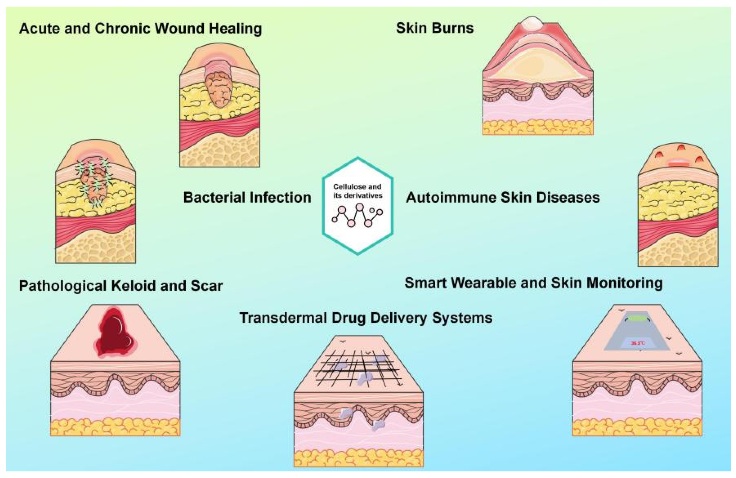
3. Applications in Various Skin Conditions
3.1. Acute and Chronic Wound Healing
3.1.1. Hemostasis and Coagulation
3.1.2. Anti-Infection and Anti-Inflammation
3.1.3. Promoting Cell Proliferation and Tissue Regeneration
3.1.4. Modulating the Wound Microenvironment
3.2. Bacterial Infectious Skin Diseases
3.3. Skin Burns
3.4. Applications in Other Conditions
3.4.1. Pathological Keloid and Scar
3.4.2. Autoimmune Skin Diseases
3.4.3. Transdermal Drug Delivery Systems
3.5. Applications in Smart Wearable and Skin Monitoring
4. Conclusions
Author Contributions
Funding
Institutional Review Board Statement
Informed Consent Statement
Data Availability Statement
Conflicts of Interest
References
- Guest, J.F.; Ayoub, N.; McIlwraith, T.; Uchegbu, I.; Gerrish, A.; Weidlich, D.; Vowden, K.; Vowden, P. Health economic burden that wounds impose on the National Health Service in the UK. BMJ Open 2015, 5, e009283. [Google Scholar] [CrossRef] [Scilit]
- Esposito, S.; Noviello, S.; Leone, S. Epidemiology and microbiology of skin and soft tissue infections. Curr. Opin. Infect. Dis. 2016, 29, 109–115. [Google Scholar] [CrossRef] [Scilit] [PubMed]
- Xue, Y.; Zhou, J.; Xu, B.N.; Li, Y.; Bao, W.; Cheng, X.L.; He, Y.; Xu, C.P.; Ren, J.; Zheng, Y.R.; et al. Global Burden of Bacterial Skin Diseases: A Systematic Analysis Combined with Sociodemographic Index, 1990–2019. Front. Med. 2022, 9, 861115. [Google Scholar] [CrossRef] [Scilit] [PubMed]
- Zhu, X.; Olsson, M.M.; Bajpai, R.; Järbrink, K.; Tang, W.E.; Car, J. Health-related quality of life and chronic wound characteristics among patients with chronic wounds treated in primary care: A cross-sectional study in Singapore. Int. Wound J. 2022, 19, 1121–1132. [Google Scholar] [CrossRef] [Scilit]
- Diban, F.; Di Lodovico, S.; Di Fermo, P.; D’Ercole, S.; D’Arcangelo, S.; Di Giulio, M.; Cellini, L. Biofilms in Chronic Wound Infections: Innovative Antimicrobial Approaches Using the In Vitro Lubbock Chronic Wound Biofilm Model. Int. J. Mol. Sci. 2023, 24, 1004. [Google Scholar] [CrossRef] [Scilit]
- Sangnim, T.; Puri, V.; Dheer, D.; Venkatesh, D.N.; Huanbutta, K.; Sharma, A. Nanomaterials in the Wound Healing Process: New Insights and Advancements. Pharmaceutics 2024, 16, 300. [Google Scholar] [CrossRef] [Scilit]
- Guo, C.; Wu, Y.; Li, W.; Wang, Y.; Kong, Q. Development of a Microenvironment-Responsive Hydrogel Promoting Chronically Infected Diabetic Wound Healing through Sequential Hemostatic, Antibacterial, and Angiogenic Activities. ACS Appl. Mater. Interfaces 2022, 14, 30480–30492. [Google Scholar] [CrossRef] [Scilit]
- Gan, S.; Zheng, Z.; Li, X.; Xu, J.; Gong, X.; Chen, S.; Zhang, P.; Chen, W. Scab-Inspired Thermoresponsive Hydrogel Dressing for Accelerated Healing of Infected Wounds. ACS Appl. Mater. Interfaces 2026, 18, 4839–4854. [Google Scholar] [CrossRef] [Scilit] [PubMed]
- Di, J.; Li, J.; Sun, C.; Xu, L.; Li, X. Advances in Cellulose-Based Hydrogels for Drug Delivery: Preparation, Modification and Challenges. Gels 2025, 11, 938. [Google Scholar] [CrossRef] [Scilit]
- Guo, W.Y.; Yuan, Q.; Huang, L.Z.; Zhang, W.; Li, D.D.; Yao, C.; Ma, M.G. Multifunctional bacterial cellulose-based organohydrogels with long-term environmental stability. J. Colloid Interface Sci. 2022, 608, 820–829. [Google Scholar] [CrossRef] [Scilit]
- Peter, Z. Order in cellulosics: Historical review of crystal structure research on cellulose. Carbohydr. Polym. 2021, 254, 117417. [Google Scholar] [CrossRef] [Scilit]
- Zennifer, A.; Senthilvelan, P.; Sethuraman, S.; Sundaramurthi, D. Key advances of carboxymethyl cellulose in tissue engineering & 3D bioprinting applications. Carbohydr. Polym. 2021, 256, 117561. [Google Scholar] [CrossRef] [Scilit]
- Heinze, T.; Siebert, M.; Berlin, P.; Koschella, A. Biofunctional Materials Based on Amino Cellulose Derivatives—A Nanobiotechnological Concept. Macromol. Biosci. 2016, 16, 10–42. [Google Scholar] [CrossRef] [Scilit] [PubMed]
- Martin, S.S.; Gan, L.; Zhang, L.; Yang, X.; Tan, Z.; Shi, H.; Long, L.; Li, H. Cellulose nanocrystal-based intelligent hydrogels: Innovations, challenges, and prospective application in advanced wound healing. Int. J. Biol. Macromol. 2025, 316, 144752. [Google Scholar] [CrossRef] [Scilit]
- Leong, M.Y.; Kong, Y.L.; Harun, M.Y.; Looi, C.Y.; Wong, W.F. Current advances of nanocellulose application in biomedical field. Carbohydr. Res. 2023, 532, 108899. [Google Scholar] [CrossRef] [Scilit] [PubMed]
- Du, H.; Liu, W.; Zhang, M.; Si, C.; Zhang, X.; Li, B. Cellulose nanocrystals and cellulose nanofibrils based hydrogels for biomedical applications. Carbohydr. Polym. 2019, 209, 130–144. [Google Scholar] [CrossRef] [Scilit]
- Rakib Hasan Khan, M.; Shankar Hazra, R.; Nair, G.; Mohammad, J.; Jiang, L.; Reindl, K.; Khalid Jawed, M.; Ganai, S.; Quadir, M. Cellulose nanofibers as Scaffold-forming materials for thin film drug delivery systems. Int. J. Pharm. 2022, 627, 122189. [Google Scholar] [CrossRef] [Scilit]
- Curvello, R.; Raghuwanshi, V.S.; Garnier, G. Engineering nanocellulose hydrogels for biomedical applications. Adv. Colloid Interface Sci. 2019, 267, 47–61. [Google Scholar] [CrossRef] [Scilit]
- Deng, W.; Tang, Y.; Mao, J.; Zhou, Y.; Chen, T.; Zhu, X. Cellulose nanofibril as a crosslinker to reinforce the sodium alginate/chitosan hydrogels. Int. J. Biol. Macromol. 2021, 189, 890–899. [Google Scholar] [CrossRef] [Scilit]
- Olmos-Juste, R.; Alonso-Lerma, B.; Pérez-Jiménez, R.; Gabilondo, N.; Eceiza, A. 3D printed alginate-cellulose nanofibers based patches for local curcumin administration. Carbohydr. Polym. 2021, 264, 118026. [Google Scholar] [CrossRef] [Scilit] [PubMed]
- Yang, M.; Ward, J.; Choy, K.L. Nature-Inspired Bacterial Cellulose/Methylglyoxal (BC/MGO) Nanocomposite for Broad-Spectrum Antimicrobial Wound Dressing. Macromol. Biosci. 2020, 20, e2000070. [Google Scholar] [CrossRef] [Scilit]
- Chen, C.; Ding, W.; Zhang, H.; Zhang, L.; Huang, Y.; Fan, M.; Yang, J.; Sun, D. Bacterial cellulose-based biomaterials: From fabrication to application. Carbohydr. Polym. 2022, 278, 118995. [Google Scholar] [CrossRef] [Scilit]
- Picheth, G.F.; Pirich, C.L.; Sierakowski, M.R.; Woehl, M.A.; Sakakibara, C.N.; de Souza, C.F.; Martin, A.A.; da Silva, R.; de Freitas, R.A. Bacterial cellulose in biomedical applications: A review. Int. J. Biol. Macromol. 2017, 104, 97–106. [Google Scholar] [CrossRef] [Scilit]
- Madub, K.; Goonoo, N.; Gimié, F.; Ait Arsa, I.; Schönherr, H.; Bhaw-Luximon, A. Green seaweeds ulvan-cellulose scaffolds enhance in vitro cell growth and in vivo angiogenesis for skin tissue engineering. Carbohydr. Polym. 2021, 251, 117025. [Google Scholar] [CrossRef] [Scilit]
- Kiiskinen, J.; Merivaara, A.; Hakkarainen, T.; Kääriäinen, M.; Miettinen, S.; Yliperttula, M.; Koivuniemi, R. Nanofibrillar cellulose wound dressing supports the growth and characteristics of human mesenchymal stem/stromal cells without cell adhesion coatings. Stem Cell Res. Ther. 2019, 10, 292. [Google Scholar] [CrossRef] [Scilit] [PubMed]
- Goh, M.; Tae, G. Mesenchymal stem cell-encapsulated cellulose nanofiber microbeads and enhanced biological activities by hyaluronic acid incorporation. Carbohydr. Polym. 2022, 280, 119026. [Google Scholar] [CrossRef] [Scilit]
- Mariia, K.; Arif, M.; Shi, J.; Song, F.; Chi, Z.; Liu, C. Novel chitosan-ulvan hydrogel reinforcement by cellulose nanocrystals with epidermal growth factor for enhanced wound healing: In vitro and in vivo analysis. Int. J. Biol. Macromol. 2021, 183, 435–446. [Google Scholar] [CrossRef] [Scilit] [PubMed]
- Vajda, J.; Bjelić, D.; Vihar, B.; Vesenjak, M.; Dubrovski, P.D.; Gradišnik, L.; Belak, M.; Maver, U.; Milojević, M. 3D printed fibroblast-laden alginate-cellulose scaffolds support extracellular matrix formation and angiogenic growth factor secretion. Int. J. Biol. Macromol. 2025, 332, 148701. [Google Scholar] [CrossRef] [Scilit]
- Picheth, G.F.; Sierakowski, M.R.; Woehl, M.A.; Ono, L.; Cofré, A.R.; Vanin, L.P.; Pontarolo, R.; De Freitas, R.A. Lysozyme-triggered epidermal growth factor release from bacterial cellulose membranes controlled by smart nanostructured films. J. Pharm. Sci. 2014, 103, 3958–3965. [Google Scholar] [CrossRef] [Scilit] [PubMed]
- Hajidariyor, T.; Nuntawad, N.; Somsaen, P.; Prukdamrongchai, R.; Cherdchoo, H.; Posoknistakul, P.; Khemthong, P.; Wanmolee, W.; Arjfuk, P.; Pongchaikul, P.; et al. Cryo-Induced Cellulose-Based Nanogel from Elaeis guineensis for Antibiotic Delivery Platform. Int. J. Mol. Sci. 2023, 24, 1230. [Google Scholar] [CrossRef] [Scilit]
- Hou, S.; Xia, Z.; Pan, J.; Wang, N.; Gao, H.; Ren, J.; Xia, X. Bacterial Cellulose Applied in Wound Dressing Materials: Production and Functional Modification—A Review. Macromol. Biosci. 2024, 24, e2300333. [Google Scholar] [CrossRef] [Scilit]
- Sharma, C.; Bhardwaj, N.K.; Pathak, P.; Dey, P.; Gautam, S.; Kumar, S.; Dutt Purohit, S. Bacterial nanocellulose by static, static intermittent fed-batch and rotary disc bioreactor-based fermentation routes using economical black tea broth medium: A comparative account. Int. J. Biol. Macromol. 2024, 277, 134228. [Google Scholar] [CrossRef] [Scilit]
- Sharma, C.; Bhardwaj, N.K. Bacterial nanocellulose: Present status, biomedical applications and future perspectives. Mater. Sci. Eng. C Mater. Biol. Appl. 2019, 104, 109963. [Google Scholar] [CrossRef] [Scilit] [PubMed]
- Portela, R.; Leal, C.R.; Almeida, P.L.; Sobral, R.G. Bacterial cellulose: A versatile biopolymer for wound dressing applications. Microb. Biotechnol. 2019, 12, 586–610. [Google Scholar] [CrossRef] [Scilit]
- Campano, C.; Rivero-Buceta, V.; Hernandez-Arriaga, A.M.; Manoli, M.T.; Prieto, M.A. Pushing the limits of bacterial cellulose for biomedicine: A review. Int. J. Biol. Macromol. 2025, 323, 146701. [Google Scholar] [CrossRef] [Scilit] [PubMed]
- Wahid, F.; Huang, L.H.; Zhao, X.Q.; Li, W.C.; Wang, Y.Y.; Jia, S.R.; Zhong, C. Bacterial cellulose and its potential for biomedical applications. Biotechnol. Adv. 2021, 53, 107856. [Google Scholar] [CrossRef] [Scilit] [PubMed]
- Venturelli, G.; Villa, F.; Petraretti, M.; Guagliano, G.; Levi, M.; Petrini, P. Bacterial Cellulose for Scalable and Sustainable Bio-Gels in the Circular Economy. Gels 2025, 11, 262. [Google Scholar] [CrossRef] [Scilit]
- Krasian, T.; Daranarong, D.; Punyodom, W.; Manokruang, K.; Somsunan, R.; Jantrawut, P.; Chaiwarit, T.; Panraksa, P.; Jantanasakulwong, K.; Rachtanapun, P.; et al. Electrospun composite membranes of ethyl cellulose and MXene (Ti3C2Tx): Biocompatible platforms for enhanced drug delivery and antibacterial wound healing. Int. J. Biol. Macromol. 2025, 287, 138596. [Google Scholar] [CrossRef] [Scilit]
- Sun, L.; Lin, J.; Zhang, L.; Chen, L.; Sun, B.; Chen, C.; Sun, D. In situ biosynthesis of functionalized fluorescent bacterial cellulose. Int. J. Biol. Macromol. 2025, 301, 140419. [Google Scholar] [CrossRef] [Scilit]
- Wang, X.L.; Liu, Z.W.; Jia, H.P.; Wu, M.Y.; Li, D.M.; Ye, X.G.; Hu, Y.; Chen, Y.; Huang, C. Production, structure, and performance of guar gum based bacterial cellulose generated from soy sauce residue hydrolysate by in-situ fermentation. Int. J. Biol. Macromol. 2025, 300, 140108. [Google Scholar] [CrossRef] [Scilit]
- Breijaert, T.C.; Fontes, M.; Fernandes, P.d.A.; Barud, H.d.S.; Ribeiro, S.J.L.; Seisenbaeva, G.A. Functionalization of bacterial nanocellulose-based wound dressing for increased drug retention. Carbohydr. Polym. Technol. Appl. 2025, 10, 100756. [Google Scholar] [CrossRef] [Scilit]
- Nascimento, A.M.d.S.S.; Silva, J.M.; de Lima, I.S.; Furtini, J.A.O.; Ribeiro, S.J.L.; Muniz, E.C.; da Silva Barud, H.; Silva-Filho, E.C. Effect of Ex Situ Modification of Bacterial Cellulose with Organosilane Coupling Agent on Drug Delivery Properties. J. Polym. Environ. 2024, 32, 4422–4439. [Google Scholar] [CrossRef] [Scilit]
- Silva, J.M.; Nunes, N.C.; Constantino, V.R.L.; Passos, A.R.; Fontes, M.L.; Marcato, P.D.; de Carvalho, G.S.G.; Lourenção Brighenti, F.; de Oliveira, A.B.; Ferrisse, T.M.; et al. Bacterial cellulose/Laponite composites as a potential new dressing with antibacterial properties. Surf. Interfaces 2025, 76, 107975. [Google Scholar] [CrossRef] [Scilit]
- Liu, L.; Kamei, K.I.; Yoshioka, M.; Nakajima, M.; Li, J.; Fujimoto, N.; Terada, S.; Tokunaga, Y.; Koyama, Y.; Sato, H.; et al. Nano-on-micro fibrous extracellular matrices for scalable expansion of human ES/iPS cells. Biomaterials 2017, 124, 47–54. [Google Scholar] [CrossRef] [Scilit] [PubMed]
- Abdel Khalek, M.A.; Abdel Gaber, S.A.; El-Domany, R.A.; El-Kemary, M.A. Photoactive electrospun cellulose acetate/polyethylene oxide/methylene blue and trilayered cellulose acetate/polyethylene oxide/silk fibroin/ciprofloxacin nanofibers for chronic wound healing. Int. J. Biol. Macromol. 2021, 193, 1752–1766. [Google Scholar] [CrossRef] [Scilit]
- Sangsefidi, F.; Tamimi, M.; Baaji, K.; Rajabi, S.; Ghadimi, T.; Zandi, M.; Pezeshki-Modaress, M. Multilayered electrospun chondroitin sulfate nanofiber impregnated with aortic extracellular matrix hydrogel for dermal regeneration: In vitro and in vivo study. Int. J. Biol. Macromol. 2025, 320, 145898. [Google Scholar] [CrossRef] [Scilit] [PubMed]
- Luo, H.; Jie, T.; Zheng, L.; Huang, C.; Chen, G.; Cui, W. Electrospun Nanofibers for Cancer Therapy. Adv. Exp. Med. Biol. 2021, 1295, 163–190. [Google Scholar] [CrossRef] [Scilit]
- Alizadeh, S.; Majidi, J.; Jahani, M.; Esmaeili, Z.; Nokhbedehghan, Z.; Aliakbar Ahovan, Z.; Nasiri, H.; Mellati, A.; Hashemi, A.; Chauhan, N.P.S.; et al. Engineering of a decellularized bovine skin coated with antibiotics-loaded electrospun fibers with synergistic antibacterial activity for the treatment of infectious wounds. Biotechnol. Bioeng. 2024, 121, 1453–1464. [Google Scholar] [CrossRef] [Scilit]
- Xu, R.; Zhang, Z.; Toftdal, M.S.; Møller, A.C.; Dagnaes-Hansen, F.; Dong, M.; Thomsen, J.S.; Brüel, A.; Chen, M. Synchronous delivery of hydroxyapatite and connective tissue growth factor derived osteoinductive peptide enhanced osteogenesis. J. Control Release 2019, 301, 129–139. [Google Scholar] [CrossRef] [Scilit]
- Son, Y.J.; Kim, W.J.; Yoo, H.S. Therapeutic applications of electrospun nanofibers for drug delivery systems. Arch. Pharm. Res. 2014, 37, 69–78. [Google Scholar] [CrossRef] [Scilit]
- Zhou, Y.; Wang, M.; Yan, C.; Liu, H.; Yu, D.G. Advances in the Application of Electrospun Drug-Loaded Nanofibers in the Treatment of Oral Ulcers. Biomolecules 2022, 12, 1254. [Google Scholar] [CrossRef] [Scilit]
- Nada, A.A.; Ali, E.A.; Soliman, A.A.F.; Shen, J.; Abou-Zeid, N.Y.; Hudson, S.M. Multi-layer dressing made of laminated electrospun nanowebs and cellulose-based adhesive for comprehensive wound care. Int. J. Biol. Macromol. 2020, 162, 629–644. [Google Scholar] [CrossRef] [Scilit] [PubMed]
- Amin, N.; Mokhter, M.A.; Salamun, N.; Wan Mahmood, W.M.A. Phosphate Adsorption from Aqueous Solution Using Electrospun Cellulose Acetate Nanofiber Membrane Modified with Graphene Oxide/Sodium Dodecyl Sulphate. Membranes 2021, 11, 546. [Google Scholar] [CrossRef] [Scilit]
- Kurečič, M.; Mohan, T.; Virant, N.; Maver, U.; Stergar, J.; Gradišnik, L.; Kleinschek, K.S.; Hribernik, S. A green approach to obtain stable and hydrophilic cellulose-based electrospun nanofibrous substrates for sustained release of therapeutic molecules. RSC Adv. 2019, 9, 21288–21301. [Google Scholar] [CrossRef] [Scilit] [PubMed]
- Phan, D.N.; Khan, M.Q.; Nguyen, V.C.; Vu-Manh, H.; Dao, A.T.; Thanh Thao, P.; Nguyen, N.M.; Le, V.T.; Ullah, A.; Khatri, M.; et al. Investigation of Mechanical, Chemical, and Antibacterial Properties of Electrospun Cellulose-Based Scaffolds Containing Orange Essential Oil and Silver Nanoparticles. Polymers 2021, 14, 85. [Google Scholar] [CrossRef] [Scilit]
- Yoo, H.S.; Kim, T.G.; Park, T.G. Surface-functionalized electrospun nanofibers for tissue engineering and drug delivery. Adv. Drug Deliv. Rev. 2009, 61, 1033–1042. [Google Scholar] [CrossRef] [Scilit] [PubMed]
- Kazsoki, A.; Szabó, P.; Domján, A.; Balázs, A.; Bozó, T.; Kellermayer, M.; Farkas, A.; Balogh-Weiser, D.; Pinke, B.; Darcsi, A.; et al. Microstructural Distinction of Electrospun Nanofibrous Drug Delivery Systems Formulated with Different Excipients. Mol. Pharm. 2018, 15, 4214–4225. [Google Scholar] [CrossRef] [Scilit]
- Fathi, H.A.; Abdelkader, A.; AbdelKarim, M.S.; Abdelaziz, A.A.; El-Mokhtar, M.A.; Allam, A.; Fetih, G.; El Badry, M.; Elsabahy, M. Electrospun vancomycin-loaded nanofibers for management of methicillin-resistant Staphylococcus aureus-induced skin infections. Int. J. Pharm. 2020, 586, 119620. [Google Scholar] [CrossRef] [Scilit]
- Seyedi, N.; Taymouri, S.; Allafchian, A.; Minaiyan, M.; Omidi, E.; Varshosaz, J. Fabrication and in-vitro and in-vivo evaluation of polyacrylonitrile and polyethylene oxide nanofibers loaded with resveratrol and silver nanoparticles for skin wound healing application. J. Biomater. Appl. 2026, 40, 830–853. [Google Scholar] [CrossRef] [Scilit]
- Tabatabaei Hosseini, B.S.; Tabatabaei Rezaei, N.; Oustadi, F.; Badv, M.; Gabriel, V.; Kim, K.; Hu, J. Development and optimization of a multifunctional cellulose-based hydrogel for enhanced crosslinking and tunability. Biomater. Adv. 2026, 179, 214490. [Google Scholar] [CrossRef] [Scilit]
- Elangwe, C.N.; Morozkina, S.N.; Olekhnovich, R.O.; Krasichkov, A.; Polyakova, V.O.; Uspenskaya, M.V. A Review on Chitosan and Cellulose Hydrogels for Wound Dressings. Polymers 2022, 14, 5163. [Google Scholar] [CrossRef] [Scilit]
- Baniasadi, H. State-of-the-art in natural hydrogel-based wound dressings: Design, functionalization, and fabrication approaches. Adv. Colloid Interface Sci. 2025, 342, 103527. [Google Scholar] [CrossRef] [Scilit] [PubMed]
- Wang, Q.; Yang, J.; Meng, J.; Yu, J.; Li, X.; Ding, B. Bioinspired hierarchical fiber-hydrogel dressing with dual immunomodulatory and angiogenic functions for diabetic wound healing. J. Control. Release 2025, 386, 114063. [Google Scholar] [CrossRef] [Scilit]
- Chang, G.; Dang, Q.; Liu, C.; Wang, X.; Song, H.; Gao, H.; Sun, H.; Zhang, B.; Cha, D. Carboxymethyl chitosan and carboxymethyl cellulose based self-healing hydrogel for accelerating diabetic wound healing. Carbohydr. Polym. 2022, 292, 119687. [Google Scholar] [CrossRef] [Scilit]
- Wang, L.; Liu, Y.; Duan, W.; Cao, F.; Lv, Q.; Zhang, S.; She, J.; Yang, L.; He, B.; Hou, Y.; et al. Sprayable oxidized cellulose nanofiber hydrogel with rapid hemostatic ability for skin wound healing. Int. J. Biol. Macromol. 2025, 310, 143264. [Google Scholar] [CrossRef] [Scilit] [PubMed]
- Jessop, Z.M.; Al-Sabah, A.; Gao, N.; Kyle, S.; Thomas, B.; Badiei, N.; Hawkins, K.; Whitaker, I.S. Printability of pulp derived crystal, fibril and blend nanocellulose-alginate bioinks for extrusion 3D bioprinting. Biofabrication 2019, 11, 045006. [Google Scholar] [CrossRef] [Scilit]
- Song, Y.E.; Eckman, N.; Sen, S.; Jons, C.K.; Saouaf, O.M.; Appel, E.A. Highly Extensible Physically Crosslinked Hydrogels for High-Speed 3D Bioprinting. Adv. Healthc. Mater. 2025, 14, e2404988. [Google Scholar] [CrossRef] [Scilit] [PubMed]
- Li, M.; Sun, L.; Liu, Z.; Shen, Z.; Cao, Y.; Han, L.; Sang, S.; Wang, J. 3D bioprinting of heterogeneous tissue-engineered skin containing human dermal fibroblasts and keratinocytes. Biomater. Sci. 2023, 11, 2461–2477. [Google Scholar] [CrossRef] [Scilit]
- Liu, H.; Xing, F.; Yu, P.; Zhe, M.; Duan, X.; Liu, M.; Xiang, Z.; Ritz, U. A review of biomacromolecule-based 3D bioprinting strategies for structure-function integrated repair of skin tissues. Int. J. Biol. Macromol. 2024, 268, 131623. [Google Scholar] [CrossRef] [Scilit]
- Liu, Y.; Wang, K.; Gao, F.; Xu, Z.; Zhao, X.; Wan, G.; Bai, X.; Li, Z.; Wang, Y. Biomedical Aerogels in Wound Healing: Therapeutic Strategies and Translational Insights. Biomater. Res. 2025, 29, 0295. [Google Scholar] [CrossRef] [Scilit]
- Gelas, L.; Budtova, T. From Cellulose Solutions to Aerogels and Xerogels: Controlling Properties for Drug Delivery. Biomacromolecules 2024, 25, 7421–7432. [Google Scholar] [CrossRef] [Scilit]
- Jeong, Y.; Patel, R.; Patel, M. Biopolymer-Based Biomimetic Aerogel for Biomedical Applications. Biomimetics 2024, 9, 397. [Google Scholar] [CrossRef] [Scilit] [PubMed]
- Tran, N.P.; Okahisa, Y.; Okubayashi, S. Ibuprofen-loaded chitosan/silk fibroin nanofibrils aerogel for drug delivery and model test for wound dressing. Next Mater. 2025, 6, 100501. [Google Scholar] [CrossRef] [Scilit]
- Rostamitabar, M.; Ghahramani, A.; Seide, G.; Jockenhoevel, S.; Ghazanfari, S. Drug loaded cellulose–chitosan aerogel microfibers for wound dressing applications. Cellulose 2022, 29, 6261–6281. [Google Scholar] [CrossRef] [Scilit]
- Li, Y.; Liu, X.; Liu, Z.; Wang, S.; Kong, F. Fabrication of controllable structure of nanocellulose composite aerogel for targeted drug delivery. Carbohydr. Polym. 2025, 358, 123518. [Google Scholar] [CrossRef] [Scilit]
- Rostamitabar, M.; Subrahmanyam, R.; Gurikov, P.; Seide, G.; Jockenhoevel, S.; Ghazanfari, S. Cellulose aerogel micro fibers for drug delivery applications. Mater. Sci. Eng. C Mater. Biol. Appl. 2021, 127, 112196. [Google Scholar] [CrossRef] [Scilit]
- Yu, S.; Budtova, T. Creating and exploring carboxymethyl cellulose aerogels as drug delivery devices. Carbohydr. Polym. 2024, 332, 121925. [Google Scholar] [CrossRef] [Scilit]
- Jose, J.; Pai, A.R.; Gopakumar, D.A.; Dalvi, Y.; Ruby, V.; Bhat, S.G.; Pasquini, D.; Kalarikkal, N.; Thomas, S. Novel 3D porous aerogels engineered at nano scale from cellulose nano fibers and curcumin: An effective treatment for chronic wounds. Carbohydr. Polym. 2022, 287, 119338. [Google Scholar] [CrossRef] [Scilit] [PubMed]
- Yu, Z.; Wu, H.; Zhang, X.; Jiang, Y.; Liu, C.; Liu, Y.; Seidi, F.; Deng, C. Antibacterial and biodegradable bandage with exudate absorption and smart monitoring for chronic wound management. J. Bioresour. Bioprod. 2025, 10, 373–385. [Google Scholar] [CrossRef] [Scilit]
- Zhang, W.; Hu, X.; Jiang, F.; Li, Y.; Chen, W.; Zhou, T. Preparation of bacterial cellulose/acrylic acid-based pH-responsive smart dressings by graft copolymerization method. J. Biomater. Sci. Polym. Ed. 2024, 35, 2767–2789. [Google Scholar] [CrossRef] [Scilit]
- Liu, Z.; Zhang, S.; Gao, C.; Meng, X.; Wang, S.; Kong, F. Temperature/pH-Responsive Carboxymethyl Cellulose/Poly (N-isopropyl acrylamide) Interpenetrating Polymer Network Aerogels for Drug Delivery Systems. Polymers 2022, 14, 1578. [Google Scholar] [CrossRef] [Scilit]
- Alsahag, M.; Alisaac, A.; Al-Hazmi, G.A.A.; Pashameah, R.A.; Attar, R.M.S.; Saad, F.A.; El-Metwaly, N.M. Preparation of carboxymethyl cellulose/polyvinyl alcohol wound dressing composite immobilized with anthocyanin extract for colorimetric monitoring of wound healing and prevention of wound infection. Int. J. Biol. Macromol. 2023, 224, 233–242. [Google Scholar] [CrossRef] [Scilit]
- Guan, C.; Li, M.; Zhou, X.; Zhou, Y.; You, Y.; Shen, H.; Song, B. 1D Nanofiber-2D Nanosheet Assembled 3D Bioinspired Dressings for Treating Exuding Infected Wounds. ACS Appl. Mater. Interfaces 2025, 17, 46820–46835. [Google Scholar] [CrossRef] [Scilit]
- Tian, X.; Wen, Y.; Zhang, Z.; Zhu, J.; Song, X.; Phan, T.T.; Li, J. Recent advances in smart hydrogels derived from polysaccharides and their applications for wound dressing and healing. Biomaterials 2025, 318, 123134. [Google Scholar] [CrossRef] [Scilit] [PubMed]
- Mottaghitalab, F.; Farokhi, M. Stimulus-responsive biomacromolecule wound dressings for enhanced drug delivery in chronic wound healing: A review. Int. J. Biol. Macromol. 2024, 281, 136496. [Google Scholar] [CrossRef] [Scilit] [PubMed]
- Du, X.; Jia, B.; Wang, W.; Zhang, C.; Liu, X.; Qu, Y.; Zhao, M.; Li, W.; Yang, Y.; Li, Y.Q. pH-switchable nanozyme cascade catalysis: A strategy for spatial-temporal modulation of pathological wound microenvironment to rescue stalled healing in diabetic ulcer. J. Nanobiotechnol. 2022, 20, 12. [Google Scholar] [CrossRef] [Scilit] [PubMed]
- Chen, T.; Yan, Y.; Zhou, X.; Liu, W.; Tan, R.; Wei, D.; Feng, Y.; Cui, Q.; Wang, W.; Zhang, R.; et al. An antioxidant hydrogel dressing with wound pH indication function prepared based on silanized bacterial nanocellulose crosslinked with beet red pigment extract. Int. J. Biol. Macromol. 2024, 269, 131824. [Google Scholar] [CrossRef] [Scilit]
- Dacrory, S.; D’Amora, U.; Longo, A.; Hasanin, M.S.; Soriente, A.; Fasolino, I.; Kamel, S.; Al-Shemy, M.T.; Ambrosio, L.; Scialla, S. Chitosan/cellulose nanocrystals/graphene oxide scaffolds as a potential pH-responsive wound dressing: Tuning physico-chemical, pro-regenerative and antimicrobial properties. Int. J. Biol. Macromol. 2024, 278, 134643. [Google Scholar] [CrossRef] [Scilit]
- Wang, Z.; Zhu, X.; Wang, X.; Shi, Z.; Zhang, Z.; Wang, Z.; Xie, X.; Li, X.; Jiang, K.; Liu, H.; et al. Self-regulating neohesperidin/silk fibroin composite aerogel for enhanced wound healing via pH-responsive release. Mater. Today Bio 2026, 36, 102637. [Google Scholar] [CrossRef] [Scilit]
- Yang, Y.; Qin, Z.; Li, J.; Zhu, M.; Lei, X.; Peng, P. A pH-responsive geniposidic acid-loaded chitosan/dialdehyde carboxymethyl cellulose Schiff base hydrogel for accelerating wound healing. Int. J. Biol. Macromol. 2026, 349, 150981. [Google Scholar] [CrossRef] [Scilit]
- Zhang, M.; Wang, Y. Citric acid-crosslinked carboxymethyl cellulose hydrogel microneedles enable gentle loading and rapid transdermal delivery of insulin. Int. J. Pharm. 2025, 686, 126350. [Google Scholar] [CrossRef] [Scilit]
- Luan, X.; Xie, G.; Tuo, J.; Liu, Y.; Li, L.; Su, Z.; Pang, J. Bioactive cellulose-based thermoresponsive hydrogel with engineered ordered channels for rapid hemostasis and tissue regeneration. Carbohydr. Polym. 2026, 373, 124583. [Google Scholar] [CrossRef] [Scilit] [PubMed]
- Yang, Z.C.; Lin, H.; Liu, G.J.; Pan, H.; Zhu, J.L.; Zhang, X.H.; Gao, F.; Wang, Z.; Wang, Z.H. CB-MNCs@ CS/HEC/GP promote wound healing in aged murine pressure ulcer model. Stem Cell Res. Ther. 2025, 16, 52. [Google Scholar] [CrossRef] [Scilit]
- Niu, S.; Liu, C.; Sun, A.; Zhang, Q.; Yan, J.; Fu, J.; Chen, H.; Dang, Q. Preparation and characterization of thermosensitive phase-transition hydrogel based on decanoic acid-modified chitosan and methyl cellulose for wound healing. Int. J. Biol. Macromol. 2025, 308, 142725. [Google Scholar] [CrossRef] [Scilit]
- Wang, Y.; Yang, P.; Wang, Y.; Chen, S.; Gan, K.; Ma, S.; Yu, J.; Liu, Y.; An, B.; Zhong, C. Thrombin-Anchored Bacterial Cellulose Dressing for Advanced Burn Wound Care. Adv. Mater. 2025, 37, e20338. [Google Scholar] [CrossRef] [Scilit]
- Hua, P.; Yang, S.; Yu, L.; Huang, Y.; Chen, M. Natural product-integrated microneedle patch for rheumatoid arthritis treatment through anti-inflammation and angiogenesis suppression. Biomater. Sci. 2025, 13, 2462–2474. [Google Scholar] [CrossRef] [Scilit] [PubMed]
- Rong, K.; Fang, Y.; Shan, R.; Zhao, R.; Qi, D.; Yin, X.; Nan, J.; Wang, Y.; Chang, S.; Dong, S. Cysteamine/Amberlyst-15-Mediated Cellulose Dissolution for Engineering Antioxidative Nanodrug in Diabetic Wound Therapy. ACS Appl. Mater. Interfaces 2025, 17, 67040–67052. [Google Scholar] [CrossRef] [Scilit]
- Sun, S.; Itagaki, S.; Fujita, M.; Yamamoto, Y.; Shiigi, H. A gold nanoparticle/cellulose nanofiber composite film for tattoo-type biosensors. Analyst 2025, 150, 5015–5022. [Google Scholar] [CrossRef] [Scilit]
- Ali, I.; Rizwan, A.; Vu, T.T.; Jo, S.H.; Oh, C.W.; Kim, Y.H.; Park, S.H.; Lim, K.T. NIR-responsive carboxymethyl-cellulose hydrogels containing thioketal-linkages for on-demand drug delivery system. Int. J. Biol. Macromol. 2024, 260, 129549. [Google Scholar] [CrossRef] [Scilit] [PubMed]
- Qu, Q.; Zhang, X.; Muhire, J.; Yang, A.; Xie, M.; Xiong, R.; Cheng, W.; Pei, D.; Huang, C. Biomimetic triggered release from hydroxyethyl cellulose @ Prussian blue microparticles for tri-modality biofilm removal. Colloids Surf. B Biointerfaces 2024, 244, 114184. [Google Scholar] [CrossRef] [Scilit]
- Yu, Z.; Chen, Z.; Xie, H.; Duan, P.; Hu, H.; Zhang, H.; Ye, X.; Yu, Y.; Cheng, Y.; Pan, Z. A metabolic reprogramming and trained immunity hydrogel mimic cluster missiles to eliminate infection and treat infected diabetic wounds. J. Nanobiotechnol. 2025, 23, 749. [Google Scholar] [CrossRef] [Scilit]
- Yin, L.; Cui, Z.; Ma, J.; Sun, W.; Zhang, Y.; Wang, Y.; Li, W.; Wang, X.; Qin, J. Mussel inspired carboxymethyl cellulose/pectin composite hydrogel with photothermal enhanced antibacterial property for burn wound healing. Carbohydr. Polym. 2025, 364, 123780. [Google Scholar] [CrossRef] [Scilit]
- Xiao, G.; Wang, Y.; Zhang, H.; Zhu, Z.; Fu, S. Cellulose nanocrystal mediated fast self-healing and shape memory conductive hydrogel for wearable strain sensors. Int. J. Biol. Macromol. 2021, 170, 272–283. [Google Scholar] [CrossRef] [Scilit]
- Wu, Y.; Hu, C.; Li, Y.; Wang, Y.; Gong, H.; Zheng, C.; Kong, Q.Q.; Yang, L.; Wang, Y. A Versatile Composite Hydrogel with Spatiotemporal Drug Delivery of Mesoporous ZnO and Recombinant Human Collagen for Diabetic Infected Wound Healing. Biomacromolecules 2024, 25, 7878–7893. [Google Scholar] [CrossRef] [Scilit] [PubMed]
- Hua, S.; Zhang, Y.; Zhu, Y.; Fu, X.; Meng, L.; Zhao, L.; Kong, L.; Pan, S.; Che, Y. Tunicate cellulose nanocrystals strengthened injectable stretchable hydrogel as multi-responsive enhanced antibacterial wound dressing for promoting diabetic wound healing. Carbohydr. Polym. 2024, 343, 122426. [Google Scholar] [CrossRef] [Scilit]
- Das, I.J.; Bal, T. pH factors in chronic wound and pH-responsive polysaccharide-based hydrogel dressings. Int. J. Biol. Macromol. 2024, 279, 135118. [Google Scholar] [CrossRef] [Scilit]
- Tricou, L.P.; Al-Hawat, M.L.; Cherifi, K.; Manrique, G.; Freedman, B.R.; Matoori, S. Wound pH-Modulating Strategies for Diabetic Wound Healing. Adv. Wound Care 2024, 13, 446–462. [Google Scholar] [CrossRef] [Scilit]
- Guo, J.; Cao, Y.; Wu, Q.Y.; Zhou, Y.M.; Cao, Y.H.; Cen, L.S. Implications of pH and Ionic Environment in Chronic Diabetic Wounds: An Overlooked Perspective. Clin. Cosmet. Investig. Dermatol. 2024, 17, 2669–2686. [Google Scholar] [CrossRef] [Scilit]
- Liu, T.; Cao, H.C.; Wang, R.; Yang, Q.; Wei, S.; Pan, P.; Shi, H. Polyphenol-hyaluronic acid-based hydrogel remodels the wound microenvironment and eliminates bacterial infection for accelerating wound healing. Int. J. Biol. Macromol. 2024, 280, 135931. [Google Scholar] [CrossRef] [Scilit] [PubMed]
- Hu, C.; Zhang, F.; Long, L.; Kong, Q.; Luo, R.; Wang, Y. Dual-responsive injectable hydrogels encapsulating drug-loaded micelles for on-demand antimicrobial activity and accelerated wound healing. J. Control. Release 2020, 324, 204–217. [Google Scholar] [CrossRef] [Scilit] [PubMed]
- Zhou, P.; Wu, S.; Hegazy, M.; Li, H.; Xu, X.; Lu, H.; Huang, X. Engineered borate ester conjugated protein-polymer nanoconjugates for pH-responsive drug delivery. Mater. Sci. Eng. C Mater. Biol. Appl. 2019, 104, 109914. [Google Scholar] [CrossRef] [Scilit] [PubMed]
- Qu, X.; Yang, Z. Synthesis of pH responsible drug delivery systems by the inclusion of a dynamic covalent bond, benzoic-imine. Acta Polym. Sin. 2011, 11, 1118–1124. [Google Scholar] [CrossRef] [Scilit]
- Jayaramudu, T.; Ko, H.-U.; Kim, H.C.; Kim, J.W.; Li, Y.; Kim, J. Transparent and semi-interpenetrating network P(vinyl alcohol)- P(Acrylic acid) hydrogels: pH responsive and electroactive application. Int. J. Smart Nano Mater. 2017, 8, 80–94. [Google Scholar] [CrossRef] [Scilit]
- Niu, Z.; Xie, M.; Wei, Z.; Guo, Y.; Han, M.; Ding, Y.; Huang, J.; Zheng, K.; Zhang, Y.; Song, Y.; et al. In Situ Structure Transformation of a Sprayed Gel for pH-Ultrasensitive Nano-Catalytic Antibacterial Therapy. Adv. Healthc. Mater. 2023, 12, e2202441. [Google Scholar] [CrossRef] [Scilit]
- Zhang, R.; Tian, Y.; Pang, L.; Xu, T.; Yu, B.; Cong, H.; Shen, Y. Wound Microenvironment-Responsive Protein Hydrogel Drug-Loaded System with Accelerating Healing and Antibacterial Property. ACS Appl. Mater. Interfaces 2022, 14, 10187–10199. [Google Scholar] [CrossRef] [Scilit]
- Lin, X.; Mao, Y.; Li, P.; Bai, Y.; Chen, T.; Wu, K.; Chen, D.; Yang, H.; Yang, L. Ultra-Conformable Ionic Skin with Multi-Modal Sensing, Broad-Spectrum Antimicrobial and Regenerative Capabilities for Smart and Expedited Wound Care. Adv. Sci. 2021, 8, 2004627. [Google Scholar] [CrossRef] [Scilit]
- Zubik, K.; Singhsa, P.; Wang, Y.; Manuspiya, H.; Narain, R. Thermo-Responsive Poly(N-Isopropylacrylamide)-Cellulose Nanocrystals Hybrid Hydrogels for Wound Dressing. Polymers 2017, 9, 119. [Google Scholar] [CrossRef] [Scilit]
- Kotova, S.; Kostjuk, S.; Rochev, Y.; Efremov, Y.; Frolova, A.; Timashev, P. Phase transition and potential biomedical applications of thermoresponsive compositions based on polysaccharides, proteins and DNA: A review. Int. J. Biol. Macromol. 2023, 249, 126054. [Google Scholar] [CrossRef] [Scilit]
- Promdontree, P.; Ounkaew, A.; Yao, Y.; Zeng, H.; Narain, R.; Ummartyotin, S. Temperature-Responsive Injectable Composite Hydrogels Based on Poly(N-Isopropylacrylamide), Chitosan, and Hemp-Derived Cellulose Nanocrystals. Polymers 2024, 16, 2984. [Google Scholar] [CrossRef] [Scilit]
- Wang, Y.; Deng, C.; Ding, Y.; Huang, S.; Chen, S.; Huang, H.; Yang, S.; Xiao, F. Metalloproteinase-responsive gelatin/polylysine hydrogel microneedles for on-demand curcumin delivery in bacteria-infected wound healing. Colloids Surf. B Biointerfaces 2025, 254, 114797. [Google Scholar] [CrossRef] [Scilit]
- Ma, X.K.; Peng, Q.; Miao, G.H.; Zhang, X.Z. Functionalized peptide hydrogels: Enabling dynamic stage-adaptive modulation for wound healing. Front. Cell Dev. Biol. 2025, 13, 1710175. [Google Scholar] [CrossRef] [Scilit]
- Zhang, L.; Luo, B.; An, Z.; Zheng, P.; Liu, Y.; Zhao, H.; Zhang, Z.; Gao, T.; Cao, Y.; Zhang, Y.; et al. MMP-Responsive Nanoparticle-Loaded, Injectable, Adhesive, Self-Healing Hydrogel Wound Dressing Based on Dynamic Covalent Bonds. Biomacromolecules 2023, 24, 5769–5779. [Google Scholar] [CrossRef] [Scilit]
- Antunes, J.; Ribeiro, A.; Lemos, D.; Miranda, T.; Santos, J.; Soares, G. Exploring Nanofibers and Hydrogels as Collagenase Carriers for the Development of Advanced Wound Dressings. Mater. Sci. Forum 2022, 1063, 43–55. [Google Scholar] [CrossRef] [Scilit]
- Jurkevicz, C.S.; Porto, F.V.A.; Tischer, C.A.; Fronza, M.; Endringer, D.C.; Ribeiro-Viana, R.M. Papain Covalent Immobilization in Bacterial Cellulose Films as a Wound Dressing. J. Pharm. Sci. 2024, 113, 427–433. [Google Scholar] [CrossRef] [Scilit]
- Senerovic, L.; Stojanović, I.; Koprivica, I.; Jonić, N.; Radulović, N.; Despotović, S.; Rokic, M.; Marković, D.; Nikolić, M.; Savić, S.; et al. Microbial nanocellulose as an effective lactonase immobilization matrix for enhanced wound healing. Int. J. Biol. Macromol. 2025, 315, 144147. [Google Scholar] [CrossRef] [Scilit]
- Pan, Y.; Chen, L.; Chen, Y.; Thomas, E.R.; Zhou, S.; Yang, Y.; Liu, K.; Wu, J.; Li, X. Mitochondrial dysfunction in diabetic ulcers: Pathophysiological mechanisms and targeted therapeutic strategies. Front. Cell Dev. Biol. 2025, 13, 1625474. [Google Scholar] [CrossRef] [Scilit] [PubMed]
- Jain, A.; Kolipaka, T.; Pandey, G.; Priyadarshinee, A.; Puri, N.; Shinde, S.; Srivastava, S. Innovative Approaches to Diabetic Wound Healing: Focusing on ROS and Redox Signals. Mol. Pharm. 2025, 22, 5738–5766. [Google Scholar] [CrossRef] [Scilit] [PubMed]
- Ukaegbu, K.; Allen, E.; Svoboda, K.K.H. Reactive Oxygen Species and Antioxidants in Wound Healing: Mechanisms and Therapeutic Potential. Int. Wound J. 2025, 22, e70330. [Google Scholar] [CrossRef] [Scilit]
- Xia, Y.; Li, X.; Huang, F.; Wu, Y.; Liu, J.; Liu, J. Design and advances in antioxidant hydrogels for ROS-induced oxidative disease. Acta Biomater. 2025, 194, 80–97. [Google Scholar] [CrossRef] [Scilit]
- Jia, X.; Dong, Y.; Lu, J.; Yang, Z.; Xu, R.; Zhang, X.; Jiao, J.; Zhang, Z.; Lin, Y.; Chu, F.; et al. A self-assembly enzyme-like hydrogel with ROS scavenging and immunomodulatory capability for microenvironment-responsive wound healing acceleration. Int. J. Pharm. 2025, 675, 125529. [Google Scholar] [CrossRef] [Scilit]
- Ding, R.; Xu, X.; Ding, H.; Chen, Z.; Xia, H.; Cao, S.; Gu, Z.; Mao, H. A Multifunctional Injectable Hydrogel Accelerates Hemostasis and Wound Healing via Synergistic Tissue Adhesion, Antimicrobial Activity, and ROS Scavenging. ACS Appl. Mater. Interfaces 2025, 17, 60255–60268. [Google Scholar] [CrossRef] [Scilit]
- Tai, Q.D.; Tang, Y.; Xie, S.T.; Ye, Y.Y.; Tang, X.; Lyu, Q.; Fan, Z.J.; Liao, Y.H. Glucose-responsive nanozyme hydrogel for glycemic control and catalytic anti-infective therapy in diabetic wound healing. Mater. Today Bio 2025, 35, 102405. [Google Scholar] [CrossRef] [Scilit] [PubMed]
- Liu, M.; Li, Z.; Ren, Q.; Lu, Z.; Zhang, Y.; Guo, Y.; Li, R.; Hu, D.; Zhang, L. A Glucose-Responsive Intelligent Antibacterial and Oxygen-Producing Hydrogel Promotes the Healing of Diabetic Wounds by Regulating Cellular Heterogeneity. Adv. Sci. 2026, 13, e17028. [Google Scholar] [CrossRef] [Scilit] [PubMed]
- Peng, H.; Ning, X.; Wei, G.; Wang, S.; Dai, G.; Ju, A. The preparations of novel cellulose/phenylboronic acid composite intelligent bio-hydrogel and its glucose, pH-responsive behaviors. Carbohydr. Polym. 2018, 195, 349–355. [Google Scholar] [CrossRef] [Scilit]
- Wangpimool, K.; George Joy, J.; Kim, J.-C. Glucose-responsive microneedle composed of hydrophobic poly(vinyl alcohol)/boric acid crosslinked by cellulose nanocrystals for controlled release. Cellulose 2025, 32, 6003–6021. [Google Scholar] [CrossRef] [Scilit]
- Chen, S.; Tang, H.; Fan, X.; Li, B.; Wang, Y.; Zhou, W.; Jiang, X.; Dong, X.; Wang, Y.; Zhao, P.; et al. Bio-orthogonal functionalization of bacterial cellulose combining metabolic glycoengineering and click chemistry. Nat. Commun. 2026, 17, 2304. [Google Scholar] [CrossRef] [Scilit] [PubMed]
- Koç, M.M.; Paksu, U.; Kurnaz Yetim, N.; Coşkun, B.; Hasanoğlu Özkan, E.; Erkovan, M. Nanoparticles in photothermal therapy-based medical and theranostic applications: An extensive review. Eur. Phys. J. Plus 2025, 140, 514. [Google Scholar] [CrossRef] [Scilit]
- Yang, Y.; Huang, X.; Huang, S.; Zhong, Q.; Zhang, J.; Huang, L.; Tan, S. Tough Conductive Hydrogel Wound Dressing with Efficient Near-Infrared Photothermal Conversion Capability. ACS Biomater. Sci. Eng. 2026, 12, 227–243. [Google Scholar] [CrossRef] [Scilit]
- Wang, P.; Miao, H.; You, Q.; Cao, L.; Liu, Y.; Hu, M.; Jiang, Y.; Miao, P.; Dong, W.F.; Li, L. A bioactive hydrogel with synergistic Photothermal-photodynamic effects for efficient antibacterial therapy and infected wound repair. J. Colloid Interface Sci. 2025, 699, 138221. [Google Scholar] [CrossRef] [Scilit]
- Xie, D.; Liu, Y.; Zhu, B.; Shen, W.; Zhang, X.; Wang, L.; Huang, C.; Zhao, R.; Pang, J. High-strength multifunctional cellulose-based hydrogels: Simultaneous achievement of supercapacitive energy storage and photothermal conversion. Int. J. Biol. Macromol. 2026, 344, 150239. [Google Scholar] [CrossRef] [Scilit]
- Chen, R.; He, Y.; Tian, L.; Meng, Y.; Chen, Z.; Ma, N.; Wang, C.; He, H. Tailoring of a specific pH-induced self-enhanced photothermal cellulose hydrogel for antibiotic-resistant bacteria-infected wound treatment. Chem. Eng. J. 2025, 513, 163025. [Google Scholar] [CrossRef] [Scilit]
- Mohanty, A.; Patra, S.; Bhutia, S.K.; Mohapatra, S. Skin-Adhesive Antibacterial Hydrogel Loaded with Biosynthesized CuS Nanoparticles for NIR Laser-Induced Wound Healing. ACS Appl. Bio Mater. 2026, 9, 412–422. [Google Scholar] [CrossRef] [Scilit]
- Li, J.; Wen, J.; Li, K.; Chen, M.; Jin, S.; Xiao, H. Synergistic effects of mineralized networks and functionalized molybdenum disulfide in a cellulose nonwoven composite for integrated mechanical, photothermal, and antibacterial performance. Appl. Surf. Sci. 2026, 726, 165880. [Google Scholar] [CrossRef] [Scilit]
- Diegelmann, R.F.; Evans, M.C. Wound healing: An overview of acute, fibrotic and delayed healing. Front. Biosci. 2004, 9, 283–289. [Google Scholar] [CrossRef] [Scilit]
- Broughton, G., 2nd; Janis, J.E.; Attinger, C.E. The basic science of wound healing. Plast. Reconstr. Surg. 2006, 117, 12s–34s. [Google Scholar] [CrossRef] [Scilit] [PubMed]
- Enoch, S.; Grey, J.E.; Harding, K.G. Recent advances and emerging treatments. BMJ 2006, 332, 962–965. [Google Scholar] [CrossRef] [Scilit] [PubMed]
- Han, G.; Ceilley, R. Chronic Wound Healing: A Review of Current Management and Treatments. Adv. Ther. 2017, 34, 599–610. [Google Scholar] [CrossRef] [Scilit] [PubMed]
- Meng, S.; Wu, H.; Xiao, D.; Lan, S.; Dong, A. Recent advances in bacterial cellulose-based antibacterial composites for infected wound therapy. Carbohydr. Polym. 2023, 316, 121082. [Google Scholar] [CrossRef] [Scilit]
- Liu, Y.; Hu, Y.; Wang, B.; Wei, Y.; Huang, D. Multifunctional cellulose based hemostatic materials: From hemostasis to multi-field applications. Carbohydr. Polym. 2025, 366, 123895. [Google Scholar] [CrossRef] [Scilit]
- Sawant, T.R.; Wang, X.; Fan, Z.; Liu, Z.; Xu, P.; Cheng, L.; Zhong, C.; Zhong, Y.; Guo, J.; Wang, Q.; et al. Interfacial property-dependent hemostatic regulation in cellulose-based scaffold: Mechanism and design strategies. Cellulose 2025, 32, 9819–9853. [Google Scholar] [CrossRef] [Scilit]
- Bukatuka, C.F.; Mbituyimana, B.; Xiao, L.; Qaed Ahmed, A.A.; Qi, F.; Adhikari, M.; Shi, Z.; Yang, G. Recent Trends in the Application of Cellulose-Based Hemostatic and Wound Healing Dressings. J. Funct. Biomater. 2025, 16, 151. [Google Scholar] [CrossRef] [Scilit]
- Kamlesh; Shakya, K.; Verma, V. Homogenized Bacterial Cellulose Incorporated Polydopamine Sponges for Rapid Hemostasis in Traumatic Wounds. Biopolymers 2026, 117, e70062. [Google Scholar] [CrossRef] [Scilit] [PubMed]
- Ma, W.; Zhang, Y.; Jiang, P.; Zhu, G.; Guo, J. Porous SiO(2)/ZnO-carboxymethyl cellulose composite hydrogels for enhanced hemostatic efficacy and antibacterial activity. Int. J. Biol. Macromol. 2025, 328, 147549. [Google Scholar] [CrossRef] [Scilit]
- Chen, L.; Xiao, Z.; Zhang, H.; Huang, C.; Liu, B.; Chen, Y.; Li, X.; Liu, X.; Wen, Q.; Shi, P.; et al. Tailored design of photoelectric-coupled Mxene-cellulose sponge dressing for programmed diabetic wound healing. Int. J. Biol. Macromol. 2025, 331, 148519. [Google Scholar] [CrossRef] [Scilit]
- Mai, Y.; Zhou, Y.; Ye, Q.; Wang, Z.; Li, C.; Chen, Y.; Xiao, H.; Hu, Y.; Li, J.; Zhang, W.; et al. Bacteria-responsive antimicrobial and hemostatic cellulose-based dressing for wound treatments. Int. J. Biol. Macromol. 2025, 315, 144272. [Google Scholar] [CrossRef] [Scilit]
- Barik, S.; Keswani, K.; Ray, P.; Chakraborty, R.; Mohini, S.; Banoth, E.; Kumar, P. Emerging Smart Biomaterials/Devices to Accelerate Chronic Wound Healing by Modulating Wound Microenvironments. Small 2025, 21, e06791. [Google Scholar] [CrossRef] [Scilit]
- Rembe, J.D.; Garabet, W.; Augustin, M.; Dissemond, J.; Ibing, W.; Schelzig, H.; Stuermer, E.K. Immunomarker profiling in human chronic wound swabs reveals IL-1 beta/IL-1RA and CXCL8/CXCL10 ratios as potential biomarkers for wound healing, infection status and regenerative stage. J. Transl. Med. 2025, 23, 407. [Google Scholar] [CrossRef] [Scilit]
- Don, T.M.; Lee, K.T.; Chen, B.Y.; Tang, S.; Huang, Y.C.; Chuang, A.E. Physicochemical properties of bacterial cellulose/phototherapeutic polypyrrole/antibacterial chitosan composite membranes and their evaluation as chronic wound dressings. Int. J. Biol. Macromol. 2025, 308, 142183. [Google Scholar] [CrossRef] [Scilit] [PubMed]
- Zhao, S.; Zhao, W.; Wang, N.; Ling, J.; Ouyang, X.K. A sustained H(2)S-releasing nanocellulose-based hydrogel with anti-inflammatory and antibacterial properties for promoting infected wound healing. Carbohydr. Polym. 2025, 355, 123424. [Google Scholar] [CrossRef] [Scilit]
- Amanat, S.; Taymouri, S.; Varshosaz, J.; Minaiyan, M.; Talebi, A. Carboxymethyl cellulose-based wafer enriched with resveratrol-loaded nanoparticles for enhanced wound healing. Drug Deliv. Transl. Res. 2020, 10, 1241–1254. [Google Scholar] [CrossRef] [Scilit] [PubMed]
- Gurtner, G.C.; Werner, S.; Barrandon, Y.; Longaker, M.T. Wound repair and regeneration. Nature 2008, 453, 314–321. [Google Scholar] [CrossRef] [Scilit] [PubMed]
- Jin, C.; Jin, Y.; Ding, Z.; Nuch, K.S.; Han, M.; Shim, J.; Chien, P.N.; Heo, C.Y. Cellular and Molecular Mechanisms of Wound Repair: From Biology to Therapeutic Innovation. Cells 2025, 14, 1850. [Google Scholar] [CrossRef] [Scilit]
- Koshy, J.T.; Reshmy, R.; Sangeetha, D.; Dalvi, Y.B.; Varghese, R.; K.S., S.; Darmenbayeva, A.; Zhussipnazarova, G.; Babu, S.S.; Philip, E.; et al. Tissue engineered implants derived from sustainable nanocellulose and polyethylene glycol (PEG) loaded with polyphenols: An in vivo study on albino rats for wound dressing. Int. J. Biol. Macromol. 2025, 315, 144415. [Google Scholar] [CrossRef] [Scilit] [PubMed]
- de Oliveira, K.P.V.; Yitayew, M.Y.; Bastos, A.P.A.; Mandrik, S.C.N.; Porto, L.M.; Tabrizian, M. Transparent 3-Layered Bacterial Nanocellulose as a Multicompartment and Biomimetic Scaffold for Co-Culturing Cells. J. Funct. Biomater. 2025, 16, 208. [Google Scholar] [CrossRef] [Scilit]
- Utoiu, E.; Manoiu, V.S.; Oprita, E.I.; Craciunescu, O. Bacterial Cellulose: A Sustainable Source for Hydrogels and 3D-Printed Scaffolds for Tissue Engineering. Gels 2024, 10, 387. [Google Scholar] [CrossRef] [Scilit]
- Pajorova, J.; Skogberg, A.; Hadraba, D.; Broz, A.; Travnickova, M.; Zikmundova, M.; Honkanen, M.; Hannula, M.; Lahtinen, P.; Tomkova, M.; et al. Cellulose Mesh with Charged Nanocellulose Coatings as a Promising Carrier of Skin and Stem Cells for Regenerative Applications. Biomacromolecules 2020, 21, 4857–4870. [Google Scholar] [CrossRef] [Scilit]
- Noda, T.; Hatakeyama, M.; Kitaoka, T. Combination of Polysaccharide Nanofibers Derived from Cellulose and Chitin Promotes the Adhesion, Migration and Proliferation of Mouse Fibroblast Cells. Nanomaterials 2022, 12, 402. [Google Scholar] [CrossRef] [Scilit]
- Liu, J.; Shi, Y.; Cheng, L.; Sun, J.; Yu, S.; Lu, X.; Biranje, S.; Xu, W.; Zhang, X.; Song, J.; et al. Growth factor functionalized biodegradable nanocellulose scaffolds for potential wound healing application. Cellulose 2021, 28, 5643–5656. [Google Scholar] [CrossRef] [Scilit]
- Emaminia, A.; Hashemnia, M.; Cheraghi, H.; Rezaei, F. A novel esculin-loaded bacterial nanocellulose wound dressing enhances cutaneous wound healing via modulation of inflammation, oxidative stress, and growth factor expression. Int. J. Biol. Macromol. 2025, 333, 148810. [Google Scholar] [CrossRef] [Scilit] [PubMed]
- Koivunotko, E.; Koivuniemi, R.; Monola, J.; Harjumäki, R.; Pridgeon, C.S.; Madetoja, M.; Linden, J.; Paasonen, L.; Laitinen, S.; Yliperttula, M. Cellulase-assisted platelet-rich plasma release from nanofibrillated cellulose hydrogel enhances wound healing. J. Control. Release 2024, 368, 397–412. [Google Scholar] [CrossRef] [Scilit]
- Tabatabaei Hosseini, B.S.; Meadows, K.; Gabriel, V.; Hu, J.; Kim, K. Biofabrication of Cellulose-based Hydrogels for Advanced Wound Healing: A Special Emphasis on 3D Bioprinting. Macromol. Biosci. 2024, 24, e2300376. [Google Scholar] [CrossRef] [Scilit] [PubMed]
- Karimi Dehkordi, N.; Minaiyan, M.; Talebi, A.; Akbari, V.; Taheri, A. Nanocrystalline cellulose-hyaluronic acid composite enriched with GM-CSF loaded chitosan nanoparticles for enhanced wound healing. Biomed. Mater. 2019, 14, 035003. [Google Scholar] [CrossRef] [Scilit]
- Bonetti, L.; De Nardo, L.; Farè, S. Thermo-Responsive Methylcellulose Hydrogels: From Design to Applications as Smart Biomaterials. Tissue Eng. Part B Rev. 2020, 27, 486–513. [Google Scholar] [CrossRef] [Scilit]
- Kumar, A.; Agrawal, A.; Mavely, L.; Bhatia, D. Characterization of a Bioactive Chitosan Dressing: A Comprehensive Solution for Different Wound Healing Phases. ACS Appl. Bio Mater. 2025, 8, 1921–1933. [Google Scholar] [CrossRef] [Scilit]
- Almasian, A.; Najafi, F.; Eftekhari, M.; Ardekani, M.R.S.; Sharifzadeh, M.; Khanavi, M. Polyurethane/carboxymethylcellulose nanofibers containing Malva sylvestris extract for healing diabetic wounds: Preparation, characterization, in vitro and in vivo studies. Mater. Sci. Eng. C Mater. Biol. Appl. 2020, 114, 111039. [Google Scholar] [CrossRef] [Scilit]
- Hivechi, A.; Yousefmoumji, H.; Bahrami, S.H.; Brouki Milan, P. Fabrication and characterization of in situ gelling oxidized carboxymethyl cellulose/gelatin nanofibers for wound healing applications. Int. J. Biol. Macromol. 2025, 298, 140033. [Google Scholar] [CrossRef] [Scilit]
- Hodel, K.V.S.; Machado, B.A.S.; Sacramento, G.D.C.; Maciel, C.A.O.; Oliveira-Junior, G.S.; Matos, B.N.; Gelfuso, G.M.; Nunes, S.B.; Barbosa, J.D.V.; Godoy, A. Active Potential of Bacterial Cellulose-Based Wound Dressing: Analysis of Its Potential for Dermal Lesion Treatment. Pharmaceutics 2022, 14, 1222. [Google Scholar] [CrossRef] [Scilit] [PubMed]
- Okunye, O.L.; Idowu, P.A.; Makinde, O.S. Evaluation of some commercial antimicrobial ointments on selected bacterial and fungal strains of clinical importance. Ann. Ib. Postgrad. Med. 2020, 18, 44–50. [Google Scholar] [PubMed]
- Nenoff, P.; Haustein, U.F.; Hittel, N. Activity of nadifloxacin (OPC-7251) and seven other antimicrobial agents against aerobic and anaerobic Gram-positive bacteria isolated from bacterial skin infections. Chemotherapy 2004, 50, 196–201. [Google Scholar] [CrossRef] [Scilit]
- Ray, P.; Singh, S.; Gupta, S. Topical antimicrobial therapy: Current status and challenges. Indian J. Med. Microbiol. 2019, 37, 299–308. [Google Scholar] [CrossRef] [Scilit]
- Zha, M.; Usatine, R. Common Skin Conditions in Children and Adolescents: Bacterial Infections. FP Essent. 2024, 541, 14–19. [Google Scholar]
- Vismara, E.; Bernardi, A.; Bongio, C.; Farè, S.; Pappalardo, S.; Serafini, A.; Pollegioni, L.; Rosini, E.; Torri, G. Bacterial Nanocellulose and Its Surface Modification by Glycidyl Methacrylate and Ethylene Glycol Dimethacrylate. Incorporation of Vancomycin and Ciprofloxacin. Nanomaterials 2019, 9, 1668. [Google Scholar] [CrossRef] [Scilit]
- Kasbi, K.; Nazemi, Z.; Janmohammadi, M.; Bahraminasab, M.; Arabhalvaei, M.; Nourbakhsh, M.S. Tuning antibiotic release from gelatin-cellulose nanocrystal hydrogel films. Int. J. Biol. Macromol. 2025, 329, 147743. [Google Scholar] [CrossRef] [Scilit]
- Hong, H.J.; Kim, J.; Kim, D.Y.; Kang, I.; Kang, H.K.; Ryu, B.G. Synthesis of carboxymethylated nanocellulose fabricated ciprofloxacine—Montmorillonite composite for sustained delivery of antibiotics. Int. J. Pharm. 2019, 567, 118502. [Google Scholar] [CrossRef] [Scilit]
- Thodikayil, A.T.; Yadav, A.; Hariprasad, P.; Saha, S. TEMPO-oxidized nanofibrillated cellulose as potential carrier for sustained antibacterial delivery. Int. J. Biol. Macromol. 2023, 254, 127604. [Google Scholar] [CrossRef] [Scilit]
- Qiu, Y.; Sun, X.; Lin, X.; Yi, W.; Jiang, J. An injectable metal nanoparticle containing cellulose derivative-based hydrogels: Evaluation of antibacterial and in vitro-vivo wound healing activity in children with burn injuries. Int. Wound J. 2022, 19, 666–678. [Google Scholar] [CrossRef] [Scilit] [PubMed]
- Gulsonbi, M.; Parthasarathy, S.; Bharat Raj, K.; Jaisankar, V. Green synthesis, characterization and drug delivery applications of a novel silver/carboxymethylcellulose—Poly(acrylamide) hydrogel nanocomposite. Ecotoxicol. Environ. Saf. 2016, 134, 421–426. [Google Scholar] [CrossRef] [Scilit]
- Ma, S.; Li, S.; Tian, M.; Dong, Z.; Wang, S.; Liu, X.; Liu, B.; Cui, H.; Ma, M.; Zheng, X.; et al. Preparation and characterization of a novel bacterial cellulose film based photothermal sensitivity Zeolitic Imidazolate Framework and acetobacter xylinum and its application in pH monitoring and antibiosis. Int. J. Biol. Macromol. 2025, 320, 145728. [Google Scholar] [CrossRef] [Scilit] [PubMed]
- Cai, Y.; Gao, H.; Qu, Y.; Sun, M.; Dong, M.; Sun, Z.; Luo, D.; Bian, H.; Dai, H.; Xu, T. Photothermal/photodynamic synergistic antibacterial Nanocellulose film modified with antioxidant MXene-PANI Nanosheets. Int. J. Biol. Macromol. 2025, 300, 140283. [Google Scholar] [CrossRef] [Scilit] [PubMed]
- Aslanli, A.; Lyagin, I.; Stepanov, N.; Presnov, D.; Efremenko, E. Bacterial Cellulose Containing Combinations of Antimicrobial Peptides with Various QQ Enzymes as a Prototype of an “Enhanced Antibacterial” Dressing: In Silico and In Vitro Data. Pharmaceutics 2020, 12, 1155. [Google Scholar] [CrossRef] [Scilit]
- Hasan, N.; Lee, J.; Ahn, H.J.; Hwang, W.R.; Bahar, M.A.; Habibie, H.; Amir, M.N.; Lallo, S.; Son, H.J.; Yoo, J.W. Nitric Oxide-Releasing Bacterial Cellulose/Chitosan Crosslinked Hydrogels for the Treatment of Polymicrobial Wound Infections. Pharmaceutics 2021, 14, 22. [Google Scholar] [CrossRef] [Scilit]
- Li, C.; Xian, J.; Hong, J.; Cao, X.; Zhang, C.; Deng, Q.; Qin, Z.; Chen, M.; Zheng, X.; Li, M.; et al. Dual photothermal nanocomposites for drug-resistant infectious wound management. Nanoscale 2022, 14, 11284–11297. [Google Scholar] [CrossRef] [Scilit]
- Shpichka, A.; Butnaru, D.; Bezrukov, E.A.; Sukhanov, R.B.; Atala, A.; Burdukovskii, V.; Zhang, Y.; Timashev, P. Skin tissue regeneration for burn injury. Stem Cell Res. Ther. 2019, 10, 94. [Google Scholar] [CrossRef] [Scilit] [PubMed]
- Rowan, M.P.; Cancio, L.C.; Elster, E.A.; Burmeister, D.M.; Rose, L.F.; Natesan, S.; Chan, R.K.; Christy, R.J.; Chung, K.K. Burn wound healing and treatment: Review and advancements. Crit. Care 2015, 19, 243. [Google Scholar] [CrossRef] [Scilit]
- Evers, L.H.; Bhavsar, D.; Mailänder, P. The biology of burn injury. Exp. Dermatol. 2010, 19, 777–783. [Google Scholar] [CrossRef] [Scilit]
- Lu, S.; Wu, H.; Ge, S.; Huang, L.; Chen, L.; Connor, C.; Guo, Z.; Jiang, Y.; Xu, B.B.; Peng, W. A Cellulose/Chitosan Dual Cross-Linked Multifunctional and Resilient Hydrogel for Emergent Open Wound Management. Adv. Healthc. Mater. 2024, 13, e2304676. [Google Scholar] [CrossRef] [Scilit]
- Jabbari, F.; Babaeipour, V. Bacterial cellulose as an ideal potential treatment for burn wounds: A comprehensive review. Wound Repair Regen. 2024, 32, 323–339. [Google Scholar] [CrossRef] [Scilit]
- Khattak, S.; Qin, X.T.; Huang, L.H.; Xie, Y.Y.; Jia, S.R.; Zhong, C. Preparation and characterization of antibacterial bacterial cellulose/chitosan hydrogels impregnated with silver sulfadiazine. Int. J. Biol. Macromol. 2021, 189, 483–493. [Google Scholar] [CrossRef] [Scilit]
- Khalid, A.; Khan, R.; Ul-Islam, M.; Khan, T.; Wahid, F. Bacterial cellulose-zinc oxide nanocomposites as a novel dressing system for burn wounds. Carbohydr. Polym. 2017, 164, 214–221. [Google Scholar] [CrossRef] [Scilit] [PubMed]
- Sajjad, W.; Khan, T.; Ul-Islam, M.; Khan, R.; Hussain, Z.; Khalid, A.; Wahid, F. Development of modified montmorillonite-bacterial cellulose nanocomposites as a novel substitute for burn skin and tissue regeneration. Carbohydr. Polym. 2019, 206, 548–556. [Google Scholar] [CrossRef] [Scilit] [PubMed]
- Das, M.; Narendren, S.; Shukla, P.; Mandal, B.; Katiyar, V. Bacterial cellulose nanofibers fabricated using electrospinning as a wound dressing material. Int. J. Biol. Macromol. 2025, 334, 148911. [Google Scholar] [CrossRef] [Scilit]
- Sajjad, W.; He, F.; Ullah, M.W.; Ikram, M.; Shah, S.M.; Khan, R.; Khan, T.; Khalid, A.; Yang, G.; Wahid, F. Fabrication of Bacterial Cellulose-Curcumin Nanocomposite as a Novel Dressing for Partial Thickness Skin Burn. Front. Bioeng. Biotechnol. 2020, 8, 553037. [Google Scholar] [CrossRef] [Scilit]
- Jiji, S.; Udhayakumar, S.; Maharajan, K.; Rose, C.; Muralidharan, C.; Kadirvelu, K. Bacterial cellulose matrix with in situ impregnation of silver nanoparticles via catecholic redox chemistry for third degree burn wound healing. Carbohydr. Polym. 2020, 245, 116573. [Google Scholar] [CrossRef] [Scilit]
- Liang, M.; Chen, Z.; Wang, F.; Liu, L.; Wei, R.; Zhang, M. Preparation of self-regulating/anti-adhesive hydrogels and their ability to promote healing in burn wounds. J. Biomed. Mater. Res. B Appl. Biomater. 2019, 107, 1471–1482. [Google Scholar] [CrossRef] [Scilit]
- Brassolatti, P.; Bossini, P.S.; Kido, H.W.; Derencio Oliveira, M.C.; Almeida-Lopes, L.; Zanardi, L.M.; Napolitano, M.A.; Retto da Silva de Avó, L.; Araújo-Moreira, F.M.; Parizotto, N.A. Photobiomodulation and bacterial cellulose membrane in the treatment of third-degree burns in rats. J. Tissue Viability 2018, 27, 249–256. [Google Scholar] [CrossRef] [Scilit]
- Bacakova, L.; Pajorova, J.; Tomkova, M.; Matejka, R.; Broz, A.; Stepanovska, J.; Prazak, S.; Skogberg, A.; Siljander, S.; Kallio, P. Applications of Nanocellulose/Nanocarbon Composites: Focus on Biotechnology and Medicine. Nanomaterials 2020, 10, 196. [Google Scholar] [CrossRef] [Scilit]
- Xu, W.; Molino, B.Z.; Cheng, F.; Molino, P.J.; Yue, Z.; Su, D.; Wang, X.; Willför, S.; Xu, C.; Wallace, G.G. On Low-Concentration Inks Formulated by Nanocellulose Assisted with Gelatin Methacrylate (GelMA) for 3D Printing toward Wound Healing Application. ACS Appl. Mater. Interfaces 2019, 11, 8838–8848. [Google Scholar] [CrossRef] [Scilit] [PubMed]
- Li, X.; Zhang, K.W.; Zhang, Z.Y.; Wu, J.J.; Yuan, Z.D.; Yuan, F.L.; Chen, J. Inhibiting dipeptidyl peptidase 4 positive fibroblasts using zinc sulfide cellulose nanofiber scaffolds to achieve scarless healing. Int. J. Biol. Macromol. 2024, 282, 137525. [Google Scholar] [CrossRef] [Scilit] [PubMed]
- Zikmundova, M.; Vereshaka, M.; Kolarova, K.; Pajorova, J.; Svorcik, V.; Bacakova, L. Effects of Bacterial Nanocellulose Loaded with Curcumin and Its Degradation Products on Human Dermal Fibroblasts. Materials 2020, 13, 4759. [Google Scholar] [CrossRef] [Scilit]
- Suo, H.; Yang, Z.R.; Du, K.; Fan, J.W.; Dong, E.; Xu, Y.; Zhang, M.; Zhou, N.; Yang, L.; Jiang, H.; et al. Pathological-microenvironment responsive injectable GelMA hydrogel with visualized biodegradation for pressure-assisted treatment of hypertrophic scars. Int. J. Biol. Macromol. 2025, 292, 139175. [Google Scholar] [CrossRef] [Scilit] [PubMed]
- da Rocha, V.M.E.; Wille, A.P.B.; Silva, A.; Gularte, M.S.; Soares, M.P.; Silveira, M.M.; Giongo, J.; Vaucher, R.A.; Spitzner, J.R.; Fajardo, A.R.; et al. Sprayable Cellulose and Mannan Nanocrystals from Ivory Nuts for Treatment of Skin Diseases in Mice. ACS Appl. Bio Mater. 2025, 8, 11019–11035. [Google Scholar] [CrossRef] [Scilit]
- Zhang, L.; Zhou, X.; Li, X.; Wang, K.; Zhou, P.; Ding, W.; Cui, J.; Qiao, Y.; Huang, S.; Luan, C.; et al. Engineered Sulfonated Bacterial Cellulose Hydrogel with Dual Bioactive-Drug Delivery Functions for Precision Treatment of Psoriasis. Biomacromolecules 2025, 26, 7974–7988. [Google Scholar] [CrossRef] [Scilit] [PubMed]
- Erdem, J.S.; Závodná, T.; Ervik, T.K.; Skare, Ø.; Hron, T.; Anmarkrud, K.H.; Kuśnierczyk, A.; Catalán, J.; Ellingsen, D.G.; Topinka, J.; et al. High aspect ratio nanomaterial-induced macrophage polarization is mediated by changes in miRNA levels. Front. Immunol. 2023, 14, 1111123. [Google Scholar] [CrossRef] [Scilit]
- Ossowicz-Rupniewska, P.; Rakoczy, R.; Nowak, A.; Konopacki, M.; Klebeko, J.; Świątek, E.; Janus, E.; Duchnik, W.; Wenelska, K.; Kucharski, Ł.; et al. Transdermal Delivery Systems for Ibuprofen and Ibuprofen Modified with Amino Acids Alkyl Esters Based on Bacterial Cellulose. Int. J. Mol. Sci. 2021, 22, 6252. [Google Scholar] [CrossRef] [Scilit] [PubMed]
- Klimek-Szczykutowicz, M.; Szopa, A.; Dziurka, M.; Komsta, Ł.; Tomczyk, M.; Ekiert, H. The Influence of Nasturtium officinale R. Br. Agar and Agitated Microshoot Culture Media on Glucosinolate and Phenolic Acid Production, and Antioxidant Activity. Biomolecules 2020, 10, 1216. [Google Scholar] [CrossRef] [Scilit]
- Bhatt, B.; Kumar, V. Regenerated Cellulose Capsules for Controlled Drug Delivery, Part 2: Modulating Membrane Permeability by Incorporation of Depolymerized Cellulose and Altering Membrane Thickness. J. Pharm. Sci. 2015, 104, 4266–4275. [Google Scholar] [CrossRef] [Scilit]
- Liu, X.; Ma, Y.; Zhang, X.; Huang, J. Cellulose nanocrystal reinforced conductive nanocomposite hydrogel with fast self-healing and self-adhesive properties for human motion sensing. Colloids Surf. A Physicochem. Eng. Asp. 2021, 613, 126076. [Google Scholar] [CrossRef] [Scilit]
- Ullah, R.; Shah, L.A.; Khan, M.T. Cellulose nanocrystals boosted hydrophobically associated self-healable conductive hydrogels for the application of strain sensors and electronic devices. Int. J. Biol. Macromol. 2024, 260, 129376. [Google Scholar] [CrossRef] [Scilit]
- Cong, C.; Wang, R.; Zhu, W.; Zheng, X.; Sun, F.; Wang, X.; Jiang, F.; Joo, S.W.; Lim, S.; Kim, S.H.; et al. Self-powered strain sensing devices with wireless transmission: DIW-printed conductive hydrogel electrodes featuring stretchable and self-healing properties. J. Colloid Interface Sci. 2025, 678, 588–598. [Google Scholar] [CrossRef] [Scilit]
- Hu, F.; Yu, D.; Dong, B.; Gong, X.; Li, Z.; Zhao, R.; Wang, Q.; Li, G.; Wang, H.; Liu, W.; et al. Antibacterial conductive hydrogels with freeze-directed microstructures reinforced by polyaniline-encapsulated bacterial cellulose for flexible sensors. Chem. Eng. J. 2025, 512, 162702. [Google Scholar] [CrossRef] [Scilit]
- Yan, H.; Gu, H.; Lu, S.; Meng, F.; Ma, Q.; Xing, X.; Pan, S.; Che, Y. Bioinspired multifunctional conductive hydrogel based on hydroxypropyl methyl cellulose for flexible sensors. Carbohydr. Polym. 2025, 368, 124192. [Google Scholar] [CrossRef] [Scilit]
- Chen, Y.; Wu, W.; Cao, X.; Li, B. Induction of polymer-grafted cellulose nanocrystals in hydrogel nanocomposites to increase anti-swelling, mechanical properties and conductive self-recovery for underwater strain sensing. Int. J. Biol. Macromol. 2024, 274, 133410. [Google Scholar] [CrossRef] [Scilit] [PubMed]
- Ji, J.; Cao, C.; Qu, R.; Zhou, N.; He, E.; Wu, M.; Xiang, H.; Ma, Z.; Liu, G.; Wei, Y. Flexible Waterborne Polyurethane-Bacterial Cellulose Films for Real-Time Physiological Monitoring. Polymers 2025, 17, 787. [Google Scholar] [CrossRef] [Scilit] [PubMed]
- Yuan, Y.; Li, Z.; Wu, L.; Cheng, X.; Deng, C.; Yu, Y.; Wang, Q.; Wang, P. Smearable CQD-entrapped hydrogel with sensitive pH response and photodynamic antibacterial properties for visual intelligent wound monitoring. Biomaterials 2025, 322, 123360. [Google Scholar] [CrossRef] [Scilit]
- Zubova, G.; Melnyk, H.; Zaets, I.; Sergeyeva, T.; Havryliuk, O.; Rogalsky, S.; Khirunenko, L.; Zaika, L.; Ruban, T.; Antonenko, S.; et al. Halochromic Bacterial Cellulose/Anthocyanins Hybrid Polymer Film with Wound-Healing Potential. Polymers 2024, 16, 2327. [Google Scholar] [CrossRef] [Scilit] [PubMed]



| Type of Responsiveness | Carrier or Wall Materials | Cargo | Responsive Unit | Experimental Category | Effect | Reference |
|---|---|---|---|---|---|---|
| pH-Responsive | silane-modified bacterial nanocellulose | beet red pigment extract (BRPE) | beet red pigment extract | in vitro | Antioxidant, monitoring wound | [87] |
| porous chitosan-based scaffolds, cellulose nanocrystals, graphene oxide | amino acid L-Arginine | -COOH, -NH2 | in vitro | Anti-bacteria, proliferation and migration of cells, collagen deposition | [88] | |
| Medulla Tetrapanacis-lignocellulosic aerogel | Neohesperidin (Ne), silk fibroin (SF) | SF | in vitro, in vivo | Antioxidant, anti-inflammatory, proliferation and migration of fibroblasts, re-epithelialization, collagen deposition, delivery system | [89] | |
| chitosan/dialdehyde carboxymethyl cellulose | geniposidic acid | Schiff base | in vitro, in vivo | Anti-bacteria, re-epithelialization, angiogenesis | [90] | |
| carboxymethyl cellulose | citric acid, insulin | -COOH | in vivo | Delivery system | [91] | |
| Temperature-responsive | poly(N-isopropylacrylamide) (PNIPAM) | water-soluble cellulose acetate (WSCA), ciprofloxacin | PNIPAM | in vitro, in vivo | Anti-bacteria, delivery system, hemostasis, re-epithelialization, collagen fiber remodeling, anti-inflammatory, reduced scar formation | [92] |
| chitosan/hydroxyethyl cellulose/glycerophosphate | Cord blood mononuclear cells (CB-MNCs) | chitosan/glycerophosphate | in vivo | Anti-inflammatory, proliferation of cells, angiogenesis, collagen synthesis | [93] | |
| decanoic acid-modified chitosan (CSDA) and methyl cellulose (MC) | / | hydrophobic interactions, hydrogen bond and electrostatic attractions | in vitro, in vivo | Proliferation and migration of L929 cells and fibroblasts, angiogenesis, collagen deposition, re-epithelialization | [94] | |
| Enzyme-Responsive | bacterial cellulose | thrombin | a recombinant thrombin-cellulose binding domain (CBD) fusion protein | in vitro, in vivo | hemostasis, angiogenesis | [95] |
| ROS-Responsive | a carboxymethyl cellulose-fabricated dissolvable microneedle (B/S-TM@MN) | berberine (Ber) and sinomenine (Sin) | thioketal bond (-S-C-S-) | in vivo | anti-inflammatory, anti-angiogenesis, delivery system | [96] |
| cysteamine-modified cellulose nanospheres (Cys-Cel NS) | solid acid Amberlyst-15 (A15) | hydrogen bonds (-H…Y) | in vitro | antioxidation | [97] | |
| Glucose-Responsive | cellulose nanofibers (CNFs) | gold nanoparticles (AuNPs), glucose oxidase (Gox) | Gox (FAD → FADH2) | in vivo | excellent conductivity and flexibility | [98] |
| Light-Responsive | norbornene modified carboxymethyl cellulose | indocyanine green (ICG), doxorubicin (DOX) | ICG | in vitro | delivery system | [99] |
| ROS, Light-Responsive | biomimetic hydroxyethyl cellulose (HEC) | Prussian blue (PB) | HEC, PB | in vitro | anti-bacteria | [100] |
| Temperature, Light-Responsive | sodium carboxymethyl cellulose (CMC), gelatin | polydopamine (PDA) calcium peroxide(CPO)-loaded polycaprolactone microspheres (CPO@PCL), vancomycin(Van)-loaded polycaprolactone microspheres (Van@PCL), and cerium oxide nanoparticles (CeNPs) | PDA, CPO@PCL, Van@PCL, CeNPs | in vitro, in vivo | Antioxidation, anti-inflammatory, anti-bacteria, trained immunity, metabolic reprogramming | [101] |
| dopamine grafted oxidized pectin, carboxymethyl cellulose bearing hydrazide groups and hemostatic polyphosphate moiety | Polydopamine-coated graphene oxide (PGO), tannic acid (TA) | PGO, TA | in vitro, in vivo | Antioxidation, anti-inflammatory, tissue regeneration, anti-bacteria, collagen deposition, angiogenesis | [102] | |
| pH, Temperature, Light-Responsive | cellulose nanocrystals grafted phenylboronic acid (CNCs-ABA), multiwalled carbon nanotubes (MWCNTs), polyvinyl alcohol (PVA) | NaOH | borate bonds (-B-O-), MWCNTs | in vitro | Conductivity enhanced, detecting human motion with superior biocompatibility and fast resistance response to applied strain | [103] |
| pH, ROS-Responsive | phenylboronic acid-grafted oxidized methylcellulose (POMC), poly(vinyl alcohol) (PVA) | type I recombinant human collagen (rhCOL1), mesoporous zinc oxide (mZnO) | boronate esters bonds (-B-O-) | in vitro, in vivo | Cell growth, angiogenesis, antibacterial, anti-inflammatory, skin regeneration | [104] |
| pH, Temperature Glucose-Responsive | phenylboronic-modified quaternized chitosan (QCS-PBA), polydopamine-coated tunicate cellulose crystals (PDAn@TCNCs) and polyvinyl alcohol (PVA) | insulin | Schiff base bonds (-C=N-) and phenylboronate esters (Ar–B(OR)2) | in vitro, in vivo | Anti-bacteria, delivery system, enhanced adhesion and antioxidation, hemostasis, angiogenesis | [105] |
Disclaimer/Publisher’s Note: The statements, opinions and data contained in all publications are solely those of the individual author(s) and contributor(s) and not of MDPI and/or the editor(s). MDPI and/or the editor(s) disclaim responsibility for any injury to people or property resulting from any ideas, methods, instructions or products referred to in the content. |
© 2026 by the authors. Licensee MDPI, Basel, Switzerland. This article is an open access article distributed under the terms and conditions of the Creative Commons Attribution (CC BY) license.
Share and Cite
Wang, S.; Wang, Y.; Guo, M. Cellulose and Its Derivatives-Based Skin Dressings: Design, Smart Advances and Applications. Pharmaceutics 2026, 18, 562. https://doi.org/10.3390/pharmaceutics18050562
Wang S, Wang Y, Guo M. Cellulose and Its Derivatives-Based Skin Dressings: Design, Smart Advances and Applications. Pharmaceutics. 2026; 18(5):562. https://doi.org/10.3390/pharmaceutics18050562
Chicago/Turabian StyleWang, Shiyan, Yu Wang, and Mengran Guo. 2026. "Cellulose and Its Derivatives-Based Skin Dressings: Design, Smart Advances and Applications" Pharmaceutics 18, no. 5: 562. https://doi.org/10.3390/pharmaceutics18050562
APA StyleWang, S., Wang, Y., & Guo, M. (2026). Cellulose and Its Derivatives-Based Skin Dressings: Design, Smart Advances and Applications. Pharmaceutics, 18(5), 562. https://doi.org/10.3390/pharmaceutics18050562