Integrative Peptide Drug Development: Chemical Engineering, AI-Driven Design, and Cell-Penetrating Peptides
 , , , , , , and
Abstract
1. Introduction
2. Overview of Peptide Therapeutics
2.1. Comparison with Small Molecules and Antibodies
2.2. Challenges in Peptide Therapeutics
2.2.1. Rapid Metabolic Turnover and Short Duration of Action
2.2.2. Physicochemical Instability and Chemical Degradation
2.2.3. Immunogenicity Constraints
3. Chemical Modification Strategies to Enhance Peptide Drug Properties
3.1. Chemical Modifications Strategies to Enhance Peptide Drug Properties
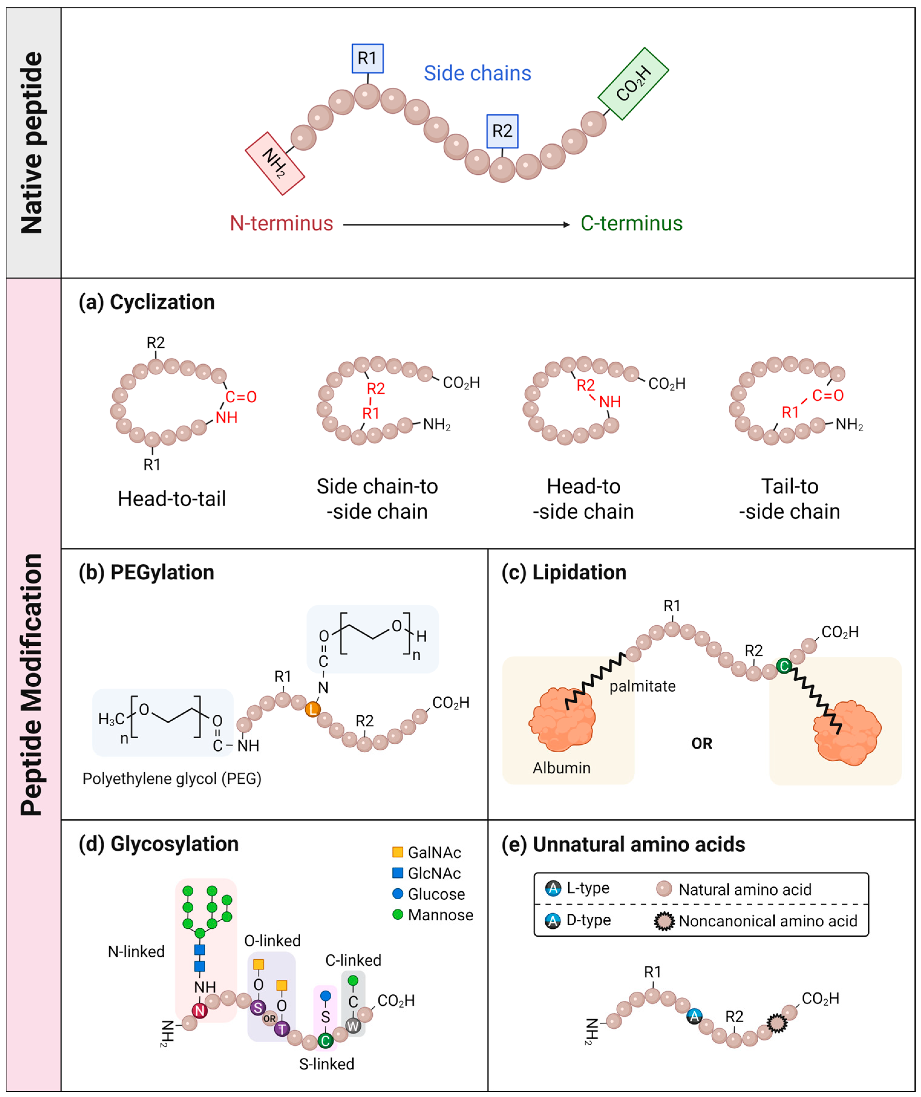
3.1.1. Cyclization
3.1.2. PEGylation
3.1.3. Lipidation
3.1.4. Glycosylation
3.1.5. D-Amino Acids and Non-Canonical Residues
4. Cell-Penetrating Peptide-Based Drug Delivery Systems
4.1. Cell-Penetrating Peptides
4.2. CPP-Based Drug Delivery to the Central Nervous System
4.3. The Pulmonary Drug Delivery System Based on CPPs
5. AI-Guided Peptide Drug Design and Predictive Modeling
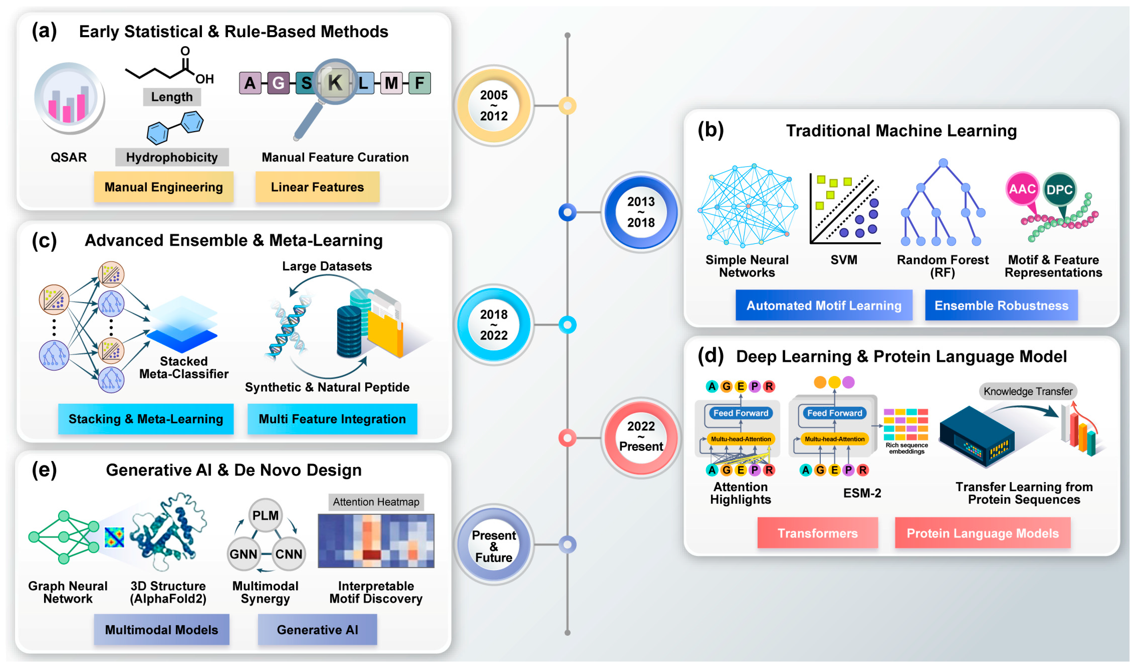
5.1. Evolution of Algorithmic Frameworks for Peptide Drug Development
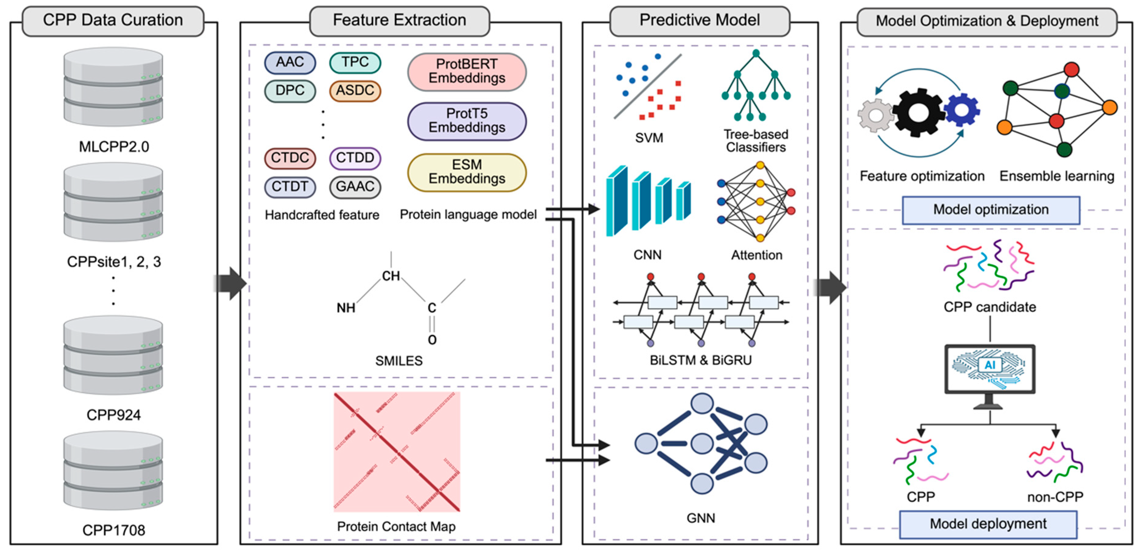
5.2. Artificial Intelligence Approaches to CPP Prediction
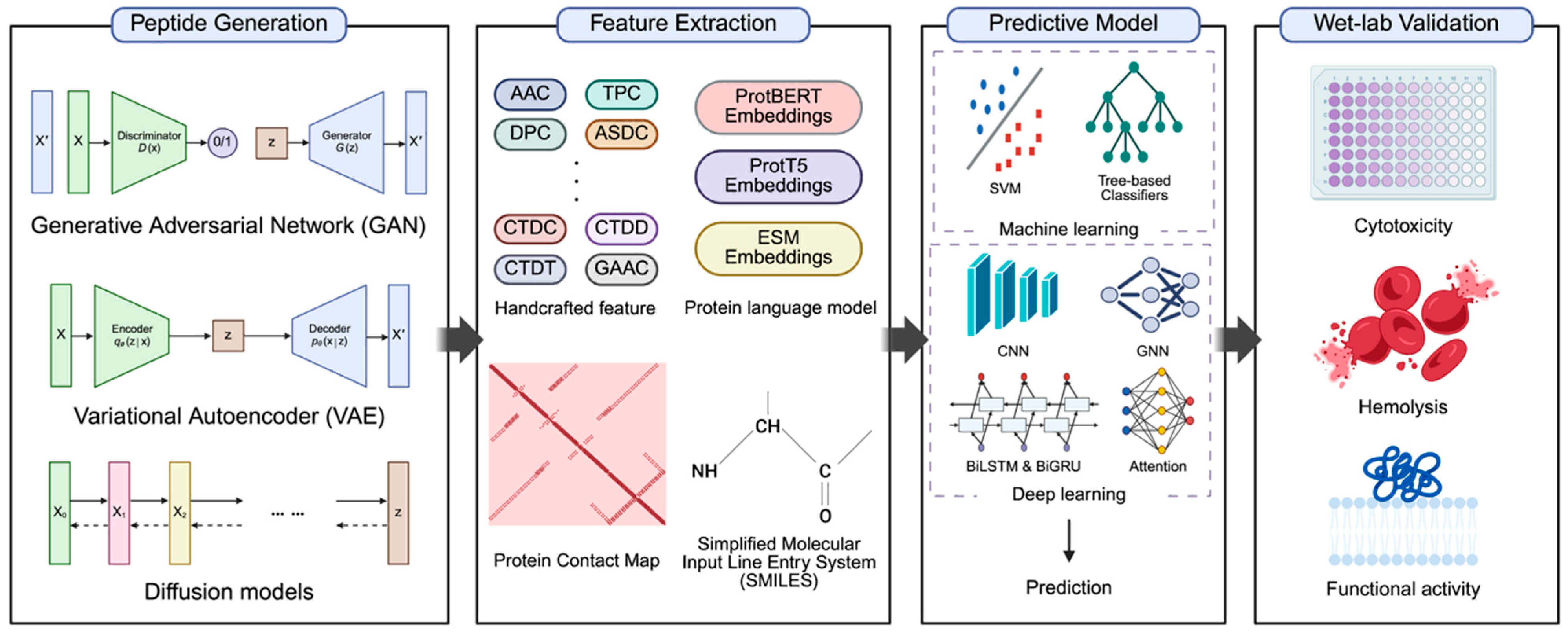
5.3. Computational Frameworks for AI-Driven Peptide Drug Discovery
6. Future Perspectives and Development Trends
7. Conclusions
Author Contributions
Funding
Institutional Review Board Statement
Informed Consent Statement
Data Availability Statement
Conflicts of Interest
Abbreviations
| AAC | Amino Acid Composition |
| ACPs | Anticancer Peptides |
| ADAs | Anti-Drug Antibodies |
| AI | Artificial Intelligence |
| Aib | α-isobutyric acid |
| AMPs | Antimicrobial Peptides |
| ASDC | Adaptive Skip Dipeptide Composition |
| AVPs | Antiviral Peptides |
| BBB | Blood–Brain Barrier |
| BPF | Binary Profile |
| BiGRU | Bidirectional Gated Recurrent Unit |
| BiLSTM | Bidirectional Long Short-Term Memory |
| CB | CatBoost |
| CD47 | Cluster of Differentiation 47 |
| CellPPD | Cell-Penetrating Peptide prediction database/tool |
| CKSAAGP | Composition of k-spaced amino acid group pairs |
| CNN | Convolutional Neural Network |
| CNS | Central Nervous System |
| CPP | Cell-Penetrating Peptide |
| CPSR | Composite Protein Sequence Representation |
| CTD | Composition Transition and Distribution |
| CTDC | Composition Transition and Distribution–Composition |
| CV | Cross-Validation |
| DPC | Dipeptide Composition |
| DL | Deep Learning |
| DPP-4 | Dipeptidyl Peptidase IV |
| DWT | Discrete Wavelet Transform |
| ESM | Evolutionary Scale Modeling |
| ESM-2 | Evolutionary Scale Modeling 2 |
| GAAC | Grouped Amino Acid Composition |
| GalNAc | N-acetylgalactosamine |
| GANs | Generative Adversarial Networks |
| GB | Gradient Boosting |
| GBM | Glioblastoma |
| GDF15 | Growth Differentiation Factor 15 |
| GIP | Glucose-Dependent Insulinotropic Polypeptide |
| GlcNAc | N-acetylglucosamine |
| GLP-1 | Glucagon-Like Peptide-1 |
| GLP-1RAs | Glucagon-Like Peptide-1 Receptor Agonists |
| GNN | Graph Neural Network |
| GPs | Glycoproteins |
| GH | Growth Hormone |
| HIV | Human Immunodeficiency Virus |
| imCNN | Improved Convolutional Neural Network |
| ITF | Information Theory Features |
| KELM | Kernel Extreme Learning Machine |
| KNN | k-Nearest Neighbor |
| LGB | Light Gradient Boosting |
| LOOCV | Leave-One-Out Cross-Validation |
| ML | Machine Learning |
| MLP | Multi-Layer Perceptron |
| mRNA | Messenger Ribonucleic Acid |
| NCAAs | Non-Canonical Amino Acids |
| NLCs | Nanostructured Lipid Carriers |
| PD-L1 | Programmed Death-Ligand 1 |
| PEI | Polyethylenimine |
| PEG | Polyethylene Glycol |
| PEO-PC7A | Amphiphilic block polymer used for pH-responsive delivery |
| PLGA | Poly(lactide-co-glycolide) |
| PLM | Protein Language Model |
| ProtBERT | Protein Bidirectional Encoder Representations from Transformers |
| ProtT5_XL_BFD | Protein text-to-text transfer transformer extra large pre-trained on Big Fantastic Database |
| PTDs | Peptide-Transduction Domains |
| QSAR | Quantitative Structure-Activity Relationship |
| RECM | Residue Energy Content Matrix |
| RF | Random Forest |
| SAAC | Split Amino Acid Composition |
| SMILES | Simplified Molecular Input Line Entry System |
| SLNs | Solid Lipid Nanoparticles |
| SVM | Support Vector Machine |
| TPC | Tripeptide Composition |
| VAE | Variational Autoencoder |
| XGB | Extreme Gradient Boosting |
References
- Wang, L.; Wang, N.; Zhang, W.; Cheng, X.; Yan, Z.; Shao, G.; Wang, X.; Wang, R.; Fu, C. Therapeutic peptides: Current applications and future directions. Signal Transduct. Target. Ther. 2022, 7, 48. [Google Scholar] [CrossRef] [Scilit]
- Liu, M.; Svirskis, D.; Proft, T.; Loh, J.; Yin, N.; Li, H.; Li, D.; Zhou, Y.; Chen, S.; Song, L.; et al. Progress in peptide and protein therapeutics: Challenges and strategies. Acta Pharm. Sin. B 2025, 15, 6342–6381. [Google Scholar] [CrossRef] [Scilit]
- Banting, F.G.; Best, C.H.; Collip, J.B.; Campbell, W.R.; Fletcher, A.A. Pancreatic Extracts in the Treatment of Diabetes Mellitus. Can. Med. Assoc. J. 1922, 12, 141–146. [Google Scholar]
- Du Vigneaud, V.; Ressler, C.; Trippett, S. The sequence of amino acids in oxytocin, with a proposal for the structure of oxytocin. J. Biol. Chem. 1953, 205, 949–957. [Google Scholar] [CrossRef] [Scilit] [PubMed]
- Du Vigneaud, V. Oxytocin, the principal oxytocic hormone of the posterior pituitary gland: Its isolation, structure, and synthesis. Experientia 1955, 9–26. [Google Scholar]
- Muttenthaler, M.; King, G.F.; Adams, D.J.; Alewood, P.F. Trends in peptide drug discovery. Nat. Rev. Drug Discov. 2021, 20, 309–325. [Google Scholar] [CrossRef] [Scilit]
- Schmid, H. Peginesatide for the treatment of renal disease-induced anemia. Expert. Opin. Pharmacother. 2013, 14, 937–948. [Google Scholar] [CrossRef] [Scilit]
- Xiao, W.; Jiang, W.; Chen, Z.; Huang, Y.; Mao, J.; Zheng, W.; Hu, Y.; Shi, J. Advance in peptide-based drug development: Delivery platforms, therapeutics and vaccines. Signal Transduct. Target. Ther. 2025, 10, 74. [Google Scholar] [CrossRef] [Scilit]
- Gare, C.L.; White, A.M.; Malins, L.R. From lead to market: Chemical approaches to transform peptides into therapeutics. Trends Biochem. Sci. 2025, 50, 467–480. [Google Scholar] [CrossRef] [Scilit] [PubMed]
- Barman, P.; Joshi, S.; Sharma, S.; Preet, S.; Sharma, S.; Saini, A. Strategic Approaches to Improvise Peptide Drugs as Next Generation Therapeutics. Int. J. Pept. Res. Ther. 2023, 29, 61. [Google Scholar] [CrossRef] [Scilit]
- Julke, E.M.; Beck-Sickinger, A.G. Peptide therapeutics: Current status and future opportunity with focus on nose-to-brain delivery☆. Peptides 2025, 188, 171404. [Google Scholar] [CrossRef] [Scilit]
- Liu, Q.K. Mechanisms of action and therapeutic applications of GLP-1 and dual GIP/GLP-1 receptor agonists. Front. Endocrinol. 2024, 15, 1431292. [Google Scholar] [CrossRef] [Scilit]
- Drucker, D.J. Advances in oral peptide therapeutics. Nat. Rev. Drug Discov. 2020, 19, 277–289. [Google Scholar] [CrossRef] [Scilit] [PubMed]
- Lau, J.; Bloch, P.; Schaffer, L.; Pettersson, I.; Spetzler, J.; Kofoed, J.; Madsen, K.; Knudsen, L.B.; McGuire, J.; Steensgaard, D.B.; et al. Discovery of the Once-Weekly Glucagon-Like Peptide-1 (GLP-1) Analogue Semaglutide. J. Med. Chem. 2015, 58, 7370–7380. [Google Scholar] [CrossRef] [Scilit] [PubMed]
- Kaspar, A.A.; Reichert, J.M. Future directions for peptide therapeutics development. Drug Discov. Today 2013, 18, 807–817. [Google Scholar] [CrossRef] [Scilit]
- Miwa, A.; Kamiya, K. Cell-Penetrating Peptide-Mediated Biomolecule Transportation in Artificial Lipid Vesicles and Living Cells. Molecules 2024, 29, 3339. [Google Scholar] [CrossRef] [Scilit] [PubMed]
- Manavalan, B.; Subramaniyam, S.; Shin, T.H.; Kim, M.O.; Lee, G. Machine-Learning-Based Prediction of Cell-Penetrating Peptides and Their Uptake Efficiency with Improved Accuracy. J. Proteome Res. 2018, 17, 2715–2726. [Google Scholar] [CrossRef] [Scilit]
- Brugnano, J.; Ward, B.C.; Panitch, A. Cell penetrating peptides can exert biological activity: A review. Biomol. Concepts 2010, 1, 109–116. [Google Scholar] [CrossRef] [Scilit]
- Ekins, S.; Puhl, A.C.; Zorn, K.M.; Lane, T.R.; Russo, D.P.; Klein, J.J.; Hickey, A.J.; Clark, A.M. Exploiting machine learning for end-to-end drug discovery and development. Nat. Mater. 2019, 18, 435–441. [Google Scholar] [CrossRef] [Scilit]
- Zhai, S.; Liu, T.; Lin, S.; Li, D.; Liu, H.; Yao, X.; Hou, T. Artificial intelligence in peptide-based drug design. Drug Discov. Today 2025, 30, 104300. [Google Scholar] [CrossRef] [Scilit]
- Manavalan, B.; Basith, S.; Lee, G. Comparative analysis of machine learning-based approaches for identifying therapeutic peptides targeting SARS-CoV-2. Brief. Bioinform. 2022, 23, bbab412. [Google Scholar]
- Baek, M.; DiMaio, F.; Anishchenko, I.; Dauparas, J.; Ovchinnikov, S.; Lee, G.R.; Wang, J.; Cong, Q.; Kinch, L.N.; Schaeffer, R.D. Accurate prediction of protein structures and interactions using a three-track neural network. Science 2021, 373, 871–876. [Google Scholar] [CrossRef] [Scilit] [PubMed]
- Jumper, J.; Evans, R.; Pritzel, A.; Green, T.; Figurnov, M.; Ronneberger, O.; Tunyasuvunakool, K.; Bates, R.; Žídek, A.; Potapenko, A. Highly accurate protein structure prediction with AlphaFold. Nature 2021, 596, 583–589. [Google Scholar] [CrossRef] [Scilit]
- Jiménez-Luna, J.; Grisoni, F.; Weskamp, N.; Schneider, G. Artificial intelligence in drug discovery: Recent advances and future perspectives. Expert. Opin. Drug Discov. 2021, 16, 949–959. [Google Scholar] [CrossRef] [Scilit]
- Askr, H.; Elgeldawi, E.; Aboul Ella, H.; Elshaier, Y.A.; Gomaa, M.M.; Hassanien, A.E. Deep learning in drug discovery: An integrative review and future challenges. Artif. Intell. Rev. 2023, 56, 5975–6037. [Google Scholar] [CrossRef] [Scilit]
- Hasan, M.M.; Schaduangrat, N.; Basith, S.; Lee, G.; Shoombuatong, W.; Manavalan, B. HLPpred-Fuse: Improved and robust prediction of hemolytic peptide and its activity by fusing multiple feature representation. Bioinformatics 2020, 36, 3350–3356. [Google Scholar] [CrossRef] [Scilit]
- Manavalan, B.; Shin, T.H.; Kim, M.O.; Lee, G. AIPpred: Sequence-based prediction of anti-inflammatory peptides using random forest. Front. Pharmacol. 2018, 9, 276. [Google Scholar] [CrossRef] [Scilit] [PubMed]
- Goles, M.; Daza, A.; Cabas-Mora, G.; Sarmiento-Varón, L.; Sepúlveda-Yañez, J.; Anvari-Kazemabad, H.; Davari, M.D.; Uribe-Paredes, R.; Olivera-Nappa, Á.; Navarrete, M.A.; et al. Peptide-based drug discovery through artificial intelligence: Towards an autonomous design of therapeutic peptides. Brief. Bioinform. 2024, 25, bbae275. [Google Scholar] [CrossRef] [Scilit] [PubMed]
- Elnaggar, A.; Heinzinger, M.; Dallago, C.; Rehawi, G.; Wang, Y.; Jones, L.; Gibbs, T.; Feher, T.; Angerer, C.; Steinegger, M. Prottrans: Toward understanding the language of life through self-supervised learning. IEEE Trans. Pattern Anal. Mach. Intell. 2021, 44, 7112–7127. [Google Scholar] [CrossRef] [Scilit]
- Hwang, J.S.; Kim, S.G.; Shin, T.H.; Jang, Y.E.; Kwon, D.H.; Lee, G. Development of Anticancer Peptides Using Artificial Intelligence and Combinational Therapy for Cancer Therapeutics. Pharmaceutics 2022, 14, 997. [Google Scholar] [CrossRef] [Scilit]
- Wang, D.; Yin, F.; Li, Z.; Zhang, Y.; Shi, C. Current progress and remaining challenges of peptide-drug conjugates (PDCs): Next generation of antibody-drug conjugates (ADCs)? J. Nanobiotechnol. 2025, 23, 305. [Google Scholar] [CrossRef] [Scilit]
- Ekambaram, S.; Dokholyan, N.V. Peptide-based drug design using generative AI. Chem. Commun. 2026, 62, 672–691. [Google Scholar] [CrossRef] [Scilit] [PubMed]
- Sun, Y.; Zhang, M.; Liu, H.; Wang, H. Emerging landscape of bioinformatics and artificial intelligence applications in cell-penetrating peptide-based delivery. Expert. Opin. Drug Deliv. 2026, 23, 495–515. [Google Scholar] [CrossRef] [Scilit]
- Sutcliffe, R.; Doherty, C.P.; Morgan, H.P.; Dunne, N.J.; Mccarthy, H.O. Strategies for the design of biomimetic cell-penetrating peptides using AI-driven in silico tools for drug delivery. Biomater. Adv. 2025, 169, 214153. [Google Scholar] [CrossRef] [Scilit] [PubMed]
- Choi, Y.; Vinks, A.A.; van der Graaf, P.H. Novel Therapeutic Modalities: The Future is Now. Clin. Pharmacol. Ther. 2023, 114, 493–496. [Google Scholar] [CrossRef] [Scilit]
- Zheng, B.; Wang, X.; Guo, M.; Tzeng, C.-M. Therapeutic Peptides: Recent Advances in Discovery, Synthesis, and Clinical Translation. Int. J. Mol. Sci. 2025, 26, 5131. [Google Scholar] [CrossRef] [Scilit]
- Henninot, A.; Collins, J.C.; Nuss, J.M. The Current State of Peptide Drug Discovery: Back to the Future? J. Med. Chem. 2018, 61, 1382–1414. [Google Scholar] [CrossRef] [Scilit] [PubMed]
- Fosgerau, K.; Hoffmann, T. Peptide therapeutics: Current status and future directions. Drug Discov. Today 2015, 20, 122–128. [Google Scholar] [CrossRef] [Scilit]
- Stein Gold, L.; Eyerich, K.; Merola, J.F.; Torres, J.; Coates, L.C.; Allegretti, J.R. Oral peptide therapeutics as an emerging treatment modality in immune-mediated inflammatory diseases: A narrative review. Adv. Ther. 2025, 42, 3158–3172. [Google Scholar] [CrossRef] [Scilit]
- Bissonnette, R.; Soung, J.; Hebert, A.A.; Pink, A.E.; Pinter, A.; Moore, A.Y.; Shi, Y.; Wang, W.-h.; Toth, D.P.; Miller-Kassamali, M. Oral icotrokinra for plaque psoriasis in adults and adolescents. N. Engl. J. Med. 2025, 393, 1784–1795. [Google Scholar] [CrossRef] [Scilit]
- Knight, B.; Tammara, B.; Modi, N.B.; Dallas, S.; Mardirosian, S.; Wang, J.; Laenen, A.; Leclercq, L.; DiLoreto, K.; Adriaenssen, L. Translational pharmacokinetics of icotrokinra, a targeted oral peptide that selectively blocks interleukin-23 receptor and inhibits signaling. Dermatol. Ther. 2025, 15, 2495–2520. [Google Scholar] [CrossRef] [Scilit]
- Fourie, A.M.; Cheng, X.; Chang, L.; Greving, C.; Li, X.; Knight, B.; Polidori, D.; Patrick, A.; Bains, T.; Steele, R. JNJ-77242113, a highly potent, selective peptide targeting the IL-23 receptor, provides robust IL-23 pathway inhibition upon oral dosing in rats and humans. Sci. Rep. 2024, 14, 17515. [Google Scholar] [CrossRef] [Scilit]
- Roopenian, D.C.; Akilesh, S. FcRn: The neonatal Fc receptor comes of age. Nat. Rev. Immunol. 2007, 7, 715–725. [Google Scholar] [CrossRef] [Scilit]
- Lau, J.L.; Dunn, M.K. Therapeutic peptides: Historical perspectives, current development trends, and future directions. Bioorg. Med. Chem. 2018, 26, 2700–2707. [Google Scholar] [CrossRef] [Scilit] [PubMed]
- Muheem, A.; Shakeel, F.; Jahangir, M.A. A review on the strategies for oral delivery of proteins and peptides and their clinical perspectives. Saudi Pharm. J. 2016, 24, 413–428. [Google Scholar] [CrossRef] [Scilit]
- Hamley, I.W. Small bioactive peptides for biomaterials design and therapeutics. Chem. Rev. 2017, 117, 14015–14041. [Google Scholar] [CrossRef] [Scilit]
- Zhao, Z.; Ukidve, A.; Kim, J.; Mitragotri, S. Targeting strategies for tissue-specific drug delivery. Cell 2020, 181, 151–167. [Google Scholar] [CrossRef] [Scilit]
- Bhagavat, R.; Sankar, S.; Srinivasan, N.; Chandra, N. An augmented pocketome: Detection and analysis of small-molecule binding pockets in proteins of known 3D structure. Structure 2018, 26, 499–512. [Google Scholar] [CrossRef] [Scilit]
- Vadevoo, S.M.P.; Gurung, S.; Lee, H.-S.; Gunassekaran, G.R.; Lee, S.-M.; Yoon, J.-W.; Lee, Y.-K.; Lee, B. Peptides as multifunctional players in cancer therapy. Exp. Mol. Med. 2023, 55, 1099–1109. [Google Scholar] [CrossRef] [Scilit] [PubMed]
- Vlieghe, P.; Lisowski, V.; Martinez, J.; Khrestchatisky, M. Synthetic therapeutic peptides: Science and market. Drug Discov. Today 2010, 15, 40–56. [Google Scholar] [CrossRef] [Scilit] [PubMed]
- Zheng, Z.; Zong, Y.; Ma, Y.; Tian, Y.; Pang, Y.; Zhang, C.; Gao, J. Glucagon-like peptide-1 receptor: Mechanisms and advances in therapy. Signal Transduct. Target. Ther. 2024, 9, 234. [Google Scholar] [CrossRef] [Scilit]
- Rai, U.; Thrimawithana, T.R.; Valery, C.; Young, S.A. Therapeutic uses of somatostatin and its analogues: Current view and potential applications. Pharmacol. Ther. 2015, 152, 98–110. [Google Scholar] [CrossRef] [Scilit]
- Drazic, A.; Winter, J. The physiological role of reversible methionine oxidation. Biochim. Biophys. Acta 2014, 1844, 1367–1382. [Google Scholar] [CrossRef] [Scilit] [PubMed]
- Irudayanathan, F.J.; Zarzar, J.; Lin, J.; Izadi, S. Deciphering deamidation and isomerization in therapeutic proteins: Effect of neighboring residue. MAbs 2022, 14, 2143006. [Google Scholar] [CrossRef] [Scilit] [PubMed]
- De Groot, A.S.; Roberts, B.J.; Mattei, A.; Lelias, S.; Boyle, C.; Martin, W.D. Immunogenicity risk assessment of synthetic peptide drugs and their impurities. Drug Discov. Today 2023, 28, 103714. [Google Scholar] [CrossRef] [Scilit]
- Achilleos, K.; Petrou, C.; Nicolaidou, V.; Sarigiannis, Y. Beyond Efficacy: Ensuring Safety in Peptide Therapeutics through Immunogenicity Assessment. J. Pept. Sci. 2025, 31, e70016. [Google Scholar] [CrossRef] [Scilit] [PubMed]
- Mullins, G.R.; Hodsdon, M.E.; Li, Y.G.; Anglin, G.; Urva, S.; Schneck, K.; Bardos, J.N.; Martins, R.F.; Brown, K.; Calderon, B. Tirzepatide Immunogenicity on Pharmacokinetics, Efficacy, and Safety: Analysis of Data From Phase 3 Studies. J. Clin. Endocrinol. Metab. 2024, 109, 361–369. [Google Scholar] [CrossRef] [Scilit]
- Bogdanowich-Knipp, S.; Jois, D.; Siahaan, T. The effect of conformation on the solution stability of linear vs. cyclic RGD peptides. J. Pept. Res. 1999, 53, 523–529. [Google Scholar] [CrossRef] [Scilit]
- Tasdemiroglu, Y.; Gourdie, R.G.; He, J.-Q. In vivo degradation forms, anti-degradation strategies, and clinical applications of therapeutic peptides in non-infectious chronic diseases. Eur. J. Pharmacol. 2022, 932, 175192. [Google Scholar] [CrossRef] [Scilit]
- Ngambenjawong, C.; Pineda, J.M.B.; Pun, S.H. Engineering an affinity-enhanced peptide through optimization of cyclization chemistry. Bioconjug. Chem. 2016, 27, 2854–2862. [Google Scholar] [CrossRef] [Scilit]
- Villequey, C.; Zurmühl, S.S.; Cramer, C.N.; Bhusan, B.; Andersen, B.; Ren, Q.; Liu, H.; Qu, X.; Yang, Y.; Pan, J. An efficient mRNA display protocol yields potent bicyclic peptide inhibitors for FGFR3c: Outperforming linear and monocyclic formats in affinity and stability. Chem. Sci. 2024, 15, 6122–6129. [Google Scholar] [CrossRef] [Scilit]
- Merz, M.L.; Habeshian, S.; Li, B.; David, J.-A.G.; Nielsen, A.L.; Ji, X.; Il Khwildy, K.; Duany Benitez, M.M.; Phothirath, P.; Heinis, C. De novo development of small cyclic peptides that are orally bioavailable. Nat. Chem. Biol. 2024, 20, 624–633. [Google Scholar] [CrossRef] [Scilit]
- Joo, S.H. Cyclic peptides as therapeutic agents and biochemical tools. Biomol. Ther. 2012, 20, 19. [Google Scholar] [CrossRef] [Scilit] [PubMed]
- Bechtler, C.; Lamers, C. Macrocyclization strategies for cyclic peptides and peptidomimetics. RSC Med. Chem. 2021, 12, 1325–1351. [Google Scholar] [CrossRef] [Scilit] [PubMed]
- White, C.J.; Yudin, A.K. Contemporary strategies for peptide macrocyclization. Nat. Chem. 2011, 3, 509–524. [Google Scholar] [CrossRef] [Scilit]
- Li, L.; Li, R.; Jiang, Y.; Chao, J.; Chen, S.; Liao, H.; Li, X. Advances in Hydrocarbon Stapled Peptides via Ring-Closing Metathesis: Synthetic Strategies, Structural Diversity, and Therapeutic Applications. ChemBioChem 2025, 26, e202500527. [Google Scholar] [CrossRef] [Scilit]
- Li, Y.; Wu, M.; Fu, Y.; Xue, J.; Yuan, F.; Qu, T.; Rissanou, A.N.; Wang, Y.; Li, X.; Hu, H. Therapeutic stapled peptides: Efficacy and molecular targets. Pharmacol. Res. 2024, 203, 107137. [Google Scholar] [CrossRef] [Scilit]
- Ferková, S.; Froehlich, U.; Nepveu-Traversy, M.-É.; Murza, A.; Azad, T.; Grandbois, M.; Sarret, P.; Lavigne, P.; Boudreault, P.-L. Comparative Analysis of Cyclization Techniques in Stapled Peptides: Structural Insights into Protein–Protein Interactions in a SARS-CoV-2 Spike RBD/hACE2 Model System. Int. J. Mol. Sci. 2023, 25, 166. [Google Scholar] [CrossRef] [Scilit]
- Walensky, L.D.; Kung, A.L.; Escher, I.; Malia, T.J.; Barbuto, S.; Wright, R.D.; Wagner, G.; Verdine, G.L.; Korsmeyer, S.J. Activation of apoptosis in vivo by a hydrocarbon-stapled BH3 helix. Science 2004, 305, 1466–1470. [Google Scholar] [CrossRef] [Scilit] [PubMed]
- Mitra, S.; Chen, M.-T.; Stedman, F.; Hernandez, J.; Kumble, G.; Kang, X.; Zhang, C.; Tang, G.; Reed, I.; Daugherty, I.Q. Cyclization of two antimicrobial peptides improves their activity. Acs Omega 2025, 10, 9728–9740. [Google Scholar] [CrossRef] [Scilit]
- Chandramohan, A.; Josien, H.; Yuen, T.Y.; Duggal, R.; Spiegelberg, D.; Yan, L.; Juang, Y.-C.A.; Ge, L.; Aronica, P.G.; Kaan, H.Y.K. Design-rules for stapled peptides with in vivo activity and their application to Mdm2/X antagonists. Nat. Commun. 2024, 15, 489. [Google Scholar] [CrossRef] [Scilit]
- Dietrich, L.; Rathmer, B.; Ewan, K.; Bange, T.; Heinrichs, S.; Dale, T.C.; Schade, D.; Grossmann, T.N. Cell permeable stapled peptide inhibitor of Wnt signaling that targets β-catenin protein-protein interactions. Cell Chem. Biol. 2017, 24, 958–968. [Google Scholar] [CrossRef] [Scilit] [PubMed]
- Jin, X.; Ding, N.; Guo, H.-Y.; Hu, Q. Macrocyclic-based strategy in drug design: From lab to the clinic. Eur. J. Med. Chem. 2024, 277, 116733. [Google Scholar] [CrossRef] [Scilit] [PubMed]
- DeLorbe, J.E.; Clements, J.H.; Whiddon, B.B.; Martin, S.F. Thermodynamic and structural effects of macrocyclic constraints in protein− ligand interactions. ACS Med. Chem. Lett. 2010, 1, 448–452. [Google Scholar] [CrossRef] [Scilit] [PubMed]
- Veronese, F.M.; Pasut, G. PEGylation, successful approach to drug delivery. Drug Discov. Today 2005, 10, 1451–1458. [Google Scholar] [CrossRef] [Scilit]
- Lawrence, P.B.; Price, J.L. How PEGylation influences protein conformational stability. Curr. Opin. Chem. Biol. 2016, 34, 88–94. [Google Scholar] [CrossRef] [Scilit]
- Cui, Q.; Xu, Q.-j.; Liu, L.; Guan, L.-l.; Jiang, X.-y.; Inam, M.; Kong, L.-c.; Ma, H.-X. Preparation, characterization and pharmacokinetic study of N-terminal PEGylated D-form antimicrobial peptide OM19r-8. J. Pharm. Sci. 2021, 110, 1111–1119. [Google Scholar] [CrossRef] [Scilit]
- Benincasa, M.; Zahariev, S.; Pelillo, C.; Milan, A.; Gennaro, R.; Scocchi, M. PEGylation of the peptide Bac7 (1–35) reduces renal clearance while retaining antibacterial activity and bacterial cell penetration capacity. Eur. J. Med. Chem. 2015, 95, 210–219. [Google Scholar]
- Li, C.; Li, T.; Tian, X.; An, W.; Wang, Z.; Han, B.; Tao, H.; Wang, J.; Wang, X. Research progress on the PEGylation of therapeutic proteins and peptides (TPPs). Front. Pharmacol. 2024, 15, 1353626. [Google Scholar] [CrossRef] [Scilit]
- Ross, R.; Leung, K.; Maamra, M.; Bennett, W.; Doyle, N.; Waters, M.; Ho, K. Binding and functional studies with the growth hormone receptor antagonist, B2036-PEG (pegvisomant), reveal effects of pegylation and evidence that it binds to a receptor dimer. J. Clin. Endocrinol. Metab. 2001, 86, 1716–1723. [Google Scholar]
- Brue, T.; Chanson, P.; Drui, D.; Cristante, J.; Espiard, S.; Peyrat, L.; Aïssani, W.; Raverot, G. Pegvisomant in managing comorbidities of acromegaly: A review. Rev. Endocr. Metab. Disord. 2025, 27, 109–122. [Google Scholar] [CrossRef] [Scilit]
- Veronese, F.M. Peptide and protein PEGylation: A review of problems and solutions. Biomaterials 2001, 22, 405–417. [Google Scholar] [CrossRef] [Scilit] [PubMed]
- Knudsen, L.B.; Lau, J. The discovery and development of liraglutide and semaglutide. Front. Endocrinol. 2019, 10, 155. [Google Scholar] [CrossRef] [Scilit] [PubMed]
- Hutchinson, J.A.; Hamley, I.W.; Torras, J.; Alemán, C.; Seitsonen, J.; Ruokolainen, J. Self-assembly of lipopeptides containing short peptide fragments derived from the gastrointestinal hormone PYY3–36: From micelles to amyloid fibrils. J. Phys. Chem. B. 2019, 123, 614–621. [Google Scholar] [CrossRef] [Scilit]
- Kurtzhals, P.; Havelund, S.; Jonassen, I.; Kiehr, B.; Larsen, U.; Ribel, U.; Markussen, J. Albumin binding of insulins acylated with fatty acids: Characterization of the ligand-protein interaction and correlation between binding affinity and timing of the insulin effect in vivo. Biochem. J. 1995, 312, 725–731. [Google Scholar] [CrossRef] [Scilit]
- Menacho-Melgar, R.; Decker, J.S.; Hennigan, J.N.; Lynch, M.D. A review of lipidation in the development of advanced protein and peptide therapeutics. J. Control. Release 2019, 295, 1–12. [Google Scholar] [CrossRef] [Scilit]
- Curry, S. Lessons from the crystallographic analysis of small molecule binding to human serum albumin. Drug Metab. Pharmacokinet. 2009, 24, 342–357. [Google Scholar] [CrossRef] [Scilit]
- Friedman, J.M. The discovery and development of GLP-1 based drugs that have revolutionized the treatment of obesity. Proc. Natl. Acad. Sci. USA 2024, 121, e2415550121. [Google Scholar] [CrossRef] [Scilit] [PubMed]
- Přáda Brichtová, E.; Edu, I.A.; Li, X.; Becher, F.; Gomes dos Santos, A.L.; Jackson, S.E. Effect of Lipidation on the Structure, Oligomerization, and Aggregation of Glucagon-like Peptide 1. Bioconjug. Chem. 2025, 36, 401–414. [Google Scholar] [CrossRef] [Scilit] [PubMed]
- Zorzi, A.; Middendorp, S.J.; Wilbs, J.; Deyle, K.; Heinis, C. Acylated heptapeptide binds albumin with high affinity and application as tag furnishes long-acting peptides. Nat. Commun. 2017, 8, 16092. [Google Scholar] [CrossRef] [Scilit]
- Madsen, T.D.; Hansen, L.H.; Hintze, J.; Ye, Z.; Jebari, S.; Andersen, D.B.; Joshi, H.J.; Ju, T.; Goetze, J.P.; Martin, C. An atlas of O-linked glycosylation on peptide hormones reveals diverse biological roles. Nat. Commun. 2020, 11, 4033. [Google Scholar] [CrossRef] [Scilit]
- Wei, Q.; Zhang, J.; Dao, Y.; Ye, M.; Liu, D.; Dong, W.; Yuan, N.; Li, H.; Song, C.; Li, M. Exploiting Complex-Type N-Glycan to Improve the in Vivo Stability of Bioactive Peptides. CCS Chem. 2023, 5, 1623–1634. [Google Scholar] [CrossRef] [Scilit]
- Chandrashekar, C.; Hossain, M.A.; Wade, J.D. Chemical glycosylation and its application to glucose homeostasis-regulating peptides. Front. Chem. 2021, 9, 650025. [Google Scholar] [CrossRef] [Scilit] [PubMed]
- Moradi, S.V.; Hussein, W.M.; Varamini, P.; Simerska, P.; Toth, I. Glycosylation, an effective synthetic strategy to improve the bioavailability of therapeutic peptides. Chem. Sci. 2016, 7, 2492–2500. [Google Scholar] [CrossRef] [Scilit]
- Cui, S.; Jin, Z.; Yu, T.; Guo, C.; He, Y.; Kan, Y.; Yan, L.; Wu, L. Effect of glycosylation on the enzymatic degradation of D-amino acid-containing peptides. Molecules 2025, 30, 441. [Google Scholar] [CrossRef] [Scilit] [PubMed]
- Bellavita, R.; Braccia, S.; Galdiero, S.; Falanga, A. Glycosylation and lipidation strategies: Approaches for improving antimicrobial peptide efficacy. Pharmaceuticals 2023, 16, 439. [Google Scholar] [CrossRef] [Scilit]
- Ochiai, H.; Shimoda, T.; Fukae, K.; Maeda, M.; Ishii, K.; Yoshida, K.; Tezuka, K.; Tazuru, K.; Saijo, H.; Asai, H. Better Peptides via Chemical Glycosylation: Somatostatin Analogues Having a Human Complex-Type N-Glycan with Improved Drug Properties. Chemistry 2023, 29, e202300111. [Google Scholar] [CrossRef] [Scilit]
- Bosques, C.J.; Imperiali, B. The interplay of glycosylation and disulfide formation influences fibrillization in a prion protein fragment. Proc. Natl. Acad. Sci. USA 2003, 100, 7593–7598. [Google Scholar] [CrossRef] [Scilit] [PubMed]
- Garton, M.; Nim, S.; Stone, T.A.; Wang, K.E.; Deber, C.M.; Kim, P.M. Method to generate highly stable D-amino acid analogs of bioactive helical peptides using a mirror image of the entire PDB. Proc. Natl. Acad. Sci. USA 2018, 115, 1505–1510. [Google Scholar] [CrossRef] [Scilit]
- Bruno, B.J.; Miller, G.D.; Lim, C.S. Basics and recent advances in peptide and protein drug delivery. Ther. Deliv. 2013, 4, 1443–1467. [Google Scholar] [CrossRef] [Scilit]
- Miyamoto, T.; Homma, H. D-Amino acid metabolism in bacteria. J. Biochem. 2021, 170, 5–13. [Google Scholar] [CrossRef] [Scilit] [PubMed]
- Seif, S.M.; Zenser, T.V.; Ciarochi, F.F.; Davis, B.B.; Robinson, A.G. DDAVP (l-Desamino-8-D-arginine-vasopressin) Treatment of Central Diabetes Insipidus—Mechanism of Prolonged Antidiuresis. J. Clin. Endocrinol. Metab. 1978, 46, 381–388. [Google Scholar] [CrossRef] [Scilit]
- Liu, M.; Li, C.; Pazgier, M.; Li, C.; Mao, Y.; Lv, Y.; Gu, B.; Wei, G.; Yuan, W.; Zhan, C. D-peptide inhibitors of the p53–MDM2 interaction for targeted molecular therapy of malignant neoplasms. Proc. Natl. Acad. Sci. USA 2010, 107, 14321–14326. [Google Scholar] [CrossRef] [Scilit]
- Yakusheva, A.; Titchenko, N.; Egorova, B.; Matazova, E.; Podkhalyuzina, N.; Osipov, V.; Khachatryan, D.; Avdeev, D.; Posypanova, G.; Kalmykov, S. From octreotide to shorter analogues: Synthesis, radiolabeling, stability. J. Label. Comp. Radiopharm. 2019, 62, 718–728. [Google Scholar] [CrossRef] [Scilit]
- Adhikari, A.; Bhattarai, B.R.; Aryal, A.; Thapa, N.; Kc, P.; Adhikari, A.; Maharjan, S.; Chanda, P.B.; Regmi, B.P.; Parajuli, N. Reprogramming natural proteins using unnatural amino acids. RSC Adv. 2021, 11, 38126–38145. [Google Scholar] [CrossRef] [Scilit]
- Castro, T.G.; Melle-Franco, M.; Sousa, C.E.; Cavaco-Paulo, A.; Marcos, J.C. Non-canonical amino acids as building blocks for peptidomimetics: Structure, function, and applications. Biomolecules 2023, 13, 981. [Google Scholar] [CrossRef] [Scilit]
- Mendel, D.; Ellman, J.; Schultz, P.G. Protein biosynthesis with conformationally restricted amino acids. J. Am. Chem. Soc. 1993, 115, 4359–4360. [Google Scholar] [CrossRef] [Scilit]
- Neumann-Staubitz, P.; Neumann, H. The use of unnatural amino acids to study and engineer protein function. Curr. Opin. Struct. Biol. 2016, 38, 119–128. [Google Scholar] [CrossRef] [Scilit] [PubMed]
- Azam, A.; Mallart, S.; Illiano, S.; Duclos, O.; Prades, C.; Maillère, B. Introduction of non-natural amino acids into T-cell epitopes to mitigate peptide-specific T-cell responses. Front. Immunol. 2021, 12, 637963. [Google Scholar] [CrossRef] [Scilit]
- Neto, T.A.P.; Sidney, J.; Grifoni, A.; Sette, A. Correlative CD4 and CD8 T-cell immunodominance in humans and mice: Implications for preclinical testing. Cell Mol. Immunol. 2023, 20, 1328–1338. [Google Scholar] [CrossRef] [Scilit]
- Mattei, A.E.; Gutierrez, A.H.; Martin, W.D.; Terry, F.E.; Roberts, B.J.; Rosenberg, A.S.; De Groot, A.S. In silico immunogenicity assessment for sequences containing unnatural amino acids: A method using existing in silico algorithm infrastructure and a vision for future enhancements. Front. Drug Discov. 2022, 2, 952326. [Google Scholar] [CrossRef] [Scilit]
- Heitz, F.; Morris, M.C.; Divita, G. Twenty years of cell-penetrating peptides: From molecular mechanisms to therapeutics. Br. J. Pharmacol. 2009, 157, 195–206. [Google Scholar] [CrossRef] [Scilit] [PubMed]
- Copolovici, D.M.; Langel, K.; Eriste, E.; Langel, U. Cell-penetrating peptides: Design, synthesis, and applications. ACS Nano 2014, 8, 1972–1994. [Google Scholar] [CrossRef] [Scilit]
- Blanco, E.; Shen, H.; Ferrari, M. Principles of nanoparticle design for overcoming biological barriers to drug delivery. Nat. Biotechnol. 2015, 33, 941–951. [Google Scholar] [CrossRef] [Scilit]
- Zhou, X.; Smith, Q.R.; Liu, X. Brain penetrating peptides and peptide–drug conjugates to overcome the blood–brain barrier and target CNS diseases. Wiley Interdiscip. Rev. Nanomed. Nanobiotechnol. 2021, 13, e1695. [Google Scholar] [CrossRef] [Scilit]
- Wen, J.; Singuru, G.; Stiltner, J.; Mishra, S.; Feldman, K.S.; McCandless, K.; Yurko, R.; Islam, K.; Frizzell, R.; Yagi, H. Novel Lung Cell-Penetrating Peptide Targets Alveolar Epithelial Type II Cells, Basal Cells, and Ionocytes. Pharmaceutics 2025, 17, 824. [Google Scholar] [CrossRef] [Scilit] [PubMed]
- Isakov, N. Cell-Penetrating Peptides as Modulators of Cell Signaling and Potential Therapeutics. 2014. Available online: https://api.semanticscholar.org/CorpusID:212457727 (accessed on 22 April 2026).
- Frankel, A.D.; Pabo, C.O. Cellular uptake of the tat protein from human immunodeficiency virus. Cell 1988, 55, 1189–1193. [Google Scholar] [CrossRef] [Scilit]
- Snyder, E.L.; Dowdy, S.F. Cell penetrating peptides in drug delivery. Pharm. Res. 2004, 21, 389–393. [Google Scholar] [CrossRef] [Scilit]
- Mi, Z.; Mai, J.; Lu, X.; Robbins, P.D. Characterization of a class of cationic peptides able to facilitate efficient protein transduction in vitro and in vivo. Mol. Ther. 2000, 2, 339–347. [Google Scholar] [CrossRef] [Scilit]
- De Coupade, C.; Fittipaldi, A.; Chagnas, V.; Michel, M.; Carlier, S.; Tasciotti, E.; Darmon, A.; Ravel, D.; Kearsey, J.; Giacca, M. Novel human-derived cell-penetrating peptides for specific subcellular delivery of therapeutic biomolecules. Biochem. J. 2005, 390, 407–418. [Google Scholar] [CrossRef] [Scilit] [PubMed]
- Wu, J.; Sahoo, J.K.; Li, Y.; Xu, Q.; Kaplan, D.L. Challenges in delivering therapeutic peptides and proteins: A silk-based solution. J. Control. Release 2022, 345, 176–189. [Google Scholar] [CrossRef] [Scilit] [PubMed]
- Behzadipour, Y.; Hemmati, S. Covalent conjugation and non-covalent complexation strategies for intracellular delivery of proteins using cell-penetrating peptides. Biomed. Pharmacother. 2024, 176, 116910. [Google Scholar] [CrossRef] [Scilit]
- Bolleddula, J.; Brady, K.; Bruin, G.; Lee, A.; Martin, J.A.; Walles, M.; Xu, K.; Yang, T.-Y.; Zhu, X.; Yu, H. Absorption, distribution, metabolism, and excretion of therapeutic proteins: Current industry practices and future perspectives. Drug Metab. Dispos. 2022, 50, 837–845. [Google Scholar] [CrossRef] [Scilit] [PubMed]
- Porello, I.; Cellesi, F. Intracellular delivery of therapeutic proteins. New advancements and future directions. Front. Bioeng. Biotechnol. 2023, 11, 1211798. [Google Scholar] [CrossRef] [Scilit]
- Hertel, S.P.; Winter, G.; Friess, W. Protein stability in pulmonary drug delivery via nebulization. Adv. Drug Deliv. Rev. 2015, 93, 79–94. [Google Scholar] [CrossRef] [Scilit]
- Campelo, S.N.; Huang, P.-H.; Buie, C.R.; Davalos, R.V. Recent advancements in electroporation technologies: From bench to clinic. Annu. Rev. Biomed. Eng. 2023, 25, 77–100. [Google Scholar] [CrossRef] [Scilit]
- Kollath, V.O.; Mullens, S.; Luyten, J.; Traina, K.; Cloots, R. Protein–calcium phosphate nanocomposites: Benchmarking protein loading via physical and chemical modifications against co-precipitation. RSC Adv. 2015, 5, 55625–55632. [Google Scholar] [CrossRef] [Scilit]
- Wang, J.; Chen, D.; Ho, E.A. Challenges in the development and establishment of exosome-based drug delivery systems. J. Control. Release 2021, 329, 894–906. [Google Scholar] [CrossRef] [Scilit]
- Meacham, J.M.; Durvasula, K.; Degertekin, F.L.; Fedorov, A.G. Physical methods for intracellular delivery: Practical aspects from laboratory use to industrial-scale processing. J. Lab. Autom. 2014, 19, 1–18. [Google Scholar] [CrossRef] [Scilit]
- Stewart, M.P.; Sharei, A.; Ding, X.; Sahay, G.; Langer, R.; Jensen, K.F. In vitro and ex vivo strategies for intracellular delivery. Nature 2016, 538, 183–192. [Google Scholar] [CrossRef] [Scilit]
- Gorantla, A.; Hall, J.T.; Troidle, A.; Janjic, J.M. Biomaterials for protein delivery: Opportunities and challenges to clinical translation. Micromachines 2024, 15, 533. [Google Scholar] [CrossRef] [Scilit]
- Berillo, D.; Yeskendir, A.; Zharkinbekov, Z.; Raziyeva, K.; Saparov, A. Peptide-based drug delivery systems. Medicina 2021, 57, 1209. [Google Scholar] [CrossRef] [Scilit]
- Wagstaff, K.M.; Jans, D.A. Protein transduction: Cell penetrating peptides and their therapeutic applications. Curr. Med. Chem. 2006, 13, 1371–1387. [Google Scholar] [CrossRef] [Scilit]
- Dinca, A.; Chien, W.-M.; Chin, M.T. Intracellular delivery of proteins with cell-penetrating peptides for therapeutic uses in human disease. Int. J. Mol. Sci. 2016, 17, 263. [Google Scholar] [CrossRef] [Scilit] [PubMed]
- Edenhofer, F. Protein transduction revisited: Novel insights into the mechanism underlying intracellular delivery of proteins. Curr. Pharm. Des. 2008, 14, 3628–3636. [Google Scholar] [CrossRef] [Scilit]
- Stiltner, J.; McCandless, K.; Zahid, M. Cell-penetrating peptides: Applications in tumor diagnosis and therapeutics. Pharmaceutics 2021, 13, 890. [Google Scholar] [CrossRef] [Scilit] [PubMed]
- Jones, S.W.; Christison, R.; Bundell, K.; Voyce, C.J.; Brockbank, S.M.; Newham, P.; Lindsay, M.A. Characterisation of cell-penetrating peptide-mediated peptide delivery. Br. J. Pharmacol. 2005, 145, 1093. [Google Scholar] [CrossRef] [Scilit] [PubMed]
- Khan, M.M.; Filipczak, N.; Torchilin, V.P. Cell penetrating peptides: A versatile vector for co-delivery of drug and genes in cancer. J. Control. Release 2021, 330, 1220–1228. [Google Scholar] [CrossRef] [Scilit]
- Sajid, M.I.; Mandal, D.; El-Sayed, N.S.; Lohan, S.; Moreno, J.; Tiwari, R.K. Oleyl conjugated histidine-arginine cell-penetrating peptides as promising agents for siRNA delivery. Pharmaceutics 2022, 14, 881. [Google Scholar] [CrossRef] [Scilit] [PubMed]
- Singh, T.; Murthy, A.S.; Yang, H.-J.; Im, J. Versatility of cell-penetrating peptides for intracellular delivery of siRNA. Drug Deliv. 2018, 25, 1996–2006. [Google Scholar] [CrossRef] [Scilit]
- Welch, J.J.; Swanekamp, R.J.; King, C.; Dean, D.A.; Nilsson, B.L. Functional delivery of siRNA by disulfide-constrained cyclic amphipathic peptides. ACS Med. Chem. Lett. 2016, 7, 584–589. [Google Scholar] [CrossRef] [Scilit] [PubMed]
- Gagat, M.; Zielińska, W.; Grzanka, A. Cell-penetrating peptides and their utility in genome function modifications. Int. J. Mol. Med. 2017, 40, 1615–1623. [Google Scholar] [CrossRef] [Scilit]
- Stewart, K.M.; Horton, K.L.; Kelley, S.O. Cell-penetrating peptides as delivery vehicles for biology and medicine. Org. Biomol. Chem. 2008, 6, 2242–2255. [Google Scholar] [CrossRef] [Scilit]
- Ivánczi, M.; Balogh, B.; Kis, L.; Mándity, I. Molecular dynamics simulations of drug-conjugated cell-penetrating peptides. Pharmaceuticals 2023, 16, 1251. [Google Scholar] [CrossRef] [Scilit]
- Feni, L. Improving Cargo Delivery in Cancer Therapy with the Help of Cell-Penetrating Peptides. Ph.D. Thesis, The Universität zu Köln, Köln, Germany, February 2019. [Google Scholar]
- Shin, M.C.; Zhang, J.; Min, K.A.; Lee, K.; Byun, Y.; David, A.E.; He, H.; Yang, V.C. Cell-penetrating peptides: Achievements and challenges in application for cancer treatment. J. Biomed. Mater. Res. A 2014, 102, 575–587. [Google Scholar] [CrossRef] [Scilit]
- Fan, B.; Jin, M.; Huang, W.; Wang, Q.; Gao, Z. The development of cell-penetrating peptides in drug delivery system. Yao Xue Xue Bao 2016, 51, 264–271. [Google Scholar]
- Foged, C.; Nielsen, H.M. Cell-penetrating peptides for drug delivery across membrane barriers. Expert. Opin. Drug Deliv. 2008, 5, 105–117. [Google Scholar] [CrossRef] [Scilit]
- Kravchenko, S.V.; Domnin, P.A.; Grishin, S.Y.; Vershinin, N.A.; Gurina, E.V.; Zakharova, A.A.; Azev, V.N.; Mustaeva, L.G.; Gorbunova, E.Y.; Kobyakova, M.I. Enhancing the antimicrobial properties of peptides through cell-penetrating peptide conjugation: A comprehensive assessment. Int. J. Mol. Sci. 2023, 24, 16723. [Google Scholar] [CrossRef] [Scilit] [PubMed]
- Gomarasca, M.; FC Martins, T.; Greune, L.; Hardwidge, P.R.; Schmidt, M.A.; Rüter, C. Bacterium-derived cell-penetrating peptides deliver gentamicin to kill intracellular pathogens. Antimicrob. Agents Chemother. 2017, 61, e02545-16. [Google Scholar] [CrossRef] [Scilit] [PubMed]
- Schafer, M.E.; Browne, H.; Goldberg, J.B.; Greenberg, D.E. Peptides and antibiotic therapy: Advances in design and delivery. Acc. Chem. Res. 2021, 54, 2377–2385. [Google Scholar] [CrossRef] [Scilit]
- Chen, H.; Battalapalli, D.; Draz, M.S.; Zhang, P.; Ruan, Z. The application of cell-penetrating-peptides in antibacterial agents. Curr. Med. Chem. 2021, 28, 5896–5925. [Google Scholar] [CrossRef] [Scilit]
- Lee, J.Y.; Lee, D.W.; Jo, B.S.; Park, K.S.; Park, Y.S.; Chung, C.P.; Park, Y.J. Engineered synthetic cell penetrating peptide with intracellular anti-inflammatory bioactivity: An in vitro and in vivo study. J. Biomed. Mater. Res. A 2021, 109, 2001–2016. [Google Scholar] [CrossRef] [Scilit]
- Lopuszynski, J.; Wang, J.; Zahid, M. Beyond Transduction: Anti-Inflammatory Effects of Cell Penetrating Peptides. Molecules 2024, 29, 4088. [Google Scholar] [CrossRef] [Scilit]
- Zorko, M.; Jones, S.; Langel, Ü. Cell-penetrating peptides in protein mimicry and cancer therapeutics. Adv. Drug Deliv. Rev. 2022, 180, 114044. [Google Scholar] [CrossRef] [Scilit]
- Borrelli, A.; Tornesello, A.L.; Tornesello, M.L.; Buonaguro, F.M. Cell penetrating peptides as molecular carriers for anti-cancer agents. Molecules 2018, 23, 295. [Google Scholar] [CrossRef] [Scilit] [PubMed]
- Lindberg, S.; Copolovici, D.M.; Langel, Ü. Therapeutic delivery opportunities, obstacles and applications for cell-penetrating peptides. Ther. Deliv. 2011, 2, 71–82. [Google Scholar] [CrossRef] [Scilit] [PubMed]
- Antoniou, X.; Borsello, T. Cell permeable peptides: A promising tool to deliver neuroprotective agents in the brain. Pharmaceuticals 2010, 3, 379–392. [Google Scholar] [CrossRef] [Scilit]
- Gori, A.; Lodigiani, G.; Colombarolli, S.G.; Bergamaschi, G.; Vitali, A. Cell penetrating peptides: Classification, mechanisms, methods of study, and applications. ChemMedChem 2023, 18, e202300236. [Google Scholar] [CrossRef] [Scilit]
- Guidotti, G.; Brambilla, L.; Rossi, D. Cell-penetrating peptides: From basic research to clinics. Trends Pharmacol. Sci. 2017, 38, 406–424. [Google Scholar] [CrossRef] [Scilit] [PubMed]
- Song, J.; Qian, Z.; Sahni, A.; Chen, K.; Pei, D. Cyclic cell-penetrating peptides with single hydrophobic groups. Chembiochem 2019, 20, 2085–2088. [Google Scholar] [CrossRef] [Scilit]
- Bató, C.; Szabó, I.; Bánóczi, Z. Enhancing cell penetration efficiency of cyclic oligoarginines using rigid scaffolds. Pharmaceutics 2023, 15, 1736. [Google Scholar] [CrossRef] [Scilit]
- Lehto, T.; Vasconcelos, L.; Margus, H.; Figueroa, R.; Pooga, M.; Hallbrink, M.; Langel, U. Saturated fatty acid analogues of cell-penetrating peptide PepFect14: Role of fatty acid modification in complexation and delivery of splice-correcting oligonucleotides. Bioconjug. Chem. 2017, 28, 782–792. [Google Scholar] [CrossRef] [Scilit] [PubMed]
- Ruseska, I.; Zimmer, A. Internalization mechanisms of cell-penetrating peptides. Beilstein J. Nanotechnol. 2020, 11, 101–123. [Google Scholar] [CrossRef] [Scilit]
- Thongpon, P.; Tang, M.; Cong, Z. Peptide-based nanoparticle for tumor therapy. Biomedicines 2025, 13, 1415. [Google Scholar] [CrossRef] [Scilit]
- Gallo, E.; Smaldone, G.; Cimmino, L.; Braile, M.; Orlandella, F.M.; Luciano, N.; Accardo, A.; Salvatore, G. Fmoc-FF Nanogel-mediated delivery of doxorubicin and curcumin in thyroid cancer cells. Pharmaceutics 2025, 17, 263. [Google Scholar] [CrossRef] [Scilit] [PubMed]
- Sumiyoshi, S.; Suyama, K.; Tanaka, N.; Andoh, T.; Nagata, A.; Tomohara, K.; Taniguchi, S.; Maeda, I.; Nose, T. Development of truncated elastin-like peptide analogues with improved temperature-response and self-assembling properties. Sci. Rep. 2022, 12, 19414. [Google Scholar] [CrossRef] [Scilit]
- Tan, L.; Huan, R.; Wu, L.F.; Bao, Y.; Ma, Y.C.; Zou, Q.L.; Yong, J. Peptide-based nanomaterials: Self-assembly and applications. Mini Rev. Med. Chem. 2023, 23, 399–411. [Google Scholar]
- Torchilin, V.P. Tat peptide-mediated intracellular delivery of pharmaceutical nanocarriers. Adv. Drug Deliv. Rev. 2008, 60, 548–558. [Google Scholar] [CrossRef] [Scilit]
- Mengozzi, A.; de Ciuceis, C.; Dell’oro, R.; Georgiopoulos, G.; Lazaridis, A.; Nosalski, R.; Pavlidis, G.; Tual-Chalot, S.; Agabiti-Rosei, C.; Anyfanti, P. The importance of microvascular inflammation in ageing and age-related diseases: A position paper from the ESH working group on small arteries, section of microvascular inflammation. J. Hypertens. 2023, 41, 1521–1543. [Google Scholar] [CrossRef] [Scilit]
- Tang, C.; Li, L.; Shi, J.; Wu, D.; Wang, M.; Wu, Y.; Yuan, X. Curcumin in age-related diseases. Pharmazie 2020, 75, 534–539. [Google Scholar] [PubMed]
- Wang, Z.; Luo, H.; Wang, H.; Xiao, M.; Jia, H.; Ren, C.; Liu, J. Peptide-based supramolecular therapeutics for fighting major diseases. Adv. Funct. Mater. 2024, 34, 2314492. [Google Scholar] [CrossRef] [Scilit]
- Iglesias, V.; Bárcenas, O.; Pintado-Grima, C.; Burdukiewicz, M.; Ventura, S. Structural information in therapeutic peptides: Emerging applications in biomedicine. FEBS Open Bio 2025, 15, 254–268. [Google Scholar] [CrossRef] [Scilit]
- Bodnar, C.N.; Watson, J.B.; Higgins, E.K.; Quan, N.; Bachstetter, A.D. Inflammatory regulation of CNS barriers after traumatic brain injury: A tale directed by interleukin-1. Front. Immunol. 2021, 12, 688254. [Google Scholar] [CrossRef] [Scilit]
- Shin, T.H.; Lee, D.Y.; Basith, S.; Manavalan, B.; Paik, M.J.; Rybinnik, I.; Mouradian, M.M.; Ahn, J.H.; Lee, G. Metabolome Changes in Cerebral Ischemia. Cells 2020, 9, 1630. [Google Scholar] [CrossRef] [Scilit]
- Wilhelm, I.; Fazakas, C.; Krizbai, I. In vitro models of the blood-brain barrier. Acta Neurobiol. Exp. 2011, 71, 113–128. [Google Scholar] [CrossRef] [Scilit] [PubMed]
- Xu, L.; Nirwane, A.; Yao, Y. Basement membrane and blood-brain barrier. Stroke Vasc. Neurol. 2018, 4, 78–82. [Google Scholar] [CrossRef] [Scilit]
- Knox, E.G.; Aburto, M.R.; Clarke, G.; Cryan, J.F.; O’Driscoll, C.M. The blood-brain barrier in aging and neurodegeneration. Mol. Psychiatry 2022, 27, 2659–2673. [Google Scholar] [CrossRef] [Scilit]
- Abbott, N.J.; Patabendige, A.A.; Dolman, D.E.; Yusof, S.R.; Begley, D.J. Structure and function of the blood–brain barrier. Neurobiol. Dis. 2010, 37, 13–25. [Google Scholar] [CrossRef] [Scilit]
- Pajouhesh, H.; Lenz, G.R. Medicinal chemical properties of successful central nervous system drugs. NeuroRx 2005, 2, 541–553. [Google Scholar] [CrossRef] [Scilit] [PubMed]
- Ding, L.; Kshirsagar, P.; Agrawal, P.; Murry, D.J. Crossing the Blood–Brain Barrier: Innovations in Receptor-and Transporter-Mediated Transcytosis Strategies. Pharmaceutics 2025, 17, 706. [Google Scholar] [CrossRef] [Scilit] [PubMed]
- Wu, D.; Chen, Q.; Chen, X.; Han, F.; Chen, Z.; Wang, Y. The blood–brain barrier: Structure, regulation and drug delivery. Signal Transduct. Target. Ther. 2023, 8, 217. [Google Scholar] [CrossRef] [Scilit]
- Mehta, P.; Soliman, A.; Rodriguez-Vera, L.; Schmidt, S.; Muniz, P.; Rodriguez, M.; Forcadell, M.; Gonzalez-Perez, E.; Vozmediano, V. Interspecies brain pbpk modeling platform to predict passive transport through the blood–brain barrier and assess target site disposition. Pharmaceutics 2024, 16, 226. [Google Scholar] [CrossRef] [Scilit]
- Ghorai, S.M.; Deep, A.; Magoo, D.; Gupta, C.; Gupta, N. Cell-penetrating and targeted peptides delivery systems as potential pharmaceutical carriers for enhanced delivery across the blood–brain barrier (BBB). Pharmaceutics 2023, 15, 1999. [Google Scholar] [CrossRef] [Scilit]
- Zhu, M.; Liu, H.; Cao, W.; Fang, Y.; Chen, Z.; Qi, X.; Luo, D.; Chen, C. Transcytosis mechanisms of cell-penetrating peptides: Cation-independent CC12 and cationic penetratin. J. Pept. Sci. 2022, 28, e3408. [Google Scholar] [CrossRef] [Scilit] [PubMed]
- Silva, S.; Almeida, A.J.; Vale, N. Combination of cell-penetrating peptides with nanoparticles for therapeutic application: A review. Biomolecules 2019, 9, 22. [Google Scholar] [CrossRef] [Scilit] [PubMed]
- Sahni, A.; Qian, Z.; Pei, D. Cell-penetrating peptides escape the endosome by inducing vesicle budding and collapse. ACS Chem. Biol. 2020, 15, 2485–2492. [Google Scholar] [CrossRef] [Scilit]
- Cao, G.; Pei, W.; Ge, H.; Liang, Q.; Luo, Y.; Sharp, F.R.; Lu, A.; Ran, R.; Graham, S.H.; Chen, J. In vivo delivery of a Bcl-xL fusion protein containing the TAT protein transduction domain protects against ischemic brain injury and neuronal apoptosis. J. Neurosci. 2002, 22, 5423–5431. [Google Scholar] [CrossRef] [Scilit] [PubMed]
- Groneberg, D.A.; Witt, C.; Wagner, U.; Chung, K.F.; Fischer, A. Fundamentals of pulmonary drug delivery. Respir. Med. 2003, 97, 382–387. [Google Scholar] [CrossRef] [Scilit]
- Gonda, I. The ascent of pulmonary drug delivery. J. Pharm. Sci. 2000, 89, 940–945. [Google Scholar] [CrossRef] [Scilit]
- Desale, K.; Kuche, K.; Jain, S. Cell-penetrating peptides (CPPs): An overview of applications for improving the potential of nanotherapeutics. Biomater. Sci. 2021, 9, 1153–1188. [Google Scholar] [CrossRef] [Scilit]
- Moschos, S.A.; Jones, S.W.; Perry, M.M.; Williams, A.E.; Erjefalt, J.S.; Turner, J.J.; Barnes, P.J.; Sproat, B.S.; Gait, M.J.; Lindsay, M.A. Lung delivery studies using siRNA conjugated to TAT (48–60) and penetratin reveal peptide induced reduction in gene expression and induction of innate immunity. Bioconjug. Chem. 2007, 18, 1450–1459. [Google Scholar] [CrossRef] [Scilit]
- Patel, L.N.; Wang, J.; Kim, K.-J.; Borok, Z.; Crandall, E.D.; Shen, W.-C. Conjugation with cationic cell-penetrating peptide increases pulmonary absorption of insulin. Mol. Pharm. 2009, 6, 492–503. [Google Scholar] [CrossRef] [Scilit]
- Jiang, J.; Zhou, Z.; Peng, T.; Huang, Z.; Pan, X.; Wu, C. Applications of AI/ML in accelerating the development of pulmonary drug delivery system. Acta Pharm. Sin. B 2025, 16, 686–708. [Google Scholar] [CrossRef] [Scilit]
- Gomes dos Reis, L.; Traini, D. Advances in the use of cell penetrating peptides for respiratory drug delivery. Expert. Opin. Drug Deliv. 2020, 17, 647–664. [Google Scholar] [CrossRef] [Scilit]
- Pilcer, G.; Amighi, K. Formulation strategy and use of excipients in pulmonary drug delivery. Int. J. Pharm. 2010, 392, 1–19. [Google Scholar] [CrossRef] [Scilit] [PubMed]
- Koppelhus, U.; Awasthi, S.K.; Zachar, V.; Holst, H.U.; Ebbesen, P.; Nielsen, P.E. Cell-dependent differential cellular uptake of PNA, peptides, and PNA-peptide conjugates. Antisense Nucleic Acid. Drug Dev. 2002, 12, 51–63. [Google Scholar] [CrossRef] [Scilit]
- Liang, Z.; Ni, R.; Zhou, J.; Mao, S. Recent advances in controlled pulmonary drug delivery. Drug Discov. Today 2015, 20, 380–389. [Google Scholar] [CrossRef] [Scilit]
- Zhou, M.; Zou, X.; Cheng, K.; Zhong, S.; Su, Y.; Wu, T.; Tao, Y.; Cong, L.; Yan, B.; Jiang, Y. The role of cell-penetrating peptides in potential anti-cancer therapy. Clin. Transl. Med. 2022, 12, e822. [Google Scholar] [CrossRef] [Scilit] [PubMed]
- Manavalan, B.; Basith, S.; Shin, T.H.; Choi, S.; Kim, M.O.; Lee, G. MLACP: Machine-learning-based prediction of anticancer peptides. Oncotarget 2017, 8, 77121. [Google Scholar] [CrossRef] [Scilit]
- Milletti, F. Cell-penetrating peptides: Classes, origin, and current landscape. Drug Discov. Today 2012, 17, 850–860. [Google Scholar] [CrossRef] [Scilit] [PubMed]
- Waghu, F.H.; Barai, R.S.; Gurung, P.; Idicula-Thomas, S. CAMPR3: A database on sequences, structures and signatures of antimicrobial peptides. Nucleic Acids Res. 2016, 44, D1094–D1097. [Google Scholar] [CrossRef] [Scilit]
- Xiao, X.; Wang, P.; Lin, W.-Z.; Jia, J.-H.; Chou, K.-C. iAMP-2L: A two-level multi-label classifier for identifying antimicrobial peptides and their functional types. Anal. Biochem. 2013, 436, 168–177. [Google Scholar] [CrossRef] [Scilit]
- Tyagi, A.; Kapoor, P.; Kumar, R.; Chaudhary, K.; Gautam, A.; Raghava, G. In silico models for designing and discovering novel anticancer peptides. Sci. Rep. 2013, 3, 2984. [Google Scholar] [CrossRef] [Scilit]
- Holton, T.A.; Pollastri, G.; Shields, D.C.; Mooney, C. CPPpred: Prediction of cell penetrating peptides. Bioinformatics 2013, 29, 3094–3096. [Google Scholar] [CrossRef] [Scilit]
- Gautam, A.; Chaudhary, K.; Kumar, R.; Sharma, A.; Kapoor, P.; Tyagi, A.; Open Source Drug Discovery Consortium; Raghava, G.P. In silico approaches for designing highly effective cell penetrating peptides. J. Transl. Med. 2013, 11, 74. [Google Scholar] [CrossRef] [Scilit] [PubMed]
- Tang, H.; Su, Z.-D.; Wei, H.-H.; Chen, W.; Lin, H. Prediction of cell-penetrating peptides with feature selection techniques. Biochem. Biophys. Res. Commun. 2016, 477, 150–154. [Google Scholar] [CrossRef] [Scilit]
- Wei, L.; Xing, P.; Su, R.; Shi, G.; Ma, Z.S.; Zou, Q. CPPred-RF: A sequence-based predictor for identifying cell-penetrating peptides and their uptake efficiency. J. Proteome Res. 2017, 16, 2044–2053. [Google Scholar] [CrossRef] [Scilit] [PubMed]
- Kwon, M.; Jang, Y.E.; Hwang, J.S.; Kim, S.G.; George, N.P.; Basith, S.; Lee, G. EnsemPred-ACP: Combining machine and deep learning to improve anticancer peptide prediction. Comput. Biol. Med. 2025, 196, 110668. [Google Scholar] [CrossRef] [Scilit]
- Fu, X.; Cai, L.; Zeng, X.; Zou, Q. StackCPPred: A stacking and pairwise energy content-based prediction of cell-penetrating peptides and their uptake efficiency. Bioinformatics 2020, 36, 3028–3034. [Google Scholar] [CrossRef] [Scilit]
- Yan, J.; Bhadra, P.; Li, A.; Sethiya, P.; Qin, L.; Tai, H.K.; Wong, K.H.; Siu, S.W. Deep-AmPEP30: Improve short antimicrobial peptides prediction with deep learning. Mol. Ther. Nucleic Acids 2020, 20, 882–894. [Google Scholar] [CrossRef] [Scilit] [PubMed]
- Agrawal, P.; Bhagat, D.; Mahalwal, M.; Sharma, N.; Raghava, G.P. AntiCP 2.0: An updated model for predicting anticancer peptides. Brief. Bioinform. 2021, 22, bbaa153. [Google Scholar] [CrossRef] [Scilit]
- Su, R.; Hu, J.; Zou, Q.; Manavalan, B.; Wei, L. Empirical comparison and analysis of web-based cell-penetrating peptide prediction tools. Brief. Bioinform. 2020, 21, 408–420. [Google Scholar] [CrossRef] [Scilit]
- Rives, A.; Meier, J.; Sercu, T.; Goyal, S.; Lin, Z.; Liu, J.; Guo, D.; Ott, M.; Zitnick, C.L.; Ma, J. Biological structure and function emerge from scaling unsupervised learning to 250 million protein sequences. Proc. Natl. Acad. Sci. USA 2021, 118, e2016239118. [Google Scholar] [CrossRef] [Scilit]
- Lin, Z.; Akin, H.; Rao, R.; Hie, B.; Zhu, Z.; Lu, W.; Smetanin, N.; Verkuil, R.; Kabeli, O.; Shmueli, Y. Evolutionary-scale prediction of atomic-level protein structure with a language model. Science 2023, 379, 1123–1130. [Google Scholar] [CrossRef] [Scilit]
- Guntuboina, C.; Das, A.; Mollaei, P.; Kim, S.; Barati Farimani, A. Peptidebert: A language model based on transformers for peptide property prediction. J. Phys. Chem. Lett. 2023, 14, 10427–10434. [Google Scholar] [CrossRef] [Scilit]
- Lee, H.; Lee, S.; Lee, I.; Nam, H. AMP-BERT: Prediction of antimicrobial peptide function based on a BERT model. Protein Sci. 2023, 32, e4529. [Google Scholar] [CrossRef] [Scilit]
- Schmirler, R.; Heinzinger, M.; Rost, B. Fine-tuning protein language models boosts predictions across diverse tasks. Nat. Commun. 2024, 15, 7407. [Google Scholar] [CrossRef] [Scilit] [PubMed]
- Watson, J.L.; Juergens, D.; Bennett, N.R.; Trippe, B.L.; Yim, J.; Eisenach, H.E.; Ahern, W.; Borst, A.J.; Ragotte, R.J.; Milles, L.F. De novo design of protein structure and function with RFdiffusion. Nature 2023, 620, 1089–1100. [Google Scholar] [CrossRef] [Scilit] [PubMed]
- Dauparas, J.; Anishchenko, I.; Bennett, N.; Bai, H.; Ragotte, R.J.; Milles, L.F.; Wicky, B.I.; Courbet, A.; de Haas, R.J.; Bethel, N. Robust deep learning–based protein sequence design using ProteinMPNN. Science 2022, 378, 49–56. [Google Scholar] [CrossRef] [Scilit] [PubMed]
- Cao, L.; Coventry, B.; Goreshnik, I.; Huang, B.; Sheffler, W.; Park, J.S.; Jude, K.M.; Marković, I.; Kadam, R.U.; Verschueren, K.H. Design of protein-binding proteins from the target structure alone. Nature 2022, 605, 551–560. [Google Scholar] [CrossRef] [Scilit]
- Kumar, V.; Agrawal, P.; Kumar, R.; Bhalla, S.; Usmani, S.S.; Varshney, G.C.; Raghava, G.P. Prediction of cell-penetrating potential of modified peptides containing natural and chemically modified residues. Front. Microbiol. 2018, 9, 725. [Google Scholar] [CrossRef] [Scilit]
- Tamás, B.; Alberts, M.; Laino, T.; Hartrampf, N. Amino acid composition drives aggregation during peptide synthesis. Nat. Chem. 2026, 18, 677–685. [Google Scholar] [CrossRef] [Scilit]
- Chen, Z.; Wang, X.; Chen, X.; Huang, J.; Wang, C.; Wang, J.; Wang, Z. Accelerating therapeutic protein design with computational approaches toward the clinical stage. Comput. Struct. Biotechnol. J. 2023, 21, 2909–2926. [Google Scholar] [CrossRef] [Scilit]
- Manavalan, B.; Patra, M.C. MLCPP 2.0: An updated cell-penetrating peptides and their uptake efficiency predictor. J. Mol. Biol. 2022, 434, 167604. [Google Scholar] [CrossRef] [Scilit]
- Gautam, A.; Singh, H.; Tyagi, A.; Chaudhary, K.; Kumar, R.; Kapoor, P.; Raghava, G.P.S. CPPsite: A curated database of cell penetrating peptides. Database 2012, 2012, bas015. [Google Scholar] [CrossRef] [Scilit] [PubMed]
- Agrawal, P.; Bhalla, S.; Usmani, S.S.; Singh, S.; Chaudhary, K.; Raghava, G.P.; Gautam, A. CPPsite 2.0: A repository of experimentally validated cell-penetrating peptides. Nucleic Acids Res. 2015, 44, D1098–D1103. [Google Scholar] [CrossRef] [Scilit]
- Bajiya, N.; Najrin, S.; Kumar, P.; Choudhury, S.; Tomer, R.; Raghava, G.P. CPPsite3: An updated large repository of experimentally validated cell-penetrating peptides. Drug Discov. Today 2025, 30, 104421. [Google Scholar] [CrossRef] [Scilit] [PubMed]
- Wei, L.; Tang, J.; Zou, Q. SkipCPP-Pred: An improved and promising sequence-based predictor for predicting cell-penetrating peptides. BMC Genom. 2017, 18, 742. [Google Scholar] [CrossRef] [Scilit] [PubMed]
- Imre, A.; Balogh, B.; Mándity, I. GraphCPP: The new state-of-the-art method for cell-penetrating peptide prediction via graph neural networks. Br. J. Pharmacol. 2025, 182, 495–509. [Google Scholar] [CrossRef] [Scilit]
- Li, W.; Godzik, A. Cd-hit: A fast program for clustering and comparing large sets of protein or nucleotide sequences. Bioinformatics 2006, 22, 1658–1659. [Google Scholar] [CrossRef] [Scilit]
- Chen, Z.; Liu, X.; Zhao, P.; Li, C.; Wang, Y.; Li, F.; Akutsu, T.; Bain, C.; Gasser, R.B.; Li, J. iFeatureOmega: An integrative platform for engineering, visualization and analysis of features from molecular sequences, structural and ligand data sets. Nucleic Acids Res. 2022, 50, W434–W447. [Google Scholar] [CrossRef] [Scilit]
- Weininger, D. SMILES, a chemical language and information system. 1. Introduction to methodology and encoding rules. J. Chem. Inf. Comput. Sci. 1988, 28, 31–36. [Google Scholar] [CrossRef] [Scilit]
- Bento, A.P.; Hersey, A.; Félix, E.; Landrum, G.; Gaulton, A.; Atkinson, F.; Bellis, L.J.; De Veij, M.; Leach, A.R. An open source chemical structure curation pipeline using RDKit. J. Cheminform. 2020, 12, 51. [Google Scholar] [CrossRef] [Scilit]
- Wu, Z.; Ramsundar, B.; Feinberg, E.N.; Gomes, J.; Geniesse, C.; Pappu, A.S.; Leswing, K.; Pande, V. MoleculeNet: A benchmark for molecular machine learning. Chem. Sci. 2018, 9, 513–530. [Google Scholar] [CrossRef] [Scilit]
- Hayes, T.; Rao, R.; Akin, H.; Sofroniew, N.J.; Oktay, D.; Lin, Z.; Verkuil, R.; Tran, V.Q.; Deaton, J.; Wiggert, M. Simulating 500 million years of evolution with a language model. Science 2025, 387, 850–858. [Google Scholar] [CrossRef] [Scilit] [PubMed]
- Qiang, X.; Zhou, C.; Ye, X.; Du, P.-f.; Su, R.; Wei, L. CPPred-FL: A sequence-based predictor for large-scale identification of cell-penetrating peptides by feature representation learning. Brief. Bioinform. 2018, 21, 11–23. [Google Scholar] [CrossRef] [Scilit]
- Pandey, P.; Patel, V.; George, N.V.; Mallajosyula, S.S. KELM-CPPpred: Kernel extreme learning machine based prediction model for cell-penetrating peptides. J. Proteome Res. 2018, 17, 3214–3222. [Google Scholar] [CrossRef] [Scilit]
- Wei, L.; Zhou, C.; Su, R.; Zou, Q. PEPred-Suite: Improved and robust prediction of therapeutic peptides using adaptive feature representation learning. Bioinformatics 2019, 35, 4272–4280. [Google Scholar] [CrossRef] [Scilit] [PubMed]
- Arif, M.; Ahmad, S.; Ali, F.; Fang, G.; Li, M.; Yu, D.-J. TargetCPP: Accurate prediction of cell-penetrating peptides from optimized multi-scale features using gradient boost decision tree. J. Comput. Aided Mol. Des. 2020, 34, 841–856. [Google Scholar] [CrossRef] [Scilit]
- de Oliveira, E.C.L.; Santana, K.; Josino, L.; Lima e Lima, A.H.; de Souza de Sales Júnior, C. Predicting cell-penetrating peptides using machine learning algorithms and navigating in their chemical space. Sci. Rep. 2021, 11, 7628. [Google Scholar] [CrossRef] [Scilit]
- Zhang, S.; Li, X. Pep-CNN: An improved convolutional neural network for predicting therapeutic peptides. Chemom. Intell. Lab. Syst. 2022, 221, 104490. [Google Scholar] [CrossRef] [Scilit]
- Zhang, X.; Wei, L.; Ye, X.; Zhang, K.; Teng, S.; Li, Z.; Jin, J.; Kim, M.J.; Sakurai, T.; Cui, L. SiameseCPP: A sequence-based Siamese network to predict cell-penetrating peptides by contrastive learning. Brief. Bioinform. 2023, 24, bbac545. [Google Scholar] [CrossRef] [Scilit] [PubMed]
- Shi, K.; Xiong, Y.; Wang, Y.; Deng, Y.; Wang, W.; Jing, B.; Gao, X. PractiCPP: A deep learning approach tailored for extremely imbalanced datasets in cell-penetrating peptide prediction. Bioinformatics 2024, 40, btae058. [Google Scholar] [CrossRef] [Scilit]
- Jang, Y.E.; Kwon, M.; Kim, S.G.; George, N.P.; Hwang, J.S.; Basith, S.; Lee, G. CPPpred-En: Ensemble framework integrating a protein language model and conventional features for highly accurate cell-penetrating peptide prediction. Comput. Biol. Med. 2025, 195, 110590. [Google Scholar] [CrossRef] [Scilit]
- Bernardes-Loch, R.M.; de Oliveira Almeida, G.; Brasiliano, I.T.; Meira, W., Jr.; Pires, D.E.V.; Baracat-Pereira, M.C.; de Azevedo Silveira, S. PerseuCPP: A machine learning strategy to predict cell-penetrating peptides and their uptake efficiency. Bioinform. Adv. 2025, 5, vbaf213. [Google Scholar] [CrossRef] [Scilit] [PubMed]
- LeCun, Y.; Boser, B.; Denker, J.S.; Henderson, D.; Howard, R.E.; Hubbard, W.; Jackel, L.D. Backpropagation applied to handwritten zip code recognition. Neural Comput. 1989, 1, 541–551. [Google Scholar] [CrossRef] [Scilit]
- Basith, S.; Manavalan, B.; Hwan Shin, T.; Lee, G. Machine intelligence in peptide therapeutics: A next-generation tool for rapid disease screening. Med. Res. Rev. 2020, 40, 1276–1314. [Google Scholar] [CrossRef] [Scilit]
- Mak, K.-K.; Pichika, M.R. Artificial intelligence in drug development: Present status and future prospects. Drug Discov. Today 2019, 24, 773–780. [Google Scholar] [CrossRef] [Scilit]
- Morris, M.C.; Deshayes, S.; Heitz, F.; Divita, G. Cell-penetrating peptides: From molecular mechanisms to therapeutics. Biol. Cell 2008, 100, 201–217. [Google Scholar] [CrossRef] [Scilit]
- Lernhardt, W.; Karlen, C.; Tinder, R.; Jenkins, I.; Ko, S.; Casazza, K.; Robinson, B.; Mathur, E.J.; Andrade, D.; Gutierrez-Castrellon, P.; et al. Peptide Therapeutics 2.0: AI-Driven Design, Sustainable Synthesis, and Next-Generation Medicine. Am. J. Biomed. Sci. Res. 2025, 28. [Google Scholar] [CrossRef]
- Yu, Y.; Zhang, Z.; Guo, H.; Ren, X.; Zhang, Y.; Meng, J.; Zhou, Y.; Han, J.; Tian, J.; Yan, W.; et al. Ai-driven de novo design of customizable membrane permeable cyclic peptides. J. Comput. Aided Mol. Des. 2025, 39, 63. [Google Scholar]
- Saxena, M.; Marron, T.U.; Kodysh, J.; Finnigan, J.P., Jr.; Onkar, S.; Kaminska, A.; Tuballes, K.; Guo, R.; Sabado, R.L.; Meseck, M.; et al. PGV001, a Multi-Peptide Personalized Neoantigen Vaccine Platform: Phase I Study in Patients with Solid and Hematologic Malignancies in the Adjuvant Setting. Cancer Discov. 2025, 15, 930–947. [Google Scholar] [CrossRef] [Scilit]
- Mørk, S.K.; Kadivar, M.; Bol, K.F.; Draghi, A.; Westergaard, M.C.W.; Skadborg, S.K.; Overgaard, N.; Sørensen, A.B.; Rasmussen, I.S.; Andreasen, L.V.; et al. Personalized therapy with peptide-based neoantigen vaccine (EVX-01) including a novel adjuvant, CAF®09b, in patients with metastatic melanoma. Oncoimmunology 2022, 11, 2023255. [Google Scholar] [CrossRef] [Scilit] [PubMed]
- Shin, T.H.; Nithiyanandam, S.; Lee, D.Y.; Kwon, D.H.; Hwang, J.S.; Kim, S.G.; Jang, Y.E.; Basith, S.; Park, S.; Mo, J.-S.; et al. Analysis of nanotoxicity with integrated omics and mechanobiology. Nanomaterials 2021, 11, 2385. [Google Scholar] [CrossRef] [Scilit] [PubMed]
- Tang, S.; Zhang, Y.; Chatterjee, P. Peptune: De novo generation of therapeutic peptides with multi-objective-guided discrete diffusion. arXiv 2025, arXiv:2412.17780v17784. [Google Scholar]



| Strategy | Mechanism | Key Advantages | Key Limitations |
|---|---|---|---|
| Cyclization (head-to-tail) | Covalent N-to-C terminal linkage; eliminates free termini | Strong exopeptidase resistance; enhanced structural rigidity; full plasma integrity over 24 h demonstrated for bicyclic formats | Reduced binding affinity if bioactive conformation not preserved; synthetic complexity; low yields |
| Cyclization (side chain/stapling) | Intramolecular cross-links via disulfide, lactam, thioether, triazole, or hydrocarbon bridges; α-helix stabilization via RCM | Preserves termini for target recognition; pre-organizes bioactive conformation; variable improvements in cell permeability | Permeability gains are scaffold- and geometry-dependent; not universally predictable; ruthenium catalysis adds synthetic burden |
| PEGylation | Covalent conjugation of polyethylene glycol; increases hydrodynamic radius | Reduced renal clearance; steric protection from proteases; low immunogenicity | Polydisperse conjugates; variable biological effects; tissue accumulation risk (macromolecular syndrome) |
| Lipidation | Fatty acid conjugation promoting reversible albumin binding | Extended half-life via albumin recycling (~20-day albumin t½); tunable lipophilicity; enhanced bioavailability | Trade-off between albumin affinity and receptor potency; requires optimization of acyl chain length and linker |
| Glycosylation | Attachment of glycan units (N-, O-, C-, or S-linked) to amino acid side chains | Improved aqueous solubility; prevents aggregation and precipitation; extended half-life; maintained receptor affinity | Heterogeneity of glycoforms; tight control over conjugation site and glycan structure is technically challenging |
| D-Amino acid substitution | Replacement of L- with D-amino acid residues; renders backbone unrecognizable to endogenous proteases | Fundamental enzymatic evasion; proteolytic resistance; nanomolar target affinity achievable; full D-peptide synthesis possible | Altered pharmacodynamics; potential immunogenic considerations; synthetic cost of D-residues |
| Non-canonical amino acids (NCAAs) | Modulation of backbone geometry, dihedral angles, and amide bond properties beyond side-chain substitution | Promotes defined secondary structures (e.g., α-helix via Aib); enhanced stability; broad structural tunability | Low translational efficiency; synthetic accessibility challenges; potential reduction in T cell antigen recognition |
| Predictor | Classifier, Years | Dataset Size | Feature Encodings | Evaluation Strategy | Accuracy (Validation/ Independent) | Reference |
|---|---|---|---|---|---|---|
| CellPPD | SVM, 2013 | 708/708 99/99 | AAC, DPC, and BPF | 5-fold CV | 0.974/0.813 | [207] |
| C2Pred | SVM, 2016 | 411/411 111/34 | DPC-based | 10-fold CV | 0.836/0.924 | [208] |
| SkipCPP-Pred | RF, 2017 | 462/462 | Adaptive k-skip-2-g | LOOCV | 0.906/– | [230] |
| CPPred-RF | RF, 2017 | 462/462 | PseAAC, ASDC, and PCP | LOOCV | 0.916/– | [209] |
| CPPred-FL | RF, 2018 | 462/462 | Compositional information, Sequence information, and Position information | 10-fold CV | 0.921/– | [238] |
| MLCPP | ERT, 2018 | 427/427 311/311 | AAC and PCP | 10-fold CV | 0.883/0.896 | [17] |
| KELM-CPPpred | KELM | 408/408 96/96 | ACC, DPC, PseAAC, and motif-based hybrid features | 10-fold CV | 0.862/0.831 | [239] |
| PEPred-Suite | RF, 2019 | 370/370 92/92 | Sequence-based features | 10-fold CV | AUC: 0.952/0.878 | [240] |
| TargetCPP | GB, 2020 | 462/462 111/34 | CPSR, CTD, SAAC, and ITF | LOOCV | 0.935/0.882 | [241] |
| StackCPPred | XGB, LGB, SVM, KNN, and RF, 2020 | 462/462 | RECM-Composition, RECM-DWT, and PseRECM | LOOCV | 0.945/– | [211] |
| BChemRF-CPPred | ANN, SVM, and GPC, 2021 | 300/300 75/75 | AAC, DPC, PseAAC, and PCP | 10-fold CV | 0.876/0.906 | [242] |
| Pep-CNN | imCNN | 370/370 92/92 | Sequence-based features | 10-fold CV | AUC: 0.993/0.988 | [243] |
| MLCPP2.0 (Layer1) | SVM, RF, AB, LGB, GB, XGB, and ERT, 2022 | 573/573 157/2184 | AAC, DPC, TPC, CKSAAGP, and PCP | 10-fold CV | 0.913/0.934 | [226] |
| SiameseCPP | Siamese neural network + Contrastive Learning, 2023 | 462/454 573/2184 | ProtBERT, One Hot Encoding, and Transformer + BiGRU encoder | 8:2 Split | 0.961/0.959 | [244] |
| PractiCPP | MLP, 2024 | 462/462 649/649,000 | Transformer encoder, Morgan Fingerprint, and ESM2 embedding | 10-fold CV | 0.956/– | [245] |
| CPPpred-En | CB, ERT, and GB, 2025 | 462/454 573/2184 | TPC, CTDC, ProtT5_XL_BFD, ESM1v, ESM1b, and ESM2 | 5-fold CV | 0.972/0.961 | [246] |
| GraphCPP | GNN, 2025 | Train: 1586 Validation: 122 Test: 121 | SMILES, and Node-wise feature embedding | 3-fold CV | –/0.795 | [231] |
| PerseuCPP | ERT, 2025 | 967/967, (157/2170, 462/462, 90/98) | AAC, DPC, TPC, CKSAAGP, and PCP | 10-fold CV | –/0.989, 0.970, 0.811 | [247] |
| Dataset | Dataset Size (CPP/Non-CPP) | Sequence Diversity | Experimental Validation | Reference |
|---|---|---|---|---|
| MLCPP2.0 | Train—573/573 Independent—157/2184 | CD-HIT 80% (Train) CD-HIT 90% (Independent—CPP) CD-HIT 70% (Independent—non-CPP) | Mixed (Experimentally validated + predicted) | [226] |
| CPPsite1 | 741 (CPP only) | Exact Duplicates removed | Experimentally validated | [227] |
| CPPsite2 | 1699 (CPP only) | Exact Duplicates removed | Experimentally validated | [228] |
| CPPsite3 | 4143 (CPP only) | Exact Duplicates removed | Experimentally validated | [229] |
| CPP924 | 462/462 | CD-HIT 80% (CPP) | Experimentally validated | [230] |
| CPP1708 | 854/854 | CD-HIT 70% | Mixed (Experimentally validated + predicted) | [231] |
Disclaimer/Publisher’s Note: The statements, opinions and data contained in all publications are solely those of the individual author(s) and contributor(s) and not of MDPI and/or the editor(s). MDPI and/or the editor(s) disclaim responsibility for any injury to people or property resulting from any ideas, methods, instructions or products referred to in the content. |
© 2026 by the authors. Licensee MDPI, Basel, Switzerland. This article is an open access article distributed under the terms and conditions of the Creative Commons Attribution (CC BY) license.
Share and Cite
Jang, Y.E.; Kwon, M.; Kwon, C.W.; Kim, S.G.; Hwang, J.S.; George, N.P.; Paik, S.R.; Misra, S.; Basith, S.; Sheen, S.S.; et al. Integrative Peptide Drug Development: Chemical Engineering, AI-Driven Design, and Cell-Penetrating Peptides. Pharmaceutics 2026, 18, 537. https://doi.org/10.3390/pharmaceutics18050537
Jang YE, Kwon M, Kwon CW, Kim SG, Hwang JS, George NP, Paik SR, Misra S, Basith S, Sheen SS, et al. Integrative Peptide Drug Development: Chemical Engineering, AI-Driven Design, and Cell-Penetrating Peptides. Pharmaceutics. 2026; 18(5):537. https://doi.org/10.3390/pharmaceutics18050537
Chicago/Turabian StyleJang, Yong Eun, Minjun Kwon, Chan Woo Kwon, Seok Gi Kim, Ji Su Hwang, Nimisha Pradeep George, Seung Ryong Paik, Sampa Misra, Shaherin Basith, Seung Soo Sheen, and et al. 2026. "Integrative Peptide Drug Development: Chemical Engineering, AI-Driven Design, and Cell-Penetrating Peptides" Pharmaceutics 18, no. 5: 537. https://doi.org/10.3390/pharmaceutics18050537
APA StyleJang, Y. E., Kwon, M., Kwon, C. W., Kim, S. G., Hwang, J. S., George, N. P., Paik, S. R., Misra, S., Basith, S., Sheen, S. S., & Lee, G. (2026). Integrative Peptide Drug Development: Chemical Engineering, AI-Driven Design, and Cell-Penetrating Peptides. Pharmaceutics, 18(5), 537. https://doi.org/10.3390/pharmaceutics18050537