Article

Strategies for Assessing Physical Compatibility of Calcium Folinate with Bicarbonate During Methotrexate Rescue Therapy in Pediatric Patients with Acute Lymphoblastic Leukemia

by
Kaveh Teimori
1,2,*,
Bjarke Strøm Larsen
2,
Mathias Buaas Austli
1,3,
Niklas Nilsson
1,3,
Ingunn Tho
2,* and
Katerina Nezvalova-Henriksen
1,2,3
1
Oslo Hospital Pharmacy, Hospital Pharmacies Enterprise, South-Eastern Norway, 0372 Oslo, Norway
2
Department of Pharmacy, University of Oslo, 0316 Oslo, Norway
3
Department of Hematology, Oslo University Hospital, South-Eastern Norway, 0372 Oslo, Norway
*
Authors to whom correspondence should be addressed.
Pharmaceutics 2025, 17(9), 1155; https://doi.org/10.3390/pharmaceutics17091155
Submission received: 18 July 2025 / Revised: 18 August 2025 / Accepted: 27 August 2025 / Published: 3 September 2025
(This article belongs to the Section Clinical Pharmaceutics)

Abstract

Background/Objectives: Acute lymphoblastic leukemia (ALL) is the most prevalent childhood cancer requiring cytotoxic methotrexate treatment. This always necessitates intravenous administration of rescue therapy consisting of calcium folinate and bicarbonate. Current recommendations advise against mixing these two drugs due to concerns regarding precipitate formation of calcium carbonate (CaCO3) that could result in catheter and capillary obstruction. These recommendations are based on drug concentrations not clinically relevant in pediatric ALL settings. Our study investigated the effect of clinically relevant calcium folinate–bicarbonate concentrations on the risk of CaCO3 precipitation. Methods: A theoretical prediction model provided estimates of final mixing concentrations in five scenarios: three simulated pediatric patient models (approx. 1, 9, and 14 years), an undiluted drug mix, and a high-risk control outlier case. Physical compatibility tests were conducted using validated methods for particle detection, complemented by Raman spectroscopy for particle identification. Results: Theoretical predictions suggested CaCO3 precipitation with elevated bicarbonate concentrations and pH levels. Our simulated patient models and high-risk control outlier case showed that CaCO3 precipitation may be avoided below certain serum methotrexate concentrations and thereby calcium folinate and bicarbonate concentrations. Physical testing demonstrated particle formation only in the undiluted mix with Raman spectroscopy confirming the finding. Conclusions: Mixing calcium folinate and bicarbonate appears safe under specific methotrexate-directed pediatric ALL treatment conditions. While high bicarbonate concentrations pose precipitation risks, protocol-based dosing regimens mitigate this. Switching to disodium folinate or using in-line filters could further enhance co-administration safety if bicarbonate concentrations exceed the safety limit suggested by our results.

Graphical Abstract

1. Introduction

The most commonly occurring childhood cancers are acute leukemias [1]. Acute lymphoblastic leukemia (ALL) accounts for almost a quarter of these diagnoses, especially in children between one and four years of age [1,2]. One of the many cytotoxic drugs used to treat ALL is the folate antimetabolite methotrexate (MTX). Supportive drugs are administered simultaneously, most intravenously (IV) [3,4,5,6,7,8,9,10,11,12,13,14,15]. The treatment regimen starts with MTX and on the second day of the treatment regimen, the widely utilized pediatric “ALL-together” protocol [15] prescribes rescue therapy, which aims at counteracting MTX toxic effects and completely clearing the cytostatic agent from the patient’s bloodstream [16]. Calcium folinate (or leucovorin calcium) [17], an MTX neutralizing agent, and a rehydration fluid with added sodium bicarbonate (NaHCO3), to increase MTX renal excretion rate by increasing the pH of urine, are standard of care for this purpose [18]. To ensure optimal calcium folinate dosing, the patient’s serum MTX concentrations (s-MTX) are regularly monitored [15,19,20].
The two solutions are administered through a centrally inserted venous catheter (CVC) [21,22] or a peripherally inserted central venous catheter (PICC) [23]. In pediatric patients, these IV catheters may consist of one or two parallel lumen (internal channels) [24], meaning there are two separate points of entry into the systemic bloodstream. Since the number of prescribed drugs typically outnumbers the available lumens, drug solutions are commonly co-infused through the same lumen and thereby mixed. In the case of ALL therapy, co-administration of calcium folinate and NaHCO3 would mean mixing calcium folinate (Ca2+) with varying proportions of bicarbonate (HCO3) and its conjugate base carbonate (CO32−). It is well known that mixing Ca2+ with species of carbonate can lead to the formation of the precipitate CaCO3 [25,26,27,28,29]. Since HCO3 and CO32− are part of the carbonic acid (H2CO3) buffer system, their concentrations both influence and depend on the pH of the resultant solution. Hence, the final pH of the mixed solutions, together with the concentrations/dose of both calcium folinate and sodium bicarbonate, have the potential to determine whether the poorly soluble calcium carbonate (CaCO3) salt will form upon mixing. If the concentrations of Ca2+ and CO32− in solution exceed the solubility limit CaCO3 (0.0166 mg/mL [30]), precipitation will occur. This may happen inside the lumen of the infusion set and could lead to either occlusion of the catheter line [31,32] or, if the particles enter the patient’s bloodstream, occlusion of the capillaries. Infusion of solid particles has been associated with fatal outcomes [33,34,35], and precipitation of solid Ca2+ drug particles due to mixing of intravenous fluids has led to the formation of emboli in both systemic and pulmonary capillaries in pediatric patients [36,37,38,39]. In fact, the mixture of calcium folinate and HCO3 has been reported to cause precipitation of CaCO3 within the infusion set in adults [40,41,42,43]. Compatibility databases therefore advise against mixing these two drugs [17,44,45], and these recommendations are adhered to by hospital pharmacists, nurses, and doctors managing the MTX rescue therapy. One often cited study, ref. [46] reported precipitation after mixing undiluted calcium folinate (10 mg/mL) and sodium bicarbonate (167 mmol/L) at a 1:1 volume mixing ratio. This corresponds to HCO3 concentrations that exceed pediatric protocol specifications [15]. Since protocol-based dosing requires administration of fluids at different rates and drug concentrations, the applicability of this finding to specific clinical cases can be impractical and limiting [47]. Additionally, the authors do not explicitly specify the nature of the precipitate [46]. This means that their conclusion leaves room for speculation as to which substance or excipient in their mixture is the culprit and which corresponding concentrations and infusion rates should be avoided. To fully predict compatibility, studies need to be performed under clinically relevant infusion conditions taking into account dilution and infusion rates, as well as changes in pH [47,48,49,50]. Due to the fear of co-administering calcium folinate and bicarbonate intravenously, pediatric wards struggle with sufficient intravenous entry points, as these two fluids are given separately. If calcium folinate and bicarbonate could be safely co-administered in the same lumen this problem would be solved.
This study investigated whether, and if so, how different y-site concentrations of Ca2+, HCO3, and CO32− dictated by the ALL-together protocol, lead to the precipitation risk of CaCO3. Firstly, a theoretical evaluation strategy was applied to estimate relevant mixing ratios and concentrations of key components in five simulated models: three simulated patient models (age approx. 1, 9, and 14 years), one undiluted mix of calcium folinate (10 mg/mL) and sodium bicarbonate (500 mmol/L, and one hypothetical high-risk control outlier case, designated “extreme patient”. Secondly, the identified mixing ratios were challenged by exploring what was happening to the estimated y-site concentrations and risk of CaCO3 precipitation in a dynamically changing environment directed by the ALL-together protocol. Finally, in vitro compatibility testing was conducted by simulating the predicted mixing ratios of the patient models and the undiluted mix, using a battery of validated methods [51] in addition to Raman spectroscopy to explore its aptness for identifying the nature of any particles discovered.

2. Materials and Methods

2.1. Test Materials

Calcium folinate 10 mg/mL, pH 6.5–8.5 [17,19,44] (Pfizer, Zaventem, Belgium), Sodium hydrogen carbonate 500 mmol/L (sodium bicarbonate), pH 7.0–8.5 [52] (B. Braun, Melsungen, Germany), sodium chloride 9 mg/mL, pH 4.5–7.0 [53] (B. Braun, Melsungen, Germany), and standard glucose 50 mg/mL solution containing sodium chloride 2.3 mg/mL (Na+ 40 mmol/L; Cl 40 mmol/L) and potassium chloride 1.5 mg/mL (K+ 20 mmol/L; Cl 20 mmol/L), pH 4 [54] (B. Braun, Melsungen, Germany) were procured from the pediatric cancer clinic at Oslo University Hospital. Anhydrous calcium carbonate (CaCO3) standard (Merck, Darmstadt, Germany) was used as reference material in the Raman analysis.
Ultra purified water (Merck Millipore, Merck, Darmstadt, Germany), termed Milli-Q® (MQ) water, was used for preparation of samples and as a particle-free reference in in vitro analyses. The MQ system included a Millipak® Express filter component containing a flat polyethersulfone (PES) 0.2 µm pore size membrane, for the removal of particles and microorganisms from the purified water.

2.2. Modelling Y-Site Concentrations in Rescue Therapy

The complex and dynamic dosing schedule [10] during induction of rescue therapy involved in the studied pediatric ALL-together protocol [15] is related to patient bodyweight (BW), body surface area (BSA), methotrexate (MTX) dose, MTX infusion rate, and s-MTX levels. Since these parameters all impact infusion rates throughout the entire treatment, the resulting y-site infusion rate ratios (i.e., mixing ratios) for calcium folinate and bicarbonate (HCO3) were estimated with a model (Figure 1) to allow for predictions of the final y-site concentrations in different patient models or cases. Initially, the MTX infusion rate was predicted based on a 24 h administration time and at a dose of 500 mg/m2 for the first hour (50 mL), followed by 4500 mg/m2 for 23 h (500 mL). The infusion rate of the rehydration fluid was adjusted to the MTX infusion rate, targeting a total rate of 3000 mL/m2/24 h (MTX + rehydration fluid), as per the protocol [15].
The calcium folinate infusion rate and dose (given until s-MTX levels reach ≤ 0.2 µmol/L [15]) was determined by patient s-MTX levels and calculated using either patient BSA (if s-MTX < 5.0 µmol/L) or BW (if s-MTX ≥ 5.0 µmol/L) (Table 1). Due to the risk of severe adverse reactions following hypercalcemia [19,55,56], the upper limit for calcium folinate dose in children is 20 mg/kg [15], while the upper limit for the infusion rate is 50 mg/m2/min [57]. Therefore, administration time was as following: bolus (2–5 min) when dosed ≤ 20 mg/kg and ≤50 mg/m2/min or short infusion (60 min) when dosed > 20 mg/kg and >50 mg/m2/min. The y-site mixing ratios between calcium folinate and HCO3-containing rehydration fluid were assumed to be directly proportional to the infusion rate ratios in the simulated models [48,49,50] (Figure 1).

2.3. Theoretical Analysis

2.3.1. Defining Patient Models

Three sets of pediatric patient models (patient model 1, 2, and 3) were initially simulated using the y-site concentrations prediction model (Figure 1). The simulated patient models represented a set of pediatric patients, specifically children of approx. 1, 9, and 14 years of age, receiving rescue therapy on the second day of the protocol (Table 2).
In addition, a mix of 20 mL undiluted calcium folinate 10 mg/mL and 20 mL sodium bicarbonate (NaHCO3) 500 mmol/L (1:1 volume mix), referred to as undiluted mix, was tested for comparison with the simulated patient models. When calcium folinate dosing was used to estimate the age, BSA, and BW values in the case of the undiluted mix, this translated into values equivalent to an 11-year-old patient with a BSA of 1.3 m2, and BW of 40 kg (Table 2). Lastly, a hypothetical high-risk outlier control case, referred to as “extreme patient”, was included in the theoretical assessments to reference changes of effects in larger and older patients. Since children can vary tremendously in attributes (BW, BSA, etc.) both within the population but also within a certain age group, the extreme patient was added to assess how more extreme attributes would affect the protocol and the precipitation risk. In practice, the parameters corresponded to a 39-year-old patient. In the unlikely event that a pediatric patient with similar attributes were to be treated, this study also captures the outlier case.
Since calcium folinate and sodium bicarbonate are dosed based on BW and BSA, the estimation of these two parameters was necessary for compatibility simulations. In all cases, model BW was assigned based on expected weight for age, using growth charts that correlate age and BW in children [58,59]. Subsequently, BSA was assigned using charts deriving from the Du Bois formula [60], which describes a nonlinear relationship between BW, patient height, and BSA, where an increase in BW and height result in a relatively smaller increase in BSA.
For reference, the Aujoulat et al. [46] study tested the compatibility by mixing 2 mL of bicarbonate 167 mmol/L with 2 mL of calcium folinate 10 mg/mL. This corresponds to a predicted concentration of 8.35 × 10⁻⁵ mol/mL HCO3 and 9.78 × 10⁻⁶ mol/mL Ca2+ in their final mix, and a 8.54 molar mixing ratio ([HCO3]/[Ca2+]).

2.3.2. Predicting Y-Site pH and CaCO3 Precipitation Risk

To evaluate the risk for CaCO3 precipitation in pediatric ALL rescue therapy, the maximum molar amounts and concentrations of HCO3 and Ca2+ ions were calculated based on predicted mixing ratios (Table 2) in simulated patient model 1–3, the undiluted mix, and the “extreme patient” case, according to the y-site concentration prediction model (Figure 1). The final pH of the mixture was then estimated based on predicted HCO3 concentration and its diprotic buffer system [61,62] (Supplementary Materials Figure S1) using the Henderson–Hasselbalch equation (Supplementary Materials Table S1). In these calculations, the conjugate acid concentration of carbonic acid (H2CO3, pKa 6.3 [63]) in the 40 mmol/L clinical NaHCO3-containing rehydration fluid solution was set to 0.11 mmol/L, which also corresponds to near-physiological pH (7.4). The resulting predicted free CO32− (pKa 10.3 [62,64]) and Ca2+ concentrations in the models and cases could then be compared with CaCO3 solubility (0.0166 mg/mL or 1.66 × 10⁻⁷ mol/mL at 20 °C [30]) in addition to their ionic products ([Ca2+] × [CO32−], KSP 3.36 × 10⁻⁹ mol2/L2 at 25 °C [65]), to evaluate whether precipitation could occur at the mixing concentrations expected at the y-site under clinical conditions. In addition to the protonation of carbonate and the solubility product, other ion-interaction effects that could have an influence on precipitation of CaCO3 were not considered in these calculations.

2.3.3. Investigation of Model Representation of Clinical Scenarios

The concentrations used when identifying mixing ratios for the simulated patient models 1–3 were based on fixed s-MTX values (Table 2). In reality, patient s-MTX levels vary throughout the course of treatment, leading to dynamic changes in calcium folinate and HCO3 [15,16,20]. In turn, this leads to variations in effective y-site mixing ratios, and thus intra-model variability regarding the potential to form CaCO3 precipitate.
In an attempt to evaluate what scenarios patient model 1–3 did account for, a series of simulations with variable s-MTX levels (constant BW and BSA maintained) were performed. The “extreme patient” outlier case was included to further reference changes of effects in patients with larger BSA and BW. The s-MTX levels were set to increase from 0.1 to 9.7 µmol/L in incremental steps of 0.1 µmol/L, in 97 steps. Correlated to this s-MTX increase, Ca2+ and HCO3 concentrations were predicted based on protocol conditions and defined limits (Table 1 and Figure 1, respectively). Accordingly, Ca2+ infusion rates were configured to two minutes for patient model 1, 2, and the “extreme patient” case at s-MTX values ≤ 4.9 µmol/mL, and five minutes for patient model 3 at s-MTX values ≤ 7.5 µmol/mL. Above these s-MTX levels, model Ca2+ administration times were configured to 60 min for all models. pH changes were predicted based on variable s-MTX values. pH values ≥ 8.3 (i.e., midpoint between pKa1 6.3 (H2CO3) and pKa2 10.3 (HCO3)), where the equilibrium shift was from HCO3 towards CO32−, was considered to be the determinant threshold value for CO32−(aq) availability, potentially leading to an expected substantial increase in CaCO3(s) formation (Supplementary Materials Figure S1). Risk of precipitation at the y-site under these simulated conditions was then explored as described in Section 2.3.2.

2.4. Physical Compatibility Testing

Physical compatibility was tested in vitro and assessed for mixed samples corresponding to mixing ratios of the simulated patient models 1–3 (Table 2). In addition, an undiluted 1:1 volume mix of calcium folinate and sodium hydrogen carbonate was tested.

2.4.1. Preparation of Samples

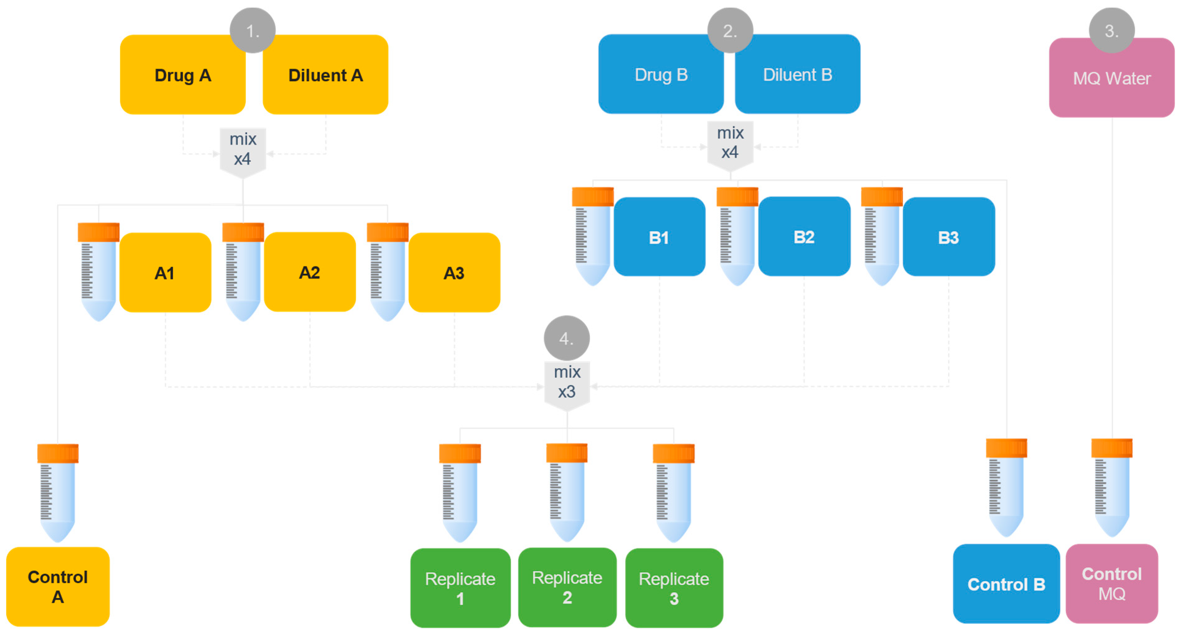
Calcium folinate 10 mg/mL in volumes of 0.73, 6.0, and 35.0 mL for simulated patient models 1–3, respectively, was diluted with sodium chloride 9 mg/mL to 40 mL in centrifuge tubes (A) (n = 4). In another tube (B), rehydration fluid was prepared by adding 3.2 mL sodium hydrogen carbonate 500 mmol/L to a standard glucose–sodium–potassium infusion solution to yield 40 mL of a rehydration fluid containing 40 mmol/L bicarbonate (n = 4). Varying proportions of A (38.8, 36.5, and 29.8 mL for patient models 1–3, respectively) to B (1.2, 3.5, and 10.2 mL for patient models 1–3, respectively) were mixed according to the mixing ratios identified in Table 2 (n = 3), simulating the y-site mixing scenario for each patient. Figure 2 illustrates the general flowchart for the testing program.
The undiluted mix of 1:1 volume ratio was prepared by adding 20 mL of calcium folinate 10 mg/mL and 20 mL sodium bicarbonate 500 mmol/L to a 40 mL centrifuge tube (n = 4). Additionally, 40 mL of unmixed A, and B, as well as MQ water served as controls throughout the analyses.
Samples were prepared in a particle-free environment inside a EuroFlow EF/S-4 safety cabinet (Clean Air®, Woerden, The Netherlands) at room temperature, according to antiseptic hospital workflow procedures [66,67]. All drug containers were carefully swirled 10 times before being used in preparations. Sterile 50 mL polypropylene (PP) centrifuge tubes with CentriStar™ PP caps (Corning™ Science, Tamaulipas, Mexico) were used as containers for the compatibility testing samples. Solutions were added to the tubes through a sterile 0.22 µm pore size syringe filter with a flat PES filter membrane (VWR International, Radnor Township, PA, USA) and effective filtration area of 2.8 cm2.

2.4.2. Analysis of Physical Compatibility Using Validated Methods

The testing program to assess physical compatibility of the prepared samples (Figure 2) was performed immediately after mixing (t0) and 4 h after mixing (t4) according to previously established methods [51]. This included measurement of pH and assessment of potential particles through visual examination using a Tyndall beam, turbidity measurements, and sub-visual particle content using light obscuration. Additionally, the Tyndall effect was explored on samples 24 h after mixing (t24). All sample containers were carefully swirled 10 times just prior to analysis.
The risk of CaCO3(s) formation and thus precipitation increases at higher pH, particularly above pH 8 (see Supplementary Materials Figure S1). Hence, the pH value of the mixed samples provides an indication of the risk of particle formation. pH was measured using an InLab® Versatile Pro electrode with a SevenCompact S220 pH-meter (Mettler-Toledo GmBH, Greifensee, Switzerland). Ionic products based on measured pH values were calculated for comparison with the calculated ionic products based on predicted pH (described in Section 2.3.2).
Tyndall beam was used to visually examine the presence of macroscopic particles in the samples, as explained earlier [49,51,68]. Mixed samples and controls were visually examined against a black background using a fiber-optic led beam (Schott KL 1600 LED, Mainz, Germany). Presence of Tyndall effect (visible coherent line through the sample) indicated particles, which may be attributed to CaCO3(s) precipitation if detected only in the mixed samples.
Sample turbidity was measured with a 2100Qis turbidimeter (Hach Lang, GmbH, Duesseldorf, Germany). An increase in Formazine Nephelometry Units (FNU) larger than 0.3 between controls and the mixed samples served as indication of particle formation in the mixed samples [51].
The sub-visual particle content was analyzed by light obscuration with the Accusizer Syringe Injection Sampler (PSS NICOMP, Billerica, MA, USA) to estimate particle size and amount. The samples were measured for three pulls of 5 mL of solution each, and the amounts of particles ≥ 5, 10 and 25 μm per mL were determined. Background count was defined as not more than 100 particles/mL ≥ 0.5 μm. As acceptance criteria, the limits for particle content in large-volume parenterals was used as described in the European Pharmacopoeia (Ph. Eur) Section 2.9.19; not more than 25 particles per mL of size ≥ 10 µm and not more than 3 particles per mL of size ≥ 25 µm. The ≥5 µm size measurement criterium was included based on the potential clinical relevance due to assumed capillary diameter size [49,51,68].
Since sub-visual particle content measurements were destructive, the testing program could not be performed on the same sample volumes. The two timepoints (t0 and t4) were therefore performed on two individual batches made in parallel. Tests on mixed samples were always compared to unmixed controls and MQ water.

2.5. Proofing Precipitate Identity Using Raman Microscopy

An alpha300 apyron confocal Raman microscopy system (WITec Wissenschaftliche Instrumente und Technologie GmbH, Ulm, Germany) equipped with a 532 nm laser source was used for microscopic and spectroscopic analysis of particle identity. Data acquisition and preprocessing was performed using the accompanying proprietary software bundle Suite SIX from WITec. The software TrueMatch v. 6.1.10.135 (WITec, Ulm, Germany) was used to compare acquired data to reference spectra.

2.5.1. Preparation of Samples for Raman Analysis

Potential precipitates were collected from the samples for analysis in the Raman microscopy system using flat filter membranes at room temperature. It has been described in the literature that CaCO3 particles have a light color with a yellowish tint [19,40,42,46] and filter membrane color was assumed to affect the background contrast and thus the visibility of particles. Therefore, three different filters of different colors (white or black) were initially tested as follows: Supor® PES (white), 0.2 µm pore size and 47 mm diameter (Pall Corporation, Ann Arbor, MI, USA) and Cyclopore™ PCTE black, 0.2 µm pore size and 25 mm diameter (Whatman™ Inc./Cytiva, Clifton, NJ, USA). The membranes were used for filtration with a 100 millibar VP100 vacuum pump (Vacuubrand GmBH, Wertheim, Germany) assembly system. Mixed samples with the highest and second highest predicted amounts of CaCO3 (undiluted mix and simulated Patient model 3), were chosen for analysis. The samples (5–10 mL) were filtered using different filter types: mixed samples from simulated patient model 3 were filtered with white PES and black PCTE membranes, A- and B-controls (Figure 2) were filtered with PCTE black membranes, and undiluted mix samples were filtered with PES membranes. Each filter was flushed through with 20–50 mL MQ water after sample filtration to remove unprecipitated material and then dried prior to Raman analysis.
Different reference materials were used to identify captured particles. Calcium carbonate standard was used to acquire a reference Raman spectrum for comparison with any precipitates collected from samples. In addition, a mixed sample corresponding to simulated patient model 3 was alkalized by addition of sodium hydroxide to pH >12 to force precipitation of CaCO3(s). This precipitate was then collected, analyzed, and used as a reference (denoted “patient model 3 alkalized”).
Reference spectra of calcium folinate and sodium hydrogen carbonate drug solutions were collected by deposition on microscope cover glass, measuring 24 × 50 × (0.13–0.17) mm (height × length × depth), (VWR International, Radnor Township, PA, USA), and allowed to air-dry prior to analysis. From these references, molecule specific high-resolution Raman spectra “fingerprints” were acquired and implemented into the local Raman spectra reference database for Hospital Pharmacies Enterprise at the Oslo University Hospital.

2.5.2. Characterizing Particle Fingerprint for Proof of Identity

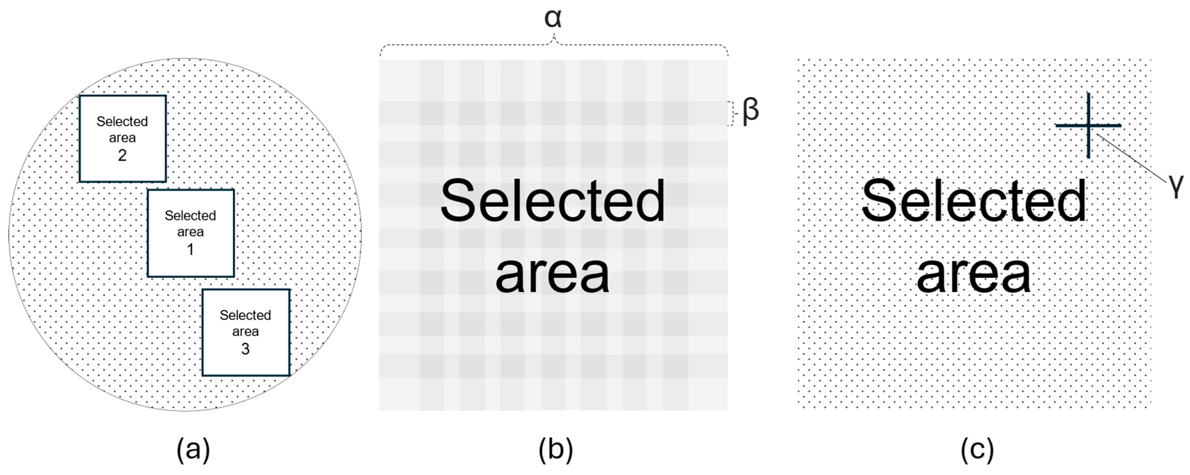
The filter membranes were first inspected using photography with the Raman microscope equipped with a 20× magnification objective (Zeiss LD Plan-Neofluar 20×/0.4) over three randomly selected areas comprising 169 stitching images (40% cross-over border) spanning 1012 × 1012 µm as illustrated in Figure 3. Particle discovery on collected images was conducted by inspecting the stitched images manually.
Raman spectra were captured at any instance of suspected particle discovery within the selected areas with a 532 nm excitation wavelength laser at 2.5 mW power using a 600 g/mm BLZ 500.00 nm grating, integration time of 0.5 s, and 50 accumulations. Raman shifts in the spectral range from 0 to 4000 cm−1 were collected. Data preprocessing was conducted using the default state software settings, which includes cosmic ray removal and background subtraction. The recorded Raman spectra were compared to reference spectra across all selected areas per sample using the software TrueMatch (WITec, Ulm, Germany). The TrueMatch software evaluated their match mathematically and assigned a hit quality index (HQI) score accordingly. Spectra demonstrating a HQI score < 50 towards reference spectra, and spectra matching with respective filter membrane material (PES, PCTE), were defined as negative analysis results with respect to the presence of CaCO3 particles.
Any samples with HQI > 50 for any of the CaCO3 reference spectra in the commercial databases (S.T. JAPAN Europe GmBH, Cologne, Germany) were recorded with a 100× objective (Zeiss LD Plan-Neofluar 100×/0.9) and further compared to the CaCO3 standard, in-house reference spectra obtained for calcium folinate, sodium bicarbonate, and “patient model 3 alkalized”.

3. Results

3.1. Theoretical Analysis

3.1.1. Predicted Ionic Yields, Molar Ratios and Risk of CaCO3 Precipitation

The predicted concentrations of calcium (Ca2+), bicarbonate (HCO3), carbonate (CO32−), and pH levels in the mixed samples, and the risk of CaCO3(s) precipitation for the simulated patient models based on their clinical parameters, dose, and infusion characteristics (Table 2) according to treatment conditions (Figure 1), are presented in Table 3. Looking at the molar HCO32−/Ca2+ mixing ratio in the simulated patient models dictated by the protocol, a slight decrease from patient model 1 (3.56) to patient model 2 (1.32) and further to patient model 3 (0.80) was detected, i.e., decreasing with increasing age and size of the patient (Table 3). An increase in molar ratio was identified for the extreme patient model (5.82) as compared to patient model 3 (oldest and largest of the pediatric models). The undiluted mix (1:1 volume mix) was found to have a molar ratio that was almost 4× higher than patient model 1 at 12.8. The molar ratio found for the “extreme patient” and the undiluted mix would be more in the comparable range as reported in the study by Aujoulat et al., where their 1:1 mixing ratio of applied undiluted drugs [46] corresponded to a molar ratio of HCO32−/Ca2+ of 8.54.
The final pH of the mixed samples was predicted to be near physiological (pH 7.4) for patient model 1, while reaching increasingly more alkaline values across the other models with higher HCO3 content (Table 3). The predicted maximum theoretical CaCO3 concentration (the concentration of the least concentrated ionic component of the salt), based on estimated CO32− concentrations from predicted pH, was lower than the solubility of CaCO3 (1.66 × 10⁻⁷ mol/mL or 0.0166 mg/mL [30]) in patient models 1–3, but higher for the undiluted mix and “extreme patient” cases. Predicted ionic products were higher than the solubility product of CaCO3 (3.36 × 10⁻⁹ mol2/L2 [65]) in all but patient model 1 (Supplementary Materials Table S1). In total, only patient model 1 was conclusively assessed to avoid precipitation of CaCO3(s) based on both theoretical evaluations.

3.1.2. Investigation of Model Representation of Clinical Scenarios

The predictions presented in Table 3, and the identified mixing ratios, were based on fixed drug concentrations deriving from fixed s-MTX values assigned to the models (Table 2). However, this represents only a snapshot of the infusion regime according to the ALL-together protocol. To challenge this method and check whether the results represent all dynamically changing concentrations and mixing ratios encountered in the ALL-together protocol, stepwise increments of s-MTX for all simulated models and the “extreme patient” case were performed. In Figure 4, the predicted fluctuations in calcium folinate, HCO3, CO32−/Ca2+ molar ratio, and maximum CaCO3 yield (based on complete deprotonation of HCO3 to CO32−) in response to increasing s-MTX are illustrated for simulated patient models 1–3 and the “extreme patient” case. An incremental increase in Ca2+ concentration is expected (Figure 4a) with an abrupt shift at s-MTX 5 µmol/L and 7.5 µmol/L, respectively, as the ALL-together protocol [15] directs changes in calcium folinate administration time (bolus 2–5 min vs. short infusion 60 min) and infusion rates. When s-MTX concentrations rise above 5 µmol/L for patient model 1, 2, and “extreme patient”, or 7.5 µmol/L for patient model 3, a large increase in HCO3 (Figure 4b) and pH is observed. Above these concentrations, predicted pH increases to above 8.3 for all patient models except patient model 3 (Figure 4c), where this shift occurs at s-MTX 7.5 µmol/L. At pH above 8.3, the fraction of dissolved carbonate in its monoprotonated form (HCO3) decreases as it is converted into the fully deprotonated form (CO32−) according to its pKa-values (6.3 and 10.3) and, therefore, there will be a significantly higher amount of dissolved CO32−(aq) available, which in turn will increase the potential for precipitation of CaCO3(s). The increased pH in the mixes (Figure 4c) therefore corresponds to an increased risk of CaCO3(s) precipitation as the concentrations of CO3−2 reach levels above the solubility limit (Figure 4d). The mixing ratios identified based on fixed concentrations for patient model 1–3 (Table 2), further used in the physical compatibility tests, can all be found at maximum theoretical CaCO3 concentrations in solution below the solubility limit (Figure 4d) and should therefore be compatible.

3.2. Physical Compatibility Testing

3.2.1. pH Measurements and Theoretical Consideration

Measured pH values in all simulated patient y-site mixes and controls are summarized in Table 4. The pH value of the mixed samples was typically between the two unmixed controls and corresponded to the mixing ratio of the two drugs. There was relatively good agreement between measured and predicted pH for patient model 1 and 2 (Table 3 and Table 4), though the measured pH was lower than the predicted values for patient model 3 and the undiluted mix.
The calculated ionic product values were consistently lower when using the measured pH (Table 4) compared to using the predicted pH (Table 3) though this did not change the final CaCO3(s) precipitation risk assessment. Only patient model 1 was found to have no risk of precipitation when comparing the calculated ionic products based on measured pH to the solubility product of CaCO3 (3.36 × 10⁻⁹ mol2/L2 [65]) (Supplementary Materials Table S1).

3.2.2. Visual Examination Using Tyndall Light

Presence of macroscopic particles was visually examined using a Tyndall beam, and any presence of visible particles was documented (Table 5). All mixes based on simulated patient model concentrations were found to be particle-free, while the undiluted mix demonstrated distinctly visible particles within the first minutes upon mixing component A (calcium folinate solution) with B (sodium bicarbonate-containing rehydration fluid). An example of the precipitated particles (Figure S3 in Supplementary Material) is shown in the image of observed particles that stem from the undiluted mix, captured by the microscopy modality of the Raman microscope (100× objective). The estimated size of the particles was in the range 10–40 µm. A few dust particles and some bubbles were observed in some of the unmixed controls, but nothing indicating that the particles in the mixed samples originated from contamination of the solutions.

3.2.3. Turbidimetry

Turbidity results are presented in Table 6. The turbidity of the mixed samples from simulated patient models resembled the turbidity of the unmixed controls, while the undiluted mixed sample demonstrated a considerably higher turbidity than its controls. An increase of more than 0.3 FNU in the mixed sample as compared to the unmixed controls was regarded as an indication of particle formation.

3.2.4. Particle Size Measurement and Quantification

From the particle content measurements (size and amounts), all mixed samples simulating patient models 1–3, and their corresponding controls (A and B), were found to be within the Ph. Eur. criteria for large-volume parenterals of maximum 25 particles/mL ≥ 10 µm and not more than 3 particles/mL ≥ 25 µm (Table 7). Also, the number of particles in the size range corresponding to the small capillaries (≥5 µm) was low for these mixed samples. However, the undiluted mix samples showed a high number of particles of all sizes at t0 and the particle count could not be measured at t4 due to detector overload. These samples were not diluted to meet the detectable range of the instrument since a dilution step could dissolve particles.

3.2.5. Characterizing Particle Fingerprint for Proof of Identity

Raman spectra of all materials were acquired and included in the local at-hospital reference database. This included reference spectra for the different filter membrane materials investigated to collect particles (PCTE and PES filter membranes), spectra for dried calcium folinate, sodium bicarbonate, powdered calcium carbonate standard, filtered sample of “patient model 3 alkalized” (i.e., forced CaCO3 precipitation) and undiluted mix samples.
From microscopy, the collected stitched images of filter membranes used to filter mixed samples corresponding to simulated patient model 3 using both PES (white) and black PCTE filter membranes exhibited particle-resembling artifacts in the images (Supplementary Materials Figure S2). These artifacts were identified as filter paper material (PES and PCTE) through analysis of the Raman spectra and the membranes did not show any visual indication of particles, suggesting that precipitation did not occur in these patient model experiments. In contrast, the PES filter used to filter the undiluted mix showed clear presence of particles on visual inspection. These particles were confirmed using Raman spectroscopy. It was found that the white background of the PES filters did not impede microscopical detection of particles. Though the undiluted mix was not tested on black PCTE filter membranes, other experiments showed that compared to the white PES filters, the black PCTE filter membranes provided a greater background contrast to artifacts. This could signify a possible advantage of the black PCTE filters over the PES filters when searching for particles.
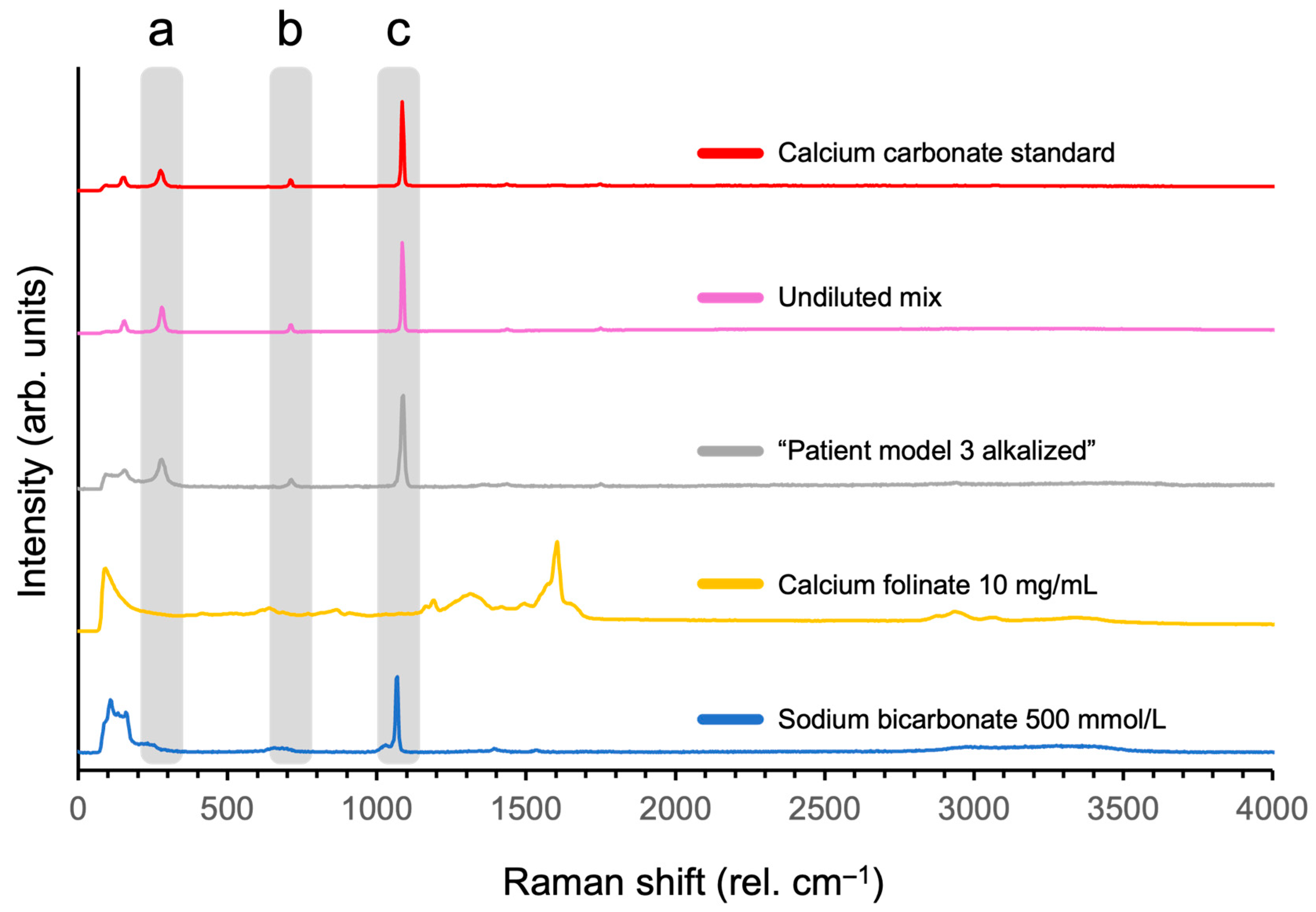
Raman spectra of CaCO3 standard and the undiluted mix sample matched with both the commercial and the in-house acquired reference spectra (HQI > 50) for calcium carbonate (Figure 5), with peak band positions at approximately 278, 712, and 1085 rel. cm −1. Also, an alkalized sample of “patient model 3” at pH > 12 showed the same peaks. The dried sample of sodium bicarbonate (500 mmol/mL) included as a reference demonstrated a peak at 1067 rel. cm−1, though this peak did not show matching spectral peaks observed in spectra of the CaCO3 containing references. The Raman specter of calcium folinate did not have these corresponding peaks; instead, it had a peak at 1603 rel. cm−1.

4. Discussion

Our study demonstrates that calcium folinate and sodium bicarbonate are physiochemically compatible when co-administered in simulated patient models corresponding to patients of approximately 1, 9, and 14 years of age (Table 2) according to the ALL-together protocol [15]. We found that an increase in patient age (BW and BSA) or s-MTX—which led to an increase in calcium folinate dose—do not necessarily result in higher Ca2+ concentrations at y-site. Instead, an increase in HCO3 concentrations and pH levels, due to protocol-directed changes in infusion rates, occurs. This fact, depending on the patient s-MTX value (Figure 4), may lead to precipitation of CaCO3(s) (Table 7) as confirmed by Raman microscopy (Figure 5). Predictive modelling does suggest that there is a certain and clinically relevant s-MTX range that allows for safe co-administration of calcium folinate and bicarbonate-containing rehydration fluid in pediatric ALL patients. This is a new finding.
The pediatric ALL-together protocol prescribes a complex and dynamic dosing regimen of calcium folinate combined with bicarbonate-containing rehydration fluid (Figure 1), during rescue therapy. According to our theoretical analysis, the HCO3 content was in surplus in relation to Ca2+ in most models; Ca2+ was in surplus in relation to the CO32− content in all models except the undiluted mix (Table 3), demonstrating that pH conditions at the y-site shifted the equilibrium towards HCO3 and not CO32−, in the diprotic H2CO3/HCO3/CO32− system (Supplementary Materials Figure S1). Only patient model 3, simulating a 14-year-old, was predicted to have more Ca2+ in relation to its HCO3 content at y-site. This model had a Ca2+ concentration more than twice the concentration of the “extreme patient” case, simulating a 39-year-old patient. We attribute this to the applied Ca2+ infusion rate limits governed by the treatment protocol (shift at s-MTX ≥ 5 µmol/L), allowing for calcium folinate bolus injection (2 min) in patient model 3, but requiring the “extreme patient” to receive an infusion of 60 min instead. This also highlights the incompatibility risk for patients being treated close to the infusion rate limit of the protocol, necessitating a shift from bolus to short infusion (Table 1). Patient models 1–3 were assessed to represent safe patient scenarios for co-administration based solely on CaCO3 solubility and the lowest concentration component, but models 2 and 3 were found to be unsafe when calculating their predicted ionic products ([Ca2+] × [CO32−]). In total, our theoretical risk assessment proposed that precipitation would not occur in patient model 1, while being inconclusive for models 2 and 3, and highly probable in the undiluted mix and the “extreme patient” case. The discrepancy between the two approaches (solubility vs. ionic product) observed here may be explained by the ionic product relying on both Ca2+ and CO32− concentrations, whereas the solubility-based approach instead relies on the limiting ingredient (either Ca2+ or CO32−) determining the maximum theoretical CaCO3 yield. Here, for all models except the undiluted mix, CO32− was the limiting ingredient and determined the estimated resulting theoretical CaCO3 concentrations in solution. Thus, the solubility-based risk assessments conducted in this study depend less on predicted Ca2+ concentrations than our assessments based on the ionic product. Additionally, as CO32− is both a pH-dependent ingredient and a buffering agent, the ionic product may be more reliant on accurate pH estimations. This suggests that accurate pH estimations (and CO32− concentration predictions) may be of great importance for precipitation risk assessments. As confirmed by our physical testing (Table 3), pH was overestimated for all patient models (Table 4), leading to overestimation of the CO32− concentrations present in the mixes, resulting in an apparent ionic product that faultily indicates a precipitation risk. The same was observed when calculating the ionic product based on measured pH (Table 3), further demonstrating the complexity involved in accurately predicting the final mix pH for compatibility assessments of systems containing acids and bases.
The overestimation of pH and precipitation risk is confirmed by our physical tests, which showed that patient models 1–3 were compatible; all results from visual observation, along with particle count, turbidity, and pH measurements met the acceptance criteria [51,69]. Only the undiluted mix sample demonstrated massive precipitation within minutes of mixing, in line with its higher HCO3 concentration resulting in a more alkaline solution and a higher concentration of CO32−. As Ca2+ was available in the solution, the increasing presence of CO32− was expected to result in CaCO3 formation and precipitation. The particle count in the undiluted mix could not be estimated due to detector overload, indicating massive precipitation.
The identity of the particles from the undiluted mix could be verified as CaCO3 using Raman spectroscopy (Figure 5), and no other salts were identified in the dried sample solutions. The 1085 rel. cm−1 peak is highly prevalent in CaCO3 samples [70], while the sodium bicarbonate sample demonstrated a close peak at 1067 rel. cm−1, possibly due to it also containing a comparable carbonate structure [71]. The shift in 18 rel. cm−1 may be explained by the absence of Ca2+ ions in that sample or the protonation of the carbonate, resulting in a difference in vibrational transitions and Raman scattering [72].
When patient model 3 was forced to precipitate in high alkaline pH (above 12), CaCO3 formation was confirmed (Figure 5). This suggests that the HCO3 and Ca2+ concentrations in patient model 3 indeed suffice for CaCO3 to form and precipitate, as long as the pH of the mixture shifts the equilibrium from HCO3 into CO32−.
Even though the in vitro tests provided clinically relevant simulations resulting in mixes absent of precipitates, they were based on fixed s-MTX levels giving fixed calcium folinate and sodium bicarbonate concentrations. However, this was expected to provide a snapshot and not the entire possible mixing panorama. In fact, our investigation of the scenario representativeness (Figure 4) demonstrated that when s-MTX increased above 5 µmol/L, only patient model 3 was estimated to remain below equilibrium conditions without precipitate. All other patient models reached the CaCO3 precipitation threshold (1.66 × 10⁻⁷ mol/mL [30]) at s-MTX 5.0 µmol/L, and patient model 3 at 7.5 µmol/L and above. At these s-MTX levels, adjusting the calcium folinate administration time from bolus (2–5 min) to short infusion (60 min) becomes necessary in order to ensure a Ca2+ load below 50 mg/m2/day [57] (Figure 1). This protocol-based change in administration time is explained by the ≥5.0 s-MTX level being the cut-off for switching to calcium folinate dosing based on BW × s-MTX instead of BSA × table value (Table 1). The changes result in dips in Ca2+ infusion rates, leading to increases in HCO3 infusion rates instead, demonstrating the importance of considering the impact of s-MTX on fluid dynamic and y-site mixing ratios. Below s-MTX 5.0 µmol/L, the investigated scenarios (Figure 4) show that patient model 3, and thus the physically tested configuration, covered the high-risk outlier control “extreme patient” case, which suggests that co-administration is safe in pediatric patients with s-MTX levels below 5.0 µmol/L. Since rescue therapy aims at lowering s-MTX over time, and levels < 5.0 µmol/L are prevalent in children [20,73,74], this could be valuable information in clinical practice.
Our findings contradict those of Aujoulat et al. [46], who observed precipitates when mixing calcium folinate 10 mg/mL with bicarbonate 167 mmol/L in a 1:1 volume mixing ratio (i.e., 2 mL of each). While attempting to simulate pediatric rescue therapy, they mixed both components undiluted, which resulted in a pH of 7.37, visually observing precipitation after 30 min. However, according to our y-site concentration prediction model (Figure 1) this mix results in effective Ca2+ concentrations that are 23% lower and HCO3 concentrations that are eight-fold higher than in patient model 3. Further, their molar mixing ratio ([HCO3]/[Ca2+]) was 8.54 compared to 0.8 in our patient model 3, emphasizing a larger availability of bicarbonate and carbonate ions in their study protocol. To put this into perspective, our estimations suggest that such a mix would reach a pH of 9.18, exceeding the ionic product of all our patient models (1–3) (6.20 × 10⁻⁵ mol2/L2) and yielding a maximum theoretical CaCO3 concentration of 9.78 × 10⁻⁶ mol/mL, which would be 103 times higher than our patient model 3 (9.48 × 10⁻⁸ mol/mL). Unexpectedly, the Aujoulat study [46] also observed precipitation when mixing calcium folinate with a rehydration fluid of similar content and pH as used in the present study, but without any added bicarbonate. This incompatibility was attributed to pH effects, without addressing whether it was CaCO3 or calcium folinate that precipitated in their study. In the present study, the observed Raman peak bands in precipitates from samples aligned well with the literature [71,75,76], and CaCO3 was successfully identified as the precipitating agent from the undiluted mix and the alkalized patient model 3 (i.e., forced CaCO3 precipitation), while no precipitates of calcium folinate or other molecules were found. Furthermore, the investigated precipitate CaCO3 in our study proved to be an apt particle for exploring the performance of Raman spectroscopy and microscopy, with its characteristic 1085 cm−1 band peak being prominent throughout the analyses. This is further supported by this band peak being recommended for determining spectral resolution by Ph. Eur. [77]. As per conventional methods [78,79,80,81,82,83,84,85], the experiments conducted by Aujoulat et al. [46] assessed one single maximal dosing scenario and mixing ratio, whereas our study may be the first to assess Ca2+ compatibility with HCO3 over a range of relevant patient scenarios, offering new insights that may instead enable safe co-administration under some common clinical circumstances.
To the best of our knowledge, this is the first study to systematically investigate the combined impact of patient clinical parameters on precipitation risk, by conducting extensive theoretical and physical compatibility testing, in an IV compatibility study. Furthermore, we augmented the analysis line-up [51] with the inclusion of an at-hospital Raman microscopy system, which allowed us to disclose the identity of formed precipitates and develop our Raman reference database further. Our approach, which mimics physiological conditions, provides clinically relevant insights that may improve multi-drug compatibility and administration guidelines in pediatric ALL treatment.
These findings highlight the importance of thoroughly understanding the impact of complex and dynamic treatment protocols on drug chemistry and pH when assessing physiochemical compatibility for clinical application [47]. Therefore, we advise future compatibility research to implement test setups that cover broader scenario representativeness, while assessing underlying mechanisms and verifying the identity of any precipitates formed. Co-administration must be conducted with utmost caution; nevertheless, our results indicate that mixing may be safe under certain conditions. If the two fluids could be given through the same venous catheter lumen (internal channel), it would reduce the number of venous access points needed to avoid mixing the drugs [86,87,88,89,90,91], enabling children to spend more time outside the clinic [43,92]. If the studied therapy were to be co-administered, it should be conducted through in-line filters to further safeguard the patients [93,94,95,96]. Such in-line filters are mandatory in pediatric care [97,98] but remain to be fully utilized in practice [99,100]. Another option for ensuring safe co-administration of bicarbonate during rescue therapy would be to choose disodium folinate [40,43,55] instead of calcium folinate. This alternative product has been shown to deliver equivalent therapy efficacy [40,101,102] and should eliminate the Ca2+ complexation-related precipitation risk altogether, but is often unavailable in the clinic, partly due to its higher pricing [43,55].
In summary, our study highlights the need for applying clinically relevant methods for compatibility assessments of parenteral drug solutions, which may contribute to optimizing administration strategies and therapeutic outcomes in pediatric cancer treatment. The Aujoulat study [46] is one of the major sources referred to when consulting calcium folinate and bicarbonate [44,45] compatibility and it has strongly impacted their co-administration practice over the years [17,44,45]. Here, instead of focusing on the worst-case scenario for mixing the two fluids, we directed our focus on the possibilities rather than limitations.

5. Limitations

One limitation of this study is that our theoretical prediction model assumed that calcium folinate is given diluted with sodium chloride 9 mg/mL as a 40 mL bolus, which in practice may vary in volume or even be given undiluted [57].
Another limitation may be that H2CO3 concentration was assigned a likely concentration (0.11 mmol/L) which may deviate from the actual concentration. In addition, the H2CO3/HCO3 buffer system was expected to be the only factors driving the final pH value, excluding the potential impact from excipients and other additives on final pH in our samples. This was conducted to simplify pH estimations and CO32− predictions, which may explain the obtained overestimations.
Our models to predict pH and the risk of CaCO3 precipitation do not take into account the fluid dynamics in the clinical setting and the effects that mixing rates would have on these parameters. Both in the case of theoretical evaluation as well as physical testing, a dynamic model would be an improvement and make the models even more clinically relevant. To account for dynamic changes in the mixing ratio, we compensated by testing different mixing ratios more frequently. We aimed at using a worst-case scenario (undiluted mix) and a possible outlier case (extreme patient) in addition to typical pediatric patients (patient models 1–3). Furthermore, we investigated the dynamic aspect of the protocol in simulations described in Section 3.2.1.
Lastly, we conducted our physical experiments in a controlled laboratory setting that does not fully replicate clinical environments where changes in final Ca2+, HCO3, and CO32− concentrations occur gradually as infusion rates are continuously adjusted.

6. Conclusions

Results from the physical compatibility tests of mixing ratios representing children of approximately 1, 9, and 14 years of age showed that doses and infusion rates of calcium folinate and bicarbonate-containing rehydration fluids in the ALL-together protocol were compatible, and the two fluids can be safely co-administered via the same lumen (internal channel) in these simulated patient models. The application of Raman spectroscopy confirmed the identity of precipitates from a non-patient model case.
Theoretical tests challenging the experimental area showed that situations where the two fluids are not compatible also do exist within the protocol framework, and precipitation of CaCO3 must be expected under some clinically relevant conditions. Risks of precipitation were identified in situations where s-MTX was above 5 µmol/L and administration of calcium folinate had to be given as short infusion (1 h) instead of a bolus injection (2–5 min). In such situations, co-administration in the same lumen must be avoided.
These novel findings, correlating the clinical parameter s-MTX levels to resulting y-site concentrations, analyzed with Raman spectroscopy, advance compatibility research by offering precise chemical insights and predictive modeling capabilities to compatibility testing, thus facilitating relevant co-administration information to pediatric rescue therapy. The study’s limitations include assumptions regarding bolus volume variability, pH predictions, exclusion of excipient effects on pH estimations, and conducting experiments in controlled environments that may not fully replicate clinical conditions.
Future research should continue to explore the interplay between clinical parameters and precipitation risk, focusing on chemical interactions when tested drugs are mixed. Compatibility information should be based on diverse, clinically relevant scenarios to become applicable and add value in pediatric oncology protocols.

Supplementary Materials

The following supporting information can be downloaded at https://www.mdpi.com/article/10.3390/pharmaceutics17091155/s1; Figure S1: Visualization of the diprotic activity of carbonic acid in relation to pH in aqueous solution; Figure S2: Proofing particle identity with Raman spectroscopy; Figure S3: Discovered particles form the undiluted mix retained by a polyethersulfone filter membrane. The image was taken with the Raman microscope equipped with the 100× objective. Estimated diameter of the largest particle approximately 35–40 µm; Table S1: Predicted pH based on calculations using the Henderson–Hasselbalch equation and ionic product calculations based on predicted and measured pH, respectively.

Author Contributions

Conceptualization, K.T., B.S.L., M.B.A., I.T. and K.N.-H.; methodology, K.T., B.S.L. and M.B.A.; software, K.T., B.S.L. and N.N.; validation, K.T., B.S.L., N.N., I.T. and K.N.-H.; formal analysis, K.T., B.S.L., N.N. and I.T.; investigation, K.T.; resources, K.T., B.S.L., M.B.A., N.N., I.T. and K.N.-H.; data curation, K.T.; writing—original draft preparation, K.T.; writing—review and editing, K.T., B.S.L., M.B.A., N.N., I.T. and K.N.-H.; visualization, K.T., B.S.L., I.T. and K.N.-H.; supervision, K.T., B.S.L., M.B.A., I.T. and K.N.-H.; project administration, K.T., I.T., K.N.-H.; funding acquisition, I.T., and K.N.-H. All authors have read and agreed to the published version of the manuscript.

Funding

This research was funded by South-Eastern Norway Regional Health Authority (project number 2022095).

Institutional Review Board Statement

This is a theoretical and in vitro experimental study involving no human or animal subjects. The study is performed according to ethical standards for laboratory experiments.

Informed Consent Statement

Not applicable.

Data Availability Statement

The original contributions presented in this study are included in the article/Supplementary Material. Further inquiries can be directed to the corresponding author(s). The raw data supporting the conclusions of this article will be made available by the authors on request.

Acknowledgments

We would like to extend our thanks to the Hospital Pharmacy Enterprise, South-Eastern Norway and all clinical pharmacists, nurses and physicians at the unit for pediatric haemato-oncology at Oslo University Hospital for continuous support. Many thanks also to Tove Larsen, Ivar Grove, and Bente Breiby (technicians at University of Oslo) for all your help in the laboratory.

Conflicts of Interest

The authors declare no conflicts of interest. The funder had no role in the design of the study; in the collection, analyses, or interpretation of data; in the writing of the manuscript, or in the decision to publish the results.

Abbreviations

The following abbreviations are used in this manuscript:
ALLAcute lymphoblastic leukemia
IVIntravenous(ly)
s-MTXSerum methotrexate concentrations
CVCCentrally inserted venous catheter
PICCPeripherally inserted central venous catheter
MQMilli-Q®
PESPolyethersulfone
BWBodyweight
BSABody surface area
PPPolypropylene
FNUFormazine Nephelometry Units
Ph. Eur.European Pharmacopoeia
PCTETrack-etched polycarbonate
HQIHit quality index
OLOverload

References

  1. Ward, E.; DeSantis, C.; Robbins, A.; Kohler, B.; Jemal, A. Childhood and adolescent cancer statistics, 2014. CA A Cancer J. Clin. 2014, 64, 83–103. [Google Scholar] [CrossRef]
  2. Surveillance Research Program, National Cancer Institute. SEER*Explorer: An Interactive Website for SEER Cancer Statistics [Internet]; Surveillance Research Program, National Cancer Institute: Rockville, MD, USA, 2024. [Google Scholar]
  3. Ali, A.A.; Charoo, N.A.; Abdallah, D.B. Pediatric drug development: Formulation considerations. Drug Dev. Ind. Pharm. 2014, 40, 1283–1299. [Google Scholar] [CrossRef]
  4. Mitchell, C.; Richards, S.; Harrison, C.J.; Eden, T. Long-term follow-up of the United Kingdom medical research council protocols for childhood acute lymphoblastic leukaemia, 1980–2001. Leukemia 2010, 24, 406–418. [Google Scholar] [CrossRef]
  5. UKALL14 Protocol, version 5.0; UCL Cancer Trials Centre: London, UK, 2012.
  6. UKALL 2019 Interim Guidelines; Children’s Cancer and Leukaemia Group (CCLG): Leicester, UK, 2019.
  7. ALLTogether Master Protocol; ALLTogether Consortium: Southampton, UK, 2020.
  8. Acute Lymphoblastic Leukaemia (ALL06) Overview. Available online: https://www.eviq.org.au/haematology-and-bmt/leukaemias/acute-lymphoblastic-leukaemia/3825-acute-lymphoblastic-leukaemia-all06-overview (accessed on 12 August 2023).
  9. Brown, P.A.; Shah, B.; Advani, A.; Aoun, P.; Boyer, M.W.; Burke, P.W.; DeAngelo, D.J.; Dinner, S.; Fathi, A.T.; Gauthier, J.; et al. Acute Lymphoblastic Leukemia, Version 2.2021, NCCN Clinical Practice Guidelines in Oncology. J. Natl. Compr. Canc Netw. 2021, 19, 1079–1109. [Google Scholar] [CrossRef]
  10. Shah, B.; Mattison, R.J.; Abboud, R.; Abdelmessieh, P.; Aldoss, I.; Burke, P.W.; DeAngelo, D.J.; Dinner, S.; Fathi, A.T.; Gauthier, J.; et al. Acute Lymphoblastic Leukemia, Version 2.2024, NCCN Clinical Practice Guidelines in Oncology. J. Natl. Compr. Canc. Netw. 2024, 22, 563–576. [Google Scholar] [CrossRef]
  11. Pediatric Acute Lymphoblastic Leukemia Treatment (PDQ®)—Health Professional Version. Available online: https://www.cancer.gov/types/leukemia/hp/child-all-treatment-pdq#_22 (accessed on 5 September 2023).
  12. Hoelzer, D.; Bassan, R.; Boissel, N.; Roddie, C.; Ribera, J.M.; Jerkeman, M. ESMO Clinical Practice Guideline interim update on the use of targeted therapy in acute lymphoblastic leukaemia. Ann. Oncol. 2024, 35, 15–28. [Google Scholar] [CrossRef]
  13. Hoelzer, D.; Bassan, R.; Dombret, H.; Fielding, A.; Ribera, J.M.; Buske, C. Acute lymphoblastic leukaemia in adult patients: ESMO Clinical Practice Guidelines for diagnosis, treatment and follow-up. Ann. Oncol. 2016, 27, v69–v82. [Google Scholar] [CrossRef]
  14. ALLTogether European Treatment Protocol for Children and Young Adults with Acute Lymphoblastic Leukemia (ALL); NordForsk: Oslo, Norway, 2023.
  15. Vestli, A.; Hellebostad, M. Ped ALL 003acd A2G Mtx 5 g/m2; Oslo University Hospital/Helse Sør-Øst: Oslo, Norway, 2021. [Google Scholar]
  16. Jiang, R.; Mei, S.; Zhao, Z. Leucovorin (folinic acid) rescue for high-dose methotrexate: A review. J. Clin. Pharm. Ther. 2022, 47, 1452–1460. [Google Scholar] [CrossRef]
  17. American Society of Health-System Pharmacists. Leucovorin Calcium. In ASHP Injectable Drug Information; American Society of Health-System Pharmacists, Ed.; American Society of Health-System Pharmacists: Bethesda, MD, USA, 2018; pp. 987–991. [Google Scholar]
  18. Sand, T.E.; Jacobsen, S. Effect of urine pH and flow on renal clearance of methotrexate. Eur. J. Clin. Pharmacol. 1981, 19, 453–456. [Google Scholar] [CrossRef]
  19. Bedford Laboratories, Leucovorin Calcium Injection USP; Bedford Laboratories: Bedford, OH, USA, 2011.
  20. Niinimäki, R.; Aarnivala, H.; Banerjee, J.; Pokka, T.; Vepsäläinen, K.; Harila-Saari, A. Reduced dose folinic acid rescue after rapid high-dose methotrexate clearance is not associated with increased toxicity in a pediatric cohort. Support. Care Cancer 2022, 30, 127–133. [Google Scholar] [CrossRef]
  21. Nunn, J.L.; Takashima, M.D.; Wray-Jones, E.M.; Soosay Raj, T.A.; Hanna, D.M.T.; Ullman, A.J. Central venous access device adverse events in pediatric patients with cancer: A systematic review and meta-analysis. Support. Care Cancer 2024, 32, 662. [Google Scholar] [CrossRef]
  22. Ullman, A.J.; Gibson, V.; Takashima, M.D.; Kleidon, T.M.; Schults, J.; Saiyed, M.; Cattanach, P.; Paterson, R.; Cooke, M.; Rickard, C.M.; et al. Pediatric central venous access devices: Practice, performance, and costs. Pediatr. Res. 2022, 92, 1381–1390. [Google Scholar] [CrossRef]
  23. Kamata, Y.; Mizuno, Y.; Okamoto, K.; Okamoto, S.; Ito, Y.; Nishigata, A. Peripherally inserted central catheters can be an alternative to tunneled central venous catheters in chemotherapy for hematological and oncological pediatric patients. Pediatr. Surg. Int. 2023, 39, 264. [Google Scholar] [CrossRef]
  24. Shulman, R.J.; Smith, E.O.; Rahman, S.; Gardner, P.; Reed, T.; Mahoney, D. Single- vs. double-lumen central venous catheters in pediatric oncology patients. Am. J. Dis. Child. 1988, 142, 893–895. [Google Scholar] [CrossRef] [PubMed]
  25. Gibbs, R.J. Mechanisms controlling world water chemistry. Science 1970, 170, 1088–1090. [Google Scholar] [CrossRef]
  26. Huang, Y.-C.; Rao, A.; Huang, S.-J.; Chang, C.-Y.; Drechsler, M.; Knaus, J.; Chan, J.C.C.; Raiteri, P.; Gale, J.D.; Gebauer, D. Uncovering the Role of Bicarbonate in Calcium Carbonate Formation at Near-Neutral pH. Angew. Chem. Int. Ed. 2021, 60, 16707–16713. [Google Scholar] [CrossRef] [PubMed]
  27. Dettori, R.; Donadio, D. Carbon dioxide, bicarbonate and carbonate ions in aqueous solutions under deep Earth conditions. Phys. Chem. Chem. Phys. 2020, 22, 10717–10725. [Google Scholar] [CrossRef] [PubMed]
  28. Al Omari, M.M.H.; Rashid, I.S.; Qinna, N.A.; Jaber, A.M.; Badwan, A.A. Chapter Two—Calcium Carbonate. In Profiles of Drug Substances, Excipients and Related Methodology, Brittain, H.G., Ed.; Academic Press: Cambridge, MA, USA, 2016; Volume 41, pp. 31–132. [Google Scholar]
  29. Ropp, R.C. Encyclopedia of the Alkaline Earth Compounds; Ropp, R.C., Ed.; Elsevier: Amsterdam, The Netherlands, 2013. [Google Scholar]
  30. KGaA, M. Safety Data Sheet: Calcium Carbonate Precipitated for Analysis, EMSURE® Reag. Ph Eur; Merck KGaA: Darmstadt, Germany, 2025. [Google Scholar]
  31. Kerner, J.A.; Garcia-Careaga, M.G.; Fisher, A.A.; Poole, R.L. Treatment of Catheter Occlusion in Pediatric Patients. J. Parenter. Enter. Nutr. 2006, 30, S73–S81. [Google Scholar] [CrossRef]
  32. Zheng, L.-y.; Xue, H.; Yuan, H.; Liu, S.-x.; Zhang, X.-y. Efficacy of management for obstruction caused by precipitated medication or lipids in central venous access devices: A systematic review and meta-analysis. J. Vasc. Access 2019, 20, 583–591. [Google Scholar] [CrossRef]
  33. Sielski, J.; Janion Sadowska, A.; Ciuraszkiewicz, K.; Janion, M. Acute pulmonary embolism following cardiac arrest in a 31 year-old female with long QT syndrome. Kardiol. Pol. 2011, 69, 590–592. [Google Scholar]
  34. Strickland, S.; Pena, E.; Walker, A.E. Fatal foreign-body granulomatous pulmonary embolization due to microcrystalline cellulose in a patient receiving total parenteral nutrition: All crystals are not what they seem. Forensic Sci. Med. Pathol. 2015, 11, 255–261. [Google Scholar] [CrossRef]
  35. McNearney, T.; Bajaj, C.; Boyars, M.; Cottingham, J.; Haque, A. Total parenteral nutrition associated crystalline precipitates resulting in pulmonary artery occlusions and alveolar granulomas. Dig. Dis. Sci. 2003, 48, 1352–1354. [Google Scholar] [CrossRef] [PubMed]
  36. Steadman, E.; Raisch, D.W.; Bennett, C.L.; Esterly, J.S.; Becker, T.; Postelnick, M.; McKoy, J.M.; Trifilio, S.; Yarnold, P.R.; Scheetz, M.H. Evaluation of a potential clinical interaction between ceftriaxone and calcium. Antimicrob. Agents Chemother. 2010, 54, 1534–1540. [Google Scholar] [CrossRef] [PubMed]
  37. Bradley, J.S.; Wassel, R.T.; Lee, L.; Nambiar, S. Intravenous ceftriaxone and calcium in the neonate: Assessing the risk for cardiopulmonary adverse events. Pediatrics 2009, 123, e609–e613. [Google Scholar] [CrossRef]
  38. Mailloux, R.J.; Delegge, M.H.; Kirby, D.F. Pulmonary Embolism as a Complication of Long-Term Total Parenteral Nutrition. J. Parenter. Enter. Nutr. 1993, 17, 578–582. [Google Scholar] [CrossRef]
  39. Christensen, M.L.; Zareie, P.; Kadiyala, B.; Bursac, Z.; Reed, M.D.; Mattison, D.R.; Davis, R.L. Concomitant ceftriaxone and intravenous calcium therapy in infants. J. Pediatr. Pharmacol. Ther. 2021, 26, 702–707. [Google Scholar] [CrossRef]
  40. Bruch, H.R.; Esser, M. Catheter occlusion by calcium carbonate during simultaneous infusion of 5-FU and calcium folinate. Onkologie 2003, 26, 469. [Google Scholar] [CrossRef]
  41. Fackler-Schwalbe, I.; Schwalbe, B.; Epple, M.; Becker, A.; Prügl, L.; Gassel, W.D.; Stoffels, D.; Südhoff, T. Occlusion of central venous port catheters after simultaneous 24 h infusions of 5-fluorouracil and calcium-folinic acid in patients with gastrointestinal cancer. Wien. Med. Wochenschr. 2004, 154, 182–185. [Google Scholar] [CrossRef]
  42. Ardalan, B.; Flores, M.R. A new complication of chemotherapy administered via permanent indwelling central venous catheter. Cancer 1995, 75, 2165–2168. [Google Scholar] [CrossRef]
  43. Ratti, M.; Hahne, J.C.; Toppo, L.; Castelli, E.; Petrelli, F.; Passalacqua, R.; Barni, S.; Tomasello, G.; Ghidini, M. Major innovations and clinical applications of disodium-levofolinate: A review of available preclinical and clinical data. Ther. Adv. Med. Oncol. 2019, 11, 1758835919853954. [Google Scholar] [CrossRef] [PubMed]
  44. Merative. Trissel’s™ 2 Clinical Pharmaceutics Database (Parenteral Compatibility) (Electronic Version); Merative: Ann Arbor, MI, USA, 2025. [Google Scholar]
  45. Vidal, H. Stabilis: The International Database on Stability and Compatibility of Injectable Drugs, 2025. Available online: https://www.stabilis.org/ (accessed on 17 March 2025).
  46. Aujoulat, P.; Coze, C.; Braguer, D.; Raybaud, C. Physico-chemical compatibility of methotrexate with co-administered drugs during cancer chemotherapy regimens. J. Pharm. Clin. 1993, 12, 31–35. [Google Scholar]
  47. Newton, D.W. Crux of drug compatibility and incompatibility. Am. J. Health-Syst. Pharm. 2010, 67, 108–112. [Google Scholar] [CrossRef]
  48. Castells Lao, G.; Rodríguez Reyes, M.; Roura Turet, J.; Prat Dot, M.; Soy Muner, D.; López Cabezas, C. Compatibility of drugs administered as Y-site infusion in intensive care units: A systematic review. Med. Intensiv. (Engl. Ed.) 2020, 44, 80–87. [Google Scholar] [CrossRef]
  49. Nilsson, N.; Storesund, I.; Tho, I.; Nezvalova-Henriksen, K. Co-administration of drugs with parenteral nutrition in the neonatal intensive care unit-physical compatibility between three components. Eur. J. Pediatr. 2022, 181, 2685–2693. [Google Scholar] [CrossRef] [PubMed]
  50. D’Huart, E.; Vigneron, J.; Demoré, B. Physical Compatibility of Intravenous Drugs Commonly Used in Intensive Care Units: An Observational Study and Physical Compatibility Laboratory Tests on Anti-Infective Drugs. Pharm. Technol. Hosp. Pharm. 2019, 4, 29–40. [Google Scholar] [CrossRef]
  51. Staven, V.; Wang, S.; Grønlie, I.; Tho, I. Development and evaluation of a test program for Y-site compatibility testing of total parenteral nutrition and intravenous drugs. Nutr. J. 2016, 15, 29. [Google Scholar] [CrossRef]
  52. Braun, B. Sodium Hydrogen Carbonate 500 mmol/L Solution for Infusion—Summary of Product Characteristics (SmPC); Electronic Medicines Compendium (EMC): Sheffield, UK, 2024; Available online: https://www.medicines.org.uk/emc/product/15216/smpc (accessed on 17 March 2025).
  53. Braun, B. Sodium Chloride 0.9% Solution for Infusion—Summary of Product Characteristics (SmPC); Electronic Medicines Compendium (EMC): Sheffield, UK, 2018; Available online: https://www.medicines.org.uk/emc/product/15145/smpc#gref (accessed on 17 March 2025).
  54. B. Braun Melsungen AG. SmPC for Kalium-Natrium-Glucose Braun Infusjonsvæske; B. Braun Melsungen AG: Melsungen, Germany, 2024. [Google Scholar]
  55. Heldrup, J.; Bleyer, A.; Ramsey, L.; Schaff, L.; Bernhardt, B.; Schwartz, S.; Chatelut, E.; Hwang, M.; Ten, C.; Guscott, M.; et al. New recommendations for reversal of high-dose methotrexate cytotoxicity with folinic acid. Cancer Chemother. Pharmacol. 2025, 95, 41. [Google Scholar] [CrossRef]
  56. Jung, Y.; Moe, K.; Torres, E.A.; Kalantar-Zadeh, K.; Hanna, R.M. Unique case of profound iatrogenic hypercalcemia in a patient with recent orthopedic prosthetic infection. Clin. Nephrol. Case Stud. 2020, 8, 91–95. [Google Scholar] [CrossRef] [PubMed]
  57. Nasjonalt Kompetansenettverk for Legemidler Til Barn. Blandekort: Kalsiumfolinat v3.0. Available online: https://www.legemidlertilbarn.no/blandekort/kalsiumfolinat (accessed on 23 October 2023).
  58. Centers for Disease Control and Prevention, National Center for Health Statistics. CDC Growth Charts. Available online: https://www.cdc.gov/growthcharts/index.htm (accessed on 15 September 2023).
  59. WHO. WHO Child Growth Standards. Available online: https://www.who.int/tools/child-growth-standards (accessed on 15 September 2023).
  60. Bois, D.D.; Bois, E.F.D. A height-weight formula to estimate the surface area of man. Exp. Biol. Med. 1916, 13, 77–78. [Google Scholar] [CrossRef]
  61. Loerting, T.; Bernard, J. Aqueous Carbonic Acid (H2CO3). ChemPhysChem 2010, 11, 2305–2309. [Google Scholar] [CrossRef]
  62. Eriksson, L. Acid/Base Calculations and Graphical Representations. 2016. Available online: https://www.lerik.se/texter/kort_jmv.pdf (accessed on 11 March 2025).
  63. NCBI. Sodium Bicarbonate Compound Summary—Dissociation Constants; NCBI: Bethesda, MD, USA, 2005. [Google Scholar]
  64. Smith, G.B.; Dezeny, G.C.; Hughes, D.L.; King, A.O.; Verhoeven, T.R. Mechanistic studies of the Suzuki cross-coupling reaction. J. Org. Chem. 1994, 59, 8151–8156. [Google Scholar] [CrossRef]
  65. Haynes, W.M. (Ed.) CRC Handbook of Chemistry and Physics, 91st ed.; CRC Press Inc.: Boca Raton, FL, USA, 2010. [Google Scholar]
  66. Oslo University Hospital (OUS). Aseptisk Arbeidsteknikk [Aseptical Work Technique for Preparing and Administering Medications for Injection and Infusion]; Oslo University Hospital: Oslo, Norway, 2025. [Google Scholar]
  67. Oslo University Hospital (OUS). Arbeid I Sikkerhetsbenk Og Avtrekksskap Ved Tilberedning Av Legemidler [Work in Safety Cabinets and Fume Hoods for Preparation of Drugs]; Oslo University Hospital: Oslo, Norway, 2025. [Google Scholar]
  68. Nezvalova-Henriksen, K.; Nilsson, N.; Østerberg, C.T.; Staven Berge, V.; Tho, I. Y-Site Physical Compatibility of Numeta G13E with Drugs Frequently Used at Neonatal Intensive Care. Pharmaceutics 2020, 12, 677. [Google Scholar] [CrossRef]
  69. EDQM. Ph.Eur. 2.9.19—Particulate Contamination: Sub-Visible Particles; EDQM: Strasbourg, France, 2021. [Google Scholar]
  70. NCBI. Calcium Carbonate Compound Summary—Raman Spectra; NCBI: Bethesda, MD, USA, 2004. [Google Scholar]
  71. NCBI. Sodium Bicarbonate Compound Summary—Raman Spectra; NCBI: Bethesda, MD, USA, 2005. [Google Scholar]
  72. Lewis, I.R.; Edwards, H. (Eds.) Handbook of Raman Spectroscopy: From the Research Laboratory to the Process Line; Marcel Dekker: New York, NY, USA, 2001. [Google Scholar]
  73. Skärby, T.; Jönsson, P.; Hjorth, L.; Behrentz, M.; Björk, O.; Forestier, E.; Jarfelt, M.; Lönnerholm, G.; Höglund, P. High-dose methotrexate: On the relationship of methotrexate elimination time vs. renal function and serum methotrexate levels in 1164 courses in 264 Swedish children with acute lymphoblastic leukaemia (ALL). Cancer Chemother. Pharmacol. 2003, 51, 311–320. [Google Scholar] [CrossRef]
  74. Skärby, T.V.; Anderson, H.; Heldrup, J.; Kanerva, J.A.; Seidel, H.; Schmiegelow, K. High leucovorin doses during high-dose methotrexate treatment may reduce the cure rate in childhood acute lymphoblastic leukemia. Leukemia 2006, 20, 1955–1962. [Google Scholar] [CrossRef]
  75. Fong, M.Y.; Nicol, M. Raman spectrum of calcium carbonate at high pressures. J. Chem. Phys. 1971, 54, 575–578. [Google Scholar] [CrossRef]
  76. Dufresne, W.J.B.; Rufledt, C.J.; Marshall, C.P. Raman spectroscopy of the eight natural carbonate minerals of calcite structure. J. Raman Spectrosc. 2018, 49, 1999–2007. [Google Scholar] [CrossRef]
  77. Commission, E.P. Revised Chapter 2.2.48 on Raman Spectroscopy, European Pharmacopoeia Supplement 10.7; European Commission: Strasbourg, France, 2021. [Google Scholar]
  78. Veggeland, T.; Brandl, M. Evaluation of a simple method for visual detection of microprecipitates in blends of parenteral drug solutions using a focused (tyndall) light beam. Int. J. Pharm. Compd. 2010, 14, 78–81. [Google Scholar] [PubMed]
  79. Dettlaff, K.; Dominiak, K.; Klimaszewska, M.; Gostynska, A. Physical Compatibility of Ibuprofen and Selected Parenteral Drugs During Simulated Y-Site Administration. Acta Pol. Pharm. 2023, 80, 255–267. [Google Scholar] [CrossRef] [PubMed]
  80. Trissel, L.A.; Gilbert, D.L.; Martinez, J.F.; Baker, M.B.; Walter, W.V.; Mirtallo, J.M. Compatibility of parenteral nutrient solutions with selected drugs during simulated Y-site administration. Am. J. Health-Syst. Pharm. 1997, 54, 1295–1300. [Google Scholar] [CrossRef]
  81. Trissel, L.A.; Gilbert, D.L.; Martinez, J.F. Compatibility of doxorubicin hydrochloride liposome injection with selected other drugs during simulated Y-site administration. Am. J. Health-Syst. Pharm. 1997, 54, 2708–2713. [Google Scholar] [CrossRef] [PubMed]
  82. Trissel, L.A.; Gilbert, D.L.; Martinez, J.F.; Kim, M.C. Compatibility of remifentanil hydrochloride with selected drugs during simulated Y-site administration. Am. J. Health-Syst. Pharm. 1997, 54, 2192–2196. [Google Scholar] [CrossRef]
  83. Trissel, L.A.; Martinez, J.F. Compatibility of thiotepa (lyophilized) with selected drugs during simulated Y-site administration. Am. J. Health-Syst. Pharm. 1996, 53, 1041–1045. [Google Scholar] [CrossRef]
  84. Allen, L.V., Jr.; Levinson, R.S.; Phisutsinthop, D. Compatibility of various admixtures with secondary additives at Y-injection sites of intravenous administration sets. Am. J. Hosp. Pharm. 1977, 34, 939–943. [Google Scholar] [CrossRef] [PubMed]
  85. Allen, L.V., Jr.; Stiles, M.L. Compatibility of various admixtures with secondary additives at Y-injection sites of intravenous administration sets. Part 2. Am. J. Hosp. Pharm. 1981, 38, 380–381. [Google Scholar] [CrossRef]
  86. Wolf, J.; Milstone, A.M. Vascular access in children to prevent and treat infectious diseases. Pediatrics 2020, 145, S290–S291. [Google Scholar] [CrossRef] [PubMed]
  87. Wolf, J.; Curtis, N.; Worth, L.J.; Flynn, P.M. Central Line–associated Bloodstream Infection in Children: An Update on Treatment. Pediatr. Infect. Dis. J. 2013, 32, 905–910. [Google Scholar] [CrossRef] [PubMed]
  88. Flynn, P.M.; Willis, B.; Gaur, A.H.; Shenep, J.L. Catheter Design Influences Recurrence of Catheter-Related Bloodstream Infection in Children With Cancer. J. Clin. Oncol. 2003, 21, 3520–3525. [Google Scholar] [CrossRef]
  89. Shenep, M.A.; Tanner, M.R.; Sun, Y.; Culley, T.; Hayden, R.T.; Flynn, P.M.; Tang, L.; Wolf, J. Catheter-Related Complications in Children With Cancer Receiving Parenteral Nutrition: Change in Risk Is Moderated by Catheter Type. J. Parenter. Enter. Nutr. 2017, 41, 1063–1071. [Google Scholar] [CrossRef]
  90. Jack, T.; Boehne, M.; Brent, B.E.; Hoy, L.; Köditz, H.; Wessel, A.; Sasse, M. In-line filtration reduces severe complications and length of stay on pediatric intensive care unit: A prospective, randomized, controlled trial. Intensive Care Med. 2012, 38, 1008–1016. [Google Scholar] [CrossRef]
  91. Ernst, F.R.; Chen, E.; Lipkin, C.; Tayama, D.; Amin, A.N. Comparison of hospital length of stay, costs, and readmissions of alteplase versus catheter replacement among patients with occluded central venous catheters. J. Hosp. Med. 2014, 9, 490–496. [Google Scholar] [CrossRef]
  92. Sandberg, K.C.; Lucien, J.N.; Stoll, D.; Yanney, E.; Mezoff, A. Decreasing Door-to-Door Times for Infliximab Infusions in a Children’s Hospital Observation Unit. Pediatr. Qual. Saf. 2019, 4, e131. [Google Scholar] [CrossRef]
  93. Perez, M.; Décaudin, B.; Abou Chahla, W.; Nelken, B.; Storme, L.; Masse, M.; Barthélémy, C.; Lebuffe, G.; Odou, P. Effectiveness of in-Line Filters to Completely Remove Particulate Contamination During a Pediatric Multidrug Infusion Protocol. Sci. Rep. 2018, 8, 7714. [Google Scholar] [CrossRef]
  94. Jack, T.; Brent, B.E.; Boehne, M.; Müller, M.; Sewald, K.; Braun, A.; Wessel, A.; Sasse, M. Analysis of particulate contaminations of infusion solutions in a pediatric intensive care unit. Intensive Care Med. 2010, 36, 707–711. [Google Scholar] [CrossRef]
  95. Falchuk, K.H.; Peterson, L.; McNeil, B.J. Microparticulate-Induced Phlebitis. N. Engl. J. Med. 1985, 312, 78–82. [Google Scholar] [CrossRef]
  96. van Lingen, R.; Baerts, W.; Marquering, A.; Ruijs, G. The use of in-line intravenous filters in sick newborn infants. Acta Paediatr. 2004, 93, 658–662. [Google Scholar] [CrossRef] [PubMed]
  97. Norsk barnelegeforenings legemiddelveileder (Norwegian Pediatric Association’s Medication Guide). Bruk av filtre hos barn [Use of Filters in Children]. In Norsk Barnelegeforenings Legemiddelveileder; Norsk Barnelegeforening: Oslo, Norway, 2005. [Google Scholar]
  98. Oslo University Hospital (OUS). Filtrering Av Injeksjons-og Infusjonsvæsker Til Barn [Filtering of Injection and Infusion Fluids for Children]; Oslo University Hospital: Oslo, Norway, 2023. [Google Scholar]
  99. Newall, F.; Ranson, K.; Robertson, J. Use of in-line filters in pediatric intravenous therapy. J. Intraven. Nurs. 1998, 21, 166–170. [Google Scholar] [PubMed]
  100. Foster, J.P.; Richards, R.; Showell, M.G.; Jones, L.J. Intravenous in-line filters for preventing morbidity and mortality in neonates. Cochrane Database Syst. Rev. 2015, 2015, Cd005248. [Google Scholar] [CrossRef]
  101. Terjung, A.; Kummer, S.; Friedrich, M. Simultaneous 24 h-infusion of high-dose 5-fluorouracil and sodium-folinate as alternative to capecitabine in advanced breast cancer. Anticancer. Res. 2014, 34, 7233–7238. [Google Scholar]
  102. Qiu, B.; Liu, G.; Wang, C.; Chen, X.; Liu, R.; Huang, Y.; Jia, Y.; Shen, J. Three-Period Bioequivalence Study of Sodium Levofolinate Injection With Calcium Levofolinate for Injection and Sodium Folinate for Injection in Healthy Chinese Subjects. Clin. Pharmacol. Drug Dev. 2023, 12, 416–423. [Google Scholar] [CrossRef] [PubMed]
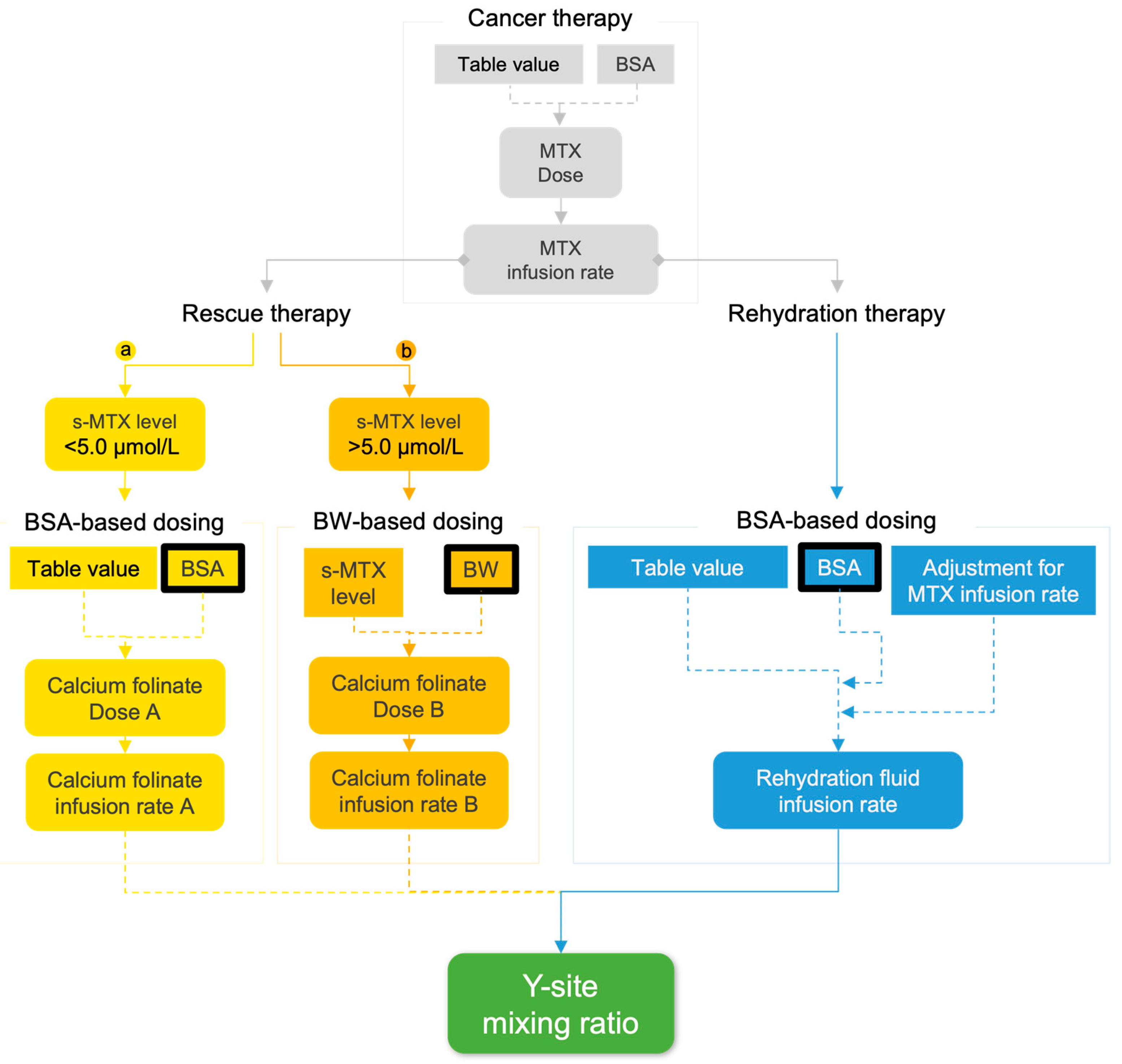
Figure 1. The complex and dynamic relationship between dosing, infusion rates, concentrations, and how they influence mixing ratios of the rescue therapy (calcium folinate) and the HCO3-containing rehydration fluid, inside the catheter lumen (internal channel) at y-site, based on the pediatric ALL-together protocol [15]. The calcium folinate dosing was based on either body surface area (BSA) in cases where serum methotrexate concentrations (s-MTX) were <5.0 µmol/L (a), or bodyweight (BW), for cases where s-MTX was >5.0 µmol/L (b). The rehydration therapy was BSA-based and was adjusted according to the MTX infusion rate to achieve a total infusion rate of 3000 mL/m2/day (MTX + rehydration fluid). Table values refer to standardized dosing and administration guidelines prescribed by the protocol [15].
Pharmaceutics 17 01155 g001
Figure 2. General compatibility testing program used for patient model 1–3, as recommended in the ALL-together treatment protocol [15]. Predicted volumes of three equal sets of A (A1, A2, and A3) and B (B1, B2, and B3) were added into three replicate tubes, representing the y-site mixing scenario for each patient. Diluent A was sodium hydrochloride 9 mg/mL and diluent B was a standard glucose–sodium–potassium infusion solution.
Pharmaceutics 17 01155 g002
Figure 3. Schematic top-down view illustrations of Raman microscopical and spectroscopical investigation of filter membrane analysis. (a) Depiction of a porous filter membrane where three selected areas (1, 2, and 3) were video-stitched by 169 images, giving three larger images. One of these images is illustrated by (b), having a 1012 µm wide side (α) with a 40% overlap (β). (c) The larger video-stitched images were then examined and targeted for Raman spectrum analysis upon suspected particle discovery at this random point (γ).
Pharmaceutics 17 01155 g003
Figure 4. Visual model of equilibrium conditions and predictive check of concentrations in simulated y-site mixes of physically tested patient models 1–3 compared to the hypothetical “extreme patient” case, for (a) Ca2+, (b) HCO3, (c) pH, and (d) CaCO3, when s-MTX increases (0.1–9.7 µmol/L). pH predictions were based on per-model predicted HCO3 concentrations, when [H2CO3] is set to 0.11 mmol/L and CaCO3 concentrations were predicted based on estimated Ca2+ or CO32− concentrations, depending on which was the least available component. Dotted s-MTX values (4d) mark levels included in physical tests (Table 2).
Pharmaceutics 17 01155 g004
Figure 5. Raman spectra of the calcium carbonate standard, undiluted mix, “patient model 3 alkalized”, calcium folinate 10 mg/mL, and sodium bicarbonate 500 mmol/L. Shaded bands (a, b, and c) highlight peaks at similar positions (rel. cm−1) across samples. Y-axis consists of an arbitrary scale.
Pharmaceutics 17 01155 g005
Table 1. Calcium folinate dosing based on patient serum methotrexate concentration (s-MTX) from hour 48 and onwards, according to the pediatric ALL-together protocol [15]. Calcium folinate is administered until s-MTX ≤ 0.2 µmol/L, as a bolus (2–5 min) when dosed ≤ 20 mg/kg [15] and ≤50 mg/m2/min [57] or short infusion (60 min) when dosed > 20 mg/kg and >50 mg/m2/min.
s-MTX
(µmol/L)
Calcium Folinate
(mg/m2)
<1.015
1.0–1.930
2.0–2.945
3.0–3.960
4.0–4.975
≥5.0s-MTX × BW *
* BW: Bodyweight.
Table 2. Assigned key clinical parameters (bodyweight, body surface area, and serum methotrexate concentration (s-MTX) of the simulated pediatric patient models 1–3 and undiluted mix, together with the predicted infusion rates of calcium folinate and HCO3-containing rehydration fluid and their estimated mixing ratios. The “extreme patient” case was added to the rest of the study lineup as a potential high-risk outlier for reference and therefore does not correspond to a typical pediatric patient.
Physical and Theoretical TestingTheoretical Testing
Patient Model 1Patient Model 2Patient Model 3Undiluted Mix aExtreme
Patient
Age (years)191411 d39
Bodyweight (kg)10285040 d118
Body surface area (m2)0.491.01.51.3 d2.6
s-MTX (µmol/L)0.93.57.05.0 d9.0
Calcium folinate dose (mg)7.35603502001062
NaHCO3 dose (mmol)0.0500.140.4110.01.41
Administration time (min)2.02.05.060 d60
Calcium folinate infusion rate (mL/h)120012004801200 d40.0
Rehydration fluid b infusion rate (mL/h)38.3116165151 d302
Mixing ratio c31.310.32.921.000.13
a 1:1 ratio of undiluted calcium folinate 10 mg/mL (20 mL) and NaHCO3 500 mmol/L (20 mL); b containing NaHCO3; c calculated as infusion rate of calcium folinate over infusion rate of NaHCO3-containing rehydration fluid; d corresponding value when extrapolating the tested calcium folinate dose (Table 1) of undiluted mix model.
Table 3. Predicted content of Ca2+(aq) from calcium folinate, and HCO3(aq) from the sodium bicarbonate-containing rehydration fluid., their relative molar ratio and pH values based on the simulated patient models 1–3, the undiluted mix, and the “extreme patient” case. Their theoretical risk for CaCO3(s) precipitation in mixed samples (40 mL) are based on solubility and ionic product (Supplementary Materials Table S1). Predictions are based on their assigned clinical parameters, dose, and infusion parameters (Table 2) and mixing protocol according to treatment conditions described in Figure 1.
Patient Model 1Patient
Model 2
Patient
Model 3
Undiluted MixExtreme
Patient
[Calcium folinate](aq) (mg/mL)0.181.376.525.003.10
[Ca2+](aq) (mol/mL)3.48 × 10−72.67 × 10−61.27 × 10−51.96 × 10−56.07 × 10⁻⁶
[HCO3](aq) (mol/mL)1.24 × 10−63.52 × 10−61.02 × 10−52.50 × 10−43.53 × 10⁻⁵
Ca2+(aq) amount (mol)1.39 × 10−51.07 × 10−45.10 × 10−47.82 × 10−42.43 × 10⁻⁴
HCO3(aq) amount (mol)4.95 × 10−51.41 × 10−44.09 × 10−41.00 × 10−21.41 × 10⁻³
HCO3/Ca2+ molar ratio3.561.320.8012.85.82
Predicted pH7.357.818.279.668.81
[CO32−](aq) (mol/mL) *1.39 × 10−91.13 × 10−89.48 × 10−85.68 × 10−51.13 × 10⁻⁶
CO32−/Ca2+ molar ratio *4.00 × 10−34.22 × 10−37.44 × 10−32.911.87 × 10−1
Maximum theoretical [CaCO3](aq or s) (mol/mL) **1.39 × 10−91.13 × 10−89.48 × 10−81.96 × 10−51.13 × 10⁻⁶
CaCO3(s) precipitation risk
based on solubility ***
nononoyesyes
Ionic product [Ca2+] × [CO32−]
(mol2/L2) *
4.85 × 10−103.02 × 10−81.21 × 10−61.11 × 10−36.88 × 10−6
CaCO3(s) precipitation risk
based on ionic product ****
noyesyesyesyes
Total assessment of CaCO3(s) precipitation risknoinconclusiveinconclusiveyesyes
* Based on predicted pH; ** Understood as the theoretical concentration of formable salt (CaCO3) in solution. This is therefore equal to the concentration of the least concentrated ionic component of the salt; *** Maximum theoretical [CaCO3](aq or s) compared to CaCO3 solubility (1.66 × 10⁻⁷ mol/mL or 0.0166 mg/mL [30]); **** Ionic product compared to solubility product (Ksp) of CaCO3 (3.36 × 10⁻⁹ mol2/L2 [65]).
Table 4. The measured pH values (mean ± SD) of mixed solutions of simulated patient model 1–3 and the undiluted mix, in addition to their corresponding control samples A-control (calcium folinate) and B-control (sodium bicarbonate-containing rehydration fluid). Ionic products based on measured pH at t0 and t4 were included for comparison with the ionic products based on predicted pH in Table 3. Detailed calculations are presented in Supplementary Materials Table S1.
y-Site MixA-Control B-Control Ionic Product *
(mol2/L2)
CaCO3(s)
Precipitation Risk **
(n = 3)(n = 1)(n = 1)
Patient model 1t07.29 ± 0.026.448.364.21 × 10−10No
t47.30 ± 0.056.458.384.31 × 10−10
pH change0.010.010.02
Patient model 2t07.63 ± 0.056.718.202.01 × 10−8Yes
t47.68 ± 0.066.698.272.26 × 10−8
pH change0.050.020.07
Patient model 3t07.73 ± 0.017.078.203.50 × 10−7Yes
t47.74 ± 0.017.058.213.58 × 10−7
pH change0.010.020.01
Undiluted mixt07.51 ± 0.037.348.187.93 × 10−6Yes
t47.98 ± 0.117.318.152.34 × 10−5
pH change0.620.040.02
* Ionic product of [Ca2+] × [CO32−], based on measured pH; ** Ionic product compared to solubility product (Ksp) of CaCO3 (3.36 × 10⁻⁹ mol2/L2 [65]).
Table 5. The results from visual examination of simulated y-site mixes using Tyndall beam of patient model 1–3 and the undiluted, in addition to their corresponding control samples, A-control (calcium folinate) and B-control (sodium bicarbonate-containing rehydration fluid), at timepoints t0, t4, and t24, respectively.
y-Site Mix
(n = 3)
A-Control
(n = 1)
B-Control
(n = 1)
t0clearclearclear
Patient model 1t4clearclearclear
t24clearclearclear
t0clearclearclear
Patient model 2t4clearclearclear
t24clearclear (dust)clear (dust)
t0clear (yellow)clear (yellow)clear
Patient model 3t4clear (yellow)clear (yellow)clear (bubbles)
t24clear (yellow)clear (yellow)clear (bubbles)
t0visible particles (yellow)clear (yellow and bubbles)clear (bubbles)
Undiluted mixt4visible particles (yellow)clear (yellow)clear
t24visible particles (yellow)clear (yellow)clear (dust)
Table 6. Turbidimetric evaluations showing formazine nephelometry units (FNU) mean ± SD values for simulated y-site mixes of patient models 1–3 and the undiluted mix, in addition to their corresponding control samples A-control (calcium folinate) and B-control (sodium bicarbonate-containing rehydration fluid), and MQ water (particle-free water) from timepoint t0 and t4.
y-Site Mix
(n = 3)
A-Control
(n = 1)
B-Control
(n = 1)
MQ Water
(n = 1)
Patient model 1t00.13 ± 0.020.120.140.11
t40.14 ± 0.020.120.170.14
Patient model 2t00.18 ± 0.100.290.160.19
t40.16 ± 0.020.210.190.18
Patient model 3t00.29 ± 0.040.480.280.27
t40.18 ± 0.060.180.170.16
Undiluted mixt062.4 ± 24.50.240.730.30
t481.4 ± 96.00.320.870.14
Table 7. Particle concentration (#amount/mL) in fractions ≥ 5 µm, ≥10 µm, and ≥25 µm in simulated y-site mixes of patient model 1–3 and undiluted mix, in addition to their corresponding control samples, A-control (calcium folinate) and B-control (sodium bicarbonate-containing rehydration fluid, at timepoints t0 and t4 given as mean ± SD. Some samples could not be measured due to detector overload (OL).
y-Site Mix (n = 9)A-Control (n = 3)B-Control (n = 3)
t0t4t0t4t0t4
Patient
model 1
≥50.22 ± 0.400.40 ± 0.410.13 ± 0.121.20 ± 0.870.33 ± 0.460.27 ± 0.46
≥100.20 ± 0.400.27 ± 0.260.13 ± 0.120.80 ± 0.690.20 ± 0.200.27 ± 0.46
≥250.09 ± 0.180.09 ± 0.150.07 ± 0.120.20 ± 0.200.00 ± 0.000.00 ± 0.00
Patient
model 2
≥51.69 ± 1.295.71 ± 3.991.20 ± 0.536.33 ± 1.101.20 ± 1.060.67 ± 0.50
≥100.22 ± 0.312.51 ± 1.980.27 ± 0.312.67 ± 1.010.13 ± 0.120.27 ± 0.12
≥250.02 ± 0.070.38 ± 0.340.07 ± 0.120.20 ± 0.200.00 ± 0.000.00 ± 0.00
Patient
model 3
≥51.58 ± 1.611.56 ± 1.060.53 ± 0.310.80 ± 0.200.40 ± 0.351.00 ± 0.87
≥100.53 ± 0.460.69 ± 0.540.33 ± 0.310.20 ± 0.350.13 ± 0.350.47 ± 0.64
≥250.11 ± 0.150.13 ± 0.170.13 ± 0.230.13 ± 0.230.00 ± 0.000.07 ± 0.12
Undiluted
mix
≥54070 ± 2320OL2.27 ± 1.17OL6.20 ± 1.51OL
≥10345 ± 160OL1.13 ± 1.01OL0.60 ± 0.20OL
≥2524.9 ± 15.1OL0.07 ± 0.12OL0.07 ± 0.12OL
Disclaimer/Publisher’s Note: The statements, opinions and data contained in all publications are solely those of the individual author(s) and contributor(s) and not of MDPI and/or the editor(s). MDPI and/or the editor(s) disclaim responsibility for any injury to people or property resulting from any ideas, methods, instructions or products referred to in the content.