Review

Therapeutic Effect of Membrane Vesicle Drug Delivery Systems in Inflammatory Bowel Disease

1
Department of Gastroenterology and Hepatology, Jinling Hospital, Affiliated Hospital of Nanjing University of Chinese Medicine, Nanjing 210000, China
2
School of Acupuncture and Tuina, School of Regimen and Rehabilitation, Nanjing University of Chinese Medicine, Nanjing 210023, China
3
School of Chinese Medicine, Nanjing University of Chinese Medicine, Nanjing 210023, China
*
Authors to whom correspondence should be addressed.
These authors contributed equally to this work.
Pharmaceutics 2025, 17(9), 1127; https://doi.org/10.3390/pharmaceutics17091127 (registering DOI)
Submission received: 8 July 2025 / Revised: 20 August 2025 / Accepted: 25 August 2025 / Published: 28 August 2025
(This article belongs to the Special Issue Advances of Membrane Vesicles in Drug Delivery Systems, 2nd Edition)

Abstract

Inflammatory bowel disease (IBD) is a chronic, heterogeneous condition characterized by recurrent intestinal inflammation and sustained mucosal barrier damage, profoundly impairing patients’ quality of life and imposing a considerable socioeconomic burden. Current therapeutic options are often constrained by low oral bioavailability, pronounced systemic toxicity, and inadequate tissue specificity, limiting their ability to achieve precise and durable efficacy. In recent years, membrane vesicle-based drug delivery systems (MV-DDSs) have shown considerable promise for precision IBD therapy owing to their excellent biocompatibility, mucosal barrier-penetrating capacity, and low immunogenicity. Building upon a systematic discussion of the roles of MV-DDSs in suppressing inflammatory signaling, modulating oxidative stress, preserving barrier integrity, reshaping the gut microbiota, and regulating programmed cell death, this review further compares the differences in key molecular targets and functional outcomes among vesicles of diverse origins and carrying distinct therapeutic payloads. These insights provide a comprehensive strategic reference and theoretical foundation for the rational design, mechanistic optimization, and clinical translation of MV-DDSs in IBD therapy.

1. Introduction

Inflammatory bowel disease (IBD) is a chronic, recurrent inflammatory disease that primarily affects the digestive system. The main subtypes of IBD are ulcerative colitis (UC) and Crohn’s disease (CD) [1]. The exact mechanisms underlying this disease have not yet been fully elucidated, but increasing evidence suggests that IBD is closely associated with abnormal immune responses, intestinal barrier dysfunction, intestinal microbiota imbalance, and complex interactions between genetic predisposition and environmental factors [2,3]. IBD affects approximately 6 million people worldwide each year [4], imposing a substantial healthcare and socioeconomic burden, increasing the risk of colorectal cancer, and severely impairing patients’ quality of life [5].
In the pathogenesis of IBD, abnormal activation of the immune system causes a persistent and excessive immune response against the host’s own intestinal tissue. This dysregulated immune response is associated with excessive production of proinflammatory cytokines and accumulation of reactive oxygen species (ROS), leading to complex amplification effects in inflammatory signaling [6]. At the same time, dysbiosis of the intestinal microbiota and dysfunction of the intestinal epithelial barrier further exacerbate the inflammatory environment, creating a vicious cycle of “barrier disruption–amplification of inflammation–progressive barrier impairment” [7]. Current standard treatment for IBD primarily involves 5-aminosalicylic acid (5-ASA) preparations, corticosteroids, immunosuppressants, and biological agents targeting inflammatory mediators [8,9]. Although these strategies are effective in the treatment of IBD to a certain extent, they still have inherent limitations, including a significant decline in curative effect in severe cases, obvious systemic toxicity, easy drug resistance, insufficient tissue specificity, and difficulty in maintaining stable therapeutic drug concentrations in inflammatory sites [10]. Consequently, repeated or intensified dosing is often required, compounding adverse effects and undermining long-term disease control [11,12].
Given the significant limitations of conventional pharmacotherapy in terms of therapeutic stability and safety, the development of drug delivery systems (DDSs) has emerged as a critical strategy to enhance the precision and effectiveness of IBD treatment [13]. In recent years, rapid advances in nanotechnology have driven the innovation of various delivery platforms. Among these, membrane vesicles (MVs) have attracted increasing attention as novel drug delivery vehicles. Owing to their unique closed bilayer architecture and programmable surface functionalization, MVs are capable of optimizing multiple aspects of drug performance, including improved physicochemical stability, prolonged systemic circulation, enhanced targeting efficiency, and controlled release profiles [14].
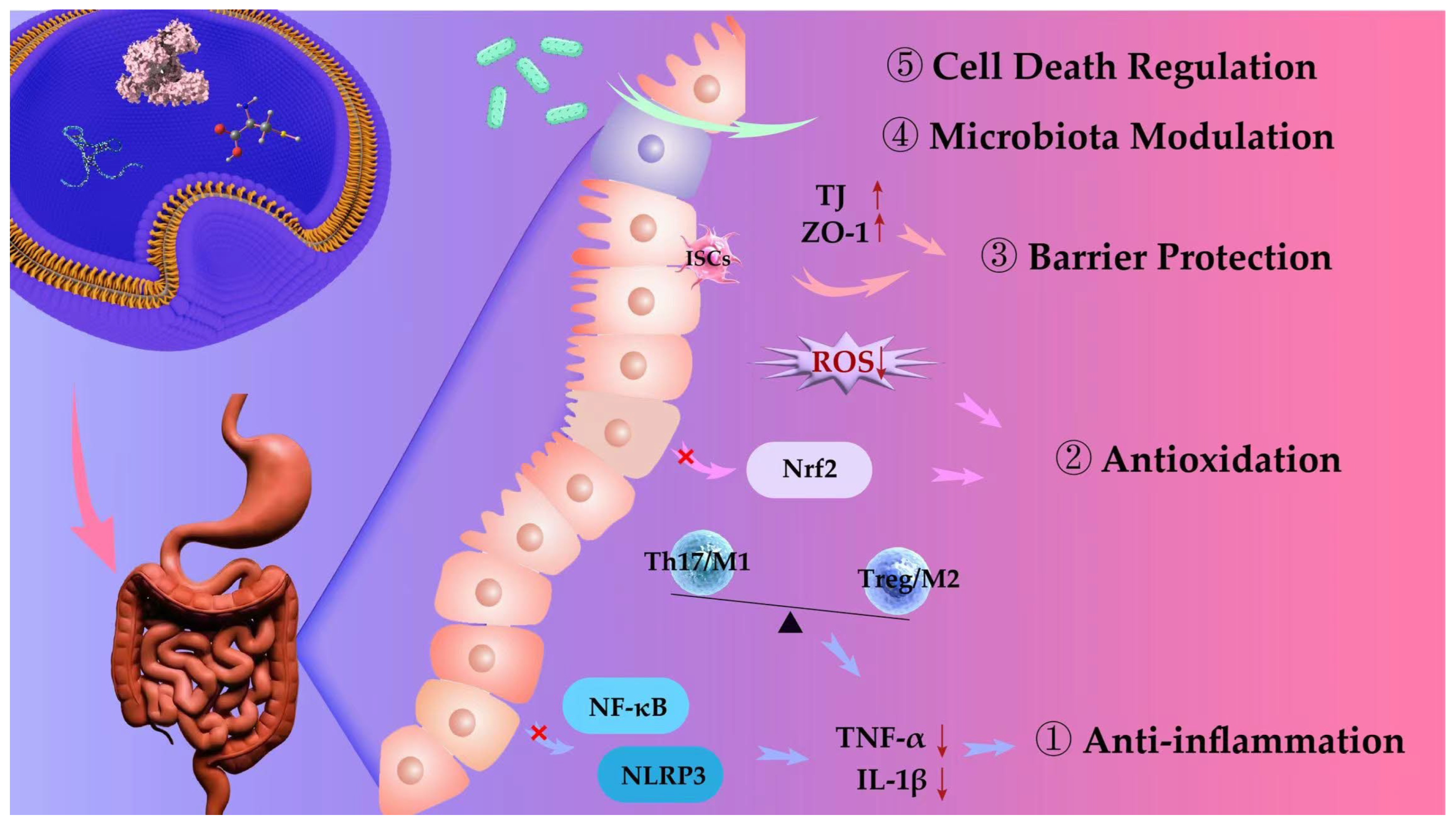
Although numerous studies have explored the drug delivery strategies and therapeutic potential of MVs derived from various sources in diseases such as cancer and atherosclerosis [15,16], there remains a lack of systematic and integrative evaluation regarding their mechanistic effects in IBD. To address this gap, this review focuses on the functional mechanisms of MV-DDSs in the treatment of IBD. Specifically, we systematically review the therapeutic mechanisms of MV-DDSs in several key pathological dimensions, including the regulation of inflammatory responses, attenuation of oxidative stress, restoration of intestinal barrier integrity, modulation of gut microbiota composition, and intervention in programmed cell death pathways. By analyzing MVs from diverse origins and their bioactive cargos, this work discovers key regulatory molecules with pivotal roles in modulation of the disease. Furthermore, we summarize current engineering strategies enhancing the targeting specificity and bioavailability of MVs (Figure 1). This review offers mechanistic insights and strategic guidance for the rational design of next-generation MV-DDSs tailored for precision therapy in IBD.

2. MVs

2.1. Overview of MVs

MVs are closed, near-spherical structures assembled from lipid bilayers or amphiphilic molecules, exhibiting membrane compositions and topological configurations that closely resemble those of natural biological membranes. This structural arrangement creates a relatively stable microenvironment within the vesicular lumen, capable of encapsulating a broad range of therapeutic cargos, including small-molecule drugs, proteins, and microRNAs, thereby markedly enhancing their physicochemical stability [17,18]. The lipid bilayer effectively shields the encapsulated agents from enzymatic degradation, pH fluctuations, and oxidative stress, prolonging their systemic half-life and preventing premature release before reaching the target site. This feature reduces non-specific toxicity to healthy tissues and ensures precise therapeutic action [19].
In terms of targeted delivery, MVs possess inherent targeting capabilities, as their surfaces are enriched with transmembrane proteins, glycoproteins, and adhesion molecules that can bind selectively to specific receptors on target cells, thereby facilitating active accumulation and uptake in desired tissues [20]. Furthermore, genetic engineering, chemical conjugation, or physical coupling strategies can be employed to graft targeting ligands (such as antibodies, peptides, or aptamers) onto the vesicle surface, further enhancing their selectivity and precision in delivering therapeutics to specific tissues or cells. These combined properties provide a robust platform for the targeted treatment of a wide spectrum of diseases.

2.2. Classification of MVs

Currently, MV-DSSs can be broadly categorized by their origin into two primary types: naturally derived MVs secreted by cells and biomimetic MVs synthesized through chemical or biophysical methods. Natural MVs can be further subdivided into three main classes based on their source: mammalian-derived membrane vesicles (MDMVs), plant-derived membrane vesicles (PDMVs), and bacterial membrane vesicles (BMVs). In contrast, artificial MVs are typically classified according to their fabrication strategies into liposomes, polymersomes, and engineered biomimetic membrane vesicles (EBMVs). Detailed information is provided in Table 1.

2.2.1. MDMVs

MDMVs have demonstrated significant therapeutic potential in the treatment of IBD, owing to their natural origin, excellent biocompatibility, and complex functional cargo. Relevant examples are summarized in Table 2. Current research has primarily focused on MDMVs derived from mesenchymal stem cells (MSCs), immune cells, and mammary gland epithelial cells. Notably, vesicles secreted by different cellular sources exhibit distinct cargo profiles, targeting capabilities, and biological functions, which collectively determine their therapeutic applicability and efficacy in specific disease contexts.
Membrane vesicles derived from MSCs (MSC-MVs) have demonstrated promising therapeutic efficacy in experimental models of IBD. While MSC-MVs from different tissue sources share common capabilities in modulating immune responses and promoting tissue repair [34,35], their biological origin confers distinct functional characteristics. For instance, bone marrow-derived MSC-MVs (BM-MSC-MVs) are widely regarded as the benchmark in regenerative medicine, due to their potent immunomodulatory and regenerative properties. These vesicles are enriched in miRNAs such as miR-21, miR-125a/b, miR-146, and miR-181, which regulate key inflammatory pathways, including TGF-β, STAT3, and NF-κB [36,37,38]. Through these mechanisms, BM-MSC-MVs facilitate M2 macrophage polarization, inhibit Th17 cell differentiation, enhance intestinal epithelial barrier integrity, and mitigate inflammatory responses [37,39]. However, their isolation involves invasive procedures, limiting clinical scalability. In contrast, adipose tissue-derived MSC-MVs (AT-MSC-MVs) are more readily obtainable and exhibit a higher proliferative potential, along with a stronger capacity to secrete anti-inflammatory cytokines [40,41,42]. In addition to suppressing classic proinflammatory mediators such as TNF-α, IL-6, and IL-17, AT-MSC-MVs also exert regulatory effects on IL-12 signaling, making them particularly suitable for treating IBD subtypes characterized by immune dysregulation [43,44,45,46]. Umbilical cord-derived MSC-MVs (UmC-MSC-MVs), on the other hand, are notable for their low immunogenicity, inherent immune privilege, and superior homing capability to inflamed tissues [47]. They exhibit enhanced tissue-repair efficacy, partly mediated by miRNA cargos such as miR-378a, which inhibit NLRP3 inflammasome activation and downstream caspase-1 cleavage, thereby preventing PANoptosis and promoting cell survival [48]. These properties make UmC-MSC-MVs especially advantageous in addressing IBD phenotypes dominated by epithelial barrier disruption.
In addition to MSC-MVs, MVs originating from immune cells also play pivotal roles in intestinal disorders, primarily by modulating innate immune responses and exerting localized anti-inflammatory effects. Neutrophil-derived MVs (PMN-MVs) have been shown to inhibit the maturation of dendritic cells while promoting the secretion of TGF-β1, thereby interrupting the amplification of immune cascades and demonstrating considerable immunoregulatory potential [49]. Meanwhile, macrophage-derived MVs (MMVs) exhibit an intrinsic capacity to home to inflamed mucosal tissues and actively regulate macrophage polarization. Specifically, they attenuate proinflammatory signaling by reducing the expression of M1-associated cytokines such as IL-1β and TNF-α, while concurrently enhancing the immunosuppressive profile of M2 macrophages. Through these dual effects, MMVs contribute significantly to the resolution of local inflammation and the promotion of mucosal repair processes [50].
Moreover, milk-derived MVs (milk-MVs) have garnered increasing attention in recent years due to their non-invasive collection, inherent biosafety, and remarkable stability in the gastrointestinal environment, making them particularly well-suited for oral drug delivery applications [51]. These vesicles naturally encapsulate a range of anti-inflammatory microRNAs, including miR-148a and let-7b, which can exert therapeutic effects through multiple mechanisms—such as modulating intestinal immune cell polarization, attenuating oxidative stress, and maintaining microbial homeostasis [52]. Owing to these properties, milk-MVs represent a promising platform for the non-parenteral treatment of chronic inflammatory diseases like IBD.
In summary, MDMVs exhibit distinct functional advantages in immune regulation, resolution of inflammation, and tissue repair. MSC-MVs are particularly well-suited for systemic immunomodulation and mucosal regeneration, whereas MVs from immune cells demonstrate superior efficacy in targeting inflamed sites and rapidly correcting immune dysregulation. In contrast, milk-MVs offer a safe and non-invasive alternative, with excellent suitability for oral administration, making them an attractive option for long-term management of chronic inflammatory conditions.
Table 2. Mechanism of MDMVs from different sources in DSS-induced IBD.
OriginDiseaseCargoTargetFunctionRef.
hucMSCsDSS-induced IBDmiR-378a-5pNLRP3 Inhibited NLRP3 inflammasome activation, reduced ASC and caspase-1, suppressed IL-1β and IL-18[53]
hucMSCsDSS-induced IBDN.D.inflammatory cytokines, macrophages Reduced TNF-α/IL-1β/IL-6/IL-7/iNOS, increased IL-10, decreased M1 macrophages, improved epithelial proliferation[54]
MSCsTNBS-induced IBDmiR-378a-3pGATA2, AQP4, PPAR-αSuppressed GATA2/AQP4/PPAR-α signaling, reduced apoptosis, improved tissue integrity[55]
hucMSCsDSS/TNBS-induced IBDTSG-6tight junction, T cells, pro-inflammatory cytokinesEnhanced tight junctions, inhibited Th17 and pro-inflammatory cytokines, promoted Th2/anti-inflammatory cytokines, improved barrier[56]
hADSCDSS-induced IBDN.D.ISCs, inflammatory cytokinesPromoted Lgr5+ ISC regeneration, reduced pro-inflammatory cytokines, increased IL-10/IL-13, protected barrier[48]
hucMSCsDSS-induced IBDmiR-326NEDD8, NF-κBInhibited NEDD8-Cullin1 binding, reduced neddylation-related enzymes (NAe1/Uba3/UBC12F/DCNL1), suppressed NF-κB, downregulated IL-1β/IL-6[57]
BMSCsDSS-induced IBDN.D.macrophage, inflammatory cytokinesPromoted M2 polarization, reduced M1 cytokines, inhibited JAK1/STAT1, activated STAT6[58]
HucMSCsDSS-induced UCmiR-23b-3pNrf2 pathwayActivated Nrf2, increased GPX4, reduced ROS/iron/lipid peroxidation, suppressed IL--1β/IL-6[59]
hPMSCsTNBS-induced IBDN.D.inflammatory cytokines, ROS, MPO, apoptosis proteins, tight junction, MMPsReduced ROS/MPO/cytokines, inhibited apoptosis, upregulated ZO-1/Claudin-1/Occludin), increased IL-10/TGF-β, improved epithelium[60]
hucMSCsDSS-induced IBDN.D.METTL3-Slc37a2-YTHDF1 axis, macrophage, inflammatory cytokinesUpregulated METTL3-m6A, stabilized Slc37a2, promoted M2 macrophages, suppressed cytokines, enhanced barrier[61]
Bovine milkDSS-induced IBDN.D.gut microbiota, T-AOCEnriched Roseburia, enhanced T-AOC, restored gut diversity[62]
Bovine milkDSS-induced IBDN.D.TLR4-NF-κB, NLRP3, Treg/Th17, microbiota, inflammatory cytokinesSuppressed TLR4-NF-κB/NLRP3, rebalanced Treg/Th17, increased Akkermansia/Enter-orhabdus, decreased Desulfovibrionaceae/Enterococcaceae, reduced IL-1β/TNF-α/IL-6/IL-17A, increased IL-10[63]
milkDSS-induced IBDN.D.TNFAIP3, COX-2, microbiota, tight junction, inflammatory cytokinesIncreased Lachnospiraceae/Rumi-nococcaceae, suppressed NF-κB via TNFAIP3, restored ZO-1, reduced IL-6/IL-1β/TNF-α[64]
milkDSS-induced IBDTGF-β1inflammatory cytokines, DNMT1/3Increased miR-320/miR-375/let-7a, downregulated DNMT1/3, increased TGF-β1, reduced IL-6/TNF-α[65]
M2b macrophagesDSS-induced IBDCCL1CCR8Promoted Th2/Treg, reduced IL-1β/IL-6/IL-17A[66]

2.2.2. BMVs

Unlike vesicles derived from other origins, BMVs not only regulate the intestinal immune system but also modulate the gut microbiota and influence the integrity of the intestinal barrier [67,68]. According to this, they play a multifaceted regulatory role in the interaction between the microbiota and the host, acting as crucial mediators. Based on their functional characteristics, BMVs can be broadly classified into three categories: probiotics, commensals, and opportunistic pathogens [69,70]. Each type of BMV exhibits unique biological effects in regulating host immune responses and maintaining the integrity of the intestinal barrier. Relevant examples are summarized in Table 3.
BMVs derived from probiotic strains, such as Escherichia coli Nissle 1917 (EcN), have been recognized as key functional carriers underpinning their antimicrobial and anti-inflammatory activities. EcN-derived outer membrane vesicles (OMVs), with diameters ranging from approximately 20 to 250 nm, can activate NF-κB and AP-1 signaling pathways in intestinal epithelial cells, thereby inducing the secretion of human β-defensin-2 (hBD-2) [71]. This process markedly enhances mucosal innate immune defenses and effectively suppresses pathogenic bacterial colonization [72,73]. Additionally, in vitro studies have shown that EcN-OMVs can trigger epithelial cell apoptosis via the PI3K/PTEN/AKT pathway, suggesting their bidirectional regulatory potential in promoting barrier repair while also contributing to epithelial renewal and homeostasis under specific conditions [74]. BMVs derived from Lactobacillus species also play a critical role in sustaining intestinal health. These vesicles are enriched with various proteins, DNA, and RNA, enabling them to modulate the composition of commensal microbiota, promote the production of anti-inflammatory cytokines, inhibit the proliferation of potentially pathogenic bacteria, and upregulate the expression of tight junction proteins. Collectively, these properties have been demonstrated to significantly attenuate inflammatory responses and mucosal injury in multiple IBD animal models [75].
BMVs derived from commensal anaerobic bacteria, such as Clostridium butyricum, have been shown to exert anti-inflammatory effects by regulating innate immune cell phenotypes. Specifically, C. butyricum-derived BMVs markedly upregulate the expression of M2 macrophage markers Arg1 and IL-10 in colonic tissues, thereby promoting polarization toward an anti-inflammatory phenotype and effectively alleviating DSS-induced colitis in mice [76]. Similarly, OMVs secreted by Bacteroides fragilis interact with dendritic cells in a TLR2-dependent manner to induce regulatory T cell (Treg) differentiation and IL-10 production, thereby enhancing mucosal immune tolerance and suppressing the progression of inflammation [77]. Notably, in genetically susceptible individuals, such as those with an ATG16L1 deficiency, this OMV-mediated immune remodeling offers a promising strategy for correcting Treg dysfunction [78].
BMVs released by certain opportunistic pathogens, including virulence-associated E. coli strains, exert complex effects within the intestinal microenvironment. Studies have demonstrated that E. coli-derived vesicles, which are enriched in respiratory chain complexes, can reduce luminal oxygen tension, re-establish anaerobic conditions, inhibit the overgrowth of Enterobacteriaceae under aerobic conditions, and mitigate DSS-induced colonic inflammation [79]. Although such effects may confer partial protective benefits, their overall impact on mucosal immunity requires further investigation to fully assess potential risks.
Overall, BMVs play a pivotal role in the onset and progression of IBD by engaging multiple targets and regulating various pathways. Compared to other sources of MVs, BMVs offer unique advantages, particularly in immune modulation and microbial–host interactions. They can effectively influence host immune responses, modulate gut microbiota, and provide significant therapeutic benefits in IBD. However, BMVs also present certain limitations, particularly due to the potential presence of endotoxins, which can trigger undesirable immune responses.
Table 3. Mechanism of BMVs from different sources in DSS-induced IBD.
OriginDiseaseCargoTargetFunctionRef.
Roseburia intestinalisDSS-induced colitisIPIDPP4Suppressed DPP4 activity, increased GLP-1, downregulated NFκB/STAT3 via PI3K, promoted Bifidobacterium[80]
EcN 1917DSS-induced colitisN.D.TLRsSuppressed IL-1β/TNF-α/IL-6/IL-17, reduced MMP-9/COX-2/iNOS, increased TFF-3[81]
Clostridium butyricumDSS-induced UCmiR-199a-3pMap3k4Suppressed MAPK/NF-κB, reduced IL-6/TNF-α/IL-1β, decreased[82]
Lactobacillus kefiranofaciensTNBS-induced IBDN.D.NF-κB p65Inhibited NF-κB p65, reduced IL-8, decreased MPO[83]
Odoribacter splanchnicusDSS-induced IBDN.D.NLRP3Suppressed NLRP3, increased IL-10, upregulated ZO-1/Occludin, reduced apoptosis[84]
Lactobacillus plantarumDSS-induced UCN.D.Gut microbiotaReduced Proteobacteria, increased Bifidobacteria/Muribaculaceae/Akkermansia, downregulated IL-6/IL-1β/TNF-α/IL-2, inhibited TLR4/MyD88/NF-κB[85]
Faecalibacterium prausnitziiDSS-induced colitisN.D.NF-κB and MAPK signaling pathwaysSuppressed NF-κB/JNK/P38 (MAPK), regulated Nrf2/HO-1, upregulated ZO-1/Occludin, reduced IL-1β/IL-2/IL-6/TNF-α/IFN-γ/GM-CSF, increased IL-4/IL-10/TGF-β[86]
Bacteroides fragilisTNBS-induced colitisPSATLR2Induced TLR2, upregulated Gadd45α, promoted CD4+CD25+Foxp3+ Tregs, suppressed TNF-α/IL-17/IFN-γ/IL-6[77]
Akkermansia muciniphilaDSS-induced colitisN.D.Tight junction proteinsSuppressed NO/TNF-α/L-1β/IL-6, upregulated ZO-1/MUC2, increased IgA and sIgA, reduced DAO/D-LA, increased Firmicutes, and decreased Proteobacteria[87]
Bacteroides fragilisDSS-induced colitismiR-5119PD-L1Delivered miR-5119 to inhibit PD-L1, suppressed GSDMD-mediated NET, promoted Lgr5+ intestinal stem cells[88]
Clostridium butyricumDSS-induced UCN.D.McrophagesPromoted M2 macrophage polarization, reduced M1 macrophage infiltration, decreased Helicobacter/Escherichia-Shigella abundance; increased Lactobacillus/Akkermansia/Bacteroides, upregulated MUC2/ZO-1[76]
Lactiplantibacillus plantarumDSS-induced colitissmall RNAs (<200 nt)Intestinal epithelial HT29 cells, pro-inflammatory cytokine IL-8Delivered small RNAs into HT29 cells, suppressed IL-8, reduced neutrophil accumulation in colonic lamina propria[89]
L. rhamnosus GGDSS-induced UCN.D.TLR4-NF-κB-NLRP3 signaling pathwaySuppressed TLR4-NF-κB-NLRP3 axis, reduced TLR4/MyD88/p65/p-p65/NLRP3/ASC, decreased TNF-α/IL-1β/IL-6/IL-2), reduced Helicobacter/Escherichia-Shigella, increased Lachnospiraceae/Akkermansia[90]
Lactobacillus kefirgranum PRCC-1301DSS-induced colitisN.D.NF-κB signaling pathwaySuppressed NF-κB by reducing phosphorylated NF-κB p65 and phosphorylated IκBα, decreased IL-2/IL-8/TNF-α, upregulated ZO-1/claudin-1/occludin[91]

2.2.3. PDMVs

PDMVs, as naturally sourced nanovesicles, offer numerous significant advantages, making them highly promising for the treatment of IBD and related disorders [92,93,94]. PDMVs exhibit excellent biocompatibility, non-immunogenicity, high stability, and a prolonged circulation time, which enables them to effectively avoid degradation during drug delivery, ensuring sustained release of therapeutic agents [95,96,97]. Relevant examples are summarized in Table 4. Compared to synthetic membrane vesicles, their production process is simpler and more cost-effective. Furthermore, due to their natural membrane structure, PDMVs offer distinct advantages in oral drug delivery, overcoming challenges faced by traditional drug delivery systems, such as poor water solubility and low bioavailability.
PDMVs are rich in various bioactive components, including lipids, proteins, and microRNAs, which play a crucial role in modulating immune responses and inflammatory cascades. For example, ginger-derived MVs, which are abundant in lipids, proteins, and microRNAs, can effectively traverse the intestinal barrier and be internalized by intestinal epithelial cells and macrophages. This process leads to the downregulation of pro-inflammatory mediators, enhancement of antioxidant responses, and promotion of epithelial regeneration, significantly improving intestinal inflammation [98]. Similarly, turmeric-derived MVs (such as TNVs and TDNPs) exhibit potent anti-inflammatory activity in DSS-induced colitis models. Oral administration of TNVs results in selective accumulation in inflamed colonic tissues, modulation of gut microbiota composition, restoration of mucosal barrier integrity, and promotion of M2 macrophage polarization, collectively ameliorating the severity of colitis [99]. TDNPs significantly inhibit the expression of pro-inflammatory cytokines (TNF-α, IL-6, IL-1β), upregulate antioxidant genes like HO-1, and suppress NF-κB signaling pathway activation. Notably, TDNPs also demonstrate exceptional targeting capabilities, being efficiently internalized by macrophages and epithelial cells in inflamed tissues, highlighting their dual immune modulation and mucosal protection properties [100].
Additionally, tea- and blueberry-derived membrane vesicles have shown significant antioxidant advantages. Tea-derived nanovesicles, rich in polyphenols and specific microRNAs, have been shown to alleviate oxidative stress in epithelial cells, thereby inhibiting inflammatory pathways and maintaining gut homeostasis [10]. Ginseng-derived MVs (GDMVs) exert antioxidant effects by activating the p62/Nrf2/Keap1 pathway. GDMVs effectively scavenge ROS in immune cells and intestinal epithelial cells, reducing oxidative stress, and enhance the antioxidant response by promoting the nuclear translocation of Nrf2. Upon activation, Nrf2 upregulates the expression of downstream antioxidant genes, such as HO-1 and GCLC, thereby maintaining cellular redox homeostasis and protecting cells from oxidative damage. GDMVs further enhance the antioxidant capacity of cells by increasing the activity of antioxidant enzymes, effectively reducing cellular damage induced by oxidative stress. In addition, GDMVs mitigate chronic intestinal inflammation by inhibiting the TLR4/MAPK signaling pathway, significantly reducing the expression of pro-inflammatory cytokines such as TNF-α, IL-6, and IL-1β, thus exerting their anti-inflammatory effects [101].
In summary, PDMVs, due to their superior biological properties, are particularly well-suited for oral drug delivery and the treatment of chronic inflammatory diseases. Compared to BMVs, PDMVs avoid the immune reactions induced by bacterial endotoxins, offering superior safety. However, PDMVs still face limitations in drug loading capacity and targeting ability, which need to be further optimized to enhance their clinical application effectiveness.
Table 4. Mechanism of PDMVs from different sources in DSS-induced IBD.
OriginDiseaseCargoTargetFunctionRef.
GarlicDSS-induced colitisN.D.TLR4/MyD88/NF-κB signaling pathway, tight junction proteins, gut microbiota, pro-inflammatory cytokines Suppressed TLR4/MyD88/NF-κB by han-miR3630-5p targeting TLR4 3′ UTR, reduced TLR4/MyD88/NF-κB p65, upregulated ZO-1/occluding/claudin-1, increased Lachnospiraceae, decreased Helicobacter/Escherichia–Shigella/Akkermansia, reduced IL-6/IL-1β/TNF-α/IFN-γ/IL-17A/NO[102]
LemonDNBS-induced colitisN.D.NF-κB signaling pathway, Nrf2 antioxidant pathway, inflammatory cytokines, gut microbiota Suppressed NF-κB, reduced IL-6/TNF-α, activated Nrf2 antioxidant pathway, reduced Pygmaiobacter/Lachnospiraceae UCG-010/Tuzzerella/Anaerofilum/Enteractinococcus/Acetatifactor, increased Enterococcus/Bacteroides_pectinophilus group/Lachnospiraceae[103]
Lycium barbarumDSS-induced UCPhosphatidylcholine, vitexin-2-O-rhamnosideInflammatory cytokines, tight junction proteins, MPOSuppressed TNF-α/IL-12, upregulated IL-10, increased occluding/ZO-1, reduced MPO[104]
TurmericDSS-induced UCN.D.Inflammatory cytokines, macrophages, tight junction proteins, gut microbiotaSuppressed TNF-α/IL-6/MCP-1, reduced CD16/32, increased CD206, upregulated ZO-1/occluding/E-cadherin, increased Akkermansia/Lactobacillus, decreased Escherichia-Shigella/Helicobacter[99]
TurmericDSS-induced UCN.D.Inflammatory cytokines, antioxidant gene, NF-κB signaling pathway, tight junction protein Suppressed NF-κB, reduced TNF-α/IL-6/IL-1β, upregulated HO-1, increased E-cadherin[100]
Tea leafDSS-induced IBDN.D.Inflammatory cytokines, ROS, antioxidant enzyme, tight junction proteins, gut microbiotaSuppressed TNF-α/IL-6/IL-12, increased IL-10, reduced ROS, upregulated HO-1/GSH, decreased MDA/MPO, upregulated ZO-1/MUC2, reduced Oscillibacter/elicobacter, increased Lachnospiraceae/kkermansia[10]
Mulberry barkDSS-induced colitisHSPA8Aryl hydrocarbon receptor (AhR), COP9 Constitutive Photomorphogenic Homolog Subunit 8 (COPS8), CUL1, anti-microbial peptides, tight junction protein, gut microbiotaActivated AhR signaling pathway via HSPA8 binding, upregulated COPS8, promoted deneddylation of CUL1, induced anti-microbial peptides secretion, upregulated ZO-1, reduced Proteobacteria/Segmented Filamentous Bacteria, increased Firmicutes[105]
AloeDSS-induced UCN.D.Oidative stress markers, tight junction proteinsReduced p-NF-κB and p-IκB, decreased TNF-α/NO/COX2/3-NT, upregulated ZO-1/claudin4/occluding/E-cadherin/γ-catenin/α-tubulin[106]
GrapeDSS-induced colitisN.D.Intestinal stem cells (Lgr5+), Wnt/β-catenin signaling pathwayActivated Wnt/β-catenin signaling (phosphorylated GSK-3β, nuclear translocation of β-catenin), upregulated intestinal stem cell markers (Lgr5, BMI1), promoted Lgr5+ stem cell proliferation[107]
GinsengDSS-induced UCGinsenosidesInflammatory cytokines, NF-κB signaling pathway, gut microbiotaSuppressed NF-κB signaling pathway activation, reduced TNF-α/IL-6/IL-17A, increased IL-10, decreased Firmicutes/Bacteroidota ratio, increased Lactobacillus, decreased Helicobacter/Ruminococcus[108]
BroccoliDSS-induced colitisN.D.Dendritic cells, AMP-activated protein kinase (AMPK), mTOR signaling pathway, inflammatory cytokinesActivated AMPK in DCs, reduced phosphorylation of p70S6K and S6, reduced TNF-α/IL-12/IFN-γ/IL-17A, increased IL-10/TGF-β[109]
GinsengDSS-induced IBDSmall RNAs, ginsenosidesIKK/IκB/NF-κB signaling pathwayActivated autophagy (upregulated LC3/Atg7/Beclin1, downregulated p-mTOR/p-AKT), increased CD206/Arg1, decreased CD86/iNOS, reduced P-IKKα/β/P-IκBα/NF-κB, reduced TNF-α/IL-1β/IL-6, increased IL-10, increased ZO-1/occludin[110]
GinsengDSS-induced IBDN.D.TLR4/MAPK signaling pathway, p62/Nrf2/Keap1 pathwaySuppressed TLR4/MAPK activation by reducing phosphorylation of ERK/JNK/p38, activated p62/Nrf2/Keap1 pathway to increase HO-1/GCLC/GCLM, decreased TNF-α/IL-6/IL-1β, enhanced ZO-1/occludin/claudin-1, promoted intestinal stem cell proliferation via Wnt/β-catenin, and reduced Firmicutes/Bacteroidetes ratio[101]

2.2.4. Liposomes

Liposomes have emerged as a versatile and promising nanocarrier platform for the treatment of IBD, offering unique advantages in drug protection, targeted delivery, and controlled release. Owing to their biocompatible phospholipid bilayer structures, liposomes can encapsulate both hydrophilic and hydrophobic therapeutic agents, thereby enhancing their bioavailability and stability in the harsh gastrointestinal environment. For instance, Xian et al. developed an oral liposomal formulation of an activatable budesonide prodrug, which demonstrated remarkable efficacy in experimental colitis models. The lipidation strategy conferred high stability under acidic gastric conditions while enabling site-specific drug release in inflamed intestinal tissues, leading to significantly improved remission of colitis and reduced systemic toxicity compared to free budesonide administration [28]. Beyond budesonide, a range of therapeutic agents, including corticosteroids, immunosuppressants, and biological molecules, have been successfully incorporated into liposomal carriers to optimize pharmacokinetics and minimize off-target effects.
Liposome can also be designed for both passive and active targeting of inflammatory mucosal sites, utilizing an enhanced permeability and retention (EPR) effect and ligand–receptor interactions to increase local accumulation of the drug. For instance, Rahman et al. reviewed cationic and polyethylene glycol (PEG)-modified liposomes, which further improved circulation times and facilitated the targeted delivery of anti-inflammatory drugs and immunomodulators to IBD lesion areas [26].
Overall, liposomal-based drug delivery systems have shown great potential in improving the safety, efficacy, and patient compliance of IBD therapies. Compared to naturally derived MVs, liposomes offer a simpler, more controllable production process suitable for large-scale manufacturing, while avoiding the ethical concerns and immune rejection associated with stem cell-derived vesicles. In comparison to polymersomes, liposomes have clear advantages in terms of biocompatibility and safety. While polymersomes offer greater flexibility in targeting and drug delivery control, their biocompatibility is inferior. The natural structure and biodegradability of liposomes make them a safe and effective platform for drug delivery. However, liposomes also have some limitations. Despite their advantages in biocompatibility and drug stability, their drug loading capacity and targeting ability are relatively weaker, particularly for certain types of macromolecular drugs. Furthermore, the surface modification of liposomes and selection of targeting ligands still require optimization to enhance their therapeutic efficacy for specific lesions.

2.2.5. Polymersomes

Polymersomes have emerged as a compelling platform for drug delivery in IBD, owing to their highly tunable physicochemical properties, excellent biocompatibility, and superior stability in the complex gastrointestinal environment. Polymersomes, typically self-assembled from amphiphilic block copolymers, possess greater mechanical strength and membrane integrity compared to conventional liposomes. This structural advantage allows them to encapsulate both hydrophilic and hydrophobic therapeutic agents while enabling multi-functional targeting and controlled release strategies [31].
Tollemeto et al. developed a pH-responsive polymersome by incorporating carboxyl functional groups into the block copolymer, which ensured structural stability under gastric acidic conditions while achieving site-specific release of immunosuppressive agents, such as mycophenolate mofetil, in the near-neutral pH environment of the small intestine [29]. In vitro and in vivo studies demonstrated that this polymersome maintained its integrity during gastric transit, thereby preventing premature drug leakage, and rapidly disassembled in the intestinal lumen, significantly enhancing drug accumulation at the target site. This approach holds promise for safer and more effective immunosuppressive therapy in patients with IBD.
In addition, polymersomes have been applied as nanocarriers to encapsulate natural antioxidants and bioactive compounds for multi-target modulation of the intestinal inflammatory microenvironment. Zhong et al. summarized advances in polymer-based nano-delivery systems for the encapsulation of anthocyanin and reported that such systems not only markedly improve the solubility and bioavailability of anthocyanins but also exert robust anti-inflammatory effects in multiple IBD models through targeted regulation of NF-κB signaling pathways, modulation of the gut microbiota, and suppression of oxidative stress [30].
In conclusion, polymersomes hold tremendous potential for the treatment of IBD owing to their customizable physicochemical properties and multifunctional stimulus-responsive release capabilities. Compared with naturally derived MVs, polymersomes offer the advantage of precisely controllable synthesis, thereby avoiding the ethical concerns and potential immune rejection issues associated with stem cell-derived vesicles. Relative to liposomes, the most prominent strengths of polymersomes lie in their superior stability and enhanced structural integrity [111,112,113]. While liposomes are widely used for the delivery of hydrophilic drugs and siRNA, they often suffer from suboptimal stability, particularly in vivo, where premature drug leakage may occur [114]. As structural analogues of liposomes, polymersomes exhibit greater mechanical robustness, enabling prolonged stability in the gastrointestinal environment and mitigating the risk of premature payload release. Nevertheless, a notable drawback of polymersomes compared to liposomes is their lower biodegradability, which may raise concerns regarding long-term safety in vivo. Therefore, while polymersomes demonstrate clear advantages in stability, drug loading capacity, and controlled release, further optimization of their biodegradability is warranted to maximize their clinical therapeutic potential.

2.2.6. EBMVs

EBMVs, as a new generation of nanoscale carrier platforms, strategically integrate the tunable physicochemical properties of synthetic nanomaterials with the targeting specificity and immunomodulatory capacity of natural cell membranes. Unlike conventional liposomes or polymersomes, EBMVs incorporate key membrane proteins—such as adhesion molecules, integrins, and immunoregulatory receptors—derived from source cells into the synthetic vesicle scaffold, thereby endowing them with highly selective bio-recognition and targeting capabilities [115]. This “hybrid” design enables both active homing to inflamed intestinal tissues and modulation of local immune responses, while retaining the structural stability, scalable manufacturing potential, and compositional controllability of synthetic carriers. A representative example is provided by the work of Corbo et al. [33], who stimulated murine leukocytes with retinoic acid to upregulate the expression of α4β7 integrin, extracted the membrane proteins, and co-assembled them with phospholipids and cholesterol to produce specialized “leukosomes.” In a DSS-induced colitis model, these reconstituted vesicles selectively accumulated at sites of inflammation, significantly suppressed the expression of pro-inflammatory cytokines (TNF-α and IL-6), and promoted mucosal healing [116,117,118]. Notably, these therapeutic effects were achieved without loading any additional pharmacological payloads, highlighting the intrinsic bioactivity conferred by the membrane proteins themselves.
EBMVs combine the controllable physicochemical characteristics, reproducible manufacturing, and high structural robustness of synthetic nanocarriers with the homologous targeting, immune evasion, and inherent bioactivity of natural cell membranes. This dual functionality positions them as a highly customizable and rationally designable platform for precision therapy in IBD, particularly in scenarios requiring both targeted delivery and inflammation microenvironment-specific immunomodulation [119]. Moreover, their modular assembly strategy allows for flexible selection of membrane sources and fine-tuning of membrane protein composition to match disease-specific therapeutic requirements.
Compared with naturally derived MVs, EBMVs circumvent batch-to-batch variability, poor scalability, and the ethical or immunogenicity concerns associated with certain cell sources, while preserving the bio-recognition and targeting features of native vesicles. Nevertheless, challenges remain in the efficient extraction of membrane proteins, maintenance of their functional integrity, and their stable integration into synthetic scaffolds. Overcoming these technical bottlenecks—through advances in membrane separation techniques, protein stabilization methods, and nano-assembly engineering—will be essential to fully realize the translational potential of EBMVs in clinical IBD therapy.

3. Therapeutic Mechanism of MV-DDSs in IBD

MV-DDSs demonstrate significant therapeutic potential for IBD, attributable to their unique biological properties and payload. Their therapeutic efficacy in IBD is manifested through five principal mechanisms: anti-inflammatory and antioxidant effects, protection of the intestinal mucosal barrier, modulation of gut microbiota, and regulation of programmed cell death pathways (Figure 2).

3.1. Anti-Inflammation

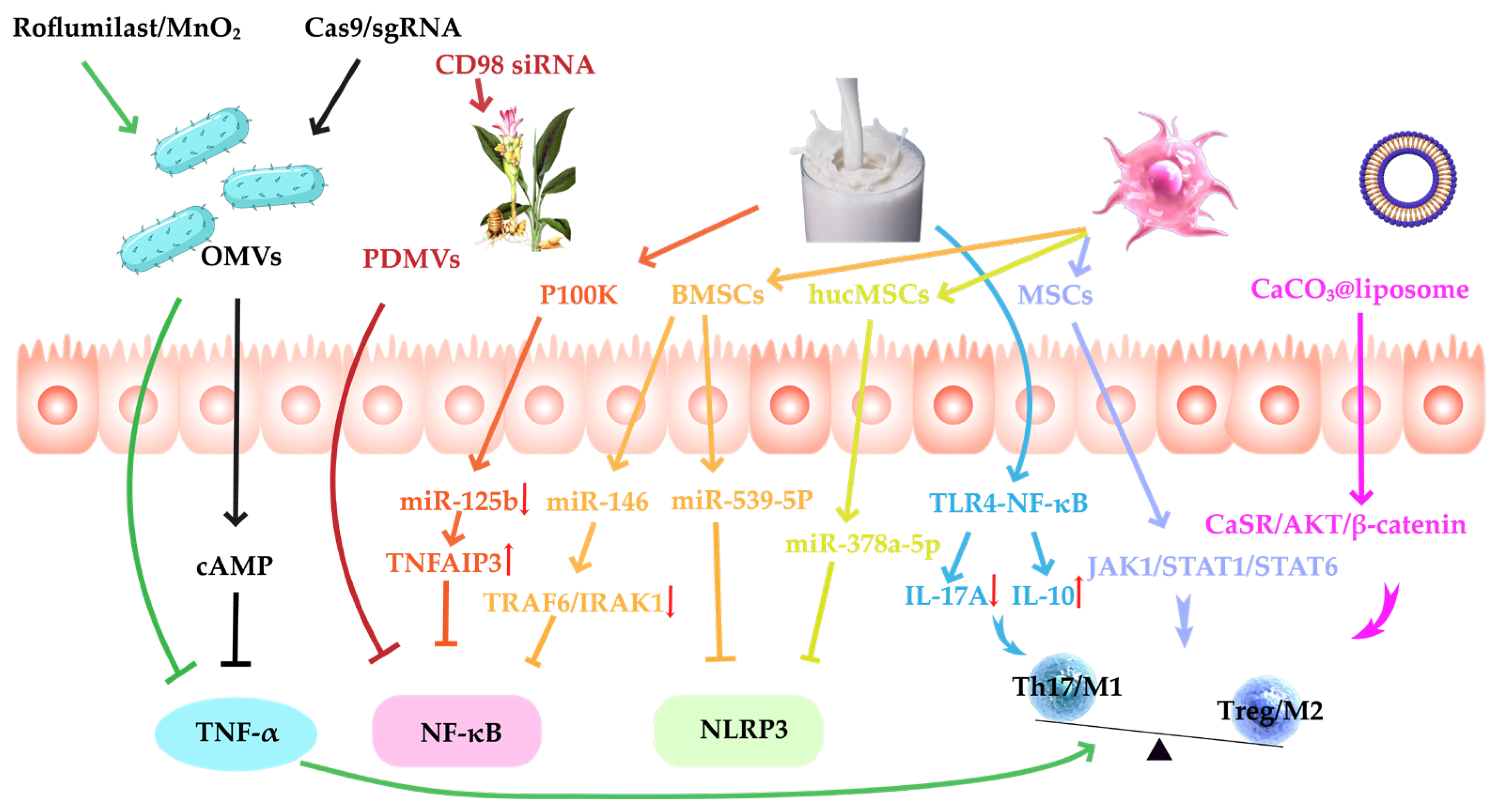
IBD is a chronic, recurrent disease driven by immune dysregulation of the intestinal mucosa. Pro-inflammatory cytokines such as TNF-α and IL-1β are significantly elevated in IBD patients. These cytokines not only directly mediate damage to intestinal epithelial cells and disruption of intestinal barrier function but also activate key inflammatory networks such as the NF-κB signaling pathway and the NLRP3 inflammasome, triggering inflammatory cascades and forming a vicious positive feedback loop that further amplifies the intensity of the immune response and pathological damage. In addition to local inflammatory cytokine storms, the chronic progression of IBD is also significantly influenced by immune imbalance. This manifests as dysregulation between innate and adaptive immune systems, such as weakened Treg function, excessive expansion of Th17 cells, or sustained maintenance of the M1 pro-inflammatory phenotype by macrophages. This imbalance not only delays the resolution of inflammation but also impedes tissue repair and the reconstruction of immune tolerance. Therefore, inhibiting the abnormal expression of key inflammatory factors, blocking their upstream signaling pathways, and restoring the homeostasis and tolerance mechanisms of the immune system are considered the three core strategies for alleviating IBD activity, slowing disease progression, and achieving long-term clinical remission (Figure 3).

3.1.1. Inhibition of Pro-Inflammatory Cytokines

In IBD, pro-inflammatory cytokines such as TNF-α, IL-1β, and IL-6 activate immune cells, promote inflammatory responses, and disrupt the intestinal epithelial barrier, leading to the loss of intestinal function and pathological changes [120]. MVs, by carrying specific cargo molecules, can effectively modulate the intestinal inflammatory response and reduce the release of pro-inflammatory cytokines, thus playing a significant role in the treatment of IBD.
TNF-α, as a key pro-inflammatory factor in IBD, not only promotes the activation and migration of immune cells but also induces damage to intestinal epithelial cells, disrupting the intestinal barrier and exacerbating the inflammatory response. According to the relevant literature, OMVs derived from EcN 1917 can accumulate in the intestinal mucosa through pathogen-associated molecular patterns (PAMPs) and, to some extent, inhibit the overexpression of TNF-α [121]. Although this intrinsic anti-inflammatory effect is limited, it still holds potential for alleviating IBD. To enhance the anti-inflammatory effects of OMVs, Song [122] et al. loaded phosphodiesterase 4 (PDE4) inhibitor roflumilast and MnO2 nanoparticles into OMVs. This combination not only upregulated cAMP synthesis and enhanced the anti-inflammatory response but also significantly suppressed the production of TNF-α and improved intestinal mucosal repair. This strategy, by altering the cargo and delivery mechanism of OMVs, significantly enhanced their therapeutic efficacy in regulating TNF-α. Furthermore, Lin [6] et al. coupled Cas9/sgRNA ribonucleoprotein nanoclusters to OMVs, enabling direct transcriptional disruption of the TNF-α gene. This approach significantly downregulated the expression of TNF-α, IL-1β, and IL-6, while upregulating IL-10. Through this gene-editing method, the anti-inflammatory effects of OMVs were further strengthened, allowing for precise targeting of pro-inflammatory factors and enhancing therapeutic outcomes. This strategy provides a more durable and stable solution for IBD treatment. Therefore, MVs-DDSs precisely regulate the release of pro-inflammatory cytokines by carrying either naturally occurring or exogenously loaded molecular cargo, thereby effectively alleviating the inflammatory response in IBD.

3.1.2. Suppression of Inflammatory Signaling Pathways

In IBD, key signaling pathways such as NF-κB and NLRP3 inflammasome are widely recognized as central molecular hubs driving the amplification of persistent inflammation and hindering the intestinal epithelial barrier’s repair [123,124]. These pathways play a decisive role in mediating the release of pro-inflammatory factors, cytokine cascade amplification, and immune dysregulation, making them critical targets in current inflammation intervention research.
NF-κB is a pivotal transcription factor that orchestrates the expression of pro-inflammatory mediators, including TNF-α, IL-1β, and IL-6. Its sustained activation creates a self-reinforcing inflammatory loop that significantly exacerbates intestinal inflammation. In the context of IBD, aberrant hyperactivation of the NF-κB pathway is a central mechanism underlying the epithelial barrier’s disruption, immune dysregulation, and chronic inflammation. Consequently, precise modulation of NF-κB activity has become a key strategy in anti-inflammatory therapeutic interventions. CD98, a transmembrane protein involved in amino acid transport and adhesion signaling, plays a critical role in facilitating upstream NF-κB activation [125]. PDMVs from ginger are enriched with natural anti-inflammatory miRNAs and bioactive small molecules. After oral administration, they effectively accumulate in the colon, suppressing CD98 expression, inhibiting NF-κB activation, and reducing the transcription and release of pro-inflammatory factors. Building on this, targeted delivery of anti-CD98 siRNA via these vesicles further silences CD98 expression, resulting in more robust NF-κB inhibition and substantial alleviation of colitis symptoms [126]. Similarly, miR-125b has been identified as a key pro-inflammatory miRNA. It enhances NF-κB signaling by repressing the translation of the anti-inflammatory protein TNFAIP3 (A20), a negative regulator of NF-κB. Milk-derived P100K MVs have been reported to effectively downregulate miR-125b, thereby relieving its inhibition on A20. This leads to a remarkable 375% increase in A20 protein expression—significantly higher than the 135% observed with P35K vesicles—ultimately resulting in suppression of NF-κB activity and attenuation of the inflammatory response [64]. In contrast to the pro-inflammatory mechanisms described above, miR-146 is widely recognized as a potent anti-inflammatory miRNA. MVs derived from BMSCs, enriched with miR-146, have demonstrated the ability to inhibit downstream NF-κB signaling by downregulating TRAF6 and IRAK1 expression and blocking phosphorylation of the NF-κB p65 subunit. This leads to a marked reduction in the production of pro-inflammatory cytokines such as TNF-α and IL-1β [36].
In addition to NF-κB, the NLRP3 inflammasome is another pivotal regulator of inflammatory responses. By sensing endogenous and exogenous signals, it activates caspase-1, which in turn cleaves and releases pro-inflammatory cytokines such as IL-1β, further exacerbating intestinal inflammation. Studies have shown that MVs derived from BMSCs primarily regulate the function of the NLRP3 inflammasome through the delivery of miR-539-5p. These BMSC-derived MVs inhibit NLRP3 activation by miR-539-5p, preventing its interaction with ASC and caspase-1, thereby reducing the release of pro-inflammatory cytokines such as IL-18 and IL-1β [127]. Similarly, hucMSC-derived exosomes deliver miR-378a-5p, which also inhibits NLRP3 activation, reduces the release of IL-1β, and alleviates colonic inflammation [53]. Collectively, these findings underscore the potent anti-inflammatory potential of MV-DDSs, which act through the precise modulation of key signaling pathways such as NF-κB and NLRP3, offering a promising therapeutic strategy for the treatment of IBD.

3.1.3. Modulation of Immune Homeostasis and Mucosal Tolerance

The imbalance of the Th17/Treg cell ratio and the polarization of macrophages directly affect the intensity and duration of immune responses, thus determining the onset and control of inflammation. In IBD, excessive Th17 cell responses and the activation of M1 macrophages are key factors in sustaining intestinal inflammation. In contrast, Treg cells and M2 macrophages help suppress excessive immune responses and restore immune homeostasis. Therefore, regulating the Th17/Treg balance and macrophage polarization has become a crucial therapeutic target in IBD treatment.
Studies have shown that milk-derived MVs inhibit the TLR4-NF-κB signaling pathway and activation of the NLRP3 inflammasome, reducing the release of pro-inflammatory factors like IL-17A and suppressing Th17 cell differentiation. At the same time, they upregulate the anti-inflammatory factor IL-10 and promote the function of IL-10+Foxp3+ Treg cells, helping restore the Th17/Treg balance [63].
In addition to regulating the Th17/Treg balance, macrophage polarization is also a critical aspect of IBD treatment. Naturally-derived MVs promote macrophage polarization to the M2b phenotype, downregulating colonic inflammation, which helps alleviate experimental colitis in mice. For example, MSC-MVs promote macrophage polarization to the M2 by downregulating the JAK1/STAT1/STAT6 signaling pathway in colonic tissue [128]. Similarly, Scutellaria baicalensis-derived MVs, carrying miR-4371c, also promote macrophage polarization to the M2 phenotype [129]. To further enhance immune modulation, researchers have shown that TNF-α-pretreated MVs upregulate miR-24-3p expression, targeting and inhibiting IRF1, which further promotes macrophage polarization from the pro-inflammatory M1 phenotype to the anti-inflammatory M2 phenotype, thereby more effectively alleviating intestinal inflammation [130]. Furthermore, the dual-drug OMVs developed by Song [122] et al. (containing roflumilast and MnO2) not only promote macrophage polarization from the pro-inflammatory M1 phenotype to the reparative M2 phenotype but also amplify cAMP-mediated anti-inflammatory effects, achieving combined inflammation control and mucosal repair. In contrast to these naturally derived MVs, CaCO3 mineral-coated liposomes release Ca2+ in response to pH changes, activating the CaSR/AKT/β-catenin signaling pathway and promoting macrophage polarization to the M2 phenotype [131]. The precision control and customizability of this liposomal system hold significant potential for IBD treatment. These studies indicate that MV-DDSs regulate immune responses through various mechanisms, particularly macrophage polarization and Th17/Treg balance, providing a multidimensional and synergistic approach for IBD therapy.

3.2. Antioxidation

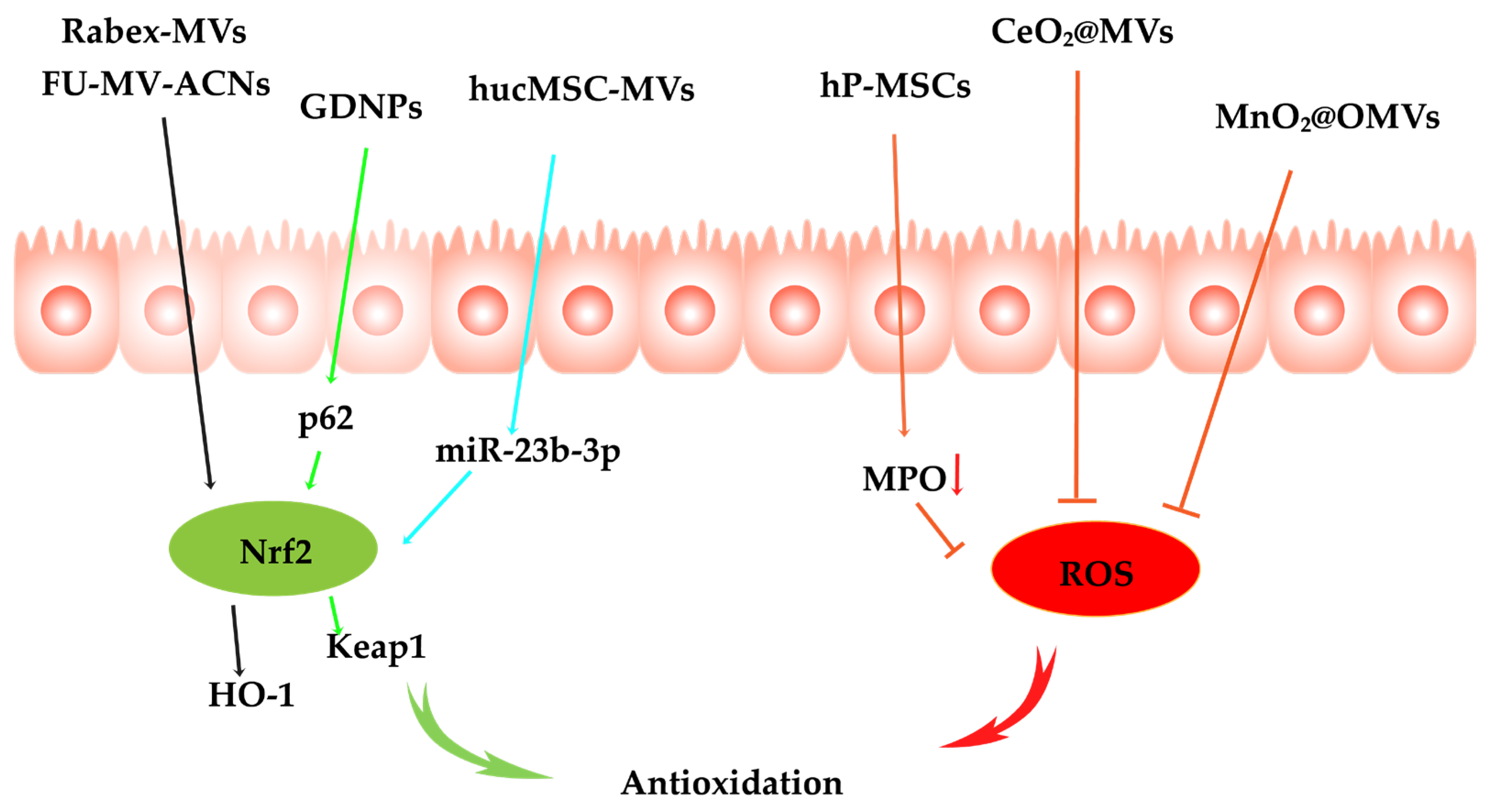
In the chronic progression of IBD, oxidative stress is widely recognized as a central mechanism driving disruption of the epithelial barrier, exacerbating mucosal inflammation, and impeding tissue repair processes. Excessive production of ROS not only directly induces apoptosis and DNA damage but also persistently activates inflammatory pathways such as NF-κB, creating a vicious cycle that underlies recurrent inflammation and tissue fibrosis. Consequently, targeted intervention in oxidative stress and effective scavenging of ROS are regarded as pivotal strategies to halt disease progression and restore intestinal homeostasis (Figure 4).

3.2.1. Oxidative Stress Signaling Pathway Inhibition

Persistent oxidative stress can markedly exacerbate intestinal immune responses, compromise the integrity of the mucosal barrier, and ultimately lead to structural and functional damage to the intestinal tissue. Nuclear factor erythroid 2-related factor 2 (Nrf2) serves as a master transcription factor in the regulation of oxidative stress responses and plays a pivotal role in maintaining redox homeostasis. As a key regulator of the antioxidant defense system, Nrf2 orchestrates the expression of downstream antioxidant enzymes—such as HO-1, NQO1, and SOD—which effectively mitigate oxidative damage. Consequently, targeting the Nrf2 signaling pathway holds considerable promise for alleviating the symptoms of IBD, attenuating inflammation, and restoring immune homeostasis.
A great deal of evidence suggests that MVs play a pivotal role in attenuating oxidative stress pathways in IBD. For instance, MVs isolated from Rabex exert antioxidant effects by activating the Nrf2/HO-1 signaling pathway, significantly alleviating oxidative stress in the intestinal environment [132]. Similarly, ginseng-derived nanoparticles (GDNPs) have been shown to enhance antioxidant defenses via the p62/Nrf2/Keap1 axis. Liu [101] et al. further demonstrated that hucMSC-MVs deliver miR-23b-3p into macrophages, thereby activating the Nrf2 antioxidant pathway. Collectively, these studies highlight the therapeutic potential of MV-based delivery systems in modulating Nrf2-mediated antioxidant responses through diverse molecular mechanisms in IBD treatment. To further enhance the efficacy of bioactive compounds in oxidative-related disorders, researchers have developed functionalized MV systems to improve their stability and antioxidative performance. In a recent study, anthocyanins (ACNs) were encapsulated into milk-derived MVs using ultrasound-assisted methods. The vesicle surface was subsequently modified with fucoidan (FU) to construct a novel FU-MV-ACNs nano-delivery platform. This system not only significantly improved the gastrointestinal stability and oral bioavailability of ACNs through membrane encapsulation, but also enhanced their targeted accumulation and cellular uptake in diseased tissues due to the mucoadhesive and sustained-release properties of FU. More importantly, FU-MV-ACNs exerted strong antioxidative effects by activating the Nrf2/HO-1 pathway, which attenuated oxidative stress and effectively protected tissue cells from ROS-induced damage [133].

3.2.2. ROS Scavenging

In the chronic progression of IBD, ROS are considered a driving factor of the inflammatory response. The excessive production of ROS not only directly induces cell death and DNA damage but also continuously activates inflammatory pathways, such as NF-κB, thereby creating a vicious cycle of recurrent inflammation and tissue fibrosis [134]. Therefore, effectively clearing ROS is considered a key strategy for halting disease progression and restoring intestinal homeostasis.
Recent evidence suggests that MV-DDSs play an important role in clearing ROS and have significant potential in treating IBD. Duan [60] et al. found that MVs derived from human placenta mesenchymal stem cells (hP-MSCs) effectively reduced ROS levels in mouse colon tissues by decreasing myeloperoxidase (MPO) activity. However, the ROS clearance ability of most membrane vesicles remains limited, failing to fully meet the therapeutic needs of IBD. Therefore, functionalized MV-DDSs based on nanoenzymes have gained increasing attention in recent years. Nanoenzymes, which exhibit peroxidase-like activity, can effectively clear ROS. Although turmeric-derived MVs naturally have mucosal enrichment and moderate anti-inflammatory effects, their efficacy in controlling oxidative stress has shown limitations. To overcome this, researchers have incorporated cerium oxide (CeO2) nanocrystals into MVs via in situ growth methods. CeO2 efficiently clears ROS by mimicking superoxide dismutase activity [135]. Similarly, Yue [136] et al. also incorporated CeO2 nanocrystals into Treg-derived MVs to further enhance their antioxidant effects. Furthermore, Li [137] et al. developed an MnO2@OMVs antioxidant drug delivery platform using EcN 1917 OMVs as a carrier. This system targets macrophages and intestinal epithelial cells through the PAMPs on OMVs, allowing the nanoenzyme to accumulate in the intestinal inflammation region. It effectively catalyzes the breakdown of excess H2O2 at the site of the lesion, significantly clearing ROS. In conclusion, through functionalized loading strategies, MV-DDSs can significantly enhance the ability to clear ROS and effectively modulate oxidative stress.

3.3. Barrier Protection

During the chronic progression of IBD, dysfunction of the intestinal barrier not only facilitates the translocation of luminal microbiota and toxins into the lamina propria—serving as a key driver of sustained mucosal immune activation—but also directly impedes epithelial regeneration and repair, thereby contributing to the relapse of patients. Therefore, promoting the expression of tight junction protein, enhancing the stability of the barrier structure, and promoting the regeneration and repair of epithelial cells are of great significance for restoring the integrity of the intestinal barrier and preventing the recurrence of diseases.

3.3.1. Tight Junction Protein Expression Enhancement

The role and importance of tight junction proteins in the treatment of IBD cannot be overstated. Tight junction proteins regulate the connections between intestinal epithelial cells, maintaining the integrity of the intestinal barrier. In IBD patients, the intestinal barrier function is often compromised, leading to increased intestinal permeability and persistent chronic inflammation. Therefore, regulating the expression of tight junction proteins is critical for restoring intestinal barrier function and alleviating IBD symptoms.
Studies have shown that BMVs have unique advantages in regulating the expression of tight junction proteins. For instance, MVs derived from EcN-1917 and ECOR63 strains can significantly upregulate the expression of ZO-1 and claudin-14 while downregulating claudin-2. This results in improved barrier function and reduced permeability. Although BMC-MVs also have a baseline ability to repair the barrier, their effect remains limited. To enhance their therapeutic potential, researchers have genetically modified MSCs via lentivirus transfection to overexpress EphB2, producing EphB2-enriched MSC-MVs. This loading strategy not only upregulated the expression of ZO-1 and occludin but also significantly reduced tight junction damage in a colitis model by inhibiting the RhoA/ROCK signaling pathway [138]. In parallel, another study demonstrated that MSC-MVs enriched with miR-181a further enhanced their ability to promote the expression of epithelial tight junction proteins. Compared to unloaded vesicles, these MVs significantly elevated the levels of ZO-1, claudin-1, and occludin, highlighting the crucial role of miRNA loading in maintaining tight junction integrity [37].

3.3.2. Epithelial Regeneration Promotion

Intestinal stem cells not only maintain the homeostasis of the intestinal epithelium but also accelerate the repair of intestinal damage by promoting epithelial cell regeneration when the intestinal barrier is compromised, thereby restoring intestinal function. During the chronic inflammation process of IBD, the normal proliferation and differentiation of intestinal stem cells are suppressed, which hinders the repair of the intestinal barrier. Therefore, promoting the proliferation and functional recovery of intestinal stem cells has become an important strategy for the treatment of IBD.
In this context, MV-DDSs have demonstrated significant effects. MVs derived from human amniotic fluid stem cells (hAFSCs) activate the Wnt/β-catenin signaling pathway and significantly upregulate the expression of the intestinal stem cell-specific marker Lgr5+ (compared to the NEC + PBS group, p < 0.0001, and the expression level was higher than the control group, p = 0.0003) [139]. Although MSC-MVs can promote the proliferation of Lgr5+ intestinal stem cells and epithelial cells, their efficacy in making repairs remains limited in highly inflammatory environments [48]. The cytokines and immune responses in the inflammatory environment can affect the function of HucMSC-MVs, restricting their repair effects in the local inflammatory areas. To overcome this limitation, researchers have designed a strategy in which MSC-MVs are functionalized with IL-33/IL1RL1 regulatory molecules. This effectively inhibits the activation of mast cells, reduces local inflammation, and creates a microenvironment conducive to epithelial repair [140]. This strategy enhances the application potential of MV-DDSs in intestinal repair.

3.4. Microbiota Modulation

During the persistent progression of IBD, intestinal dysbiosis is increasingly recognized not merely as a concomitant phenomenon but as a central driver of mucosal barrier disruption, immune dysregulation, and recurrent inflammation [141]. Historically, therapeutic strategies have primarily focused on suppressing inflammatory responses, while effective restoration of microbial homeostasis has often been overlooked—a gap that contributes to sustained expansion of pathogenic bacteria and depletion of beneficial species, ultimately compromising the durability of long-term treatment. Therefore, modulating the gut microbiota has become a key strategy in the treatment of IBD.
In recent years, MV-DDS has emerged as a promising tool for microbiota modulation due to its targeted delivery, mucosal accumulation, and the ability to encapsulate various bioactive cargos. Gu [37] et al. discovered that RNA-181a in MSC-MVs increased the abundance of lactobacilli while reducing the abundance of Bacteroides, thereby improving the gut microbiota’s composition and alleviating experimental colitis symptoms. Unlike MSC-MVs, ginger-derived MVs carry miR-167a. To enhance its effect, the researchers specifically extracted miR-167a and re-encapsulated it into ginger-derived MVs. After oral administration, these vesicles were efficiently internalized by lactobacilli and mediated the upregulation of SpaC mRNA expression, thus supporting stable and persistent colonization along the colonic mucosa. In the DSS-induced colitis model, this approach resulted in more than a twofold increase in lactobacilli abundance, a significant rise in IL-22 levels, and a nearly 40% reduction in the inflammation score, while scrambled RNA-loaded vesicles provided only limited protection under the same conditions [142]. Another study further emphasized the value of pretreatment methods. LPS-pretreated MSC-MVs selectively enhanced the proliferation of beneficial Clostridia_UCG 014 while reducing the colonization density of Streptococcus and Enterococcus [143]. This “preprogramming” strategy proved to be superior to unmodified vesicles in promoting microbiota homeostasis and alleviating colitis pathology. Notably, the different vesicle sources and cargo configurations appeared to play complementary roles: PDMVs primarily regulate bacterial adhesion genes and colonization, while MSC-MVs are involved in host immune signaling and reshape the nutritional microenvironment to achieve multidimensional microbiota modulation.
These studies demonstrate the potential effects of MV-DDS on regulating the intestinal microbiota. In the future, MV-DDS may enable precise regulation of the intestinal microbiota through functional design, and may also enhance the therapeutic effects of IBD by synergizing with other treatments.

3.5. Cell Death Regulation

Programmed cell death (PCD) pathways, including apoptosis, pyroptosis, necroptosis, PANoptosis, ferroptosis, and autophagy, play crucial roles in intracellular and extracellular signal transduction, immune response, repair mechanisms, and intestinal barrier function. In IBD, excessive or dysregulated PCD is one of the primary causes of intestinal damage, inflammation, and barrier dysfunction [144,145]. Therefore, modulating these PCD offers new strategies and directions for the treatment of IBD.
Apoptosis, as a classical form of PCD, is typically driven by the mitochondrial pathway and the activation of caspase family proteins, leading to orderly cell death. Excessive apoptosis of intestinal epithelial cells can result in the disruption of intestinal barrier function, thereby exacerbating the progression of IBD. Based on this, Li [55] et al. investigated natural MVs derived from MSCs and engineered them to enrich miR-378a-3p, constructing a delivery platform to intervene in the apoptosis of colonic epithelial cells in UC. MSC-MVs themselves exhibit certain biocompatibility and mucosal delivery capabilities, but their inhibitory effect on epithelial cell apoptosis is relatively limited. By transfecting MSCs with miR-378a-3p, they produced MVs enriched with this miRNA, significantly enhancing their ability to regulate mucosal homeostasis. Mechanistic studies revealed that miR-378a-3p-loaded MVs primarily exert their effects by downregulating the expression of GATA2, further inhibiting the upregulation of AQP4 and PPAR-α, thereby reducing intestinal epithelial cell apoptosis.
Pyroptosis is an inflammatory form of PCD activated by inflammasomes. The activation of the NLRP3 inflammasome promotes the activation of caspase-1, which subsequently cleaves Gasdermin D, leading to the rupturing of cell membrane and the release of inflammatory cytokines. miR-539-5p-enriched BMSCs-MVs block this process by targeting and inhibiting NLRP3-inflammasome. It significantly reduces IL-1β and IL-18 levels and effectively improves inflammatory lesions in DSS-induced IBD models [127]. Li [146] et al. further explored the potential of MVs derived from induced pluripotent stem cell-derived mesenchymal stem cells (iPSC-MSCs) in regulating epithelial cell pyroptosis. Although MVs were effectively internalized by intestinal epithelial cells and showed mild inhibition of pyroptosis-related proteins (such as GSDMD-N and caspase-1), their impact on pyroptosis inhibition remained limited. To enhance this effect, the researchers genetically engineered iPSC-MSCs to overexpress circ-CCND1, producing extracellular vesicles enriched with this circular RNA (MVs-oe-CCND1). Upon delivery, circ-CCND1 significantly downregulated KDM6B protein and ELF3 mRNA levels, increased H3K27me3 modification, de-repressed miR-342-3p, and established a negative feedback loop, further inhibiting KDM6B expression. Through this mechanism, MVs-oe-CCND1, compared to control vesicles, significantly reduced the levels of GSDMD-N and IL-1β, decreased pyroptosis by approximately 60%, and markedly improved the histological scoring of the intestinal mucosa in a DSS-induced colitis model.
As research advances, increasing evidence suggests that different forms of cell death are not isolated but instead interact significantly with each other. Based on these extensive signaling crosstalks, the concept of “PANoptosis” was introduced in 2019: a cell death mode driven by the PANoptosome multiprotein complex that integrates key signaling features of apoptosis, pyroptosis, and necroptosis within the same intestinal epithelial cell, providing an explanation for the comprehensive pathological features of UC mucosal damage [147,148]. As a result, PANoptosis has become a critical target in the treatment of IBD. Gong [149] et al. engineered Treg-derived MVs by loading them with selenium (Se) and modifying them with mitochondrial-targeting peptide SS-31. This system not only utilizes SS-31 for inflammation-specific release but also inhibits PANoptosis by reducing excessive ROS in macrophages, significantly alleviating inflammation and intestinal damage in a DSS-induced UC model.
Additionally, ferroptosis, an emerging form of PCD, has garnered increasing attention in recent years in the context of IBD research. Ferroptosis primarily occurs due to the excessive accumulation of intracellular iron, which catalyzes the production of ROS, leading to lipid peroxidation and subsequent cell damage. MVs derived from endometrial regenerative cells have been shown to downregulate ACSL4, a key regulator of ferroptosis, while upregulating GPX4 antioxidant activity. This effectively inhibits lipid peroxidation and ferroptosis in intestinal epithelial cells, thereby promoting mucosal repair and reducing inflammation [150].
Above the studies have revealed the complex role of PCD in IBD and its potential therapeutic value. MV-DDSs, derived from various sources, can effectively target and regulate PCD by carrying key molecules. In the future, by utilizing functionalized MV-DDSs to achieve more precise modulation of these PCDs and to enable the coordinated regulation of multiple PCDs, novel therapeutic strategies for the treatment of IBD may be developed.

4. Delivery Strategies of MV-DDSs in IBD

In IBD, MV-DDSs offer a diverse range of administration routes, each demonstrating distinct advantages and facing specific challenges depending on their characteristics and clinical application scenarios (Table 5). Common delivery strategies include intravenous injection, rectal administration, oral delivery, and intraperitoneal injection. Among these, oral delivery has been the most extensively investigated, particularly in the context of plant-derived vesicles and bacterial extracellular vesicles. Owing to its convenience and high patient compliance, oral administration is regarded as the preferred approach for long-term IBD management [95,151]. However, despite the ability of plant- and bacteria-derived vesicles to accumulate efficiently in the colon and suppress pro-inflammatory cytokine expression, they still face substantial degradation in the gastrointestinal environment due to gastric acid and digestive enzymes, ultimately compromising their therapeutic bioactivity [27]. To address these limitations, Deng [14] et al. developed an effective self-assembled layer-by-layer (LbL) MV delivery system for oral administration, enabling targeted release of MVs at inflamed colonic sites to improve UC therapy. In this strategy, the outer shell of MSC-MVs was engineered using biocompatible and biodegradable polymers—N-(2-hydroxy)propyl-3-trimethylammonium chitosan chloride (HTCC) and oxidized konjac glucomannan (OKGM). These materials were selected to facilitate colonic targeting while simultaneously protecting the MVs from enzymatic degradation in the upper gastrointestinal tract. Compared to intravenous delivery, the orally administered LbL-EVs achieved significantly greater therapeutic efficacy in alleviating UC symptoms, despite utilizing only half the dose of EVs.
Rectal administration delivers therapeutics directly to the colon and rectum, thereby bypassing gastrointestinal degradation and first-pass hepatic metabolism. This approach enhances local drug concentrations and improves therapeutic efficacy, making it particularly suitable for patients requiring localized treatment while minimizing systemic side effects. However, rectal delivery also presents certain limitations, primarily including the short retention time of the administered drug—due to potential expulsion by defecation or flatulence—and the incomplete coverage of the proximal colon. To address these challenges, Nie [152] et al. developed a bioadhesive microcarrier system. They encapsulated IL-27-loaded MSC-MVs within a dopamine methacrylamide (DMA)-modified hydrogel matrix, fabricated using microfluidic techniques. This hydrogel exhibited excellent mucosal adhesion properties. Following rectal administration, the catechol groups of DMA formed hydrogen bonds with amino groups on the colonic mucosal surface, ensuring firm anchoring of the formulation at the site of colonic inflammation and reducing the likelihood of drug loss through intestinal transit. Moreover, the crosslinked network structure of the hydrogel effectively protected the embedded MVs from enzymatic degradation, allowing for sustained and localized release of IL-27. This strategy significantly alleviated inflammatory responses and promoted epithelial barrier repair.
Intravenous injection, as a systemic administration route, offers the advantages of rapid onset and broad pharmacological action, making it particularly suitable for acute inflammatory episodes and systemic immunomodulation in IBD treatment. However, this route generally lacks tissue specificity, limiting drug accumulation in inflamed colonic regions and increasing the risk of off-target effects in non-diseased tissues. To overcome these limitations, Gong [149] et al. engineered regulatory Treg-MVs by decorating their surface with the mitochondria-targeting tetrapeptide SS-31. This peptide possesses a positive charge and an aromatic structure, enabling selective binding to cardiolipin located on the inner mitochondrial membrane. Through this design, the targeted delivery of MVs to mitochondria was significantly enhanced. Upon delivery, the SS-31-modified Treg-MVs effectively suppressed PANoptosis by attenuating excessive ROS.
Intraperitoneal (i.p.) injection is a commonly employed drug delivery route in experimental models of IBD. By allowing direct entry into the systemic circulation, i.p. administration avoids the destructive effects of gastric acid and digestive enzymes on vesicular structures, thereby preserving the biological activity of nucleic acid or protein cargos encapsulated within membrane vesicles. Nevertheless, as a non-targeted, systemic delivery strategy, intraperitoneal injection has inherent limitations in achieving a site-specific accumulation of drugs. To overcome this challenge and achieve precise delivery to inflamed tissues, Du [153] et al. innovatively employed M1-MVs as inflammation-homing carriers. These M1-MVs exhibit a high surface expression of the cc chemokine receptor 2 (CCR2), which specifically recognizes and binds to monocyte chemoattractant protein-1 (MCP-1), a chemokine markedly upregulated at inflammatory sites. Through the CCR2/MCP-1 axis, M1-MVs are actively recruited to regions of high MCP-1 expression, enabling inflammation-targeted accumulation. Following i.p. administration, the siIRF5 cargo delivered by M1-MVs successfully downregulated the expression of pro-inflammatory transcription factor IRF5 within macrophages, thereby inhibiting M1 polarization and promoting M2 phenotype transition. This phenotypic shift alleviates the inflammatory microenvironment and mitigates disease progression. Moreover, to further enhance tissue-specific delivery, membrane vesicles can be functionalized with targeting ligands such as hyaluronic acid or bioactive peptides, providing opportunities for synergistic targeting and expanding their therapeutic potential in complex inflammatory disorders [154,155].
Various delivery routes—including oral, rectal, intravenous, and i.p. administration—each offer distinct advantages and are suited to specific clinical scenarios, while also presenting unique technical challenges. Through the rational selection of membrane vesicle types and engineering optimization of delivery systems, these strategies are progressively overcoming the inherent limitations of conventional drug administration. Such advances have markedly enhanced the targeting specificity and therapeutic efficacy of MV-DDSs in the treatment of IBD, providing robust technological support for precise delivery and disease-specific intervention.
Table 5. Delivery strategies of MV-DDSs in IBD.
Delivery RouteVesicle TypesAdvantagesLimitationsRef.
OralLiposomes
  • Patient-friendly administration
  • Protect drugs from premature degradation
  • High biocompatibility and loading capacity
  • Effective delivery to intestinal tract
  • Susceptible to gastric acid and enzymes
  • Low stability without protective coating
  • Limited absorption of hydrophobic drugs
[28]
IntravenousLiposomes
  • Rapid systemic distribution
  • Active and passive targeting (e.g., EPR effect)
  • Ability to load hydrophilic and hydrophobic drugs
  • Rapid clearance by RES
  • Possible immunogenicity
  • High production cost
[29]
OralPolymersomes
  • pH-responsive release in intestine
  • Enhanced protection of drugs
  • High encapsulation efficiency
  • Controlled release at target site
  • Complex synthesis
  • Potential instability during GI transit
  • Limited clinical validation
[26]
IntravenousEBMVs
  • Selective targeting of inflamed intestine
  • Surface α4β7 integrin enables adhesion
  • Intrinsic anti-inflammatory activity
  • Reduction in inflammatory cytokine expression
  • Risk of immune reactions
[33]
OralPDMVs
  • Excellent food-derived origin
  • High biocompatibility
  • Stability in the gastrointestinal tract
  • Difficult to purify on a large scale
  • Partial degradation by digestive enzymes remains possible
  • Limited intestinal absorption efficiency
[32]
OralBMVs
  • Intrinsic mucosal adhesion
  • Modulation of gut microbiota
  • Oral convenience
  • Potential immunostimulation
  • Enzyme degradation risk
[156]

5. Conclusions and Outlook

As summarized in the preceding sections, MV-DDSs have demonstrated remarkable promise in the treatment of IBD, by enabling targeted delivery of anti-inflammatory agents, nucleic acids, antioxidants, and biologics, while also modulating immune responses, maintaining intestinal barrier integrity, and regulating programmed cell death pathways. However, despite their compelling preclinical efficacy, the translation of these potent vesicle-based systems into clinically meaningful therapies for IBD remains elusive, largely due to unresolved technical, safety, and regulatory barriers. Addressing these issues is imperative for realizing their therapeutic potential in human patients.
  • Standardization and scalability of production. The heterogeneity of vesicle sources—including plant-derived vesicles, bacterial outer membrane vesicles, milk-derived vesicles, and engineered biomimetic vesicles—poses difficulties in achieving reproducible large-scale production with consistent physicochemical properties and bioactivity. Future efforts should focus on establishing robust manufacturing processes and standardized quality control protocols.
  • Biosafety and immunogenicity. Despite their generally favorable biocompatibility, the residual immunogenic components in certain vesicles, such as endotoxins in bacterial vesicles or allergenic proteins in milk-derived vesicles, remain a safety concern. Rigorous purification, detoxification, and characterization strategies are needed to ensure low immunogenicity without compromising therapeutic efficacy.
  • Precise targeting and controlled release. Although vesicles exhibit inherent tropism to inflamed intestinal tissues, strategies to further improve site-specific delivery and stimuli-responsive release—such as engineering vesicles to respond to local oxidative stress or pH gradients—require continued optimization.
  • Cargo loading efficiency and stability. Efficient encapsulation and stable retention of diverse therapeutic cargos, especially sensitive biological molecules like siRNA, peptides, or probiotics, remain technical bottlenecks. The development of advanced loading techniques and stabilizing formulations will be pivotal to maximize their therapeutic potential.
  • In vivo tracking and pharmacokinetics. The complex intestinal microenvironment and dynamic mucus barrier present obstacles for vesicle trafficking and persistence in vivo. New imaging modalities and tracing methods are necessary to elucidate vesicles’ biodistribution, clearance, and mechanisms of action in real time.
  • Regulatory pathways and translational readiness. Given the novelty and complexity of MV-DDSs, regulatory frameworks governing their safety, efficacy, and manufacturing standards are still evolving. Close collaboration among academic researchers, industry partners, and regulatory agencies will be essential to accelerate their clinical development.
In light of these challenges, targeted measures such as establishing unified manufacturing standards, integrating advanced bioengineering for precision delivery, and implementing rigorous preclinical-to-clinical validation pipelines will be key to bridging the gap between laboratory success and patient benefit. Overall, MVs-DDSs represent a versatile and innovative therapeutic platform with the potential to reshape IBD management. Future interdisciplinary research—uniting material science, microbiology, immunology, and clinical medicine—is expected to overcome the current barriers, enabling the development of safe, precise, and effective next-generation MV-DDSs for inflammatory bowel disease.

Author Contributions

Writing—original draft preparation, Z.Z.; writing—review and editing, Z.Z., Z.L. and R.G.; supervision, Y.G. and R.G.; funding acquisition, R.G., Z.L. and Y.G. All authors have read and agreed to the published version of the manuscript.

Funding

This research was funded by 75th Batch of the China Postdoctoral Science Foundation (Project No.: 2024M754279), Jiangsu Natural Science Foundation (Project No.: BK20240738), Youth Talent Project of Jiangsu Provincial Science and Technology Development Program for Traditional Chinese Medicine (Project No.: QN202206), and General Program of Basic Science (Natural Science) Research in Institutions of Higher Education of Jiangsu Province (Project No.: 24KJB360004).

Acknowledgments

During the preparation of this manuscript/study, the author(s) used ChatGPT 4.0 (OpenAI) to help improve its grammatical accuracy and structural coherence. The authors reviewed and edited the results and take full responsibility for the content of this publication.

Conflicts of Interest

The authors declare no conflicts of interest. The funders had no role in the design of this study; in the collection, analyses, or interpretation of data; in the writing of the manuscript; or in the decision to publish the results.

Abbreviations

The following abbreviations are used in this manuscript:
IBDInflammatory bowel disease
CDCrohn’s disease
UCUlcerative colitis
MV-DDSsMembrane vesicle-based drug delivery systems
MDMVsMammalian-derived membrane vesicles
PDMVsPlant-derived membrane vesicles
BMVsBacterial membrane vesicles
EBMVsEngineered biomimetic membrane vesicles
DDSsDrug delivery systems
MVsMembrane vesicles
OMVsOuter membrane vesicles
ROSReactive oxygen species
5-ASA5-aminosalicylic acid
MSCsMesenchymal stem cells
EcNEscherichia coli Nissle
EPREnhanced permeability and retention
PEGPolyethylene glycol
PAMPsPathogen-associated molecular patterns
PDE4Phosphodiesterase 4
Nrf2Nuclear factor erythroid 2-related factor 2
ACNsAnthocyanins
FUFucoidan
MPOMyeloperoxidase
CeO2Cerium oxide
hAFSCshuman amniotic fluid stem cells
PCDProgrammed cell death
SeSelenium
LbLLayer-by-layer
HTCCN-(2-hydroxy)propyl-3-trimethylammonium chitosan chloride
OKGMOxidized konjac glucomannan
DMADopamine methacrylamide
i.p.Intraperitoneal
CCR2cc chemokine receptor 2
MCP-1Monocyte chemoattractant protein-1

References

  1. Graham, D.B.; Xavier, R.J. Pathway paradigms revealed from the genetics of inflammatory bowel disease. Nature 2020, 578, 527–539. [Google Scholar] [CrossRef] [PubMed]
  2. Mentella, M.C.; Scaldaferri, F.; Pizzoferrato, M.; Gasbarrini, A.; Miggiano, G.A.D. Nutrition, IBD and Gut Microbiota: A Review. Nutrients 2020, 12, 944. [Google Scholar] [CrossRef] [PubMed]
  3. Ananthakrishnan, A.N.; Bernstein, C.N.; Iliopoulos, D.; Macpherson, A.; Neurath, M.F.; Ali, R.A.R.; Vavricka, S.R.; Fiocchi, C. Environmental triggers in IBD: A review of progress and evidence. Nat. Rev. Gastroenterol. Hepatol. 2018, 15, 39–49. [Google Scholar] [CrossRef]
  4. Annese, V.; Annese, M. Precision Medicine in Inflammatory Bowel Disease. Diagnostics 2023, 13, 2797. [Google Scholar] [CrossRef] [PubMed]
  5. Ko, I.K.; Kim, B.-G.; Awadallah, A.; Mikulan, J.; Lin, P.; Letterio, J.J.; Dennis, J.E. Targeting Improves MSC Treatment of Inflammatory Bowel Disease. Mol. Ther. 2010, 18, 1365–1372. [Google Scholar] [CrossRef]
  6. Lin, S.; Han, S.; Wang, X.; Wang, X.; Shi, X.; He, Z.; Sun, M.; Sun, J. Oral Microto-Nano Genome-Editing System Enabling Targeted Delivery and Conditional Activation of CRISPR-Cas9 for Gene Therapy of Inflammatory Bowel Disease. ACS Nano 2024, 18, 25657–25670. [Google Scholar] [CrossRef]
  7. Wang, X.; Ni, J.; You, Y.; Feng, G.; Zhang, S.; Bao, W.; Hou, H.; Li, H.; Liu, L.; Zheng, M.; et al. SNX10-mediated LPS sensing causes intestinal barrier dysfunction via a caspase-5-dependent signaling cascade. EMBO J. 2021, 40, e108080. [Google Scholar] [CrossRef]
  8. Gecse, K.B.; Vermeire, S. Differential diagnosis of inflammatory bowel disease: Imitations and complications. Lancet Gastroenterol. Hepatol. 2018, 3, 644–653. [Google Scholar] [CrossRef]
  9. Saadah, O.I.; Alameel, T.; Al Sarkhy, A.; Hasosah, M.; Al-Hussaini, A.; Almadi, M.A.; Al-Bawardy, B.; Altuwaijri, T.A.; Aledreesi, M.; Bakkari, S.A.; et al. Saudi consensus guidance for the diagnosis and management of inflammatory bowel disease in children and adolescents. Saudi J. Gastroenterol. 2025, 31, 107–136. [Google Scholar] [CrossRef]
  10. Zu, M.; Xie, D.; Canup, B.S.B.; Chen, N.; Wang, Y.; Sun, R.; Zhang, Z.; Fu, Y.; Dai, F.; Xiao, B. ‘Green’ nanotherapeutics from tea leaves for orally targeted prevention and alleviation of colon diseases. Biomaterials 2021, 279, 121178. [Google Scholar] [CrossRef]
  11. Rogler, G.; Singh, A.; Kavanaugh, A.; Rubin, D.T. Extraintestinal Manifestations of Inflammatory Bowel Disease: Current Concepts, Treatment, and Implications for Disease Management. Gastroenterology 2021, 161, 1118–1132. [Google Scholar] [CrossRef] [PubMed]
  12. Du, L.; Ha, C. Epidemiology and Pathogenesis of Ulcerative Colitis. Gastroenterol. Clin. N. Am. 2020, 49, 643–654. [Google Scholar] [CrossRef] [PubMed]
  13. Zhang, Y.; Wang, T.; Sun, M.; Song, Y.; Huang, X.; Zhang, S.; Yang, J.; Zhang, J. Advanced Nanomedicine: Redefining Therapeutic Paradigms for Inflammatory Bowel Disease. Adv. Healthc. Mater. 2023, 12, e2300069. [Google Scholar] [CrossRef] [PubMed]
  14. Deng, C.; Hu, Y.; Conceicao, M.; Wood, M.J.A.; Zhong, H.; Wang, Y.; Shao, P.; Chen, J.; Qiu, L. Oral delivery of layer-by-layer coated exosomes for colitis therapy. J. Control. Release 2023, 354, 635–650. [Google Scholar] [CrossRef]
  15. Song, G.; Zeng, C.; Li, J.; Liu, J.; Zhao, J.; Liu, B.; Fan, J.; Xie, H. Exosome-based nanomedicines for digestive system tumors therapy. Nanomedicine 2025, 20, 1167–1180. [Google Scholar] [CrossRef]
  16. Zhang, Y.; Zhang, W.; Wu, Z.; Chen, Y. Diversity of extracellular vesicle sources in atherosclerosis: Role and therapeutic application. Angiogenesis 2025, 28, 34. [Google Scholar] [CrossRef]
  17. Yu, Y.; Lin, S.; Chen, Z.; Qin, B.; He, Z.; Cheng, M.; Sun, M.; Sun, J. Bacteria-driven bio-therapy: From fundamental studies to clinical trials. Nano Today 2023, 48, 101731. [Google Scholar] [CrossRef]
  18. Geng, T.; Ding, L.; Liu, M.; Zou, X.; Gu, Z.; Lin, H.; Sun, L. Preservation of extracellular vesicles for drug delivery: A comparative evaluation of storage buffers. J. Drug Deliv. Sci. Technol. 2025, 107, 106850. [Google Scholar] [CrossRef]
  19. Glassman, P.M.; Hood, E.D.; Ferguson, L.T.; Zhao, Z.; Siegel, D.L.; Mitragotri, S.; Brenner, J.S.; Muzykantov, V.R. Red blood cells: The metamorphosis of a neglected carrier into the natural mothership for artificial nanocarriers. Adv. Drug Deliv. Rev. 2021, 178, 113992. [Google Scholar] [CrossRef]
  20. Lei, P.; Yu, H.; Ma, J.; Du, J.; Fang, Y.; Yang, Q.; Zhang, K.; Luo, L.; Jin, L.; Wu, W.; et al. Cell membrane nanomaterials composed of phospholipids and glycoproteins for drug delivery in inflammatory bowel disease: A review. Int. J. Biol. Macromol. 2023, 249, 126000. [Google Scholar] [CrossRef]
  21. Liu, R. A promising area of research in medicine: Recent advances in properties and applications of Lactobacillus-derived exosomes. Front. Microbiol. 2024, 15, 1266510. [Google Scholar] [CrossRef] [PubMed]
  22. Barathan, M.; Ng, S.L.; Lokanathan, Y.; Ng, M.H.; Law, J.X. Milk-Derived Extracellular Vesicles: A Novel Perspective on Comparative Therapeutics and Targeted Nanocarrier Application. Vaccines 2024, 12, 1282. [Google Scholar] [CrossRef] [PubMed]
  23. Kameli, N.; Borman, R.; Lopez-lglesias, C.; Savelkoul, P.; Stassen, F.R.M. Characterization of Feces-Derived Bacterial Membrane Vesicles and the Impact of Their Origin on the Inflammatory Response. Front. Cell. Infect. Microbiol. 2021, 11, 1282. [Google Scholar] [CrossRef] [PubMed]
  24. Tan, X.; Gao, B.; Xu, Y.; Zhao, Q.; Jiang, J.; Sun, D.; Zhang, Y.; Zhou, S.; Fan, J.-B.; Zhang, M.; et al. Atractylodes macrocephala-derived extracellular vesicles-like particles enhance the recovery of ulcerative colitis by remodeling intestinal microecological balance. J. Nanobiotechnol. 2025, 23, 433. [Google Scholar] [CrossRef]
  25. Di Gioia, S.; Hossain, M.N.; Conese, M. Biological properties and therapeutic effects of plant-derived nanovesicles. Open Med. 2020, 15, 1096–1122. [Google Scholar] [CrossRef]
  26. Rahman, M.; Kumar, V.; Beg, S.; Sharma, G.; Katare, O.P.; Anwar, F. Emergence of liposome as targeted magic bullet for inflammatory disorders: Current state of the art. Artif. Cells Nanomed. Biotechnol. 2016, 44, 1597–1608. [Google Scholar] [CrossRef]
  27. Yang, D.; Zeng, H.; Zhuang, Y.; Jiang, S.; Pan, W. Nanodrug delivery materials for digestive system diseases. APL Mater. 2024, 12, 060603. [Google Scholar] [CrossRef]
  28. Xian, S.; Zhu, J.; Wang, Y.; Song, H.; Wang, H. Oral liposomal delivery of an activatable budesonide prodrug reduces colitis in experimental mice. Drug Deliv. 2023, 30, 2183821. [Google Scholar] [CrossRef]
  29. Tollemeto, M.; Ursulska, S.; Welzen, P.L.W.; Thamdrup, L.H.E.; Permlid, A.M.; Li, Y.; Soufi, G.; Padial, T.P.; Christensen, J.B.; Nielsen, L.H.; et al. Tailored Polymersomes for Enhanced Oral Drug Delivery: pH-Sensitive Systems for Intestinal Delivery of Immunosuppressants. Small 2024, 20, e2403640. [Google Scholar] [CrossRef]
  30. Zhong, H.; Luo, X.; Abdullah, X.; Liu, X.; Hussain, M.; Guan, R. Nano-targeted delivery system: A promising strategy of anthocyanin encapsulation for treating intestinal inflammation. Crit. Rev. Food Sci. Nutr. 2025, 65, 1–22. [Google Scholar] [CrossRef]
  31. Xu, X.; Yang, W.; Liang, Q.; Shi, Y.; Zhang, W.; Wang, X.; Meng, F.; Zhong, Z.; Yin, L. Efficient and targeted drug/siRNA co-delivery mediated by reversibly crosslinked polymersomes toward anti-inflammatory treatment of ulcerative colitis (UC). Nano Res. 2019, 12, 659–667. [Google Scholar] [CrossRef]
  32. Yang, W.; Lin, P.; Gao, R.; Fang, Z.; Wang, Z.; Ma, Z.; Shi, J.; Yu, W. Cell-derived biomimetic drug delivery system for inflammatory bowel disease therapy. Mater. Today Bio 2024, 29, 101332. [Google Scholar] [CrossRef]
  33. Corbo, C.; Cromer, W.E.; Molinaro, R.; Toledano Furman, N.E.; Hartman, K.A.; De Rosa, E.; Boada, C.; Wang, X.; Zawieja, D.C.; Agostini, M.; et al. Engineered biomimetic nanovesicles show intrinsic anti-inflammatory properties for the treatment of inflammatory bowel diseases. Nanoscale 2017, 9, 14581–14591. [Google Scholar] [CrossRef]
  34. Gao, F.; Chiu, S.M.; Motan, D.A.L.; Zhang, Z.; Chen, L.; Ji, H.L.; Tse, H.F.; Fu, Q.L.; Lian, Q. Mesenchymal stem cells and immunomodulation: Current status and future prospects. Cell Death Dis. 2016, 7, e2062. [Google Scholar] [CrossRef] [PubMed]
  35. Dabrowska, S.; Andrzejewska, A.; Janowski, M.; Lukomska, B. Immunomodulatory and Regenerative Effects of Mesenchymal Stem Cells and Extracellular Vesicles: Therapeutic Outlook for Inflammatory and Degenerative Diseases. Front. Immunol. 2021, 11, 591065. [Google Scholar] [CrossRef] [PubMed]
  36. Wu, H.; Fan, H.; Shou, Z.; Xu, M.; Chen, Q.; Ai, C.; Dong, Y.; Liu, Y.; Nan, Z.; Wang, Y.; et al. Extracellular vesicles containing miR-146a attenuate experimental colitis by targeting TRAF6 and IRAK1. Int. Immunopharmacol. 2019, 68, 204–212. [Google Scholar] [CrossRef] [PubMed]
  37. Gu, L.; Ren, F.; Fang, X.; Yuan, L.; Liu, G.; Wang, S. Exosomal MicroRNA-181a Derived From Mesenchymal Stem Cells Improves Gut Microbiota Composition, Barrier Function, and Inflammatory Status in an Experimental Colitis Model. Front. Med. 2021, 8, 660614. [Google Scholar] [CrossRef]
  38. Ihara, S.; Hirata, Y.; Koike, K. TGF-β in inflammatory bowel disease: A key regulator of immune cells, epithelium, and the intestinal microbiota. J. Gastroenterol. 2017, 52, 777–787. [Google Scholar] [CrossRef]
  39. Yang, R.; Huang, H.; Cui, S.; Zhou, Y.; Zhang, T.; Zhou, Y. IFN-γ promoted exosomes from mesenchymal stem cells to attenuate colitis via miR-125a and miR-125b. Cell Death Dis. 2020, 11, 603. [Google Scholar] [CrossRef]
  40. El-Badawy, A.; Amer, M.; Abdelbaset, R.; Sherif, S.N.; Abo-Elela, M.; Ghallab, Y.H.; Abdelhamid, H.; Ismail, Y.; El-Badri, N. Adipose Stem Cells Display Higher Regenerative Capacities and More Adaptable Electro-Kinetic Properties Compared to Bone Marrow-Derived Mesenchymal Stromal Cells. Sci. Rep. 2016, 6, 37801. [Google Scholar] [CrossRef]
  41. Guneta, V.; Tan, N.S.; Chan, S.K.J.; Tanavde, V.; Lim, T.C.; Wong, T.C.M.; Choong, C. Comparative study of adipose-derived stem cells and bone marrow-derived stem cells in similar microenvironmental conditions. Exp. Cell Res. 2016, 348, 155–164. [Google Scholar] [CrossRef] [PubMed]
  42. Hong, P.; Yang, H.; Wu, Y.; Li, K.; Tang, Z. The functions and clinical application potential of exosomes derived from adipose mesenchymal stem cells: A comprehensive review. Stem Cell Res. Ther. 2019, 10, 242. [Google Scholar] [CrossRef] [PubMed]
  43. Heidari, N.; Abbasi-Kenarsari, H.; Namaki, S.; Baghaei, K.; Zali, M.R.; Ghaffari Khaligh, S.; Hashemi, S.M. Adipose-derived mesenchymal stem cell-secreted exosome alleviates dextran sulfate sodium-induced acute colitis by Treg cell induction and inflammatory cytokine reduction. J. Cell. Physiol. 2021, 236, 5906–5920. [Google Scholar] [CrossRef] [PubMed]
  44. Lee, S.; Heo, J.; Ahn, E.-K.; Kim, J.H.; Kim, Y.-H.; Chang, H.-K.; Lee, S.-J.; Kim, J.; Park, S.-J. Conditioned secretome of adipose-derived stem cells improves dextran sulfate sodium-induced colitis in mice. World J. Gastroenterol. 2021, 27, 3342–3356. [Google Scholar] [CrossRef]
  45. Tian, J.; Zhu, Q.; Zhang, Y.; Bian, Q.; Hong, Y.; Shen, Z.; Xu, H.; Rui, K.; Yin, K.; Wang, S. Olfactory Ecto-Mesenchymal Stem Cell-Derived Exosomes Ameliorate Experimental Colitis via Modulating Th1/Th17 and Treg Cell Responses. Front. Immunol. 2020, 11, 598322. [Google Scholar] [CrossRef]
  46. Li, Y.; Altemus, J.; Lightner, A.L. Mesenchymal stem cells and acellular products attenuate murine induced colitis. Stem Cell Res. Ther. 2020, 11, 515. [Google Scholar] [CrossRef]
  47. Li, X.; Bai, J.; Ji, X.; Li, R.; Xuan, Y.; Wang, Y. Comprehensive characterization of four different populations of human mesenchymal stem cells as regards their immune properties, proliferation and differentiation. Int. J. Mol. Med. 2014, 34, 695–704. [Google Scholar] [CrossRef]
  48. Yu, H.; Yang, X.; Xiao, X.; Xu, M.; Yang, Y.; Xue, C.; Li, X.; Wang, S.; Zhao, R.C. Human Adipose Mesenchymal Stem Cell-derived Exosomes Protect Mice from DSS-Induced Inflammatory Bowel Disease by Promoting Intestinal-stem-cell and Epithelial Regeneration. Aging Dis. 2021, 12, 1423–1437. [Google Scholar] [CrossRef]
  49. Wang, S.; Song, Y.; Wang, Z.; Chang, X.; Wu, H.; Yan, Z.; Wu, J.; He, Z.; Kang, L.; Hu, W.; et al. Neutrophil-derived PAD4 induces citrullination of CKMT1 exacerbates mucosal inflammation in inflammatory bowel disease. Cell. Mol. Immunol. 2024, 21, 620–633. [Google Scholar] [CrossRef]
  50. Cheng, J.; Zhang, Y.; Ma, L.; Du, W.; Zhang, Q.; Gao, R.; Zhao, X.; Chen, Y.; Jiang, L.; Li, X.; et al. Macrophage-Derived Extracellular Vesicles-Coated Palladium Nanoformulations Modulate Inflammatory and Immune Homeostasis for Targeting Therapy of Ulcerative Colitis. Adv. Sci. 2023, 10, e2304002. [Google Scholar] [CrossRef]
  51. Han, G.; Kim, H.; Jang, H.; Kim, E.S.; Kim, S.H.; Yang, Y. Oral TNF-α siRNA delivery via milk-derived exosomes for effective treatment of inflammatory bowel disease. Bioact. Mater. 2024, 34, 138–149. [Google Scholar] [CrossRef] [PubMed]
  52. van Herwijnen, M.J.C.; Driedonks, T.A.P.; Snoek, B.L.; Kroon, A.M.T.; Kleinjan, M.; Jorritsma, R.; Pieterse, C.M.J.; Nolte-‘t Hoen, E.N.M.; Wauben, M.H.M. Abundantly Present miRNAs in Milk-Derived Extracellular Vesicles Are Conserved Between Mammals. Front. Nutr. 2018, 5, 81. [Google Scholar] [CrossRef] [PubMed]
  53. Cai, X.; Zhang, Z.-Y.; Yuan, J.-T.; Ocansey, D.K.W.; Tu, Q.; Zhang, X.; Qian, H.; Xu, W.-R.; Qiu, W.; Mao, F. hucMSC-derived exosomes attenuate colitis by regulating macrophage pyroptosis via the miR-378a-5p/NLRP3 axis. Stem Cell Res. Ther. 2021, 12, 416. [Google Scholar] [CrossRef]
  54. Mao, F.; Wu, Y.; Tang, X.; Kang, J.; Zhang, B.; Yan, Y.; Qian, H.; Zhang, X.; Xu, W. Exosomes Derived from Human Umbilical Cord Mesenchymal Stem Cells Relieve Inflammatory Bowel Disease in Mice. BioMed Res. Int. 2017, 2017, 5356760. [Google Scholar] [CrossRef]
  55. Li, P.; Zhang, H.-Y.; Gao, J.-Z.; Du, W.-Q.; Tang, D.; Wang, W.; Wang, L.-H. Mesenchymal stem cells-derived extracellular vesicles containing miR-378a-3p inhibit the occurrence of inflammatory bowel disease by targeting GATA2. J. Cell. Mol. Med. 2022, 26, 3133–3146. [Google Scholar] [CrossRef]
  56. Yang, S.; Liang, X.; Song, J.; Li, C.; Liu, A.; Luo, Y.; Ma, H.; Tan, Y.; Zhang, X. A novel therapeutic approach for inflammatory bowel disease by exosomes derived from human umbilical cord mesenchymal stem cells to repair intestinal barrier via TSG-6. Stem Cell Res. Ther. 2021, 12, 315. [Google Scholar] [CrossRef]
  57. Wang, G.; Yuan, J.; Cai, X.; Xu, Z.; Wang, J.; Ocansey, D.K.W.; Yan, Y.; Qian, H.; Zhang, X.; Xu, W.; et al. HucMSC-exosomes carrying miR-326 inhibit neddylation to relieve inflammatory bowel disease in mice. Clin. Transl. Med. 2020, 10, e113. [Google Scholar] [CrossRef]
  58. Cao, L.; Xu, H.; Wang, G.; Liu, M.; Tian, D.; Yuan, Z. Extracellular vesicles derived from bone marrow mesenchymal stem cells attenuate dextran sodium sulfate-induced ulcerative colitis by promoting M2 macrophage polarization. Int. Immunopharmacol. 2019, 72, 264–274. [Google Scholar] [CrossRef]
  59. Liu, L.; Ye, Y.; Feng, Z.; Cao, X.; Zhao, J. Human Umbilical Cord Mesenchymal Stem Cells Derived Exosomes Alleviate Ulcerative Colitis by Inhibiting Macrophage Ferroptosis Via miR-23b-3p/Nrf2 Pathway. Gastroenterology 2023, 164, S695–S696. [Google Scholar] [CrossRef]
  60. Duan, L.; Huang, H.; Zhao, X.; Zhou, M.; Chen, S.; Wang, C.; Han, Z.; Han, Z.-C.; Guo, Z.; Li, Z.; et al. Extracellular vesicles derived from human placental mesenchymal stem cells alleviate experimental colitis in mice by inhibiting inflammation and oxidative stress. Int. J. Mol. Med. 2020, 46, 1551–1561. [Google Scholar] [CrossRef]
  61. Xu, X.; Peng, J.; Wang, N.; Ocansey, D.K.W.; Zhang, X.; Mao, F. hucMSC-Ex alleviates inflammatory bowel disease in mice by enhancing M2-type macrophage polarization via the METTL3-Slc37a2-YTHDF1 axis. Cell Biol. Toxicol. 2024, 40, 74. [Google Scholar] [CrossRef] [PubMed]
  62. Li, T.; Chen, X.; Qi, Q.; Feng, X. Bovine Milk Derived Exosomes Affect Gut Microbiota of DSS-Induced Colitis Mice. Indian J. Microbiol. 2024, 64, 100–109. [Google Scholar] [CrossRef] [PubMed]
  63. Tong, L.; Hao, H.; Zhang, Z.; Lv, Y.; Liang, X.; Liu, Q.; Liu, T.; Gong, P.; Zhang, L.; Cao, F.; et al. Milk-derived extracellular vesicles alleviate ulcerative colitis by regulating the gut immunity and reshaping the microbiota. Theranostics 2021, 11, 8570–8586. [Google Scholar] [CrossRef]
  64. Benmoussa, A.; Diallo, I.; Salem, M.; Michel, S.; Gilbert, C.; Sevigny, J.; Provost, P. Concentrates of two subsets of extracellular vesicles from cow’s milk modulate symptoms and inflammation in experimental colitis. Sci. Rep. 2019, 9, 14661. [Google Scholar] [CrossRef]
  65. Reif, S.; Elbaum-Shiff, Y.; Koroukhov, N.; Shilo, I.; Musseri, M.; Golan-Gerstl, R. Cow and Human Milk-Derived Exosomes Ameliorate Colitis in DSS Murine Model. Nutrients 2020, 12, 2589. [Google Scholar] [CrossRef]
  66. Yang, R.; Liao, Y.; Wang, L.; He, P.; Hu, Y.; Yuan, D.; Wu, Z.; Sun, X. Exosomes Derived From M2b Macrophages Attenuate DSS-Induced Colitis. Front. Immunol. 2019, 10, 2346. [Google Scholar] [CrossRef] [PubMed]
  67. Gierynska, M.; Szulc-Dabrowska, L.; Struzik, J.; Mielcarska, M.B.; Gregorczyk-Zboroch, K.P. Integrity of the Intestinal Barrier: The Involvement of Epithelial Cells and Microbiota-A Mutual Relationship. Animals 2022, 12, 145. [Google Scholar] [CrossRef]
  68. Di Tommaso, N.; Gasbarrini, A.; Ponziani, F.R. Intestinal Barrier in Human Health and Disease. Int. J. Environ. Res. Public Health 2021, 18, 12836. [Google Scholar] [CrossRef]
  69. Kang, C.-S.; Ban, M.; Choi, E.-J.; Moon, H.-G.; Jeon, J.-S.; Kim, D.-K.; Park, S.-K.; Jeon, S.G.; Roh, T.-Y.; Myung, S.-J.; et al. Extracellular Vesicles Derived from Gut Microbiota, Especially Akkermansia muciniphila, Protect the Progression of Dextran Sulfate Sodium-Induced Colitis. PLoS ONE 2013, 8, e76520. [Google Scholar] [CrossRef]
  70. Qiao, K.; Chen, C.; Liu, H.; Qin, Y.; Liu, H. Pinin Induces Epithelial-to-Mesenchymal Transition in Hepatocellular Carcinoma by Regulating m6A Modification. J. Oncol. 2021, 2021, 7529164. [Google Scholar] [CrossRef]
  71. Li, Y.; Zhao, X.; Wang, D.; Wang, R.; Zhu, R.; You, X.; Liu, X.; Li, Y. Probiotic nanovesicles encapsulating baicalin: A strategy to overcome colorectal cancer. Lett. Appl. Microbiol. 2025, 78, ovae117. [Google Scholar] [CrossRef]
  72. Behrouzi, A.; Mazaheri, H.; Falsafi, S.; Tavassol, Z.H.; Moshiri, A.; Siadat, S.D. Intestinal effect of the probiotic Escherichia coli strain Nissle 1917 and its OMV. J. Diabetes Metab. Disord. 2020, 19, 597–604. [Google Scholar] [CrossRef]
  73. Chen, H.; Lei, P.; Ji, H.; Yang, Q.; Peng, B.; Ma, J.; Fang, Y.; Qu, L.; Li, H.; Wu, W.; et al. Advances in Escherichia coli Nissle 1917 as a customizable drug delivery system for disease treatment and diagnosis strategies. Mater. Today Bio 2023, 18, 100543. [Google Scholar] [CrossRef] [PubMed]
  74. Wehkamp, J.; Harder, J.; Wehkamp, K.; Wehkamp-von Meissner, B.; Schlee, M.; Enders, C.; Sonnenborn, U.; Nuding, S.; Bengmark, S.; Fellermann, K.; et al. NF-κB- and AP-1-mediated induction of human beta defensin-2 in intestinal epithelial cells by Escherichia coli Nissle 1917: A novel effect of a probiotic bacterium. Infect. Immun. 2004, 72, 5750–5758. [Google Scholar] [CrossRef] [PubMed]
  75. Choi, J.H.; Moon, C.M.; Shin, T.-S.; Kim, E.K.; McDowell, A.; Jo, M.-K.; Joo, Y.H.; Kim, S.-E.; Jung, H.-K.; Shim, K.-N.; et al. Lactobacillus paracasei-derived extracellular vesicles attenuate the intestinal inflammatory response by augmenting the endoplasmic reticulum stress pathway. Exp. Mol. Med. 2020, 52, 423–437. [Google Scholar] [CrossRef] [PubMed]
  76. Liang, L.; Yang, C.; Liu, L.; Mai, G.; Li, H.; Wu, L.; Jin, M.; Chen, Y. Commensal bacteria-derived extracellular vesicles suppress ulcerative colitis through regulating the macrophages polarization and remodeling the gut microbiota. Microb. Cell Factories 2022, 21, 88. [Google Scholar] [CrossRef]
  77. Shen, Y.; Torchia, M.L.G.; Lawson, G.W.; Karp, C.L.; Ashwell, J.D.; Mazmanian, S.K. Outer Membrane Vesicles of a Human Commensal Mediate Immune Regulation and Disease Protection. Cell Host Microb. 2012, 12, 509–520. [Google Scholar] [CrossRef]
  78. Chu, H.; Khosravi, A.; Kusumawardhani, I.P.; Kwon, A.H.K.; Vasconcelos, A.C.; Cunha, L.D.; Mayer, A.E.; Shen, Y.; Wu, W.-L.; Kambal, A.; et al. Gene-microbiota interactions contribute to the pathogenesis of inflammatory bowel disease. Science 2016, 352, 1116–1120. [Google Scholar] [CrossRef]
  79. Pitt, N.; Morrissette, M.; Gates, M.F.; Bargabos, R.; Krumpoch, M.; Hawkins, B.; Lewis, K. Bacterial membrane vesicles restore gut anaerobiosis. NPJ Biofilms Microbiomes 2025, 11, 48. [Google Scholar] [CrossRef]
  80. Han, H.S.; Hwang, S.; Choi, S.Y.; Hitayezu, E.; Humphrey, M.A.; Enkhbayar, A.; Song, D.-G.; Kim, M.; Park, J.-S.; Park, Y.-T.; et al. Roseburia intestinalis-derived extracellular vesicles ameliorate colitis by modulating intestinal barrier, microbiome, and inflammatory responses. J. Extracell. Vesicles 2024, 13, e12487. [Google Scholar] [CrossRef]
  81. Fabrega, M.-J.; Rodriguez-Nogales, A.; Garrido-Mesa, J.; Algieri, F.; Badia, J.; Gimenez, R.; Galvez, J.; Baldoma, L. Intestinal Anti-inflammatory Effects of Outer Membrane Vesicles from Escherichia coli Nissle 1917 in DSS-Experimental Colitis in Mice. Front. Microbiol. 2017, 8, 1274. [Google Scholar] [CrossRef]
  82. Ma, L.; Lyu, W.; Song, Y.; Chen, K.; Lv, L.; Yang, H.; Wang, W.; Xiao, Y. Anti-Inflammatory Effect of Clostridium butyricum-Derived Extracellular Vesicles in Ulcerative Colitis: Impact on Host microRNAs Expressions and Gut Microbiome Profiles. Mol. Nutr. Food Res. 2023, 67, e2200884. [Google Scholar] [CrossRef]
  83. Seo, M.K.; Park, E.J.; Ko, S.Y.; Choi, E.W.; Kim, S. Therapeutic effects of kefir grain Lactobacillus-derived extracellular vesicles in mice with 2,4,6-trinitrobenzene sulfonic acid-induced inflammatory bowel disease. J. Dairy Sci. 2018, 101, 8662–8671. [Google Scholar] [CrossRef]
  84. Zhuang, J.; Zhuang, Z.; Chen, B.; Yang, Y.; Chen, H.; Guan, G. Odoribacter splanchnicus-derived extracellular vesicles alleviate inflammatory bowel disease by modulating gastrointestinal inflammation and intestinal barrier function via the NLRP3 inflammasome suppression. Mol. Med. 2025, 31, 56. [Google Scholar] [CrossRef] [PubMed]
  85. Hao, H.; Zhang, X.; Tong, L.; Liu, Q.; Liang, X.; Bu, Y.; Gong, P.; Liu, T.; Zhang, L.; Xia, Y.; et al. Effect of Extracellular Vesicles Derived from Lactobacillus plantarum Q7 on Gut Microbiota and Ulcerative Colitis in Mice. Front. Immunol. 2021, 12, 777147. [Google Scholar] [CrossRef] [PubMed]
  86. Ye, L.; Wang, Y.; Xiao, F.; Wang, X.; Li, X.; Cao, R.; Zhang, J.; Zhang, T.F. Prausnitzii-derived extracellular vesicles attenuate experimental colitis by regulating intestinal homeostasis in mice. Microb. Cell Factories 2023, 22, 235. [Google Scholar] [CrossRef] [PubMed]
  87. Zheng, T.; Hao, H.; Liu, Q.; Li, J.; Yao, Y.; Liu, Y.; Zhang, T.; Zhang, Z.; Yi, H. Effect of Extracelluar Vesicles Derived from Akkermansia muciniphila on Intestinal Barrier in Colitis Mice. Nutrients 2023, 15, 4722. [Google Scholar] [CrossRef]
  88. Yang, Y.; Yang, L.; Yang, Y.; Deng, H.; Su, S.; Xia, Y.; Su, J.; Liu, Y.; Wu, J.; Zhang, J.; et al. Bacteroides fragilis-Derived Outer Membrane Vesicles Deliver MiR-5119 and Alleviate Colitis by Targeting PD-L1 to Inhibit GSDMD-Mediated Neutrophil Extracellular Trap Formation. Adv. Sci. 2025, 12, e00781. [Google Scholar] [CrossRef]
  89. Yamasaki-Yashiki, S.; Kawashima, F.; Saika, A.; Hosomi, R.; Kunisawa, J.; Katakura, Y. RNA-Based Anti-Inflammatory Effects of Membrane Vesicles Derived from Lactiplantibacillus plantarum. Foods 2024, 13, 967. [Google Scholar] [CrossRef]
  90. Tong, L.; Zhang, X.; Hao, H.; Liu, Q.; Zhou, Z.; Liang, X.; Liu, T.; Gong, P.; Zhang, L.; Zhai, Z.; et al. Lactobacillus rhamnosus GG Derived Extracellular Vesicles Modulate Gut Microbiota and Attenuate Inflammatory in DSS-Induced Colitis Mice. Nutrients 2021, 13, 3319. [Google Scholar] [CrossRef]
  91. Kang, E.A.; Choi, H.-I.; Hong, S.W.; Kang, S.; Jegal, H.-Y.; Choi, E.W.; Park, B.-S.; Kim, J.S. Extracellular Vesicles Derived from Kefir Grain Lactobacillus Ameliorate Intestinal Inflammation via Regulation of Proinflammatory Pathway and Tight Junction Integrity. Biomedicines 2020, 8, 522. [Google Scholar] [CrossRef]
  92. Gupta, M.; Mishra, V.; Gulati, M.; Kapoor, B.; Kaur, A.; Gupta, R.; Tambuwala, M.M. Natural compounds as safe therapeutic options for ulcerative colitis. Inflammopharmacology 2022, 30, 397–434. [Google Scholar] [CrossRef]
  93. Liu, T.; Ning, Z.; Liu, P.; Gao, H. Cassane diterpenoid ameliorates dextran sulfate sodium-induced experimental colitis by regulating gut microbiota and suppressing tryptophan metabolism. Front. Immunol. 2023, 13, 1045901. [Google Scholar] [CrossRef]
  94. Yuan, S.; Li, Y.; Li, J.; Xue, J.-C.; Wang, Q.; Hou, X.-T.; Meng, H.; Nan, J.-X.; Zhang, Q.-G. Traditional Chinese Medicine and Natural Products: Potential Approaches for Inflammatory Bowel Disease. Front. Pharmacol. 2022, 13, 892790. [Google Scholar] [CrossRef] [PubMed]
  95. Dad, H.A.; Gu, T.-W.; Zhu, A.-Q.; Huang, L.-Q.; Peng, L.-H. Plant Exosome-like Nanovesicles: Emerging Therapeutics and Drug Delivery Nanoplatforms. Mol. Ther. 2021, 29, 13–31. [Google Scholar] [CrossRef] [PubMed]
  96. De Jong, W.H.; Borm, P.J.A. Drug delivery and nanoparticles: Applications and hazards. Int. J. Nanomed. 2008, 3, 133–149. [Google Scholar] [CrossRef] [PubMed]
  97. Kim, K.; Park, J.; Sohn, Y.; Oh, C.-E.; Park, J.-H.; Yuk, J.-M.; Yeon, J.-H. Stability of Plant Leaf-Derived Extracellular Vesicles According to Preservative and Storage Temperature. Pharmaceutics 2022, 14, 457. [Google Scholar] [CrossRef]
  98. Zhuang, X.; Deng, Z.-B.; Mu, J.; Zhang, L.; Yan, J.; Miller, D.; Feng, W.; McClain, C.J.; Zhang, H.-G. Ginger-derived nanoparticles protect against alcohol-induced liver damage. J. Extracell. Vesicles 2015, 4, 28713. [Google Scholar] [CrossRef]
  99. Gao, C.; Zhou, Y.; Chen, Z.; Li, H.; Xiao, Y.; Hao, W.; Zhu, Y.; Vong, C.T.; Farag, M.A.; Wang, Y.; et al. Turmeric-derived nanovesicles as novel nanobiologics for targeted therapy of ulcerative colitis. Theranostics 2022, 12, 5596–5614. [Google Scholar] [CrossRef]
  100. Liu, C.; Yan, X.; Zhang, Y.; Yang, M.; Ma, Y.; Zhang, Y.; Xu, Q.; Tu, K.; Zhang, M. Oral administration of turmeric-derived exosome-like nanovesicles with anti-inflammatory and pro-resolving bioactions for murine colitis therapy. J. Nanobiotechnol. 2022, 20, 206. [Google Scholar] [CrossRef]
  101. Yang, S.; Li, W.; Bai, X.; Di Nunzio, G.; Fan, L.; Zhao, Y.; Ren, L.; Zhao, R.; Bian, S.; Liu, M.; et al. Ginseng-derived nanoparticles alleviate inflammatory bowel disease via the TLR4/MAPK and p62/Nrf2/Keap1 pathways. J. Nanobiotechnol. 2024, 22, 48. [Google Scholar] [CrossRef]
  102. Zhu, Z.; Liao, L.; Gao, M.; Liu, Q. Garlic-derived exosome-like nanovesicles alleviate dextran sulphate sodium-induced mouse colitis via the TLR4/MyD88/NF-κB pathway and gut microbiota modulation. Food Funct. 2023, 14, 7520–7534. [Google Scholar] [CrossRef] [PubMed]
  103. Tinnirello, V.; Zizzo, M.G.; Conigliaro, A.; Tabone, M.; Ganji, N.R.; Cicio, A.; Bressa, C.; Larrosa, M.; Rappa, F.; Vergilio, G.; et al. Industrial-produced lemon nanovesicles ameliorate experimental colitis-associated damages in rats via the activation of anti-inflammatory and antioxidant responses and microbiota modification. Biomed. Pharmacother. 2024, 174, 116514. [Google Scholar] [CrossRef] [PubMed]
  104. Zu, M.; Song, H.; Zhang, J.; Chen, Q.; Deng, S.; Canup, B.S.B.; Yuan, Y.; Xiao, B. Lycium barbarum lipid-based edible nanoparticles protect against experimental colitis. Colloids Surf. B Biointerfaces 2020, 187, 110747. [Google Scholar] [CrossRef]
  105. Sriwastva, M.K.; Deng, Z.-B.; Wang, B.; Teng, Y.; Kumar, A.; Sundaram, K.; Mu, J.; Lei, C.; Dryden, G.W.; Xu, F.; et al. Exosome-like nanoparticles from Mulberry bark prevent DSS-induced colitis via the AhR/COPS8 pathway. Embo Rep. 2022, 23, e53365. [Google Scholar] [CrossRef] [PubMed]
  106. Choi, S.-H.; Eom, J.-Y.; Kim, H.-J.; Seo, W.; Kwun, H.-J.; Kim, D.-K.; Kim, J.; Cho, Y.-E. Aloe-derived nanovesicles attenuate inflammation and enhance tight junction proteins for acute colitis treatment. Biomater. Sci. 2023, 11, 5490–5501. [Google Scholar] [CrossRef]
  107. Ju, S.; Mu, J.; Dokland, T.; Zhuang, X.; Wang, Q.; Jiang, H.; Xiang, X.; Deng, Z.-B.; Wang, B.; Zhang, L.; et al. Grape Exosome-like Nanoparticles Induce Intestinal Stem Cells and Protect Mice from DSS-Induced Colitis. Mol. Ther. 2013, 21, 1345–1357. [Google Scholar] [CrossRef]
  108. Kim, J.; Zhang, S.; Zhu, Y.; Wang, R.; Wang, J. Amelioration of colitis progression by ginseng-derived exosome-like nanoparticles through suppression of inflammatory cytokines. J. Ginseng Res. 2023, 47, 627–637. [Google Scholar] [CrossRef]
  109. Deng, Z.; Rong, Y.; Teng, Y.; Mu, J.; Zhuang, X.; Sannykutty, A.; Zhang, L.; Yan, J.; Miller, D.; Suttles, J.; et al. Broccoli-derived nanoparticle inhibits mouse colitis by activating dendritic cell AMP-activated protein kinase. J. Immunol. 2017, 25, 1641–1654. [Google Scholar]
  110. Yang, S.; Fan, L.; Yin, L.; Zhao, Y.; Li, W.; Zhao, R.; Jia, X.; Dong, F.; Zheng, Z.; Zhao, D.; et al. Ginseng exosomes modulate M1/M2 polarisation by activating autophagy and target IKK/IкB/NF-кB to alleviate inflammatory bowel disease. J. Nanobiotechnol. 2025, 23, 198. [Google Scholar] [CrossRef]
  111. Wong, C.K.; Laos, A.J.; Soeriyadi, A.H.; Wiedenmann, J.; Curmi, P.M.G.; Gooding, J.J.; Marquis, C.P.; Stenzel, M.H.; Thordarson, P. Polymersomes Prepared from Thermoresponsive Fluorescent Protein-Polymer Bioconjugates: Capture of and Report on Drug and Protein Payloads. Angew. Chem.-Int. Ed. 2015, 54, 5317–5322. [Google Scholar] [CrossRef] [PubMed]
  112. Zou, Y.; Zheng, M.; Yang, W.; Meng, F.; Miyata, K.; Kim, H.J.; Kataoka, K.; Zhong, Z. Virus-Mimicking Chimaeric Polymersomes Boost Targeted Cancer siRNA Therapy In Vivo. Adv. Mater. 2017, 29, 1703285. [Google Scholar] [CrossRef] [PubMed]
  113. Negut, I.; Bita, B. Polymersomes as Innovative, Stimuli-Responsive Platforms for Cancer Therapy. Pharmaceutics 2024, 16, 463. [Google Scholar] [CrossRef]
  114. Deng, C.; Jiang, Y.; Cheng, R.; Meng, F.; Zhong, Z. Biodegradable polymeric micelles for targeted and controlled anticancer drug delivery: Promises, progress and prospects. Nano Today 2012, 7, 467–480. [Google Scholar] [CrossRef]
  115. Molinaro, R.; Corbo, C.; Martinez, J.O.; Taraballi, F.; Evangelopoulos, M.; Minardi, S.; Yazdi, I.K.; Zhao, P.; De Rosa, E.; Sherman, M.B.; et al. Biomimetic proteolipid vesicles for targeting inflamed tissues. Nat. Mater. 2016, 15, 1037–1046. [Google Scholar] [CrossRef]
  116. Steidler, L.; Neirynck, S.; Huyghebaert, N.; Snoeck, V.; Vermeire, A.; Goddeeris, B.; Cox, E.; Remon, J.P.; Remaut, E. Biological containment of genetically modified Lactococcus lactis for intestinal delivery of human interleukin 10. Nat. Biotechnol. 2003, 21, 785–789. [Google Scholar] [CrossRef]
  117. Braat, H.; Rottiers, P.; Hommes, D.W.; Huyghebaert, N.; Remaut, E.; Remon, J.-P.; Van Deventer, S.J.H.; Neirynck, S.; Peppelenbosch, M.P.; Steidler, L. A phase I trial with Transgenic bacteria expressing interleukin-10 in Crohn’s disease. Clin. Gastroenterol. Hepatol. 2006, 4, 754–759. [Google Scholar] [CrossRef]
  118. Hanson, M.L.; Hixon, J.A.; Li, W.; Felber, B.K.; Anver, M.R.; Stewart, C.A.; Janelsins, B.M.; Datta, S.K.; Shen, W.; McLean, M.H.; et al. Oral Delivery of IL-27 Recombinant Bacteria Attenuates Immune Colitis in Mice. Gastroenterology 2014, 146, 210–221.e13. [Google Scholar] [CrossRef]
  119. Wang, D.; Jiang, Q.; Shen, R.; Peng, L.; Zhou, W.; Meng, T.; Hu, F.; Wang, J.; Yuan, H. ROS-responsive nanoparticles targeting inflamed colon for synergistic therapy of inflammatory bowel disease via barrier repair and anti-inflammation. Nano Res. 2024, 17, 5409–5423. [Google Scholar] [CrossRef]
  120. Hummel, T.; Tak, E.; Maurice-Stam, H.; Benninga, M.A.; Kindermann, A.; Grootenhuis, M.A. The Course of Life and Health-Related Quality of Life of Adolescents with Inflammatory Bowel Disease. Gastroenterology 2011, 140, S511. [Google Scholar] [CrossRef]
  121. Han, L.; Liu, X.-W.; Li, W.-X.; Zang, T.; Li, Z.-P.; Deng, Y.; Tan, L.-L.; Liang, D.-S.; Zhang, X.-J.; Shen, X.-F.; et al. Innovative probiotic Escherichia coli nissle 1917-derived outer membrane vesicles coupled microspheres demonstrate anti-inflammatory and barrier-enhancing properties in colitis. Chem. Eng. J. 2024, 487, 150711. [Google Scholar] [CrossRef]
  122. Song, C.; Wu, J.; Wu, J.; Wang, F. MnO2 and roflumilast-loaded probiotic membrane vesicles mitigate experimental colitis by synergistically augmenting cAMP in macrophage. J. Nanobiotechnol. 2024, 22, 294. [Google Scholar] [CrossRef] [PubMed]
  123. Kumar, A.; Chatterjee, I.; Gujral, T.; Alakkam, A.; Coffing, H.; Anbazhagan, A.N.; Borthakur, A.; Saksena, S.; Gill, R.K.; Alrefai, W.A.; et al. Activation of Nuclear Factor—κB by Tumor Necrosis Factor in Intestinal Epithelial Cells and Mouse Intestinal Epithelia Reduces Expression of the Chloride Transporter SLC26A3. Gastroenterology 2017, 153, 1338–1350.e3. [Google Scholar] [CrossRef]
  124. Song, Y.; Zhao, Y.; Ma, Y.; Wang, Z.; Rong, L.; Wang, B.; Zhang, N. Biological functions of NLRP3 inflammasome: A therapeutic target in inflammatory bowel disease. Cytokine Growth Factor Rev. 2021, 60, 61–75. [Google Scholar] [CrossRef]
  125. Stylianou, P.; Rushwan, S.; Wang, W.; Miah, M.A.; Darweesh, O.; Mackinnon, A.; Roach, K.M.; Hitchman, C.J.; Gonchar, O.; Thorpe, S.; et al. CD98 is critical for a conserved inflammatory response to diverse injury stimuli relevant to IPF exacerbations and COVID pneumonitis. bioRxiv 2022. [Google Scholar] [CrossRef]
  126. Zhang, M.; Wang, X.; Han, M.K.; Collins, J.F.; Merlin, D. Oral administration of ginger-derived nanolipids loaded with siRNA as a novel approach for efficient siRNA drug delivery to treat ulcerative colitis. Nanomedicine 2017, 12, 1927–1943. [Google Scholar] [CrossRef]
  127. Wang, D.; Xue, H.; Tan, J.; Liu, P.; Qiao, C.; Pang, C.; Zhang, L. Bone marrow mesenchymal stem cells-derived exosomes containing miR-539-5p inhibit pyroptosis through NLRP3/caspase-1 signalling to alleviate inflammatory bowel disease. Inflamm. Res. 2022, 71, 833–846. [Google Scholar] [CrossRef]
  128. Arabpour, M.; Saghazadeh, A.; Rezaei, N. Anti-inflammatory and M2 macrophage polarization-promoting effect of mesenchymal stem cell-derived exosomes. Int. Immunopharmacol. 2021, 97, 107823. [Google Scholar] [CrossRef]
  129. Zhang, M.; Xu, X.; Su, L.; Zeng, Y.; Lin, J.; Li, W.; Zou, Y.; Li, S.; Lin, B.; Li, Z.; et al. Oral administration of Sophora Flavescens-derived exosomes-like nanovesicles carrying CX5461 ameliorates DSS-induced colitis in mice. J Nanobiotechnol. 2024, 22, 607. [Google Scholar] [CrossRef]
  130. Xu, H.; Fu, J.; Chen, L.; Zhou, S.; Fang, Y.; Zhang, Q.; Chen, X.; Yuan, L.; Li, Y.; Xu, Z.; et al. TNF-α Enhances the Therapeutic Effects of MenSC-Derived Small Extracellular Vesicles on Inflammatory Bowel Disease through Macrophage Polarization by miR-24-3p. Stem Cells Int. 2023, 2023, 2988907. [Google Scholar] [CrossRef]
  131. Zhao, Y.; Yin, W.; Yang, Z.; Sun, J.; Chang, J.; Huang, L.; Xue, L.; Zhang, X.; Zhi, H.; Chen, S.; et al. Nanotechnology-enabled M2 macrophage polarization and ferroptosis inhibition for targeted inflammatory bowel disease treatment. J. Control. Release 2024, 367, 339–353. [Google Scholar] [CrossRef]
  132. Kang, S.J.; Lee, J.H.; Rhee, W.J. Engineered plant-derived extracellular vesicles for targeted regulation and treatment of colitis-associated inflammation. Theranostics 2024, 14, 5643–5661. [Google Scholar] [CrossRef]
  133. Jiang, Q.; Wang, L.; Si, X.; Cui, H.; Li, J.; Tian, J.; Bao, Y.; Xie, X.; Xu, A.; Sun, Y.; et al. An anthocyanins-loaded extracellular vesicles coated with fucoidan nano-delivery system for enhancing dietary defense against inflammation-related colon injury. Int. J. Biol. Macromol. 2025, 318, 144948. [Google Scholar] [CrossRef]
  134. Ho, G.-T.; Theiss, A.L. Mitochondria and Inflammatory Bowel Diseases: Toward a Stratified Therapeutic Intervention. Annu. Rev. Physiol. 2022, 84, 435–459. [Google Scholar] [CrossRef]
  135. Lu, G.; Lu, S.; Dai, H.; Zhang, F.; Wang, X.; Li, W.; Mei, L.; Tan, H. Engineered Turmeric-Derived Nanovesicles for Ulcerative Colitis Therapy by Attenuating Oxidative Stress and Alleviating Inflammation. Mol. Pharm. 2025, 22, 2159–2167. [Google Scholar] [CrossRef]
  136. Yue, S.; Gong, L.; Tan, Y.; Zhang, X.; Liao, F. Cerium Oxide-Loaded Exosomes Derived from Regulatory T Cells Ameliorate Inflammatory Bowel Disease by Scavenging Reactive Oxygen Species and Modulating the Inflammatory Response. J. Inflamm. Res. 2025, 18, 4395–4408. [Google Scholar] [CrossRef]
  137. Li, J.; Sun, M.; Liu, L.; Yang, W.; Sun, A.; Yu, J.; Liu, D.; Zhao, W.; Cheng, M.; He, Z.; et al. Nanoprobiotics for Remolding the Pro-inflammatory Microenvironment and Microbiome in the Treatment of Colitis. Nano Lett. 2023, 23, 8593–8601. [Google Scholar] [CrossRef] [PubMed]
  138. Chu, S.; Yu, T.; Wang, W.; Wu, H.; Zhu, F.; Wei, C.; Gao, F.; Liu, C.; Fan, H. Exosomes derived from EphB2-overexpressing bone marrow mesenchymal stem cells regulate immune balance and repair barrier function. Biotechnol. Lett. 2023, 45, 601–617. [Google Scholar] [CrossRef] [PubMed]
  139. O’Connell, J.S.; Lee, C.; Farhat, N.; Antounians, L.; Zani, A.; Li, B.; Pierro, A. Administration of extracellular vesicles derived from human amniotic fluid stem cells: A new treatment for necrotizing enterocolitis. Pediatr. Surg. Int. 2021, 37, 301–309. [Google Scholar] [CrossRef]
  140. Wei, Z.; Tang, X.; Yi, C.; Ocansey, D.K.W.; Mao, F.; Mao, Z. HucMSC-Ex alleviates DSS-induced colitis in mice by decreasing mast cell activation via the IL-33/ST2 axis. Am. J. Transl. Res. 2024, 16, 2727–2744. [Google Scholar] [CrossRef] [PubMed]
  141. Weiss, G.A.; Hennet, T. Mechanisms and consequences of intestinal dysbiosis. Cell. Mol. Life Sci. 2017, 74, 2959–2977. [Google Scholar] [CrossRef]
  142. Teng, Y.; Ren, Y.; Sayed, M.; Hu, X.; Lei, C.; Kumar, A.; Hutchins, E.; Mu, J.; Deng, Z.; Luo, C.; et al. Plant-Derived Exosomal MicroRNAs Shape the Gut Microbiota. Cell Host Microbe 2018, 24, 637–652.e8. [Google Scholar] [CrossRef] [PubMed]
  143. Li, Z.; Liu, L.; Sun, Y.; Liu, X.; Zhang, P.; Wang, Y.; Ding, G. Mesenchymal stem/stromal cells-derived exosomes: Possible therapeutic mechanism in inflammatory bowel disease. Hum. Cell 2025, 38, 111. [Google Scholar] [CrossRef] [PubMed]
  144. Levine, A.D. Apoptosis: Implications for inflammatory bowel disease. Inflamm. Bowel Dis. 2000, 6, 191–205. [Google Scholar] [CrossRef]
  145. Xiao, Z.; Xie, J.; Zhao, X.; Chen, X.; Lu, Y.; Xu, Y.; Wu, M.; An, L.; Li, Q. Role of Pyroptosis in inflammatory bowel disease. Int. Immunopharmacol. 2025, 155, 114619. [Google Scholar] [CrossRef]
  146. Li, L.; Lam, I.; Wang, J.; Yu, H.; Chan, C.; Cai, S. Epigenetic mechanism of iPSC-MSC-EVs in colonic epithelial cell pyroptosis in ulcerative colitis cell models via modulation of ELF3/miR-342-3p/ KDM6B axis and histone methylation. Int. Immunopharmacol. 2025, 157, 114704. [Google Scholar] [CrossRef]
  147. Zhao, C.; Lin, S. PANoptosis in intestinal epithelium: Its significance in inflammatory bowel disease and a potential novel therapeutic target for natural products. Front. Immunol. 2025, 15, 1507065. [Google Scholar] [CrossRef]
  148. Tan, C.; Xiang, Z.; Wang, S.; He, H.; Li, X.; Xu, M.; Guo, X.; Pu, Y.; Zhen, J.; Dong, W. Diosmin alleviates colitis by inhibiting PANoptosis of intestinal epithelial cells and regulating gut microbiota and metabolites. Phytomedicine 2025, 141, 156671. [Google Scholar] [CrossRef]
  149. Gong, W.; Liu, Z.; Wang, Y.; Huang, W.; Yang, K.; Gao, Z.; Guo, K.; Xiao, Z.; Zhao, W. Reprogramming of Treg cell-derived small extracellular vesicles effectively prevents intestinal inflammation from PANoptosis by blocking mitochondrial oxidative stress. Trends Biotechnol. 2025, 43, 893–917. [Google Scholar] [CrossRef]
  150. Zhu, Y.; Qin, H.; Sun, C.; Shao, B.; Li, G.; Qin, Y.; Kong, D.; Ren, S.; Wang, H.; Wang, Z.; et al. Endometrial Regenerative Cell-Derived Exosomes Attenuate Experimental Colitis through Downregulation of Intestine Ferroptosis. Stem Cells Int. 2022, 2022, 3014123. [Google Scholar] [CrossRef]
  151. Tong, Q.; Li, K.; Huang, F.; Dai, Y.; Zhang, T.; Muaibati, M.; Abuduyilimu, A.; Huang, X. Extracellular vesicles hybrid plasmid-loaded lipid nanovesicles for synergistic cancer immunotherapy. Mater. Today Bio 2023, 23, 100845. [Google Scholar] [CrossRef]
  152. Nie, M.; Huang, D.; Chen, G.; Zhao, Y.; Sun, L. Bioadhesive Microcarriers Encapsulated with IL-27 High Expressive MSC Extracellular Vesicles for Inflammatory Bowel Disease Treatment. Adv. Sci. 2023, 10, e2303349. [Google Scholar] [CrossRef] [PubMed]
  153. Du, R.; Chen, S.; Han, C.; He, Z.; Pei, H.; Yang, Y. M1 intestinal macrophages-derived exosomes promote colitis progression and mucosal barrier injury. Aging 2024, 16, 5703–5710. [Google Scholar] [CrossRef] [PubMed]
  154. Chen, R.; Lin, X.; Wang, Q.; An, X.; Zhao, X.; Lin, Y.; Sun, T.; Yan, C.; Cai, A.; Cao, W.; et al. Dual-targeting celecoxib nanoparticles protect intestinal epithelium and regulate macrophage polarization for ulcerative colitis treatment. Chem. Eng. J. 2023, 452, 139445. [Google Scholar] [CrossRef]
  155. Sun, S.; Lv, W.; Li, S.; Zhang, Q.; He, W.; Min, Z.; Teng, C.; Chen, Y.; Liu, L.; Yin, J.; et al. Smart Liposomal Nanocarrier Enhanced the Treatment of Ischemic Stroke through Neutrophil Extracellular Traps and Cyclic Guanosine Monophosphate-Adenosine Monophosphate Synthase-Stimulator of Interferon Genes (cGAS-STING) Pathway Inhibition of Ischemic Penumbra. ACS Nano 2023, 17, 17845–17857. [Google Scholar] [CrossRef]
  156. Wu, J.; Ye, W.; Yu, J.; Zhou, T.; Zhou, N.; Ng, D.K.P.; Li, Z. Engineered bacteria and bacterial derivatives as advanced therapeutics for inflammatory bowel disease. Essays Biochem. 2025. [Google Scholar] [CrossRef] [PubMed]
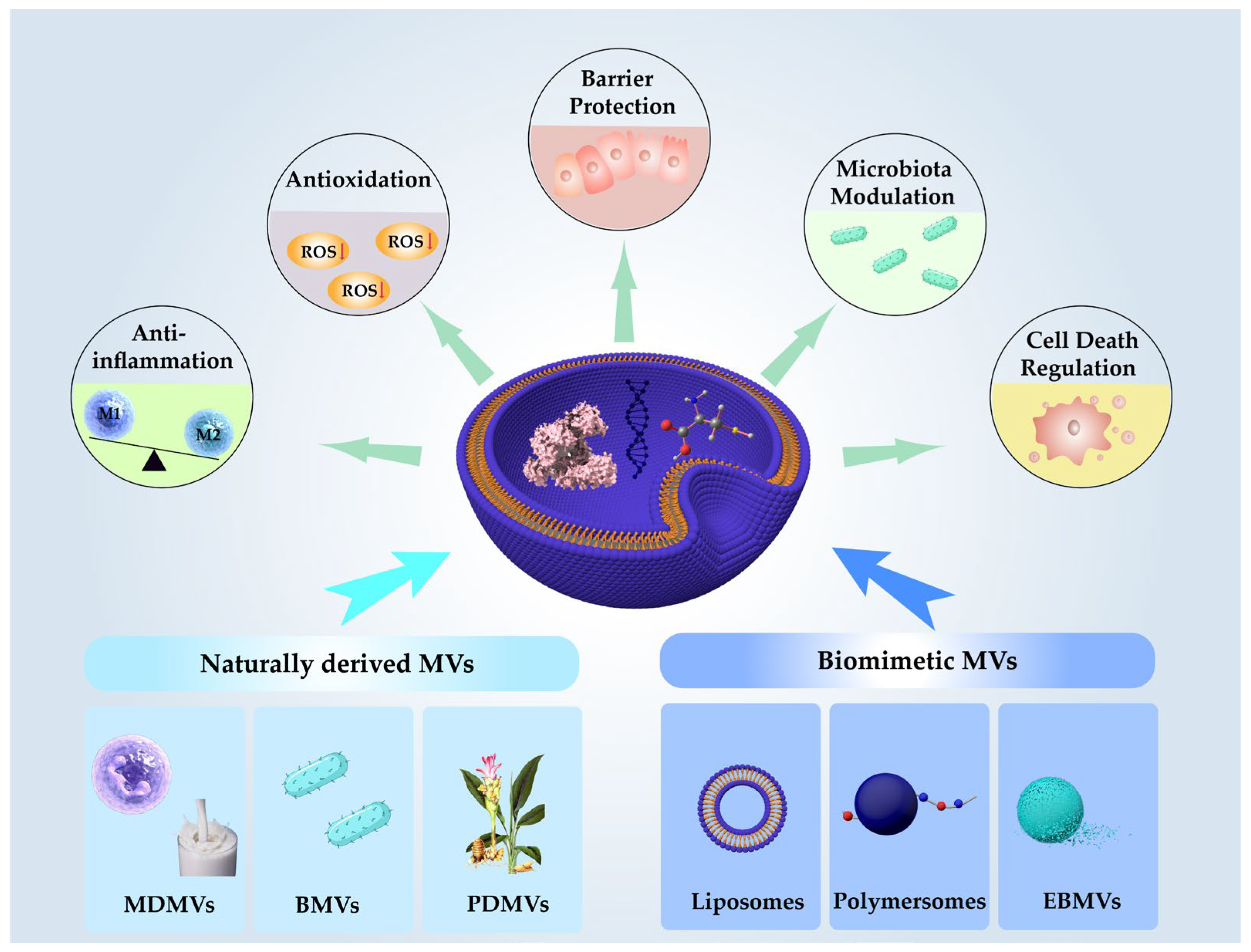
Figure 1. Origin of MV-DDSs and therapeutic mechanism in IBD. MV-DDSs can be classified according to their origin into mammalian-derived membrane vesicles (MDMVs), plant-derived membrane vesicles (PDMVs), bacterial membrane vesicles (BMVs), as well as liposomes, polymersomes, and engineered biomimetic membrane vesicles (EBMVs). These vesicles are capable of encapsulating a wide range of therapeutic cargos, including proteins, nucleic acids, and small-molecule drugs. In IBD therapy, MV-DDSs exert their effects through multiple mechanisms, such as suppressing inflammatory responses, scavenging ROS to alleviate oxidative stress, protecting intestinal barrier integrity, modulating gut microbiota composition, and regulating programmed cell death pathways.
Pharmaceutics 17 01127 g001
Figure 2. Therapeutic effect of MV-DDSs in IBD. In this figure, red arrows are used to denote the change trends of target substances: upward red arrows stand for an increase, and downward red arrows signify a decrease.
Pharmaceutics 17 01127 g002
Figure 3. Anti-inflammatory mechanism of MV-DDSs in IBD. Colors of lines are used to distinguish pathways of MV-DDSs from different sources.
Pharmaceutics 17 01127 g003
Figure 4. Antioxidant mechanism of MV-DDSs in IBD.
Pharmaceutics 17 01127 g004
Table 1. Related information on different MVs.
CategorySourceSize (nm)MarkersBiogenesisFeaturesRefs.
MDMVsMammalian cells30–150 CD9, CD63, CD81Endosomal budding to form multivesicular bodies, followed by membrane fusion
  • high biocompatibility;
  • low immunogenicity;
  • intrinsic targeting;
[21,22]
BMVsBacteria (Gram-negative, Gram-positive)10–400Gram-negative: LPS, OmpA/OmpC/OmpF;
Gram-positive: LTA, Peptidoglycan
Outward budding from bacterial membrane
  • strong immune stimulation;
  • vaccine adjuvant potential
[23,24]
PDNVsPlants (fruits, vegetables, herbs, etc.)50–500PM H+-ATPase, PATL1Plasma membrane invagination and exocytosis
  • excellent biocompatibility;
  • specific targeting;
  • acid-resistant;
[25]
LiposomesPhospholipids, cholesterol50–200DSPC, PEG-DSPESelf-assembly in aqueous solution
  • high drug loading;
  • immune evasion;
  • high stability;
  • controlled release;
  • combines natural biofunctionality with tunable synthetic properties
[26,27,28]
Polymersomessynthetic amphiphilic block copolymers70–300PEG-PLASelf-assembly in aqueous solution[29,30,31]
EBMVsArtificial assembly (cell membrane + synthetic materials)20–300Related to molecular signatures of parent cell membranesCoating nanoparticle cores with natural or hybrid membrane[32,33]
Disclaimer/Publisher’s Note: The statements, opinions and data contained in all publications are solely those of the individual author(s) and contributor(s) and not of MDPI and/or the editor(s). MDPI and/or the editor(s) disclaim responsibility for any injury to people or property resulting from any ideas, methods, instructions or products referred to in the content.