Article

Dendrimer-Derived Mimics of Host Defense Peptides Selectively Disrupt Cancer Cell Membranes for Melanoma Therapy

1
Translational Medical Center for Stem Cell Therapy, Department of Dermatology, Tongji Hospital, School of Medicine, Tongji University, Shanghai 200331, China
2
School of Material Science and Engineering, Tongji University, 4800 Caoan Road, Shanghai 201804, China
3
Laboratory Animal Center of Tongji University, Tongji University, Shanghai 200092, China
4
Experimental Teaching Center for Medicine and Life Science, School of Medicine, Tongji University, Shanghai 200331, China
5
Department of Dermatology, The First Affiliated Hospital of Soochow University, Suzhou 215031, China
*
Authors to whom correspondence should be addressed.
These authors contributed equally to this work.
Pharmaceutics 2025, 17(3), 361; https://doi.org/10.3390/pharmaceutics17030361
Submission received: 27 January 2025 / Revised: 18 February 2025 / Accepted: 7 March 2025 / Published: 12 March 2025
(This article belongs to the Special Issue Advanced Nanopharmaceuticals for Anticancer Therapy)

Abstract

:
Background: Melanoma is one of the most common malignancies, posing a significant health threat to patients, particularly in advanced stages due to its high aggressiveness. Chemotherapy agents with biocompatibility and low susceptibility to induce resistance are required for systematic management. Methods: Dendrimer-derived mimics (DMs) of host defense peptides (HDPs), which were constructed by a dendrimer core and optimized ratios of the hydrophobic arm, were used to treat A375 cells and HaCaT cells as the control. Live/dead staining, flow cytometry, and scanning electron microscopy (SEM) were conducted to analyze the anticancer mechanism. Mice with subcutaneous tumors were used to test the antitumor activity and toxicity in vivo. Results: DMs exhibited enhanced activity against A375 cells with remarkable selectivity, which mimics the action of natural HDPs and can cause damage to cell membranes. DMs can effectively inhibit solid tumor growth with minimal systemic toxicity and no adverse effects on healthy tissues. Conclusion: All the findings highlight DMs as promising anticancer candidates with significant potential for systemic melanoma therapy.

1. Introduction

Melanoma is a highly aggressive skin cancer with a rising global incidence, making it a significant clinical challenge [1,2,3]. Advanced-stage melanoma is particularly concerning, because its metastasis drastically reduces patients’ median survival rates [4,5,6]. Despite advancements in immunotherapy that have mitigated melanoma-related mortality, systematic management remains inadequate for refractory and relapsed cases due to the lack of effective chemotherapy options [7,8,9]. This limitation largely stems from the unfavorable profiles of traditional chemotherapeutic agents, such as dacarbazine, which demonstrate high systemic toxicity, the possibility to induce drug resistance, and poor tumor site accumulation [10,11]. Addressing these shortcomings is crucial for the development of novel therapeutic agents to combat melanoma more effectively.
Host defense peptides (HDPs) have emerged as promising anticancer candidates due to their unique membranolytic mechanism against bacteria and even cancer cells [12,13,14]. Unlike normal cells, cancer cell membranes display an excess of negative charges which were attributed to the abnormal externalization of phosphatidylserine on the outer leaflet [15,16]. This distinct feature enables cationic HDPs to selectively bind to cancer cells via electrostatic interactions, subsequently disrupting the cell membrane and inducing lysis [16]. This mechanism not only provides HDPs with potent anticancer activity but also reduces the possibility of developing resistance compared to traditional chemotherapy agents [16]. However, these advantages are counterbalanced by significant challenges, including low productivity, systemic toxicity, and poor enzymatic stability, which severely limit their clinical translation [17,18].
Cationic amphiphilic polymers have been recently designed to mimic the membranolytic mechanism of HDPs while addressing their inherent limitations [19,20]. Among all the biomaterials, dendrimers as three-dimensional, highly branched polymers stand out as superior biomaterials due to their unique structural and functional advantages [21]. Compared to linear polymers, dendrimers offer an extended in vivo half-life, enhanced binding affinity to cell membranes, and potential immunomodulatory effects, making them particularly well-suited candidates for biomedical applications [22,23]. However, reports on the dendrimer-based HDP mimics remain scarce, and their effect against different cancer cells has yet to be comprehensively explored.
In our previous work [24], we developed dendrimer-derived mimics (DMs) of HDPs, featuring a hydrophilic dendronized polylysine core and hydrophobic polycaprolactone (PCL) arms. These DMs demonstrated significant potential in treating cancer, and we hypothesized that they could achieve superior efficacy in treating superficial tumors, such as skin cancer. Herein, we systematically evaluated the anticancer activity of dendronized polylysine with varying generations and DMs against melanoma A375 cells. To assess their selectivity, cytotoxicity against HaCaT cells was also examined. This investigation elucidated the relationship between dendrimer structure and its anticancer activity. Mechanistic studies, including scanning electron microscopy (SEM), live/dead staining, and flow cytometry, revealed the unique membranolytic action of our DMs. Furthermore, in vivo anticancer efficacy was assessed using subcutaneous melanoma models in nude mice (Figure 1). Our DMs demonstrated potent anticancer activity and remarkable selectivity, highlighting their potential as promising candidates for melanoma treatment and underscoring the utility of dendrimer-based structures as HDP mimics.

2. Materials and Methods

2.1. Materials

The dendronized polylysine cores (DG1, DG2, and DG3), PCL, and a series of DG3Mn (n = 2, 4, 6, 8, where n represents the number of PCL arms modified on DG3, Figure S1 and Figure 1a) were synthesized following previously reported methods [24]. Briefly, the dendronized polylysine cores were prepared with stepwise conjugation of activated lysine to the preceding generation. Subsequently, PCL arms functionalized with isocyanate groups were obtained via ring-opening polymerization and were conjugated to DG3 through efficient reactions with amino groups, yielding the DG3Mn series. For subsequent experiments, the dendrimer-derived mimics (DMs) were prepared by dissolving 10 mg of each DM in 1 mL of phosphate-buffered saline (PBS; 1×, Corning, Corning, NY, USA). The prepared solutions were filtered using a 0.22 µm sterile filter (Taitan, Shanghai, China) to ensure sterility and uniformity before use.

2.2. Cell Culture

The human malignant melanoma A375 cell line and normal human keratinocyte HaCaT cell line were obtained from the National Collection of Authenticated Cell Cultures (Shanghai, China). A375 cells and HaCaT cells were cultured in Dulbecco’s modified eagle medium (DMEM; Gibco, Carlsbad, CA, USA) supplemented with 10% fetal bovine serum (FBS; Gibco, Carlsbad, CA, USA) and 1% penicillin/streptomycin (P/S; Gibco, Carlsbad, CA, USA). Cells were maintained in an incubator at 37 °C with 5% CO₂. The culture medium was refreshed every 48 h to sustain optimal cell growth. For subculturing, cells were detached using 0.05% trypsin-EDTA solution (Gibco, Carlsbad, CA, USA) when reaching approximately 80% confluence, with a split ratio of 1:3. Only low-passage cells (passages 2–6) of HaCaT and A375 were used in all subsequent experiments to ensure reproducibility and minimize phenotypic drift.

2.3. CCK-8 Analysis

The cytotoxicity of DMs was evaluated using the cell counting kit-8 (CCK-8, Dojindo, Kumamoto, Japan) assay. A375 cells and HaCaT cells were seeded into 96-well plates at a density of about 5 × 10³ cells/well in 100 µL of culture medium, with five replicates for each experimental group. After an initial incubation for 24 h to allow cell attachment, the DMs were diluted in the respective culture medium to final concentrations of 0, 8, 16, 32, 64, 128, and 256 μg/mL. The cells were treated with the DMs at these concentrations for 6, 24, 48, and 72 h. At each time point, 10 µL of CCK-8 solution was added to each well and incubated at 37 °C for an additional 2.5 h. The absorbance at 450 nm was measured using a microplate reader (Thermo Scientific Varioskan Flash, Waltham, MA, USA). Untreated cells served as the negative control, and the medium without cells was used as the blank control. The cell viability was calculated as a percentage of cell viability compared to the control group.

2.4. Live/Dead Staining Analysis

Live/dead staining was performed to evaluate the effects of DG3M2 (A dendronized polylysine cores DG3 with two modified PCL arms) on cell viability and its potential to cause membrane damage. A375 cells and HaCaT cells (5 × 10⁴ cells/well) were seeded into 24-well plates and allowed to attach for 24 h before treatment. For dose-dependent analysis, cells were treated with DG3M2 at final concentrations of 0, 8, 16, 32, 64, 128, and 256 μg/mL for 12 h. For time-dependent analysis, cells were treated with 64 μg/mL DG3M2 for various time intervals (30, 60, 120, 180, and 240 min). After treatment, the cells were stained with a live/dead viability kit (Calcein AM/PI; KeyGen Biotech, Nanjing, China). Fluorescent images of live (Calcein AM, green fluorescence) and dead (PI, red fluorescence) cells were captured using an inverted phase-contrast fluorescence microscope (Nikon Eclipse Ti-S, Tokyo, Japan).

2.5. Flow Cytometry Analysis of Apoptosis

To evaluate cell apoptosis, 5 × 10⁵ A375 cells were seeded into 6-well plates and cultured for 24 h to allow attachment. DG3M2 at various concentrations (0, 8, 16, 32, 64, and 128 μg/mL) was added to the wells and incubated with the cells for 12 h. After treatment, the cells were collected and washed twice with cold phosphate-buffered saline. Cell apoptosis was assessed using the Annexin V-FITC/PI Apoptosis Detection Kit (Yeasen, Shanghai, China). Briefly, cells were incubated with 5 µL of annexin V-FITC and 5 µL of propidium iodide (PI) for 15 min at room temperature in the dark. Fluorescence signals were analyzed using a fluorescence-activated cell sorter (FACS; BD FACSAria III, BD Biosciences, San Jose, CA, USA) to distinguish live, early apoptotic, and late apoptotic/necrotic cells.

2.6. Scanning Electron Microscopy (SEM) Analysis

To evaluate the effects of DMs on cancer cell membranes, A375 cells (5 × 10⁴ cells/well) were seeded onto sterilized glass coverslips in 24-well plates and cultured for 24 h to allow attachment. The cells were then treated with 64 μg/mL DMs for 12 h. After treatment, the cells were fixed with 4% paraformaldehyde (PFA; Sigma-Aldrich, St. Louis, MO, USA) at room temperature for 20 min and washed three times with PBS to remove residual fixative. Gradient ethanol dehydration was performed sequentially with 70%, 80%, 95%, and 100% ethanol, each for 15 min. Dehydrated samples were dried and sputter-coated with a layer of gold to enhance conductivity. The samples were then examined using a scanning electron microscope (SEM, Zeiss, Berlin, Germany) to observe morphological changes in the cell membrane.

2.7. In Vivo Anticancer Analysis

A total of six female BALB/c nude mice (6 weeks old, body weight 18–20 g) were purchased from the Experimental Animal Center at Tongji University (Shanghai, China). All animal studies were conducted following the protocols approved by the Animal Experimental Ethical Inspection Committee of the Laboratory Animal Centre, Tongji University (Approval No. TJAA00422103). To establish subcutaneous melanoma models, 5 × 10⁶ A375 cells in 100 µL PBS were injected subcutaneously into the right flank of each mouse. Tumor dimensions were measured using a vernier caliper, with the tumor volume calculated using the following formula:
Tumor Volume = 1 2 × ( long axis ) × ( short axis ) 2
Once the average tumor volume reached approximately 30 mm3, the mice were randomly divided into two groups (n = 3 per group). One group received intraperitoneal injections of DMs (10 mg/kg in PBS), while the control group received an equivalent volume of PBS. Injections were administered every two days for a total of 28 days. Body weight and tumor dimensions were recorded before each injection to monitor systemic toxicity and tumor progression. At the end of the treatment, mice were euthanized humanely, and tumors were excised and weighed. Tissues were fixed in 4% paraformaldehyde, embedded in paraffin, sectioned, and stained with hematoxylin and eosin (H&E) for histopathological evaluation to assess systemic toxicity.

2.8. Statistical Analysis

Statistical analyses were performed using SPSS 25.0 (IBM Corporation, Armonk, NY, USA) and OriginPro (OriginLab Corporation, Northampton, MA, USA). Quantitative data are presented as mean ± standard deviation (SD) and were derived from at least three independent experiments. Comparisons between two groups were conducted using the Student’s t-test, while multiple group comparisons were analyzed by one-way analysis of variance (ANOVA) followed by post hoc tests when applicable. Statistical significance was set at p < 0.05. In figures, levels of significance are denoted as follows: p < 0.05 (*), p < 0.01 (**), and p < 0.001 (***).

3. Results

3.1. Potent Anticancer Activity and Remarkable Selectivity of DMs

To evaluate the relationship between cytotoxicity and dendrimer structure, the viability of A375 cells and HaCaT cells was assessed using the CCK-8 assay after treatment with dendronized polylysine core of varying generations (DG1, DG2, and DG3). As shown in Figure 2b and Figure S2, ε-polylysine, used as the backbone for dendronized polypeptide preparation, exhibited minimal toxicity toward A375 cells, with cell viability remaining at 90.6% ± 5.68%, even at the high concentration tested (256 μg/mL, 72 h). In contrast, dendronized polylysine cores (DG1, DG2, and DG3) showed increased cytotoxicity with higher generations. The viabilities of A375 cells decreased to 18.6% ± 4.23%, 12.6% ± 0.61%, and 7.5% ± 0.26%, respectively. A similar trend was observed in HaCaT cells, where viabilities decreased to 57.3% ± 2.43%, 52.7% ± 3.16%, and 37.0% ± 0.84%, respectively.
Based on the strongest cytotoxicity observed for DG3, dendronized polylysine mimics (DMs) were constructed starting from DG3. However, due to DG3’s low selectivity, PCL with excellent biocompatibility was incorporated into the structure. The resulting products, named DG3Mn (where n = 2, 4, 6, 8), were tested for cytotoxicity against A375 and HaCaT cells (Figure 2c). DMs displayed sustained capabilities to inhibit cancer cell growth over time, as the viability of A375 cells continued to decrease during the 72 h incubation period (Figure S3). Moreover, The half-maximal inhibitory concentrations (IC50) after a 48 h incubation were calculated using linear interpolation. As shown in Figure 2d, DG3M2, DG3M4, DG3M6, and DG3M8 exhibited IC50 values of 12.17 ± 4.74, 39.23 ± 5.93, 47.14 ± 2.97, and 47.25 ± 7.21 μg/mL against A375 cells, respectively. In contrast, DMs demonstrated significantly lower cytotoxicity against HaCaT cells. Specifically, DG3M2 and DG3M8 had IC50 values were greater than 256 μg/mL, while DG3M4 and DG3M6 exhibited relatively higher cytotoxicity, with IC50 values of 212.25 ± 5.94 and 62.25 ± 1.32 μg/mL, respectively. Specifically, selectivity indexes for each DM were calculated as the ratio of IC50 values against HaCaT cells to IC50 values against A375 cells. DG3M2 showed the highest selectivity (20.5), while the other products, including DG3, showed lower selectivity (below 5.41).

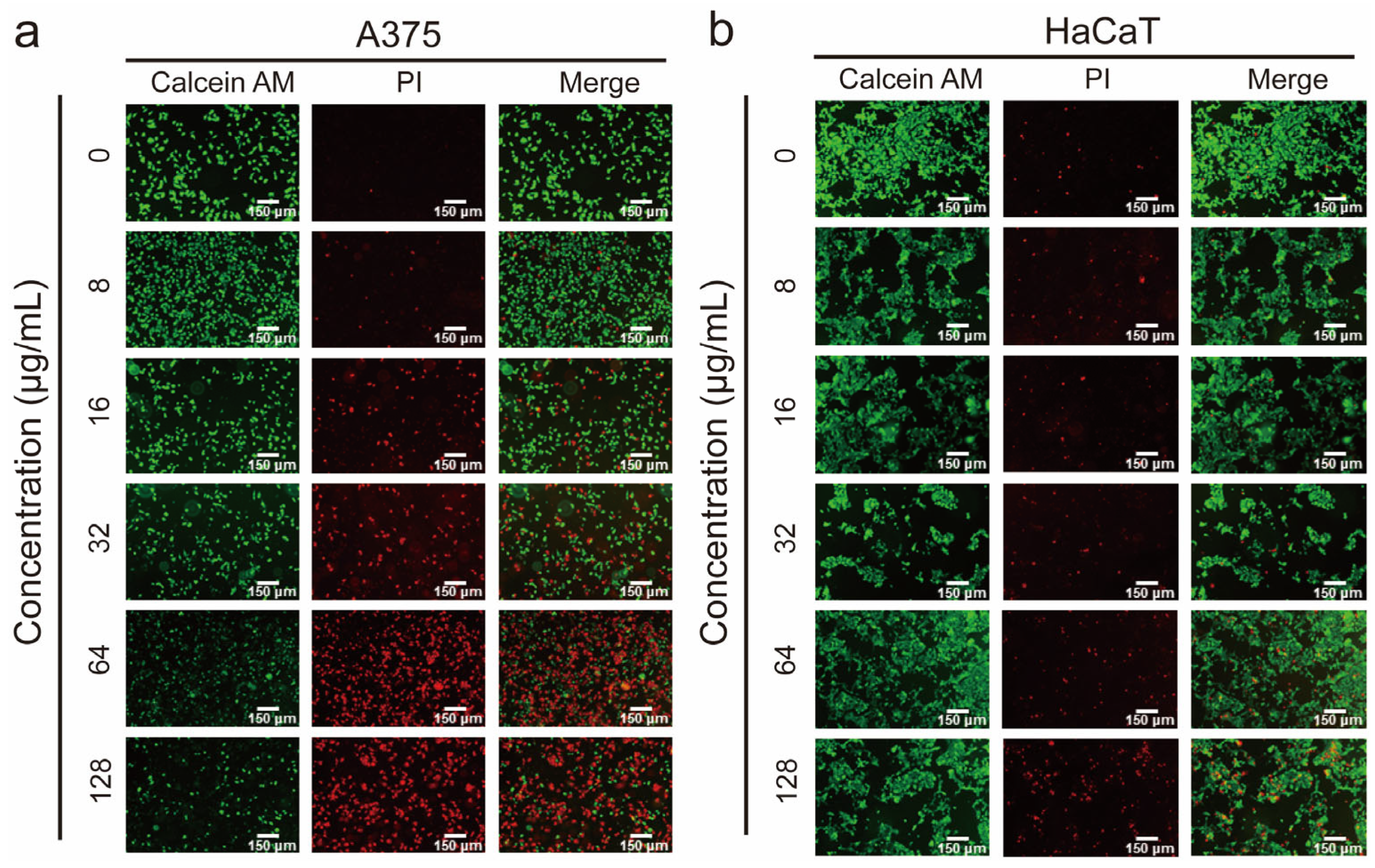
3.2. Demonstration of the Membranolytic Mechanism via the Live/Dead Staining Analysis

The outstanding performance of DMs against malignant cells in vitro motivated us to investigate whether their anticancer mechanism successfully mimics that of natural HDPs. Calcein AM was used to stain viable cells, while PI labeled the DNA of dead cells with disrupted membranes. Both concentration-dependent and time-dependent live/dead staining assays were performed. As shown in Figure 3a, the ratio of dead A375 cells increased with rising concentrations of DG3M2, ranging from 8 μg/mL to 128 μg/mL. Nearly 85% of A375 cells were dead after 12 h of treatment at the highest concentration. In contrast, while a similar concentration-dependent trend was observed, less than 30% of HaCaT cells were killed under the same conditions (Figure 3b). These findings align closely with the CCK-8 results, reaffirming the selective cytotoxicity of DG3M2 toward cancer cells. As for the killing kinetics assay, approximately 10% of A375 cells died within the initial 30 min of DG3M2 treatment, with complete eradication of viable cells achieved within 240 min (Figure S4). In contrast, HaCaT cells were minimally affected during this period, with less than 10% cell death observed even after 240 min. These results emphasized that DG3M2 showed a preference for disrupting cancer cell membranes.

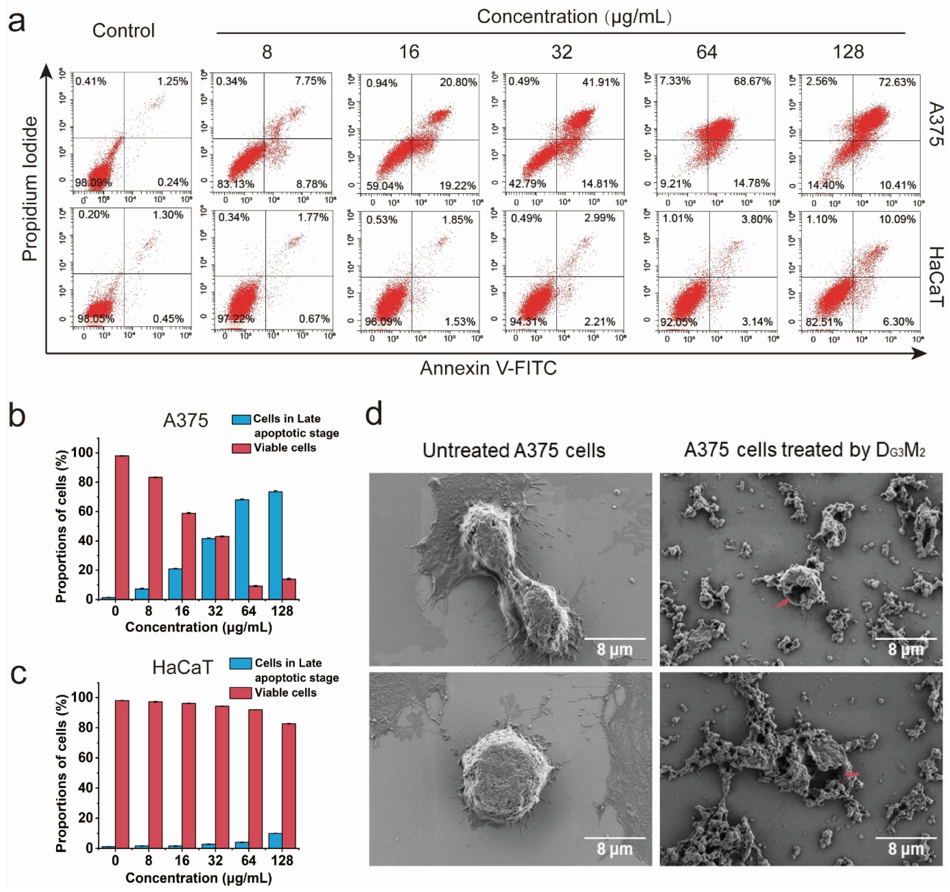
3.3. The Membranolytic Mechanism Indicated by Flow Cytometry Analysis

Flow cytometry with annexinV-FITC/PI double staining was utilized to distinguish viable, early apoptotic, late apoptotic, and necrotic cells following DG3M2 treatment (Figure 4a). As shown in Figure 4b, the results demonstrated a significant reduction in the proportion of viable A375 cells, decreasing from 83.4% ± 0.27% to 14.0% ± 0.55% as the DG3M2 concentration increased from 8 μg/mL to 128 μg/mL. Most affected cancer cells were classified as late apoptotic, with their proportion rising from 7.23% ± 0.46% to 73.5% ± 0.85%. In contrast, early apoptotic and necrotic cells consistently remained at low levels across all concentrations. For HaCaT cells (Figure 4c), the proportion of viable cells remained as high as 82.51%, even at the maximum concentration of 128 μg/mL, supporting the observed selectivity of DG3M2.

3.4. Clear Membrane Lysis Observed by SEM Analysis

To gain deeper insights into the membranolytic mechanism, the optimized product DG3M2 was used to treat melanoma cells, and the resulting membrane alterations were observed using SEM. After 12 h of incubation, significant morphological changes were evident in A375 cells treated with DG3M2 (Figure 4d). Untreated A375 cells adhered firmly to the plate, exhibiting an oval shape with intact cell membranes and abundant filopodia. In stark contrast, cells treated with DG3M2 displayed severe membrane damage, including the formation of large holes in the cell membrane, through which intracellular contents visibly leaked out. Additionally, DG3M2 caused noticeable shrinkage of the filopodia, suggesting its potential to impair tumor cell invasiveness. Furthermore, numerous small vesicles were observed on the plate surface, likely remnants shed from the disrupted cell membranes. These findings provide compelling evidence that DG3M2 exerts its anticancer effects via a membranolytic mechanism, effectively mimicking the natural action of HDPs as our intended design.

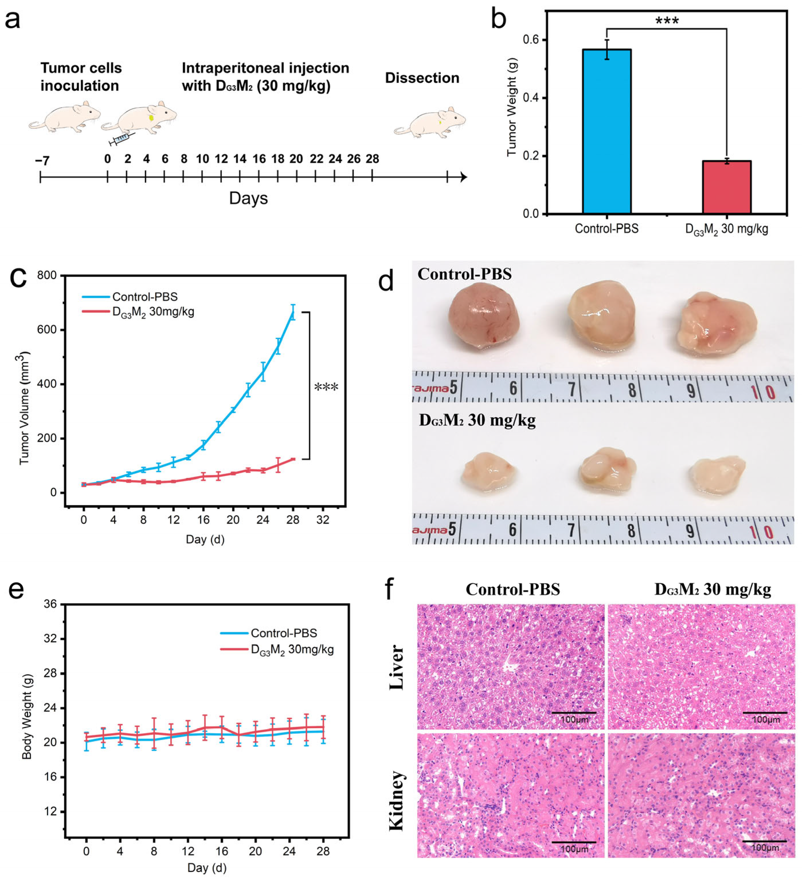
3.5. The Capability of DMs to Inhibit Tumor Growth in Vivo with Low Tissue Toxicity

The in vivo anticancer efficacy and safety profile of DG3M2 were evaluated using a subcutaneous tumor model in nude mice. A375 cells were injected into the mice to establish tumors, and treatments commenced when tumor volumes reached approximately 30 mm3. Mice were randomly assigned to two groups (n = 3 per group) and treated with DG3M2 or PBS via intraperitoneal injection for 28 days (Figure 5a). Tumor growth in the PBS-treated control group progressed rapidly, with tumor volumes exceeding 665 mm3 (Figure 5c), whereas the DG3M2-treated group exhibited significantly reduced tumor growth, maintaining volumes below 125 mm3 throughout the experiment. At the end of the study, tumor weights were measured, revealing a 68% reduction in the DG3M2 group (0.183 g, Figure 5b) compared to the control group. Mice body weights remained stable in both groups, indicating no apparent systemic toxicity associated with DG3M2 treatment (Figure 5e). Furthermore, hematoxylin and eosin (H&E) staining of liver and kidney tissues showed no histological abnormalities after 28 days of treatment (Figure 5f), further confirming the biocompatibility of DG3M2.

4. Discussion

Traditional chemotherapy often falls short in providing systematic treatment of skin cancer [25,26], underscoring the urgent need to develop novel anticancer agents with enhanced selectivity and biocompatibility. In our previous work [24], dendronized polylysine mimics (DMs) of natural HDPs demonstrated activity against bacterial pathogens and A549 lung cancer cells. However, their efficacy required further optimization. In this study, we explored the potential of DMs as selective and effective therapeutic agents for melanoma, offering a promising approach to address this unmet clinical need.
Our findings demonstrate a clear correlation between the generation of the dendronized polylysine core and its cytotoxicity. Among DG1, DG2, and DG3, the latter exhibited the strongest anticancer activity, which can be attributed to the increase in terminal amino groups. This trend aligns with observations for other cationic polymers, such as polyethyleneimine (PEI), where higher amino group density enhances cationic charge [27,28]. The increased charge density facilitates stronger interactions with negatively charged cancer cell membranes, driving the observed cytotoxicity. However, DG3 showed poor selectivity, significantly affecting both A375 melanoma cells and HaCaT keratinocytes which have become the key limitation for designing anticancer agents. To address this, PCL arms were incorporated to improve the selectivity of the dendronized polylysine core, and DMs were constructed.
For the anticancer assay, DG3M2 demonstrated the most potent activity against A375 cells, with the lowest IC50 (12.17 μg/mL). As the ratio of PCL increased, anticancer activity gradually decreased. This observation suggests that a low proportion of hydrophobic residues optimally enhances interactions between DMs and the lipid components of cancer cell membranes. However, excessive PCL incorporation fails to further improve these interactions and instead limits the solubility of the resulting polymer. Consequently, DG3M8 exhibited the weakest anticancer activity among the tested derivatives. Regarding cytotoxicity against HaCaT cells, DG3M2 showed a marked improvement in biocompatibility compared to DG3, attributable to the intrinsic biocompatibility of PCL. In addition, DG3M2 also performed better compared to chemotherapy drugs [29] and natural HDPs [30,31]. However, further increases in hydrophobic residues appeared to compromise biocompatibility, likely due to their enhanced interactions with normal cell membranes. Overall, the PCL arms demonstrated both positive and negative effects; they improved biocompatibility but also disrupted cell membranes and adversely affected water solubility. These complex interactions led to variations in selectivity, with DG3M2 exhibiting superior selectivity (20.5) compared to the core DG3 and other DMs. This structure–activity relationship highlights the importance of balancing hydrophobic and hydrophilic components to optimize the HDP mimics. Additionally, the anticancer activity of DG3M2 against A375 cells was higher than its activity against A549 cells, as previously reported [24]. This finding underscores the potential of DMs as promising agents for skin cancer treatment.
The anticancer mechanism of DMs was elucidated through live/dead staining, SEM analysis, and flow cytometry assays. Live/dead staining demonstrated that DG3M2 induced cancer cell death in a concentration- and time-dependent manner while sparing normal cells, consistent with the CCK-8 assay results. The red fluorescence observed with PI staining indicated membrane damage in cancer cells, as PI is impermeable to intact membranes [32]. SEM analysis provided direct morphological evidence of membranolytic activity, revealing large pores, leakage of cellular contents, and disruption of filopodia in DG3M2-treated A375 cells. Compared to the membrane changes observed in A549 cells in our previous study [24], a larger scale of membrane lysis was evident in A375 cells, likely attributable to differences in membrane composition. This suggests that A375 cells are more susceptible to disruption by HDP mimics, further supporting their potential as systemic agents in skin cancer treatment. Flow cytometry further confirmed that DG3M2 predominantly induced late apoptosis, with minimal involvement of early apoptotic or necrotic pathways. Membrane disruption facilitated PI penetration and labeling of intracellular components, allowing clear identification of late apoptotic cells [33]. Collectively, these findings demonstrate that DG3M2 exerts its anticancer effects through a unique membranolytic mechanism, aligning with its design to mimic the anticancer processes of natural HDPs.
The potent in vitro anticancer activity of DG3M2 translated effectively to an in vivo model. DG3M2 significantly inhibited tumor growth in A375-bearing nude mice without causing systemic toxicity, as evidenced by stable body weight and unaltered liver and kidney histology. Tumor volumes in the DG3M2 group were consistently suppressed, with a 68% reduction in tumor weight compared to the PBS control group. These results highlight the favorable pharmacokinetics, stability, and biocompatibility of DG3M2, which are critical attributes for clinical translation [34].
Moreover, DG3M2’s ability to suppress tumor growth while sparing healthy tissues suggests an optimal balance between efficacy and safety, which is often a significant challenge in cancer therapy. These results also underline DG3M2’s favorable pharmacokinetics and stability in vivo, potentially attributable to its dendronized structure and the incorporation of biocompatible PCL arms. The enhanced selectivity observed in vitro was consistently reflected in vivo, reinforcing its suitability for targeted melanoma treatment. We believe that the application of DMs in a preoperative context to systematically manage the tumor mass before surgical procedures may offer a strategic advantage in optimizing therapeutic efficacy and patient outcomes. Overall, these findings position DG3M2 as a promising candidate for further preclinical and clinical evaluations aimed at developing effective and safe therapies for melanoma.

5. Conclusions

Melanoma treatment requires not only effective chemotherapy but also agents that can minimize resistance development and exhibit low systemic toxicity. In this study, we explored dendrimer-derived mimics (DMs) of HDPs, which demonstrated potent anticancer activity against A375 melanoma cells. The dendronized polylysine core was shown to exhibit generation-dependent cytotoxicity, highlighting the critical role of dendrimer generation in enhancing anticancer effects. After modification with PCL arms, the biocompatibility of DMs was significantly improved, leading to increased selectivity for cancer cells over normal cells. Among the various derivatives, DG3M2 emerged as the most promising candidate, demonstrating the ability to induce membrane pore formation and lysis, effectively mimicking the membrane-disrupting mechanism of natural HDPs. This unique membranolytic action enabled DG3M2 to efficiently eliminate melanoma cells while sparing normal cells. In vivo, DG3M2 successfully inhibited tumor growth in a melanoma-bearing nude mice model with minimal systemic toxicity and no observable damage to vital organs, such as the liver and kidneys. Additionally, when compared to its activity against A549 lung cancer cells, DG3M2 showed superior anticancer effects against A375 melanoma cells, underscoring its potential for targeted melanoma therapy. Overall, these findings suggest that DMs, particularly DG3M2, hold significant promise as selective, biocompatible anticancer agents for melanoma treatment, warranting further investigation in preclinical and clinical studies.

Supplementary Materials

The following supporting information can be downloaded at: https://www.mdpi.com/article/10.3390/pharmaceutics17030361/s1, Figure S1: The chemical structure and schematic representation of DMs. Figure S2: Cytotoxicity of ε-polylysine, DG1, and DG2 as determined by CCK-8 assay; Figure S3: Inhibition rate of A375 and HaCaT cells after treatment with DMs at different concentrations and incubation times. Figure S4: The killing kinetic assay of DMs against A375 (a) and HaCaT cells (b) as determined by live/dead staining assay.

Author Contributions

Conceptualization, C.Z. and J.H.; Formal analysis, Y.Q.; Funding acquisition, C.Z. and J.H.; Investigation, Y.Q., X.L., D.Y., C.S., J.Z., J.X., Y.Z. (Yan Zhao), L.Z., H.K., M.Z. and Y.Z. (Yueqian Zhu); Methodology, Y.Q., X.L., D.Y., C.S., J.Z., J.X., Y.Z. (Yan Zhao), L.Z., H.K. and M.Z.; Resources, Y.Z. (Yueqian Zhu) and J.H.; Software, Y.Q.; Supervision, C.Z. and J.H.; Visualization, Y.Q; Writing—original draft, Y.Q.; Project administration, J.H. Writing—review and editing, J.H. All authors have read and agreed to the published version of the manuscript.

Funding

This work is supported by the Science Project of Shanghai Municipal Health Commission (Grant Number 202040027).

Institutional Review Board Statement

The animal study protocol was approved by the Animal Experimental Ethical Inspection Committee of the Laboratory Animal Centre, Tongji University (Approval No. TJAA00422103).

Informed Consent Statement

Not applicable.

Data Availability Statement

The original contributions presented in this study are included in the article/Supplementary Material. Further inquiries can be directed to the corresponding authors.

Conflicts of Interest

The authors declare no conflicts of interest.

References

  1. Arnold, M.; Singh, D.; Laversanne, M.; Vignat, J.; Vaccarella, S.; Meheus, F.; Cust, A.E.; de Vries, E.; Whiteman, D.C.; Bray, F. Global Burden of Cutaneous Melanoma in 2020 and Projections to 2040. JAMA Dermatol. 2022, 158, 495–503. [Google Scholar] [CrossRef] [PubMed]
  2. Ferlay, J.; Colombet, M.; Soerjomataram, I.; Parkin, D.M.; Piñeros, M.; Znaor, A.; Bray, F. Cancer statistics for the year 2020: An overview. Int. J. Cancer 2021, 149, 778–789. [Google Scholar] [CrossRef] [PubMed]
  3. Lin, L.F.; Li, Z.Y.; Yan, L.; Liu, Y.L.; Yang, H.J.; Li, H. Global, regional, and national cancer incidence and death for 29 cancer groups in 2019 and trends analysis of the global cancer burden, 1990–2019. J. Hematol. Oncol. 2021, 14, 197. [Google Scholar] [CrossRef] [PubMed]
  4. Zhang, Y.L.; Hou, J.B.; Shi, S.M.; Du, J.; Liu, Y.D.; Huang, P.; Li, Q.; Liu, L.C.; Hu, H.R.; Ji, Y.C.; et al. CSN6 promotes melanoma proliferation and metastasis by controlling the UBR5-mediated ubiquitination and degradation of CDK9. Cell Death Dis. 2021, 12, 118. [Google Scholar] [CrossRef]
  5. Shirley, C.A.; Chhabra, G.; Amiri, D.; Chang, H.; Ahmad, N. Immune escape and metastasis mechanisms in melanoma: Breaking down the dichotomy. Front. Immunol. 2024, 15, 1336023. [Google Scholar] [CrossRef]
  6. Leonardi, G.C.; Falzone, L.; Salemi, R.; Zanghì, A.; Spandidos, D.A.; McCubrey, J.A.; Candido, S.; Libra, M. Cutaneous melanoma: From pathogenesis to therapy (Review). Int. J. Oncol. 2018, 52, 1071–1080. [Google Scholar] [CrossRef]
  7. Curti, B.D.; Faries, M.B. Recent Advances in the Treatment of Melanoma. N. Engl. J. Med. 2021, 384, 2229–2240. [Google Scholar] [CrossRef]
  8. Rui, R.; Zhou, L.Q.; He, S.M. Cancer immunotherapies: Advances and bottlenecks. Front. Immunol. 2023, 14, 1212476. [Google Scholar] [CrossRef]
  9. Pham, J.P.; Joshua, A.M.; da Silva, I.P.; Dummer, R.; Goldinger, S.M. Chemotherapy in Cutaneous Melanoma: Is There Still a Role? Curr. Oncol. Rep. 2023, 25, 609–621. [Google Scholar] [CrossRef]
  10. Liu, Q.; Das, M.; Liu, Y.; Huang, L. Targeted drug delivery to melanoma. Adv. Drug Deliv. Rev. 2018, 127, 208–221. [Google Scholar] [CrossRef]
  11. Mundra, V.; Li, W.; Mahato, R.I. Nanoparticle-mediated drug delivery for treating melanoma. Nanomedicine 2015, 10, 2613–2633. [Google Scholar] [CrossRef] [PubMed]
  12. Lu, F.T.; Zhu, Y.K.; Zhang, G.D.; Liu, Z.P. Renovation as innovation: Repurposing human antibacterial peptide LL-37 for cancer therapy. Front. Pharmacol. 2022, 13, 944147. [Google Scholar] [CrossRef] [PubMed]
  13. Riedl, S.; Zweytick, D.; Lohner, K. Membrane-active host defense peptides—Challenges and perspectives for the development of novel anticancer drugs. Chem. Phys. Lipids 2011, 164, 766–781. [Google Scholar] [CrossRef]
  14. Kardani, K.; Bolhassani, A. Antimicrobial/anticancer peptides: Bioactive molecules and therapeutic agents. Immunotherapy 2021, 13, 669–684. [Google Scholar] [CrossRef]
  15. Szlasa, W.; Zendran, I.; Zalesinska, A.; Tarek, M.; Kulbacka, J. Lipid composition of the cancer cell membrane. J. Bioenerg. Biomembr. 2020, 52, 321–342. [Google Scholar] [CrossRef]
  16. Tornesello, A.L.; Borrelli, A.; Buonaguro, L.; Buonaguro, F.M.; Tornesello, M.L. Antimicrobial Peptides as Anticancer Agents: Functional Properties and Biological Activities. Molecules 2020, 25, 2850. [Google Scholar] [CrossRef]
  17. Quemé-Peña, M.; Ricci, M.; Juhász, T.; Horváti, K.; Bosze, S.; Biri-Kovács, B.; Szeder, B.; Zsila, F.; Beke-Somfai, T. Old Polyanionic Drug Suramin Suppresses Detrimental Cytotoxicity of the Host Defense Peptide LL-37. ACS Pharmacol. Transl. Sci. 2021, 4, 155–167. [Google Scholar] [CrossRef]
  18. Mookherjee, N.; Anderson, M.A.; Haagsman, H.P.; Davidson, D.J. Antimicrobial host defence peptides: Functions and clinical potential. Nat. Rev. Drug Discov. 2020, 19, 311–332. [Google Scholar] [CrossRef]
  19. Ergene, C.; Yasuhara, K.; Palermo, E.F. Biomimetic antimicrobial polymers: Recent advances in molecular design. Polym. Chem. 2018, 9, 2407–2427. [Google Scholar] [CrossRef]
  20. Kalelkar, P.P.; Riddick, M.; García, A.J. Biomaterial-based antimicrobial therapies for the treatment of bacterial infections. Nat. Rev. Mater. 2022, 7, 39–54. [Google Scholar] [CrossRef] [PubMed]
  21. Dias, A.P.; Santos, S.D.; da Silva, J.V.; Parise, R.; Ferreira, E.I.; El Seoud, O.; Giarolla, J. Dendrimers in the context of nanomedicine. Int. J. Pharm. 2020, 573, 118814. [Google Scholar] [CrossRef] [PubMed]
  22. Chauhan, A.S. Dendrimers for Drug Delivery. Molecules 2018, 23, 938. [Google Scholar] [CrossRef] [PubMed]
  23. Wang, J.; Li, B.X.; Qiu, L.; Qiao, X.; Yang, H. Dendrimer-based drug delivery systems: History, challenges, and latest developments. J. Biol. Eng. 2022, 16, 18. [Google Scholar] [CrossRef] [PubMed]
  24. Qian, Y.S.; Yang, D.J.; Zhu, J.M.; Huang, S.T.; Chen, S.J.; Zeng, J.; Xu, J.; He, J.; Zhou, C.C. Mimics of Host Defense Peptides Derived from Dendronized Polylysines for Antibacterial and Anticancer Therapy. ACS Macro Lett. 2024, 13, 1156–1163. [Google Scholar] [CrossRef] [PubMed]
  25. Burton, K.A.; Ashack, K.A.; Khachemoune, A. Cutaneous Squamous Cell Carcinoma: A Review of High-Risk and Metastatic Disease. Am. J. Clin. Dermatol. 2016, 17, 491–508. [Google Scholar] [CrossRef]
  26. Seidel, J.A.; Otsuka, A.; Kabashima, K. Anti-PD-1 and Anti-CTLA-4 Therapies in Cancer: Mechanisms of Action, efficacy, and Limitations. Front. Oncol. 2018, 8, 86. [Google Scholar] [CrossRef]
  27. Janaszewska, A.; Lazniewska, J.; Trzepinski, P.; Marcinkowska, M.; Klajnert-Maculewicz, B. Cytotoxicity of Dendrimers. Biomolecules 2019, 9, 330. [Google Scholar] [CrossRef]
  28. Chis, A.A.; Dobrea, C.; Morgovan, C.; Arseniu, A.M.; Rus, L.L.; Butuca, A.; Juncan, A.M.; Totan, M.; Vonica-Tincu, A.L.; Cormos, G.; et al. Applications and Limitations of Dendrimers in Biomedicine. Molecules 2020, 25, 3982. [Google Scholar] [CrossRef]
  29. Tian, Y.H.; Li, S.P.; Song, J.; Ji, T.J.; Zhu, M.T.; Anderson, G.J.; Wei, J.Y.; Nie, G.J. A doxorubicin delivery platform using engineered natural membrane vesicle exosomes for targeted tumor therapy. Biomaterials 2014, 35, 2383–2390. [Google Scholar] [CrossRef]
  30. Schweizer, F. Cationic amphiphilic peptides with cancer-selective toxicity. Eur. J. Pharmacol. 2009, 625, 190–194. [Google Scholar] [CrossRef]
  31. Gajski, G.; Garaj-Vrhovac, V. Melittin: A lytic peptide with anticancer properties. Environ. Toxicol. Pharmacol. 2013, 36, 697–705. [Google Scholar] [CrossRef] [PubMed]
  32. Zhou, X.Y.; He, J.; Zhou, C.C. Strategies from nature: Polycaprolactone-based mimetic antimicrobial peptide block copolymers with low cytotoxicity and excellent antibacterial efficiency. Polym. Chem. 2019, 10, 945–953. [Google Scholar] [CrossRef]
  33. Shao, N.; Yuan, L.; Ma, P.C.; Zhou, M.; Xiao, X.M.; Cong, Z.H.; Wu, Y.M.; Xiao, G.H.; Fei, J.; Liu, R.H. Heterochiral β-Peptide Polymers Combating Multidrug-Resistant Cancers Effectively without Inducing Drug Resistance. J. Am. Chem. Soc. 2022, 144, 7283–7294. [Google Scholar] [CrossRef] [PubMed]
  34. Kumar, P.; Kizhakkedathu, J.N.; Straus, S.K. Antimicrobial Peptides: Diversity, Mechanism of Action and Strategies to Improve the Activity and Biocompatibility In Vivo. Biomolecules 2018, 8, 4. [Google Scholar] [CrossRef]
Figure 1. Scheme of the membranolytic mechanism of DMs against A375 cells. (a) Schematic representation of DMs. DMs destroy the membrane of A375 cells by inducing pore formation and membrane contraction. (b) DMs with cationic charges absorb the cell membrane via electronic interaction as the cancer cell membrane possesses negative charges. (c) DMs further insert into the cell membrane, causing pores as a consequence.
Pharmaceutics 17 00361 g001
Figure 2. Cytotoxicity of dendronized polylysine core and DMs against A375 and HaCaT cells as determined by CCK-8 assay. (a) Schematic representation of DMs constructed by dendronized polylysine core and varying numbers of PCL arms. (b) Cell viability of A375 and HaCaT cells following treatment with dendronized polylysine core (256 μg/mL) for 72 h. (c) Cell viability of A375 and HaCaT cells treated with DMs at various concentrations and incubation times. (d) IC50 values of DMs against A375 and HaCaT cells and the selectivity index, calculated as the ratio of IC50 against HaCaT cells to IC50 against A375 cells.
Pharmaceutics 17 00361 g002
Figure 3. Concentration-dependent live/dead staining analysis of DMs against A375 (a) and HaCaT cells (b). Cells were treated with DMs at concentrations of 0, 8, 16, 32, 64, and 128 μg/mL for 6 h.
Pharmaceutics 17 00361 g003
Figure 4. Flow cytometry analysis (annexin V/PI) of A375 and HaCaT cells after treatment with DG3M2 at various concentrations for 6 h. (a) Flow cytometry plots of A375 and HaCaT cells after treatment with DG3M2. (b) Proportion of late apoptotic and viable A375 cells after the treatment at different concentrations. (c) Proportion of late apoptotic and viable HaCaT cells after the treatment at different concentrations. (d) Morphological changes of A375 cancer cells before and after treatment with DG3M2. Treatment with DG3M2 significantly compromised cell membrane integrity (red arrows), resulting in observable morphological alterations.
Pharmaceutics 17 00361 g004
Figure 5. In vivo anticancer analysis of DG3M2 against A375 tumors. (a) Construction of mice model and the treatment of DG3M2 (30 mg/kg) (b) Tumor weight measured after the completion of the experiment, p < 0.001 (***). (c) Average tumor volume in different treatment groups throughout the experiment, p < 0.001 (***). (d) Photographs of dissected tumor tissues from mice after 28 days of treatment. (e) Body weight changes of mice during the experimental period. (f) H&E staining of liver and kidney tissues to assess potential organ toxicity.
Pharmaceutics 17 00361 g005
Disclaimer/Publisher’s Note: The statements, opinions and data contained in all publications are solely those of the individual author(s) and contributor(s) and not of MDPI and/or the editor(s). MDPI and/or the editor(s) disclaim responsibility for any injury to people or property resulting from any ideas, methods, instructions or products referred to in the content.

