

Polysaccharide-Based Coatings as Drug Delivery Systems


Abstract
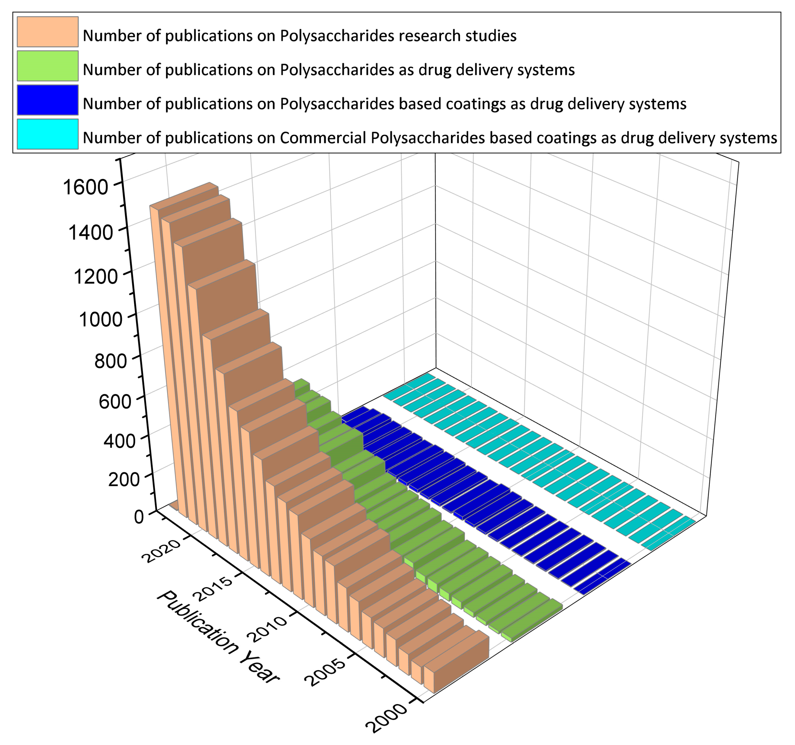
:1. Introduction: Background, Significance, and Justification
2. Polysaccharide Administration Routes in Drug Delivery Systems
- (i.)
- Oral administration: Polysaccharides can be formulated into nanoparticles/microparticles or films for oral drug delivery [7]. These particles can protect the drug from degradation in the gastrointestinal tract and enhance its absorption. Polysaccharide particles can adhere to mucosal surfaces, facilitating drug transport through the mucosal epithelium and improving bioavailability [7].
- (ii.)
- Parenteral administration: Polysaccharide-based delivery systems can be administered via injection, allowing for direct delivery into the bloodstream [7]. This route enables rapid drug distribution and targeted delivery to specific tissues. Polysaccharide nanoparticles can circulate in the bloodstream for extended periods, avoiding rapid clearance by the reticuloendothelial system [7]. This prolonged circulation can enhance drug efficacy and reduce the frequency of administration.
- (iii.)
- Topical administration: Polysaccharides can be incorporated into creams, gels, or patches for topical drugs [7]. When applied to the skin, polysaccharide-based formulations can provide sustained release of drugs, improving their therapeutic effect. The mucoadhesive properties of certain polysaccharides can enhance drug retention at the site of application, prolonging drug release and absorption [7].
- (iv.)
- Targeted delivery: Polysaccharides offer the advantage of tissue-specific targeting in drug delivery [9]. By modifying the surface of polysaccharide particles or conjugating targeting ligands, drugs can be delivered to specific tissues or cells of interest. This targeted delivery approach improves drug efficacy and reduces off-target effects.
- (v.)
- Combination with other delivery systems: Polysaccharides can be combined with other drug delivery systems, such as liposomes or nanoparticles, to enhance their absorption properties and functionality [7]. This combination can provide synergistic effects, such as improved drug stability, controlled release, and enhanced targeting capabilities.
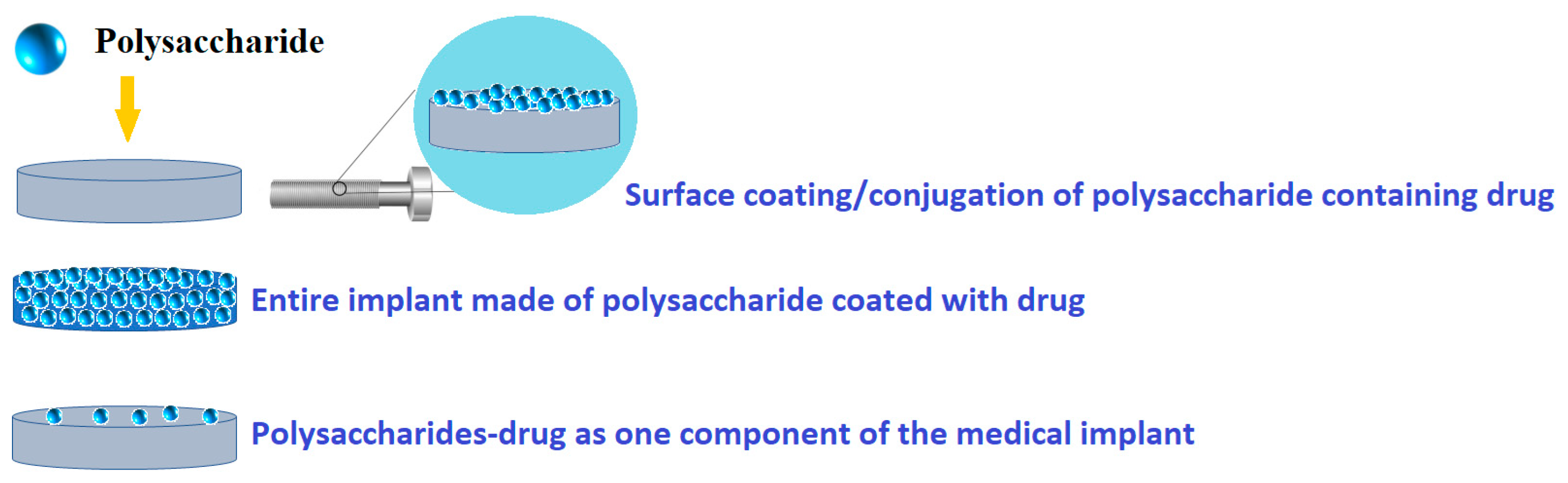
Polysaccharide-Based Coatings Administration Routes in Drug Delivery Systems
- (i)
- Coating of the drug core: In this approach, the drug is coated with a polysaccharide material that can be broken down by the acid-mediated cleavage. This allows for the controlled release of the drug at the desired site [7].
- (ii)
- Embedding of the drug in a biodegradable matrix: Polysaccharides can be used to form a matrix in which the drug is embedded. The matrix slowly degrades, releasing the drug over time [18].
- (iii)
- Formulation of drug-saccharide conjugates (prodrugs): Polysaccharides can be chemically modified to form conjugates with drugs, resulting prodrugs. These prodrugs can be designed to release the active drug molecules, improving drug targeting and reducing systemic side effects [19].
3. Overview on Polysaccharide-Based Drug Delivery Systems
3.1. Polysaccharide-Based Drug Delivery Systems Characteristics
- (i)
- Biocompatibility and Biodegradability: Contrary to many synthetic polymers, polysaccharides have very low (if any) toxicity levels [38]. It is worth noting that many polysaccharides can be broken down by enzymes, resulting in the release of their monomer or oligomer building blocks. This process allows for the recycling of these building blocks for various purposes, such as storage, structural support, and cell signaling [39]. It is worth mentioning that other polysaccharides are particularly susceptible to degradation by lysosomal enzymes after endocytosis, including esterases, glycosidases and proteases [40]. Thus, enzymatic degradation offers a proper way to release therapeutics from polysaccharide-based carrier systems [41].
- (ii)
- Bioactivity: Numerous polysaccharides possess inherent bioactivity, notably mucoadhesion, anti-inflammatory, and antimicrobial properties. Mucoadhesion refers to the bond between a substance and mucosal layer, such as in the gastrointestinal tract, nasal pathway, or airway [42]. Some polysaccharides such as chitosan have antimicrobial properties, while others such as heparin are known to help in reducing inflammation.
- (iii)
- Solubility: Sometimes, solubility can be changed by adjusting the monomer (basic units of polymers) structure. For instance, it is possible to alter the solubility of chitosan in acidic conditions. Polysaccharides have functional groups (hydroxyl and amine groups) along their backbone, which usually result in high solubility in water [43]. For instance, when the degree of deacetylation is higher, it results in more protonated free amino groups along the polysaccharide backbone. This leads to improved solubility [44].
- (iv)
- Ease of Modification: Polysaccharides can be easily modified. Glucose-based polysaccharides such as amylose, amylopectin, glycogen, and cellulose have many free reactive hydroxyl groups [45]. There are some polysaccharides that have hydroxyl and carboxylic acid groups, which can be readily altered/modified [46].
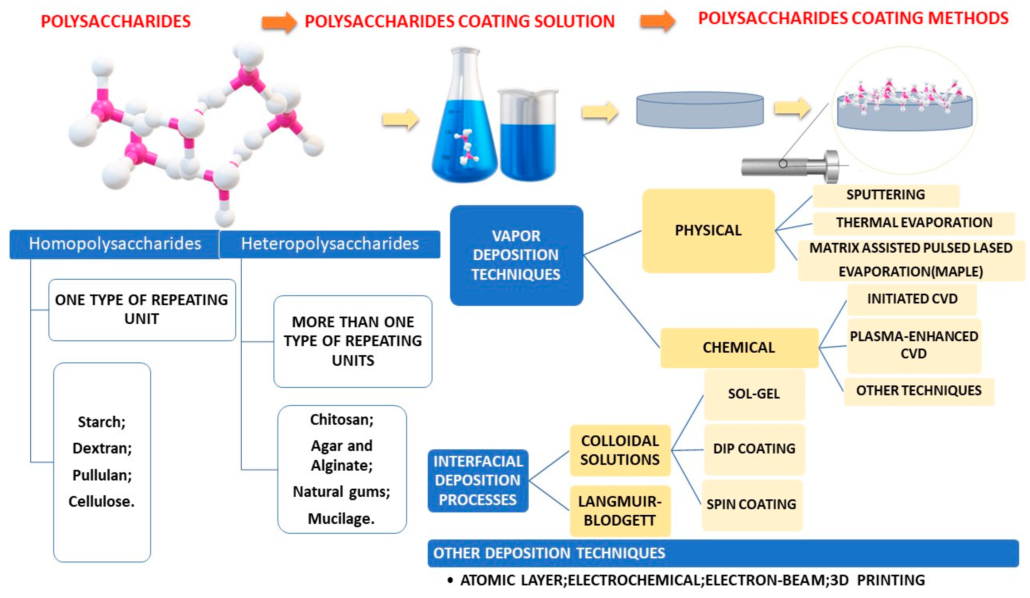
3.2. Polysaccharide-Based Drug Delivery Systems Classification
- − Plant/algal
- − Starch, which includes amylose, amylopectin, cellulose, agar, alginate, and carrageenan.
- − Pectin and konjac, as well as guar gum.
- − Animal-based substances, such as chitin/chitosan, and hyaluronic acid.
- − Bacterial substances, such as xanthan, dextran, levan, and curdlan.
- − Fungal substances, including pullulan and yeast glucans.
3.3. Starch-Based Coatings as Drug Delivery Systems
3.4. Chitosan-Based Coatings as Drug Delivery Systems
3.5. Dextran-Based Coatings as Drug Delivery Systems
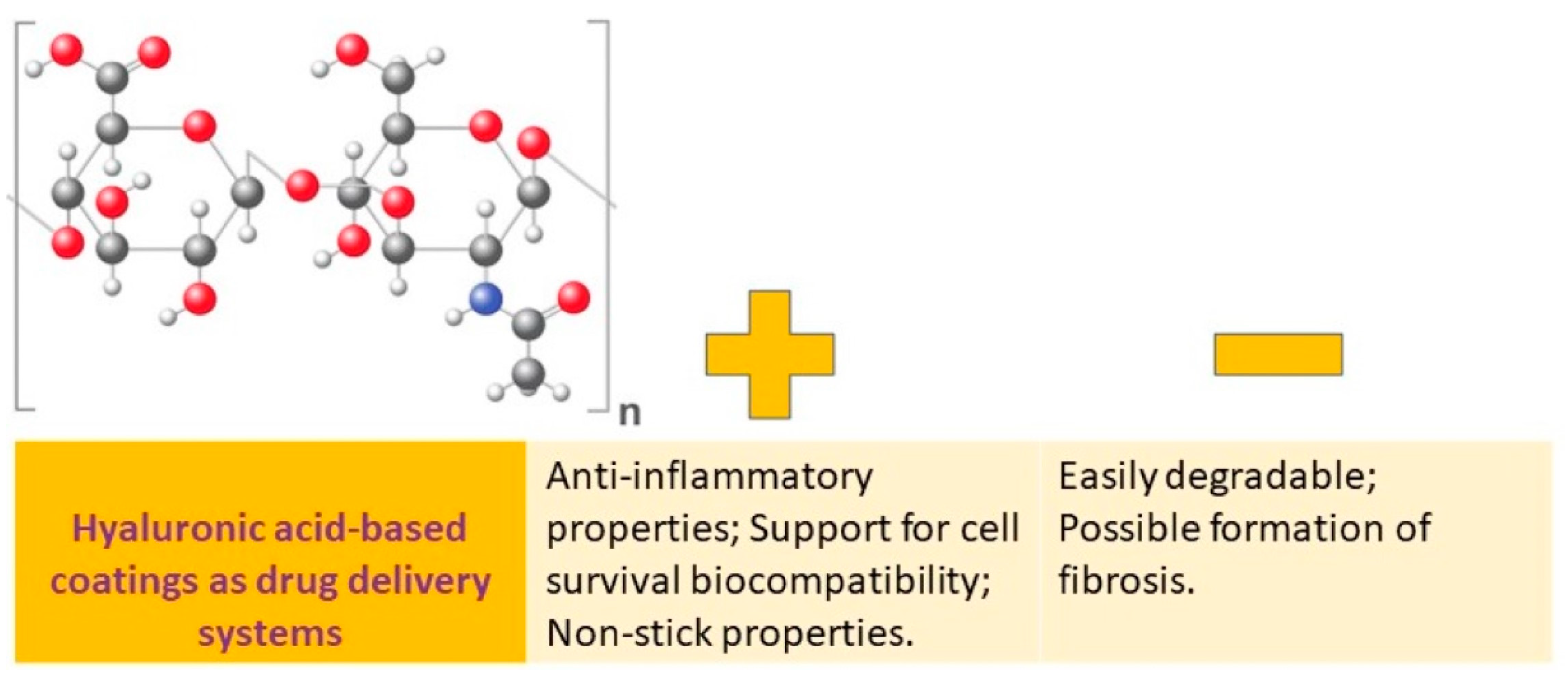
3.6. Hyaluronic Acid-Based Coatings as Drug Delivery Systems
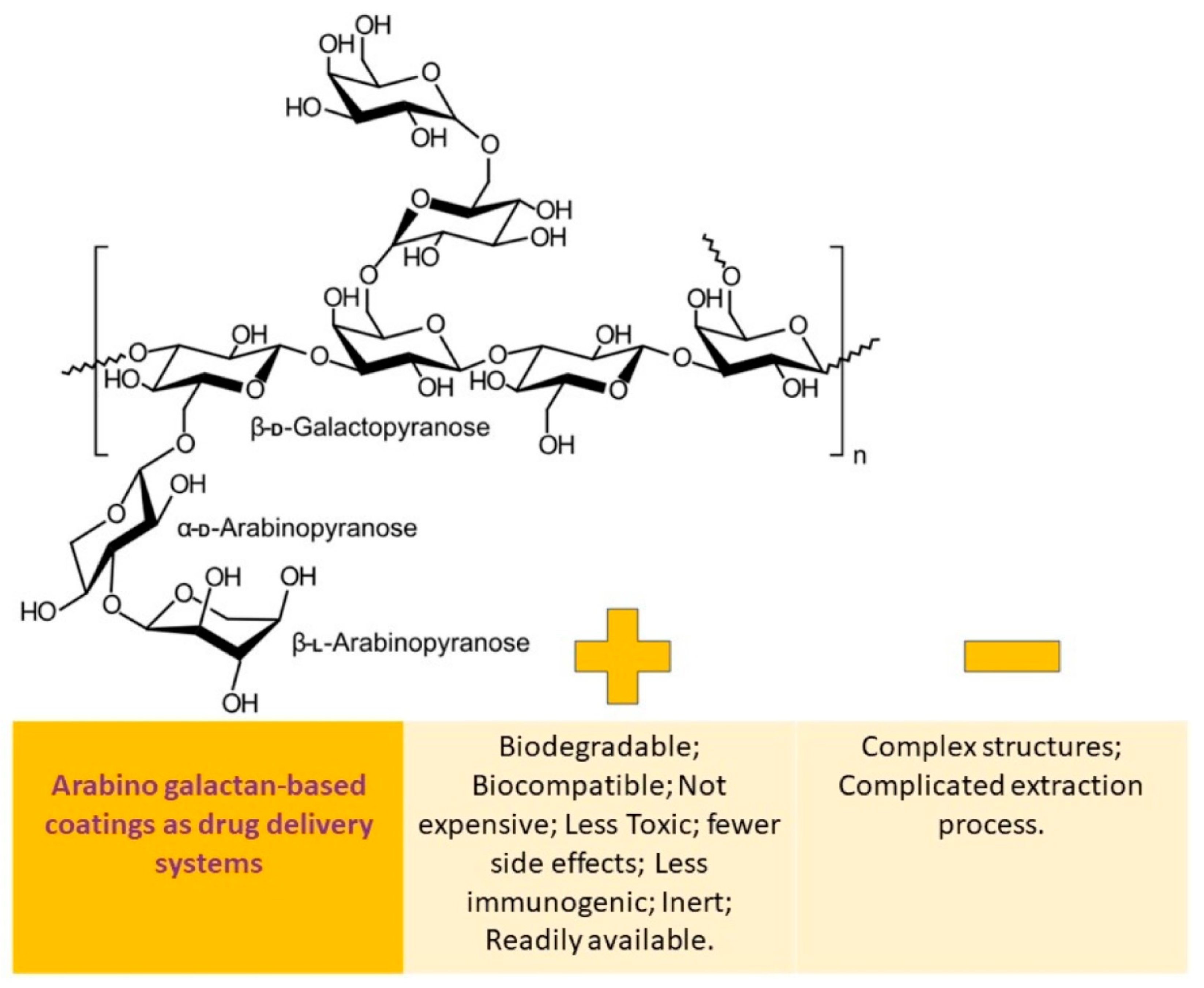
3.7. Arabinogalactan-Based Coatings as Drug Delivery Systems
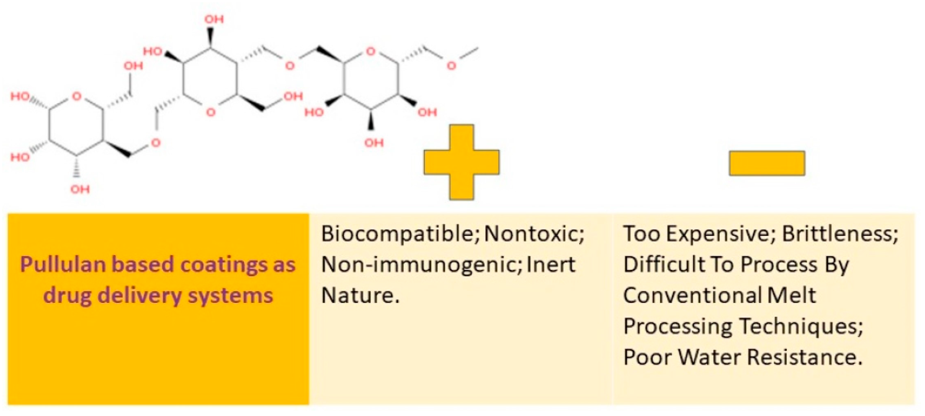
3.8. Pullulan-Based Coatings as Drug Delivery Systems
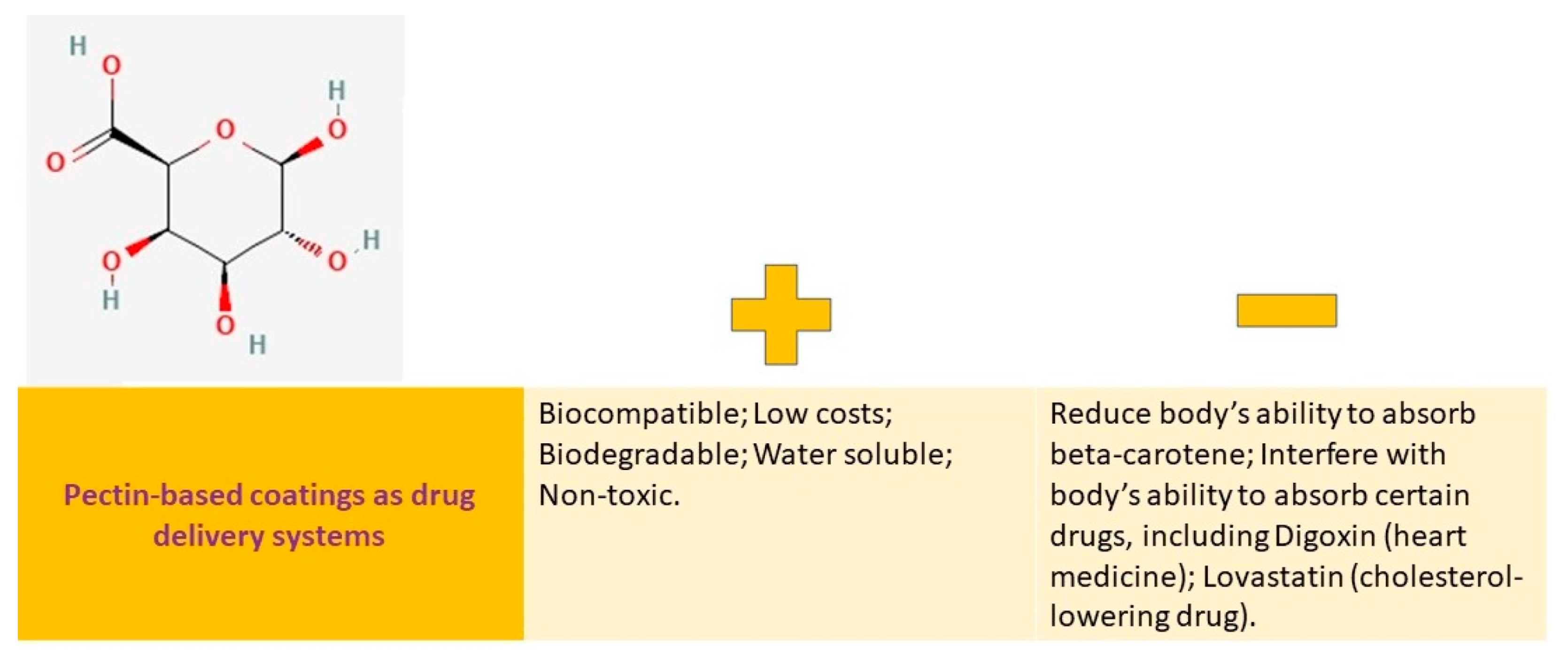
3.9. Pectin-Based Coatings as Drug Delivery Systems
4. Important Methods in Polysaccharide-Based Coatings Characterization
- (i)
- Sugar content identification. A developer–sulfuric acid method is often used to measure the sugar content in the sample. When sulfuric acid is used to break down monosaccharides, polysaccharides, and their derivatives, they are converted into aldehyde derivatives, which can then be combined with phenols or aromatic amines to generate colored compounds. By measuring the intensity of these colors, the amount of polysaccharides present can be estimated. These colorimetric methods are efficient, precise, and reliable, with the added benefit of stable coloration [135].
- (ii)
- Molecular weight evaluation. Measuring the molecular weight of polysaccharides is challenging, and there is no single absolute method. Instead, the statistical average method is typically used. In the past, methods such as osmotic pressure, terminal group analysis, viscosity measurement, and light scattering were commonly used, but they were complex and prone to errors. At present, the most commonly used methods are gel filtration and high-performance liquid chromatography, which require standard polysaccharides of known molecular weight as a reference. Mass spectrometry can be used for polysaccharides with a molecular weight of less than 50,000 [136].
- (iii)
- Component identification. There are three main methods for analyzing polysaccharide composition: chemical, physical (instrumental), and biological. Chemical analysis involves acid hydrolysis, neutralization, and filtration. Following this, paper chromatography (PC), thin layer chromatography (TLC), gas chromatography (GC), liquid chromatography (HPLC), or ion chromatography are used for analysis. Some widely used instrumental analyses include spectrophotometry, infrared spectroscopy, nuclear magnetic resonance, and mass spectrometry (MS) [137].
- (iv)
- Structure investigations. Compared to proteins, polysaccharides have more intricate macromolecular structures. Identifying these structures can be complicated due to the diversity of monosaccharides, linking methods, and the complexity of the branching chains. Currently, the primary focus of polysaccharide structure determination is analyzing the molecular weight range of polysaccharides, the type, proportion, and linking order of monosaccharides, and the configuration of glycosidic bonds. Common methods for structural analysis include periodate oxidation, Smith degradation, and methylation reaction. In recent years, advanced instruments such as ultraviolet have greatly improved analysis [138].
5. Challenges towards Commercialization of Polysaccharide-Based Drug Delivery Systems
- (i)
- Polysaccharides are often heterogeneous, meaning that they can have variations in their repeating units and structures. This complexity poses challenges in terms of characterization, standardization, and quality control. The variations in polysaccharide structures can affect their functional properties and, of course, the coating characteristic, making it necessary to develop reliable and efficient methods to analyze and characterize either pristine and polysaccharide-based coatings [163].
- (ii)
- Even the fact that polysaccharides are renewable resources (being typically extracted from natural sources such as plants, algae, and microorganisms) represents a big plus, the extraction process can be complex and may require optimization in order to ensure high yields and purity of polysaccharides. Additionally, purification steps may be necessary to remove impurities and contaminants [164]. Developing efficient and cost-effective extraction and purification methods is crucial for the commercial production of polysaccharides and polysaccharide-based coatings.
- (iii)
- Scaling up the production of polysaccharides from laboratory to industrial levels can be challenging. Factors such as yield, cost, and scalability need to be considered. The production processes should be optimized to ensure consistent quality, high yields, and affordable cost [163]. Strategies such as fermentation, enzymatic synthesis, and biotechnology approaches may be employed to enhance polysaccharide production [163].
- (iv)
- Polysaccharides may exhibit different properties depending on their structures and interactions with other components. Formulating polysaccharides into stable products such as gels, films, or coatings, requires accurate understanding their physicochemical properties and optimizing the formulation parameters [163]. Stability during storage, transportation, and use, is also a critical factor to consider for commercialization.
- (v)
- Polysaccharides intended for commercial use may need to comply with regulatory requirements and safety standards. These requirements may vary, depending on the intended application and region in which the polysaccharides will be used. Safety assessments, including toxicity and allergenicity studies, may be necessary to ensure the safety of polysaccharide-based products [163].
- (vi)
- The commercial success of polysaccharides depends on market acceptance and cost considerations. Polysaccharides should offer unique functional properties and advantages compared to existing alternatives. The cost of production, including raw materials, extraction, purification, and formulation, should be competitive in the market [165].
6. Conclusions and Future Perspectives
Author Contributions
Funding
Institutional Review Board Statement
Informed Consent Statement
Data Availability Statement
Conflicts of Interest
Abbreviations
| 1,4-Butanediol diglycidyl ether | (Ti-HABDDE) |
| 1-Phenyl-3-methyl-5-pyrazolone | (PMP) |
| 2,4-Dinitrochlorobenzene | (DNCB) |
| 2-Chloro-N,N-diethylethylamine | (DE-AE) |
| 3-Hydroxy-3-methylglutaryl coenzyme A | (HMG-CoA) |
| 45S5 Bioglass | (BG) |
| 5-Fluorouracil | (5-FU) |
| Acetylsalicylic acid (aspirin) | (ASA) |
| Adamantane-modified hyaluronic acid | (HAD) |
| Alpha-smooth muscle actin, commonly known as actin alpha 2, Acta2 | α-SMA |
| Aptamer | AS-42 |
| Arabinogalactan | (AG) |
| Asialoglycoprotein receptor | (ASGPR-1) |
| Atomic force microscopy | (AFM) |
| Atopic dermatitis | (AD) |
| Bacterial cellulose | (BC) |
| Beta-cyclodextrin-modified silk fibroin | (SCD) |
| Bone morphogenetic protein 2 | BMP2 |
| Cancer drug methotrexate | (MTX) |
| Carbon tetrachloride | (CCl4) |
| Carboxymethyl starch | (CMS) |
| Carboxymethyl pullulan | (CMP) |
| Carboxymethyl dextran | (CMD) |
| Cefazolin/chitosan | (P-Z@C) |
| Cefazolin/chitosan crosslinked by calcium phosphate | (P-Z@C/CP) |
| Chitosan/carboxymethyl pullulan | (CCMP) |
| Chitosan/carboxymethyl pullulan polyelectrolyte complex | (PEC) |
| Chitosan/carboxymethyl pullulan polyelectrolyte complex (PEC) loaded with 45S5 Bioglass | (CCMPBG) |
| Cisplatin–arabinogalactan–aptamer | (Cis-AG-Ap) |
| Collagen-1-alpha | COL1A1 |
| Corn starch acetates | (SA) |
| Crosslinked MC-loaded nanocells | (MC@(ODA-CMD)CL |
| DEAE-modified pullulan | (PULL-DEAE) |
| Defensive Antibacterial Coating | DAC® |
| Degree of methoxylation | (DM) |
| Dextran-coated cerium-doped hydroxyapatite (Ca10−xCex(PO4)6(OH)2) with x = 0.05 | (5CeHAp-D) |
| Dextran-coated cerium-doped hydroxyapatite (Ca10-−xCex(PO4)6(OH)2) with x = 0.1 | (10CeHAp-D) |
| Differential refractive index detector | (RID) |
| Divinyl sulfone | (Ti-HADVS) |
| Dosage forms | (FDs) |
| Drug Delivery System | (DDS) |
| Dynamic light scattering | (DLS) |
| D-α-tocopheryl polyethylene glycol succinate | (TPGS) |
| Encapsulation efficiency | (EE) |
| Endosomal cleavable peptide | (GFLG) |
| Energy-dispersive X-ray spectroscopy | (EDX) |
| Essential oil | (EO) |
| Fibrosis-mediating proteins | PDGFRB, TIMP-1, TLR-9 and TGFβ |
| Folate receptors | (FR) |
| Folic acid | (FA) |
| Fourier transform infrared spectroscopy | (FTIR) |
| Gas chromatography | (GC) |
| Gas chromatography–mass spectrometry | GC-MS |
| Gastrointestinal | (GI) |
| Gel permeation chromatography | GPC |
| Glow discharge optical emission spectroscopy | (GDOES) |
| Grafted copolymer pullulan–DEAE-g-poly(Z-1-lysine) | (PULL–DEAE-g-PZLL) |
| Heat shock proteins | (HSP) |
| High-performance liquid chromatography | (HPLC) |
| Highly methoxylated pectin | (HMP) |
| High-performance anion exchange chromatography combined with pulsed amperometric detection | (HPAEC-PAD) |
| High-performance capillary electrophoresis | (HPCE) |
| High-performance exclusion chromatography | (HPSEC) |
| High-performance gel permeation chromatography | (HPGPC) |
| Human liver cancer cell line | (HepG2) |
| Human serum albumin | (HSA) |
| Hyaluronic acid | (HA) |
| Hydroxypropyl methylcellulose | (HPMC) |
| Hypromellose Acetate Succinate | (HPMCAS) |
| Ibuprofen | (IBU) |
| Lactobionic acid | (LA) |
| Lactobionic acid–adipic acid dihydrazide conjugate | (LAD) |
| Lauroylated AG | (LAG) |
| Lauroylated PL | (LPL) |
| Layer-by-layer | (LbL) |
| Levofloxacin | (Levo) |
| Local drug delivery | (LDD) |
| Mammalian cells | CHO |
| Mass spectrometry | (MS) |
| Matrix-assisted pulsed laser evaporation | (MAPLE) |
| MC-loaded CMD-based nanocells coated on a MAO-Ti surface | (MC@(ODA-CMD)CL-Ti) |
| Metallographic microscopy | (MM) |
| Microarcoxidized titanium | (MAO-Ti) |
| Minimum inhibitory concentration | (MIC) |
| Minocycline | (MC) |
| Molecular weights | (MW) |
| Multiangle laser scattering detector | (MALLS) |
| Nanocomposites | (NCs) |
| Nanoparticles | (NPs) |
| Nanostructured lipid carriers | (NLCs) |
| N-carbobenzyloxy-1-lysine-N-carboxyanhydride | (Lys(Z)-NCA) |
| Nuclear magnetic resonance | (NMR) |
| Octadecylamine | (ODA) |
| Palmitoylated PL | (PPL) |
| Paper chromatography | (PC) |
| Platelet-derived growth factor receptor beta | PDGFRB |
| Platelet-derived growth factor receptor beta | (PDGFRB) |
| Poly(glycerol adipate) | (PGA) |
| Poly(methyl methacrylate) | (PMMA) |
| Polyamide 12 | (PA12) |
| Polycaprolactone | (PCL) |
| Polysaccharide arabinogalactan | (AG) |
| Pomegranate peel extract | (PPE) |
| Porous starch | (PS) |
| Pullulan | (PL) |
| Pullulan–polyethyleneimine | (Pul-PEI) |
| Pullulan–tris(2-aminoethyl)amine | (Pul-TAEA) |
| Pulsed direct current | (DC) |
| Quercetin | (Quer) |
| Salicylic acid | (SA) |
| Scanning electron microscopy | (SEM) |
| Silver nanoparticles | (AgNPs) |
| Staphylococcus aureus | (S. aureus) |
| The most biologically relevant receptor | (CD44) |
| The receptor for hyaluronan-mediated motility | (RHAMM) |
| Thin-layer chromatography | (TLC) |
| Tissue inhibitors of metalloproteinases | (TIMPs) |
| Transmission electron microscopy | (TEM) |
| Triblock copolymer | (Pluronic F127) |
| Palmitoylated AG | (PAG) |
References
- Li, Z.; Bratlie, K.M. The influence of polysaccharides-based material on macrophage phenotypes. Macromol. Biosci. 2021, 21, 2100031. [Google Scholar] [CrossRef]
- Popyrina, T.N.; Demina, T.S.; Akopova, T.A. Polysaccharide-based films: From packaging materials to functional food. J. Food Sci. Technol. 2022, 1–12. [Google Scholar] [CrossRef]
- Kumar, N. Polysaccharide-based component and their relevance in edible film/coating: A review. Nutr. Food Sci. 2019, 49, 793–823. [Google Scholar] [CrossRef]
- Cazón, P.; Velazquez, G.; Ramírez, J.A.; Vázquez, M. Polysaccharide-based films and coatings for food packaging: A review. Food Hydrocoll. 2017, 68, 136–148. [Google Scholar] [CrossRef]
- Web of Sience. Available online: http://apps.webofknowledge.com (accessed on 7 February 2023).
- Maver, T.; Mohan, T.; Gradišnik, L.; Finšgar, M.; Stana Kleinschek, K.; Maver, U. Polysaccharide thin solid films for analgesic drug delivery and growth of human skin cells. Front. Chem. 2019, 7, 217. [Google Scholar] [CrossRef] [PubMed]
- Barclay, T.G.; Day, C.M.; Petrovsky, N.; Garg, S. Review of polysaccharide particle-based functional drug delivery. Carbohydr. Polym. 2019, 221, 94–112. [Google Scholar] [CrossRef] [PubMed]
- Li, J.; Xiang, H.; Zhang, Q.; Miao, X. Polysaccharide-Based Transdermal Drug Delivery. Pharmaceuticals 2022, 15, 602. [Google Scholar] [CrossRef]
- Gopinath, V.; Saravanan, S.; Al-Maleki, A.; Ramesh, M.; Vadivelu, J. A review of natural polysaccharides for drug delivery applications: Special focus on cellulose, starch and glycogen. Biomed. Pharmacother. 2018, 107, 96–108. [Google Scholar] [CrossRef]
- Antosik, A.K.; Miądlicki, P.; Wilpiszewska, K.; Markowska-Szczupak, A.; Koren, Z.C.; Wróblewska, A. Polysaccharide films modified by compounds of natural origin and silver having potential medical applications. Cellulose 2021, 28, 7257–7271. [Google Scholar] [CrossRef]
- Hashizume, M.; Murata, Y.; Iijima, K.; Shibata, T. Drug loading and release behaviors of freestanding polysaccharide composite films. Polym. J. 2016, 48, 545–550. [Google Scholar] [CrossRef]
- Thang, N.H.; Chien, T.B.; Cuong, D.X. Polymer-Based Hydrogels Applied in Drug Delivery: An Overview. Gels 2023, 9, 523. [Google Scholar] [CrossRef]
- Zelikin, A.N. Drug releasing polymer thin films: New era of surface-mediated drug delivery. ACS Nano 2010, 4, 2494–2509. [Google Scholar] [CrossRef] [PubMed]
- Salawi, A. An insight into preparatory methods and characterization of orodispersible film—A review. Pharmaceuticals 2022, 15, 844. [Google Scholar] [CrossRef]
- Laubach, J.; Joseph, M.; Brenza, T.; Gadhamshetty, V.; Sani, R.K. Exopolysaccharide and biopolymer-derived films as tools for transdermal drug delivery. J. Control. Release 2021, 329, 971–987. [Google Scholar] [CrossRef] [PubMed]
- Park, S.; Han, U.; Choi, D.; Hong, J. Layer-by-layer assembled polymeric thin films as prospective drug delivery carriers: Design and applications. Biomater. Res. 2018, 22, 29. [Google Scholar] [CrossRef]
- Karki, S.; Kim, H.; Na, S.-J.; Shin, D.; Jo, K.; Lee, J. Thin films as an emerging platform for drug delivery. Asian J. Pharm. Sci. 2016, 11, 559–574. [Google Scholar] [CrossRef]
- Salawi, A. Pharmaceutical Coating and Its Different Approaches, a Review. Polymers 2022, 14, 3318. [Google Scholar] [CrossRef]
- Kurczewska, J. Recent reports on polysaccharide-based materials for drug delivery. Polymers 2022, 14, 4189. [Google Scholar] [CrossRef] [PubMed]
- Maiti, S.; Jana, S. Polysaccharide Carriers for Drug Delivery; Woodhead Publishing: Oxford, UK, 2019. [Google Scholar]
- Pushpamalar, J.; Veeramachineni, A.K.; Owh, C.; Loh, X.J. Biodegradable polysaccharides for controlled drug delivery. ChemPlusChem 2016, 81, 504–514. [Google Scholar] [CrossRef]
- Posocco, B.; Dreussi, E.; De Santa, J.; Toffoli, G.; Abrami, M.; Musiani, F.; Grassi, M.; Farra, R.; Tonon, F.; Grassi, G. Polysaccharides for the delivery of antitumor drugs. Materials 2015, 8, 2569–2615. [Google Scholar] [CrossRef]
- Stiff-Roberts, A.D.; Ge, W. Organic/hybrid thin films deposited by matrix-assisted pulsed laser evaporation (MAPLE). Appl. Phys. Rev. 2017, 4, 041303. [Google Scholar] [CrossRef]
- Takeuchi, I. Combinatorial pulsed laser deposition. In Pulsed Laser Deposition of Thin Films: Applications-Led Growth of Functional Materials; John Wiley & Sons, Inc.: Hoboken, NJ, USA, 2007; pp. 161–175. [Google Scholar]
- Li, X.; Wang, L.; Yu, X.; Feng, Y.; Wang, C.; Yang, K.; Su, D. Tantalum coating on porous Ti6Al4V scaffold using chemical vapor deposition and preliminary biological evaluation. Mater. Sci. Eng. C 2013, 33, 2987–2994. [Google Scholar] [CrossRef] [PubMed]
- Abegunde, O.O.; Akinlabi, E.T.; Oladijo, O.P.; Akinlabi, S.; Ude, A.U. Overview of thin film deposition techniques. AIMS Mater. Sci. 2019, 6, 174–199. [Google Scholar] [CrossRef]
- Sakka, S. Sol-gel technology as reflected in journal of sol-gel science and technology. J. Sol-Gel Sci. Technol. 2003, 26, 29–33. [Google Scholar] [CrossRef]
- Kausar, A. Survey on Langmuir–Blodgett films of polymer and polymeric composite. Polym.-Plast. Technol. Eng. 2017, 56, 932–945. [Google Scholar] [CrossRef]
- Parsons, G.N.; Atanasov, S.E.; Dandley, E.C.; Devine, C.K.; Gong, B.; Jur, J.S.; Lee, K.; Oldham, C.J.; Peng, Q.; Spagnola, J.C. Mechanisms and reactions during atomic layer deposition on polymers. Coord. Chem. Rev. 2013, 257, 3323–3331. [Google Scholar] [CrossRef]
- Xu, J.; Hsu, S.-h. Enhancement of Cell Behavior by the Polysaccharide Extract of Arthrospira and Potential Biomedical Applications. Molecules 2023, 28, 732. [Google Scholar] [CrossRef]
- Kocira, A.; Kozłowicz, K.; Panasiewicz, K.; Staniak, M.; Szpunar-Krok, E.; Hortyńska, P. Polysaccharides as edible films and coatings: Characteristics and influence on fruit and vegetable quality—A review. Agronomy 2021, 11, 813. [Google Scholar] [CrossRef]
- Vickers, N.J. Animal communication: When i’m calling you, will you answer too? Curr. Biol. 2017, 27, R713–R715. [Google Scholar] [CrossRef]
- Orbach, H.; Matok, I.; Gorodischer, R.; Sheiner, E.; Daniel, S.; Wiznitzer, A.; Koren, G.; Levy, A. Hypertension and antihypertensive drugs in pregnancy and perinatal outcomes. Am. J. Obstet. Gynecol. 2013, 208, e301.e1–e301.e6. [Google Scholar] [CrossRef] [PubMed]
- Li, M.; Chen, H.; Peng, D.; Lu, X.; Kong, J.; Luo, S.; Li, S.; Tan, C.; Wang, Y. FU-coating pH-sensitive liposomes for improving the release of gemcitabine by endosome escape in pancreatic cancer cells. J. Drug Deliv. Sci. Technol. 2023, 80, 104135. [Google Scholar] [CrossRef]
- Wang, K.; Yu, Y.; Li, W.; Li, D.; Li, H. Preparation of fully bio-based multilayers composed of heparin-like carboxymethylcellulose sodium and chitosan to functionalize poly (l-lactic acid) film for cardiovascular implant applications. Int. J. Biol. Macromol. 2023, 231, 123285. [Google Scholar] [CrossRef]
- Raeisi, F.; Raeisi, E. Mini review of polysaccharide nanoparticles and drug delivery process. Adv. Appl. NanoBio-Technol. 2020, 1, 33–44. [Google Scholar]
- Sun, Y.; Jing, X.; Ma, X.; Feng, Y.; Hu, H. Versatile types of polysaccharide-based drug delivery systems: From strategic design to cancer therapy. Int. J. Mol. Sci. 2020, 21, 9159. [Google Scholar] [CrossRef]
- Awad, A.; Madla, C.M.; McCoubrey, L.E.; Ferraro, F.; Gavins, F.K.; Buanz, A.; Gaisford, S.; Orlu, M.; Siepmann, F.; Siepmann, J. Clinical translation of advanced colonic drug delivery technologies. Adv. Drug Deliv. Rev. 2022, 181, 114076. [Google Scholar] [CrossRef] [PubMed]
- Wagh, O.V.; Shendge, R.; Kardile, S.S. Formulation and evaluation of effective colon targeted drug delivery of naproxen: A pellet based tablet. Int. J. Early Child. 2022, 14, 2688–2699. [Google Scholar] [CrossRef]
- Peer, D.; Karp, J.M.; Hong, S.; Farokhzad, O.C.; Margalit, R.; Langer, R. Nanocarriers as an emerging platform for cancer therapy. Nat. Nanotechnol. 2007, 2, 751–760. [Google Scholar] [CrossRef]
- Jain, A.; Gupta, Y.; Jain, S.K. Perspectives of biodegradable natural polysaccharides for site-specific drug delivery to the colon. J. Pharm. Pharm. Sci. 2007, 10, 86–128. [Google Scholar] [PubMed]
- Liu, Z.; Jiao, Y.; Wang, Y.; Zhou, C.; Zhang, Z. Polysaccharides-based nanoparticles as drug delivery systems. Adv. Drug Deliv. Rev. 2008, 60, 1650–1662. [Google Scholar] [CrossRef]
- Saravanakumar, G.; Jo, D.-G.; H Park, J. Polysaccharide-based nanoparticles: A versatile platform for drug delivery and biomedical imaging. Curr. Med. Chem. 2012, 19, 3212–3229. [Google Scholar] [CrossRef]
- Elbialy, N.S.; Mohamed, N. Fabrication of the quaternary nanocomplex curcumin-casein-alginate-chitosan as a potential oral delivery system for cancer nutraceutical therapy. J. Drug Deliv. Sci. Technol. 2022, 70, 103226. [Google Scholar] [CrossRef]
- Morris, G.A.; Kök, S.M.; Harding, S.E.; Adams, G.G. Polysaccharide drug delivery systems based on pectin and chitosan. Biotechnol. Genet. Eng. Rev. 2010, 27, 257–284. [Google Scholar] [CrossRef] [PubMed]
- Alonso-Sande, M.; Teijeiro-Osorio, D.; Remuñán-López, C.; Alonso, M. Glucomannan, a promising polysaccharide for biopharmaceutical purposes. Eur. J. Pharm. Biopharm. 2009, 72, 453–462. [Google Scholar] [CrossRef]
- Ju, D.-B.; Lee, J.-C.; Hwang, S.-K.; Cho, C.-S.; Kim, H.-J. Progress of Polysaccharide-Contained Polyurethanes for Biomedical Applications. Tissue Eng. Regen. Med. 2022, 19, 891–912. [Google Scholar] [CrossRef]
- Zhang, M.; Ma, H.; Wang, X.; Yu, B.; Cong, H.; Shen, Y. Polysaccharide-based nanocarriers for efficient transvascular drug delivery. J. Control. Release 2023, 354, 167–187. [Google Scholar] [CrossRef]
- Chen, L.; Li, X.; Li, L.; Guo, S. Acetylated starch-based biodegradable materials with potential biomedical applications as drug delivery systems. Curr. Appl. Phys. 2007, 7, e90–e93. [Google Scholar] [CrossRef]
- Santander-Ortega, M.; Stauner, T.; Loretz, B.; Ortega-Vinuesa, J.L.; Bastos-González, D.; Wenz, G.; Schaefer, U.F.; Lehr, C.-M. Nanoparticles made from novel starch derivatives for transdermal drug delivery. J. Control. Release 2010, 141, 85–92. [Google Scholar] [CrossRef] [PubMed]
- Soares, I.r.; Faria, J.; Marques, A.; Ribeiro, I.A.; Baleizão, C.; Bettencourt, A.; Ferreira, I.M.; Baptista, A.C. Drug Delivery from PCL/Chitosan Multilayer Coatings for Metallic Implants. ACS Omega 2022, 7, 23096–23106. [Google Scholar] [CrossRef]
- Feng, C.; Li, J.; Kong, M.; Liu, Y.; Cheng, X.J.; Li, Y.; Park, H.J.; Chen, X.G. Surface charge effect on mucoadhesion of chitosan based nanogels for local anti-colorectal cancer drug delivery. Colloids Surf. B Biointerfaces 2015, 128, 439–447. [Google Scholar] [CrossRef]
- Schuetz, C.A.; Juillerat-Jeanneret, L.; Kaeuper, P.; Wandrey, C. Cell response to the exposure to chitosan-TPP//alginate nanogels. Biomacromolecules 2011, 12, 4153–4161. [Google Scholar] [CrossRef] [PubMed]
- Bodnar, M.; Hartmann, J.F.; Borbely, J. Preparation and characterization of chitosan-based nanoparticles. Biomacromolecules 2005, 6, 2521–2527. [Google Scholar] [CrossRef]
- Huh, M.S.; Lee, E.J.; Koo, H.; Yhee, J.Y.; Oh, K.S.; Son, S.; Lee, S.; Kim, S.H.; Kwon, I.C.; Kim, K. Polysaccharide-based nanoparticles for gene delivery. In Polymeric Gene Delivery Systems; Springer: Cham, Switzerland, 2018; pp. 65–83. [Google Scholar]
- Ye, W.; Zhou, M.; Zhang, L.; Yu, J.; Fan, J.; Wei, H. Carboxymethyl Dextran-Based Nanomicelle Coatings on Microarc Oxidized Titanium Surface for Percutaneous Implants: Drug Release, Antibacterial Properties, and Biocompatibility. BioMed Res. Int. 2022, 2022, 9225647. [Google Scholar] [CrossRef] [PubMed]
- Xu, W.; Ding, J.; Xiao, C.; Li, L.; Zhuang, X.; Chen, X. Versatile preparation of intracellular-acidity-sensitive oxime-linked polysaccharide-doxorubicin conjugate for malignancy therapeutic. Biomaterials 2015, 54, 72–86. [Google Scholar] [CrossRef]
- Drago, L.; Boot, W.; Dimas, K.; Malizos, K.; Hänsch, G.M.; Stuyck, J.; Gawlitta, D.; Romanò, C.L. Does implant coating with antibacterial-loaded hydrogel reduce bacterial colonization and biofilm formation in vitro? Clin. Orthop. Relat. Res.® 2014, 472, 3311–3323. [Google Scholar] [CrossRef]
- Tripodo, G.; Trapani, A.; Torre, M.L.; Giammona, G.; Trapani, G.; Mandracchia, D. Hyaluronic acid and its derivatives in drug delivery and imaging: Recent advances and challenges. Eur. J. Pharm. Biopharm. 2015, 97, 400–416. [Google Scholar] [CrossRef]
- Deng, X.; Cao, M.; Zhang, J.; Hu, K.; Yin, Z.; Zhou, Z.; Xiao, X.; Yang, Y.; Sheng, W.; Wu, Y. Hyaluronic acid-chitosan nanoparticles for co-delivery of MiR-34a and doxorubicin in therapy against triple negative breast cancer. Biomaterials 2014, 35, 4333–4344. [Google Scholar] [CrossRef]
- Tan, H.; Rubin, J.P.; Marra, K.G. Direct Synthesis of Biodegradable Polysaccharide Derivative Hydrogels through Aqueous Diels-Alder Chemistry. Macromol. Rapid Commun. 2011, 32, 905–911. [Google Scholar] [CrossRef]
- Zamay, T.N.; Starkov, A.K.; Kolovskaya, O.S.; Zamay, G.S.; Veprintsev, D.V.; Luzan, N.; Nikolaeva, E.D.; Lukyanenko, K.A.; Artyushenko, P.V.; Shchugoreva, I.A. Nucleic Acid Aptamers Increase the Anticancer Efficiency and Reduce the Toxicity of Cisplatin-Arabinogalactan Conjugates In Vivo. Nucleic Acid Ther. 2022, 32, 497–506. [Google Scholar] [CrossRef] [PubMed]
- Zhang, Z.; Yang, L.; Hou, J.; Xia, X.; Wang, J.; Ning, Q.; Jiang, S. Promising positive liver targeting delivery system based on arabinogalactan-anchored polymeric micelles of norcantharidin. Artif. Cells Nanomed. Biotechnol. 2018, 46, 630–640. [Google Scholar] [CrossRef]
- Gehrcke, M.; Martins, C.C.; de Bastos Brum, T.; da Rosa, L.S.; Luchese, C.; Wilhelm, E.A.; Soares, F.Z.M.; Cruz, L. Novel Pullulan/Gellan Gum Bilayer Film as a Vehicle for Silibinin-Loaded Nanocapsules in the Topical Treatment of Atopic Dermatitis. Pharmaceutics 2022, 14, 2352. [Google Scholar] [CrossRef] [PubMed]
- Singh, R.S.; Kaur, N.; Rana, V.; Kennedy, J.F. Pullulan: A novel molecule for biomedical applications. Carbohydr. Polym. 2017, 171, 102–121. [Google Scholar] [CrossRef]
- Kang, J.-H.; Tachibana, Y.; Kamata, W.; Mahara, A.; Harada-Shiba, M.; Yamaoka, T. Liver-targeted siRNA delivery by polyethylenimine (PEI)-pullulan carrier. Bioorganic Med. Chem. 2010, 18, 3946–3950. [Google Scholar] [CrossRef] [PubMed]
- Swierczewska, M.; Han, H.S.; Kim, K.; Park, J.; Lee, S. Polysaccharide-based nanoparticles for theranostic nanomedicine. Adv. Drug Deliv. Rev. 2016, 99, 70–84. [Google Scholar] [CrossRef]
- Potaś, J.; Szymańska, E.; Wróblewska, M.; Kurowska, I.; Maciejczyk, M.; Basa, A.; Wolska, E.; Wilczewska, A.Z.; Winnicka, K. Multilayer films based on chitosan/pectin polyelectrolyte complexes as novel platforms for buccal administration of clotrimazole. Pharmaceutics 2021, 13, 1588. [Google Scholar] [CrossRef] [PubMed]
- Neut, C.; Wils, D.; Siepmann, F.; Deremaux, L.; Desreumaux, P.; Siepmann, J. Characterization of ethylcellulose: Starch-based film coatings for colon targeting. Drug Dev. Ind. Pharm. 2009, 35, 1190–1200. [Google Scholar]
- Li, Q.; Fan, X.; Pan, X.; Yu, Y.; Jian, L.; Zhang, Y.; Yin, T.; He, H.; Tang, X.; Jin, J. S/O/W microparticles prepared with hydroxyethyl starch-based emulsifier showed reduced macrophage affinity. Colloids Surf. B Biointerfaces 2022, 220, 112917. [Google Scholar] [CrossRef]
- Hataminia, F.; Majidi, R.F.; Shabankareh, A.N.T.; Ghanbari, H. Green synthesis of oxidized starch with a novel catalyst based on Fe3O4 nanoparticles and H2O2 reagent to form thermoplastic as a stable gel coating on the cardiovascular stents. Int. J. Biol. Macromol. 2022, 219, 290–303. [Google Scholar] [CrossRef]
- Micale, N.; Citarella, A.; Molonia, M.S.; Speciale, A.; Cimino, F.; Saija, A.; Cristani, M. Hydrogels for the delivery of plant-derived (poly) phenols. Molecules 2020, 25, 3254. [Google Scholar] [CrossRef]
- Heller, J.; Pangburn, S.; Roskos, K. Development of enzymatically degradable protective coatings for use in triggered drug delivery systems: Derivatized starch hydrogels. Biomaterials 1990, 11, 345–350. [Google Scholar] [CrossRef]
- Hou, X.; Wang, H.; Shi, Y.; Yue, Z. Recent advances of antibacterial starch-based materials. Carbohydr. Polym. 2022, 302, 120392. [Google Scholar] [CrossRef]
- Yasar, H.; Ho, D.-K.; De Rossi, C.; Herrmann, J.; Gordon, S.; Loretz, B.; Lehr, C.-M. Starch-chitosan polyplexes: A versatile carrier system for anti-infectives and gene delivery. Polymers 2018, 10, 252. [Google Scholar] [CrossRef]
- Lemos, P.V.F.; Marcelino, H.R.; Cardoso, L.G.; de Souza, C.O.; Druzian, J.I. Starch chemical modifications applied to drug delivery systems: From fundamentals to FDA-approved raw materials. Int. J. Biol. Macromol. 2021, 184, 218–234. [Google Scholar] [CrossRef]
- Pires, P.C.; Mascarenhas-Melo, F.; Pedrosa, K.; Lopes, D.; Lopes, J.; Macário-Soares, A.; Peixoto, D.; Giram, P.S.; Veiga, F.; Paiva-Santos, A.C. Polymer-based biomaterials for pharmaceutical and biomedical applications: A focus on topical drug administration. Eur. Polym. J. 2023, 187, 111868. [Google Scholar] [CrossRef]
- Vilivalam, V.D.; Illum, L.; Iqbal, K. Starch capsules: An alternative system for oral drug delivery. Pharm. Sci. Technol. Today 2000, 3, 64–69. [Google Scholar] [CrossRef] [PubMed]
- Vestri, A.; Pearce, A.K.; Cavanagh, R.; Styliari, I.D.; Sanders, C.; Couturaud, B.; Schenone, S.; Taresco, V.; Jakobsen, R.R.; Howdle, S.M. Starch/poly (glycerol-adipate) nanocomposites: A novel oral drug delivery device. Coatings 2020, 10, 125. [Google Scholar] [CrossRef]
- Moghadam, M.; Dorraji, M.S.S.; Dodangeh, F.; Ashjari, H.R.; Mousavi, S.N.; Rasoulifard, M.H. Design of a new light curable starch-based hydrogel drug delivery system to improve the release rate of quercetin as a poorly water-soluble drug. Eur. J. Pharm. Sci. 2022, 174, 106191. [Google Scholar] [CrossRef] [PubMed]
- Zhao, B.; Du, J.; Zhang, Y.; Gu, Z.; Li, Z.; Cheng, L.; Li, C.; Hong, Y. Polysaccharide-coated porous starch-based oral carrier for paclitaxel: Adsorption and sustained release in colon. Carbohydr. Polym. 2022, 291, 119571. [Google Scholar] [CrossRef] [PubMed]
- Costa, N.N.; de Faria Lopes, L.; Ferreira, D.F.; de Prado, E.M.L.; Severi, J.A.; Resende, J.A.; de Paula Careta, F.; Ferreira, M.C.P.; Carreira, L.G.; de Souza, S.O.L. Polymeric films containing pomegranate peel extract based on PVA/starch/PAA blends for use as wound dressing: In vitro analysis and physicochemical evaluation. Mater. Sci. Eng. C 2020, 109, 110643. [Google Scholar] [CrossRef]
- Oliyaei, N.; Moosavi-Nasab, M.; Tamaddon, A.M.; Tanideh, N. Antidiabetic effect of fucoxanthin extracted from Sargassum angustifolium on streptozotocin-nicotinamide-induced type 2 diabetic mice. Food Sci. Nutr. 2021, 9, 3521–3529. [Google Scholar] [CrossRef]
- Khubiev, O.M.; Egorov, A.R.; Kirichuk, A.A.; Khrustalev, V.N.; Tskhovrebov, A.G.; Kritchenkov, A.S. Chitosan-Based Antibacterial Films for Biomedical and Food Applications. Int. J. Mol. Sci. 2023, 24, 10738. [Google Scholar] [CrossRef]
- Visan, A.I.; Ristoscu, C.; Popescu-Pelin, G.; Sopronyi, M.; Matei, C.E.; Socol, G.; Chifiriuc, M.C.; Bleotu, C.; Grossin, D.; Brouillet, F. Composite Drug Delivery System Based on Amorphous Calcium Phosphate–Chitosan: An Efficient Antimicrobial Platform for Extended Release of Tetracycline. Pharmaceutics 2021, 13, 1659. [Google Scholar] [CrossRef]
- Mady, M.M.; Darwish, M.M. Effect of chitosan coating on the characteristics of DPPC liposomes. J. Adv. Res. 2010, 1, 187–191. [Google Scholar] [CrossRef]
- Sadeghi, M.; Tajzadeh, P.; Zarei-Ghanavati, S.; Arefnezhad, M. Chitosan-coated contact lens-based ophthalmic drug delivery system to manage Acanthamoeba keratitis: A preliminary hypothesis. Med. Hypothesis Discov. Innov. Optom. 2021, 2, 114–118. [Google Scholar] [CrossRef]
- Taherian, A.; Esfandiari, N.; Rouhani, S. Breast cancer drug delivery by novel drug-loaded chitosan-coated magnetic nanoparticles. Cancer Nanotechnol. 2021, 12, 15. [Google Scholar] [CrossRef]
- Song, T.-Y.; Wang, Y.-H.; Chien, H.-W.; Ma, C.-H.; Lee, C.-L.; Ou, S.-F. Synthesis of cross-linked chitosan by calcium phosphate as long-term drug delivery coating with cytocompatibility. Prog. Org. Coat. 2022, 173, 107162. [Google Scholar] [CrossRef]
- Elzoheiry, A.; Ayad, E.; Omar, N.; Elbakry, K.; Hyder, A. Anti-liver fibrosis activity of curcumin/chitosan-coated green silver nanoparticles. Sci. Rep. 2022, 12, 18403. [Google Scholar] [CrossRef]
- Araújo, J.M.S.; de Siqueira, A.C.P.; Blank, A.F.; Narain, N.; de Aquino Santana, L.C.L. A cassava starch–chitosan edible coating enriched with Lippia sidoides Cham. essential oil and pomegranate peel extract for preservation of Italian tomatoes (Lycopersicon esculentum Mill.) stored at room temperature. Food Bioprocess Technol. 2018, 11, 1750–1760. [Google Scholar] [CrossRef]
- Thatiparti, T.R.; Shoffstall, A.J.; Von Recum, H.A. Cyclodextrin-based device coatings for affinity-based release of antibiotics. Biomaterials 2010, 31, 2335–2347. [Google Scholar] [CrossRef] [PubMed]
- Croitoru, A.-M.; Karaçelebi, Y.; Saatcioglu, E.; Altan, E.; Ulag, S.; Aydoğan, H.K.; Sahin, A.; Motelica, L.; Oprea, O.; Tihauan, B.-M. Electrically triggered drug delivery from novel electrospun poly (lactic acid)/graphene oxide/quercetin fibrous scaffolds for wound dressing applications. Pharmaceutics 2021, 13, 957. [Google Scholar] [CrossRef]
- Viet Nguyen, K.; Laidmäe, I.; Kogermann, K.; Lust, A.; Meos, A.; Viet Ho, D.; Raal, A.; Heinämäki, J.; Thi Nguyen, H. Preformulation study of electrospun haemanthamine-loaded amphiphilic nanofibers intended for a solid template for self-assembled liposomes. Pharmaceutics 2019, 11, 499. [Google Scholar] [CrossRef] [PubMed]
- Predoi, D.; Ciobanu, C.S.; Radu, M.; Costache, M.; Dinischiotu, A.; Popescu, C.; Axente, E.; Mihailescu, I.; Gyorgy, E. Hybrid dextran-iron oxide thin films deposited by laser techniques for biomedical applications. Mater. Sci. Eng. C 2012, 32, 296–302. [Google Scholar] [CrossRef]
- Ciobanu, C.S.; Nica, I.C.; Dinischiotu, A.; Iconaru, S.L.; Chapon, P.; Bita, B.; Trusca, R.; Groza, A.; Predoi, D. Novel Dextran Coated Cerium Doped Hydroxyapatite Thin Films. Polymers 2022, 14, 1826. [Google Scholar] [CrossRef] [PubMed]
- Gao, S.; Torrente-Rodríguez, R.M.; Pedrero, M.; Pingarrón, J.M.; Campuzano, S.; Rocha-Martin, J.; Guisán, J.M. Dextran-coated nanoparticles as immunosensing platforms: Consideration of polyaldehyde density, nanoparticle size and functionality. Talanta 2022, 247, 123549. [Google Scholar] [CrossRef] [PubMed]
- Díaz-Montes, E.; Yáñez-Fernández, J.; Castro-Muñoz, R. Dextran/chitosan blend film fabrication for bio-packaging of mushrooms (Agaricus bisporus). J. Food Process. Preserv. 2021, 45, e15489. [Google Scholar] [CrossRef]
- Brunsen, A.; Ritz, U.; Mateescu, A.; Höfer, I.; Frank, P.; Menges, B.; Hofmann, A.; Rommens, P.; Knoll, W.; Jonas, U. Photocrosslinkable dextran hydrogel films as substrates for osteoblast and endothelial cell growth. J. Mater. Chem. 2012, 22, 19590–19604. [Google Scholar] [CrossRef]
- Buckley, C.; Murphy, E.J.; Montgomery, T.R.; Major, I. Hyaluronic acid: A review of the drug delivery capabilities of this naturally occurring polysaccharide. Polymers 2022, 14, 3442. [Google Scholar] [CrossRef]
- Zhao, B.; Li, Y.-P.; Wang, Q.; Ren, Y.; Zheng, Z.-L.; Bai, M.-H.; Lv, J.-C.; Li, K.; Xu, J.-Z.; Li, Z.-M. Ultra-slippery, nonirritating, and anti-inflammatory hyaluronic acid-based coating to mitigate intubation injury. Chem. Eng. J. 2022, 427, 130911. [Google Scholar] [CrossRef]
- Del Olmo, J.A.; Alonso, J.M.; Sáez-Martínez, V.; Benito-Cid, S.; Pérez-González, R.; Vilas-Vilela, J.L.; Pérez-Álvarez, L. Hyaluronic acid-based hydrogel coatings on Ti6Al4V implantable biomaterial with multifunctional antibacterial activity. Carbohydr. Polym. 2023, 301, 120366. [Google Scholar] [CrossRef]
- Xuan, H.; Tang, X.; Zhu, Y.; Ling, J.; Yang, Y. Freestanding hyaluronic acid/silk-based self-healing coating toward tissue repair with antibacterial surface. ACS Appl. Bio Mater. 2020, 3, 1628–1635. [Google Scholar] [CrossRef]
- Gaetano, G.; Giuseppe, P.; Salvatore, P.F.; Susanna, M.; Sara, S.; Luca, R.C. Chapter Hyaluronic-Based Antibacterial Hydrogel Coating for Implantable Biomaterials in Orthopedics and Trauma: From Basic Research to Clinical Applications. 2018. Available online: https://library.oapen.org/handle/20.500.12657/49243 (accessed on 19 May 2023).
- Saha, I.; Rai, V.K. Hyaluronic acid based microneedle array: Recent applications in drug delivery and cosmetology. Carbohydr. Polym. 2021, 267, 118168. [Google Scholar] [CrossRef]
- Katsumi, H.; Liu, S.; Tanaka, Y.; Hitomi, K.; Hayashi, R.; Hirai, Y.; Kusamori, K.; Quan, Y.-s.; Kamiyama, F.; Sakane, T. Development of a novel self-dissolving microneedle array of alendronate, a nitrogen-containing bisphosphonate: Evaluation of transdermal absorption, safety, and pharmacological effects after application in rats. J. Pharm. Sci. 2012, 101, 3230–3238. [Google Scholar] [CrossRef] [PubMed]
- Liu, S.; Jin, M.-n.; Quan, Y.-s.; Kamiyama, F.; Katsumi, H.; Sakane, T.; Yamamoto, A. The development and characteristics of novel microneedle arrays fabricated from hyaluronic acid, and their application in the transdermal delivery of insulin. J. Control. Release 2012, 161, 933–941. [Google Scholar] [CrossRef]
- Petersen, S.; Kaule, S.; Teske, M.; Minrath, I.; Schmitz, K.-P.; Sternberg, K. Development and in vitro characterization of hyaluronic acid-based coatings for implant-associated local drug delivery systems. J. Chem. 2013, 2013, 587875. [Google Scholar] [CrossRef]
- Avramoff, A.; Khan, W.; Mizrahi, B.; Domb, A.J. Preparation and characterization of a novel once-daily formulation of diltiazem using arabinogalactan as a channeling agent. J. Appl. Polym. Sci. 2012, 126, E197–E203. [Google Scholar] [CrossRef]
- Assaraf, Y.G. The role of multidrug resistance efflux transporters in antifolate resistance and folate homeostasis. Drug Resist. Updates 2006, 9, 227–246. [Google Scholar] [CrossRef] [PubMed]
- Pinhassi, R.I.; Assaraf, Y.G.; Farber, S.; Stark, M.; Ickowicz, D.; Drori, S.; Domb, A.J.; Livney, Y.D. Arabinogalactan− folic acid− drug conjugate for targeted delivery and target-activated release of anticancer drugs to folate receptor-overexpressing cells. Biomacromolecules 2010, 11, 294–303. [Google Scholar] [CrossRef]
- Thakare, K.N. Solid Dosage form Containing Arabinogalactan. Patent No. US10994013, 4 May 2021. [Google Scholar]
- Warrier, D.U.; Dhanabalan, A.K.; Krishnasamy, G.; Kolge, H.; Ghormade, V.; Gupta, C.R.; Ambre, P.K.; Shinde, U.A. Novel derivatives of arabinogalactan, pullulan & lactobionic acid for targeting asialoglycoprotein receptor: Biomolecular interaction, synthesis & evaluation. Int. J. Biol. Macromol. 2022, 207, 683–699. [Google Scholar]
- Chistyachenko, Y.S.; Dushkin, A.V.; Polyakov, N.E.; Khvostov, M.V.; Tolstikova, T.G.; Tolstikov, G.A.; Lyakhov, N.Z. Polysaccharide arabinogalactan from larch Larix sibirica as carrier for molecules of salicylic and acetylsalicylic acid: Preparation, physicochemical and pharmacological study. Drug Deliv. 2015, 22, 400–407. [Google Scholar] [CrossRef]
- Khvostov, M.V.; Borisov, S.A.; Tolstikova, T.G.; Dushkin, A.V.; Tsyrenova, B.D.; Chistyachenko, Y.S.; Polyakov, N.E.; Dultseva, G.G.; Onischuk, A.A.; An’kov, S.V. Supramolecular complex of ibuprofen with larch polysaccharide arabinogalactan: Studies on bioavailability and pharmacokinetics. Eur. J. Drug Metab. Pharmacokinet. 2017, 42, 431–440. [Google Scholar] [CrossRef]
- Carvalho, L.T.; Teixeira, A.J.R.; Moraes, R.M.; Barbosa, R.F.; Queiroz, R.C.; Tada, D.B.; Mulinari, D.R.; Rosa, D.S.; Ré, M.I.; Medeiros, S.F. Preparation and characterization of cationic pullulan-based polymers with hydrophilic or amphiphilic characteristics for drug delivery. React. Funct. Polym. 2022, 181, 105441. [Google Scholar] [CrossRef]
- Jelinek, M.; Cristescu, R.; Axente, E.; Kocourek, T.; Dybal, J.; Remsa, J.; Plestil, J.; Mihaiescu, D.; Albulescu, M.; Buruiana, T. Matrix assisted pulsed laser evaporation of cinnamate-pullulan and tosylate-pullulan polysaccharide derivative thin films for pharmaceutical applications. Appl. Surf. Sci. 2007, 253, 7755–7760. [Google Scholar] [CrossRef]
- Cristescu, R.; Dorcioman, G.; Ristoscu, C.; Axente, E.; Grigorescu, S.; Moldovan, A.; Mihailescu, I.; Kocourek, T.; Jelinek, M.; Albulescu, M. Matrix assisted pulsed laser evaporation processing of triacetate-pullulan polysaccharide thin films for drug delivery systems. Appl. Surf. Sci. 2006, 252, 4647–4651. [Google Scholar] [CrossRef]
- Le, N.-M.N.; Le-Vinh, B.; Friedl, J.D.; Jalil, A.; Kali, G.; Bernkop-Schnürch, A. Polyaminated pullulan, a new biodegradable and cationic pullulan derivative for mucosal drug delivery. Carbohydr. Polym. 2022, 282, 119143. [Google Scholar] [CrossRef] [PubMed]
- Soubhagya, A.; Balagangadharan, K.; Selvamurugan, N.; Sathya Seeli, D.; Prabaharan, M. Preparation and characterization of chitosan/carboxymethyl pullulan/bioglass composite films for wound healing. J. Biomater. Appl. 2022, 36, 1151–1163. [Google Scholar] [CrossRef] [PubMed]
- Andrighetti, T.T.; Jacques, M.C.; Schmitt, C.; Antunes, A.S.; de Barros, C.F.; de Freitas, C.M.D.; Bunhak, É.J.; De Lima, I.A. Polymeric pullulan films incorporated with extract of Cyclospermum leptophyllum (pers.) Sprague for healing purposes. Res. Soc. Dev. 2022, 11, e209111537082. [Google Scholar] [CrossRef]
- Popescu, R.A.; Tăbăran, F.A.; Bogdan, S.; Fărcăṣanu, A.; Purdoiu, R.; Magyari, K.; Vulpoi, A.; Dreancă, A.; Sevastre, B.; Simon, S. Bone regeneration response in an experimental long bone defect orthotopically implanted with alginate-pullulan-glass-ceramic composite scaffolds. J. Biomed. Mater. Res. Part B Appl. Biomater. 2020, 108, 1129–1140. [Google Scholar] [CrossRef] [PubMed]
- Sriganaranjan, P.; Leopold, C.S. Effect of Pullulan as Additive to the Synthetic Polymeric Coating Blend Eudragit® NM-L55 on the Properties of the Resulting Films. J. Pharm. Sci. 2020, 109, 2166–2172. [Google Scholar] [CrossRef]
- Ramalingam, A.; Kuppusamy, M.; Sambandam, S.; Medimagh, M.; Oyeneyin, O.E.; Shanmugasundaram, A.; Issaoui, N.; Ojo, N.D. Synthesis, spectroscopic, topological, hirshfeld surface analysis, and anti-COVID-19 molecular docking investigation of isopropyl 1-benzoyl-4-(benzoyloxy)-2, 6-diphenyl-1, 2, 5, 6-tetrahydropyridine-3-carboxylate. Heliyon 2022, 8, e10831. [Google Scholar] [CrossRef]
- Kedir, W.M.; Deresa, E.M.; Diriba, T.F. Pharmaceutical and drug delivery applications of pectin and its modified nanocomposites. Heliyon 2022, 8, e10654. [Google Scholar] [CrossRef]
- Hosseini, S.A.; Hoseini, S.J.; Askari, V.R.; Salarinia, R.; Ebrahimzadeh-Bideskan, A.; Tara, F.; Kermani, F.; Nazarnezhad, S.; Kargozar, S. Pectin-reinforced electrospun nanofibers: Fabrication and characterization of highly biocompatible mats for wound healing applications. J. Drug Deliv. Sci. Technol. 2022, 77, 103916. [Google Scholar] [CrossRef]
- Wanasawas, P.; Mitrevej, A.; Sinchaipanid, N. Influence of In Situ Calcium Pectinate Coating on Metoprolol Tartrate Pellets for Controlled Release and Colon-Specific Drug Delivery. Pharmaceutics 2022, 14, 1061. [Google Scholar] [CrossRef] [PubMed]
- Shehata, E.M.; Gowayed, M.A.; El-Ganainy, S.O.; Sheta, E.; Elnaggar, Y.S.; Abdallah, O.Y. Pectin coated nanostructured lipid carriers for targeted piperine delivery to hepatocellular carcinoma. Int. J. Pharm. 2022, 619, 121712. [Google Scholar] [CrossRef] [PubMed]
- Yan, J.-K.; Qiu, W.-Y.; Wang, Y.-Y.; Wu, J.-Y. Biocompatible polyelectrolyte complex nanoparticles from lactoferrin and pectin as potential vehicles for antioxidative curcumin. J. Agric. Food Chem. 2017, 65, 5720–5730. [Google Scholar] [CrossRef]
- Murata, Y.; Maida, C.; Kofuji, K. Drug release profiles and disintegration properties of pectin films. Materials 2019, 12, 355. [Google Scholar] [CrossRef] [PubMed]
- Cacicedo, M.L.; Islan, G.A.; Drachemberg, M.F.; Alvarez, V.A.; Bartel, L.C.; Bolzán, A.D.; Castro, G.R. Hybrid bacterial cellulose–pectin films for delivery of bioactive molecules. New J. Chem. 2018, 42, 7457–7467. [Google Scholar] [CrossRef]
- He, W.; Du, Q.; Cao, D.-Y.; Xiang, B.; Fan, L.-F. Study on colon-specific pectin/ethylcellulose film-coated 5-fluorouracil pellets in rats. Int. J. Pharm. 2008, 348, 35–45. [Google Scholar] [CrossRef]
- Hiorth, M.; Skøien, T.; Sande, S.A. Immersion coating of pellet cores consisting of chitosan and calcium intended for colon drug delivery. Eur. J. Pharm. Biopharm. 2010, 75, 245–253. [Google Scholar] [CrossRef]
- Hu, D.j.; Cheong, K.l.; Zhao, J.; Li, S.p. Chromatography in characterization of polysaccharides from medicinal plants and fungi. J. Sep. Sci. 2013, 36, 1–19. [Google Scholar] [CrossRef]
- Li, L.-F.; Zhang, Q.-W.; Han, Q.-B. Recent advances in qualitative and quantitative analysis of polysaccharides in natural medicines: A critical review. J. Pharm. Biomed. Anal. 2022, 220, 115016. [Google Scholar] [CrossRef]
- Oberlerchner, J.T.; Rosenau, T.; Potthast, A. Overview of Methods for the Direct Molar Mass Determination of Cellulose. Molecules 2015, 20, 10313–10341. [Google Scholar] [CrossRef]
- Mitić, Ž.; Stolić, A.; Stojanović, S.; Najman, S.; Ignjatović, N.; Nikolić, G.; Trajanović, M. Instrumental methods and techniques for structural and physicochemical characterization of biomaterials and bone tissue: A review. Mater. Sci. Eng. C 2017, 79, 930–949. [Google Scholar] [CrossRef]
- Herget, S.; Ranzinger, R.; Thomson, R.; Frank, M.; von der Lieth, C.W. Introduction to carbohydrate structure and diversity. In Bioinformatics for Glycobiology and Glycomics: An Introduction; John Wiley & Sons: Hoboken, NJ, USA, 2009; pp. 21–47. [Google Scholar] [CrossRef]
- Gericke, M.; Schulze, P.; Heinze, T. Nanoparticles based on hydrophobic polysaccharide derivatives—Formation principles, characterization techniques, and biomedical applications. Macromol. Biosci. 2020, 20, 1900415. [Google Scholar] [CrossRef]
- Ren, Y.; Bai, Y.; Zhang, Z.; Cai, W.; Del Rio Flores, A. The preparation and structure analysis methods of natural polysaccharides of plants and fungi: A review of recent development. Molecules 2019, 24, 3122. [Google Scholar] [CrossRef]
- Xie, J.-H.; Shen, M.-Y.; Nie, S.-P.; Liu, X.; Zhang, H.; Xie, M.-Y. Analysis of monosaccharide composition of Cyclocarya paliurus polysaccharide with anion exchange chromatography. Carbohydr. Polym. 2013, 98, 976–981. [Google Scholar] [CrossRef] [PubMed]
- Zeng, H.; Miao, S.; Zhang, Y.; Lin, S.; Jian, Y.; Tian, Y.; Zheng, B. Isolation, preliminary structural characterization and hypolipidemic effect of polysaccharide fractions from Fortunella margarita (Lour.) Swingle. Food Hydrocoll. 2016, 52, 126–136. [Google Scholar] [CrossRef]
- Castro, L.S.E.P.W.; de Sousa Pinheiro, T.; Castro, A.J.G.; da Silva Nascimento Santos, M.; Soriano, E.M.; Leite, E.L. Potential anti-angiogenic, antiproliferative, antioxidant, and anticoagulant activity of anionic polysaccharides, fucans, extracted from brown algae Lobophora variegata. J. Appl. Phycol. 2015, 27, 1315–1325. [Google Scholar] [CrossRef]
- Mirhosseini, H.; Amid, B.T.; Cheong, K.W. Effect of different drying methods on chemical and molecular structure of heteropolysaccharide–protein gum from durian seed. Food Hydrocoll. 2013, 31, 210–219. [Google Scholar] [CrossRef]
- Xu, Y.; Liu, G.; Yu, Z.; Song, X.; Li, X.; Yang, Y.; Wang, L.; Liu, L.; Dai, J. Purification, characterization and antiglycation activity of a novel polysaccharide from black currant. Food Chem. 2016, 199, 694–701. [Google Scholar] [CrossRef]
- Nwokocha, L.M.; Williams, P.A. Rheological characterization of the galactomannan from Leucaena leucocephala seed. Carbohydr. Polym. 2012, 90, 833–838. [Google Scholar] [CrossRef]
- Guo, Q.; Cui, S.W.; Wang, Q.; Hu, X.; Kang, J.; Yada, R.Y. Structural characterization of a low-molecular-weight heteropolysaccharide (glucomannan) isolated from Artemisia sphaerocephala Krasch. Carbohydr. Res. 2012, 350, 31–39. [Google Scholar] [CrossRef]
- Ma, X.-L.; Feng Song, F.; Zhang, H.; Huan, X.; Ya Li, S. Compositional monosaccharide analysis of Morus nigra Linn by HPLC and HPCE quantitative determination and comparison of polysaccharide from Morus nigra Linn by HPCE and HPLC. Curr. Pharm. Anal. 2017, 13, 433–437. [Google Scholar] [CrossRef] [PubMed]
- Salave, S.; Rana, D.; Sharma, A.; Bharathi, K.; Gupta, R.; Khode, S.; Benival, D.; Kommineni, N. Polysaccharide based implantable drug delivery: Development strategies, regulatory requirements, and future perspectives. Polysaccharides 2022, 3, 625–654. [Google Scholar] [CrossRef]
- Zero. European Medicines Agency. Available online: https://www.ema.europa.eu/en/medicines/human/ (accessed on 4 March 2023).
- First. European Medicines Agency. Available online: https://www.ema.europa.eu/en/medicines/human/EPAR/noxafil (accessed on 4 March 2023).
- Third. European Medicines Agency. Available online: https://www.ema.europa.eu/en/medicines/human/EPAR/kalydeco (accessed on 4 March 2023).
- Forth. European Medicines Agency. Available online: https://www.ema.europa.eu/en/medicines/human/referrals/crestor-5-mg (accessed on 4 March 2023).
- Five. European Medicines Agency. Available online: https://www.ema.europa.eu/en/medicines/human/EPAR/votubia (accessed on 4 March 2023).
- Six. European Medicines Agency. Available online: https://www.ema.europa.eu/en/medicines/human/EPAR/zepatier (accessed on 4 March 2023).
- Seven. European Medicines Agency. Available online: https://www.ema.europa.eu/en/medicines/human/EPAR/envarsus (accessed on 4 March 2023).
- Chudzik, S.J.; Missling, J.J. Biodegradable Hydrophobic Polysaccharide-Based Coatings. Patent No. US20070218102A1, 20 September 2007. [Google Scholar]
- Haeusler, O.; Wils, D.; Siepmann, J.; Karrout, Y. Water Insoluble Polymer: Indigestible Water-Soluble Polysaccharide Film Coatings for Colon Targeting. Patent No. US9107819B2, 18 August 2015. [Google Scholar]
- Hirakura, T.; Nakamura, T.; Shimoboji, T. Hyaluronic Acid Modification Products and Drug Carriers Using Them. Patent No. US7767806B2, 3 August 2010. [Google Scholar]
- Tsiros, D.; Nugent, M. Compositions and Methods for Treatment of Angiogenesis Related Diseases. Patent No. WO2020219497A1, 29 October 2020. [Google Scholar]
- Luo, J.; Lee, Q.Y.M.; Wang, H. Drug Coated Medical Devices. Patent No. CN107206129B, 2 March 2021. [Google Scholar]
- Wilson, G. Medical Implants with Polysaccharide Drug Eluting Coatings. Patent No. US7939096B2, 10 May 2011. [Google Scholar]
- Lovegrove, A.; Edwards, C.; De Noni, I.; Patel, H.; El, S.; Grassby, T.; Zielke, C.; Ulmius, M.; Nilsson, L.; Butterworth, P. Role of polysaccharides in food, digestion, and health. Crit. Rev. Food Sci. Nutr. 2017, 57, 237–253. [Google Scholar] [CrossRef] [PubMed]
- Vanderford, N.L.; Marcinkowski, E. A case study of the impediments to the commercialization of research at the University of Kentucky. F1000Research 2015, 4, 133. [Google Scholar] [CrossRef] [PubMed]
- Ullrich, M. Bacterial Polysaccharides: Current Innovations and Future Trends. 2009. Available online: https://lccn.loc.gov/2009483172 (accessed on 19 May 2023).
- Van Steenwijk, H.P.; Bast, A.; De Boer, A. Immunomodulating effects of fungal beta-glucans: From traditional use to medicine. Nutrients 2021, 13, 1333. [Google Scholar] [CrossRef] [PubMed]
- Schierano, G.; Canuto, R.A.; Mauthe von Degerfeld, M.; Navone, R.; Peirone, B.; Preti, G.; Muzio, G. Role of rhBMP-7, fibronectin, and type I collagen in dental implant osseointegration process: An initial pilot study on minipig animals. Materials 2021, 14, 2185. [Google Scholar] [CrossRef]
- Manivasagam, V.K.; Sabino, R.M.; Kantam, P.; Popat, K.C. Surface modification strategies to improve titanium hemocompatibility: A comprehensive review. Mater. Adv. 2021, 2, 5824–5842. [Google Scholar] [CrossRef]
- López-Valverde, N.; Aragoneses, J.; López-Valverde, A.; Rodríguez, C.; de Sousa, B.M.; Aragoneses, J.M. Role of chitosan in titanium coatings. trends and new generations of coatings. Front. Bioeng. Biotechnol. 2022, 10, 907589. [Google Scholar] [CrossRef]
- Kunrath, M.F.; Campos, M.M. Metallic-nanoparticle release systems for biomedical implant surfaces: Effectiveness and safety. Nanotoxicology 2021, 15, 721–739. [Google Scholar] [CrossRef]


| Deposition Technique | Advantages | Disadvantages | |
|---|---|---|---|
| Vapor Deposition Techniques | Physical Ex.: matrix-assisted pulsed laser evaporation (MAPLE) | Applied to both organic and inorganic coatings, multilayers and multi structures, nanoparticulate films, and thickness control [23]. | Generation of micrometer-sized droplets and particulates on surfaces, small covering areas [24]. |
| Chemical | Very high deposition rate and can produce thick coatings [25]. | It required a very high temperature [26]. | |
| Interfacial Deposition Processes | Colloidal Solutions Ex.: sol–gel | Available for both organic and inorganic coatings, simple operation, high versatility [26]. | Poor adhesion to substrates, difficulty in producing multilayers due to solvent issues, and difficulty in generating gradient coatings [27]. |
| Langmuir-Blodgett | Possibility to assemble monolayers, high spatial coverage [28]. | Limited to very thin films, typically used for organic coatings only [28]. | |
| Other Deposition Techniques | Ex.: atomic layer deposition | High-quality films could be produced at low temperature [29]. | Very high energy waste rate [29]. |
| Polysaccharide | Formulation and/or Administration Route of Delivery System. Stage of Development | Drug | References |
|---|---|---|---|
| Starch | Coatings: oral drug delivery systems for in vitro drug release assays. | Bovine Serum Albumin | [49] |
| Hydrophobic modification attaches and stabilizes hydrophobic drugs (transdermal drug delivery systems based on nanoparticles for in vitro drug release studies). | TestosteroneCaffeine Flufenamic acid | [50] | |
| Cationic Chitosan | Coatings: on metallic implants for in vitro drug release tests. | Vancomycin or daptomycin | [51] |
| Mucoadhesive nanocarriers for in vivo intestinal adhesion and permeation study. Polyelectrolyte hydrogel. Charge tailored to the active substance binding. Stabilization of labile active substances in gastric disorders. Chitosan provides mucosal binding and transmucosal transport. | DNA/RNA Therapeutic peptides and proteins Doxorubicin | [52] | |
| Nanogels for cytotoxicity evaluations. Ionic crosslinking ammonium groups promote transmucosal delivery. | Model macromolecule therapeutics Furosemide Doxorubicin Triclosan | [53] | |
| Colloid systems based on chitosan for physicochemical characterization. Adjustable size drug release particles. | [54] | ||
| Neutral Dextran | Nanoparticles for gene delivery; in vivo studies. This has been adapted for use with polyelectrolyte hydrogel. The tuning of the charge for drug binding has been optimized. | Therapeutic peptides and proteins Doxorubicin | [55] |
| Coatings: on percutaneous implants, in vitro studies. | Minocycline | [56] | |
| Nanoparticle polysaccharide–drug conjugates for in vivo antitumor efficacy, modified for amphiphilic self-assembly. Covalently bound drugs. | Curcumin DoxorubicinBortezomib | [57] | |
| Anionic Hyaluronic Acid | Coatings: on implant coating; in vitro tests. | Vancomycin or tobramycin | [58] |
| Different formulations as nanoparticles, micelles, nanogels, liposomes, and nanocapsules have been used as vehicles, following various administration routes such as parenteral, ocular, dermal, nasal, and oral. In vitro and in vivo studies. Amphiphilic self-assembly. Target cancer cells. | Salinomycin EtoposideDoxorubicinCurcuminPaclitaxel | [59] | |
| Injection for in vitro and in vivo signal intensity induced by GCHN. Polyelectrolyte hydrogel. Target cancer cells | MicroRNA Doxorubicin | [60] | |
| Biodegradable hydrogels for cytocompatibility study. Covalently crosslinked biodegradable hydrogel. | Therapeutic peptides and proteins | [61] | |
| Arabinogalactan | Coatings: coating of cisplatin with polysaccharides and specific carriers for targeted delivery for in vivo antitumor effect. | Cisplatin | [62] |
| Drug delivery systems with hepatocyte asialoglycoprotein receptor binding targeting the liver for in vivo antitumor efficacy. Self-assembled particles. Binds asialoglycoprotein receptors. Targets liver and cancer tumors. | Norcantharidin | [63] | |
| Neutral Pullulan | Coatings: gellan gum/pullulan bilayer film containing silibinin-loaded nanocapsules for topical treatment of atopic dermatitis; in vivo study. | Silibinin | [64] |
| Therapeutic molecules to directly target various body organs such as liver, lungs, brain, spleen; proficient gene carrier for in vitro studies. Self-assembly of amphiphiles can bind and safeguard hydrophobic drugs and dyes. | Model protein IRDye 800 | [65] | |
| Hydrodynamics- or nonhydrodynamics-based injection for in vivo studies. PEI modification assembles into particles. Targets asialoglycoprotein receptors in the liver | siRNA | [66] | |
| Anionic GumsPectin | Polysaccharide-based theranostic systems—nanoparticles—for in vivo studies Delivery of substances orally to the colon. Degradation caused by gut bacteria. | Protein and polypeptide drugs | [67] |
| Coatings: buccal films; mucosal delivery of drugs for in vitro drug release studies. | Clotrimazole | [68] |
| Polysaccharide Coatings as Drug Delivery Systems | Advantages | Disadvantages |
|---|---|---|
| Starch-Based Coatings as Drug Delivery Systems | Natural, renewable available, nontoxic, biodegradable, low cost, biocompatibility, tailorable characteristics [69]. | Hydrophilicity, fragility, high viscosity, poor mechanical properties, poor resistance to external factors [70]. |
| Polysaccharide Coatings as Drug Delivery Systems | Advantages | Disadvantages |
|---|---|---|
| Chitosan-Based Coatings as Drug Delivery Systems | Less toxicity for drug administration, enhanced biocompatibility, process stability, site-specific drug targeting, possess mucoadhesive character, therapeutic index of the drug is increased [84]. | Less mechanical resistance, difficulty in controlling pore size, may contract, low solubility in neutral and alkaline pH, preparation by crosslinking can affect intrinsic properties of chitosan [84]. |
| Polysaccharide Coatings as Drug Delivery Systems | Advantages | Disadvantages |
|---|---|---|
| Dextran-Based Coatings as Drug Delivery Systems | Low cost, biodegradability, biocompatibility, nontoxicity, decreased blood viscosity, decreased platelet adhesiveness [92]. | Briefer volume expansion; the highest incidence of anaphylactic reactions; interferes with blood grouping, clotting; antiplatelet; worsens renal failure; hyperviscosity syndrome in renal tubules [92]. |
| Identification of Primary Structure of Polysaccharides | Principle of the Method |
|---|---|
| Detection of polysaccharide homogeneity | Chromatography An example could be gel permeation chromatography and HPLC in combination with a differential refractive index detection (RID). To conduct HPLC, the polysaccharide is initially broken down into monosaccharides and chemically modified, such as by adding a fluorescent group to enhance detection sensitivity. This is achieved through the use of a derivative reagent, with 1-phenyl-3-methyl-5-pyrazolone (PMP) being a commonly utilized option [141]. Gel permeation chromatography is a method that can be used to determine the molecular weight of a polysaccharide [142]. In the gel column, polysaccharides with varying masses can move at varying speeds [142]. Therefore, an appropriate flow rate can be used to elute different components of the polysaccharide sample. The absorbance curve is plotted using the tube number and absorbance measurement as the ordinates. When a single and symmetrical peak appears using this method, the component is typically considered to be a homogeneous polysaccharide [140]. One way to measure the amount of polysaccharides present is through the use of the phenol–sulfuric acid method [140]. |
| Other Methods: Polyacrylamide gel electrophoresis and cellulose acetate membrane electrophoresis are used in conjunction with GPC to confirm the purity of polysaccharides and determine their molecular weight [143]. | |
| Determination of molecular weight of polysaccharides | High Performance Liquid Chromatography Some examples of chromatography techniques that offer advantages such as speed, high resolution, and reproducibility while detecting polysaccharide homogeneity are high-performance gel permeation chromatography (HPGPC) and high-performance exclusion chromatography (HPSEC) [144]. Some commonly used column types for scientific analysis are μ-Bondagel, TSK, Sephadex, and Superose. The mobile phases used in these columns include water, buffer, or aqueous organic solvent. Various detectors are also used such as refractive index refractometers, evaporative light scattering detectors, and multiangle excitation diffusers [145]. |
| Gel Permeation Chromatography In certain practical applications, GPC is utilized in conjunction with a multiangle laser scattering detector (MALLS) [146]. This technique boasts high accuracy and precision, without the need for calibration, using a reference material. | |
| Mass Spectrometry MALDI-TOF-MS is a common tool for analyzing biological macromolecules. Various techniques, including collision-induced cleavage, electron transfer cleavage, electron capture cleavage, post-source decay, and other post-source cleavage methods are utilized to determine the molecular weight of polysaccharides and identify structural fragments [147]. To achieve the desired outcome, the concentration of the sample and the matrix are chosen based on the structure of the polysaccharide during the measurement process. | |
| Determination of monosaccharide composition | High-Performance Liquid Phase Chromatography (HPLC) |
| High-Performance Capillary Electrophoresis (HPCE) To analyze the polysaccharide, it is first labeled with an acidic reagent, and then its composition is detected using a laser-induced fluorescence detector. It has been found that polysaccharides can be broken down into monosaccharides through acid hydrolysis and then derivatized using PMP [140]. The sample is then examined by the device. The mole percentage of monosaccharides can be calculated from the peak area [148]. | |
| Ion Chromatography By using high-performance anion exchange chromatography in conjunction with pulsed amperometric detection (HPAEC-PAD), it is possible to analyze hydrolytic products directly without needing to derive samples [140]. When hydrolyzed polysaccharides are treated with a pH > 12 elution solution, the resulting monosaccharides and oligosaccharides can be separated into anions. This process involves exchanging and distributing the polysaccharides on a high-efficiency anion exchange resin and detecting them with a pulsed amperometric detector. The device used for this purpose contains an electrochemical detector that has a Au working electrode and a Ag reference electrode. A step-gradient elution is performed using a solution of pure water, sodium hydroxide, and sodium acetate. Comparing the monosaccharide composition with standard samples such as glucose, fructose, galactose, and mannose helps to determine the composition [135]. | |
| Formation Of Methyl Glycosides When analyzing the monosaccharide composition with GC-MS, it is important to measure the content of uronic acid separately. Uronic acid is present in polysaccharide structures and cannot be analyzed as part of the monosaccharide analysis [135,140]. |
| Marketed Product | Drug | Polysaccharide Carrier | Description | Reference |
|---|---|---|---|---|
| NOXAFIL (manufacturer: Merch Sharp & Dohme Ltd.; year of approval: 2014) | Posaconazole | Hypromellose acetate succinate (HPMCAS) | A tablet with a delayed release that resists gastric acid was produced. It contains a substance that restricts posaconazole release when pH is low but releases it when pH is neutral. | [151] |
| KALYDECO (manufacturer: Abbott Laboratories; year of approval: 2012) | Ivacaftor | Hypromellose acetate succinate (HPMCAS) | Indication for cystic fibrosis How it works? Cystic fibrosis is caused by changes in the CFTR gene, which affects the production of the CFTR protein that controls mucus and digestive juices. Kalydeco, containing ivacaftor, helps to alleviate symptoms by boosting the protein’s activity. | [152] |
| CRESTOR (manufacturer: Astra Zeneca; year of approval: 2003) | Rosuvastatin | Hydroxypropyl methylcellulose (HPMC) | Indication for use as a hypolipidemic agent How it works? Crestor, also known as rosuvastatin calcium, is a type of medication called a selective 3-hydroxy-3-methylglutaryl coenzyme A (HMG-CoA) reductase inhibitor or statin. It has been approved for managing dyslipidemia, which is a condition characterized by abnormal levels of lipids in the blood. | [153] |
| Votubia (manufacturer: Novartis; year of approval: 2015) | Everolimus | Hydroxypropyl methylcellulose | Indication for treatment of benign (noncancerous) tumors caused by the genetic disease tuberous sclerosis. How it works? Votubia is an antitumor drug that blocks an enzyme called mTOR, which is more active in tumor cells of patients with SEGA or renal angiomyolipoma. It inhibits tumor cell division and reduces their blood supply by targeting cell division control and the growth of blood vessels. It is not fully understood how Votubia works to prevent seizures in patients with tuberous sclerosis, but mTOR is believed to play a role. | [154] |
| Zepatier (manufacturer: Merck; year of approval: 2016) | Elbasvir/Grazoprevir | HPMC, D-α-tocopheryl polyethylene glycol succinate (TPGS), and copovidone | This treatment is intended for adults and children aged 12 and above, who weigh at least 30 kg, and are suffering from chronic hepatitis C. This is an infectious disease that affects the liver and is caused by the hepatitis C virus. How it works? Zepatier contains elbasvir and grazoprevir, which are active substances that prevent hepatitis C virus from multiplying. Elbasvir targets the protein NS5A, while grazoprevir targets the enzyme NS3/4A protease. By blocking the action of these proteins and enzymes, Zepatier effectively stops the virus from infecting new cells. | [155] |
| Envarsus (manufacturer: Veloxis Pharmaceuticals; year of approval: 2015) | Tacrolimus | Poloxamer/HPMC | Indication for prevention of organ rejection in adult patients who have undergone kidney or liver transplants. It can also be used to treat rejection when other medications have failed. How it works? Envarsus is an immunosuppressive medicine that decreases the activity of T cells in the immune system to prevent organ rejection. | [156] |
| Marketed Patent/Technology | Polysaccharide-Based Drug Delivery Systems | Reference |
|---|---|---|
| Water insoluble polymer: indigestible water-soluble polysaccharide film coatings for colon targeting. Patent no. US9107819B2. 18 August 2015 | This formulation contains prednisolone sodium metasulfobenzoate, which is surrounded by a coating made up of glassy amylose, ethyl cellulose, and dibutyl sebacate. The ratio of amylose to ethyl cellulose falls between 1:3.5 and 1:4.5, and the amylose used is derived from either corn or maize. | [158] |
| Hyaluronic acid modification products and drug carrier using them. Patent no. US7767806B2 3 August 2010 | This product is a modified version of hyaluronic acid that includes a polymer bonded to it. The polymer can be polylactic acid, polyglycolic acid, or lactic acid–glycolic acid copolymer. This modified hyaluronic acid serves as an efficient carrier for low molecular weight drugs, providing sustained-release over a long period of time, controlling blood residence, and being well-dispersible in an aqueous solution. Moreover, it has excellent biocompatibility. | [159] |
| Tsiros, D. and Nugent, M.A. Compositions and methods for the treatment of angiogenesis-related diseases (2020). Patent no. WO2020219497A1 29 October 2020 | Avastin, also known as Bevacizumab, is an antibody that works by binding to VEGF and preventing it from binding to VEGF receptors on endothelial cells. It is commonly used to treat cancers and other diseases that involve excessive growth of blood vessels. To enhance its anti-VEGF activity, Avastin was conjugated with either biotin or streptavidin, and biotin–heparin was utilized to bring the two molecules closer together through biotin–streptavidin binding. | [160] |
| Drug-coated medical devices. Patent no. CN107206129B 2 March 2021 | A drug-coated balloon is designed to enhance the efficiency of drug transfer and reduce drug loss. These balloons are primarily made up of a single active pharmaceutical layer that uses small, hydrophilic molecules as carriers. Examples of these carriers include urea, sorbitol, polysorbate, micelles, oil or lipid vehicles, contrast agents, iopromide, iodophorol, resveratrol, surfactants, and sodium alginate polysaccharide. It is important to note that increasing the efficiency of drug transfer can sometimes lead to a higher amount of drug loss, and vice versa. | [161] |
| Medical implants with polysaccharide drug-eluting coatings Patent number: US7939096B2 10 May 2011 Publication of US7939096B2 | A medical implant has a bioerodible metal section and a protective layer. The layer has a therapeutic substance and a polysaccharide matrix, cross-linked with metal cations. Upon insertion, the substance is released, and the metal dissolves to recrosslink the matrix. | [162] |
Disclaimer/Publisher’s Note: The statements, opinions and data contained in all publications are solely those of the individual author(s) and contributor(s) and not of MDPI and/or the editor(s). MDPI and/or the editor(s) disclaim responsibility for any injury to people or property resulting from any ideas, methods, instructions or products referred to in the content. |
© 2023 by the authors. Licensee MDPI, Basel, Switzerland. This article is an open access article distributed under the terms and conditions of the Creative Commons Attribution (CC BY) license (https://creativecommons.org/licenses/by/4.0/).
Share and Cite
Visan, A.I.; Cristescu, R. Polysaccharide-Based Coatings as Drug Delivery Systems. Pharmaceutics 2023, 15, 2227. https://doi.org/10.3390/pharmaceutics15092227
Visan AI, Cristescu R. Polysaccharide-Based Coatings as Drug Delivery Systems. Pharmaceutics. 2023; 15(9):2227. https://doi.org/10.3390/pharmaceutics15092227
Chicago/Turabian StyleVisan, Anita Ioana, and Rodica Cristescu. 2023. "Polysaccharide-Based Coatings as Drug Delivery Systems" Pharmaceutics 15, no. 9: 2227. https://doi.org/10.3390/pharmaceutics15092227
APA StyleVisan, A. I., & Cristescu, R. (2023). Polysaccharide-Based Coatings as Drug Delivery Systems. Pharmaceutics, 15(9), 2227. https://doi.org/10.3390/pharmaceutics15092227