Review

Copolymer Micelles: A Focus on Recent Advances for Stimulus-Responsive Delivery of Proteins and Peptides

by
Thomas Trimaille
1,* and
Bernard Verrier
2
1
Ingénierie des Matériaux Polymères, Univ Lyon, CNRS, Université Claude Bernard Lyon 1, INSA Lyon, Université Jean Monnet, UMR 5223, CEDEX, 69622 Villeurbanne, France
2
Laboratoire de Biologie Tissulaire et d’Ingénierie Thérapeutique, Univ Lyon, CNRS, Université Claude Bernard Lyon 1, UMR 5305, 7 Passage du Vercors, CEDEX 07, 69367 Lyon, France
*
Author to whom correspondence should be addressed.
Pharmaceutics 2023, 15(10), 2481; https://doi.org/10.3390/pharmaceutics15102481
Submission received: 9 September 2023 / Revised: 9 October 2023 / Accepted: 11 October 2023 / Published: 17 October 2023
(This article belongs to the Special Issue Application of Polymeric Micelles for Drug and Gene Delivery)

Abstract

:
Historically used for the delivery of hydrophobic drugs through core encapsulation, amphiphilic copolymer micelles have also more recently appeared as potent nano-systems to deliver protein and peptide therapeutics. In addition to ease and reproducibility of preparation, micelles are chemically versatile as hydrophobic/hydrophilic segments can be tuned to afford protein immobilization through different approaches, including non-covalent interactions (e.g., electrostatic, hydrophobic) and covalent conjugation, while generally maintaining protein biological activity. Similar to many other drugs, protein/peptide delivery is increasingly focused on stimuli-responsive nano-systems able to afford triggered and controlled release in time and space, thereby improving therapeutic efficacy and limiting side effects. This short review discusses advances in the design of such micelles over the past decade, with an emphasis on stimuli-responsive properties for optimized protein/peptide delivery.

1. Introduction

Proteins and peptides constitute a major class of therapeutics, representing one of the fastest-growing sectors in the pharmaceutical industry, with hundreds of protein-based drugs approved and in clinical trials [1,2]. Being molecules typically produced by the body to ensure normal function, they have an inherent “non-toxic” profile. However, their biological function is closely related to their tertiary structure, which is fragile and easily altered. In addition, they have numerous charged groups and (in most cases) high hydrophilicity that can disfavor cell membrane permeability for reaching their target. Finally, they have a short half-life in vivo, impeding prolonged therapeutic effects [3].
Many nano-based delivery systems have been developed with more or less success to overcome these problems [4], including polymer nanoparticles [5,6,7,8] or micelles [9,10,11,12,13], polymersomes [14,15], lipid-based nanoparticles [16,17,18], hybrid lipid/polymer particles [19], and inorganic particles [20]. For example, particles from FDA-approved aliphatic polyesters such as polylactide (PLA) and poly(lactide-co-glycolide) (PLGA) have been extensively used for protein delivery, with several formulations marketed [4]. However, encapsulation of such hydrophilic macromolecules requires a process of double emulsion (water/oil/water) for particle elaboration, during which the integrity of the protein is partly altered as described more than 20 years ago [21]. In addition, the protein loading is rather low in this process. Lipid-based nanoparticles (liposomes or lipid nanoparticles) have also been described historically [16,22]. While the chemical engineering of these carriers has greatly evolved, as shown by the success of mRNA vaccines in the context of COVID-19, issues remain regarding storage stability and the achievable low protein loadings [23].
Among the abovementioned systems, micelle structures, arising from the self-assembly of amphiphilic copolymers, have gained considerable interest for protein/peptide delivery over the past decades [8,24,25]. They indeed present advantageous features including low size (<100 nm), stealth character (hydrophilic corona), ease of preparation, good reproducibility, and particularly high chemical versatility, as hydrophobic/hydrophilic segments can be tuned to exhibit suitable core design, surface charge, and functionalities for conjugation of biomolecules (drug, targeting moieties). Notably, such a tunability in copolymer design has largely benefited from the ground-breaking progress in controlled polymerization techniques (i.e., atom transfer radical polymerization (ATRP), reversible addition fragmentation transfer (RAFT), and nitroxide mediated polymerization (NMP)) since the end of 1990s, making possible many block copolymer natures which were until then unachievable, and with a high degree of control over chain, length, composition, and functionality [26,27]. Micelles have been traditionally regarded as choice carriers for hydrophobic drugs, such as many anticancer drugs, through physical encapsulation in the micelle core [28,29]. However, innovative strategies have emerged to load hydrophilic compounds such as proteins/peptides, either covalently or non-covalently (electrostatic, hydrophobic interactions, H-bonds), taking advantage of their versatility in polymer design [8,25]. Specifically, micelles here present a strong asset by being able to combine both the hydrophobic drug and protein, having complementary therapeutic effects to synergistically enhance treatment efficiency, particularly in the context of cancer [30].
The major challenge of drug delivery is promoting precise, selective delivery to the target, to avoid undesired side effects and maximize the therapeutic effect at minimal doses. In this context, besides introducing on carriers specific ligands of cell receptors (“active targeting” strategy) [31,32], the approach based on stimuli-responsive (“smart”) delivery systems to trigger drug release has currently been receiving tremendous attention since 2013 [33]. Proteins and peptides, as a highly attractive class of drugs, are no exception to this trend. Precisely, copolymer micelles are relevant for the purpose of smart, “on demand” delivery because their chemical flexibility allows them to easily build up and tune copolymer architectures with adapted stimulus-responsive behavior [34], which motivated the present review as none so far dealt with this topic. Undoubtedly, pH has been, to date, the most investigated internal stimulus to trigger drug delivery, not only for anticancer therapy (as there is an acidic pH in tumor environments) [35,36], but also in other pathological contexts, such as ischemia [37], bacterial biofilms [38], and inflammation [39], where pH is lower than that observed in normal tissues. Furthermore, as pH decreases following endocytosis in the endosomal compartments, this stimulus can be used to improve endosomal escape (e.g., through the proton sponge effect) and the protein drug is released intracellularly to reach its target [40]. This intracellular delivery is particularly important in the case of antigen proteins (in antigen-presenting cells, APCs) for eliciting a cellular immune response, which is highly desired in vaccine development against infectious diseases and cancer [41]. Other stimuli such as temperature [42], redox [43], and more recently enzymes [44] have also appeared as interesting stimuli to trigger drug release, taking advantage of hyperthermia, a reductive intracellular environment, and upregulation of enzymes in pathological sites, respectively. In this review, we aim at discussing the advances in stimulus-responsive micelles for protein/peptide delivery over the past decade. It should be mentioned that the delivery of nucleic acids with smart micelles, recently widely investigated, will deserve another review.

2. Non-Covalent Association to Micelles

Proteins can be loaded on/in micelles through a non-covalent approach. The general advantage is a simple and benign process as compared to a covalent strategy, for which possibly deleterious reaction conditions (e.g., toxic coupling agents, irradiation) can lead to partial protein denaturation or undesired side reactions. Nevertheless, it is well known that even passive (i.e., non-covalent) immobilization of proteins can have a dramatic effect on biological activity as the tertiary structure can be altered. Still, proteins released from micelles present a generally well-conserved biological activity. Non-covalent association generally involves hydrogen bonding, a hydrophobic effect, and electrostatic interactions. The latter has been particularly used to develop pH-triggered release systems, by controlling the attraction/repulsion electrostatic forces between the protein and dedicated polyelectrolyte as a function of pH. Temperature-sensitive as well as multiple stimuli-responsive micelles have also been reported.

2.1. pH-Repsonsive Micelles

Polyion complex (PIC) micelles are attractive systems for pH-triggered release properties. Historically developed by Kataoka’s team [45,46], they are formed through electrostatic interactions when a diblock copolymer containing a hydrophilic neutral block (e.g., PEG) and an ionic block is mixed with an oppositely charged polyelectrolyte. Such systems have been largely developed for the loading and delivery of proteins, which can present a polyelectrolyte behavior in given pH conditions depending on their isoelectric point pI (cationic for pH < pI, anionic for pH > pI) (Figure 1, Table 1) [25]. The Kataoka group prepared micelles using a cationic poly[N-{N′-(2-aminoethyl)-2-aminoethyl}aspartamide]-PEG (pAsp(DET)-PEG) block copolymer and cytochrome C (Cyt C) protein made anionic by modifying its numerous lysine amines with citraconic anhydride (leading to carboxylate groups). PIC micelles were obtained at neutral pH with a core formed following electrostatic interactions between cationic pAsp (DET) and anionic protein, thus protecting them from degradation (Figure 1a). Interestingly, upon acidification in the endosomes, acid-triggered hydrolysis of the citraconic amides regenerated the initial protein with its strong cationic character, enabling its release from the PIC in the cytoplasm upon electrostatic repulsion [47]. This intracellular delivery was helped by the endosome-destabilization activity of the pAsp(DET) [48]. The same approach was used to develop PIC micelles based on a poly(L-lysine)-PEG block copolymer and myoglobin protein [49]. Poly(L-lysine) was made anionic through the reaction of its amines (45%) with carboxydimethyl-maleic anhydride (CDM), generating carboxylate groups, and PIC micelles were thus formed through electrostatic interactions with myoglobin (Figure 1b). The micelles were stable at pH 7.4, and micelle dissociation and protein release occurred at pH 6.5 (70% released in 24 h, against less than 20% at pH 7.4), due to the acid-mediated cleavage of the CDM amides. The PIC micelles also showed improved half-life in the blood.
PIC micelles were more recently designed as a block copolymer made by RAFT based on a PEG-block and a block bearing pendant carboxylate groups complexed with a cationic protein (lysozyme) (Figure 1b) [50]. Interestingly, the authors showed that the introduction of alkyl spacers between the vinyl backbone and the carboxylate groups improved the micelle stability in physiological conditions, but also provoked an increase in pKa of carboxylates (up to 6.3) and thus an enhanced protein release upon acidification in endosomes (pH~6), due to protonation of the carboxylates.
pH-responsive micelles used for the physical loading of proteins can also be typically based on diblock copolymers composed of a hydrophilic block (e.g., PEG) and a block with pH-dependent properties (hydrophobic/hydrophilic) which will generally induce micelle disassembly and endosomal escape capacity (Figure 2, Table 1). Poly(L-histidine) (PLH) has been a typical example of a reported pH-responsive polymer block [51]. Hydrophobic at physiological pH 7.4, it becomes protonated and thus hydrophilic at the relevant pH range (from 7.4 to 5) thanks to a rather low pKa (~6.8), promoting micelle disassembly. In addition, it has interesting endosomolytic properties through the proton sponge effect thanks to its buffering capacity [51,52].
Table 1. pH-responsive micelles for protein/peptide delivery through non-covalent approach: PIC (Figure 1) and standard micelles with the pH-responsive block (Figure 2).
CopolymerProtein/Peptide (pI)InteractionsResultsRef.
PIC micelles
pAsp(DET)-PEGCytochrome C a (3.7)electrostaticImproved cytoplasm delivery[47]
Poly(L-lysine) a-PEGMyoglobin (7)electrostaticImproved half-life in blood[49]
Polycarboxylate-PEGLysozyme (11)electrostaticImproved release upon cell uptake[50]
Standard micelles
Poly(β-amino ester)-PEGBSA (5)
HSA (5)
hydrophobic/H-bond/electrostaticImproved delivery in acidic tissues (ischemic area)[53]
[54]
Poly(urethane amino sulfamethazine)SDF-1α (9.8)hydrophobic/H-bond/electrostaticEnhanced neurogenesis and angiogenesis (cerebral ischemia)[55]
phosphatidylethanolamine-PEGE5 peptide (11.4) belectrostaticImproved CXCR4 targeting by E5 antagonist (cancer)[56]
Polyglutamate-PEGBSA (5)electrostaticEfficient delivery in cells[57]
Polyhistidine-polyglutamateGranzyme B (9.6) celectrostaticImproved anti-tumor efficiency[58]
Poly(lactide-co-glycolide)-PEI aAntimicrobial peptide delectrostaticBiofilm eradication[59]
a Made anionic (carboxylates) through acid labile link. b Co-delivery with hydrophobic doxorubicin drug. c Co-delivery with hydrophobic docetaxel drug. d Conjugated to photosensitizer chlorin e6 for photodynamic therapy.
D. S. Lee’s group developed poly(β-amino ester)-g-PEG (PAE-g-PEG) copolymers exhibiting pH sensitivity (Figure 2) [53]. Micelles were formed at neutral pH due to the hydrophobic character of PAE, and at a pH of below 7, protonation of the tertiary amine groups led to a fully soluble copolymer and thus micelle disassembly, releasing albumin protein. The team optimized this copolymer by introducing imidazole groups in the PAE segment for improving protein association through hydrogen bonding and hydrophobic interactions and applied this carrier in a cerebral ischemia model known to exhibit an acidic environment. The rats intravenously injected with micelles loaded with Cy5.5 fluorophore-labeled albumin showed improved fluorescence in the ischemic area compared to those injected with free fluorophore-labeled albumin [54] due to micelle disassembly in an acidic environment. To note, the secondary structure of the protein was not altered by pH, as shown by circular dichroism. A similar copolymer with additional sulfamethazine units was also developed, again in a stroke context [55]. At the physiological pH of 7.4, the copolymer formed micelles encapsulating positively charged protein (SDF-1α, Stromal cell-derived factor-1α) by ionic interactions with negatively charged sulfamethazine groups. When pH decreased, ionization of the tertiary amines of the polymer led to micelle disassembly and effective release of the protein. Again, increased amounts of protein (SDF-1α) were found in the ischemic region when delivered with this copolymer carrier. Furthermore, the delivered SDF-1α enhanced neurogenesis and angiogenesis [55].
More classical amphiphilic compounds, such as phosphatidylethanolamine-PEG (PE-PEG) [60,61], have been also used to promote the pH-triggered release of proteins. Micelles from this polymer were loaded with E5 peptide, an antagonist of the CXCR4 receptor for preventing tumor metastasis [56]. The protein was encapsulated through electrostatic interactions between a slightly negatively charged polymer (phosphate groups) and a positively charged E5 peptide (isoelectric point (pI ) = 11.4) as well as hydrophobic interactions. A faster protein release was observed at pH 5 compared to pH 7.4 due to electrostatic repulsion occurring between the protein and polymer (pI = 5.9), which turned positively charged at pH 5. Typically, 80% of E5 was released in 24 h at pH 5, compared to 30% at pH 7.4. Interestingly, doxorubicin (Dox, pI: 9.06) could be co-loaded with the protein in the micelles and also released in a pH-sensitive manner. The micelles improved the targeting efficiency for CXCR4 and the efficiency of the Dox drug.
pH-sensitive micelles based on amphiphilic poly(ethylene glycol)-b-poly(L-glutamate-g-tyramine) (PEG-b-P(GA-g-Tyr)) were also developed [57]. A PEG-b-PGA copolymer was first synthesized through ring-opening polymerization (ROP) of γ-benzyl-L-glutamate-and further deprotection of the benzyl groups for subsequent grafting of tyramine on some carboxylic acids of the polyglutamic acid (PGA) block in the presence of coupling agents (EDC/NHS). Then, the pendant tyramine groups were crosslinked by an HRP-catalyzed oxidation reaction in a very dilute hydrogen peroxide solution to form core-crosslinked nanogels. BSA protein could be efficiently loaded in such a system at pH 7.4, which was surprising at first sight, as PGA and BSA are both overall negatively charged (pKa and pI of about 4.5–5). The authors explained this phenomenon by the presence of patches of positive charges on BSA even above its pI (as reported by the Ballauff group back in 2003 [62]), allowing electrostatic interactions with the negatively charged carboxyl groups of PGA. The protein was released in the pH-dependent manner, in higher amounts at pH 6.8 compared to pH 7.4, which was attributed to the protonation of the carboxyl groups of PGA, weakening the electrostatic interactions with positive patches of the protein.

2.2. pH-Repsonsive Micelles in Biomaterials

Increasing research works are being devoted to micelle embedding in biomaterials to improve local treatments regarding controlled release. pH-responsive micelles based on a poly (γ-glutamic acid)-b-poly(L-histidine) (PGA-PLH) copolymer loaded with both docetaxel and Granzyme B (GrB) protein as anticancer drugs were incorporated into a thermosensitive PEG-based hydrogel [58]. The cationic protein (pI~9.6) was loaded in the micelles at neutral pH through electrostatic interactions with the negatively charged PGA corona, while docetaxel was encapsulated in the neutral, hydrophobic PLH core of the micelles (Figure 2). The system, gelling in situ at body temperature, was injected peritumorally and degraded by proteinase, releasing the micelles that accumulated deeply in tumor cells. Upon acidification in the endosomes, protonation of the imidazole amine groups of the PLH block (pKa~6.8) provoked the disassembly of the micelles and endolysosomal escape (via the proton sponge effect) to release both drugs, showing a synergistic antitumor efficacy [58].
More recently, Lei et al. embedded pH-sensitive micelles in a microneedle patch made of hyaluronic acid to release an antimicrobial cationic peptide (conjugated to a photosensitizer chorine e6) preferentially in the acidic environment of a bacterial biofilm [59]. The micelles were based on poly(lactide-co-glycolide)-polyethyleneimine (PLGA-PEI), whose primary amines were functionalized with 2,3-dimaleic anhydride (DA) through amidation. This provided negatively charged micelles at the surface (due to a carboxylate group arising from DA) which became positively charged in the acidic environment due to cleavage of the pH-labile amide group from DA, thereby releasing the loaded peptide. This system showed excellent antimicrobial activity on biofilm-infected diabetic mice. Interestingly, a similar approach of negative-to-positive charge reversal induced in mildly acidic conditions (with maleic anhydride derivative) was used to recover the cationic character of antimicrobial poly(L-ornithine) at the micelle surface and thus cytotoxic efficiency selectively on tumor cells [63].

2.3. Thermo-Repsonsive Micelles

Thermo-responsive micelles for protein delivery have gained interest over the last few years. For example, radical copolymerization of 1-butyl-3-vinylimidazolium bromide and NIPAAm gave copolymers presenting an LCST (38.2 °C) higher than that of pure PNIPAAm (32 °C). Anionic BSA could be loaded in the copolymer micelles through electrostatic interactions with cationic imidazolium groups at body temperature and released (73% of the entrapped amount) in the temperature range of 38–42 °C (near LCST), following size contraction. This offers a nice perspective on the delivery of proteins in a hyperthermic treatment context [64].
Thermal stimulus is also interesting in developing injectable hydrogels from micelles [65,66]. The group of Mallapragada developed an amphiphilic pentablock copolymer consisting of poly(diethylaminoethyl methacrylate) (PDEA) segments at each extremity of triblock Pluronic F127 [67,68,69]. The micelles of the copolymer formed a gel at body temperature, as did unmodified Pluronic F127, through micelle association (while PDEA could also induce pH-sensitive behavior). For vaccination purposes, this system could be easily loaded with protein antigen (ovalbumin, OVA) by simple mixing at room temperature, and used as an injectable system forming an antigen depot in vivo with persistence at the site of injection for over 50 days. An improved immune response was observed (fivefold higher than that of antigen alone) until 6 weeks after administration. Notably, secondary and tertiary structures of the protein as well as its antigenicity were shown to be highly preserved. In addition, to circumvent the poor long-term stability of injectable hydrogels based on Pluronic F127 micelles, Lee et al. synthesized adamantane-conjugated Pluronic F127 and mixed them with cyclodextrin polymers to induce additional host-guest interactions [70]. The subcutaneously injected hydrogel remained stable for up to 30 days and allowed sustained release of the loaded protein such as insulin, whose profile could be controlled by varying the cyclodextrin content in the hydrogel.

2.4. Multiple Stimuli-Responsive Micelles

To improve their efficiency in delivering proteins to their target with optimal therapeutic effect, micelles can be designed to present responsiveness to multiple stimuli of interest.
Dual pH/sugar-responsive PIC micelles were for example developed by Ren et al. [71], using electrostatically driven self-assembly of both PEG-polylysine (cationic) and PEG-polyglutamic acid (anionic) copolymers at neutral pH. Notably, the polyglutamic acid and polylysine blocks presented phenylboronic acid and catechol groups, respectively, to induce additional core-crosslinking through boronic acid-catechol interactions, and thus improved stability and avoided the premature release of loaded protein. The Cytochrome C protein (cationic) was loaded through electrostatic interactions with the polyanion block and could be burst released in response to the endosomal acidic pH, as well as the excess of fructose sugar, due to its binding affinity with boronic groups, breaking the boronate ester crosslinks.
Dual pH/enzyme-responsive micelles such as a PEG-b-poly(2-(diisopropylamino)ethyl-methacrylate (PDPA) copolymer have recently been described for tumor vaccine purposes (Figure 2) [72]. The PDPA block contained an imidazoquinoline adjuvant as pendant groups, coupled to the backbone via a peptidic linker (GFLG) sensitive to the cathepsin B enzyme. Micelles encapsulating the ovalbumin (OVA) peptide antigen (SIINFEKL sequence) were formed. After phagocytosis of the micelles by APCs (particularly dendritic cells, DCs), the acidic environment of the endosomes led to micelle disassembly following protonation of the amines of the PDPA block, thereby releasing the antigen. Degradation of the GFLG linker by the cathepsin B present in the endosomes induced the release of imidazoquinoline, which could efficiently interact with its TLR7/8 receptor (in the inner membrane of the endosomes), and thus stimulate/maturate the DCs. This resulted in an improved cellular immune response against B16-OVA in tumor-bearing mice.
Triple pH/temperature/reduction-sensitive nanogels were developed by Li et al. using an original micelle-mediated strategy [73]. First, an amphiphilic conjugate consisting of an anionic phospholipid linked with thermoresponsive PNIPAAm was synthesized and self-assembled into micelles above the lower critical solution temperature (LCST ~ 32 °C) of PNIPAAm. The hydrophobic PNIPAAm and distearoyl groups of the phospholipid formed the core, while its phosphate groups composed the corona. Chelation of the phosphate with Ca2+ ions induced the formation of nanoclusters through micelle bridging. These nanoclusters were further core-crosslinked using a reduction-sensitive cysteamine (H2N–(CH2)2–S–S–(CH2)2–NH2) by reaction of its amines with the N-succinimidyl-activated esters present along the PNIPAAm chains, and calcium ions were further removed by dialysis against EDTA. An anionic nanogel was thus obtained, and efficiently loaded at neutral pH with proteins presenting with a cationic character (pI > 8, e.g., lysozyme, Cytochrome C, RNAse A). The authors showed a temperature-dependent release of RNAse A protein. However, the pH- and reduction-responsiveness of this nanogel was only studied for the release of small drugs (doxorubicin).

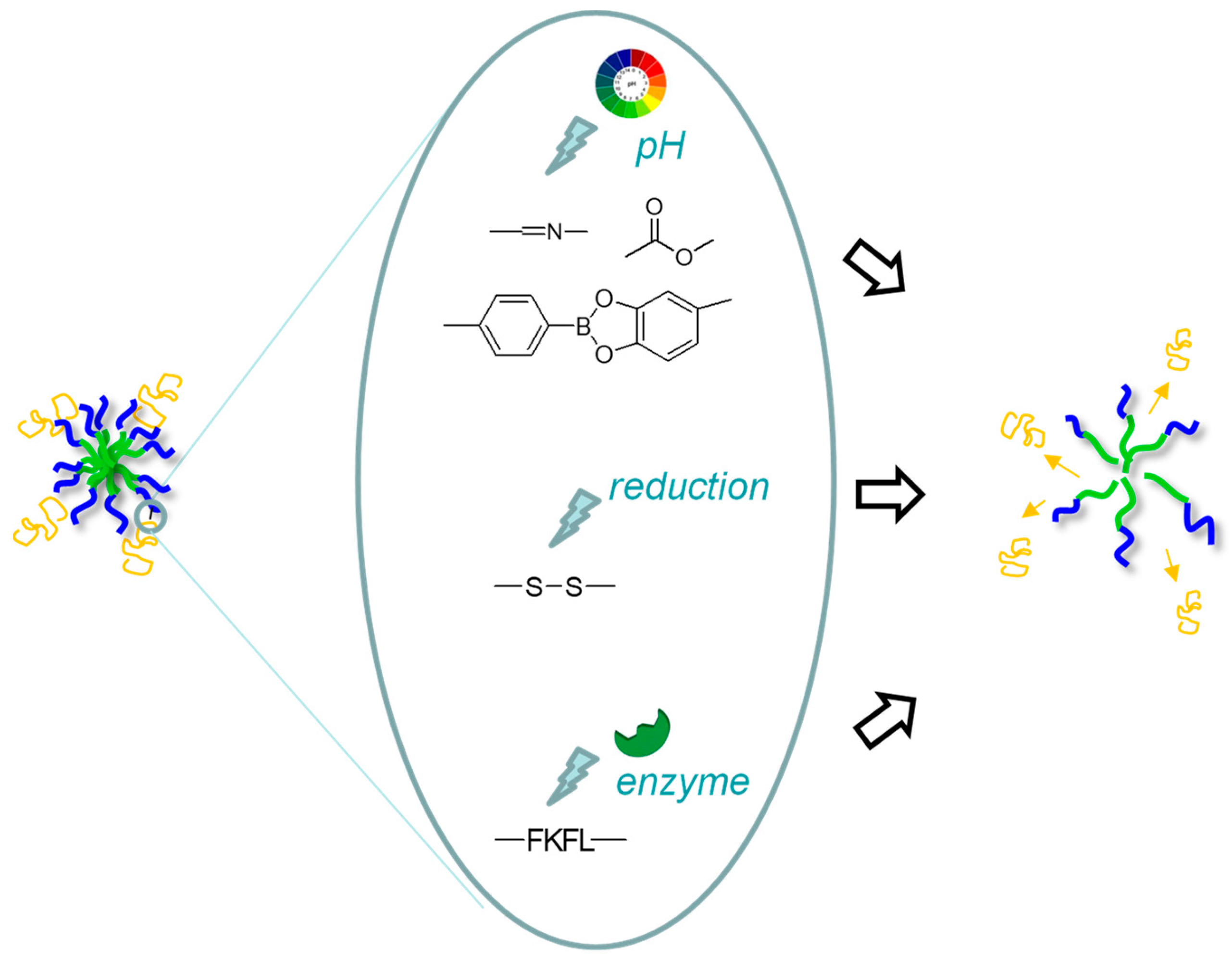
3. Covalent Association to Micelles

Covalent immobilization of proteins/peptides on micelles generally has the advantage of better stability over the non-covalent approach, avoiding premature release. Many micelles have been covalently bound to proteins/peptides through “conventional”, i.e., non-reversible chemistries such as amide bond formation [13,74,75,76], Michael additions (thiol-acrylate, thiol-maleimide) [77], or azide-alkyne click chemistry [78]. Research has more recently been dedicated to the use of stimulus-cleavable covalent bindings for triggering intracellular protein release with better control. Micelles coupled to proteins/peptides through pH, reduction, and enzyme-sensitive linkages have been particularly investigated (Figure 3, Table 2).

3.1. pH-Sensitive Linkages

In early works, Liu et al. have developed an aldehyde-terminated triblock copolymer of oligo(ethylene glycol)methyl ether methacrylate (OEGMA), 2-(dimethylamino)ethyl methacrylate (DMA), and 2-(diethylamino)-ethyl methacrylate (DEA) (namely, an Ald-POEGMA-b-PDMA-b-PDEA copolymer) which was used to form crosslinked micelles showing pH-dependent swelling due to the pH-sensitive PDEA block core. The aldehyde at the end of the hydrophilic shell block could be coupled to the amines of the model protein (lysozyme) to form imine pH-labile binding (Figure 3, top). However, the ability for the triggered release of the protein upon hydrolysis of the imine in mildly acidic conditions was not studied [79].
Yuba et al. have developed PLA-PEG-PLA block copolymers whose hydroxyl end-groups were conjugated to lisinopril dipeptide by ester formation in the presence of carbodiimide/DMAP coupling agents (Figure 3, top). Peptide release was not observed at pH 7.4 while 53% release occurred at pH 4, which was attributed to the acid-triggered hydrolysis of ester bonds. Interestingly, lisinopril physically entrapped in the micelles as a control was significantly released at pH 7.4, while it was negligible for the conjugated drug, showing the relevance of a covalent strategy for the stability of drug loading at physiological conditions [80].
Recently, Zhang et al. have designed smart micelles of a triblock copolymer conjugated to ribonuclease A (RNAse A) protein through a pH-reversible catechol-phenylboronic acid linkage (Figure 3, top) and encapsulating doxorubicin (Dox) [81]. The copolymer was composed of (i) a hydrophilic PEG block, (ii) an intermediate block functionalized with pendant catechol groups for conjugation of phenylboronic acid functionalized RNAse A, and (iii) a pH-sensitive block containing tertiary amine pendant groups forming the micelle core. Upon cell internalization, the latter block became hydrophilic under the acidic environment in the endosomes (due to protonation of the amine groups), inducing micelle disassembly and Dox release as well as endosomal escape through the proton sponge effect. The release of RNAse A also occurred through acidic pH-triggered cleavage of phenylboronic acid–catechol linkages. Such enhanced intracellular releases of both RNAse A and Dox synergistically enhanced anticancer efficacy in vitro and in vivo.

3.2. Reduction-Sensitive Linkages

Recent research has increasingly focused on the conjugation of proteins on polymer micelles through disulfide bonds to trigger their release in reductive intracellular environments. In vaccine delivery, this is of particular interest to promote the release of protein antigen in the cytosol of APCs, favoring its processing through the MHC I pathway and thus a CD8+ cellular immune response, which is highly desirable in many infectious diseases and cancer [82,94,95,96]. Since 2014, Stayton’s team has developed micelles from block amphiphilic copolymer-bearing pendant reactive pyridyl disulfide groups for protein-antigen conjugation through their thiol cysteine (disulfide formation, Figure 3, middle) and pH-responsive DMA units, i.e., bringing membrane disruptive properties at endosomal pH (protonation of tertiary amine units) to promote endosomal escape and intracellular delivery of the peptide/protein antigen [83].
Interestingly, this copolymer platform can be tuned to introduce additional immunostimulating molecules, namely ligands of APC (DC) receptors, to further improve immune response. For example, the OVA antigen-conjugated copolymer micelles could be electrostatically complexed with an immunostimulating molecule (anionic CpG) [84], resulting in an enhanced CD8+ T cell response. Sevimli et al. used a similar copolymer approach of a disulfide-conjugated antigen and pH-responsiveness but tuned the core block to be more hydrophobic to enhance the encapsulation of the immunostimulating imiquimod molecule (hydrophobic, ligand of TLR7 of DCs). The co-delivered antigen/imiquimod with micelles improved CD8+ T cell as well as IgG antibody responses [82]. Finally, a mannose ligand was also conjugated on such OVA antigen copolymer micelles to enhance DC targeting through the mannose receptor [85], inducing improved DC activation and a higher antitumor immunity in OVA-tumor-bearing mice compared to the free peptide antigen.
Notably, improvements have recently been achieved on the copolymer micelles of the Stayton group by conjugating a lytic peptide (melittin) on the micelles through disulfide bonds, which further enhanced endosomal escape and cytotoxic T cell response [86,87].
The pH-sensitive copolymer nanoplatform developed by the Stayton team was also exploited in the anticancer drug delivery context using disulfide conjugation of a proapoptotic peptide drug for effective intracellular delivery [88]. In addition, a targeting property could be introduced by binding an anti-CD22 antibody (labelled with streptavidin) on biotin end-groups of the hydrophilic block copolymer. Tumor growth was suppressed for the duration of treatment and prolonged survival in a human B-cell lymphoma xenograft mouse model was observed. In the same anticancer context, Morales-Cruz et al. have constructed a copolymer PEG-b-PLGA functionalized at the PLGA end-group by pyridyldisulfide moieties for conjugation of the cytochrome C (Cyt C) protein through a disulfide bond, and at the PEG end-group by folic acid for targeting properties. The copolymer micelles exhibited excellent stability under extracellular physiological conditions, whereas once in the intracellular reducing environment, Cyt C was released from the conjugate. The micelles promoted endosomal escape, and their potential for antitumor therapy was shown in a brain tumor model back in 2016 [89].

3.3. Enzyme-Sensitive Linkages

Enzyme as a stimulus for triggering the release of drugs is probably the most currently investigated, particularly in the cancer treatment context. Indeed, specific enzymes are highly expressed in particular tumor tissues [97]. The copolymer nanoplatform developed by Stayton’s team was tuned to introduce an enzyme-cleavable peptide sequence (FKFL) between the hydrophilic block and the anti-cancer proapoptotic peptide (Figure 3, bottom), instead of the disulfide linkage previously used [90]. This sequence was efficiently cleaved by the cathepsin B protease enzyme present in intracellular compartments (upregulated in many cancers), enhancing the quantity of peptide reaching tumor intracellular targets. The micelles successfully induced apoptosis in SKOV3 ovarian cancer cells in comparison to micelles from the control polymer containing a scrambled peptide sequence [90].
Su et al. prepared micelles from an azide-modified polyethylene glycol-block-polyaspartic acid(benzylamine) (azide-PEG-b-PAsp(Bz)) with encapsulated zinc phthalocyanine (ZnPC) photosensitizer for photodynamic therapy. The azide end-groups at the micelle surface were used to couple a PD-L1 antibody to block the PD-1/PD-L1 pathway and thus reactivate the immune CD8+ T cells to attack the tumor. Interestingly, the antibody was coupled through a matrix metalloprotease 2 (MMP2) enzyme-sensitive peptide, promoting antibody-triggered release in the tumor environment (rich in MM2). ZnPC was further released intracellularly and both drugs synergistically improved the antitumor effect through this immuno-photodynamic therapeutic approach [91]. Shi et al. designed amphiphiles composed of (i) a hydrophobic palmitic acid and (ii) a cationic cell-penetrating peptide (CPP) bound to a targeting peptide moiety through an MMP2-sensitive peptide linker. Self-assembling into fiber nanostructures with surface targeting moieties was obtained, which upon cleavage of the MMP2 in the tumor environment, turned to micelles presenting CPP at their surface to facilitate cell penetration across the cell membrane [98].
MMP enzymes are also upregulated in inflammatory environments, for example in the heart following myocardial infarction. Nguyen et al. developed, in this context, peptide-polymer amphiphiles based on a polynorbornene backbone and MMP-cleavable hydrophilic peptide brushes, forming micelles of about 20 nm [92]. After intravenous injection in rats, the micelles entered the infarct tissue, in which upregulation of MMP induced peptide cleavage and transition into an aggregated-like network, allowing specific accumulation in this tissue for drug delivery. The authors have very recently greatly improved the biodegradability of these amphiphiles by introducing degradable phosphoramidate segments [93].

4. Conclusions and Outlooks

Over the past decade, polymeric micelles have clearly appeared as attractive and relevant carriers to trigger the precise delivery of proteins/peptides to the targeted site, either through non-covalent or covalent approaches. This is largely attributed to the high flexibility in design of the copolymers, which can be tuned to present controllable charge states as well as suitable stimuli-responsive functionalities for transiently immobilizing proteins/peptides. Such success can be tempered regarding some aspects, within the context of this review: to date, PEG is still mainly used as a hydrophilic segment, which raises increasing concerns of hypersensitivity and accelerated blood clearance [99,100,101]. Another limitation is that the smart self-assembly is strongly dependent on the protein properties (e.g., pI). However, it is important to point out that this field is rapidly evolving. Indeed, many pioneer works were performed with model proteins (e.g., BSA, or ovalbumin antigen in the case of vaccine delivery) to establish the proof of concept of the smart releasing system, mainly exemplified by in vitro studies. Nowadays, more relevant and diverse proteins of interest requiring a delivery system are currently envisioned with ad hoc and well-defined copolymer micelle design, and investigated through deeper in vivo studies, as highlighted in this review. The convincing biological performance obtained in many pathologic contexts (e.g., vaccines, cancer, inflammation) will foster works closer to the clinical application. Undoubtedly, these versatile nano-systems, with their inherent ease of preparation, scalability, and high reproducibility (moreover improvable with the current inputs of microfluidics techniques) will play a major role in the development of the next generation of protein therapeutics.

Author Contributions

Conceptualization, T.T. and B.V.; writing—original draft preparation, T.T.; writing—review and editing, T.T. and B.V.; supervision, T.T. All authors have read and agreed to the published version of the manuscript.

Funding

This work was funded by ANR (LipoFishVac project, ANR-21-CE35-0019) to T.T. and B.V.

Conflicts of Interest

The authors declare no conflict of interest.

References

  1. Carton, J.M.; Strohl, W.R. Protein Therapeutics (Introduction to Biopharmaceuticals). In Introduction to Biological and Small Molecule Drug Research and Development; Elsevier: Amsterdam, The Netherlands, 2013; pp. 127–159. ISBN 978-0-12-397176-0. [Google Scholar]
  2. Ebrahimi, S.B.; Samanta, D. Engineering Protein-Based Therapeutics through Structural and Chemical Design. Nat. Commun. 2023, 14, 2411. [Google Scholar] [CrossRef] [PubMed]
  3. Zaman, R.; Islam, R.A.; Ibnat, N.; Othman, I.; Zaini, A.; Lee, C.Y.; Chowdhury, E.H. Current Strategies in Extending Half-Lives of Therapeutic Proteins. J. Control. Release 2019, 301, 176–189. [Google Scholar] [CrossRef] [PubMed]
  4. Yu, M.; Wu, J.; Shi, J.; Farokhzad, O.C. Nanotechnology for Protein Delivery: Overview and Perspectives. J. Control. Release 2016, 240, 24–37. [Google Scholar] [CrossRef] [PubMed]
  5. Vila, A.; Sánchez, A.; Tobio, M.; Calvo, P.; Alonso, M.J. Design of Biodegradable Particles for Protein Delivery. J. Control. Release 2002, 78, 15–24. [Google Scholar] [CrossRef]
  6. Lamalle-Bernard, D.; Munier, S.; Compagnon, C.; Charles, M.-H.; Kalyanaraman, V.S.; Delair, T.; Verrier, B.; Ataman-Önal, Y. Coadsorption of HIV-1 P24 and Gp120 Proteins to Surfactant-Free Anionic PLA Nanoparticles Preserves Antigenicity and Immunogenicity. J. Control. Release 2006, 115, 57–67. [Google Scholar] [CrossRef]
  7. Zhao, H.; Lin, Z.Y.; Yildirimer, L.; Dhinakar, A.; Zhao, X.; Wu, J. Polymer-Based Nanoparticles for Protein Delivery: Design, Strategies and Applications. J. Mater. Chem. B 2016, 4, 4060–4071. [Google Scholar] [CrossRef]
  8. Vardaxi, A.; Kafetzi, M.; Pispas, S. Polymeric Nanostructures Containing Proteins and Peptides for Pharmaceutical Applications. Polymers 2022, 14, 777. [Google Scholar] [CrossRef] [PubMed]
  9. Cabral, H.; Miyata, K.; Osada, K.; Kataoka, K. Block Copolymer Micelles in Nanomedicine Applications. Chem. Rev. 2018, 118, 6844–6892. [Google Scholar] [CrossRef]
  10. Trimaille, T.; Verrier, B. Micelle-Based Adjuvants for Subunit Vaccine Delivery. Vaccines 2015, 3, 803–813. [Google Scholar] [CrossRef] [PubMed]
  11. Wang, J.; Li, S.; Chen, T.; Xian, W.; Zhang, H.; Wu, L.; Zhu, W.; Zeng, Q. Nanoscale Cationic Micelles of Amphiphilic Copolymers Based on Star-Shaped PLGA and PEI Cross-Linked PEG for Protein Delivery Application. J. Mater. Sci. Mater. Med. 2019, 30, 93. [Google Scholar] [CrossRef]
  12. Shi, Y.; Zhu, H.; Ren, Y.; Li, K.; Tian, B.; Han, J.; Feng, D. Preparation of Protein-Loaded PEG-PLA Micelles and the Effects of Ultrasonication on Particle Size. Colloid Polym. Sci. 2017, 295, 259–266. [Google Scholar] [CrossRef]
  13. Lamrayah, M.; Phelip, C.; Coiffier, C.; Lacroix, C.; Willemin, T.; Trimaille, T.; Verrier, B. A Polylactide-Based Micellar Adjuvant Improves the Intensity and Quality of Immune Response. Pharmaceutics 2022, 14, 107. [Google Scholar] [CrossRef]
  14. Iqbal, S.; Blenner, M.; Alexander-Bryant, A.; Larsen, J. Polymersomes for Therapeutic Delivery of Protein and Nucleic Acid Macromolecules: From Design to Therapeutic Applications. Biomacromolecules 2020, 21, 1327–1350. [Google Scholar] [CrossRef]
  15. Nomani, A.; Nosrati, H.; Manjili, H.; Khesalpour, L.; Danafar, H. Preparation and Characterization of Copolymeric Polymersomes for Protein Delivery. Drug Res. 2017, 67, 458–465. [Google Scholar] [CrossRef]
  16. Martins, S.; Sarmento, B.; Ferreira, D.C.; Souto, E.B. Lipid-Based Colloidal Carriers for Peptide and Protein Delivery--Liposomes versus Lipid Nanoparticles. Int. J. Nanomed. 2007, 2, 595–607. [Google Scholar]
  17. Samimi, S.; Maghsoudnia, N.; Eftekhari, R.B.; Dorkoosh, F. Lipid-Based Nanoparticles for Drug Delivery Systems. In Characterization and Biology of Nanomaterials for Drug Delivery; Elsevier: Amsterdam, The Netherlands, 2019; pp. 47–76. ISBN 978-0-12-814031-4. [Google Scholar]
  18. Chatzikleanthous, D.; O’Hagan, D.T.; Adamo, R. Lipid-Based Nanoparticles for Delivery of Vaccine Adjuvants and Antigens: Toward Multicomponent Vaccines. Mol. Pharm. 2021, 18, 2867–2888. [Google Scholar] [CrossRef] [PubMed]
  19. Wu, C.; Mu, H. Lipid and PLGA Microparticles for Sustained Delivery of Protein and Peptide Drugs. Pharm. Nanotechnol. 2020, 8, 22–32. [Google Scholar] [CrossRef] [PubMed]
  20. Scaletti, F.; Hardie, J.; Lee, Y.-W.; Luther, D.C.; Ray, M.; Rotello, V.M. Protein Delivery into Cells Using Inorganic Nanoparticle–Protein Supramolecular Assemblies. Chem. Soc. Rev. 2018, 47, 3421–3432. [Google Scholar] [CrossRef]
  21. van de Weert, M.; Hennink, W.E.; Jiskoot, W. Protein Instability in Poly(Lactic-Co-Glycolic Acid) Microparticles. Pharm. Res. 2000, 17, 1159–1167. [Google Scholar] [CrossRef]
  22. Mehnert, W. Solid Lipid Nanoparticles Production, Characterization and Applications. Adv. Drug Deliv. Rev. 2001, 47, 165–196. [Google Scholar] [CrossRef]
  23. Ball, R.; Bajaj, P.; Whitehead, K. Achieving Long-Term Stability of Lipid Nanoparticles: Examining the Effect of pH, Temperature, and Lyophilization. Int. J. Nanomed. 2016, 12, 305–315. [Google Scholar] [CrossRef]
  24. Stevens, C.A.; Kaur, K.; Klok, H.-A. Self-Assembly of Protein-Polymer Conjugates for Drug Delivery. Adv. Drug Deliv. Rev. 2021, 174, 447–460. [Google Scholar] [CrossRef] [PubMed]
  25. Chen, F.; Stenzel, M.H. Polyion Complex Micelles for Protein Delivery. Aust. J. Chem. 2018, 71, 768. [Google Scholar] [CrossRef]
  26. Yildirim, I.; Weber, C.; Schubert, U.S. Old Meets New: Combination of PLA and RDRP to Obtain Sophisticated Macromolecular Architectures. Prog. Polym. Sci. 2018, 76, 111–150. [Google Scholar] [CrossRef]
  27. Palmiero, U.C.; Sponchioni, M.; Manfredini, N.; Maraldi, M.; Moscatelli, D. Strategies to Combine ROP with ATRP or RAFT Polymerization for the Synthesis of Biodegradable Polymeric Nanoparticles for Biomedical Applications. Polym. Chem. 2018, 9, 4084–4099. [Google Scholar] [CrossRef]
  28. Savić, R.; Eisenberg, A.; Maysinger, D. Block Copolymer Micelles as Delivery Vehicles of Hydrophobic Drugs: Micelle–Cell Interactions. J. Drug Target. 2006, 14, 343–355. [Google Scholar] [CrossRef]
  29. Ahmad, Z.; Shah, A.; Siddiq, M.; Kraatz, H.-B. Polymeric Micelles as Drug Delivery Vehicles. RSC Adv. 2014, 4, 17028–17038. [Google Scholar] [CrossRef]
  30. Sun, J.-H.; Liang, X.; Cai, M.; Yan, L.; Chen, Z.; Guo, L.; Jing, L.; Wang, Y.; Zhou, D. Protein-Crowned Micelles for Targeted and Synergistic Tumor-Associated Macrophage Reprogramming to Enhance Cancer Treatment. Nano Lett. 2022, 22, 4410–4420. [Google Scholar] [CrossRef]
  31. Kedar, U.; Phutane, P.; Shidhaye, S.; Kadam, V. Advances in Polymeric Micelles for Drug Delivery and Tumor Targeting. Nanomed. Nanotechnol. Biol. Med. 2010, 6, 714–729. [Google Scholar] [CrossRef]
  32. Lee, E. Polymeric Micelle for Tumor pH and Folate-Mediated Targeting. J. Control. Release 2003, 91, 103–113. [Google Scholar] [CrossRef]
  33. Mura, S.; Nicolas, J.; Couvreur, P. Stimuli-Responsive Nanocarriers for Drug Delivery. Nat. Mater. 2013, 12, 991–1003. [Google Scholar] [CrossRef] [PubMed]
  34. Zhang, Q.; Re Ko, N.; Kwon Oh, J. Recent Advances in Stimuli-Responsive Degradable Block Copolymer Micelles: Synthesis and Controlled Drug Delivery Applications. Chem. Commun. 2012, 48, 7542. [Google Scholar] [CrossRef] [PubMed]
  35. Manchun, S.; Dass, C.R.; Sriamornsak, P. Targeted Therapy for Cancer Using pH-Responsive Nanocarrier Systems. Life Sci. 2012, 90, 381–387. [Google Scholar] [CrossRef] [PubMed]
  36. AlSawaftah, N.M.; Awad, N.S.; Pitt, W.G.; Husseini, G.A. pH-Responsive Nanocarriers in Cancer Therapy. Polymers 2022, 14, 936. [Google Scholar] [CrossRef] [PubMed]
  37. Cheng, Y.; Cheng, A.; Jia, Y.; Yang, L.; Ning, Y.; Xu, L.; Zhong, Y.; Zhuang, Z.; Guan, J.; Zhang, X.; et al. pH-Responsive Multifunctional Theranostic Rapamycin-Loaded Nanoparticles for Imaging and Treatment of Acute Ischemic Stroke. ACS Appl. Mater. Interfaces 2021, 13, 56909–56922. [Google Scholar] [CrossRef] [PubMed]
  38. Peng, D.; Liu, G.; He, Y.; Gao, P.; Gou, S.; Wu, J.; Yu, J.; Liu, P.; Cai, K. Fabrication of a pH-Responsive Core–Shell Nanosystem with a Low-Temperature Photothermal Therapy Effect for Treating Bacterial Biofilm Infection. Biomater. Sci. 2021, 9, 7483–7491. [Google Scholar] [CrossRef]
  39. Zhang, C.Y.; Lin, W.; Gao, J.; Shi, X.; Davaritouchaee, M.; Nielsen, A.E.; Mancini, R.J.; Wang, Z. pH-Responsive Nanoparticles Targeted to Lungs for Improved Therapy of Acute Lung Inflammation/Injury. ACS Appl. Mater. Interfaces 2019, 11, 16380–16390. [Google Scholar] [CrossRef]
  40. Tayo, L.L. Stimuli-Responsive Nanocarriers for Intracellular Delivery. Biophys. Rev. 2017, 9, 931–940. [Google Scholar] [CrossRef]
  41. Miyazaki, M.; Yuba, E.; Hayashi, H.; Harada, A.; Kono, K. Development of pH-Responsive Hyaluronic Acid-Based Antigen Carriers for Induction of Antigen-Specific Cellular Immune Responses. ACS Biomater. Sci. Eng. 2019, 5, 5790–5797. [Google Scholar] [CrossRef]
  42. Schmaljohann, D. Thermo- and pH-Responsive Polymers in Drug Delivery. Adv. Drug Deliv. Rev. 2006, 58, 1655–1670. [Google Scholar] [CrossRef]
  43. Abed, H.F.; Abuwatfa, W.H.; Husseini, G.A. Redox-Responsive Drug Delivery Systems: A Chemical Perspective. Nanomaterials 2022, 12, 3183. [Google Scholar] [CrossRef]
  44. Hu, Q.; Katti, P.S.; Gu, Z. Enzyme-Responsive Nanomaterials for Controlled Drug Delivery. Nanoscale 2014, 6, 12273–12286. [Google Scholar] [CrossRef]
  45. Harada, A.; Kataoka, K. Formation of Polyion Complex Micelles in an Aqueous Milieu from a Pair of Oppositely-Charged Block Copolymers with Poly(Ethylene Glycol) Segments. Macromolecules 1995, 28, 5294–5299. [Google Scholar] [CrossRef]
  46. Lee, Y.; Kataoka, K. Biosignal-Sensitive Polyion Complex Micelles for the Delivery of Biopharmaceuticals. Soft Matter 2009, 5, 3810. [Google Scholar] [CrossRef]
  47. Lee, Y.; Ishii, T.; Cabral, H.; Kim, H.J.; Seo, J.-H.; Nishiyama, N.; Oshima, H.; Osada, K.; Kataoka, K. Charge-Conversional Polyionic Complex Micelles-Efficient Nanocarriers for Protein Delivery into Cytoplasm. Angew. Chem. Int. Ed. 2009, 48, 5309–5312. [Google Scholar] [CrossRef]
  48. Miyata, K.; Oba, M.; Nakanishi, M.; Fukushima, S.; Yamasaki, Y.; Koyama, H.; Nishiyama, N.; Kataoka, K. Polyplexes from Poly(Aspartamide) Bearing 1,2-Diaminoethane Side Chains Induce pH-Selective, Endosomal Membrane Destabilization with Amplified Transfection and Negligible Cytotoxicity. J. Am. Chem. Soc. 2008, 130, 16287–16294. [Google Scholar] [CrossRef] [PubMed]
  49. Tao, A.; Huang, G.L.; Igarashi, K.; Hong, T.; Liao, S.; Stellacci, F.; Matsumoto, Y.; Yamasoba, T.; Kataoka, K.; Cabral, H. Polymeric Micelles Loading Proteins through Concurrent Ion Complexation and pH-Cleavable Covalent Bonding for In Vivo Delivery. Macromol. Biosci. 2020, 20, 1900161. [Google Scholar] [CrossRef]
  50. Li, K.; Chen, F.; Wang, Y.; Stenzel, M.H.; Chapman, R. Polyion Complex Micelles for Protein Delivery Benefit from Flexible Hydrophobic Spacers in the Binding Group. Macromol. Rapid Commun. 2020, 41, 2000208. [Google Scholar] [CrossRef]
  51. Zhang, X.; Chen, D.; Ba, S.; Zhu, J.; Zhang, J.; Hong, W.; Zhao, X.; Hu, H.; Qiao, M. Poly(l-Histidine) Based Triblock Copolymers: pH Induced Reassembly of Copolymer Micelles and Mechanism Underlying Endolysosomal Escape for Intracellular Delivery. Biomacromolecules 2014, 15, 4032–4045. [Google Scholar] [CrossRef]
  52. Lee, E.S.; Shin, H.J.; Na, K.; Bae, Y.H. Poly(l-Histidine)–PEG Block Copolymer Micelles and pH-Induced Destabilization. J. Control. Release 2003, 90, 363–374. [Google Scholar] [CrossRef]
  53. Kim, M.S.; Gao, G.H.; Kang, S.W.; Lee, D.S. Evaluation of pH-Sensitive Poly(β-Amino Ester)-Graft-Poly(Ethylene Glycol) and Its Usefulness as a pH-Sensor and Protein Carrier. Macromol. Biosci. 2011, 11, 946–951. [Google Scholar] [CrossRef]
  54. Gao, G.H.; Park, M.J.; Li, Y.; Im, G.H.; Kim, J.-H.; Kim, H.N.; Lee, J.W.; Jeon, P.; Bang, O.Y.; Lee, J.H.; et al. The Use of pH-Sensitive Positively Charged Polymeric Micelles for Protein Delivery. Biomaterials 2012, 33, 9157–9164. [Google Scholar] [CrossRef]
  55. Kim, D.H.; Seo, Y.K.; Thambi, T.; Moon, G.J.; Son, J.P.; Li, G.; Park, J.H.; Lee, J.H.; Kim, H.H.; Lee, D.S.; et al. Enhancing Neurogenesis and Angiogenesis with Target Delivery of Stromal Cell Derived Factor-1α Using a Dual Ionic pH-Sensitive Copolymer. Biomaterials 2015, 61, 115–125. [Google Scholar] [CrossRef] [PubMed]
  56. Fang, X.; Xie, H.; Duan, H.; Li, P.; Yousaf, M.; Xu, H.; Yang, Y.; Wang, C. Anti-Tumor Activity of Nanomicelles Encapsulating CXCR4 Peptide Antagonist E5. PLoS ONE 2017, 12, e0182697. [Google Scholar] [CrossRef] [PubMed]
  57. Zeng, Z.; She, Y.; Peng, Z.; Wei, J.; He, X. Enzyme-Mediated in Situ Formation of pH-Sensitive Nanogels for Proteins Delivery. RSC Adv. 2016, 6, 8032–8042. [Google Scholar] [CrossRef]
  58. Pang, X.; Liang, S.; Wang, T.; Yu, S.; Yang, R.; Hou, T.; Liu, Y.; He, C.; Zhang, N. Engineering Thermo-pH Dual Responsive Hydrogel for Enhanced Tumor Accumulation, Penetration, and Chemo-Protein Combination Therapy. Int. J. Nanomed. 2020, 15, 4739–4752. [Google Scholar] [CrossRef] [PubMed]
  59. Lei, X.-L.; Cheng, K.; Li, Y.; Zhong, Z.-T.; Hou, X.-L.; Song, L.-B.; Zhang, F.; Wang, J.-H.; Zhao, Y.-D.; Xu, Q.-R. The Eradication of Biofilm for Therapy of Bacterial Infected Chronic Wound Based on pH-Responsive Micelle of Antimicrobial Peptide Derived Biodegradable Microneedle Patch. Chem. Eng. J. 2023, 462, 142222. [Google Scholar] [CrossRef]
  60. Tang, N.; Du, G.; Wang, N.; Liu, C.; Hang, H.; Liang, W. Improving Penetration in Tumors with Nanoassemblies of Phospholipids and Doxorubicin. J. Natl. Cancer Inst. 2007, 99, 1004–1015. [Google Scholar] [CrossRef]
  61. Wei, X.; Wang, Y.; Zeng, W.; Huang, F.; Qin, L.; Zhang, C.; Liang, W. Stability Influences the Biodistribution, Toxicity, and Anti-Tumor Activity of Doxorubicin Encapsulated in PEG-PE Micelles in Mice. Pharm. Res. 2012, 29, 1977–1989. [Google Scholar] [CrossRef]
  62. Wittemann, A.; Haupt, B.; Ballauff, M. Adsorption of Proteins on Spherical Polyelectrolyte Brushes in Aqueous Solution. Phys. Chem. Chem. Phys. 2003, 5, 1671–1677. [Google Scholar] [CrossRef]
  63. Pan, M.; Lu, C.; Zhang, W.; Huang, H.; Shi, X.; Tang, S.; Liu, D. Poly(l-Ornithine)-Based Polymeric Micelles as pH-Responsive Macromolecular Anticancer Agents. Pharmaceutics 2023, 15, 1307. [Google Scholar] [CrossRef]
  64. Seo, J.-W.; Hwang, J.-Y.; Shin, U.S. Ionic Liquid-Doped and p-NIPAAm-Based Copolymer (p-NIBIm): Extraordinary Drug-Entrapping and -Releasing Behaviors at 38–42 °C. RSC Adv. 2014, 4, 26738–26747. [Google Scholar] [CrossRef]
  65. Bonacucina, G.; Cespi, M.; Mencarelli, G.; Giorgioni, G.; Palmieri, G.F. Thermosensitive Self-Assembling Block Copolymers as Drug Delivery Systems. Polymers 2011, 3, 779–811. [Google Scholar] [CrossRef]
  66. Huynh, C.T.; Nguyen, M.K.; Lee, D.S. Injectable Block Copolymer Hydrogels: Achievements and Future Challenges for Biomedical Applications. Macromolecules 2011, 44, 6629–6636. [Google Scholar] [CrossRef]
  67. Determan, M.D.; Guo, L.; Lo, C.-T.; Thiyagarajan, P.; Mallapragada, S.K. P H- and Temperature-Dependent Phase Behavior of a PEO-PPO-PEO-Based Pentablock Copolymer in Aqueous Media. Phys. Rev. E 2008, 78, 021802. [Google Scholar] [CrossRef]
  68. Adams, J.R.; Haughney, S.L.; Mallapragada, S.K. Effective Polymer Adjuvants for Sustained Delivery of Protein Subunit Vaccines. Acta Biomater. 2015, 14, 104–114. [Google Scholar] [CrossRef] [PubMed]
  69. Senapati, S.; Darling, R.J.; Loh, D.; Schneider, I.C.; Wannemuehler, M.J.; Narasimhan, B.; Mallapragada, S.K. Pentablock Copolymer Micelle Nanoadjuvants Enhance Cytosolic Delivery of Antigen and Improve Vaccine Efficacy While Inducing Low Inflammation. ACS Biomater. Sci. Eng. 2019, 5, 1332–1342. [Google Scholar] [CrossRef]
  70. Lee, S.Y.; Jeon, S.I.; Sim, S.B.; Byun, Y.; Ahn, C.-H. A Supramolecular Host-Guest Interaction-Mediated Injectable Hydrogel System with Enhanced Stability and Sustained Protein Release. Acta Biomater. 2021, 131, 286–301. [Google Scholar] [CrossRef] [PubMed]
  71. Ren, J.; Zhang, Y.; Zhang, J.; Gao, H.; Liu, G.; Ma, R.; An, Y.; Kong, D.; Shi, L. pH/Sugar Dual Responsive Core-Cross-Linked PIC Micelles for Enhanced Intracellular Protein Delivery. Biomacromolecules 2013, 14, 3434–3443. [Google Scholar] [CrossRef]
  72. Xia, H.; Qin, M.; Wang, Z.; Wang, Y.; Chen, B.; Wan, F.; Tang, M.; Pan, X.; Yang, Y.; Liu, J.; et al. A pH-/Enzyme-Responsive Nanoparticle Selectively Targets Endosomal Toll-like Receptors to Potentiate Robust Cancer Vaccination. Nano Lett. 2022, 22, 2978–2987. [Google Scholar] [CrossRef] [PubMed]
  73. Li, Z.; Jiang, Y.; Zhao, H.; Liu, L. Ca 2+ -Chelation-Induced Fabrication of Multistimuli-Responsive Charged Nanogels from Phospholipid–Polymer Conjugates and Use for Drug/Protein Loading. Langmuir 2022, 38, 6612–6622. [Google Scholar] [CrossRef] [PubMed]
  74. Vanparijs, N.; De Coen, R.; Laplace, D.; Louage, B.; Maji, S.; Lybaert, L.; Hoogenboom, R.; De Geest, B.G. Transiently Responsive Protein–Polymer Conjugates via a ‘Grafting-from’ RAFT Approach for Intracellular Co-Delivery of Proteins and Immune-Modulators. Chem. Commun. 2015, 51, 13972–13975. [Google Scholar] [CrossRef] [PubMed]
  75. Zhang, Q.; Vanparijs, N.; Louage, B.; De Geest, B.G.; Hoogenboom, R. Dual pH- and Temperature-Responsive RAFT-Based Block Co-Polymer Micelles and Polymer–Protein Conjugates with Transient Solubility. Polym. Chem. 2014, 5, 1140–1144. [Google Scholar] [CrossRef]
  76. Lacroix, C.; Humanes, A.; Coiffier, C.; Gigmes, D.; Verrier, B.; Trimaille, T. Polylactide-Based Reactive Micelles as a Robust Platform for mRNA Delivery. Pharm. Res. 2020, 37, 30. [Google Scholar] [CrossRef]
  77. Qiao, Z.-Y.; Hou, C.-Y.; Zhang, D.; Liu, Y.; Lin, Y.-X.; An, H.-W.; Li, X.-J.; Wang, H. Self-Assembly of Cytotoxic Peptide Conjugated Poly(β-Amino Ester)s for Synergistic Cancer Chemotherapy. J. Mater. Chem. B 2015, 3, 2943–2953. [Google Scholar] [CrossRef] [PubMed]
  78. Florinas, S.; Liu, M.; Fleming, R.; Van Vlerken-Ysla, L.; Ayriss, J.; Gilbreth, R.; Dimasi, N.; Gao, C.; Wu, H.; Xu, Z.-Q.; et al. A Nanoparticle Platform to Evaluate Bioconjugation and Receptor-Mediated Cell Uptake Using Cross-Linked Polyion Complex Micelles Bearing Antibody Fragments. Biomacromolecules 2016, 17, 1818–1833. [Google Scholar] [CrossRef]
  79. Liu, H.; Jiang, X.; Fan, J.; Wang, G.; Liu, S. Aldehyde Surface-Functionalized Shell Cross-Linked Micelles with pH-Tunable Core Swellability and Their Bioconjugation with Lysozyme. Macromolecules 2007, 40, 9074–9083. [Google Scholar] [CrossRef]
  80. Danafar, H.; Rostamizadeh, K.; Davaran, S.; Hamidi, M. Drug-Conjugated PLA–PEG–PLA Copolymers: A Novel Approach for Controlled Delivery of Hydrophilic Drugs by Micelle Formation. Pharm. Dev. Technol. 2017, 22, 947–957. [Google Scholar] [CrossRef]
  81. Zhang, P.; Zhang, Y.; Ding, X.; Shen, W.; Li, M.; Wagner, E.; Xiao, C.; Chen, X. A Multistage Cooperative Nanoplatform Enables Intracellular Co-Delivery of Proteins and Chemotherapeutics for Cancer Therapy. Adv. Mater. 2020, 32, 2000013. [Google Scholar] [CrossRef]
  82. Sevimli, S.; Knight, F.C.; Gilchuk, P.; Joyce, S.; Wilson, J.T. Fatty Acid-Mimetic Micelles for Dual Delivery of Antigens and Imidazoquinoline Adjuvants. ACS Biomater. Sci. Eng. 2017, 3, 179–194. [Google Scholar] [CrossRef]
  83. Keller, S.; Wilson, J.T.; Patilea, G.I.; Kern, H.B.; Convertine, A.J.; Stayton, P.S. Neutral Polymer Micelle Carriers with pH-Responsive, Endosome-Releasing Activity Modulate Antigen Trafficking to Enhance CD8+ T Cell Responses. J. Control. Release 2014, 191, 24–33. [Google Scholar] [CrossRef] [PubMed]
  84. Wilson, J.T.; Keller, S.; Manganiello, M.J.; Cheng, C.; Lee, C.-C.; Opara, C.; Convertine, A.; Stayton, P.S. pH-Responsive Nanoparticle Vaccines for Dual-Delivery of Antigens and Immunostimulatory Oligonucleotides. ACS Nano 2013, 7, 3912–3925. [Google Scholar] [CrossRef] [PubMed]
  85. Lv, S.; Song, K.; Yen, A.; Peeler, D.J.; Nguyen, D.C.; Olshefsky, A.; Sylvestre, M.; Srinivasan, S.; Stayton, P.S.; Pun, S.H. Well-Defined Mannosylated Polymer for Peptide Vaccine Delivery with Enhanced Antitumor Immunity. Adv Healthc. Mater. 2022, 11, 2101651. [Google Scholar] [CrossRef]
  86. Peeler, D.J.; Yen, A.; Luera, N.; Stayton, P.S.; Pun, S.H. Lytic Polyplex Vaccines Enhance Antigen-Specific Cytotoxic T Cell Response through Induction of Local Cell Death. Adv. Ther. 2021, 4, 2100005. [Google Scholar] [CrossRef]
  87. Song, K.; Nguyen, D.C.; Luu, T.; Yazdani, O.; Roy, D.; Stayton, P.S.; Pun, S.H. A Mannosylated Polymer with Endosomal Release Properties for Peptide Antigen Delivery. J. Control. Release 2023, 356, 232–241. [Google Scholar] [CrossRef] [PubMed]
  88. Berguig, G.Y.; Convertine, A.J.; Frayo, S.; Kern, H.B.; Procko, E.; Roy, D.; Srinivasan, S.; Margineantu, D.H.; Booth, G.; Palanca-Wessels, M.C.; et al. Intracellular Delivery System for Antibody–Peptide Drug Conjugates. Mol. Ther. 2015, 23, 907–917. [Google Scholar] [CrossRef]
  89. Morales-Cruz, M.; Cruz-Montañez, A.; Figueroa, C.M.; González-Robles, T.; Davila, J.; Inyushin, M.; Loza-Rosas, S.A.; Molina, A.M.; Muñoz-Perez, L.; Kucheryavykh, L.Y.; et al. Combining Stimulus-Triggered Release and Active Targeting Strategies Improves Cytotoxicity of Cytochrome c Nanoparticles in Tumor Cells. Mol. Pharm. 2016, 13, 2844–2854. [Google Scholar] [CrossRef]
  90. Kern, H.B.; Srinivasan, S.; Convertine, A.J.; Hockenbery, D.; Press, O.W.; Stayton, P.S. Enzyme-Cleavable Polymeric Micelles for the Intracellular Delivery of Proapoptotic Peptides. Mol. Pharm. 2017, 14, 1450–1459. [Google Scholar] [CrossRef]
  91. Su, Z.; Xiao, Z.; Huang, J.; Wang, Y.; An, Y.; Xiao, H.; Peng, Y.; Pang, P.; Han, S.; Zhu, K.; et al. Dual-Sensitive PEG-Sheddable Nanodrug Hierarchically Incorporating PD-L1 Antibody and Zinc Phthalocyanine for Improved Immuno-Photodynamic Therapy. ACS Appl. Mater. Interfaces 2021, 13, 12845–12856. [Google Scholar] [CrossRef]
  92. Nguyen, M.M.; Carlini, A.S.; Chien, M.-P.; Sonnenberg, S.; Luo, C.; Braden, R.L.; Osborn, K.G.; Li, Y.; Gianneschi, N.C.; Christman, K.L. Enzyme-Responsive Nanoparticles for Targeted Accumulation and Prolonged Retention in Heart Tissue after Myocardial Infarction. Adv. Mater. 2015, 27, 5547–5552. [Google Scholar] [CrossRef]
  93. Liang, Y.; Sullivan, H.L.; Carrow, K.; Mesfin, J.M.; Korpanty, J.; Worthington, K.; Luo, C.; Christman, K.L.; Gianneschi, N.C. Inflammation-Responsive Micellar Nanoparticles from Degradable Polyphosphoramidates for Targeted Delivery to Myocardial Infarction. J. Am. Chem. Soc. 2023, 145, 11185–11194. [Google Scholar] [CrossRef]
  94. Heffernan, M.J.; Murthy, N. Disulfide-Crosslinked Polyion Micelles for Delivery of Protein Therapeutics. Ann. Biomed. Eng. 2009, 37, 1993–2002. [Google Scholar] [CrossRef] [PubMed]
  95. Sun, J.; Chen, X.; Wei, J.; Yan, L.; Jing, X. Application of the Biodegradable Diblock Copolymer Poly(L-Lactide)-Block-Poly(L-Cysteine): Drug Delivery and Protein Conjugation. J. Appl. Polym. Sci. 2010, 118, 1738–1742. [Google Scholar] [CrossRef]
  96. Eby, J.K.; Dane, K.Y.; O’Neil, C.P.; Hirosue, S.; Swartz, M.A.; Hubbell, J.A. Polymer Micelles with Pyridyl Disulfide-Coupled Antigen Travel through Lymphatics and Show Enhanced Cellular Responses Following Immunization. Acta Biomater. 2012, 8, 3210–3217. [Google Scholar] [CrossRef]
  97. Shahriari, M.; Zahiri, M.; Abnous, K.; Taghdisi, S.M.; Ramezani, M.; Alibolandi, M. Enzyme Responsive Drug Delivery Systems in Cancer Treatment. J. Control. Release 2019, 308, 172–189. [Google Scholar] [CrossRef] [PubMed]
  98. Shi, Y.; Hu, Y.; Ochbaum, G.; Lin, R.; Bitton, R.; Cui, H.; Azevedo, H.S. Enzymatic Activation of Cell-Penetrating Peptides in Self-Assembled Nanostructures Triggers Fibre-to-Micelle Morphological Transition. Chem. Commun. 2017, 53, 7037–7040. [Google Scholar] [CrossRef]
  99. Cox, F.; Khalib, K.; Conlon, N. PEG That Reaction: A Case Series of Allergy to Polyethylene Glycol. J. Clin. Pharmacol. 2021, 61, 832–835. [Google Scholar] [CrossRef]
  100. Liu, M.; Chu, Y.; Liu, H.; Su, Y.; Zhang, Q.; Jiao, J.; Liu, M.; Ding, J.; Liu, M.; Hu, Y.; et al. Accelerated Blood Clearance of Nanoemulsions Modified with PEG-Cholesterol and PEG-Phospholipid Derivatives in Rats: The Effect of PEG-Lipid Linkages and PEG Molecular Weights. Mol. Pharm. 2020, 17, 1059–1070. [Google Scholar] [CrossRef]
  101. Knop, K.; Hoogenboom, R.; Fischer, D.; Schubert, U.S. Poly(Ethylene Glycol) in Drug Delivery: Pros and Cons as Well as Potential Alternatives. Angew. Chem. Int. Ed. 2010, 49, 6288–6308. [Google Scholar] [CrossRef]
Figure 1. pH-responsive micelles based on polyion complex (PIC) micelles (the box shows the typical cationic and anionic segments used).
Pharmaceutics 15 02481 g001
Figure 2. pH-responsive micelles based on block copolymers with a pH-responsive block (the box presents the typical pH responsive segments used).
Pharmaceutics 15 02481 g002
Figure 3. Micelles developed with stimuli (pH, reduction, enzyme)-responsive linkages to trigger protein delivery.
Pharmaceutics 15 02481 g003
Table 2. pH-, reduction-, and enzyme-responsive micelles for protein/peptide delivery through the covalent approach (see Figure 3).
CopolymerProtein/PeptideStimulusLinkageResultsRef.
POEGMA-PDMA-PDEALysozymepHImineBioconjugation assessment[79]
PLA-PEG-PLALisinoprilpHEsterImproved drug loading stability at pH 7.4 (and release at pH 4)[80]
Polyglutamate-PEG basedRibonuclease A apHphenylboronic acid–catecholEnhanced intracellular release, enhanced anticancer efficacy[81]
PDPA/PDMA basedOvalbumin
Lytic peptide
ReductionDisulfideImproved immune responses (vaccines)[82,83,84,85,86,87]
Proapoptotic peptide (BIM)ReductionDisulfideSuppression of tumor growth[88]
Poly(lactide-co-glycolide)-PEGCytochrome CReductionDisulfideEndosomal escape, potential for antitumor therapy (brain model)[89]
PDMA/PDPA basedProapoptotic peptide (BIM)Enzyme (cathepsin B)FKFL cleavable
sequence
Improved intracellular delivery, Successful cancer cell apoptosis[90]
PAsp(Bz)-PEGPD-L1 antibody bEnzyme (MMP2)GGPLGVRGG cleavable sequenceImproved intracellular delivery, improved antitumor effect[91]
Polynorbornene based-Enzyme (MMP2/9)GPLGLAG cleavable sequenceSpecific accumulation in infarcted heart[92,93]
a Co-delivery with hydrophobic doxorubicin drug. b Co-delivery with hydrophobic Zn phthalocyanine photosensitizer.
Disclaimer/Publisher’s Note: The statements, opinions and data contained in all publications are solely those of the individual author(s) and contributor(s) and not of MDPI and/or the editor(s). MDPI and/or the editor(s) disclaim responsibility for any injury to people or property resulting from any ideas, methods, instructions or products referred to in the content.

