Review

Towards Personalized Allele-Specific Antisense Oligonucleotide Therapies for Toxic Gain-of-Function Neurodegenerative Diseases

1
German Center for Neurodegenerative Diseases (DZNE), 72076 Tübingen, Germany
2
Hertie Institute for Clinical Brain Research and Department of Neurology, University of Tübingen, 72076 Tübingen, Germany
3
Graduate School of Cellular and Molecular Neuroscience, University of Tübingen, 72076 Tübingen, Germany
*
Author to whom correspondence should be addressed.
Pharmaceutics 2022, 14(8), 1708; https://doi.org/10.3390/pharmaceutics14081708
Submission received: 24 June 2022 / Revised: 11 August 2022 / Accepted: 12 August 2022 / Published: 16 August 2022

Abstract

:
Antisense oligonucleotides (ASOs) are single-stranded nucleic acid strings that can be used to selectively modify protein synthesis by binding complementary (pre-)mRNA sequences. By specific arrangements of DNA and RNA into a chain of nucleic acids and additional modifications of the backbone, sugar, and base, the specificity and functionality of the designed ASOs can be adjusted. Thereby cellular uptake, toxicity, and nuclease resistance, as well as binding affinity and specificity to its target (pre-)mRNA, can be modified. Several neurodegenerative diseases are caused by autosomal dominant toxic gain-of-function mutations, which lead to toxic protein products driving disease progression. ASOs targeting such mutations—or even more comprehensively, associated variants, such as single nucleotide polymorphisms (SNPs)—promise a selective degradation of the mutant (pre-)mRNA while sparing the wild type allele. By this approach, protein expression from the wild type strand is preserved, and side effects from an unselective knockdown of both alleles can be prevented. This makes allele-specific targeting strategies a focus for future personalized therapies. Here, we provide an overview of current strategies to develop personalized, allele-specific ASO therapies for the treatment of neurodegenerative diseases, such Huntington’s disease (HD) and spinocerebellar ataxia type 3 (SCA3/MJD).

1. Introduction

Antisense oligonucleotides (ASO) are short, modified single-stranded DNA, RNA, or hybrid DNA-RNA sequences that bind complementary cellular RNAs, such as (pre-)mRNAs, or noncoding RNAs, such as microRNAs, thereby influencing their further processing. In the central dogma of protein synthesis, such an ASO–(pre-)mRNA interaction would prevent the translation of a potentially toxic gene variant. Continuous new modifications of ASOs at their sugar, backbone, or base aim to improve pharmacokinetics and dynamics (reviewed in [1,2,3,4,5]). Thereby, it becomes possible to develop ASOs that target the (pre-)mRNA in an allele-specific manner. This can be achieved by, e.g., selectively targeting single nucleotide polymorphisms (SNP) on the mutant allele, thus providing an approach for a personalized allele-specific treatment [6,7].
Huntington’s disease (HD), amyotrophic lateral sclerosis (ALS), Parkinson’s disease (PD), Alzheimer’s disease (AD) and spinocerebellar ataxia (SCA) are neurodegenerative diseases in which a toxic version of a protein can lead to abnormal protein aggregation [8,9,10]. In such cases, an altered sequence or post-translational modification leads to a conformational or structural change and an increased risk for aggregation and mislocalization [11,12,13,14]. Even though the cause of several monogenetic neurodegenerative diseases driven by toxic gain-of-function mechanisms has known for years or decades [15,16,17,18], there is so far no cure, and current treatments only mitigate specific symptoms, but do not stop or slow down the progression of the disease [19]. One reason why no suitable therapy options have been found so far might be that the toxic gain-of-function often leads to a misprocessing of the protein, which manifests as an aggregation that impairs multiple cellular pathways, leading to neurotoxic effects [11,20,21].
In monogenetic disorders with dominant inheritance, a genetic diagnosis can potentially be made early in life, even before a clinical manifestation of the disease. This offers, in principle, the chance to prevent disease manifestation, if treatment becomes available that “silences” the toxic gain-of-function mutations before the disease-causing effects can no longer be offset.
One possibility to develop a causal therapy could be to degrade the disease protein. This could ideally involve strategies to selectively degrade the toxic protein variant while preserving the wild type form of the protein. ASOs could play a key role in such a therapeutic strategy. In autosomal dominant inherited neurodegenerative diseases with toxic gain-of-function mutations, minor differences between wild type and pathogenic alleles (e.g., SNPs) could be used to selectively degrade the mutant (pre-)mRNA and by this, prevent expression of the toxic protein variant while preserving expression of the healthy copy from the wild type allele [6,7]. Here, we describe the current state and possibilities of personalized, allele-specific research and treatment in neurodegenerative diseases caused by toxic gain-of-function mutations.

2. Antisense Oligonucleotides

Antisense oligonucleotides are composed of a strand of DNA or RNA as an initial structure that binds via Watson–Crick hybridization to its complementary RNA strand. The internalization of ASOs is not necessarily linked to a single pathway. Uptake is mostly receptor-dependent with subsequent intracellular trafficking from early endosomes to late endosomes, Golgi apparatus, and lysosomes. However, to fulfill its function in the cytoplasm and nucleus, the ASO must escape the endosome in a process that is likely associated with endosomal transport proteins [22,23]. A similar transport capability for ASOs is not described for the blood–brain barrier (BBB), making it possible to specifically target the brain by intrathecal application [3]. Length and nucleotide arrangement define the mode of action, as well as the target specificity. The arrangement of nucleotides can be a modified RNA strand, a mixmer design, with various alignments of DNA and RNA nucleotides, or a gapmer design, with a central DNA area that is flanked by RNA-based nucleotide wings [24,25,26,27]. Generally, two modes of action can be distinguished (Figure 1A). In the first, modified RNA strands and mixmers act as a steric block. They bind to their target pre-mRNA and thus prevent further processing or modulate splicing [28] (Figure 1B). Binding to complementary pre-mRNA at splice sites can lead to either exon skipping or exon inclusion. Therefore, ASOs can specifically be used to generate a spliced mRNA lacking exons, resulting in frameshifts, mismatches, or early stop codons leading to nonsense-mediated mRNA decay (NMD). In addition, deep intronic splice variants and non-productive transcripts that are pathologically integrated can be targeted by this approach [3,29,30]. The second mode of action of ASOs is via RNase H-dependent degradation. For this, gapmers consisting of flanking RNA wings with a central DNA portion are mainly used. Binding of DNA to its target (pre-)mRNA in the nucleus or cytoplasm recruits RNase H and leads to the degradation of the complementary counterpart [3,30,31,32,33], (Figure 1C). By modifying the backbone, sugar, and/or base of the designed ASO, its characteristics, including stability, longevity, toxicity, target affinity, and specificity, can be modulated (Table 1).

3. Backbone Modifications

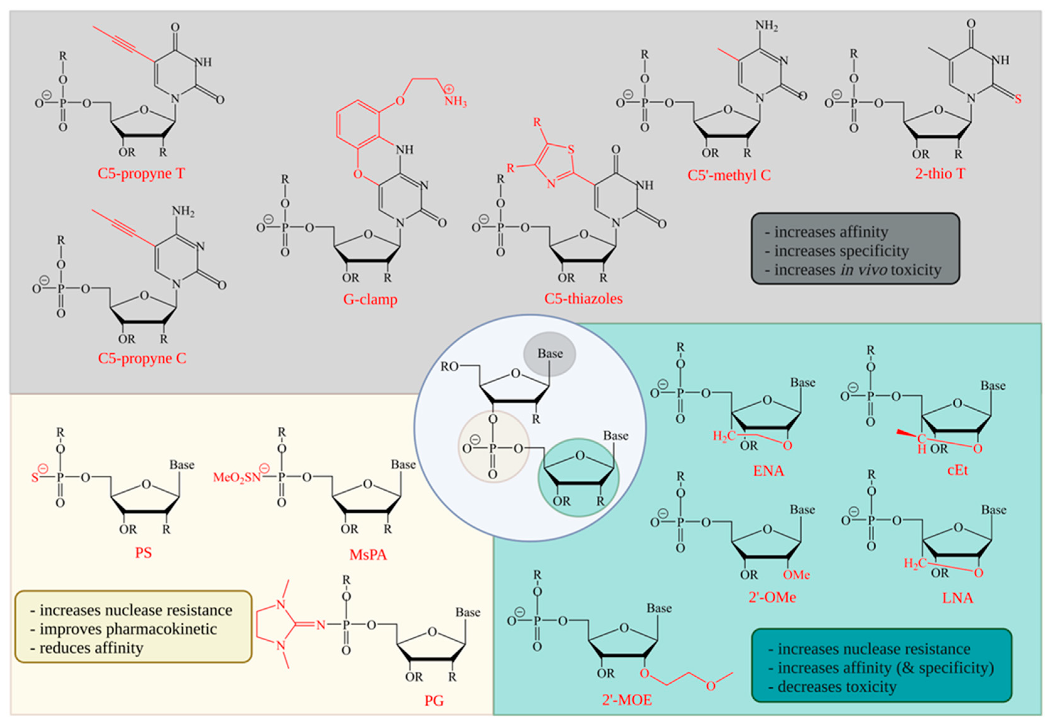
The most common modification of the backbone is a replacement of a phosphodiester (PO) with a phosphorothioate (PS) bond (Figure 2). This increases nuclease stability and cellular uptake; in addition, the interaction with albumin allows the ASO to be transported within the cerebrospinal fluid (CSF) or blood plasma [61,62,63]. ASO–protein bindings are particularly important with regard to pharmacokinetic properties, since these interactions can delay renal clearance [64]. Since this clearing mechanism is independent from the brain, the main focus on backbone modifications of ASOs for treatments of the central nervous system is on the improved uptake and transport associated with protein binding [61]. The interaction with intracellular proteins is potentially associated with an increase in toxicity, which can be reduced by a site-specific introduction of a neutral PO linkage at position 2 or 3 in the DNA gap [35,36]. However, this modification also results in a reduction of affinity, with a reduction in melting temperature of −0.45 to −1 °C per nucleotide [34]. Another aspect of a sulfur modification is that the negative charge is exclusively on the sulfur atom. This leads to different possible chiralities between the phosphorus and the sulfur atoms [65]. The effects of stereoisomerism of PS on the stability and target cleavage site are currently under debate.
RNase H interacts with the DNA:RNA junction at three consecutive nucleotides. Iwamoto et al. proposed a stereochemical PS backbone of 3′-SpSpRp-5′ in the center of the DNA gapmer as the most suitable site, with the cleavage site located two nucleotides upstream from the Rp with respect to the DNA [66]. This pattern and its capabilities could not be confirmed by Østergaard and colleagues. However, they showed that a single Rp junction at position 7 in a Sp DNA gap, resulted in a single cleavage site, located two nucleotides downstream on the DNA [37]. The potential to predefine cleavage sites could increase the focus for nucleotide-specific modifications to specific positions, thus increasing the impact of modifications at specific sites.
A relatively novel modification of the backbone is the use of mesylphosphoramidate (MsPA) [67,68]. Stetsenko et al. described positive effects on RNase H activity when DNA-ASOs were modified with MsPA compared to PS-modified DNA-ASOs [39]. Ionis Pharmaceuticals could not confirm these positive effects when DNA gapmer ASOs were uniformly modified with MsPA. Slight improvement in RNase H activity could be observed when a maximum of 5 PS bonds in the DNA gap were replaced by MsPA, or Gap 2′-OMe PS ASOs were modified with MsPA at two positions in the 3′ wing, or the 5′ DNA gap [40,69]. Additionally, MsPA positively influences toxicity and half-life of ASOs. An improvement in the stability of nucleases could be obtained when 4 PS bonds of the DNA gap were replaced by MsPA. Even more important might be the effect of MsPA on the interactions with proteins. These interactions could already be substantially reduced by 2 MsPA instead of PS in the DNA gap and could thus become particularly important if protein-associated toxicity needs to be reduced [39,40]. A recent publication indicates the interaction between the design of ASOs and the ionotropic glutamate α-amino-3-hydroxy-5-methyl-4-isoxazolepropionic acid receptor (AMPAR). It has been shown that an increasing number of G bases in an ASO gapmer increases the risk of AMPAR-associated neurotoxicity [70]. Therefore, it will be interesting to see if MsPA modifications around the Gs have an impact on the interaction with AMPAR, thereby modifying potential toxicity. In vivo studies showed a superior effect of MsPA over PS in a fully modified ASO, when delivered in folate-containing liposomes. MsPA showed an improved tumor tissue penetration and therefore, improved action and retention of tumor growth, by targeting miR-21 [71].
Recent studies suggest that the understanding of stereochemical effects and further modifications, such as by using phosphoryl guanidine diester (PG), can improve the pharmacological effects [72,73,74]. PG was also incorporated into the backbone of an ASO in a recent HD trial (NCT05032196) [75]. The modification itself was already introduced in an oligonucleotide in 2014 [76]. PGs have a strong nuclease resistance and a reduced toxicity, but also an increased reduction in affinity and a loss of RNase H recognition, when present in DNA gaps. With their good nuclease resistance, it might be beneficial to use this modification at the flanking regions of an ASO [41,42,43]. Used in steric block ASOs, PGs can improve the activity and uptake of the corresponding ASO and suppress target gene expression [44].

4. Sugar Modifications

Modifications of the sugar of a nucleotide are mostly accompanied by a modification at the 2′ C atom or a total loss of the ring structure. In both cases, the molecular character of the DNA is modified to an extent that it is not recognized by RNase H [77,78,79] (Figure 2). Therefore, such modifications are not possible in the central DNA portion of a gapmer. In general, sugar modifications in steric block ASOs, or the flanking RNA part of gapmers, improve nuclease resistance and RNA affinity [31,51]. The idea of improving the affinity of antisense strands to their target is already decades old. In 1987, Inoue et al. used a 9-nucleotide-long gapmer with two or three flanking 2′O-methylnucleotides in which the RNase H cleavage site was specifically located in the unmodified, central DNA [79]. This modification was also part of the first approved aptamer drug, pegaptanib, in 2004 [80]. Binding to RNA, this modification has minor effects on affinity, increasing melting temperature by 0 to +1.3 °C per nucleotide [27,34,38,46].
The 2′-O-methoxyethylnucleotide (2′MOE) modifications are more widely used. The 2′-MOE modifications have an even higher affinity than other modifications, as well as a good nuclear resistance, with an increase in melting temperature of +0.9 to +1.9 °C per nucleotide [34,46,52,53,81]. One of the most prominent uses of 2′MOE in ASOs is in nusinersen (Spinraza), which was FDA approved in 2016. Nusinersen is an end-to-end 2′MOE modified, 18-nucleotide-long steric block ASO that is used for the treatment of spinal muscular atrophy (SMA) [82]. A similarly prominent ASO example in which 2′MOE modifications were applied, but with a different function, is in the development of tominersen as potential treatment for Huntington’s disease (HD). The gapmer ASO, developed by Roche and IONIS Pharmaceuticals, consists of a 10-nucleotide-long DNA-gap and 5 flanking 2′-O-methoxyethylnucleotides on both wings. In March 2021, the phase III trial was stopped due to risk/benefit concerns; nonetheless, a reduction in huntingtin protein was achieved and is thus proof of the principle of using ASOs with mechanism of action via RNase H degradation [2,83,84].
In another group of sugar modifications, a linkage between the 2′-O and the 4′-C results in a C3′-endo sugar pucker of the furanose. The so-called locked nucleic acids (LNA) change the helix structure towards A-type, with respect to a DNA:RNA binding. This prevents the duplex from RNase H cleavage [85,86]. LNA:RNA bindings have a high affinity, with an increase of up to 9 °C per nucleotide with respect to DNA:RNA bindings [27,45,46,47,48,49,50]. In addition, LNA provide increased protection against endonuclease (nuclease P1) and 3′exonucleases (snake venom phosphodiesterase, SVPD) compared to DNA [49,51]. Three LNA modified nucleotides, each at the 3′ and 5′ ends, increase the half-life of the respective gapmer in human serum by 10-fold compared to unmodified DNA oligonucleotides [27]. LNAs also exhibit increased potency compared to 2′MOEs, although this is also associated with increased toxicity [87].
Like LNA, 2′4′-constrained 2′-O-ethyl (2′-cEt) are part of the bridged nucleic acids (BNA). The 2′-cEt have an additional methyl group at the connecting C-atom. Since the stereochemistry of the methyl group can vary in its connection to the C atom, they are referred to as enantiomers S-cEt and R-cEt [88]. Compared to LNAs, both analogs showed comparable thermal stability, potency, and increased nuclease stability against SVPD [26,51].
Modifications of the sugar are particularly good at increasing affinity and nuclease resistance. However, since they cannot be used in the DNA gap, their influence on the cleavage site and the specificity of individual bonds is limited.

5. Base Modifications

Base modifications may have the greatest potential to increase specificity of a single base, but they also present the greatest challenges. Although an adjustment can lead to an increase in the affinity of the base, it can also lead to an enhanced affinity for non-complementary bases, leading to an elevated likelihood of mismatches. The purpose of base modulation is to increase the specificity of the entire ASO, but most importantly, of a single base to its complementary counterpart. The smallest variants of the modification of bases are methylations, such as the 5′-methylation of cytidines (Figure 2). This methylation can reduce immunostimulatory effects, especially of CpG dinucleotides, and increase nuclease stability and thermal stability, with an increase of up to 1.1 °C per nucleotide [25,34,89]. C5′-methylations of cytidine and uracil are used throughout the molecule in some steric block ASOs, such as Spinraza, and RNase H-activating ASOs [3]. A slightly larger modification is C5′-propynyl thymine and cytosine. Compared to the single C5′-methylation, C5′propynyls increase thermal stability by +0.9 to +2.6 °C per nucleotide. Depending on their position and stereochemistry, they may even improve specificity [34,54,55,57,58,59]. The 5′thiazole pyrimidine analogues have similar characteristics for base stacking, and they improve thermal stability, with +1.7 to +2.2 °C per base [59].
A further step towards allele-selective oligonucleotide modifications could be achieved by 2′-thio pyrimidine modifications. Østergaard et al. showed that by incorporating a single 2′-thio deoxythymidine (2′-thio dT) into the DNA gap of an ASO, the affinity, but also the specificity, could be increased. For a HTT SNP-targeting ASO, the difference between melting temperatures was highest (5.6 °C) when the 2′-thio dT modification was at the position of the SNP [60]. Another well-studied pyrimidine analogue is the G-clamp, a cytosine that is scaled up to a phenoxazine with 9′-O-aminoethyl. The G-clamp and newer approaches, like G8AE-clamp, or guadino-G-clamp, improve binding affinity and specificity to guanine by making additional use of the Hoogsteen binding site, forming a fourth hydrogen bond to guanine [56,90,91,92]. By this, the melting temperature can be increased by +4 to +18 °C per nucleotide [46,56], especially when the G-clamp is flanked by a 5′-cytidine. In addition, the G-clamp has fluorescent properties and a 3′exonuclease resistance [56]. Increased specificity was shown in a DNA-ASO consisting of 10 nucleotides. Comparisons between 5′methylcytosine and G-clamp modifications resulted in an increase in thermal stability by 18 °C with the G-clamp. Although the melting temperature of all mismatch partners also increased compared with 5′-methylcytosine, the difference in melting temperature between match and mismatch was greatest for the G-clamp, suggesting increased specificity [55]. At the same time, the incorporation of a single G-clamp appears to have little or no effect on RNase H cleavage [46,55]. It remains to be shown if specific base modifications, like the G-clamp, can be used to improve SNP-based allele-specific targeting approaches.

6. Selective vs. Non-Selective Targeting Strategies

Dominant inherited diseases are often caused by heterozygous mutations, with a toxic gain-of-function. Non-selective targeting of the diseased gene would reduce both the toxic and the wild type variant and therefore, diminish the function of the protein. With a selective targeting of the mutated allele, the toxic protein load could be reduced, while preserving the physiological function of the wild type protein. Even though tominersen, a non-selective HTT ASO, showed dose-dependent reduction in HTT in the phase I/IIa trial [93], the phase III trial was stopped because of unfavorable effects outweighing potential benefits [84,94,95,96]. Previous animal studies suggested that the toxic effect of mtHTT is greater than the impairment resulting from a total loss of HTT [97]. However, it is hard to predict the ideal knockdown efficiency for nonselective degradation strategies, since it is difficult to estimate the right balance between necessary reduction in toxic mtHTT and a negative treatment response due to the loss of wtHTT [2]. Similarly, in another CAG repeat disease, SCA3, very promising non-allele-specific reduction in ATXN3 has been achieved in humanized SCA3 mice, as well as patient-derived hESCs [98,99,100]. Recently, SCA3 ASOs have just entered phase I (NCT05160558).
Table 2 gives an overview of current clinical trials using ASOs in neurodegenerative diseases caused by toxic gain-of-function mutations (reviewed in [8,101]). Treating these diseases with non-selective ASOs would result in a reduction in the mutated and wild type protein with the aim of decreasing the toxic effects. At the same time, potential protective and physiologically necessary functions of the wild type protein are also reduced. Even though some functions of proteins causing neurodegeneration are known, there are still uncertainties (Table 2). An unspecific knockdown of such a protein may have serious, unforeseeable consequences. Specifically targeting the mutant allele is supposed to result in a selective degradation of the toxic protein and preservation of the healthy allele. One of the most approachable uses of an ASO that aims for allele-specificity would be to target the mutation directly. In the case of HD or polyQ SCAs, this would be the expanded CAG repeat. An approach to circumvent the toxic effect of the expanded CAG repeat is alternative splicing. Toonen et al. masked splicing signals of the SCA3 pre-mRNA, resulting in a shortened protein version that bypasses the CAG repeat containing exon 10 and most parts of exon 11 due to an early stop codon. This modified protein showed beneficial effects on pathogenicity, with a detectable in vivo longevity of 2.5 months [102]. Even though the approach was specific for CAG toxicity, the effects were not purely allele-specific, since both alleles are targeted with these steric block ASOs. One way to directly eliminate allele-selective toxicity of the expanded CAG repeat is to use ASOs that directly target the CAG repeat at the (pre-)mRNA level. Since several proteins physiologically possess CAG repeats, a single ASO could be used for treating several different CAG repeat diseases. Likewise, the number of potential off-targets may increase, since such a CAG-specific ASO could potentially bind to several genes with CAG repeats. Evers et al. observed that such an ASO, with 21 nucleotides targeting the CAG repeat, showed allele-specific reduction not only in HTT, but also in other CAG repeat diseases, such as SCA1, 3, and DRPLA. No reduction was detected in potential CAG repeats containing off-targets, such as SCA2, ZNF384, or TATA box binding protein (TBP) [103]. Other publications with predominantly steric block ASOs show similar success in HD, SCA1, and SCA3, with dose-dependent, allele-specific reductions of up to 6.6-fold. It is believed that these steric block ASOs bind more efficient to the expanded CAG repeat containing mRNA compared to the shorter WT version, thus providing translation in an allele-specific manner [104,105,106]. A potential disadvantage of directly targeting the CAG repeat could be that every third nucleotide of the ASO that targets the CAG repeat is a guanine. Recent studies showed that in vitro and in vivo neurotoxicity is associated with the number and position of Gs within an ASO. The risk for a potential neurotoxicity increases with the number of Gs within the ASO sequence.
A less strong association to neurotoxicity was shown for the distance from the 3′-end to the first guanine. In contrast, increasing the number of As in an ASO had positive effects on the risk of potential neurotoxicity [70].
Like CAG repeat disorders, ASOs were also tested targeting other repeat diseases, such as C9ORF72 in ALS/FTD patient cells. Different ASOs were designed targeting either the C9ORF72 repeat (GGGGCC) itself or exonic and intronic regions up- and downstream of the repeat. Experiments on patient-derived cells and in vivo experiments showed mainly positive and allele-specific effects [130,131]. Recent publications also confirmed the possibility of achieving effective reduction via the RNase H-mediated degradation of C9ORF72, targeting either the repeat or intronic sequences of the gene [73,132]. Treatment of a patient with C9-ALS/FTD showed a marked reduction in polyGP-DPR (dipeptide repeat protein), with no medical or neurological adverse effects [132].
Success in patients was also reported with ASO treatment of other genes in the ALS/FTD spectrum. Treatment of an ALS patient carrying the FUSP525L mutation with ION363 (jacifusen) resulted in a mutant-specific reduction of FUS protein and its aggregation, reaching clinical phase III trials (Table 2) [113]. Additionally, an ASO that targets superoxide dismutase 1 (SOD1) also reached phase III in clinical studies [115,130,133]. Clinical trials have also been initiated in Alzheimer’s disease using ASOs targeting MAPT. Preclinical studies in mice led to a significant reduction in human TAU, aggregation, and cell loss, resulting in a lifespan extension [134]. Testing of the gapmer ASO IONIS-MAPTRx (NCT03186989) in non-human primates showed a MAPT mRNA reduction of nearly 80%. These promising preclinical experiments resulted in the initiation of a first clinical trial for patients with mild AD [123,135]. Further potential targets for AD treatment are other genes that are directly or indirectly related to Alzheimer’s disease, such as APP and APOER2. The mRNA concentrations of these genes could also be reduced via steric block ASOs and may have beneficial effects on the course of AD [136,137].
For Parkinson’s disease, preclinical experiments in rodents achieved promising results in gapmer ASO treatment of different PD-associated genes [125,138,139]. Phase I and II clinical trials against both LRRK2 and SCNA are ongoing (NCT03976349, NCT04165486).

7. SNP-Based Allele-Specific Treatment Strategies

Steric block ASOs that modulate splicing can be used to generate a semi-functional or non-functional protein. This shortened protein version lacks the toxic gain-of-function mutation of interest. Nevertheless, the total functional protein concentration is also lowered in this approach. Comparable effects can be achieved by non-allele-specific degradation via RNase H. Alternatively, an allele-specific degradation by targeting an SNP could surpass these non-allele-selective approaches. If the wild type allele and the disease-associated allele differ in one base at a specific site, this site might be a potential target for an allele-specific treatment strategy that aims to degrade the toxic allele at the (pre-)mRNA level. The association of a targetable SNP with the disease causing mutation might be ideal, but is not essential for such an approach. Even distant SNPs that are present in coding or non-coding regions of the disease gene far from the mutation can be used as a target to tag the disease-associated allele for degradation. The same principle can be used for steric block ASOs. Distant SNPs involved in splicing that are located in the same gene as the disease mutation could be used to disrupt the splicing machinery at the mutant allele.
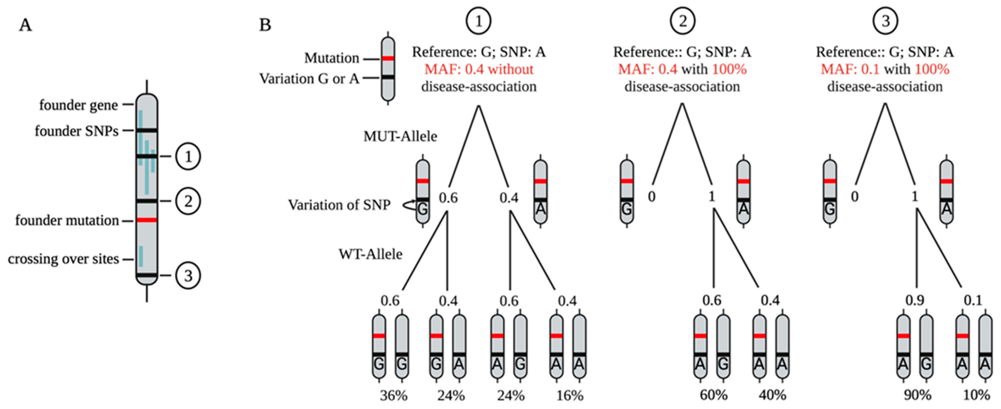
In 2014, Skotte et al. published an approach to use HD-associated SNPs for allele-specific RNase H-mediated degradation of mtHTT. They evaluated 50 previously published HD-associated SNPs for targetability [54,140,141]. A total of 4 SNPs were identified in which ASOs showed increased degradation. Further optimization of the ASO sequence increased selectivity from 2.4-fold to over 100-fold and reduced the IC50 value against mtHTT from over 400 nM to single digit nM values for an ASO targeting the SNP rs7685686_A. To achieve this, the modifications of the flanking wings played an important role, as well as the shortening of the length of the ASO and the length of the DNA gap region. It was shown that a shorter gap, due to the shortened binding region of RNase H, resulted in enhanced allele-selective degradation. Furthermore, this study demonstrated that the SNP does not need to be in the central position of the DNA gap to achieve allele-specific degradation. By targeting the selected SNP, it would be possible to treat 48.7% of the analyzed HD population allele-specifically and another 44.9% in a non-allele-specific manner [7]. In vivo follow-up studies of this ASO in humanized HD mice (Hu97/18) confirmed the allele-specific lowering of mtHTT and showed positive effects in behavioral tests [142,143]. In follow-up studies, Kay et al. precisely analyzed 63 SNPs in HTT from people of Canadian, Swedish, French, Italian, Korean, Japanese, Chinese, and European-Canadian ethnicity, revealing three major gene-spanning haplotype groups. Differences between controls and patients of each ethnic group were used to identify the most suitable SNPs for an allele-specific treatment approach, including the analysis of the association of some SNPs to the expanded CAG repeat [144,145]. In addition to SNPs, HD-associated insertion–deletion variations (indels) can potentially be used as targets for an allele-specific approach [146]. The identification and characterization of disease-associated SNPs is a crucial step towards the design of ASOs that can potentially be used to treat the majority of patients in an allele-specific manner (Figure 3).
In a recent study, our group made use of an already known disease-associated SNP in ATXN3 for an allele-specific ASO-based treatment in spinocerebellar ataxia type 3 (SCA3). Different ASOs were designed targeting a variant that is present in ~70% of SCA3 patients [6,147]. By establishing an in vitro platform using patient-derived iPSC-based cortical neurons, we were able to identify an ASO which leads to an allele-specific reduction in the glutamine-expanded allele by up to 75%. A one-time application showed significant allele-specific reduction for more than 7 weeks [6].
Together, these studies demonstrate that rare neurodegenerative diseases could play an important pioneering role in the development of allele-specific ASOs, especially when different modifications need to be tested in an in vitro screening. For this, an easily detectable and robust readout to quantify the allele-specific targeting efficiency is a crucial prerequisite. In the case of SCA3, iPSC-based patient-derived neuronal cultures are an ideal tool. Due to the repeat expansion, the mutant and wild type protein differ not only functionally, but also in their molecular mass. This difference can be used to display both forms separately at the protein level. Experience gained from an allele-specific design for ASOs targeting in HTT and SCA3 could then be transferred to diseases where allele-specific readout poses significant hurdles.

8. Target-Based ASO Design

One of the biggest challenges is to find a suitable target for an allele-specific ASO approach. If the variant that causes the disease can be directly targeted, it is usually well described. This becomes more difficult if new targets or SNPs need to be identified. As described, these can be SNPs that are located in the disease-causing gene, but are not causally related to the disease. To identify SNPs that are unrelated to the disease per se, but are in close linkage disequilibrium (LD) with a disease-associated mutation (point mutations or repeats), long-coding sequencing of larger cohorts would be of help (Figure 3). Alternatively, larger databases such as https://www.ensembl.org/index.html (accessed on 23 June 2022) or https://bravo.sph.umich.edu/freeze8/hg38/ (accessed on 23 June 2022) can be used to identify a list of candidate SNPs. Suitable new targets should have a high MAF or a high association with the disease mutation (Figure 3). A hint could be given by the MAF or the linkage disequilibrium (LD) of the potential candidate SNP with the disease-causing variant (https://ldlink.nci.nih.gov/?tab=home (accessed on 23 June 2022)) [148]. Once a list of candidate SNPs has been generated, other factors can help with further selection. The target area of the (pre-)mRNA needs to be accessible for the ASO. Computations of potential 2D structures can predict easily or poorly accessible areas (http://rna.tbi.univie.ac.at (accessed on 23 June 2022)) [149]. Short sequence patterns within an ASO are expected to have positive or negative effects on ASO activity [150]. The probability of complementary off-targets can be calculated via GGGenome (https://gggenome.dbcls.jp (accessed on 23 June 2022)). Especially for an allele-specific approach, it is advantageous if the sequence is as short as possible. With each additional nucleotide, the affinity increases, while the contribution of the individual base to the total melting temperature decreases. Thus, the influence of a single match or mismatch decreases the longer an ASO is. In addition, base variation may also play a role. Some allelic SNP constellations might be more suitable for allele-specific targeting than others. Cytosines may be more suitable as mismatch partners, as they show a greater difference in melting temperature compared to the match [151,152]. In addition, neurotoxic effects of ASOs play an important role, especially for later application. Thus, the number and position of G’s in an ASO sequence should be taken into account [70]. Once an appropriate SNP has been identified and the ASO designed, a suitable model is needed. For in vitro experiments, cells must be heterozygous for the targeted SNP and should ideally reflect the primarily affected organ/cell type. iPSC-derived cell cultures are particularly well suited for this purpose (Figure 4). They offer the advantage that they directly reflect a patient’s heterozygous genotype and can be differentiated into any cell type of interest, including neuronal subtypes affected by the disease (reviewed in [153,154]). Therapeutic effects and off-targets can easily be analyzed by transcriptomic and/or proteomic studies. (Neuro)toxicity can be quantified by, e.g., LDH assays or calcium oscillation measurements.
Another challenge is to find a suitable in vivo model, mainly mouse strains. This model should carry the target SNP on the same allele that bears the disease-associated variant. Lentiviral transduction of a humanized mutant allele with the respective target SNP has the advantage that the inserted allele and its SNPs can easily be adapted and that wild type mice can be used. Such transductions with the target allele can be performed prior to an ASO application [155,156]. In vivo mouse experiments are of particular importance for testing the in vivo functionality and acute toxicity of ASOs. Due to genomic differences between mice and humans, mouse models are less suitable for testing off-targets. Therefore, in vivo off-target studies need to be performed in non-human primates. Brain organoids from human iPSC may become increasingly important, especially in early in vitro off-target screenings (Figure 4). These self-assembled three-dimensional structures resemble the embryonic human brain and include multiple neural cell types, yielding the potential to extend the time of an ASO treatment in a multicellular system, which might be especially important for neurodegenerative diseases (reviewed in [157,158,159]).

9. The Challenge of Readout

In addition to the identification of appropriate SNPs, the identification of suitable readouts to prove allele-specificity is another challenge that varies in difficulty between diseases. As previously mentioned, repeat expansion disorders, such as HD [7] or DRPLA [160], bear the advantage of different molecular weight between the wild type and expanded disease protein that can be used to discriminate the respective protein. In SCA3, the wild type and mutant allele typically differ by about 40 CAG repeats, resulting in an almost 6 kDa difference in molecular weight at the protein level. Thus, both proteins can easily be separated and discriminated by Western blot testing (WB) [6,161]. In other CAG repeat expansion diseases, such as SCA1, 2, or 6, absolute size differences are smaller, making it more challenging to separate the two proteins by WB. Longer runtimes and adjustment of gel type and gradient could help in solving this problem. Alternatively, proteins or mRNA could be predigested so that the difference in glutamine repeats relative to the total peptide becomes more distinct [103]. TR-FRET assays can also be used to discriminate between mutant and wild type protein. In this assay, a fluorophore coupled antibody binds specifically to the expanded polyQ part of the mutant protein, while a second antibody binds to a different part of the protein. Only when both antibodies bind does an energy transfer occur from one antibody to the other, resulting in a fluorescent signal that can be detected and quantified. A second pair of antibodies that binds both versions of the protein is used to determine total protein concentration [162].
A more elaborated approach would be to artificially create an in vitro model by transfecting cells with either the wild type sequence or a sequence with the corresponding SNP. Subsequently, cells could be treated with ASOs, which are then examined for their allele-specific effects. A fluorescence tag could help not only to control the transfection, but also to distinguish the transfected allele from the endogenous variant. Alternatively, cells could be transfected with both variants simultaneously, if the two variants have distinct tags. This would allow for studying allele-specific effects upon an ASO treatment directly in a cell culture model and later on, in vivo. A benefit of this approach could be that multiple SNP targets could be inserted into the mutant allele variant. Therefore, cooperative ASO effects could also be part of an in vitro investigation. With this approach, multiple ASOs initiate degradation of the same target by binding to different positions.
An indirect approach to test allele specificity of an ASO could be an RNase H assay. Both variants of the target RNA need to be incubated as a duplex with the corresponding ASO (with and without mismatch) and RNase H. In a fluorometric RNase H assay, the released DNA of the gapmer binds with a N-methyl mesoporphyrin IX (NMM), forming a G-quadruplex with a fluorescence signal [163], which can be amplified by using a catalytic hairpin assembly [164]. In a next step, the efficacy could be tested in vitro and in vivo.

10. Conclusions

Currently, no allele-specific ASO has been approved for the treatment of a neurodegenerative disease. However, an allele-selective approach with ASOs via RNase H degradation could have great potential, especially for toxic gain-of-function mutations. If successful, the toxic allele would be eliminated, and the wild type allele could continue to maintain its physiological function. In such an approach, all coding or non-coding SNPs that are on the same allele as the disease-causing variant are potential candidates for a personalized therapeutic approach [6,144,145], (Figure 3). One reason why current research is still in its early stages might be that the allele-specific readout for most diseases has major hurdles. Repeat expansion diseases, in which a readout might be established by a different molecular weight between the wild type and mutant protein, could be flagship projects for the development of allele-specific targeting therapies using ASOs.
To standardize the identification of targetable candidate SNPs for a specific disease, long-read sequencing can help (reviewed in [165]) to directly prove the disease-association of a candidate SNP. In case of a high association of a candidate SNP with the disease-causing mutation, these SNPs would be ideal starting points to design and test allele-specific ASOs, including different modifications to increase allele-specificity. These ASOs could then be used to treat the majority of patients in an allele-specific manner.
Regarding ASO modifications, much time and effort has been spent on sugar phosphate backbone modifications. Since base modifications, such as G-clamp, can have the greatest impact on melting temperature per nucleotide [46,56] and therefore, the specificity and affinity of a single nucleotide, these modifications might be important to improve allele-selectivity. Another potential research focus for the future might be to analyze the cooperative effects of ASOs and whether there are ways to improve the administration of multiple different ASOs to enhance target engagement [166]. By administering a combination of ASOs, lower concentrations of each individual oligonucleotide could be used, reducing potential off-target effects of every single ASO. Thus, depending on the SNPs on the disease-associated allele, each patient could be given an ASO cocktail that specifically targets the mutant (pre)-mRNA for degradation through multiple binding sites.
Another hurdle is to establish a standardized workflow to identify and characterize ASOs in preclinical trials. We here propose a workflow for the design and preclinical testing of allele-specific ASOs (Figure 4). After successful target evaluation, several lead ASOs could be tested in parallel. The most effective of these will be further modified to ideally exclude toxicity and increase affinity, specificity, and longevity. Modified ASOs should then be characterized in vitro in 2D and 3D human cell culture models, as well as in vivo models. This includes effect-strength, allele-specificity, longevity, off-target effects, and (neuro)toxicity. While many challenges remain regarding allele-specific ASO therapies, allele-specific ASO strategies provide a great opportunity for future therapeutic approaches, especially for toxic gain-of-function neurodegenerative diseases.

Author Contributions

Conceptualization, J.H. and S.H.; writing—original draft preparation, J.H.; writing—review and editing, S.H. and L.S.; visualization, J.H. and S.H.; supervision, S.H. and L.S.; project administration, S.H. and L.S.; funding acquisition, S.H. and L.S. All authors have read and agreed to the published version of the manuscript.

Funding

The work of L.S. has been supported by the German Ministry of Education and Research (BMBF grant 01ED16028).

Institutional Review Board Statement

Not applicable.

Informed Consent Statement

Not applicable.

Data Availability Statement

Not applicable.

Acknowledgments

Figures were created with BioRender.com. We acknowledge support from the Open Access Publishing Fund of the University of Tübingen.

Conflicts of Interest

The authors declare no conflict of interest.

References

  1. Testa, C.M. Antisense Oligonucleotide Therapeutics for Neurodegenerative Disorders. Curr. Geriatr. Rep. 2022, 11, 19–32. [Google Scholar] [CrossRef]
  2. Rook, M.E.; Southwell, A.L. Antisense Oligonucleotide Therapy: From Design to the Huntington Disease Clinic. BioDrugs 2022, 36, 105–119. [Google Scholar] [CrossRef] [PubMed]
  3. Roberts, T.C.; Langer, R.; Wood, M.J.A. Advances in oligonucleotide drug delivery. Nat. Rev. Drug Discov. 2020, 19, 673–694. [Google Scholar] [CrossRef] [PubMed]
  4. Crooke, S.T.; Baker, B.F.; Crooke, R.M.; Liang, X.-H. Antisense technology: An overview and prospectus. Nat. Rev. Drug Discov. 2021, 20, 427–453. [Google Scholar] [CrossRef] [PubMed]
  5. Scoles, D.R.; Minikel, E.V.; Pulst, S.M. Antisense oligonucleotides: A primer. Neurol. Genet. 2019, 5, e323. [Google Scholar] [CrossRef] [PubMed]
  6. Hauser, S.; Helm, J.; Kraft, M.; Korneck, M.; Hübener-Schmid, J.; Schöls, L. Allele-specific targeting of mutant ataxin-3 by antisense oligonucleotides in SCA3-iPSC-derived neurons. Mol. Ther.-Nucleic Acids 2022, 27, 99–108. [Google Scholar] [CrossRef] [PubMed]
  7. Skotte, N.H.; Southwell, A.L.; Østergaard, M.E.; Carroll, J.B.; Warby, S.C.; Doty, C.N.; Petoukhov, E.; Vaid, K.; Kordasiewicz, H.; Watt, A.T. Allele-specific suppression of mutant huntingtin using antisense oligonucleotides: Providing a therapeutic option for all Huntington disease patients. PLoS ONE 2014, 9, e107434. [Google Scholar] [CrossRef]
  8. Taylor, J.P.; Hardy, J.; Fischbeck, K.H. Toxic Proteins in Neurodegenerative Disease. Science 2002, 296, 1991–1995. [Google Scholar] [CrossRef]
  9. Scekic-Zahirovic, J.; Sendscheid, O.; El Oussini, H.; Jambeau, M.; Sun, Y.; Mersmann, S.; Wagner, M.; Dieterlé, S.; Sinniger, J.; Dirrig-Grosch, S. Toxic gain of function from mutant FUS protein is crucial to trigger cell autonomous motor neuron loss. EMBO J. 2016, 35, 1077–1097. [Google Scholar] [CrossRef]
  10. Yankner, B.A.; Lu, T. Amyloid β-protein toxicity and the pathogenesis of Alzheimer disease. J. Biol. Chem. 2009, 284, 4755–4759. [Google Scholar] [CrossRef]
  11. Paulson, H.L.; Perez, M.; Trottier, Y.; Trojanowski, J.; Subramony, S.; Das, S.; Vig, P.; Mandel, J.-L.; Fischbeck, K.; Pittman, R. Intranuclear inclusions of expanded polyglutamine protein in spinocerebellar ataxia type 3. Neuron 1997, 19, 333–344. [Google Scholar] [CrossRef]
  12. Ishikawa, K.; Nagai, Y. Molecular mechanisms and future therapeutics for spinocerebellar ataxia type 31 (SCA31). Neurotherapeutics 2019, 16, 1106–1114. [Google Scholar] [CrossRef] [PubMed]
  13. Chou, C.-C.; Zhang, Y.; Umoh, M.E.; Vaughan, S.W.; Lorenzini, I.; Liu, F.; Sayegh, M.; Donlin-Asp, P.G.; Chen, Y.H.; Duong, D.M. TDP-43 pathology disrupts nuclear pore complexes and nucleocytoplasmic transport in ALS/FTD. Nat. Neurosci. 2018, 21, 228–239. [Google Scholar] [CrossRef] [PubMed]
  14. Grima, J.C.; Daigle, J.G.; Arbez, N.; Cunningham, K.C.; Zhang, K.; Ochaba, J.; Geater, C.; Morozko, E.; Stocksdale, J.; Glatzer, J.C. Mutant huntingtin disrupts the nuclear pore complex. Neuron 2017, 94, 93–107.e6. [Google Scholar] [CrossRef] [PubMed]
  15. MacDonald, M.E.; Novelletto, A.; Lin, C.; Tagle, D.; Barnes, G.; Bates, G.; Taylor, S.; Allitto, B.; Altherr, M.; Myers, R. The Huntington’s disease candidate region exhibits many different haplotypes. Nat. Genet. 1992, 1, 99–103. [Google Scholar] [CrossRef] [PubMed]
  16. Simon-Sanchez, J.; Schulte, C.; Bras, J.M.; Sharma, M.; Gibbs, J.R.; Berg, D.; Paisan-Ruiz, C.; Lichtner, P.; Scholz, S.W.; Hernandez, D.G. Genome-wide association study reveals genetic risk underlying Parkinson’s disease. Nat. Genet. 2009, 41, 1308–1312. [Google Scholar] [CrossRef]
  17. Romanul, F.C.; Fowler, H.L.; Radvany, J.; Feldman, R.G.; Feingold, M. Azorean disease of the nervous system. N. Engl. J. Med. 1977, 296, 1505–1508. [Google Scholar] [CrossRef]
  18. Morita, M.; Al-Chalabi, A.; Andersen, P.; Hosler, B.; Sapp, P.; Englund, E.; Mitchell, J.; Habgood, J.; De Belleroche, J.; Xi, J. A locus on chromosome 9p confers susceptibility to ALS and frontotemporal dementia. Neurology 2006, 66, 839–844. [Google Scholar] [CrossRef]
  19. Chandran, J.S.; Scarrott, J.M.; Shaw, P.J.; Azzouz, M. Gene therapy in the nervous system: Failures and successes. Pers. Med. 2017, 1007, 241–257. [Google Scholar]
  20. Seidel, K.; Schöls, L.; Nuber, S.; Petrasch-Parwez, E.; Gierga, K.; Wszolek, Z.; Dickson, D.; Gai, W.P.; Bornemann, A.; Riess, O. First appraisal of brain pathology owing to A30P mutant alpha-synuclein. Ann. Neurol. 2010, 67, 684–689. [Google Scholar] [CrossRef]
  21. Metzger, S.; Rong, J.; Nguyen, H.-P.; Cape, A.; Tomiuk, J.; Soehn, A.S.; Propping, P.; Freudenberg-Hua, Y.; Freudenberg, J.; Tong, L.; et al. Huntingtin-associated protein-1 is a modifier of the age-at-onset of Huntington’s disease. Hum. Mol. Genet. 2008, 17, 1137–1146. [Google Scholar] [CrossRef] [PubMed]
  22. Juliano, R.L. Chemical Manipulation of the Endosome Trafficking Machinery: Implications for Oligonucleotide Delivery. Biomedicines 2021, 9, 512. [Google Scholar] [CrossRef]
  23. Crooke, S.T.; Wang, S.; Vickers, T.A.; Shen, W.; Liang, X.-h. Cellular uptake and trafficking of antisense oligonucleotides. Nat. Biotechnol. 2017, 35, 230–237. [Google Scholar] [CrossRef] [PubMed]
  24. Geary, R.S.; Norris, D.; Yu, R.; Bennett, C.F. Pharmacokinetics, biodistribution and cell uptake of antisense oligonucleotides. Adv. Drug Deliv. Rev. 2015, 87, 46–51. [Google Scholar] [CrossRef]
  25. Wan, W.B.; Seth, P.P. The Medicinal Chemistry of Therapeutic Oligonucleotides. J. Med. Chem. 2016, 59, 9645–9667. [Google Scholar] [CrossRef]
  26. Seth, P.P.; Siwkowski, A.; Allerson, C.R.; Vasquez, G.; Lee, S.; Prakash, T.P.; Wancewicz, E.V.; Witchell, D.; Swayze, E.E. Short Antisense Oligonucleotides with Novel 2′−4′ Conformationaly Restricted Nucleoside Analogues Show Improved Potency without Increased Toxicity in Animals. J. Med. Chem. 2009, 52, 10–13. [Google Scholar] [CrossRef]
  27. Kurreck, J.; Wyszko, E.; Gillen, C.; Erdmann, V.A. Design of antisense oligonucleotides stabilized by locked nucleic acids. Nucleic Acids Res. 2002, 30, 1911–1918. [Google Scholar] [CrossRef] [PubMed]
  28. Lu-Nguyen, N.; Malerba, A.; Herath, S.; Dickson, G.; Popplewell, L. Systemic antisense therapeutics inhibiting DUX4 expression ameliorates FSHD-like pathology in an FSHD mouse model. Hum. Mol. Genet. 2021, 30, 1398–1412. [Google Scholar] [CrossRef]
  29. Synofzik, M.; van Roon-Mom, W.M.; Marckmann, G.; van Duyvenvoorde, H.A.; Graessner, H.; Schüle, R.; Aartsma-Rus, A. Preparing n-of-1 antisense oligonucleotide treatments for rare neurological diseases in Europe: Genetic, regulatory, and ethical perspectives. Nucleic Acid Ther. 2022, 32, 83–94. [Google Scholar] [CrossRef]
  30. Crooke, S.T. Molecular mechanisms of antisense oligonucleotides. Nucleic Acid Ther. 2017, 27, 70–77. [Google Scholar] [CrossRef]
  31. Lim, K.R.Q.; Yokota, T. Invention and early history of gapmers. Gapmers 2020, 2176, 3–19. [Google Scholar]
  32. Lima, W.; Wu, H.; Crooke, S.T. The RNase H mechanism. Antisense Drug Technol. Princ. Strateg. Appl. 2007, 2, 47–74. [Google Scholar]
  33. Liang, X.-H.; Sun, H.; Nichols, J.G.; Crooke, S.T. RNase H1-dependent antisense oligonucleotides are robustly active in directing RNA cleavage in both the cytoplasm and the nucleus. Mol. Ther. 2017, 25, 2075–2092. [Google Scholar] [CrossRef]
  34. Freier, S.M.; Altmann, K.H. The ups and downs of nucleic acid duplex stability: Structure-stability studies on chemically-modified DNA:RNA duplexes. Nucleic Acids Res. 1997, 25, 4429–4443. [Google Scholar] [CrossRef]
  35. Migawa, M.T.; Shen, W.; Wan, W.B.; Vasquez, G.; Oestergaard, M.E.; Low, A.; De Hoyos, C.L.; Gupta, R.; Murray, S.; Tanowitz, M.; et al. Site-specific replacement of phosphorothioate with alkyl phosphonate linkages enhances the therapeutic profile of gapmer ASOs by modulating interactions with cellular proteins. Nucleic Acids Res. 2019, 47, 5465–5479. [Google Scholar] [CrossRef] [PubMed]
  36. Shen, W.; De Hoyos, C.L.; Migawa, M.T.; Vickers, T.A.; Sun, H.; Low, A.; Bell, T.A., 3rd; Rahdar, M.; Mukhopadhyay, S.; Hart, C.E.; et al. Chemical modification of PS-ASO therapeutics reduces cellular protein-binding and improves the therapeutic index. Nat. Biotechnol. 2019, 37, 640–650. [Google Scholar] [CrossRef]
  37. Østergaard, M.E.; De Hoyos, C.L.; Wan, W.B.; Shen, W.; Low, A.; Berdeja, A.; Vasquez, G.; Murray, S.; Migawa, M.T.; Liang, X.-h.; et al. Understanding the effect of controlling phosphorothioate chirality in the DNA gap on the potency and safety of gapmer antisense oligonucleotides. Nucleic Acids Res. 2020, 48, 1691–1700. [Google Scholar] [CrossRef]
  38. Stein, D.; Foster, E.; Huang, S.B.; Weller, D.; Summerton, J. A specificity comparison of four antisense types: Morpholino, 2’-O-methyl RNA, DNA, and phosphorothioate DNA. Antisense Nucleic Acid Drug Dev. 1997, 7, 151–157. [Google Scholar] [CrossRef]
  39. Miroshnichenko, S.K.; Patutina, O.A.; Burakova, E.A.; Chelobanov, B.P.; Fokina, A.A.; Vlassov, V.V.; Altman, S.; Zenkova, M.A.; Stetsenko, D.A. Mesyl phosphoramidate antisense oligonucleotides as an alternative to phosphorothioates with improved biochemical and biological properties. Proc. Natl. Acad. Sci. USA 2019, 116, 1229–1234. [Google Scholar] [CrossRef]
  40. Anderson, B.A.; Freestone, G.C.; Low, A.; De-Hoyos, C.L.; Iii, W.J.D.; Østergaard, M.E.; Migawa, M.T.; Fazio, M.; Wan, W.B.; Berdeja, A. Towards next generation antisense oligonucleotides: Mesylphosphoramidate modification improves therapeutic index and duration of effect of gapmer antisense oligonucleotides. Nucleic Acids Res. 2021, 49, 9026–9041. [Google Scholar] [CrossRef]
  41. Lomzov, A.A.; Kupryushkin, M.S.; Shernyukov, A.V.; Nekrasov, M.D.; Dovydenko, I.S.; Stetsenko, D.A.; Pyshnyi, D.V. Diastereomers of a mono-substituted phosphoryl guanidine trideoxyribonucleotide: Isolation and properties. Biochem. Biophys. Res. Commun. 2019, 513, 807–811. [Google Scholar] [CrossRef] [PubMed]
  42. Kupryushkin, M.S.; Filatov, A.V.; Mironova, N.L.; Patutina, O.A.; Chernikov, I.V.; Chernolovskaya, E.L.; Zenkova, M.A.; Pyshnyi, D.V.; Stetsenko, D.A.; Altman, S. Antisense oligonucleotide gapmers containing phosphoryl guanidine groups reverse MDR1-mediated multiple drug resistance of tumor cells. Mol. Ther.-Nucleic Acids 2022, 27, 211–226. [Google Scholar] [CrossRef] [PubMed]
  43. Dyudeeva, E.; Kupryushkin, M.; Lomzov, A.; Pyshnaya, B.; Pyshnyi, D. Physicochemical properties of the phosphoryl guanidine oligodeoxyribonucleotide analogs. Russ. J. Bioorg. Chem. 2019, 45, 709–718. [Google Scholar] [CrossRef]
  44. Skvortsova, Y.V.; Salina, E.G.; Burakova, E.A.; Bychenko, O.S.; Stetsenko, D.A.; Azhikina, T.L. A new antisense phosphoryl guanidine oligo-2′-O-methylribonucleotide penetrates into intracellular mycobacteria and suppresses target gene expression. Front. Pharmacol. 2019, 10, 1049. [Google Scholar] [CrossRef] [PubMed]
  45. Bondensgaard, K.; Petersen, M.; Singh, S.K.; Rajwanshi, V.K.; Kumar, R.; Wengel, J.; Jacobsen, J.P. Structural studies of LNA: RNA duplexes by NMR: Conformations and implications for RNase H activity. Chem.-Eur. J. 2000, 6, 2687–2695. [Google Scholar] [CrossRef]
  46. Stanton, R.; Sciabola, S.; Salatto, C.; Weng, Y.; Moshinsky, D.; Little, J.; Walters, E.; Kreeger, J.; DiMattia, D.; Chen, T. Chemical modification study of antisense gapmers. Nucleic Acid Ther. 2012, 22, 344–359. [Google Scholar] [CrossRef]
  47. Singh, S.K.; Koshkin, A.A.; Wengel, J.; Nielsen, P. LNA (locked nucleic acids): Synthesis and high-affinity nucleic acid recognition. Chem. Commun. 1998, 455–456. [Google Scholar] [CrossRef]
  48. Koshkin, A.A.; Singh, S.K.; Nielsen, P.; Rajwanshi, V.K.; Kumar, R.; Meldgaard, M.; Olsen, C.E.; Wengel, J. LNA (Locked Nucleic Acids): Synthesis of the adenine, cytosine, guanine, 5-methylcytosine, thymine and uracil bicyclonucleoside monomers, oligomerisation, and unprecedented nucleic acid recognition. Tetrahedron 1998, 54, 3607–3630. [Google Scholar] [CrossRef]
  49. Morita, K.; Hasegawa, C.; Kaneko, M.; Tsutsumi, S.; Sone, J.; Ishikawa, T.; Imanishi, T.; Koizumi, M. 2′-O, 4′-C-ethylene-bridged nucleic acids (ENA): Highly nuclease-resistant and thermodynamically stable oligonucleotides for antisense drug. Bioorg. Med. Chem. Lett. 2002, 12, 73–76. [Google Scholar] [CrossRef]
  50. Obika, S.; Nanbu, D.; Hari, Y.; Andoh, J.-i.; Morio, K.-i.; Doi, T.; Imanishi, T. Stability and structural features of the duplexes containing nucleoside analogues with a fixed N-type conformation, 2′-O, 4′-C-methyleneribonucleosides. Tetrahedron Lett. 1998, 39, 5401–5404. [Google Scholar] [CrossRef]
  51. Seth, P.P.; Vasquez, G.; Allerson, C.A.; Berdeja, A.; Gaus, H.; Kinberger, G.A.; Prakash, T.P.; Migawa, M.T.; Bhat, B.; Swayze, E.E. Synthesis and biophysical evaluation of 2′, 4′-constrained 2′ O-methoxyethyl and 2′, 4′-constrained 2′ O-ethyl nucleic acid analogues. J. Org. Chem. 2010, 75, 1569–1581. [Google Scholar] [CrossRef] [PubMed]
  52. Egli, M.; Minasov, G.; Tereshko, V.; Pallan, P.S.; Teplova, M.; Inamati, G.B.; Lesnik, E.A.; Owens, S.R.; Ross, B.S.; Prakash, T.P.; et al. Probing the influence of stereoelectronic effects on the biophysical properties of oligonucleotides: Comprehensive analysis of the RNA affinity, nuclease resistance, and crystal structure of ten 2’-O-ribonucleic acid modifications. Biochemistry 2005, 44, 9045–9057. [Google Scholar] [CrossRef] [PubMed]
  53. Teplova, M.; Minasov, G.; Tereshko, V.; Inamati, G.B.; Cook, P.D.; Manoharan, M.; Egli, M. Crystal structure and improved antisense properties of 2’-O-(2-methoxyethyl)-RNA. Nat. Struct. Biol. 1999, 6, 535–539. [Google Scholar] [CrossRef]
  54. Østergaard, M.E.; Southwell, A.L.; Kordasiewicz, H.; Watt, A.T.; Skotte, N.H.; Doty, C.N.; Vaid, K.; Villanueva, E.B.; Swayze, E.E.; Frank Bennett, C. Rational design of antisense oligonucleotides targeting single nucleotide polymorphisms for potent and allele selective suppression of mutant Huntingtin in the CNS. Nucleic Acids Res. 2013, 41, 9634–9650. [Google Scholar] [CrossRef] [PubMed]
  55. Seitz, O. Chemically Modified Antisense Oligonucleotides—Recent Improvements of RNA Binding and Ribonuclease H Recruitment. Angew. Chem. Int. Ed. 1999, 38, 3466–3469. [Google Scholar] [CrossRef]
  56. Ortega, J.-A.; Blas, J.R.; Orozco, M.; Grandas, A.; Pedroso, E.; Robles, J. Binding affinities of oligonucleotides and PNAs containing phenoxazine and G-clamp cytosine analogues are unusually sequence-dependent. Org. Lett. 2007, 9, 4503–4506. [Google Scholar] [CrossRef]
  57. Froehler, B.C.; Wadwani, S.; Terhorst, T.J.; Gerrard, S.R. Oligodeoxynucleotides containing C-5 propyne analogs of 2′-deoxyuridine and 2′-deoxycytidine. Tetrahedron Lett. 1992, 33, 5307–5310. [Google Scholar] [CrossRef]
  58. Herdewijn, P. Heterocyclic modifications of oligonucleotides and antisense technology. Antisense Nucleic Acid Drug Dev. 2000, 10, 297–310. [Google Scholar] [CrossRef]
  59. Gutierrez, A.J.; Froehler, B.C. RNA duplex formation by oligodeoxynucleotides containing C-5 alkyne and C-5 thiazole substituted deoxyuridine analogs. Tetrahedron Lett. 1996, 37, 3959–3962. [Google Scholar] [CrossRef]
  60. Østergaard, M.E.; Kumar, P.; Nichols, J.; Watt, A.; Sharma, P.K.; Nielsen, P.; Seth, P.P. Allele-selective inhibition of mutant Huntingtin with 2-thio-and C5-triazolylphenyl-deoxythymidine-modified antisense oligonucleotides. Nucleic Acid Ther. 2015, 25, 266–274. [Google Scholar] [CrossRef]
  61. Dowdy, S.F. Overcoming cellular barriers for RNA therapeutics. Nat. Biotechnol. 2017, 35, 222–229. [Google Scholar] [CrossRef] [PubMed]
  62. Crooke, S.T.; Vickers, T.A.; Liang, X.-h. Phosphorothioate modified oligonucleotide–protein interactions. Nucleic Acids Res. 2020, 48, 5235–5253. [Google Scholar] [CrossRef] [PubMed]
  63. Putney, S.D.; Benkovic, S.J.; Schimmel, P.R. A DNA fragment with an alpha-phosphorothioate nucleotide at one end is asymmetrically blocked from digestion by exonuclease III and can be replicated in vivo. Proc. Natl. Acad. Sci. USA 1981, 78, 7350–7354. [Google Scholar] [CrossRef] [PubMed]
  64. Laurent, Q.; Martinent, R.; Moreau, D.; Winssinger, N.; Sakai, N.; Matile, S. Oligonucleotide Phosphorothioates Enter Cells by Thiol-Mediated Uptake. Angew. Chem. 2021, 133, 19250–19254. [Google Scholar] [CrossRef]
  65. Eckstein, F. Phosphorothioate oligodeoxynucleotides: What is their origin and what is unique about them? Antisense Nucleic Acid Drug Dev. 2000, 10, 117–121. [Google Scholar] [CrossRef]
  66. Iwamoto, N.; Butler, D.C.; Svrzikapa, N.; Mohapatra, S.; Zlatev, I.; Sah, D.W.; Standley, S.M.; Lu, G.; Apponi, L.H.; Frank-Kamenetsky, M. Control of phosphorothioate stereochemistry substantially increases the efficacy of antisense oligonucleotides. Nat. Biotechnol. 2017, 35, 845–851. [Google Scholar] [CrossRef]
  67. Chelobanov, B.P.; Burakova, E.A.; Prokhorova, D.V.; Fokina, A.A.; Stetsenko, D.A. New oligodeoxynucleotide derivatives containing N-(methanesulfonyl)-phosphoramidate (mesyl phosphoramidate) internucleotide group. Russ. J. Bioorganic Chem. 2017, 43, 664–668. [Google Scholar] [CrossRef]
  68. Prokhorova, D.; Chelobanov, B.; Burakova, E.; Fokina, A.; Stetsenko, D. New oligodeoxyribonucleotide derivatives bearing internucleotide N-tosyl phosphoramidate groups: Synthesis and complementary binding to DNA and RNA. Russ. J. Bioorganic Chem. 2017, 43, 38–42. [Google Scholar] [CrossRef]
  69. Zhang, L.; Liang, X.-h.; De Hoyos, C.L.; Migawa, M.; Nichols, J.G.; Freestone, G.; Tian, J.; Seth, P.P.; Crooke, S.T. The Combination of Mesyl-Phosphoramidate Inter-Nucleotide Linkages and 2′-O-Methyl in Selected Positions in the Antisense Oligonucleotide Enhances the Performance of RNaseH1 Active PS-ASOs. Nucleic Acid Ther. 2022. [Google Scholar] [CrossRef]
  70. Hagedorn, P.H.; Brown, J.M.; Easton, A.; Pierdomenico, M.; Jones, K.; Olson, R.E.; Mercer, S.E.; Li, D.; Loy, J.; Høg, A.M.; et al. Acute Neurotoxicity of Antisense Oligonucleotides After Intracerebroventricular Injection Into Mouse Brain Can Be Predicted from Sequence Features. Nucleic Acid Ther. 2022, 32, 151–162. [Google Scholar] [CrossRef]
  71. Patutina, O.A.; Gaponova, S.K.; Sen’kova, A.V.; Savin, I.A.; Gladkikh, D.V.; Burakova, E.A.; Fokina, A.A.; Maslov, M.A.; Shmendel’, E.V.; Wood, M.J. Mesyl phosphoramidate backbone modified antisense oligonucleotides targeting miR-21 with enhanced in vivo therapeutic potency. Proc. Natl. Acad. Sci. USA 2020, 117, 32370–32379. [Google Scholar] [CrossRef] [PubMed]
  72. Wave Life Science Provides Update on Phase 1b/2a PRECISION-HD Trials. Available online: https://ir.wavelifesciences.com/node/9126/pdf (accessed on 23 June 2022).
  73. Liu, Y.; Dodart, J.-C.; Tran, H.; Berkovitch, S.; Braun, M.; Byrne, M.; Durbin, A.F.; Hu, X.S.; Iwamoto, N.; Jang, H.G. Variant-selective stereopure oligonucleotides protect against pathologies associated with C9orf72-repeat expansion in preclinical models. Nat. Commun. 2021, 12, 847. [Google Scholar] [CrossRef] [PubMed]
  74. Kandasamy, P.; McClorey, G.; Shimizu, M.; Kothari, N.; Alam, R.; Iwamoto, N.; Kumarasamy, J.; Bommineni, G.R.; Bezigian, A.; Chivatakarn, O. Control of backbone chemistry and chirality boost oligonucleotide splice switching activity. Nucleic Acids Res. 2022, 50, 5443–5466. [Google Scholar] [CrossRef]
  75. Brunet de Courssou, J.-B.; Durr, A.; Adams, D.; Corvol, J.-C.; Mariani, L.-L. Antisense therapies in neurological diseases. Brain 2022, 145, 816–831. [Google Scholar] [CrossRef]
  76. Kupryushkin, M.; Pyshnyi, D.; Stetsenko, D. Phosphoryl guanidines: A new type of nucleic acid analogues. Acta Nat. 2014, 6, 116–118. [Google Scholar] [CrossRef]
  77. Toulme, J.; Di Primo, C.; Moreau, S. Modulation of RNA function by oligonucleotides recognizing RNA structure. Prog. Nucleic Acid Res. Mol. Biol. 2001, 69, 1–46. [Google Scholar]
  78. Lima, W.F.; Crooke, S.T. Binding affinity and specificity of Escherichia coli RNase H1: Impact on the kinetics of catalysis of antisense Oligonucleotide—RNA hybrids. Biochemistry 1997, 36, 390–398. [Google Scholar] [CrossRef] [PubMed]
  79. Inoue, H.; Hayase, Y.; Iwai, S.; Ohtsuka, E. Sequence-dependent hydrolysis of RNA using modified oligonucleotide splints and RNase H. FEBS Lett. 1987, 215, 327–330. [Google Scholar] [CrossRef]
  80. Burmeister, P.E.; Lewis, S.D.; Silva, R.F.; Preiss, J.R.; Horwitz, L.R.; Pendergrast, P.S.; McCauley, T.G.; Kurz, J.C.; Epstein, D.M.; Wilson, C.; et al. Direct in vitro selection of a 2’-O-methyl aptamer to VEGF. Chem. Biol. 2005, 12, 25–33. [Google Scholar] [CrossRef] [PubMed]
  81. De Mesmaeker, A.; Haener, R.; Martin, P.; Moser, H.E. Antisense oligonucleotides. Acc. Chem. Res. 1995, 28, 366–374. [Google Scholar] [CrossRef]
  82. Hoy, S.M. Nusinersen: First Global Approval. Drugs 2017, 77, 473–479. [Google Scholar] [CrossRef] [PubMed]
  83. Khorkova, O.; Hsiao, J.; Wahlestedt, C. Nucleic Acid–Based Therapeutics in Orphan Neurological Disorders: Recent Developments. Front. Mol. Biosci. 2021, 8, 643681. [Google Scholar] [CrossRef] [PubMed]
  84. Schobel, S.A. Preliminary results from GENERATION HD1, a phase III trial of tominersen in individuals with manifest HD. In Proceedings of the CHDI 16th Annual HD Therapeutics Conference, Online, 27–29 April 2021. [Google Scholar]
  85. Obika, S.; Nanbu, D.; Hari, Y.; Morio, K.-i.; In, Y.; Ishida, T.; Imanishi, T. Synthesis of 2′-O, 4′-C-methyleneuridine and-cytidine. Novel bicyclic nucleosides having a fixed C3,-endo sugar puckering. Tetrahedron Lett. 1997, 38, 8735–8738. [Google Scholar] [CrossRef]
  86. Nielsen, K.E.; Rasmussen, J.; Kumar, R.; Wengel, J.; Jacobsen, J.P.; Petersen, M. NMR studies of fully modified locked nucleic acid (LNA) hybrids: Solution structure of an LNA: RNA hybrid and characterization of an LNA: DNA hybrid. Bioconjug. Chem. 2004, 15, 449–457. [Google Scholar] [CrossRef]
  87. Swayze, E.E.; Siwkowski, A.M.; Wancewicz, E.V.; Migawa, M.T.; Wyrzykiewicz, T.K.; Hung, G.; Monia, B.P.; Bennett, C.F. Antisense oligonucleotides containing locked nucleic acid improve potency but cause significant hepatotoxicity in animals. Nucleic Acids Res. 2007, 35, 687–700. [Google Scholar] [CrossRef]
  88. Seth, P.P.; Siwkowski, A.; Allerson, C.R.; Vasquez, G.; Lee, S.; Prakash, T.P.; Kinberger, G.; Migawa, M.T.; Gaus, H.; Bhat, B.; et al. Design, synthesis and evaluation of constrained methoxyethyl (cMOE) and constrained ethyl (cEt) nucleoside analogs. Nucleic Acids Symp. Ser. 2008, 553–554. [Google Scholar] [CrossRef]
  89. Krieg, A.M. Antiinfective applications of toll-like receptor 9 agonists. Proc. Am. Thorac. Soc. 2007, 4, 289–294. [Google Scholar] [CrossRef] [PubMed]
  90. Varizhuk, A.M.; Zatsepin, T.S.; Golovin, A.V.; Belyaev, E.S.; Kostyukevich, Y.I.; Dedkov, V.G.; Shipulin, G.A.; Shpakovski, G.V.; Aralov, A.V. Synthesis of oligonucleotides containing novel G-clamp analogue with C8-tethered group in phenoxazine ring: Implication to qPCR detection of the low-copy Kemerovo virus dsRNA. Bioorg. Med. Chem. 2017, 25, 3597–3605. [Google Scholar] [CrossRef]
  91. Egli, M.; Manoharan, M. Re-engineering RNA molecules into therapeutic agents. Acc. Chem. Res. 2019, 52, 1036–1047. [Google Scholar] [CrossRef]
  92. Wilds, C.J.; Maier, M.A.; Tereshko, V.; Manoharan, M.; Egli, M. Direct observation of a cytosine analogue that forms five hydrogen bonds to guanosine: Guanidino G-clamp. Angew. Chem. Int. Ed. 2002, 41, 115–117. [Google Scholar] [CrossRef]
  93. Tabrizi, S.J.; Leavitt, B.R.; Landwehrmeyer, G.B.; Wild, E.J.; Saft, C.; Barker, R.A.; Blair, N.F.; Craufurd, D.; Priller, J.; Rickards, H. Targeting huntingtin expression in patients with Huntington’s disease. N. Engl. J. Med. 2019, 380, 2307–2316. [Google Scholar] [CrossRef] [PubMed]
  94. Stoker, T.B.; Andresen, K.E.; Barker, R.A. Hydrocephalus complicating intrathecal antisense oligonucleotide therapy for Huntington’s disease. Mov. Disord. 2021, 36, 263. [Google Scholar] [CrossRef] [PubMed]
  95. Kwon, D. Failure of genetic therapies for Huntington’s devastates community. Nature 2021, 593, 180. [Google Scholar] [CrossRef] [PubMed]
  96. Arnold, C. Record number of gene-therapy trials, despite setbacks. Nat. Med. 2021, 27, 1312–1315. [Google Scholar] [CrossRef]
  97. Bečanović, K.; Nørremølle, A.; Neal, S.J.; Kay, C.; Collins, J.A.; Arenillas, D.; Lilja, T.; Gaudenzi, G.; Manoharan, S.; Doty, C.N.; et al. A SNP in the HTT promoter alters NF-κB binding and is a bidirectional genetic modifier of Huntington disease. Nat. Neurosci. 2015, 18, 807–816. [Google Scholar] [CrossRef]
  98. Moore, L.R.; Keller, L.; Bushart, D.D.; Delatorre, R.G.; Li, D.; McLoughlin, H.S.; do Carmo Costa, M.; Shakkottai, V.G.; Smith, G.D.; Paulson, H.L. Antisense oligonucleotide therapy rescues aggresome formation in a novel spinocerebellar ataxia type 3 human embryonic stem cell line. Stem Cell Res. 2019, 39, 101504. [Google Scholar] [CrossRef] [PubMed]
  99. Moore, L.R.; Rajpal, G.; Dillingham, I.T.; Qutob, M.; Blumenstein, K.G.; Gattis, D.; Hung, G.; Kordasiewicz, H.B.; Paulson, H.L.; McLoughlin, H.S. Evaluation of antisense oligonucleotides targeting ATXN3 in SCA3 mouse models. Mol. Ther.-Nucleic Acids 2017, 7, 200–210. [Google Scholar] [CrossRef]
  100. McLoughlin, H.S.; Moore, L.R.; Chopra, R.; Komlo, R.; McKenzie, M.; Blumenstein, K.G.; Zhao, H.; Kordasiewicz, H.B.; Shakkottai, V.G.; Paulson, H.L. Oligonucleotide therapy mitigates disease in spinocerebellar ataxia type 3 mice. Ann. Neurol. 2018, 84, 64–77. [Google Scholar] [CrossRef]
  101. Winklhofer, K.F.; Tatzelt, J.; Haass, C. The two faces of protein misfolding: Gain-and loss-of-function in neurodegenerative diseases. EMBO J. 2008, 27, 336–349. [Google Scholar] [CrossRef]
  102. Toonen, L.J.; Rigo, F.; van Attikum, H.; van Roon-Mom, W.M. Antisense oligonucleotide-mediated removal of the polyglutamine repeat in spinocerebellar ataxia type 3 mice. Mol. Ther.-Nucleic Acids 2017, 8, 232–242. [Google Scholar] [CrossRef]
  103. Evers, M.M.; Pepers, B.A.; van Deutekom, J.C.; Mulders, S.A.; den Dunnen, J.T.; Aartsma-Rus, A.; van Ommen, G.-J.B.; van Roon-Mom, W.M. Targeting several CAG expansion diseases by a single antisense oligonucleotide. PLoS ONE 2011, 6, e24308. [Google Scholar] [CrossRef] [PubMed]
  104. Hu, J.; Matsui, M.; Gagnon, K.T.; Schwartz, J.C.; Gabillet, S.; Arar, K.; Wu, J.; Bezprozvanny, I.; Corey, D.R. Allele-specific silencing of mutant huntingtin and ataxin-3 genes by targeting expanded CAG repeats in mRNAs. Nat. Biotechnol. 2009, 27, 478–484. [Google Scholar] [CrossRef] [PubMed]
  105. Gagnon, K.T.; Pendergraff, H.M.; Deleavey, G.F.; Swayze, E.E.; Potier, P.; Randolph, J.; Roesch, E.B.; Chattopadhyaya, J.; Damha, M.J.; Bennett, C.F.; et al. Allele-Selective Inhibition of Mutant Huntingtin Expression with Antisense Oligonucleotides Targeting the Expanded CAG Repeat. Biochemistry 2010, 49, 10166–10178. [Google Scholar] [CrossRef]
  106. Kourkouta, E.; Weij, R.; González-Barriga, A.; Mulder, M.; Verheul, R.; Bosgra, S.; Groenendaal, B.; Puoliväli, J.; Toivanen, J.; van Deutekom, J.C. Suppression of mutant protein expression in SCA3 and SCA1 mice using a CAG repeat-targeting antisense oligonucleotide. Mol. Ther.-Nucleic Acids 2019, 17, 601–614. [Google Scholar] [CrossRef]
  107. Saudou, F.; Humbert, S. The biology of huntingtin. Neuron 2016, 89, 910–926. [Google Scholar] [CrossRef] [PubMed]
  108. Freier, S.M. Compositions and Their Uses Directed to Huntingtin. U.S. Patent 8952145B2, 10 February 2015. [Google Scholar]
  109. Brown, J.M.; Berkovitch, S.S.-M.; Iwamoto, N.; Vargeese, C.; Aklilu, K.M.; Frank-Kamenetsky, M.D.; Brown, D.P. Oligonucleotide Composistions and Methods Thereof. U.S. Patent 20220098585A1, 30 January 2020. [Google Scholar]
  110. Nolan, M.; Talbot, K.; Ansorge, O. Pathogenesis of FUS-associated ALS and FTD: Insights from rodent models. Acta Neuropathol. Commun. 2016, 4, 99. [Google Scholar] [CrossRef]
  111. Conte, A.; Lattante, S.; Zollino, M.; Marangi, G.; Luigetti, M.; Del Grande, A.; Servidei, S.; Trombetta, F.; Sabatelli, M. P525L FUS mutation is consistently associated with a severe form of juvenile amyotrophic lateral sclerosis. Neuromuscul. Disord. 2012, 22, 73–75. [Google Scholar] [CrossRef]
  112. Zhang, W.; Jiao, B.; Xiao, T.; Liu, X.; Liao, X.; Xiao, X.; Guo, L.; Yuan, Z.; Yan, X.; Tang, B.; et al. Association of rare variants in neurodegenerative genes with familial Alzheimer’s disease. Ann. Clin. Transl. Neurol. 2020, 7, 1985–1995. [Google Scholar] [CrossRef]
  113. Korobeynikov, V.A.; Lyashchenko, A.K.; Blanco-Redondo, B.; Jafar-Nejad, P.; Shneider, N.A. Antisense oligonucleotide silencing of FUS expression as a therapeutic approach in amyotrophic lateral sclerosis. Nat. Med. 2022, 28, 104–116. [Google Scholar] [CrossRef]
  114. McCord, J.M.; Fridovich, I. Superoxide dismutase: An enzymic function for erythrocuprein (hemocuprein). J. Biol. Chem. 1969, 244, 6049–6055. [Google Scholar] [CrossRef]
  115. Miller, T.; Cudkowicz, M.; Shaw, P.J.; Andersen, P.M.; Atassi, N.; Bucelli, R.C.; Genge, A.; Glass, J.; Ladha, S.; Ludolph, A.L.; et al. Phase 1–2 Trial of Antisense Oligonucleotide Tofersen for SOD1 ALS. N. Engl. J. Med. 2020, 383, 109–119. [Google Scholar] [CrossRef] [PubMed]
  116. Bennett, C.F.; Kenneth, W.D. Antisense Modulation of Superoxide Dismutase 1, Soluble Expression. U.S. Patent 8993529B2, 21 June 2011. [Google Scholar]
  117. DeJesus-Hernandez, M.; Mackenzie, I.R.; Boeve, B.F.; Boxer, A.L.; Baker, M.; Rutherford, N.J.; Nicholson, A.M.; Finch, N.A.; Flynn, H.; Adamson, J. Expanded GGGGCC hexanucleotide repeat in noncoding region of C9ORF72 causes chromosome 9p-linked FTD and ALS. Neuron 2011, 72, 245–256. [Google Scholar] [CrossRef] [PubMed]
  118. Wave_Life_Sciences. Available online: https://ir.wavelifesciences.com/static-files/4d0a51d3-35bc-471e-b5d8-b395ac5c7702 (accessed on 24 May 2022).
  119. Liu, Y.; Andreucci, A.; Iwamoto, N.; Yin, Y.; Yang, H.; Liu, F.; Bulychev, A.; Hu, X.S.; Lin, X.; Lamore, S. Preclinical evaluation of WVE-004, aninvestigational stereopure oligonucleotide forthe treatment of C9orf72-associated ALS or FTD. Mol. Ther.-Nucleic Acids 2022, 28, 558–570. [Google Scholar] [CrossRef] [PubMed]
  120. Albrecht, M.; Golatta, M.; Wüllner, U.; Lengauer, T. Structural and functional analysis of ataxin-2 and ataxin-3. Eur. J. Biochem. 2004, 271, 3155–3170. [Google Scholar] [CrossRef]
  121. IONIS. Available online: https://www.ionispharma.com/ionis-innovation/pipeline/ (accessed on 24 May 2022).
  122. Rademakers, R.; Cruts, M.; Van Broeckhoven, C. The role of tau (MAPT) in frontotemporal dementia and related tauopathies. Hum. Mutat. 2004, 24, 277–295. [Google Scholar] [CrossRef]
  123. Lane, R.M.; Kordasiewicz, H.B.; Smith, A.; Mignon, L.; Miller, T.; Narayanan, P.; Swayze, E.; Norris, D.; Fitzsimmons, B.; Bennett, C.F. Rationale for, and Development of, Ionis-MAPTRx, the First Tau-Lowering Antisense Oligonucleotide, in Patients with Mild AD. In Proceedings of the 142nd Annual Meeting of the American Neurological Association, San Diego, CA, USA, 15–17 October 2017; p. S151. [Google Scholar]
  124. Usmani, A.; Shavarebi, F.; Hiniker, A. The Cell Biology of LRRK2 in Parkinson’s Disease. Mol. Cell. Biol. 2021, 41, e00660-20. [Google Scholar] [CrossRef]
  125. Zhao, H.T.; John, N.; Delic, V.; Ikeda-Lee, K.; Kim, A.; Weihofen, A.; Swayze, E.E.; Kordasiewicz, H.B.; West, A.B.; Volpicelli-Daley, L.A. LRRK2 antisense oligonucleotides ameliorate α-synuclein inclusion formation in a Parkinson’s disease mouse model. Mol. Ther.-Nucleic Acids 2017, 8, 508–519. [Google Scholar] [CrossRef]
  126. Burré, J.; Sharma, M.; Tsetsenis, T.; Buchman, V.; Etherton, M.R.; Südhof, T.C. α-Synuclein Promotes SNARE-Complex Assembly in Vivo and in Vitro. Science 2010, 329, 1663–1667. [Google Scholar] [CrossRef]
  127. Alarcón-Arís, D.; Recasens, A.; Galofré, M.; Carballo-Carbajal, I.; Zacchi, N.; Ruiz-Bronchal, E.; Pavia-Collado, R.; Chica, R.; Ferrés-Coy, A.; Santos, M. Selective α-synuclein knockdown in monoamine neurons by intranasal oligonucleotide delivery: Potential therapy for Parkinson’s disease. Mol. Ther. 2018, 26, 550–567. [Google Scholar] [CrossRef]
  128. Hagemann, T.L. Alexander disease: Models, mechanisms, and medicine. Curr. Opin. Neurobiol. 2022, 72, 140–147. [Google Scholar] [CrossRef] [PubMed]
  129. Hagemann, T.L.; Powers, B.; Mazur, C.; Kim, A.; Wheeler, S.; Hung, G.; Swayze, E.; Messing, A. Antisense suppression of glial fibrillary acidic protein as a treatment for Alexander disease. Ann. Neurol. 2018, 83, 27–39. [Google Scholar] [CrossRef] [PubMed]
  130. Smith, R.A.; Miller, T.M.; Yamanaka, K.; Monia, B.P.; Condon, T.P.; Hung, G.; Lobsiger, C.S.; Ward, C.M.; McAlonis-Downes, M.; Wei, H.; et al. Antisense oligonucleotide therapy for neurodegenerative disease. J. Clin. Investig. 2006, 116, 2290–2296. [Google Scholar] [CrossRef]
  131. Donnelly, C.J.; Zhang, P.-W.; Pham, J.T.; Haeusler, A.R.; Mistry, N.A.; Vidensky, S.; Daley, E.L.; Poth, E.M.; Hoover, B.; Fines, D.M. RNA toxicity from the ALS/FTD C9ORF72 expansion is mitigated by antisense intervention. Neuron 2013, 80, 415–428. [Google Scholar] [CrossRef]
  132. Tran, H.; Moazami, M.P.; Yang, H.; McKenna-Yasek, D.; Douthwright, C.L.; Pinto, C.; Metterville, J.; Shin, M.; Sanil, N.; Dooley, C. Suppression of mutant C9orf72 expression by a potent mixed backbone antisense oligonucleotide. Nat. Med. 2022, 28, 117–124. [Google Scholar] [CrossRef] [PubMed]
  133. Miller, T.M.; Pestronk, A.; David, W.; Rothstein, J.; Simpson, E.; Appel, S.H.; Andres, P.L.; Mahoney, K.; Allred, P.; Alexander, K. An antisense oligonucleotide against SOD1 delivered intrathecally for patients with SOD1 familial amyotrophic lateral sclerosis: A phase 1, randomised, first-in-man study. Lancet Neurol. 2013, 12, 435–442. [Google Scholar] [CrossRef]
  134. DeVos, S.L.; Miller, R.L.; Schoch, K.M.; Holmes, B.B.; Kebodeaux, C.S.; Wegener, A.J.; Chen, G.; Shen, T.; Tran, H.; Nichols, B.; et al. Tau reduction prevents neuronal loss and reverses pathological tau deposition and seeding in mice with tauopathy. Sci. Transl. Med. 2017, 9, eaag0481. [Google Scholar] [CrossRef]
  135. Mignon, L.; Kordasiewicz, H.; Lane, R.; Smith, A.; Miller, T.; Narayanan, P.; Swayze, E.; Norris, D.; Fitzsimmons, B.; Bennett, F. Design of the first-in-human study of IONIS-MAPTRx, a tau-lowering antisense oligonucleotide, in patients with Alzheimer disease (S2. 006). Neurology 2018, 90, S2.006. [Google Scholar]
  136. Chang, J.L.; Hinrich, A.J.; Roman, B.; Norrbom, M.; Rigo, F.; Marr, R.A.; Norstrom, E.M.; Hastings, M.L. Targeting amyloid-β precursor protein, APP, splicing with antisense oligonucleotides reduces toxic amyloid-β production. Mol. Ther. 2018, 26, 1539–1551. [Google Scholar] [CrossRef]
  137. Hinrich, A.J.; Jodelka, F.M.; Chang, J.L.; Brutman, D.; Bruno, A.M.; Briggs, C.A.; James, B.D.; Stutzmann, G.E.; Bennett, D.A.; Miller, S.A.; et al. Therapeutic correction of ApoER2 splicing in Alzheimer’s disease mice using antisense oligonucleotides. EMBO Mol. Med. 2016, 8, 328–345. [Google Scholar] [CrossRef]
  138. Uehara, T.; Choong, C.-J.; Nakamori, M.; Hayakawa, H.; Nishiyama, K.; Kasahara, Y.; Baba, K.; Nagata, T.; Yokota, T.; Tsuda, H.; et al. Amido-bridged nucleic acid (AmNA)-modified antisense oligonucleotides targeting α-synuclein as a novel therapy for Parkinson’s disease. Sci. Rep. 2019, 9, 7567. [Google Scholar] [CrossRef] [PubMed]
  139. Cole, T.A.; Zhao, H.; Collier, T.J.; Sandoval, I.; Sortwell, C.E.; Steece-Collier, K.; Daley, B.F.; Booms, A.; Lipton, J.; Welch, M. α-Synuclein antisense oligonucleotides as a disease-modifying therapy for Parkinson’s disease. JCI Insight 2021, 6, e135633. [Google Scholar] [CrossRef] [PubMed]
  140. Carroll, J.B.; Warby, S.C.; Southwell, A.L.; Doty, C.N.; Greenlee, S.; Skotte, N.; Hung, G.; Bennett, C.F.; Freier, S.M.; Hayden, M.R. Potent and selective antisense oligonucleotides targeting single-nucleotide polymorphisms in the Huntington disease gene/allele-specific silencing of mutant huntingtin. Mol. Ther. 2011, 19, 2178–2185. [Google Scholar] [CrossRef] [PubMed]
  141. Bennett, C.F.; Hayden, M.R.; Freier, S.M.; Greenlee, S.; Carroll, J.B.; Warby, S.C.; Swayze, E.E. Selective Reduction of Allelic Variants. U.S. Patent 20200377946, 14 January 2020. [Google Scholar]
  142. Southwell, A.L.; Kordasiewicz, H.B.; Langbehn, D.R.; Skotte, N.H.; Parsons, M.P.; Villanueva, E.B.; Caron, N.S.; Østergaard, M.E.; Anderson, L.M.; Xie, Y.; et al. Huntingtin suppression restores cognitive function in a mouse model of Huntington’s disease. Sci. Transl. Med. 2018, 10, eaar3959. [Google Scholar] [CrossRef] [PubMed]
  143. Southwell, A.L.; Skotte, N.H.; Kordasiewicz, H.B.; Østergaard, M.E.; Watt, A.T.; Carroll, J.B.; Doty, C.N.; Villanueva, E.B.; Petoukhov, E.; Vaid, K. In vivo evaluation of candidate allele-specific mutant huntingtin gene silencing antisense oligonucleotides. Mol. Ther. 2014, 22, 2093–2106. [Google Scholar] [CrossRef]
  144. Kay, C.; Collins, J.A.; Caron, N.S.; de Andrade Agostinho, L.; Findlay-Black, H.; Casal, L.; Sumathipala, D.; Dissanayake, V.H.; Cornejo-Olivas, M.; Baine, F. A comprehensive haplotype-targeting strategy for allele-specific HTT suppression in Huntington disease. Am. J. Hum. Genet. 2019, 105, 1112–1125. [Google Scholar] [CrossRef]
  145. Kay, C.; Collins, J.A.; Skotte, N.H.; Southwell, A.L.; Warby, S.C.; Caron, N.S.; Doty, C.N.; Nguyen, B.; Griguoli, A.; Ross, C.J. Huntingtin haplotypes provide prioritized target panels for allele-specific silencing in Huntington disease patients of European ancestry. Mol. Ther. 2015, 23, 1759–1771. [Google Scholar] [CrossRef]
  146. Shin, J.W.; Shin, A.; Park, S.S.; Lee, J.-M. Haplotype-specific insertion-deletion variations for allele-specific targeting in Huntington’s disease. Mol. Ther.-Methods Clin. Dev. 2022, 25, 84–95. [Google Scholar] [CrossRef]
  147. Gaspar, C.; Lopes-Cendes, I.; Hayes, S.; Goto, J.; Arvidsson, K.; Dias, A.; Silveira, I.; Maciel, P.; Coutinho, P.; Lima, M. Ancestral origins of the Machado-Joseph disease mutation: A worldwide haplotype study. Am. J. Hum. Genet. 2001, 68, 523–528. [Google Scholar] [CrossRef]
  148. Machiela, M.J.; Chanock, S.J. LDlink: A web-based application for exploring population-specific haplotype structure and linking correlated alleles of possible functional variants. Bioinformatics 2015, 31, 3555–3557. [Google Scholar] [CrossRef]
  149. Gruber, A.R.; Bernhart, S.H.; Lorenz, R. The ViennaRNA web services. In RNA Bioinformatics; Springer: Berlin/Heidelberg, Germany, 2015; pp. 307–326. [Google Scholar]
  150. Matveeva, O.; Tsodikov, A.; Giddings, M.; Freier, S.; Wyatt, J.; Spiridonov, A.a.; Shabalina, S.; Gesteland, R.; Atkins, J. Identification of sequence motifs in oligonucleotides whose presence is correlated with antisense activity. Nucleic Acids Res. 2000, 28, 2862–2865. [Google Scholar] [CrossRef] [PubMed]
  151. Wick, L.M.; Rouillard, J.M.; Whittam, T.S.; Gulari, E.; Tiedje, J.M.; Hashsham, S.A. On-chip non-equilibrium dissociation curves and dissociation rate constants as methods to assess specificity of oligonucleotide probes. Nucleic Acids Res. 2006, 34, e26. [Google Scholar] [CrossRef] [PubMed]
  152. Naiser, T.; Ehler, O.; Kayser, J.; Mai, T.; Michel, W.; Ott, A. Impact of point-mutations on the hybridization affinity of surface-bound DNA/DNA and RNA/DNA oligonucleotide-duplexes: Comparison of single base mismatches and base bulges. BMC Biotechnol. 2008, 8, 48. [Google Scholar] [CrossRef] [PubMed]
  153. Yousefi, N.; Abdollahii, S.; Kouhbanani, M.A.J.; Hassanzadeh, A. Induced pluripotent stem cells (iPSCs) as game-changing tools in the treatment of neurodegenerative disease: Mirage or reality? J. Cell. Physiol. 2020, 235, 9166–9184. [Google Scholar] [CrossRef]
  154. Pasteuning-Vuhman, S.; de Jongh, R.; Timmers, A.; Pasterkamp, R.J. Towards advanced iPSC-based drug development for neurodegenerative disease. Trends Mol. Med. 2021, 27, 263–279. [Google Scholar] [CrossRef]
  155. Gonçalves, N.; Simões, A.T.; Cunha, R.A.; de Almeida, L.P. Caffeine and adenosine A2A receptor inactivation decrease striatal neuropathology in a lentiviral-based model of Machado–Joseph disease. Ann. Neurol. 2013, 73, 655–666. [Google Scholar] [CrossRef]
  156. Nobre, R.J.; Lobo, D.D.; Henriques, C.; Duarte, S.P.; Lopes, S.M.; Silva, A.C.; Lopes, M.M.; Mariet, F.; Schwarz, L.K.; Baatje, M. miRNA-Mediated Knockdown of ATXN3 Alleviates Molecular Disease Hallmarks in a Mouse Model for Spinocerebellar Ataxia Type 3. Nucleic Acid Ther. 2022, 32, 194–205. [Google Scholar] [CrossRef]
  157. Grenier, K.; Kao, J.; Diamandis, P. Three-dimensional modeling of human neurodegeneration: Brain organoids coming of age. Mol. Psychiatry 2020, 25, 254–274. [Google Scholar] [CrossRef]
  158. Slanzi, A.; Iannoto, G.; Rossi, B.; Zenaro, E.; Constantin, G. In vitro models of neurodegenerative diseases. Front. Cell Dev. Biol. 2020, 8, 328. [Google Scholar] [CrossRef]
  159. Venkataraman, L.; Fair, S.R.; McElroy, C.A.; Hester, M.E.; Fu, H. Modeling neurodegenerative diseases with cerebral organoids and other three-dimensional culture systems: Focus on Alzheimer’s disease. Stem Cell Rev. Rep. 2022, 18, 696–717. [Google Scholar] [CrossRef]
  160. Hu, J.; Liu, J.; Narayanannair, K.J.; Lackey, J.G.; Kuchimanchi, S.; Rajeev, K.G.; Manoharan, M.; Swayze, E.E.; Lima, W.F.; Prakash, T.P. Allele-selective inhibition of mutant atrophin-1 expression by duplex and single-stranded RNAs. Biochemistry 2014, 53, 4510–4518. [Google Scholar] [CrossRef] [PubMed]
  161. Hu, J.; Gagnon, K.T.; Liu, J.; Watts, J.K.; Syeda-Nawaz, J.; Bennett, C.F.; Swayze, E.E.; Randolph, J.; Chattopadhyaya, J.; Corey, D.R. Allele-selective inhibition of ataxin-3 (ATX3) expression by antisense oligomers and duplex RNAs. Biol. Chem. 2011, 392, 315–325. [Google Scholar] [CrossRef] [PubMed]
  162. Gonsior, K.; Kaucher, G.A.; Pelz, P.; Schumann, D.; Gansel, M.; Kuhs, S.; Klockgether, T.; Forlani, S.; Durr, A.; Hauser, S. PolyQ-expanded ataxin-3 protein levels in peripheral blood mononuclear cells correlate with clinical parameters in SCA3: A pilot study. J. Neurol. 2021, 268, 1304–1315. [Google Scholar] [CrossRef] [PubMed]
  163. Hu, D.; Pu, F.; Huang, Z.; Ren, J.; Qu, X. A Quadruplex-Based, Label-Free, and Real-Time Fluorescence Assay for RNase H Activity and Inhibition. Chem.-Eur. J. 2010, 16, 2605–2610. [Google Scholar] [CrossRef]
  164. Lee, C.Y.; Jang, H.; Park, K.S.; Park, H.G. A label-free and enzyme-free signal amplification strategy for a sensitive RNase H activity assay. Nanoscale 2017, 9, 16149–16153. [Google Scholar] [CrossRef]
  165. Kono, N.; Arakawa, K. Nanopore sequencing: Review of potential applications in functional genomics. Dev. Growth Differ. 2019, 61, 316–326. [Google Scholar] [CrossRef]
  166. Echigoya, Y.; Lim, K.R.Q.; Melo, D.; Bao, B.; Trieu, N.; Mizobe, Y.; Maruyama, R.; Mamchaoui, K.; Tanihata, J.; Aoki, Y. Exons 45–55 skipping using mutation-tailored cocktails of antisense morpholinos in the DMD gene. Mol. Ther. 2019, 27, 2005–2017. [Google Scholar] [CrossRef]
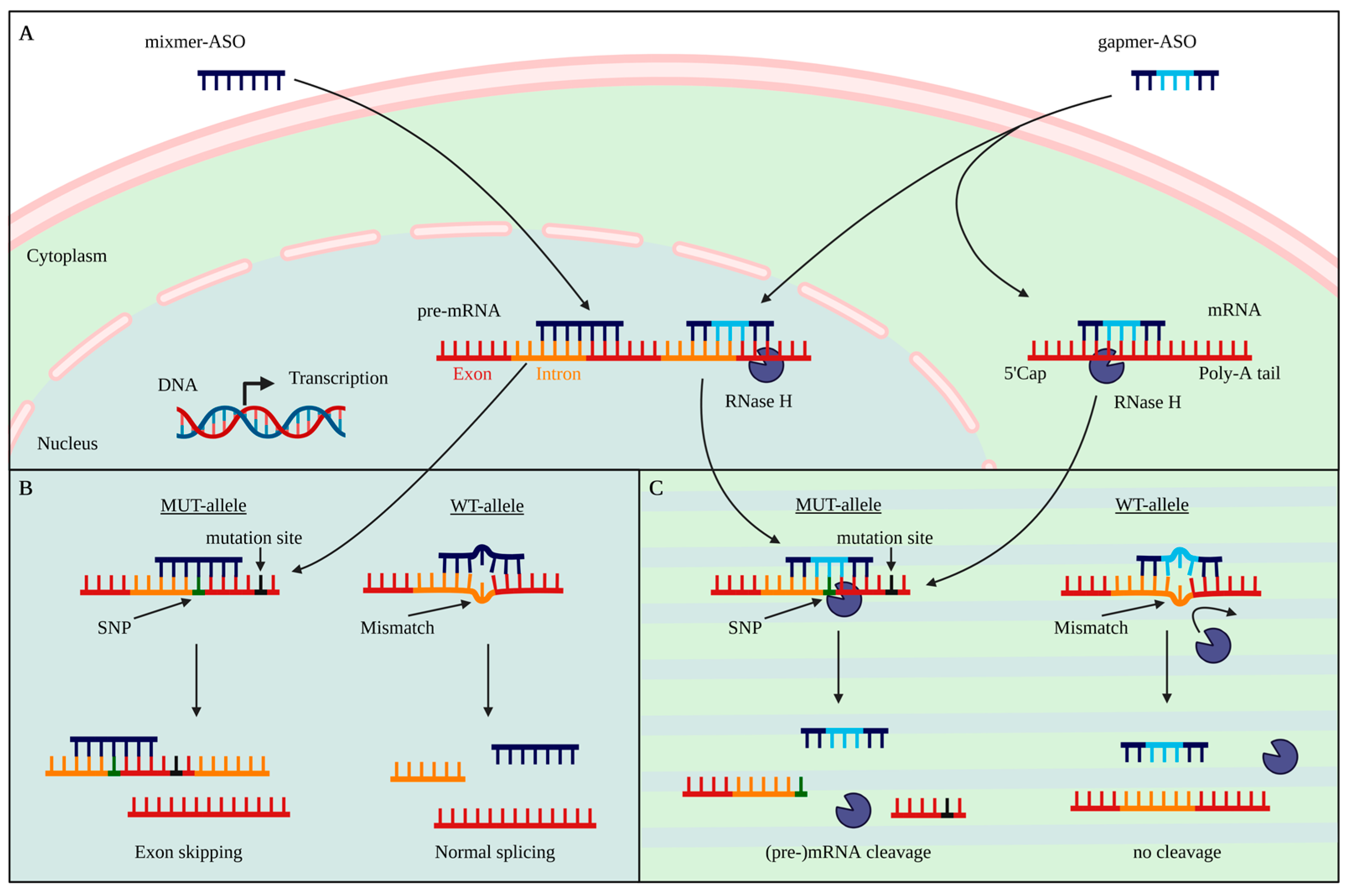
Figure 1. Mode of action of allele-specific antisense oligonucleotide strategies. (A) Mixmer ASOs and gapmer ASOs can be transported through cell membranes and nuclear pores to target (pre-)mRNA. (B) Mixmers act as a steric block that binds to the target pre-mRNA and can modulate the splicing process. Steric block ASOs can be used to selectively target the SNP region and therefore, modulate splicing. Binding to the WT allele has a lower affinity compared to the MUT allele, due to a mismatch. This results in a less effective splicing modulation. (C) Gapmer ASOs can either bind intronic regions or exonic regions of (pre)-mRNA in the nucleus or cytoplasm. Binding of gapmers with their target (pre-)mRNA recruits RNase H. The DNA:RNA binding leads to a degradation of the (pre-)mRNA. SNPs that appear on the same allele as the disease-causing mutation, such as a CAG repeat expansion, can be directly targeted with an ASO, potentially leading to a mutant-specific degradation. The mismatch on the WT allele results in a conformational change that leads to a reduced cleavage capacity.
Pharmaceutics 14 01708 g001
Figure 2. Common ASO nucleotide modifications. ASOs can be optimized at different positions of the nucleotide. Modifications at the phosphate (yellow) reduce affinity, but have positive effects on immunoreactivity and nuclease resistance and thus, also on longevity. Sugar modifications (turquoise) can increase nuclease resistance, affinity, and specificity and decrease toxicity. Disadvantageously, 2′O modifications cause the loss of deoxyribose character and cannot be recognized by RNase H. Therefore, these modifications cannot be implemented in the DNA gap region of gapmer ASOs. Base modifications (gray) may have the greatest potential for increasing the specificity of individual bases, but are also associated with an increased toxicity in vivo. cET = constrained ethylbridged nucleic acid; ENA = 2′O,4′-C-ethylene bridged nucleic acid; LNA = locked nucleic acid; MsPA = mesyl phosphoramidate; PG = phosphoryl guanidine; PS = phosphorothioate; 2′-MOE = 2′-O-methoxyethyl; 2′-OMe = 2′-O-methyl.
Pharmaceutics 14 01708 g002
Figure 3. Schematic description of the association between the mutation site and non-disease-causing SNPs. (A) Exemplary representation of a gene with its founder mutation (red) and individual additional variants/SNPs (black). Vertical blue shadings exemplarily indicate common crossover sites. The more crossovers occur within a region, the less the association between different regions of the gene, and the linkage disequilibrium (LD) decreases. (B) Numbers 1, 2, and 3 show potential SNPs in the founder gene with different association strengths to the mutation site after some generations. SNP 1 has a MAF of 0.4 and is located within a region with several common crossover sites. Therefore, the SNP shows no association to the founder mutation, even though the founder gene carries SNP 1. In a patient cohort, SNP 1 is therefore similarly distributed to a control cohort. A total of 60% of the patients carry a G in SNP 1 on the same allele as the founder mutation, and 40% carry an A. The WT allele of the patients show the same distribution in SNP 1. With an ASO that specifically targets SNP 1_A, 24% of the cohort (A/G) could be treated as allele-specifical, and an additional 16% (A/A) as non-allele-specific. SNP 2 has a MAF of 0.4 but is 100% associated with the disease mutation. Therefore, every patient carries an A in SNP 2, along with the mutation on the MUT allele. With an ASO that targets SNP 2_A, 60% of the patients (A/G) could be treated as allele-specific and 40% as non-allele-specific. SNP 3 appears rarely in a population with an MAF of 0.1, but has an association of 100% to the mutation site. Every patient carries an A in SNP 3 on the same allele as the disease mutation. With an ASO that targets SNP 3_A, 90% of the patients (A/G) can be treated as allele-specific, and the remaining 10% as non-allele-specific. These example pedigrees show the importance of long coding sequencing of the whole genes of patient cohorts. Targeting rare SNPs that have a high disease association might be an optimal target. Identification of these rare, disease-associated SNPs reduces the number of ASOs needed to identify suitable ASOs for an allele-specific approach in a whole patient cohort. MAF = minor allele frequency; SNP = single nucleotide polymorphism.
Pharmaceutics 14 01708 g003
Figure 4. Workflow of in vitro testing and optimization of allele-specific ASOs. For allele-specific testing of ASOs, the association of the target with the mutation and the distribution in the population and patients should be known or determined. One or more lead ASOs could be tested at different positions, giving a first indication of the targetability of the (pre-)mRNA position. In further in vitro experiments, the lead ASO can be further modified to decrease toxicity and increase longevity, affinity, and allele-specificity. Different model systems have different advantages regarding evaluation of ASOs. IPSC-derived 2D models are a fast and cost-efficient tool to test the effectiveness of an ASO in a model with the genotype of a patient. Nevertheless, more complex systems, such as brain organoids, provide more precise information about the effects on the pathomechanism, such as aggregate formation, but also provide the opportunity to investigate off-targets in several neural cell types in parallel. Mice are still the model of choice to investigate (acute) toxicity and specificity in an in vivo system.
Pharmaceutics 14 01708 g004
Table 1. Antisense oligonucleotide modifications and properties.
ModificationTm per NucleotideReferencesSpecificGeneral
Backbone
Phosphorothioate (PS)0.45 to 1 °C[27,34,35,36,37,38,39]
  • Increases toxicity
  • Improves RNase H recognition
  • Increases nuclease resistance
  • Improves pharmacokinetics
  • Reduces affinity
  • Stereoisomers
Mesylphosphoramidate (MsPA)1.3 to +1.1 °C (with respect to PS)[39,40]
  • Reduces toxicity
  • Number and position influences RNase H activation
  • Incorporation in gap reduces protein binding
  • Nuclease resistance (MsPA > PS)
Phosphoryl guanidines (PG)1.2 to 0 °C[41,42,43,44]
  • Reduces toxicity
  • Prevents RNase H binding, when used in DNA-gap
  • Nuclease resistance (PG > PS)
  • Reduced cellular uptake compared to PS
Sugar
Locked nucleic acids (LNA)+1.5 to +9.1 °C[27,45,46,47,48,49,50,51]
  • Can improve specificity
  • Increases affinity
  • Increases nuclease resistance
  • Decreases toxicity
  • Not compatible with DNA-gap
2’O-methyl (2’-OMe)0 to +1.3 °C[27,34,38,46]
2’-O-methoxyethyl (2’-MOE)+0.9 to +1.9 °C[34,46,51,52,53]
  • Improves cellular uptake
2’4’-constrained 2’-O-ethyl
(2’-cEt)
+4.7 to +6.1 °C[51,54]
  • Can improve specificity
  • Exonuclease resistance cET > LNA
  • Stereoisomers
  • Can increase specificity
2’-O,4’-C-ethylene-bridged
nucleic acid (ENA)
+5.2 °C[49]
  • Exonuclease resistance ENA > LNA
Base
G-clamp+4 to +18 °C[46,55,56]
  • Can improve specificity
  • Reduces RNase H activity
  • Increases affinity
  • Can induce toxicity
C5-propyne C+1.5 to 1.6 °C[55,57,58,59]
  • Reduces RNase H activity
  • Increases resistance
C5-propyne T+0.9 to +2.6 °C[34,54,55,57,58,59]
  • Reduces RNase H activity
  • Increases resistance
2-thio-thymidine+0.3 to +1.8 °C[54,60]
  • Can improve specificity
5’-thiazole analogues+1.7 to 2.2 °C[58,59]
  • RNA affinity (thiazole > propyne)
5-Methyl cytosine0 to +1.1 °C[25,34]
Table 2. Clinical trials of antisense oligonucleotides in toxic gain-of-function neurodegenerative diseases.
Disease 1 TargetCellular FunctionASOPhaseASO Type/ModificationsRef./Clinical Trial
HD–HTTBrain development, involved in vesicle trafficking and recycling, cell division, ciliogenesis, autophagy, development [107]Tominersen, IONIS-HTTRxPhase III
halted (03/21)
Non-allele-specific, PS 2′-MOE[2,84,108]
NCT03842969
HD–HTTWVE-003 (WVE-120101 & 120102: suspended)Phase I/IIAllele-specific, PS stereopure[2,72,109]
NCT05032196
ALS/FTD–FUSDNA/RNA metabolism [110]Jacifusen/ION36Phase IIIMutation-specific (p.P525L), PS 2′-MOE[111,112,113]
NCT04768972
ALS–SOD1Antioxidant [114]Tofersen/IONIS-SOD1Rx (BIIB067)Phase IIINon-allele-specific, PS 2′-MOE[115,116]
NCT02623699
NCT03070119
ALS/FTD–C9ORF72Repeat in noncoding region [117]IONIS-C9Rx (BIIB078)Phase I
discontinued (03/22)
Non-allele specific, PS 2′-MOENCT03626012
NCT04288856
ALS/FTD–C9ORF72WVE004Phase I/IIAllele-specific (Targeting V1 and V3 transcript), PS PG stereopure[118,119]
NCT04931862
ALS/SCA2–ATXN2RNA metabolism [120]ION541 (BIIB105)Phase I/IIPS 2′-MOE[83,121]
NCT04494256
AD/FTD–MAPT (TAU)Stabilizing & promotion of microtubule assembly [122]IONIS-MAPTRx (BIIB080)Phase IIPS 2′-MOE[123]
SCA3–ATXN3Deubiquitinase [120]ION260 (BIIB132)Phase INon-allele-specific, PS 2′-MOE[100,121]
NCT05160558
PD–LRRK2Kinase involved in lysosomal processes, autophagy, mitophagy, vesicle trafficking [124]ION859 (BIIB094)Phase I/IIPS 2′-MOE[125]
NCT03976349
PD–SNCAPresynaptic protein, involved in SNARE complex assembly [126]ION464 (BIIB101)Phase IIPS 2′-MOE[127]
NCT04165486
Alexander disease–GFAPIntermediate filament [128]Zilganersen, ION373Phase IINon-allele-specific, PS 2′-MOE[129]
NCT04849741
CAS2305355-56-8
Publisher’s Note: MDPI stays neutral with regard to jurisdictional claims in published maps and institutional affiliations.