Review

Selective Activation of the Wnt-Signaling Pathway as a Novel Therapy for the Treatment of Diabetic Retinopathy and Other Retinal Vascular Diseases

Surrozen Operating, Inc., 171 Oyster Point Blvd, Suite 400, South San Francisco, CA 94080, USA
*
Author to whom correspondence should be addressed.
Pharmaceutics 2022, 14(11), 2476; https://doi.org/10.3390/pharmaceutics14112476
Submission received: 28 September 2022 / Revised: 7 November 2022 / Accepted: 14 November 2022 / Published: 16 November 2022

Abstract

:
Retinal ischemia, often associated with various disorders such as diabetic retinopathy (DR), retinal vein occlusion, glaucoma, optic neuropathies, stroke, and other retinopathies, is a major cause of visual impairment and blindness worldwide. As proper blood supply to the retina is critical to maintain its high metabolic demand, any impediment to blood flow can lead to a decrease in oxygen supply, resulting in retinal ischemia. In the pathogenesis of DR, including diabetic macular edema (DME), elevated blood glucose leads to blood-retina barrier (BRB) disruptions, vascular leakage, and capillary occlusion and dropouts, causing insufficient delivery of oxygen to the retina, and ultimately resulting in visual impairment. Other potential causes of DR include neuronal dysfunction in the absence of vascular defect, genetic, and environmental factors. The exact disease progression remains unclear and varies from patient to patient. Vascular leakage leading to edema clearly links to visual impairment and remains an important target for therapy. Despite recent advances in the treatment of DME and DR with anti-VEGFs, effective therapies with new mechanisms of action to address current treatment limitations regarding vessel regeneration and reperfusion of ischemic retinal areas are still needed. The Wnt signaling pathway plays a critical role in proper vascular development and maintenance in the retina, and thus provides a novel therapeutic approach for the treatment of diabetic and other retinopathies. In this review, we summarize the potential of this pathway to address treatment gaps with current therapies, its promise as a novel and potentially disease modifying therapy for patients with DR and opportunities in other retinal vascular diseases.

1. Introduction

Diabetic retinopathy (DR) remains a leading cause of vison loss in many developed countries and is the most common, specific, and severe microvascular ocular complication of diabetes. According to The International Diabetes Federation, 463 million people were diagnosed with diabetes in 2019 [1]. About one-third of them have some signs of DR and nearly 10% of diabetes patients have vision-threatening DR [2,3]. Chronic hyperglycemia and other causative risk factors such as hypertension are believed to initiate a series of biochemical and pathophysiological changes that ultimately lead to microvascular damage and retinal dysfunction [4,5,6]. In the pathogenesis of DR, including diabetic macular edema (DME), elevated blood glucose can affect the lining of the blood vessels inducing pericyte loss, endothelial cell damage, and basement membrane thickening, which can lead to blood-retina barrier (BRB) disruptions, vascular leakage, and insufficient delivery of oxygen to the retina, and ultimately results in visual impairment [7,8,9]. The vascular leakage and pathologic angiogenesis in the oxygen-deprived retina are often associated with hypoxia-induced overproduction of multiple signaling factors, including vascular endothelial growth factor A (VEGF-A), angiopoeitin-2, angiopoietin-like 4, tissue inhibitor matrix metalloproteinase 1, and hypoxia-inducible factor 1-alpha [10].
Vascular endothelial growth factor (VEGF) is a prominent factor inducing pathological neovascularization and altering retinal capillary permeability [11,12,13]. As a result, anti-VEGF agents such as ranibizumab and aflibercept have been developed to control intraocular vascular abnormalities and are currently considered the standard of care (SOC) in addition to laser photocoagulation in the treatment of DME and wet type age-related macular degeneration (wAMD) [14]. However, real-world data suggest that patients with DME receive fewer anti-VEGF injections in clinical practice compared with randomized clinical trials, resulting in visual acuity outcomes falling short of clinical trial results [15]. Thus, there remains significant need for therapies with improved durability to reduce treatment burden and potentially improve real-world patient outcomes. To that end, considerable efforts are being focused on improving efficacy durability of VEGF pathway inhibitors. For instance, the Port Delivery System with ranibizumab, an intraocular drug delivery system designed for the continuous delivery of ranibizumab (anti-VEGF) into the vitreous for 6 months and beyond (instead of monthly intravitreal [IVT] injection), was recently approved by the Food and Drug Administration (FDA) for the treatment of wAMD and DME [16]. Other pathways beyond VEGF signaling have also been extensively explored. Of these, the Tie2 signaling pathway represents a novel therapeutic target as Faricimab, the first bispecific antibody that targets Ang-2 and VEGF-A designed for intraocular use, has been recently approved by the FDA for the treatment of DME [17] and wAMD [18]. Table 1 includes a partial list of VEGF-binding and non VEGF-binding drugs that are being developed as potential therapies for the treatment of wAMD and DME.
However, as these therapies focus on the vascular leakage and inflammation component of the disease, their impact on vessel regeneration and reperfusion of ischemic retinal areas are minimal. Since vessel regeneration and a reduction of the retinal ischemic area could potentially lead to a durable reduction in hypoxia-induced signaling factors such as VEGF, effective therapies with this mechanism of action would be highly desirable. The Wnt signaling pathway is essential for embryonic development and in adults for tissue homeostasis and injury repair. This pathway has been shown to be critical for proper vascular development and for the maintenance of BRB in the adult retina [28,29]. Dysregulated Wnt signaling has been suggested to contribute to the pathophysiology of DR [30] and modulation of Wnt signaling has been reported to have a beneficial role in various preclinical models. Here, we provide perspective on a new therapeutic strategy that selectively activates the Wnt/β-catenin signaling pathway in retinal endothelial cells as a potential therapy for DR and other retinal vascular diseases.

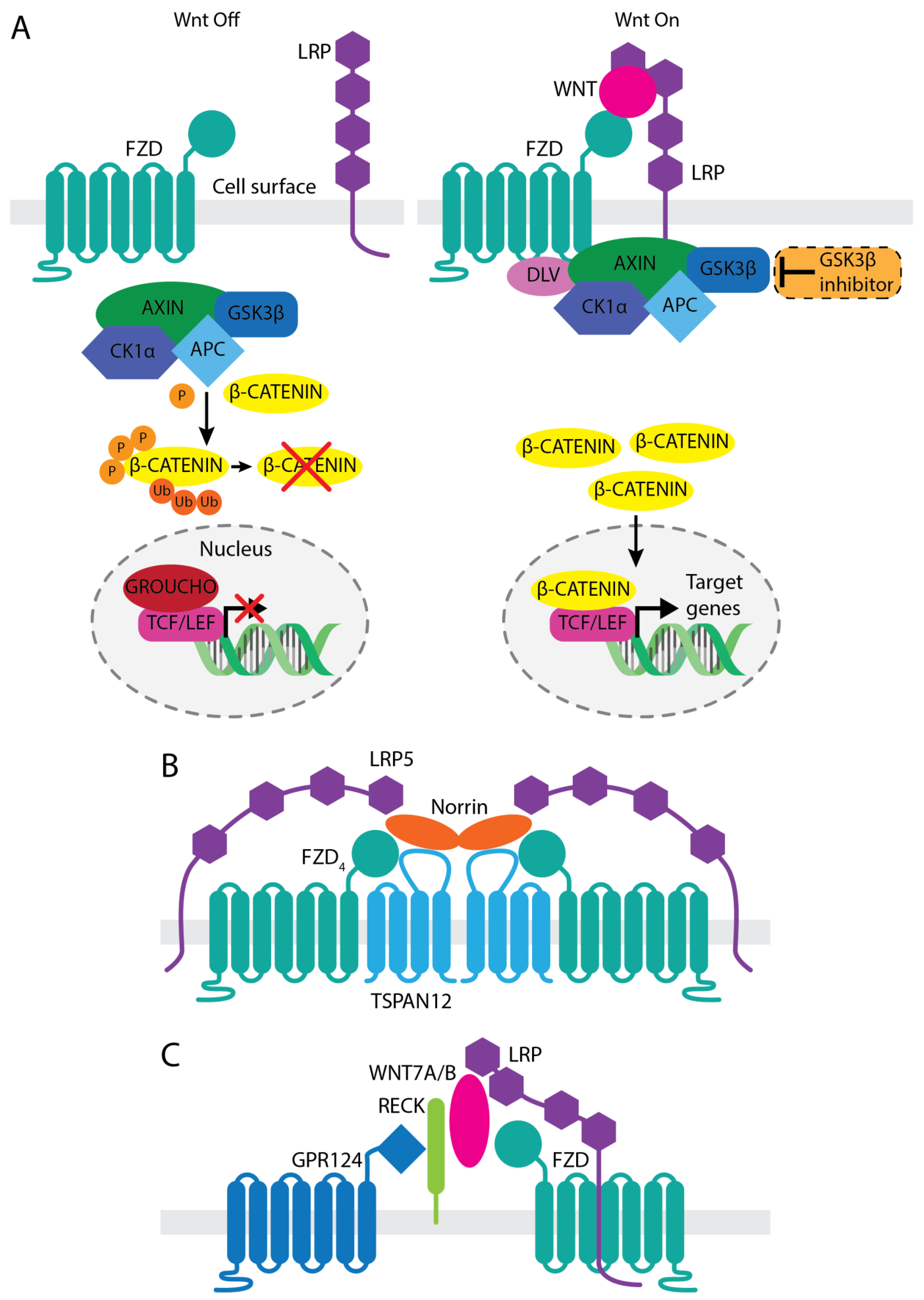
2. Involvement of Wnt/β-Catenin Signaling in Vascular Development and Function

The Wnt signal transduction cascade is a key driver of numerous biological events throughout the life of all animals [31]. The activation of the intracellular signaling cascade initiates when the secreted lipid-modified ligands of the Wnt family bind to surface receptors on target cells. Wnt pathway activation elicits a plethora of cellular responses ranging from cell fate determination, proliferation and migration, body axis patterning during embryonic development to the maintenance of adult tissue stem cells and the regulation of tissue homeostasis and regeneration [32,33]. More in-depth reviews on Wnt signaling can be found elsewhere [32,33,34,35,36,37,38].
The canonical Wnt signaling pathway, referred here as Wnt/β-catenin pathway is the most widely studied Wnt signaling pathway and it involves WNT binding to a heterodimeric receptor complex comprised of a frizzled (FZD) family of 7-pass transmembrane receptors and a single pass transmembrane co-receptor, low-density lipo-protein receptor-related protein (LRP). There are 19 mammalian WNTs, 10 FZDs (FZD1–10), and 2 LRPs (LRP5 and LRP6) [32,39]. Following WNT binding, clustering of FZD and LRP results in conformational changes, and subsequent phosphorylation of the receptors. The scaffold protein AXIN, part of the β-catenin destruction complex consisting of adenomatous polyposis coli (APC) along with the serine-threonine kinases casein kinase 1 alpha (CK1α) and glycogen synthase kinase 3 beta (GSK3β), is then sequestered to the cytoplasmic tail of LRP via the FZD-bound disheveled (DVL). As the destruction complex is recruited to the membrane, the transcription factor β-catenin begins to accumulate in the cytoplasm, and subsequently translocates to the nucleus where it binds to T cell factor (TCF)/lymphoid enhancer-binding factor (LEF) transcription factors, inducing downstream transactivation of Wnt target genes (Figure 1A).
Significant evidence has demonstrated essential roles for Wnt/β-catenin signaling in both retinal and central nervous system (CNS) angiogenesis during development and in establishing and maintaining BRB/blood-brain-barrier (BBB) function in adults. In the retina, Norrin, encoded by the NDP (Norrie Disease Protein) gene, is secreted by Müller glia and endothelial cells, and plays a critical role in the patterning of the retinal vasculature during development and the establishment of the BRB during adulthood [40,41,42,43]. Norrin, which could be considered an atypical WNT ligand [44,45], is a secreted 131-amino acid protein from the cysteine-knot growth factor superfamily. Norrin forms a dimer that binds with high affinity and specificity to FZD4 and LRP5 in the presence of the Norrin-specific co-receptor tetraspanin 12 (TSPAN12), forming a ternary complex that activates the Wnt/β-catenin signal in retinal endothelial cells [46,47] (Figure 1B). NDP mutations result in Norrie disease, an X-linked genetic disorder characterized by hypovascularization of the retina, retinal detachment, and severe visual impairments or loss of vision [48,49]. FZD4, LRP5, or TSPAN12 mutations are also the causes of a spectrum of related congenital retinopathies such as osteoporosis-pseudoglioma syndrome, familial exudative vitreoretinopathy (FEVR), and Coats disease, each of which share resembling phenotypes with Norrie disease. Mutations in Fzd4, Lrp5, Tspan12, or Ndp in mice result in retinopathies with remarkable similarity to human diseases, including aberrant development of the retinal vasculature, BRB defects, and impaired vision [33,50,51,52,53].
While Wnt signaling mediated through GPR124-RECK-WNT7A/B axis (Figure 1C) is critical for proper CNS angiogenesis and BBB formation, the Norrin/FZD4/LRP5/TSPAN12 axis also contributes [47]. Genetic mutant mice of either Ndp, Fzd4, Lrp5, or Tspan12 share phenotypically similar defects in retina and brain vasculature [40,42,46,54]. Knockout (KO) mice show not only retinal vascular hemorrhage, but also BBB breakdown in the cerebellum, which is induced by decreases in endothelial cell tight junction proteins and increases in plasmalemma vesicle associated protein (Plvap) [43,55]. Norrin is also expressed in the inner ear for the maintenance of the stria vascularis, a highly vascularized tissue that produces endolymph in the cochlea [56,57]. Norrin loss of function showed progressive hearing loss with stria vascularis impairment, as seen in Norrie disease patients [56]. Recent research further showed that Ndp KO mice have malformations of the microvasculature in the stria vascularis, inducing loss of vessel integrity, resulting in hearing loss [58]. These findings demonstrate the importance of the Norrin/FZD4/LRP5/TSPAN12 signaling axis in the development and maintenance of proper functioning vessels in certain organs, including the retina.
Additional roles for Wnt signaling during the development of the retinal vascular system have also been reported. The hyaloid vasculature, a transient embryonic circulatory system that provides oxygen and nutrients to the developing fetal eye, regresses concurrently with the growing and maturing retinal vasculature. This regression of hyaloid vessels occurs immediately after birth in mice and at the fifth month of gestation in humans. Impaired hyaloid regression can lead to an ocular pathology called Persistent Fetal Vasculature (PFV) in humans, which may cause cataracts, glaucoma, intraocular hemorrhages, retinal detachment, and visual impairment [59,60]. Wnt7b is required for hyaloid regression and is expressed in macrophages [61].
Wnt/β-catenin signaling also plays a critical role in the maintenance of barrier function in both the retinal and the CNS vasculature in adults. Acute loss of Fzd4 in adult mice results in the loss of barrier function in the retina as well as in the cerebellum and olfactory bulb. Acute induction of Norrin in adult Ndp null mice restores cerebellar barrier function. Moreover, constitutively active β-catenin in endothelial cells in the choroid plexus causes upregulation of claudin-5 (Cldn5), a marker normally expressed in an intact BBB, and downregulation of Plvap, a marker of fenestrated BBB [29,62]. Furthermore, Wnt signaling is found to be downregulated in highly permeable capillaries such as the circumventricular organs in the brain and the choriocapillaris as well as the ciliary body and choroid plexus in the retina. Upregulation of Wnt signaling in these permeable endothelial cells causes them to partially convert to a barrier-like state [29].
Given the essential roles of Wnt/β-catenin signaling in the development of the retinal vessels and the maintenance of adult vessel barrier functions, it is not surprising that dysregulated Wnt/β-catenin signaling has been observed in ocular diseases. For example, total β-catenin levels were reported to be elevated in non-proliferative DR patients [63] while the levels of Dickkopf1, an endogenous Wnt signaling inhibitor, was significantly higher in the vitreous of DR patients over that in non-DR groups [64]. Furthermore, both inhibition and activation of this pathway have been reported to have benefits in various disease models [65,66,67,68,69,70]. However, the contribution of Wnt/β-catenin signaling to the pathogenesis of retinal vascular diseases remains to be fully defined.

3. Therapeutic Approaches Targeting Wnt Activation

The fact that Norrin/FZD4/LRP5/TSPAN12 signaling pathway plays an indispensable role in retinal vascular development and BRB integrity suggests that modulating this pathway may have therapeutic potential to treat retinal vascular diseases. Indeed, overexpression of lens-derived Norrin via gene delivery not only completely restored normal vascular development in Norrin-deficient (Ndpy/−) mice [71], but it also induced proper vessel regrowth and prevented the pathologic neovascular tuft generation in a mouse retinal ischemic model of oxygen induced retinopathy (OIR) [70]. The potential therapeutic effects in the OIR mice were also observed by treatment with exogenous recombinant Norrin [72]. These results imply that ectopic delivery of Norrin protein or its functional mimetic can have a potential to restore physiological angiogenesis in ischemic retinopathy.
In addition to inducing proper vessel regeneration, Norrin also reduced retinal vascular permeability [43,69,73]. Expressions of key regulators for vascular barrier functions in BRB/BBB such as major facilitator superfamily domain containing 2 (Mfsd2), Cldn5, and zonula occludens-1 (Zo1) are upregulated by FZD4 signaling activation, whereas the expression of Plvap is downregulated [29,43,64,68,73], indicating that the FZD4 signaling inhibits vascular leakage. In addition, the intravitreal injection of Norrin in streptozotocin-induced diabetic rats partially, but significantly reduced retinal leakage [69]. All these findings demonstrate the effectiveness of Wnt/β-catenin pathway activation in reducing abnormal vascular permeability. The combination of reducing vascular leakage and building normal vessels is an exciting concept that can be tested for retinopathy. Therefore, Norrin/FZD4-specific Wnt/β-catenin pathway is a potential target as a new therapeutic strategy for the treatment of retinal vascular diseases including DR.

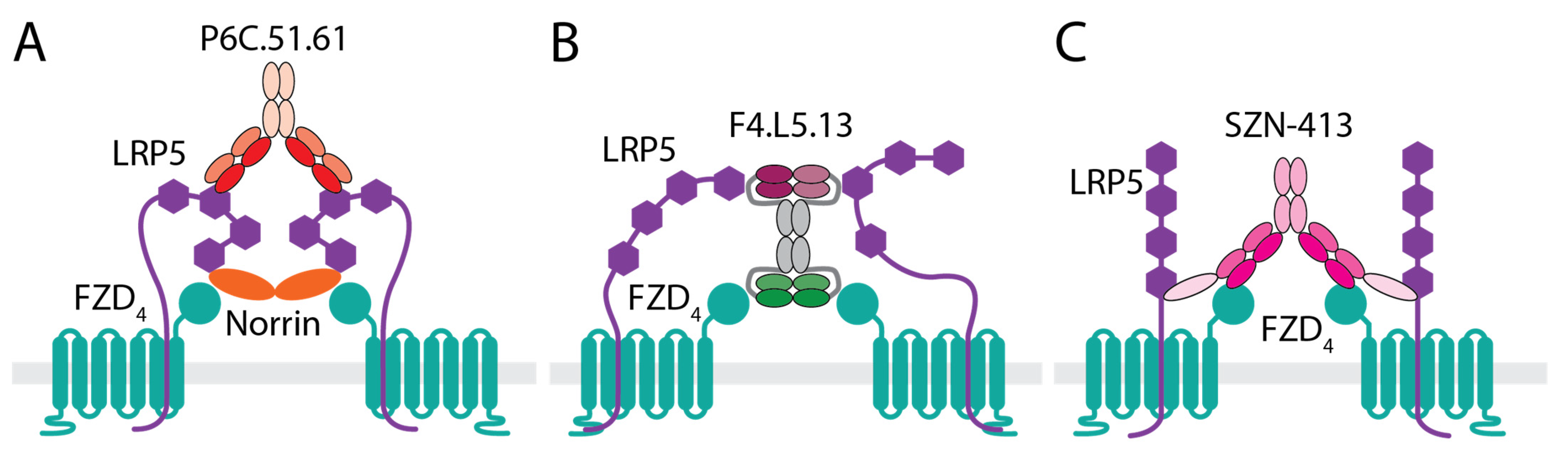
3.1. Anti-LRP5 Antibody That Enhances Norrin Signaling

Although Norrin/FZD4/LRP5/TSPAN12 signaling pathway has a significant potential to treat retinal vascular disease, successful pharmacologic FZD4 activation through recombinant Norrin for therapeutic BRB modulation and retinal vessel regeneration has remained elusive. Norrin is poorly secreted and is highly associated with the extracellular matrix [74], making it difficult to produce and unsuitable as a therapeutic agent for DR. In addition, Norrin activates Wnt/β-catenin signaling through binding and formation of a ternary complex with receptors FZD4, LRP5/6, and TSPAN12 [75,76]. Since many human inherited vitreoretinopathies have mutations in these receptors that affect interactions with Norrin or the assembly of the ternary complex by Norrin, it is unlikely that Norrin itself would be beneficial for individuals harboring these mutations. Therefore, additional approaches will need to be explored to circumvent challenges associated with Norrin and to expand the potential uses to broader retinal vascular diseases.
Activation of Wnt/β-catenin requires the binding of WNT ligands to FZD receptors and to the co-receptors LRP5/6. WNTs bind to the cysteine-rich domain (CRD) on the N-terminus of FZD via two binding sites that have been described as “thumb” (site 1 on FZD) and “index finger” (site 2 on FZD), and the critical palmitoleoyl group on WNT is involved in site 1 binding [77]. LRP5/6 are single-pass transmembrane proteins that contain four tandem YWTD-type β-propeller domains with each followed by an epidermal growth factor (EGF)-like domain, called E1-E4 from N- to C-terminus of the extracellular domain (ECD) [78]. Different WNTs bind different LRP ECD regions with WNT1, 2, 2B, 6, 8A, 9A, 9B, 10B binding to E1E2 and WNT3, 3A binding to E3E4 [78]. It has been reported that bivalent antibodies binding to E1E2 of LRP6 while inhibiting the binding and signaling of E1E2 engaging WNTs, enhance signaling of E3E4-binding WNTs and vice versa [79,80].
Norrin engages FZD4 on site 2 and LRP5/6 on the E1E2 domain [75,76]. Similar to what has been observed for WNT ligands, an anti-LRP5 E3E4 antibody, P6C.51.61 (Figure 2A), identified through phage display campaign, enhanced Norrin signaling in HEK293T and human retinal microvascular endothelial cells (HRMEC) in vitro (US10035854). Interestingly, P6C.51.61 also rescued a FZD4 mutation, M157V [40] and a Norrin mutation, C95R [46], both identified in FEVR patients that affected either interaction with Norrin or Norrin dimerization, respectively. These results suggest that the clustering of LRP5 by an antibody that binds complementary to Norrin could strengthen ternary ligand/receptor complex formation. Given the critical role of TSPAN12 in Norrin function, mutations in TSPAN12 have been identified in human FEVR patients [81], and Tspan12 KO mice show stunted retinal vessel development [46]. P6C.51.61 partially rescued the vascular defect and minimally restored formation of capillary network in the retina of Tspan12 KO mice (US10035854). Treatment with P6C.51.61 in a mouse OIR model also showed trends of reduced pathological neovascular tufts formation and partially induced vascular regrowth in the vaso-obliterated avascular area. These results support the approach of activating Norrin/FZD4 signaling for the treatment of retinopathy. However, one of the limitations of this enhancer approach is the reliance on the presence of endogenous ligands. In addition, other Wnt ligands such as Wnt3a, Wnt7a, and Wnt10a are present in the vitreous humor, and some have been reported to be upregulated in OIR [65]. Therefore, it is unclear if the in vivo effects observed with P6C.51.61 are due to its ability to enhance Norrin signaling or to enhance the signaling of these other Wnts present in the vitreous humor that bind Lrp5 E1E2, or even due to inhibition of Wnts that bind Lrp5 E3E4. Therefore, more definitive evidence is still needed to understand whether activation of Wnt signaling in vascular endothelial cells would be beneficial in retinopathy. In addition, a more specific approach may be desirable to reduce potential off-target effects that may complicate data interpretation and therapeutic development.

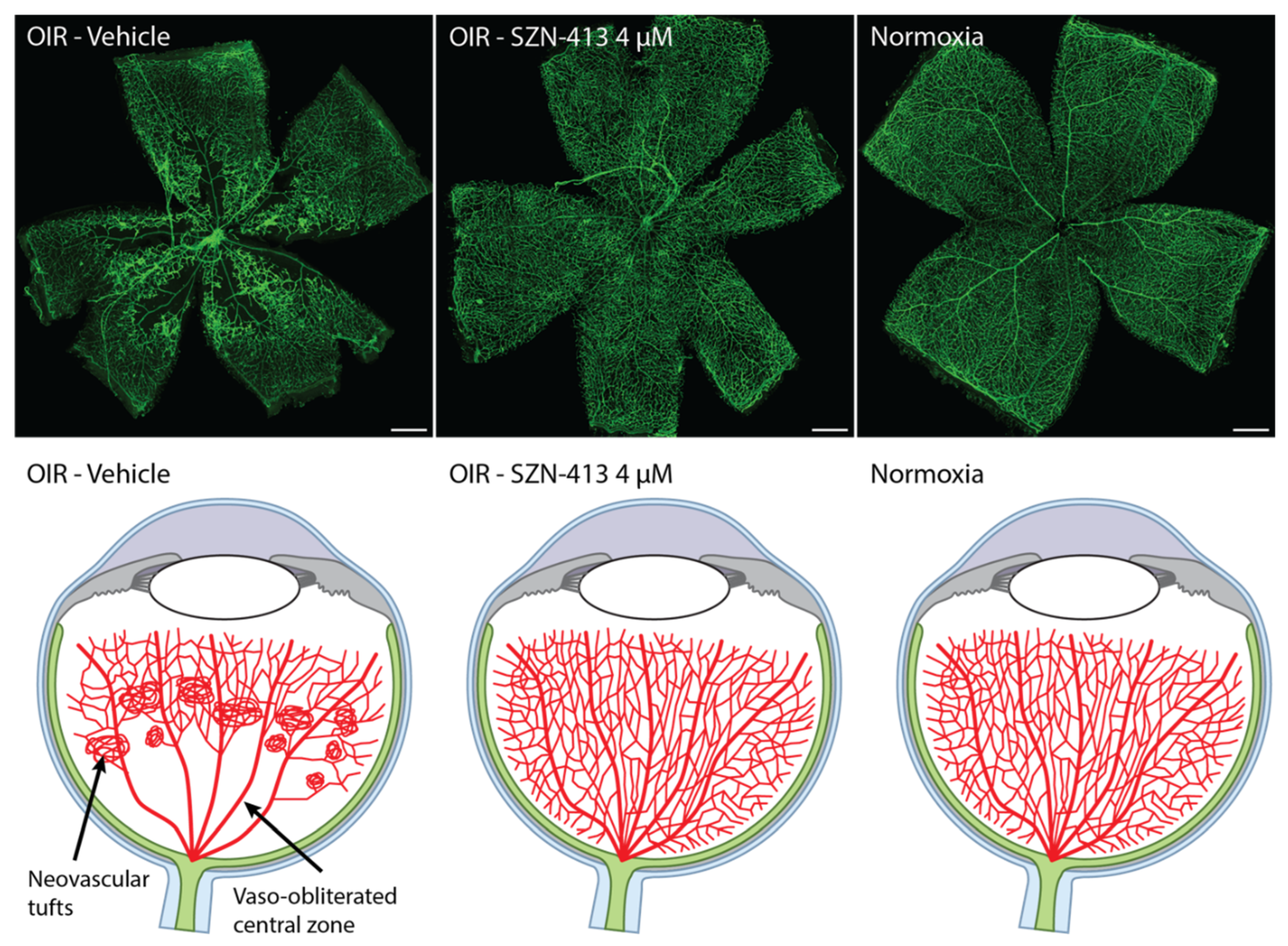
3.2. WNT Mimetics That Activate FZD4/LRP5

Another way to enhance Wnt signaling in the retina is through the direct pharmacological activation of FZD4/LRP5 mediated Wnt/β-catenin signaling in endothelial cells. Unfortunately, WNT ligands are even more difficult to work with than Norrin due to their lipid modification that makes them insoluble and difficult to express, and their lack of FZD selectivity. However, recent breakthroughs have allowed for the development of FZD-specific WNT-mimetic molecules based on various protein scaffolds [82,83,84,85]. All these approaches utilize bispecific molecules that can simultaneously bind to FZDs and LRPs to induce Wnt/β-catenin signaling. In theory, these approaches can generate molecules with better biophysical properties and with increased receptor specificity that would address the many challenges associated with natural WNTs.
One approach to design WNT mimetics is based on a tetravalent diabody format where two diabodies, each having two binding sites for FZD or LRP, are tethered on two ends of an Fc fragment [83]. Because of this modular design, up to four different binders could be combined into one molecule to achieve the desired receptor and epitope specificity. One WNT mimetic molecule of this format, F4L5.13 (Figure 2B), was constructed by combining a diabody having two binding sites with mono specificity for FZD4 with another diabody containing an LRP5 E1 and an LRP5 E3 binder in the tetravalent trispecific configuration [68]. F4L5.13 was reported to specifically activate FZD4-mediated Wnt/β-catenin signaling to a similar extent as recombinant Norrin in HEK293 cells. Unlike Norrin, F4L5.13-induced signaling is not TSPAN12-dependent. Treatment of a mouse brain endothelial cell line (bEnd.3 cells) in vitro by F4L5.13 led to rescue of tight junction proteins Zo-1, Cldn3, and Cldn5 expression on the cell surface that was reduced by the pretreatment of these cells with VEGF and led to reduced permeability of the cells in vitro [68]. Since F4L5.13 does not depend on TSPAN12 for signaling, treatment of Tspan12 KO mice with F4L5.13 restored retinal angiogenesis and barrier function as indicated by increased Cldn5 and decreased Plvap expression. Similar to what was observed with anti-LRP5 Norrin enhancer antibody described above, F4L5.13 treatment also reduced pathological neovascular tufts formation in the mouse OIR model [68]. However, the ischemic area of the retina was not restored by F4L5.13 treatment as no significant effect on the avascular area was observed. It is unclear why F4L5.13 had no impact on avascular area reduction, which was observed by the treatment of the anti-LRP5 E3E4 antibody, P6C.51.61. Perhaps the potency of F4L5.13 needs further improvements as a high vitreous concentration, 500 nM, was necessary to show a significant reduction in neovascularization [68].
Another approach to WNT mimetic generation has focused on other antibody-based formats. Using scFv based fragments, different design parameters, including valency, linker lengths, relative orientation, geometry, stoichiometry, and receptor specificity, were first systematically evaluated [84]. This led to the identification that multivalent binding to FZDs and LRPs was a critical requirement for maximal Wnt/β-catenin signaling with the optimal ratio of two FZD binding arms with one or two LRP binding arms [84]. Subsequently, these design principles were shown to apply to other multivalent antibody formats such as the tetravalent bispecific VHH-IgG format [86], which are highly stable and suitable for large scale manufacturing of drug products.
Recently, a FZD4/LRP5 specific WNT mimetic molecule, SZN-413 (Figure 2C), was constructed based on the tetravalent bispecific VHH-IgG format [87]. The IgG portion of SZN-413 binds FZD4 with high affinity (with KD < pM) and the VHH domains bind LRP5. SZN-413 is highly potent, capable of activating Wnt/β-catenin signaling in vascular endothelial cells such as HRMEC and bEnd.3 cells in vitro and its potency is several orders of magnitude higher than that of recombinant Norrin or WNT3A in vitro. SZN-413 has biophysical and chemical characteristics that translate to desirable solubility, stability, and large scale manufacturability. Similar to F4L5.13, the bispecificity toward FZD4/LRP5 may have the advantage of averting promiscuous activation of other FZD family members in the vitreous space as both FZD4 and LRP5 are enriched in retinal endothelial cells vs. other cell types [87]. SZN-413 increased CLDN5 and ZO-1 protein levels in HRMEC [87]. The upregulated expression of known BBB/BRB endothelial cell transcripts, LEF1 and MFSD2A [29,73], by SZN-413 further supports that SZN-413 can increase vascular integrity through upregulation of barrier function proteins. In a mouse ischemic retinopathy OIR model, SZN-413 reduced pathological neovascular tufts formation to a higher extent than aflibercept [87]. Furthermore, SZN-413 treatment reduced the size of avascular area significantly more than aflibercept and almost completely restored the normal vessels in the vaso-obliterated central zone in the retina (Figure 3). This implicates that the novel FZD4/LRP5 specific WNT mimetic molecule may be able to transform pathologic neovascularization into physiological neovascularization, reducing retinal ischemia and the ensuing overproduction of signaling molecules responsible for the development of pathologic neovascularization in the proliferative stage of diabetic retinopathy. This regeneration of vessels could be disease modifying, and has the potential to have a durable treatment effect; however, this will need to be confirmed in the clinic. In addition, dose–response data have shown that efficacy could be obtained at concentrations as low as 40 nM with a maximum effect at or above 4 µM suggesting that high doses may further extend the durability provided safety is not compromised. More research on the efficacy duration of SZN-413 is needed to determine the dose and delivery frequency of the drug. Even though both SZN-413 and F4L5.13 should be based on a similar mechanism of action the avascular area size reduction was not observed with F4L5.13 in OIR [68]. The reason for this difference is unclear; however, the results may suggest that appropriate molecular format, geometry and potency may be critical to induce appropriate levels of Wnt/β-catenin signaling in a therapeutic setting.
Although inhibition of VEGF-driven retinal leakage is a major consideration for retinopathy treatment, studies to date have not sufficiently provided direct evidence showing WNT ligands can successfully perform this function in vivo. Recently, Diaz-Coranguez et al. showed that intravitreal injection of recombinant Norrin can inhibit the VEGF-driven retinal leakage in rats [69]. Consistent with this finding, Nguyen et al. showed that the SZN-413, a FZD4/LRP5 specific WNT mimetic, inhibits the VEGF-driven retinal leakage in rabbit eyes [87]. Moreover, its efficacy was observed at vitreous humor VEGF concentrations >600 µg/mL (1 µg recombinant hVEGF165, intravitreally injected into rabbit eye); considering that the VEGF concentration seen in proliferative DR or DME patients is less than 10 ng/mL in vitreous humor [88,89,90], SZN-413 may have therapeutic potential in VEGF-driven retinal leakage diseases such as DR.

3.3. GSK-3β Inhibitor

GSK-3β is a key intracellular component of the Wnt/β-catenin signaling pathway. In the absence of WNT ligands, a destruction complex consisting of AXIN/GSK-3β/CK1/APC phosphorylates β-catenin leading to its ubiquitination and degradation. Upon ligand binding to FZD/LRP, the destruction complex containing GSK-3β is recruited to the intracellular side of the receptor complex on the membrane via DVL and AXIN, leading to stabilization of β-catenin and subsequent activation of Wnt target genes [33]. Therefore, inhibition of GSK-3β can lead to stabilization of β-catenin and activation of Wnt/β-catenin signaling (Figure 1A). It was reported that hypoxia increases GSK-3β activity in HRMEC leading to a reduction in β-catenin and its associated impairment in cell/cell junctions, and inhibition of GSK-3β kinase activity by small molecules or peptides improved capillary morphogenesis in a 3D collagen assay in vitro [91]. In a mouse ischemic retinopathy OIR model, GSK-3β inhibitors reduced abnormal vascular tufts, reduced vascular leakage, improved proper re-vascularization, reduced avascular area, and improved retina perfusion similar to what was observed with SZN-413 discussed above [91]. Furthermore, another GSK-3β inhibitor, lithium chloride, also partially rescued the retinal vascular developmental defects in several FEVR models [92,93,94]. A bell-shaped curve was observed both in vitro and in vivo for GSK-3β inhibitors where the beneficial effects were lost at high compound concentrations, which may contribute to the lack of efficacy of some GSK-3β inhibitors in certain studies, suggesting that a careful titration of doses may be necessary to achieve meaningful efficacy for this approach [91,95]. The cause for the bell-shaped dose–response curve is not clear. Therapeutic development for retinopathy based on GSK-3β inhibitors is challenging given the short half-life of these small molecules or peptide inhibitors and the broad impact inhibition of GSK-3 serine threonine kinase activity may have due to its cross talk with many signaling pathways in addition to Wnt [96]. Nonetheless, the reported effects of GSK-3β inhibitors on endothelial cells in vitro and in vivo are consistent with the hypothesis that activation of Wnt/β-catenin signaling could be a novel approach to treat retinal vascular diseases such as DR.

4. Conclusions and Future Perspectives

Wnt/β-catenin signaling mediated through the Norrin/FZD4/LRP5/TSPAN12 axis has been shown to be essential for the proper formation of retinal vascular structure during development, and for the maintenance of proper vascular structure and function in adults. Activation of this pathway using several different approaches, including the use of Norrin, Norrin enhancing anti-LRP5 antibodies, FZD4 activating WNT mimetics, and GSK-3β inhibitors all suggest that activation of Wnt/β-catenin signaling could be a novel strategy for the treatment of retinal vascular diseases such as DR. Common findings from these Wnt activators are an increase in tight junction protein expression, a reduction in vascular leakage in vitro and/or in vivo, and a reduction in pathological neovascular tufts formation in the ischemic mouse OIR model. These findings are consistent with the established role of Wnt/β-catenin signaling in tight junction protein expression and the maintenance of the BRB in adult vessels [29]. In addition, SZN-413 directly suppressed vascular leakage in a rabbit VEGF-induced leakage model [87], suggesting that this mechanism of action can directly counter the effects of VEGF on the integrity of the retinal vessels.
The effects on vessel regeneration are somewhat variable between these Wnt activating approaches. While F4L5.13 rescued the vascular insufficiency in Tspan12 KO mice, it did not induce vessel regrowth in the ischemic mouse OIR model [68] whereas GSK-3β inhibitors [91] and the Norrin enhancing anti-LRP5 antibody, P6C.51.61 (US10035854) partially reduced the avascular area, and SZN-413 fully regenerated vessels in the vaso-obliterated central zone in the OIR [87]. It is not clear what caused the differences in vessel regeneration observed between the different compounds; it could be possible that specificity and signaling strength may contribute to the differences. Furthermore, clinical studies will be needed to determine if the results observed in the OIR model will translate to humans. Nevertheless, the ability of SZN-413 to fully regenerate vessels in the ischemic OIR model is exciting as it suggests that this mechanism has the potential to address the retinal non-perfusion and ischemia that is not effectively addressed with the current therapies, and therefore could provide a treatment option that can simultaneously address both retinal non-perfusion and vascular leakage in disease. The ability of Wnt/β-catenin signaling to induce proper healing and regeneration has been observed in other tissues as well [86,97,98]. For example, in the intestine, a WNT mimetic not only induced progenitor cell proliferation, but also induced differentiation, setting the tissue onto a normal healing progress, and fully restored the normal architecture of the intestinal structure [98]. The collective observations from the studies summarized here may also suggest a similar effect of the Wnt/β-catenin signaling in the retina where the induction of this signaling pathway (mediated through FZD4) shuts down the vicious cycle of vascular break down, hypoxia, production of factors inducing pathological angiogenesis, and restores normal tissue healing process and regenerates physiological vasculatures. It would be exciting to explore whether these healing effects are indeed observed in the clinical setting. This novel mechanism of action of FZD4/LRP5 specific WNT mimetic opens new possibilities to treat diabetic and potentially other retinopathies. Although choroidal vessels do not have tight junctions, the observation that activation of Wnt/β-catenin signaling in choroidal endothelial cells reduces permeability [29] offers an intriguing possibility that FZD4 activating WNT mimetics may also provide a benefit for wAMD.
FZD4 activating WNT mimetics could also have benefits in treating inherited human vitreopathies. Many mutations identified in these patients are located in TSPAN12, FZD4, or LRP5 that affect Norrin binding or Norrin induced receptor complex formation [99]; therefore, it is unlikely that Norrin treatments would be effective in correcting these genetic defects. As demonstrated by the rescuing of the FZD4 M157V and Norrin C95R FEVR mutants by P6C.51.61 and since FZD4 activating WNT mimetics do not rely on TSPAN12 to signal and could bind to different regions on FZD4 and LRP5 from Norrin, FZD4 activating WNT mimetics could effectively induce Wnt/β-catenin signaling in individuals harboring these mutations and offer a potential option for treating these inherited retinal vascular diseases.
While human genetics highlight the importance of FZD4/LRP5 in retinal vascular endothelial biology, and expression analysis suggest that FZD4/LRP5 may provide more selectivity toward the retinal endothelial cells over other cell types in the vitreous space, other FZDs have been reported to be expressed on retinal endothelial cells [100]. Future research could also explore these other receptors and understand whether there are advantages to target other FZDs beyond FZD4. Given the critical role of Wnt/β-catenin on brain endothelial function and maintenance of the BBB, FZD4 activating WNT mimetics could find broader applications for conditions characterized by a disrupted BBB such as stroke, epilepsy, and certain neurodegenerative diseases. In conclusion, modulation of Wnt/β-catenin signaling, especially mediated through FZD4 activating WNT mimetics, may offer a novel therapeutic approach for retinal vascular diseases such as DR and other various vascular diseases.

Author Contributions

Conceptualization, Y.L.; writing—original draft preparation, H.N., S.-J.L. and Y.L.; writing—review and editing, H.N., S.-J.L. and Y.L.; supervision, Y.L. All authors have read and agreed to the published version of the manuscript.

Funding

All studies were funded by Surrozen Operating, Inc.

Institutional Review Board Statement

Not applicable.

Informed Consent Statement

Not applicable.

Data Availability Statement

Not applicable.

Acknowledgments

The authors would like to thank Geertrui Vanhove, Wen-Chen Yeh, and Craig Parker for critical reading of the manuscript and Harnisha Dalwadi (all with Surrozen) for editorial support.

Conflicts of Interest

All authors are current full-time employees of Surrozen Operating, Inc. A patent application is pending for the work described in this manuscript.

References

  1. Teo, Z.L.; Tham, Y.C.; Yu, M.; Chee, M.L.; Rim, T.H.; Cheung, N.; Bikbov, M.M.; Wang, Y.X.; Tang, Y.; Lu, Y.; et al. Global Prevalence of Diabetic Retinopathy and Projection of Burden through 2045: Systematic Review and Meta-analysis. Ophthalmology 2021, 128, 1580–1591. [Google Scholar] [CrossRef] [PubMed]
  2. Takkar, B.; Sheemar, A.; Jayasudha, R.; Soni, D.; Narayanan, R.; Venkatesh, P.; Shivaji, S.; Das, T. Unconventional avenues to decelerated diabetic retinopathy. Surv. Ophthalmol. 2022, 67, 1574–1592. [Google Scholar] [CrossRef] [PubMed]
  3. Yau, J.W.; Rogers, S.L.; Kawasaki, R.; Lamoureux, E.L.; Kowalski, J.W.; Bek, T.; Chen, S.J.; Dekker, J.M.; Fletcher, A.; Grauslund, J.; et al. Global prevalence and major risk factors of diabetic retinopathy. Diabetes Care 2012, 35, 556–564. [Google Scholar] [CrossRef] [PubMed] [Green Version]
  4. Cheung, N.; Mitchell, P.; Wong, T.Y. Diabetic retinopathy. Lancet 2010, 376, 124–136. [Google Scholar] [CrossRef]
  5. Horton, W.B.; Barrett, E.J. Microvascular Dysfunction in Diabetes Mellitus and Cardiometabolic Disease. Endocr. Rev. 2021, 42, 29–55. [Google Scholar] [CrossRef]
  6. Kusuhara, S.; Fukushima, Y.; Ogura, S.; Inoue, N.; Uemura, A. Pathophysiology of Diabetic Retinopathy: The Old and the New. Diabetes Metab. J. 2018, 42, 364–376. [Google Scholar] [CrossRef]
  7. Das, A. Diabetic Retinopathy: Battling the Global Epidemic. Investig. Ophthalmol. Vis. Sci. 2016, 57, 6669–6682. [Google Scholar] [CrossRef] [Green Version]
  8. Roy, S.; Kim, D. Retinal capillary basement membrane thickening: Role in the pathogenesis of diabetic retinopathy. Prog. Retin. Eye Res. 2021, 82, 100903. [Google Scholar] [CrossRef]
  9. Spencer, B.G.; Estevez, J.J.; Liu, E.; Craig, J.E.; Finnie, J.W. Pericytes, inflammation, and diabetic retinopathy. Inflammopharmacology 2020, 28, 697–709. [Google Scholar] [CrossRef]
  10. Wykoff, C.C.; Yu, H.J.; Avery, R.L.; Ehlers, J.P.; Tadayoni, R.; Sadda, S.R. Retinal non-perfusion in diabetic retinopathy. Eye 2022, 36, 249–256. [Google Scholar] [CrossRef]
  11. Apte, R.S.; Chen, D.S.; Ferrara, N. VEGF in Signaling and Disease: Beyond Discovery and Development. Cell 2019, 176, 1248–1264. [Google Scholar] [CrossRef] [PubMed] [Green Version]
  12. Arrigo, A.; Aragona, E.; Bandello, F. VEGF-targeting drugs for the treatment of retinal neovascularization in diabetic retinopathy. Ann. Med. 2022, 54, 1089–1111. [Google Scholar] [CrossRef] [PubMed]
  13. Penn, J.S.; Madan, A.; Caldwell, R.B.; Bartoli, M.; Caldwell, R.W.; Hartnett, M.E. Vascular endothelial growth factor in eye disease. Prog. Retin. Eye Res. 2008, 27, 331–371. [Google Scholar] [CrossRef] [Green Version]
  14. Simo, R.; Hernandez, C. Intravitreous anti-VEGF for diabetic retinopathy: Hopes and fears for a new therapeutic strategy. Diabetologia 2008, 51, 1574–1580. [Google Scholar] [CrossRef]
  15. Naujokaitis, T.; Balciuniene, V.J. Real-World Treatment Patterns and Vision Outcomes with Ranibizumab for Diabetic Macular Edema. J. Ophthalmol. 2021, 2021, 8825082. [Google Scholar] [CrossRef] [PubMed]
  16. Holekamp, N.M.; Campochiaro, P.A.; Chang, M.A.; Miller, D.; Pieramici, D.; Adamis, A.P.; Brittain, C.; Evans, E.; Kaufman, D.; Maass, K.F.; et al. Archway Randomized Phase 3 Trial of the Port Delivery System with Ranibizumab for Neovascular Age-Related Macular Degeneration. Ophthalmology 2022, 129, 295–307. [Google Scholar] [CrossRef] [PubMed]
  17. Wykoff, C.C.; Abreu, F.; Adamis, A.P.; Basu, K.; Eichenbaum, D.A.; Haskova, Z.; Lin, H.; Loewenstein, A.; Mohan, S.; Pearce, I.A.; et al. Efficacy, durability, and safety of intravitreal faricimab with extended dosing up to every 16 weeks in patients with diabetic macular oedema (YOSEMITE and RHINE): Two randomised, double-masked, phase 3 trials. Lancet 2022, 399, 741–755. [Google Scholar] [CrossRef]
  18. Heier, J.S.; Khanani, A.M.; Quezada Ruiz, C.; Basu, K.; Ferrone, P.J.; Brittain, C.; Figueroa, M.S.; Lin, H.; Holz, F.G.; Patel, V.; et al. Efficacy, durability, and safety of intravitreal faricimab up to every 16 weeks for neovascular age-related macular degeneration (TENAYA and LUCERNE): Two randomised, double-masked, phase 3, non-inferiority trials. Lancet 2022, 399, 729–740. [Google Scholar] [CrossRef]
  19. Grishanin, R.; Vuillemenot, B.; Sharma, P.; Keravala, A.; Greengard, J.; Gelfman, C.; Blumenkrantz, M.; Lawrence, M.; Hu, W.; Kiss, S.; et al. Preclinical Evaluation of ADVM-022, A Novel Gene Therapy Approach to Treating Wet Age-Related Macular Degeneration. Mol. Ther. 2019, 27, 118–129. [Google Scholar] [CrossRef] [Green Version]
  20. Chandrasekaran, P.R.; Madanagopalan, V.G. KSI-301: Antibody biopolymer conjugate in retinal disorders. Ther. Adv. Ophthalmol. 2021, 13, 25158414211027708. [Google Scholar] [CrossRef]
  21. Kunimoto, D.; Ohji, M.; Maturi, R.K.; Sekiryu, T.; Wang, Y.; Pan, G.; Li, X.Y.; Schneider, S.; Bamboo and Cypress Study Groups. Evaluation of Abicipar Pegol (an Anti-VEGF DARPin Therapeutic) in Patients With Neovascular Age-Related Macular Degeneration: Studies in Japan and the United States. Ophthalmic. Surg. Lasers Imaging Retina 2019, 50, e10–e22. [Google Scholar] [CrossRef] [PubMed] [Green Version]
  22. Silva, R.L.E.; Kanan, Y.; Mirando, A.C.; Kim, J.; Shmueli, R.B.; Lorenc, V.E.; Fortmann, S.D.; Sciamanna, J.; Pandey, N.B.; Green, J.J.; et al. Tyrosine kinase blocking collagen IV-derived peptide suppresses ocular neovascularization and vascular leakage. Sci. Transl. Med. 2017, 9, eaai8030. [Google Scholar] [CrossRef] [PubMed] [Green Version]
  23. Brown, D.M.; Boyer, D.S.; Csaky, K.; Vitti, R.; Perlee, L.; Chu, K.W.; Asmus, F.; Leal, S.; Zeitz, O.; Cheng, Y.; et al. Intravitreal Nesvacumab (Antiangiopoietin 2) plus Aflibercept in Diabetic Macular Edema: Phase 2 RUBY Randomized Trial. Retina 2022, 42, 1111–1120. [Google Scholar] [CrossRef] [PubMed]
  24. Campochiaro, P.A.; Khanani, A.; Singer, M.; Patel, S.; Boyer, D.; Dugel, P.; Kherani, S.; Withers, B.; Gambino, L.; Peters, K.; et al. Enhanced Benefit in Diabetic Macular Edema from AKB-9778 Tie2 Activation Combined with Vascular Endothelial Growth Factor Suppression. Ophthalmology 2016, 123, 1722–1730. [Google Scholar] [CrossRef]
  25. Shaw, L.T.; Mackin, A.; Shah, R.; Jain, S.; Jain, P.; Nayak, R.; Hariprasad, S.M. Risuteganib—A novel integrin inhibitor for the treatment of non-exudative (dry) age-related macular degeneration and diabetic macular edema. Expert Opin. Investig. Drugs 2020, 29, 547–554. [Google Scholar] [CrossRef]
  26. Pfeiffer, N.; Voykov, B.; Renieri, G.; Bell, K.; Richter, P.; Weigel, M.; Thieme, H.; Wilhelm, B.; Lorenz, K.; Feindor, M.; et al. First-in-human phase I study of ISTH0036, an antisense oligonucleotide selectively targeting transforming growth factor beta 2 (TGF-beta2), in subjects with open-angle glaucoma undergoing glaucoma filtration surgery. PLoS ONE 2017, 12, e0188899. [Google Scholar] [CrossRef] [Green Version]
  27. Mundo, L.; Tosi, G.M.; Lazzi, S.; Pertile, G.; Parolini, B.; Neri, G.; Posarelli, M.; De Benedetto, E.; Bacci, T.; Silvestri, E.; et al. LRG1 Expression Is Elevated in the Eyes of Patients with Neovascular Age-Related Macular Degeneration. Int. J. Mol. Sci. 2021, 22, 8879. [Google Scholar] [CrossRef]
  28. Martowicz, A.; Trusohamn, M.; Jensen, N.; Wisniewska-Kruk, J.; Corada, M.; Ning, F.C.; Kele, J.; Dejana, E.; Nyqvist, D. Endothelial Beta-Catenin Signaling Supports Postnatal Brain and Retinal Angiogenesis by Promoting Sprouting, Tip Cell Formation, and VEGFR (Vascular Endothelial Growth Factor Receptor) 2 Expression. Arterioscler Thromb. Vasc. Biol. 2019, 39, 2273–2288. [Google Scholar] [CrossRef] [Green Version]
  29. Wang, Y.; Sabbagh, M.F.; Gu, X.; Rattner, A.; Williams, J.; Nathans, J. Beta-catenin signaling regulates barrier-specific gene expression in circumventricular organ and ocular vasculatures. Elife 2019, 8, e43257. [Google Scholar] [CrossRef]
  30. Chen, Q.; Ma, J.X. Canonical Wnt signaling in diabetic retinopathy. Vision Res. 2017, 139, 47–58. [Google Scholar] [CrossRef]
  31. Loh, K.M.; van Amerongen, R.; Nusse, R. Generating Cellular Diversity and Spatial Form: Wnt Signaling and the Evolution of Multicellular Animals. Dev. Cell 2016, 38, 643–655. [Google Scholar] [CrossRef] [PubMed] [Green Version]
  32. Rim, E.Y.; Clevers, H.; Nusse, R. The Wnt Pathway: From Signaling Mechanisms to Synthetic Modulators. Annu. Rev. Biochem. 2022, 91, 571–598. [Google Scholar] [CrossRef] [PubMed]
  33. Nusse, R.; Clevers, H. Wnt/beta-Catenin Signaling, Disease, and Emerging Therapeutic Modalities. Cell 2017, 169, 985–999. [Google Scholar] [CrossRef] [PubMed] [Green Version]
  34. Clevers, H.; Nusse, R. Wnt/beta-catenin signaling and disease. Cell 2012, 149, 1192–1205. [Google Scholar] [CrossRef] [Green Version]
  35. Nusse, R.; Varmus, H. Three decades of Wnts: A personal perspective on how a scientific field developed. EMBO J. 2012, 31, 2670–2684. [Google Scholar] [CrossRef] [Green Version]
  36. Clevers, H.; Loh, K.M.; Nusse, R. Stem cell signaling. An integral program for tissue renewal and regeneration: Wnt signaling and stem cell control. Science 2014, 346, 1248012. [Google Scholar] [CrossRef]
  37. Van Amerongen, R. Celebrating Discoveries in Wnt Signaling: How One Man Gave Wings to an Entire Field. Cell 2020, 181, 487–491. [Google Scholar] [CrossRef]
  38. Niehrs, C. The role of Xenopus developmental biology in unraveling Wnt signalling and antero-posterior axis formation. Dev. Biol. 2022, 482, 1–6. [Google Scholar] [CrossRef]
  39. Schulte, G.; Wright, S.C. Frizzleds as GPCRs—More Conventional Than We Thought! Trends Pharmacol. Sci. 2018, 39, 828–842. [Google Scholar] [CrossRef]
  40. Xu, Q.; Wang, Y.; Dabdoub, A.; Smallwood, P.M.; Williams, J.; Woods, C.; Kelley, M.W.; Jiang, L.; Tasman, W.; Zhang, K.; et al. Vascular development in the retina and inner ear: Control by Norrin and Frizzled-4, a high-affinity ligand-receptor pair. Cell 2004, 116, 883–895. [Google Scholar] [CrossRef]
  41. Ye, X.; Wang, Y.; Cahill, H.; Yu, M.; Badea, T.C.; Smallwood, P.M.; Peachey, N.S.; Nathans, J. Norrin, frizzled-4, and Lrp5 signaling in endothelial cells controls a genetic program for retinal vascularization. Cell 2009, 139, 285–298. [Google Scholar] [CrossRef] [PubMed] [Green Version]
  42. Richter, M.; Gottanka, J.; May, C.A.; Welge-Lussen, U.; Berger, W.; Lutjen-Drecoll, E. Retinal vasculature changes in Norrie disease mice. Investig. Ophthalmol. Vis. Sci. 1998, 39, 2450–2457. [Google Scholar]
  43. Wang, Y.; Cho, C.; Williams, J.; Smallwood, P.M.; Zhang, C.; Junge, H.J.; Nathans, J. Interplay of the Norrin and Wnt7a/Wnt7b signaling systems in blood-brain barrier and blood-retina barrier development and maintenance. Proc. Natl. Acad. Sci. USA 2018, 115, E11827–E11836. [Google Scholar] [CrossRef] [Green Version]
  44. Agostino, M.; Pohl, S.O. The structural biology of canonical Wnt signalling. Biochem. Soc. Trans. 2020, 48, 1765–1780. [Google Scholar] [CrossRef] [PubMed]
  45. DeBruine, Z.J.; Xu, H.E.; Melcher, K. Assembly and architecture of the Wnt/beta-catenin signalosome at the membrane. Br. J. Pharmacol. 2017, 174, 4564–4574. [Google Scholar] [CrossRef]
  46. Junge, H.J.; Yang, S.; Burton, J.B.; Paes, K.; Shu, X.; French, D.M.; Costa, M.; Rice, D.S.; Ye, W. TSPAN12 regulates retinal vascular development by promoting Norrin- but not Wnt-induced FZD4/beta-catenin signaling. Cell 2009, 139, 299–311. [Google Scholar] [CrossRef] [PubMed] [Green Version]
  47. Lai, M.B.; Zhang, C.; Shi, J.; Johnson, V.; Khandan, L.; McVey, J.; Klymkowsky, M.W.; Chen, Z.; Junge, H.J. TSPAN12 Is a Norrin Co-receptor that Amplifies Frizzled4 Ligand Selectivity and Signaling. Cell Rep. 2017, 19, 2809–2822. [Google Scholar] [CrossRef] [Green Version]
  48. Wawrzynski, J.; Patel, A.; Badran, A.; Dowell, I.; Henderson, R.; Sowden, J.C. Spectrum of Mutations in NDP Resulting in Ocular Disease; a Systematic Review. Front. Genet. 2022, 13, 884722. [Google Scholar] [CrossRef]
  49. Hendrickx, M.; Leyns, L. Non-conventional Frizzled ligands and Wnt receptors. Dev. Growth Differ. 2008, 50, 229–243. [Google Scholar] [CrossRef]
  50. Liebner, S.; Plate, K.H. Differentiation of the brain vasculature: The answer came blowing by the Wnt. J. Angiogenes Res. 2010, 2, 1. [Google Scholar] [CrossRef] [Green Version]
  51. Rattner, A.; Wang, Y.; Nathans, J. Signaling Pathways in Neurovascular Development. Annu. Rev. Neurosci. 2022, 45, 87–108. [Google Scholar] [CrossRef] [PubMed]
  52. Noda, M.; Vallon, M.; Kuo, C.J. The Wnt7’s Tale: A story of an orphan who finds her tie to a famous family. Cancer Sci. 2016, 107, 576–582. [Google Scholar] [CrossRef] [PubMed] [Green Version]
  53. Drenser, K.A. Wnt signaling pathway in retinal vascularization. Eye Brain 2016, 8, 141–146. [Google Scholar] [CrossRef] [Green Version]
  54. Kato, M.; Patel, M.S.; Levasseur, R.; Lobov, I.; Chang, B.H.; Glass, D.A., 2nd; Hartmann, C.; Li, L.; Hwang, T.H.; Brayton, C.F.; et al. Cbfa1-independent decrease in osteoblast proliferation, osteopenia, and persistent embryonic eye vascularization in mice deficient in Lrp5, a Wnt coreceptor. J. Cell Biol. 2002, 157, 303–314. [Google Scholar] [CrossRef] [PubMed]
  55. Wang, Y.; Rattner, A.; Zhou, Y.; Williams, J.; Smallwood, P.M.; Nathans, J. Norrin/Frizzled4 signaling in retinal vascular development and blood brain barrier plasticity. Cell 2012, 151, 1332–1344. [Google Scholar] [CrossRef] [PubMed] [Green Version]
  56. Rehm, H.L.; Zhang, D.S.; Brown, M.C.; Burgess, B.; Halpin, C.; Berger, W.; Morton, C.C.; Corey, D.P.; Chen, Z.Y. Vascular defects and sensorineural deafness in a mouse model of Norrie disease. J. Neurosci. 2002, 22, 4286–4292. [Google Scholar] [CrossRef]
  57. Ye, X.; Smallwood, P.; Nathans, J. Expression of the Norrie disease gene (Ndp) in developing and adult mouse eye, ear, and brain. Gene. Expr. Patterns 2011, 11, 151–155. [Google Scholar] [CrossRef] [PubMed] [Green Version]
  58. Bryant, D.; Pauzuolyte, V.; Ingham, N.J.; Patel, A.; Pagarkar, W.; Anderson, L.A.; Smith, K.E.; Moulding, D.A.; Leong, Y.C.; Jafree, D.J.; et al. The timing of auditory sensory deficits in Norrie disease has implications for therapeutic intervention. JCI Insight 2022, 7, e148586. [Google Scholar] [CrossRef]
  59. Silbert, M.; Gurwood, A.S. Persistent hyperplastic primary vitreous. Clin. Eye Vis. Care 2000, 12, 131–137. [Google Scholar] [CrossRef]
  60. Thomas, D.M.; Kannabiran, C.; Balasubramanian, D. Identification of Key Genes and Pathways in Persistent Hyperplastic Primary Vitreous of the Eye Using Bioinformatic Analysis. Front. Med. 2021, 8, 690594. [Google Scholar] [CrossRef]
  61. Lobov, I.B.; Rao, S.; Carroll, T.J.; Vallance, J.E.; Ito, M.; Ondr, J.K.; Kurup, S.; Glass, D.A.; Patel, M.S.; Shu, W.; et al. WNT7b mediates macrophage-induced programmed cell death in patterning of the vasculature. Nature 2005, 437, 417–421. [Google Scholar] [CrossRef] [PubMed]
  62. Zhou, Y.; Wang, Y.; Tischfield, M.; Williams, J.; Smallwood, P.M.; Rattner, A.; Taketo, M.M.; Nathans, J. Canonical WNT signaling components in vascular development and barrier formation. J. Clin. Investig. 2014, 124, 3825–3846. [Google Scholar] [CrossRef] [PubMed] [Green Version]
  63. Chen, Y.; Hu, Y.; Zhou, T.; Zhou, K.K.; Mott, R.; Wu, M.; Boulton, M.; Lyons, T.J.; Gao, G.; Ma, J.X. Activation of the Wnt pathway plays a pathogenic role in diabetic retinopathy in humans and animal models. Am. J. Pathol. 2009, 175, 2676–2685. [Google Scholar] [CrossRef] [PubMed] [Green Version]
  64. Qiu, F.; He, J.; Zhou, Y.; Bai, X.; Wu, G.; Wang, X.; Liu, Z.; Chen, Y.; Ma, J.X.; Liu, Z. Plasma and vitreous fluid levels of Dickkopf-1 in patients with diabetic retinopathy. Eye 2014, 28, 402–409. [Google Scholar] [CrossRef]
  65. Chen, J.; Stahl, A.; Krah, N.M.; Seaward, M.R.; Dennison, R.J.; Sapieha, P.; Hua, J.; Hatton, C.J.; Juan, A.M.; Aderman, C.M.; et al. Wnt signaling mediates pathological vascular growth in proliferative retinopathy. Circulation 2011, 124, 1871–1881. [Google Scholar] [CrossRef] [Green Version]
  66. Bucher, F.; Zhang, D.; Aguilar, E.; Sakimoto, S.; Diaz-Aguilar, S.; Rosenfeld, M.; Zha, Z.; Zhang, H.; Friedlander, M.; Yea, K. Antibody-Mediated Inhibition of Tspan12 Ameliorates Vasoproliferative Retinopathy Through Suppression of beta-Catenin Signaling. Circulation 2017, 136, 180–195. [Google Scholar] [CrossRef]
  67. Bats, M.L.; Bougaran, P.; Peghaire, C.; Gueniot, F.; Abelanet, A.; Chan, H.; Seguy, C.; Jeanningros, S.; Jaspard-Vinassa, B.; Couffinhal, T.; et al. Therapies targeting Frizzled-7/beta-catenin pathway prevent the development of pathological angiogenesis in an ischemic retinopathy model. FASEB J. 2020, 34, 1288–1303. [Google Scholar] [CrossRef] [Green Version]
  68. Chidiac, R.; Abedin, M.; Macleod, G.; Yang, A.; Thibeault, P.E.; Blazer, L.L.; Adams, J.J.; Zhang, L.; Roehrich, H.; Jo, H.N.; et al. A Norrin/Wnt surrogate antibody stimulates endothelial cell barrier function and rescues retinopathy. EMBO Mol. Med. 2021, 13, e13977. [Google Scholar] [CrossRef]
  69. Diaz-Coranguez, M.; Lin, C.M.; Liebner, S.; Antonetti, D.A. Norrin restores blood-retinal barrier properties after vascular endothelial growth factor-induced permeability. J. Biol. Chem. 2020, 295, 4647–4660. [Google Scholar] [CrossRef] [Green Version]
  70. Ohlmann, A.; Seitz, R.; Braunger, B.; Seitz, D.; Bosl, M.R.; Tamm, E.R. Norrin promotes vascular regrowth after oxygen-induced retinal vessel loss and suppresses retinopathy in mice. J. Neurosci. 2010, 30, 183–193. [Google Scholar] [CrossRef] [Green Version]
  71. Ohlmann, A.; Scholz, M.; Goldwich, A.; Chauhan, B.K.; Hudl, K.; Ohlmann, A.V.; Zrenner, E.; Berger, W.; Cvekl, A.; Seeliger, M.W.; et al. Ectopic norrin induces growth of ocular capillaries and restores normal retinal angiogenesis in Norrie disease mutant mice. J. Neurosci. 2005, 25, 1701–1710. [Google Scholar] [CrossRef]
  72. Tokunaga, C.C.; Chen, Y.H.; Dailey, W.; Cheng, M.; Drenser, K.A. Retinal vascular rescue of oxygen-induced retinopathy in mice by norrin. Investig. Ophthalmol. Vis. Sci. 2013, 54, 222–229. [Google Scholar] [CrossRef] [PubMed] [Green Version]
  73. Wang, Z.; Liu, C.H.; Huang, S.; Fu, Z.; Tomita, Y.; Britton, W.R.; Cho, S.S.; Chen, C.T.; Sun, Y.; Ma, J.X.; et al. Wnt signaling activates MFSD2A to suppress vascular endothelial transcytosis and maintain blood-retinal barrier. Sci. Adv. 2020, 6, eaba7457. [Google Scholar] [CrossRef] [PubMed]
  74. Niehrs, C. Norrin and frizzled; a new vein for the eye. Dev. Cell 2004, 6, 453–454. [Google Scholar] [CrossRef] [Green Version]
  75. Ke, J.; Harikumar, K.G.; Erice, C.; Chen, C.; Gu, X.; Wang, L.; Parker, N.; Cheng, Z.; Xu, W.; Williams, B.O.; et al. Structure and function of Norrin in assembly and activation of a Frizzled 4-Lrp5/6 complex. Genes. Dev. 2013, 27, 2305–2319. [Google Scholar] [CrossRef] [Green Version]
  76. Chang, T.H.; Hsieh, F.L.; Zebisch, M.; Harlos, K.; Elegheert, J.; Jones, E.Y. Structure and functional properties of Norrin mimic Wnt for signalling with Frizzled4, Lrp5/6, and proteoglycan. Elife 2015, 4, e06554. [Google Scholar] [CrossRef]
  77. Janda, C.Y.; Waghray, D.; Levin, A.M.; Thomas, C.; Garcia, K.C. Structural basis of Wnt recognition by Frizzled. Science 2012, 337, 59–64. [Google Scholar] [CrossRef] [Green Version]
  78. Ren, Q.; Chen, J.; Liu, Y. LRP5 and LRP6 in Wnt Signaling: Similarity and Divergence. Front. Cell Dev. Biol. 2021, 9, 670960. [Google Scholar] [CrossRef]
  79. Gong, Y.; Bourhis, E.; Chiu, C.; Stawicki, S.; DeAlmeida, V.I.; Liu, B.Y.; Phamluong, K.; Cao, T.C.; Carano, R.A.; Ernst, J.A.; et al. Wnt isoform-specific interactions with coreceptor specify inhibition or potentiation of signaling by LRP6 antibodies. PLoS ONE 2010, 5, e12682. [Google Scholar] [CrossRef] [Green Version]
  80. Ettenberg, S.A.; Charlat, O.; Daley, M.P.; Liu, S.; Vincent, K.J.; Stuart, D.D.; Schuller, A.G.; Yuan, J.; Ospina, B.; Green, J.; et al. Inhibition of tumorigenesis driven by different Wnt proteins requires blockade of distinct ligand-binding regions by LRP6 antibodies. Proc. Natl. Acad. Sci. USA 2010, 107, 15473–15478. [Google Scholar] [CrossRef] [Green Version]
  81. Poulter, J.A.; Ali, M.; Gilmour, D.F.; Rice, A.; Kondo, H.; Hayashi, K.; Mackey, D.A.; Kearns, L.S.; Ruddle, J.B.; Craig, J.E.; et al. Mutations in TSPAN12 cause autosomal-dominant familial exudative vitreoretinopathy. Am. J. Hum. Genet. 2010, 86, 248–253. [Google Scholar] [CrossRef] [PubMed]
  82. Janda, C.Y.; Dang, L.T.; You, C.; Chang, J.; de Lau, W.; Zhong, Z.A.; Yan, K.S.; Marecic, O.; Siepe, D.; Li, X.; et al. Surrogate Wnt agonists that phenocopy canonical Wnt and beta-catenin signalling. Nature 2017, 545, 234–237. [Google Scholar] [CrossRef] [PubMed]
  83. Tao, Y.; Mis, M.; Blazer, L.; Ustav, M.J.; Steinhart, Z.; Chidiac, R.; Kubarakos, E.; O’Brien, S.; Wang, X.; Jarvik, N.; et al. Tailored tetravalent antibodies potently and specifically activate Wnt/Frizzled pathways in cells, organoids and mice. Elife 2019, 8, e46134. [Google Scholar] [CrossRef] [PubMed]
  84. Chen, H.; Lu, C.; Ouyang, B.; Zhang, H.; Huang, Z.; Bhatia, D.; Lee, S.J.; Shah, D.; Sura, A.; Yeh, W.C.; et al. Development of Potent, Selective Surrogate WNT Molecules and Their Application in Defining Frizzled Requirements. Cell Chem. Biol. 2020, 27, 598–609 e594. [Google Scholar] [CrossRef]
  85. Miao, Y.; Ha, A.; de Lau, W.; Yuki, K.; Santos, A.J.M.; You, C.; Geurts, M.H.; Puschhof, J.; Pleguezuelos-Manzano, C.; Peng, W.C.; et al. Next-Generation Surrogate Wnts Support Organoid Growth and Deconvolute Frizzled Pleiotropy In Vivo. Cell Stem. Cell 2020, 27, 840–851 e846. [Google Scholar] [CrossRef]
  86. Fowler, T.W.; Mitchell, T.L.; Janda, C.Y.; Xie, L.; Tu, S.; Chen, H.; Zhang, H.; Ye, J.; Ouyang, B.; Yuan, T.Z.; et al. Development of selective bispecific Wnt mimetics for bone loss and repair. Nat. Commun. 2021, 12, 3247. [Google Scholar] [CrossRef]
  87. Nguyen, H.; Chen, H.; Vuppalapaty, M.; Whisler, E.; Logas, K.R.; Sampathkumar, P.; Fletcher, R.B.; Sura, A.; Suen, N.; Gupta, S.; et al. SZN-413, a FZD4 Agonist, as a Potential Novel Therapeutic for the Treatment of Diabetic Retinopathy. Transl. Vis. Sci. Technol. 2022, 11, 19. [Google Scholar] [CrossRef]
  88. Aiello, L.P.; Avery, R.L.; Arrigg, P.G.; Keyt, B.A.; Jampel, H.D.; Shah, S.T.; Pasquale, L.R.; Thieme, H.; Iwamoto, M.A.; Park, J.E.; et al. Vascular endothelial growth factor in ocular fluid of patients with diabetic retinopathy and other retinal disorders. N Engl. J. Med. 1994, 331, 1480–1487. [Google Scholar] [CrossRef]
  89. Shimada, H.; Akaza, E.; Yuzawa, M.; Kawashima, M. Concentration gradient of vascular endothelial growth factor in the vitreous of eyes with diabetic macular edema. Investig. Ophthalmol. Vis. Sci. 2009, 50, 2953–2955. [Google Scholar] [CrossRef]
  90. Wang, J.; Chen, S.; Jiang, F.; You, C.; Mao, C.; Yu, J.; Han, J.; Zhang, Z.; Yan, H. Vitreous and plasma VEGF levels as predictive factors in the progression of proliferative diabetic retinopathy after vitrectomy. PLoS ONE 2014, 9, e110531. [Google Scholar] [CrossRef] [Green Version]
  91. Hoang, M.V.; Smith, L.E.; Senger, D.R. Moderate GSK-3beta inhibition improves neovascular architecture, reduces vascular leakage, and reduces retinal hypoxia in a model of ischemic retinopathy. Angiogenesis 2010, 13, 269–277. [Google Scholar] [CrossRef] [PubMed]
  92. Wang, Z.; Liu, C.H.; Sun, Y.; Gong, Y.; Favazza, T.L.; Morss, P.C.; Saba, N.J.; Fredrick, T.W.; He, X.; Akula, J.D.; et al. Pharmacologic Activation of Wnt Signaling by Lithium Normalizes Retinal Vasculature in a Murine Model of Familial Exudative Vitreoretinopathy. Am. J. Pathol. 2016, 186, 2588–2600. [Google Scholar] [CrossRef] [PubMed] [Green Version]
  93. He, Y.; Yang, M.; Zhao, R.; Peng, L.; Dai, E.; Huang, L.; Zhao, P.; Li, S.; Yang, Z. Novel truncating variants in CTNNB1 cause familial exudative vitreoretinopathy. J. Med. Genet. 2022. [Google Scholar] [CrossRef] [PubMed]
  94. Yang, M.; Li, S.; Huang, L.; Zhao, R.; Dai, E.; Jiang, X.; He, Y.; Lu, J.; Peng, L.; Liu, W.; et al. CTNND1 variants cause familial exudative vitreoretinopathy through the Wnt/cadherin axis. JCI Insight 2022, 7, e158428. [Google Scholar] [CrossRef]
  95. Zeilbeck, L.F.; Muller, B.; Knobloch, V.; Tamm, E.R.; Ohlmann, A. Differential angiogenic properties of lithium chloride in vitro and in vivo. PLoS ONE 2014, 9, e95546. [Google Scholar] [CrossRef] [Green Version]
  96. Rayasam, G.V.; Tulasi, V.K.; Sodhi, R.; Davis, J.A.; Ray, A. Glycogen synthase kinase 3: More than a namesake. Br. J. Pharmacol. 2009, 156, 885–898. [Google Scholar] [CrossRef] [Green Version]
  97. Zhang, Z.; Broderick, C.; Nishimoto, M.; Yamaguchi, T.; Lee, S.J.; Zhang, H.; Chen, H.; Patel, M.; Ye, J.; Ponce, A.; et al. Tissue-targeted R-spondin mimetics for liver regeneration. Sci. Rep. 2020, 10, 13951. [Google Scholar] [CrossRef]
  98. Xie, L.; Fletcher, R.B.; Bhatia, D.; Shah, D.; Phipps, J.; Deshmukh, S.; Zhang, H.; Ye, J.; Lee, S.; Le, L.; et al. Robust Colonic Epithelial Regeneration and Amelioration of Colitis via FZD-Specific Activation of Wnt Signaling. Cell Mol. Gastroenterol. Hepatol. 2022, 14, 435–464. [Google Scholar] [CrossRef]
  99. Xiao, H.; Tong, Y.; Zhu, Y.; Peng, M. Familial Exudative Vitreoretinopathy-Related Disease-Causing Genes and Norrin/beta-Catenin Signal Pathway: Structure, Function, and Mutation Spectrums. J. Ophthalmol. 2019, 2019, 5782536. [Google Scholar] [CrossRef] [Green Version]
  100. Orozco, L.D.; Chen, H.H.; Cox, C.; Katschke, K.J., Jr.; Arceo, R.; Espiritu, C.; Caplazi, P.; Nghiem, S.S.; Chen, Y.J.; Modrusan, Z.; et al. Integration of eQTL and a Single-Cell Atlas in the Human Eye Identifies Causal Genes for Age-Related Macular Degeneration. Cell Rep. 2020, 30, 1246–1259 e1246. [Google Scholar] [CrossRef]
Figure 1. (A) WNT/β-catenin signal transduction. (B) Norrin signaling complex. (C) GPR124-RECK-WNT7A/B signaling complex. The drawings are for illustration purpose only, not drawn to scale nor are the epitopes/binding regions precisely mapped.
Pharmaceutics 14 02476 g001
Figure 2. Different approaches to enhance or activate Wnt signaling. (A) Anti-LRP5 P6C.51.61 antibody. (B) FZD4-LRP5 bispecific F4.L5.13 antibody. (C) FZD4-LRP5 bispecific SZN-413 antibody. The drawings are for illustration purpose only, not drawn to scale nor are the epitopes/binding regions precisely mapped.
Pharmaceutics 14 02476 g002
Figure 3. SZN-413 regenerates vessels in the vaso-obliterated central zone and inhibits neovascular tuft formation in OIR retina.
Pharmaceutics 14 02476 g003
Table 1. Pipeline drugs in clinical studies for the treatment of wAMD and DME.
Signaling PathwayDrugTargetDrug TypeReference
VEGF-bindingPort Delivery System with ranibizumabVEGF-AMonoclonal antibody Fab[16]
ADVM-022VEGF-A, B, Placenta growth factorAAV-7m8 vector coding aflibercept[19]
KSI-301VEGF-AAntibody biopolymer conjugate[20]
Abicipar pegolVEGF-ASmall proteins that contain engineered ankyrin repeat domain[21]
FaricimabVEGF-A, Ang-2Humanized full-length bispecific IgG1 antibody that selectively neutralizes VEGF-A and Ang-2 [17,18]
Non VEGF-bindingAXT107IntegrinPeptide[22]
NesvacumabAng-2Monoclonal antibody[23]
RazuprotafibTie2Small molecule[24]
RisuteganibIntegrinPeptide[25]
Anti-LRG1Leucine-rich alpha-2-glycoprotein 1 (LRG1)Antibody[26]
THR-149Plasma kallikreinPeptide[27]
Publisher’s Note: MDPI stays neutral with regard to jurisdictional claims in published maps and institutional affiliations.

