Review

The Battle Between Japanese Encephalitis Virus and Host Innate Immune System: Evasion and Response


1
Institute of Animal Husbandry and Veterinary Medicine, Beijing Academy of Agricultural and Forestry Sciences, Beijing 100097, China
2
China Animal Husbandry Industry Co., Ltd., Beijing 100095, China
3
Beijing Key Laboratory of Traditional Chinese Veterinary Medicine, College of Animal Science and Technology, Beijing University of Agriculture, 7 Beinong Road, Changping District, Beijing 102206, China
*
Authors to whom correspondence should be addressed.
These authors contributed equally to this work.
Viruses 2026, 18(9), 975; https://doi.org/10.3390/v18090975
Submission received: 1 July 2026 / Revised: 31 August 2026 / Accepted: 2 September 2026 / Published: 4 September 2026

Abstract

Japanese encephalitis (JE) is a natural zoonotic disease caused by the Japanese encephalitis virus (JEV), which poses potential threats to human health and the pig farming industry. To establish infection, JEV must overcome the innate immune responses and complete its lifecycle in new hosts. Notably, the direct virus-induced neuronal cell death and an uncontrolled neuroinflammatory response jointly lead to the pathogenesis of JEV. In this review, we will focus on the innate immune response to JEV infection and the viral immune evasion strategies, such as escaping recognition or inhibiting the production of antiviral factors. Generally, JEV exploits four innate immune pathways, including type I interferon, interleukins, programmed cell death, and autophagy, to facilitate self-replication or exacerbate disease. Moreover, host microRNAs modulated during JEV infection have emerged as key regulators of this virus–host interplay. Therefore, a full understanding of how the immune system reacts to JEV infection and how the virus evades innate immune clearance will help develop effective vaccines or antiviral therapies.

1. Japanese Encephalitis Virus (JEV)

1.1. Epidemiology

Japanese encephalitis (JE) was reported in 1871 in Japan, and its causative agent, Japanese encephalitis virus (JEV), was first isolated in human brain tissue in 1935 [1]. In the following years, Korea (1933), China (1940), India (1955), and other Asian countries reported JE cases successively [2]. JEV is a leading cause of viral encephalitis and there are approximately 68,000 cases of JE annually and at least 13,000 deaths according to the World Health Organization (WHO) report [3,4]. Among these cases, the fatality rate for patients with encephalitis is approximately 30%, and approximately 30~50% of cases have permanent neurological or psychiatric sequelae, especially in children [3,5]. The clinical spectrum of JEV infection ranges from asymptomatic infection to fatal encephalitis. After an incubation period of 4–14 days, symptomatic patients typically develop a febrile prodrome characterized by headache, vomiting, and myalgia, which can progress to acute encephalitis with altered consciousness, seizures, and focal neurological deficits; a case-fatality rate of 20–30% and long-term neuropsychiatric sequelae in 30–50% of survivors remain major public health concerns [6].
As a dual-host virus, JEV is transmitted horizontally between arthropods and vertebrates [4]. Mosquitoes are recognized as the primary vectors and mainly transmit JEV to pigs, humans, and other animals, although JEV RNA has also been detected in host-questing tick nymphs in certain reports [7]; it remains an important zoonotic mosquito-borne orthoflavivirus. Pigs, the terminal host, usually develop high viral titers and prolonged viremia that facilitates virus transmission to mosquitoes [8]. Infection of pregnant sows can cause miscarriage or lead to mummified, weak, or stillborn piglets. In addition, humans and horses are considered to be terminal hosts because JEV infection in these species results in insufficient viremia to infect mosquitoes taking a blood meal [9]. Many factors such as the expanding global population, and climate fluctuations have contributed to the increased prevalence of JEV. To date, almost 2 billion people in endemic countries face the constant threat of JEV. To reduce the impact of JEV infection, a variety of vaccines, including live-attenuated, inactivated, and chimeric vaccines, have been applied in many countries and regions [10]. Although vaccination has contributed substantially to controlling JEV infection worldwide, the infection risks remain high in some developing countries and areas. The persistently high infection risk despite successful vaccination can be attributed to incomplete vaccine coverage in some regions, the presence of amplifying hosts such as pigs, and climate-driven changes in mosquito vector distribution [6].

1.2. The Virion

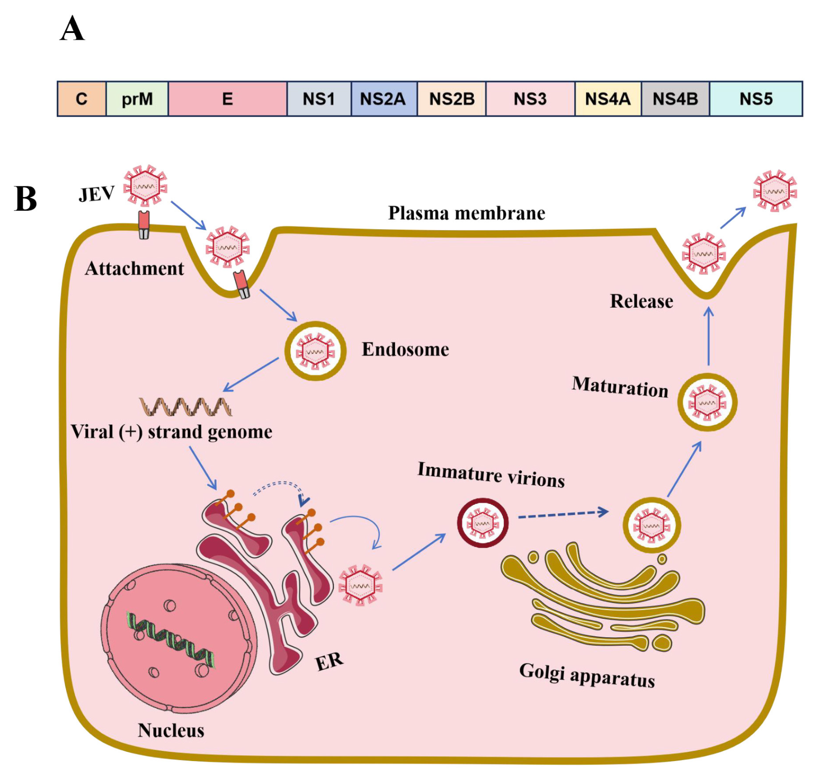
JEV is a member of the genus Orthoflavivirus [11] in the family Flaviviridae [12]. Its positive-sense single-stranded RNA genome consists of 11,000 nucleotides and encodes three structural proteins, including the enveloped protein (E), the membrane protein (M), and the capsid protein (C), as well as seven non-structural proteins named NS1, NS2A/2B, NS3, NS4A/4B, and NS5 (Figure 1) [13]. Based on the nucleotide sequence of the E gene, JEV can be further divided into five genotypes: GI-GV [1]. A detailed phylogenetic analysis of the current major global JEV strains showed that JEV originated from the same ancestor in the Indonesia–Malaysia region, and genotype I JEV has gradually replaced genotype III to become the predominant epidemic strain in the last two decades in China and even the whole of Asia [14].
The E and M proteins form heterodimers that are embedded in the viral envelope, whereas the nucleocapsid, composed of the capsid protein and the viral RNA genome, is located beneath the envelope [17]. The E protein is involved in cellular attachment and entry, and thus it is the primary target of neutralizing antibodies [18]. Meanwhile, some studies have reported that the E protein is involved in binding to the surface receptors of host cells and plays an important role in mediating virus-cell membrane fusion [19]. The role of prM in JEV entry is still largely unknown, and the lack of prM in pseudotyped viruses has no significant influence on virus entry [20]. The C protein is a highly basic protein and associates with RNA to form the nucleocapsid to mediate viral assembly and protect the viral RNA genome.
The NS proteins are responsible for viral replication and interact with host factors to modulate immune responses. NS1 is a glycoprotein that exists in both intracellular and secreted forms. NS2B and NS3 form a protease complex that processes the viral polyprotein, with NS3 acting as a bifunctional protein that harbors a proteolytic domain at its N-terminus and a helicase domain at its C-terminus. NS5 is a highly conserved bifunctional protein that consists of an MTase domain and an RdRp domain, located at the N-terminal and C-terminal respectively. Their roles in regulating immune responses will be discussed in the following sections.

1.3. Viral Life Cycle

JEV has a broad host tropism, and various receptors have been identified to be involved in virus attachment and entry so far, such as heparan sulfate proteoglycans, glycosaminoglycans, laminin, and other host factors, including vimentin, nucleolin, glucose-regulated protein 78 (GRP78), and the recently identified porcine entry factor CD161 [4]. Viral entry is the first step to establishing infection, and several studies have shown that the viral endocytic routes varied for different cell types. JEV hijacks the clathrin-mediated endocytic pathway and the clathrin-independent endocytosis pathway in epithelial cells and neuronal cells, respectively, after interaction with attachment receptors and entry receptors [15,16]. After internalization, virions are delivered to early endosomes and subsequently trafficked to late endosomes, where the acidic pH triggers membrane fusion and genome release into the cytoplasm for RNA synthesis [21]. Once the positive-strand RNA is released into the cytoplasm, it is translated into a polyprotein and cleaved into at least 10 proteins. The viral NS proteins (NS4A, NS4B, NS1, NS2A, NS2B, NS3, and NS5) recruit the viral genome to form a replication compartment associated with the endoplasmic reticulum (ER). The C protein forms dimers and associates with viral (+) strand RNA to bud into ER membranes containing the E-prM proteins. The immature virus particles are transported to the cell surface by the secretory pathway, where viral particles undergo a series of maturation steps, including glycosylation of prM and E, low-pH-induced rearrangement of E-prM, and prM cleavage. Mature virus particles are transported to the cell surface in vesicles and released from the cell surface by exocytosis [22] (Figure 1).

2. Innate Immune Response

2.1. Viral Sensing

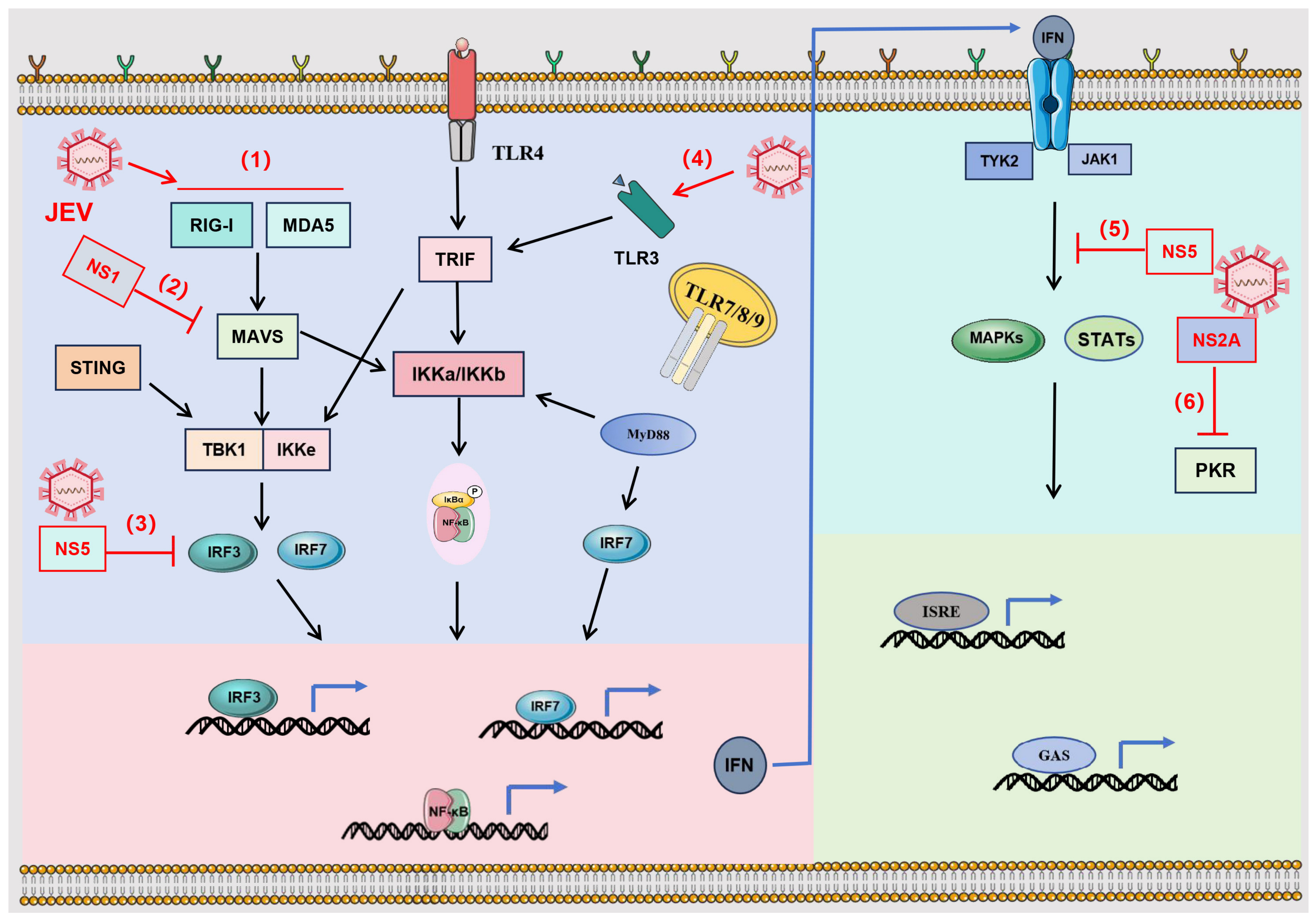
Various cell types can provide favorable environments for JEV genome replication and viral protein translation. They are recognized as foreign invaders by pattern recognition receptors (PRRs) to trigger a variety of host immune responses [23]. In mammalian cells, there are at least four major types of well-characterized PRRs, including Toll-like receptors (TLRs), Retinoic acid-inducible gene I (RIG-I)-like receptors (RLRs), cyclic GMP-AMP synthase (cGAS), and cytoplasmic NOD-like receptors (NLRs) [24,25,26,27]. PRR recognition will trigger the production of cytokines and chemokines, which induce an antiviral state. During JEV infection, the nucleic acid sensors, including RIG-I and melanoma differentiation-associated protein 5 (MDA5), along with endosomal TLR3 and TLR4, are engaged in innate immune activation (Figure 2) [28].
As viral RNA sensors, TLRs can recognize microbes in different cellular locations. TLRs 1, 2, 4, 5, and 6 are expressed on the plasma membrane to recognize various pathogen-associated molecular patterns (PAMPs) in the extracellular environment, such as bacterial lipopolysaccharide (LPS) and lipoteichoic acid. In contrast, TLRs 3, 7, 8, and 9 are mainly expressed on the endosomal and lysosomal membranes, where they detect several different nucleic acids [35]. Double-stranded RNAs (dsRNAs) are generated in the cytoplasm during the life cycle of most RNA viruses, including JEV, whose replicative intermediates are sensed by RIG-I and MDA5; single-stranded RNAs in the endosomes are likewise potent activators of the innate immune response. Studies have reported that TLR3-deficient mice had higher levels of pro-inflammatory cytokines and were extremely vulnerable to JEV by using several TLR-deficient mouse strains (TLR2, TLR3, TLR4, TLR7, TLR9) [36,37,38,39], indicating TLR3 is an antiviral factor.
TLR4 signals through both myeloid differentiation primary response 88 (MyD88) and TIR-domain-containing adapter-inducing interferon-β (TRIF) and could induce the expression of both type I IFN (IFN-I) and inflammatory genes. In addition, TLR4, located on the cell surface, has been shown to detect viral components such as envelope glycoproteins to modulate immune response [40,41]. Similarly, TLR7 has also been shown to be involved in regulating the expression of IFN-I and pro-inflammatory cytokines in response to the orthoflaviviruses Dengue virus (DENV), West Nile virus (WNV), and Zika virus (ZIKV) infection [24,36,42,43,44].
RLRs are cytosolic sensors of viral RNA that respond to viral nucleic acids, and RIG-I as well as MDA5 are two best-characterized RLRs that recognize double- and single-stranded RNA [45,46,47,48]. Notably, whereas the involvement of TLR7 in the recognition of DENV, WNV, and ZIKV is well documented, direct evidence for TLR7-mediated sensing of JEV is still limited; by contrast, the protective role of TLR3 against JEV has been established using TLR-deficient mouse models [36,37,38,39].

2.2. IFN-I Response

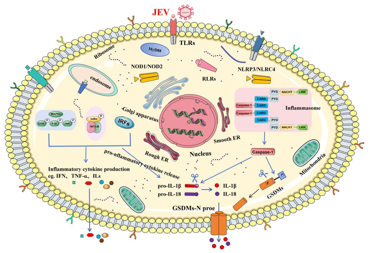
The innate immune system is the first line of defense against virus propagation. During orthoflavivirus infection, viral-encoded PAMPs, including dsRNA and viral proteins, could be recognized by a variety of PRRs. PRRs serve as sentinels to detect invaders and induce the production of IFNs and inflammatory cytokines (Figure 3). JEV showed robust and prolonged viral replication in the intestine, spleen, liver, kidney, and other abdominal organs in IFN-deficient mice [49], indicating host IFN-I signaling was the major barrier to the pathogenicity of JEV. Similarly, IFN-I signal-competent mice died after a prolonged neurological illness, but IFN-I signal-incompetent mice all succumbed without neurological signs [50]. The activation of TLR3 and RLRs induces the production of IFN-I (IFN-α and IFN-β), whose most important action is to inhibit viral replication. They bind the receptors IFNAR1 or IFNAR2 to activate JAK1 and tyrosine kinase 2, leading to the phosphorylation and dimerization of STAT1 and STAT2, which then forms a complex with IRF9 to induce the expression of antiviral proteins, including protein kinase R (PKR), the 2’,5’-oligoadenylate synthetase (OAS)/RNase L system, myxovirus resistance protein 1 (Mx1), ISG15, and viperin. In addition, IFN-I can enhance the cytotoxicity of NK cells and upregulate MHC-I molecule expression to clear viral infection. Notably, IFN-γ is the principal macrophage-activating cytokine and serves critical functions in immunity against intracellular microbes. Larena M et al. reported that IFN-γ, secreted by T cells, is critical for recovery from primary JEV infection by suppressing virus growth and promoting virus clearance in the CNS [51,52].

2.3. Inflammatory Cytokines

One of the earliest innate immune responses to infection is the production of cytokines, which are critical for the acute inflammatory response (Figure 3). JEV pathogenicity and clinical symptoms are highly related to inflammation processes and subsequent breaches in the blood–brain barrier (BBB). However, the JEV infection-induced inflammation is a double-edged sword: on the one hand, the inflammatory response is the first line of defense against viral infection; on the other hand, the excessive or prolonged inflammatory response can cause immunologic injury instead, especially triggering the disruption of the BBB and the cell death of microglial cells [53,54]. In this section, we focus on the overall cytokine response elicited by JEV infection as part of the host innate immune defense, whereas Section 3.2 addresses how the virus actively manipulates cytokine production to evade immune clearance.
The virus completes its lifecycle in host cells and its proteins and RNAs can be detected by PRRs to trigger innate immune response and elevate the production of various kinds of interleukins. Elevated IL-6 levels are associated with disruption of BBB integrity and increased disease severity [55,56], and IL-6/IL-8 levels correlate with the severity of orthoflavivirus-associated neurological disease (detailed in Section 3.2) [57]. Meanwhile, anti-inflammatory cytokines are also induced to maintain homeostasis and prevent the inflammatory storm and inflammation-associated brain damage. For example, IL-10, as a negative immune regulator, inhibits the activation of macrophages and dendritic cells and the subsequent production of inflammatory cytokines.

2.4. Regulation of Host microRNAs During JEV Infection

MicroRNAs (miRNAs) are small non-coding RNAs of approximately 18–25 nucleotides that post-transcriptionally regulate gene expression, usually by binding to the 3′ untranslated regions (UTRs) of target mRNAs. Accumulating evidence indicates that host miRNAs are dynamically regulated during JEV infection and play dual roles in the virus–host interplay [58]. On the one hand, some miRNAs or miRNA-sponging transcripts exert antiviral effects. For example, a circular RNA (circRNA) network that functions as a miRNA sponge was reported to play a potential antiviral role in the early stage of JEV infection in the mouse brain [59]. On the other hand, JEV can hijack the host miRNA machinery to promote viral replication and evade innate immune responses. JEV NS1 was shown to indirectly inhibit MAVS activation through miR-22, thereby suppressing type I interferon production [30] (Figure 2). More recently, the long non-coding RNA JINR1 was reported to promote JEV infection and virus-induced cell death by regulating the miR-216b-5p/GRP78 and miR-1-3p/DDX5 axes [60], and miR-9-5p was found to regulate the transcription factor Onecut2 in JEV-infected neural stem/progenitor cells, affecting neuronal development [61]. Collectively, these findings highlight miRNAs as critical regulators of the JEV-host innate immune interaction and as promising targets for antiviral intervention [58,62].

3. Innate Immune Evasion

3.1. Inhibition and Evasion of IFN-I

The major way in which the innate immune system deals with viral infections is to induce the expression of IFN. Studies have found that JEV NS1 could inhibit the production of IFN-I by targeting MAVS [30]. JEV subgenomic flavivirus RNA (sfRNA) reduced the phosphorylation and nuclear translocation of IRF3, as well as the expression of downstream IFN-β [63]. Similarly, ZIKV sfRNA could inhibit IFN-β promoter activation mediated by RIG-I or MDA5 [64]. Interestingly, the DNA sensor cGAS-STING axis can also be effectively activated during JEV infection [65], and cGAS has also been proven to detect and limit flavivirus infection [66,67]. Recently, several new viral antagonists have been identified: JEV NS1 inhibits IFN-β production by interacting with the host RNA helicase DDX3X [68]; JEV NS4B suppresses IFN-β induction by targeting TLR3 and TRIF [69]; JEV NS1 and NS4B synergistically impair TLR3 signaling to promote viral replication [32]; and JEV NS5 degrades TRAF3 in cooperation with the host factor TUFM to suppress type I interferon induction [70].
To promote its replication, JEV has evolved multiple strategies for immune evasion to escape the innate immune responses (Figure 2). The NS5 protein, which is highly conserved among orthoflaviviruses, has been shown to regulate IFN production and signaling [33,71,72,73]: NS5 could inhibit the activation of transcription factors IRF3 and JAK-STAT to block the IFN-I signaling pathway [31,33]. In addition, JEV NS1 inhibits IFN-β production by enhancing CDK1 phosphorylation and contributes to immune evasion; JEV NS2A not only represses IFN-α production but specifically blocks PKR activation to inhibit eIF2α phosphorylation and enhances viral replication [34]. In addition, many in vitro studies have reported that JEV replication could also hamper the IFN responses by regulating ISRE and GAS-driven genes [74,75].

3.2. Regulation of Interleukins

In contrast to the general cytokine response described in Section 2.3, JEV actively manipulates the production and function of interleukins to subvert immune clearance and promote pathogenesis. JEV has the propensity to infect neuronal and glial cells of the brain, triggering microglial activation and the generation of pro-inflammatory cytokines [53]. Wang et al. reported that JEV infection markedly elevated IL-1α release to devastate the BBB and promoted viral neuroinvasion, indicating that IL-1α plays a crucial role in augmenting JEV-associated pathophysiology [76]. Similarly, the effect of IL-1α could be attenuated by its antagonist [76]. In addition, JEV infection could activate the inflammasome to promote the generation of IL-1β and IL-18 in microglia and astrocyte cells, which in turn differentially regulate the release of other cytokines and chemokines from these cells [77,78]. JEV infection also results in the release of a variety of chemokines and inflammatory factors, including IL-6, which plays a pivotal role in the enhancement of endothelial permeability and may be related to endothelial barrier damage [51,79,80]. In addition, the elevated levels of pro-inflammatory cytokines and the altered permeability of the BBB may be positively related. The high amount of IL-6 can digest BBB tight junctions, leading to CNS inflammation [55,56]. Another similar study reported that in patients with neurological diseases caused by orthoflavivirus infection, the production of IL-6 and IL-8 increased with the severity of JEV infection [57].
Notably, there are also anti-inflammatory cytokines, such as IL-4 and IL-10, to prevent the inflammatory storm and inflammation-associated brain damage during JEV infection. These anti-inflammatory cytokines are negatively associated with neuronal death, and the expression of both was reduced during the progression of JEV infection [81]. IL-10 acts as an immune regulator to protect tissues from damage caused by over-adaptive immune responses and pro-inflammatory mediators. A reduction in JEV-mediated IL-10 generation may be associated with microglial activation and neuronal death [82,83].

3.3. Other Immune Evasion Mechanisms

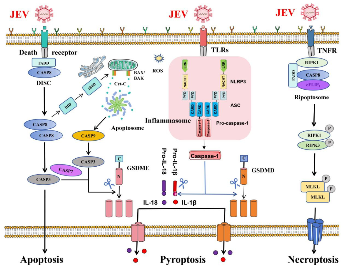
Programmed cell death, although an important host defense, can be subverted by many intracellular pathogens to favor viral dissemination. In the context of JEV infection, the virus can cross the BBB and efficiently replicate in astrocytes and microglia, leading to neuronal death through multiple mechanisms [53]. In this process, a variety of pro-inflammatory cytokines drive cells to undergo apoptosis, pyroptosis, and necroptosis (Figure 4) [84].
Apoptosis is a major kind of cell death and driven by the initiator caspase-8, -9, and -10, as well as the cleavage executor caspase-3 and -7. Among them, caspase-3 and -8 can cleave and activate gasdermin E (GSDME) or GSDMD, respectively, thereby leading to inflammatory cell death [85,86]. It is reported that apoptosis always occurs during JEV infection in neuronal and non-neuronal cells through multiple signal pathways [87]. There are three extensively studied apoptosis pathways according to their initiator caspases [88]. On the one hand, JEV infection could induce mitochondrial-mediated apoptosis through the proapoptotic protein BAX [89] and also induce apoptosis by the IRE1/JNK pathway of ER stress [90]. On the other hand, JEV inhibits the STAT3-Foxo-Bcl-6/p21 pathway to trigger apoptosis [91]. Regarding viral proteins, NS3 of JEV induces cell apoptosis by activating caspase-3 or caspase-8, causing extensive damage [92,93]; the M protein of JEV, WNV, and Yellow fever virus (YFV) also has pro-apoptotic properties [94,95]. In addition, JEV-NS4B could also activate the PERK-ATF4-CHOP pathway to lead to apoptosis in response to ER stress [96]. Furthermore, to promote its replication, JEV infection activates PI3K signaling to block caspase-dependent apoptosis at the early stage of virus infection [97].
Pyroptosis is another form of cell death induced by pro-inflammatory cytokines in microglia during JEV infection [98]. JEV infection could activate the Src/Ras/Raf/ERK/NF-κB signaling axis in the neuron/glial co-culture system in a ROS-dependent manner [99]. Meanwhile, when cells were treated with inhibitors targeting PTK, Ras, and ERK pathways, the production of JEV-induced pro-inflammatory cytokines was significantly inhibited, similar to alleviating neurotoxicity [99]. The activation of neurotoxic microglia and subsequent inflammatory responses are strongly correlated with JEV-induced CCR2 expression [100]. Furthermore, ROS and K+ efflux induce NLRP3 inflammasome assembly in JEV-infected mouse microglia and brain tissue, leading to caspase-1 activation and cytokine maturation [77].
Necroptosis is a regulated form of necrotic cell death mediated by the kinase activities of receptor-interacting protein kinase 1 (RIPK1) and RIPK3, which phosphorylate mixed lineage kinase domain-like protein (MLKL), leading to MLKL oligomerization and plasma membrane pore formation [101]. During JEV infection, transcriptomic analysis of JEV-infected macrophages revealed activation of the necroptotic pathway, which was confirmed by immunofluorescent staining with specific markers [102]. In addition, necroptosis is associated with neuronal loss during JEV infection, providing evidence that necroptosis participates in the pathogenesis of JEV [103].

4. Future Directions and Therapeutic Prospects

4.1. Unresolved Questions and Knowledge Gaps

Despite substantial progress in understanding JEV-host innate immune interactions, several key questions remain unresolved. First, although multiple attachment and entry factors have been identified in vitro, the physiologically relevant receptor(s) mediating JEV entry in vivo remain to be defined [104]. Second, the precise role of the cGAS-STING axis in JEV sensing and pathogenesis warrants further investigation, as both antiviral and pro-inflammatory activities have been proposed [65,66,67]. Third, the in vivo functions of most JEV-regulated host microRNAs and long non-coding RNAs remain largely unexplored, and gain- or loss-of-function studies in animal models are needed to establish their physiological relevance [58,59,60,61]. Finally, the molecular mechanisms by which JEV breaches the BBB and the relative contributions of neuroinflammation versus direct viral cytopathology to neurological sequelae are still incompletely understood [80].

4.2. Developments in Emergency Antiviral Therapy

Because JE progresses rapidly and no specific antiviral drug is currently licensed, there is an urgent need for effective emergency therapies [105]. Several promising directions are under active investigation. Small-molecule inhibitors that target viral enzymes or host factors essential for viral entry and replication are being developed; for example, bergamottin, a bioactive component of bergamot, was recently reported to dually inhibit JEV internalization and genome replication in vitro [106]. Host-directed strategies, such as targeting the entry factor CD161, have also been proposed to inhibit JEV infection [107]. In addition, RNA interference (RNAi)-based therapeutics, including siRNAs and miRNAs directed against conserved viral genomic regions, represent a rapidly advancing modality with multiple delivery platforms under evaluation [62]. Given that antibodies cannot readily access the CNS, anti-inflammatory and neuroprotective approaches that temper the excessive inflammatory response while preserving antiviral immunity may complement direct-acting antivirals and help reduce mortality and neurological sequelae [105].

4.3. Future Research Directions

Beyond immediate therapeutic goals, several research avenues deserve priority. From a mechanistic perspective, systematic dissection of the JEV-encoded proteins that antagonize innate immune signaling (e.g., NS1, NS2A, NS4B, and NS5) continues to reveal new immune-evasion nodes, as exemplified by recent findings on NS1-DDX3X [68], NS4B-TLR3/TRIF [69], and NS5-mediated TRAF3 degradation [70]. The application of high-throughput technologies, including single-cell and spatial transcriptomics, will help define cell-type-specific innate immune responses during JEV neuroinfection. Our group has recently demonstrated that JEV NS1 evades the type I interferon response by interacting with the host helicase DDX3X [68], and we are currently constructing recombinant JEV mutants and using reverse genetics approaches to map the viral determinants of innate immune evasion and neurovirulence. We anticipate that integrating these mechanistic insights with vaccine and therapeutic development will ultimately reduce the burden of this devastating disease.
In summary, the interplay between JEV and the host innate immune system is a dynamic battlefield in which the virus employs multiple strategies to escape recognition and antagonize antiviral effectors, while the host mounts multilayered innate defenses. A comprehensive understanding of these mechanisms not only deepens our insight into viral pathogenesis but also provides a rational basis for the development of effective vaccines, immunomodulatory interventions, and antiviral therapies for Japanese encephalitis.

Author Contributions

T.L.: writing—original draft preparation, formal analysis. S.Z.: writing—original draft preparation. S.X.: resources. H.J.: investigation. D.C.: writing—review and editing. Z.Z.: writing—review and editing, validation. All authors have read and agreed to the published version of the manuscript.

Funding

This research was supported by the R&D Program of the Beijing Municipal Education Commission (KM202310020004), the Public Institution Project of Institute of Animal Husbandry and Veterinary Medicine (XMS202509), and the Special Program on Science and Technology Innovation Capacity Building of BAAFS (KJCX20230424).

Data Availability Statement

No new data were created or analyzed in this study. Data sharing is not applicable to this article.

Acknowledgments

During the preparation of this work, the authors used OpenAI ChatGPT (GPT-5.6 Sol) and OpenAI Codex for literature search, English-language translation and polishing, and manuscript organization. The authors have reviewed the applicable terms of use for the AI tools employed and confirm their suitability for publication.

Conflicts of Interest

Authors Shucheng Zong and Dengjin Chen were employed by China Animal Husbandry Industry Co., Ltd. The re-maining authors declare that the research was conducted in the absence of any commercial or financial relation-ships that could be construed as a potential conflict of interest.

References

  1. Solomon, T.; Ni, H.; Beasley, D.W.; Ekkelenkamp, M.; Cardosa, M.J.; Barrett, A.D. Origin and evolution of Japanese encephalitis virus in southeast Asia. J. Virol. 2003, 77, 3091–3098. [Google Scholar] [CrossRef] [Scilit] [PubMed]
  2. Quan, T.M.; Thao, T.T.N.; Duy, N.M.; Nhat, T.M.; Clapham, H. Estimates of the global burden of Japanese encephalitis and the impact of vaccination from 2000–2015. eLife 2020, 9, e51027. [Google Scholar] [CrossRef] [Scilit] [PubMed]
  3. Campbell, G.L.; Hills, S.L.; Fischer, M.; Jacobson, J.A.; Hoke, C.H.; Hombach, J.M.; Marfin, A.A.; Solomon, T.; Tsai, T.F.; Tsu, V.D.; et al. Estimated global incidence of Japanese encephalitis: A systematic review. Bull. World Health Organ. 2011, 89, 766–774. [Google Scholar] [CrossRef] [Scilit] [PubMed]
  4. Sharma, K.B.; Vrati, S.; Kalia, M. Pathobiology of Japanese encephalitis virus infection. Mol. Asp. Med. 2021, 81, 100994. [Google Scholar] [CrossRef] [Scilit] [PubMed]
  5. Mansfield, K.L.; Horton, D.L.; Johnson, N.; Li, L.; Barrett, A.D.T.; Smith, D.J.; Galbraith, S.E.; Solomon, T.; Fooks, A.R. Flavivirus-induced antibody cross-reactivity. J. Gen. Virol. 2011, 92, 2821–2829. [Google Scholar] [CrossRef] [Scilit] [PubMed]
  6. Hills, S.L.; Vu, H.M.; Pitisuttithum, P.; Lim, C.K.; Goodman, C.H.; Hombach, J.; Solomon, T. Japanese encephalitis: Current status, recent progress, and persistent challenges. PLoS Negl. Trop. Dis. 2026, 20, e0014641. [Google Scholar] [CrossRef] [Scilit] [PubMed]
  7. Kobayashi, D.; Kuwata, R.; Kimura, T.; Faizah, A.N.; Azerigyik, F.A.; Higa, Y.; Hayashi, T.; Sawabe, K.; Isawa, H. Detection of Japanese Encephalitis Virus RNA in Host-Questing Ticks in Japan, 2019–2020. Am. J. Trop. Med. Hyg. 2022, 106, 1725–1728. [Google Scholar] [CrossRef] [Scilit] [PubMed]
  8. Yun, S.I.; Lee, Y.M. Japanese encephalitis: The virus and vaccines. Hum. Vaccines Immunother. 2014, 10, 263–279. [Google Scholar] [CrossRef] [Scilit] [PubMed]
  9. Hubalek, Z.; Rudolf, I.; Nowotny, N. Arboviruses pathogenic for domestic and wild animals. Adv. Virus Res. 2014, 89, 201–275. [Google Scholar] [CrossRef] [Scilit] [PubMed]
  10. Hegde, N.R.; Gore, M.M. Japanese encephalitis vaccines: Immunogenicity, protective efficacy, effectiveness, and impact on the burden of disease. Hum. Vaccines Immunother. 2017, 13, 1320–1337. [Google Scholar] [CrossRef] [Scilit] [PubMed]
  11. Postler, T.S.; Beer, M.; Blitvich, B.J.; Bukh, J.; de Lamballerie, X.; Drexler, J.F.; Imrie, A.; Kapoor, A.; Karganova, G.G.; Lemey, P.; et al. Renaming of the genus Flavivirus to Orthoflavivirus and extension of binomial species names within the family Flaviviridae. Arch. Virol. 2023, 168, 224. [Google Scholar] [CrossRef] [Scilit] [PubMed]
  12. Billoir, F.; de Chesse, R.; Tolou, H.; de Micco, P.; Gould, E.A.; de Lamballerie, X. Phylogeny of the genus Flavivirus using complete coding sequences of arthropod-borne viruses and viruses with no known vector. J. Gen. Virol. 2000, 81, 781–790. [Google Scholar] [CrossRef] [Scilit] [PubMed]
  13. Erlanger, T.E.; Weiss, S.; Keiser, J.; Utzinger, J.; Wiedenmayer, K. Past, present, and future of Japanese encephalitis. Emerg. Infect. Dis. 2009, 15, 1–7. [Google Scholar] [CrossRef] [Scilit] [PubMed]
  14. Li, C.; Di, D.; Huang, H.; Wang, X.; Xia, Q.; Ma, X.; Liu, K.; Li, B.; Shao, D.; Qiu, Y.; et al. NS5-V372A and NS5-H386Y variations are responsible for differences in interferon α/β induction and co-contribute to the replication advantage of Japanese encephalitis virus genotype I over genotype III in ducklings. PLoS Pathog. 2020, 16, e1008773. [Google Scholar] [CrossRef] [Scilit] [PubMed]
  15. Khasa, R.; Sharma, P.; Vaidya, A.; Vrati, S.; Kalia, M. Proteins involved in actin filament organization are key host factors for Japanese encephalitis virus life-cycle in human neuronal cells. Microb. Pathog. 2020, 149, 104565. [Google Scholar] [CrossRef] [Scilit] [PubMed]
  16. Khasa, R.; Vaidya, A.; Vrati, S.; Kalia, M. Membrane trafficking RNA interference screen identifies a crucial role of the clathrin endocytic pathway and ARP2/3 complex for Japanese encephalitis virus infection in HeLa cells. J. Gen. Virol. 2019, 100, 176–186. [Google Scholar] [CrossRef] [Scilit] [PubMed]
  17. Zhu, Y.; He, Z.; Qi, Z. Virus-host Interactions in Early Japanese Encephalitis Virus Infection. Virus Res. 2023, 331, 199120. [Google Scholar] [CrossRef] [Scilit] [PubMed]
  18. Kumar, S.; Verma, A.; Yadav, P.; Dubey, S.K.; Azhar, E.I.; Maitra, S.S.; Dwivedi, V.D. Molecular pathogenesis of Japanese encephalitis and possible therapeutic strategies. Arch. Virol. 2022, 167, 1739–1762. [Google Scholar] [CrossRef] [Scilit] [PubMed]
  19. Tripathi, N.K.; Shrivastava, A.; Biswal, K.C.; Rao, P.V. Development of a pilot-scale production process and characterization of a recombinant Japanese encephalitis virus envelope domain III protein expressed in Escherichia coli. Appl. Microbiol. Biotechnol. 2012, 95, 1179–1189. [Google Scholar] [CrossRef] [Scilit] [PubMed]
  20. Lee, H.J.; Min, K.I.; Lee, J.; Kang, S.H.; Jeon, W.; Nam, J.H.; Ju, Y.R.; Kim, Y.B. The prM-independent packaging of pseudotyped Japanese encephalitis virus. Virol. J. 2009, 6, 115. [Google Scholar] [CrossRef] [Scilit] [PubMed]
  21. Yun, S.I.; Lee, Y.M. Early Events in Japanese Encephalitis Virus Infection: Viral Entry. Pathogens 2018, 7, 68. [Google Scholar] [CrossRef] [Scilit] [PubMed]
  22. Mohsin, F.; Suleman, S.; Anzar, N.; Narang, J.; Wadhwa, S. A review on Japanese Encephalitis virus emergence, pathogenesis and detection: From conventional diagnostics to emerging rapid detection techniques. Int. J. Biol. Macromol. 2022, 217, 435–448. [Google Scholar] [CrossRef] [Scilit] [PubMed]
  23. Koyama, S.; Ishii, K.J.; Coban, C.; Akira, S. Innate immune response to viral infection. Cytokine 2008, 43, 336–341. [Google Scholar] [CrossRef] [Scilit] [PubMed]
  24. Town, T.; Bai, F.; Wang, T.; Kaplan, A.T.; Qian, F.; Montgomery, R.R.; Anderson, J.F.; Flavell, R.A.; Fikrig, E. Toll-like receptor 7 mitigates lethal West Nile encephalitis via interleukin 23-dependent immune cell infiltration and homing. Immunity 2009, 30, 242–253. [Google Scholar] [CrossRef] [Scilit] [PubMed]
  25. Stone, A.E.L.; Green, R.; Wilkins, C.; Hemann, E.A.; Gale, M., Jr. RIG-I-like receptors direct inflammatory macrophage polarization against West Nile virus infection. Nat. Commun. 2019, 10, 3649. [Google Scholar] [CrossRef] [Scilit] [PubMed]
  26. Hornung, V.; Ellegast, J.; Kim, S.; Brzozka, K.; Jung, A.; Kato, H.; Poeck, H.; Akira, S.; Conzelmann, K.K.; Schlee, M.; et al. 5′-Triphosphate RNA is the ligand for RIG-I. Science 2006, 314, 994–997. [Google Scholar] [CrossRef] [Scilit] [PubMed]
  27. Ramos, H.J.; Lanteri, M.C.; Blahnik, G.; Negash, A.; Suthar, M.S.; Brassil, M.M.; Sodhi, K.; Treuting, P.M.; Busch, M.P.; Norris, P.J.; et al. IL-1beta signaling promotes CNS-intrinsic immune control of West Nile virus infection. PLoS Pathog. 2012, 8, e1003039. [Google Scholar] [CrossRef] [Scilit] [PubMed]
  28. Kaur, S.; Tripathi, S. Innate Immune Subversion Strategies of Human Flaviviruses. Crit. Rev. Immunol. 2021, 41, 27–42. [Google Scholar] [CrossRef] [Scilit] [PubMed]
  29. Zheng, B.; Wang, X.; Liu, Y.; Li, Y.; Long, S.; Gu, C.; Ye, J.; Xie, S.; Cao, S. Japanese Encephalitis Virus infection induces inflammation of swine testis through RIG-I-NF-kB signaling pathway. Vet. Microbiol. 2019, 238, 108430. [Google Scholar] [CrossRef] [Scilit] [PubMed]
  30. Zhou, D.; Li, Q.; Jia, F.; Zhang, L.; Wan, S.; Li, Y.; Song, Y.; Chen, H.; Cao, S.; Ye, J. The Japanese Encephalitis Virus NS1’ Protein Inhibits Type I IFN Production by Targeting MAVS. J. Immunol. 2020, 204, 1287–1298. [Google Scholar] [CrossRef] [Scilit] [PubMed]
  31. Ye, J.; Chen, Z.; Li, Y.; Zhao, Z.; He, W.; Zohaib, A.; Song, Y.; Deng, C.; Zhang, B.; Chen, H.; et al. Japanese Encephalitis Virus NS5 Inhibits Type I Interferon (IFN) Production by Blocking the Nuclear Translocation of IFN Regulatory Factor 3 and NF-kappaB. J. Virol. 2017, 91, e00039-17. [Google Scholar] [CrossRef] [Scilit] [PubMed]
  32. Zeng, Q.; He, M.; Li, M.; Yang, D.; Wang, L.; Hao, C.; Wang, Z.; Zhou, M.; Zhu, T.; Niu, X.; et al. Japanese encephalitis virus NS1 and NS4B synergistically target TLR3 signaling to promote viral replication. mBio 2026, 17, e0360425. [Google Scholar] [CrossRef] [Scilit] [PubMed]
  33. Best, S.M. The Many Faces of the Flavivirus NS5 Protein in Antagonism of Type I Interferon Signaling. J. Virol. 2017, 91, e01970-16. [Google Scholar] [CrossRef] [Scilit] [PubMed]
  34. Tu, Y.C.; Yu, C.Y.; Liang, J.J.; Lin, E.; Liao, C.L.; Lin, Y.L. Blocking double-stranded RNA-activated protein kinase PKR by Japanese encephalitis virus nonstructural protein 2A. J. Virol. 2012, 86, 10347–10358. [Google Scholar] [CrossRef] [Scilit] [PubMed]
  35. Blasius, A.L.; Beutler, B. Intracellular toll-like receptors. Immunity 2010, 32, 305–315. [Google Scholar] [CrossRef] [Scilit] [PubMed]
  36. Awais, M.; Wang, K.; Lin, X.; Qian, W.; Zhang, N.; Wang, C.; Wang, K.; Zhao, L.; Fu, Z.F.; Cui, M. TLR7 Deficiency Leads to TLR8 Compensative Regulation of Immune Response against JEV in Mice. Front. Immunol. 2017, 8, 160. [Google Scholar] [CrossRef] [Scilit] [PubMed]
  37. Scholle, F.; Mason, P.W. West Nile virus replication interferes with both poly(I:C)-induced interferon gene transcription and response to interferon treatment. Virology 2005, 342, 77–87. [Google Scholar] [CrossRef] [Scilit] [PubMed]
  38. Daffis, S.; Samuel, M.A.; Suthar, M.S.; Gale, M., Jr.; Diamond, M.S. Toll-like receptor 3 has a protective role against West Nile virus infection. J. Virol. 2008, 82, 10349–10358. [Google Scholar] [CrossRef] [Scilit] [PubMed]
  39. Aleyas, A.G.; George, J.A.; Han, Y.W.; Rahman, M.M.; Kim, S.J.; Han, S.B.; Kim, B.S.; Kim, K.; Eo, S.K. Functional modulation of dendritic cells and macrophages by Japanese encephalitis virus through MyD88 adaptor molecule-dependent and -independent pathways. J. Immunol. 2009, 183, 2462–2474. [Google Scholar] [CrossRef] [Scilit] [PubMed]
  40. Okumura, A.; Pitha, P.M.; Yoshimura, A.; Harty, R.N. Interaction between Ebola virus glycoprotein and host toll-like receptor 4 leads to induction of proinflammatory cytokines and SOCS1. J. Virol. 2010, 84, 27–33. [Google Scholar] [CrossRef] [Scilit] [PubMed]
  41. Kurt-Jones, E.A.; Popova, L.; Kwinn, L.; Haynes, L.M.; Jones, L.P.; Tripp, R.A.; Walsh, E.E.; Freeman, M.W.; Golenbock, D.T.; Anderson, L.J.; et al. Pattern recognition receptors TLR4 and CD14 mediate response to respiratory syncytial virus. Nat. Immunol. 2000, 1, 398–401. [Google Scholar] [CrossRef] [Scilit] [PubMed]
  42. Wang, J.P.; Liu, P.; Latz, E.; Golenbock, D.T.; Finberg, R.W.; Libraty, D.H. Flavivirus activation of plasmacytoid dendritic cells delineates key elements of TLR7 signaling beyond endosomal recognition. J. Immunol. 2006, 177, 7114–7121. [Google Scholar] [CrossRef] [Scilit] [PubMed]
  43. Baker, D.G.; Woods, T.A.; Butchi, N.B.; Morgan, T.M.; Taylor, R.T.; Sunyakumthorn, P.; Mukherjee, P.; Lubick, K.J.; Best, S.M.; Peterson, K.E. Toll-like receptor 7 suppresses virus replication in neurons but does not affect viral pathogenesis in a mouse model of Langat virus infection. J. Gen. Virol. 2013, 94, 336–347. [Google Scholar] [CrossRef] [Scilit] [PubMed]
  44. Vanwalscappel, B.; Tada, T.; Landau, N.R. Toll-like receptor agonist R848 blocks Zika virus replication by inducing the antiviral protein viperin. Virology 2018, 522, 199–208. [Google Scholar] [CrossRef] [Scilit] [PubMed]
  45. Straub, S.; Sampaio, N.G. Activation of cytosolic RNA sensors by endogenous ligands: Roles in disease pathogenesis. Front. Immunol. 2023, 14, 1092790. [Google Scholar] [CrossRef] [Scilit] [PubMed]
  46. Errett, J.S.; Suthar, M.S.; McMillan, A.; Diamond, M.S.; Gale, M., Jr. The essential, nonredundant roles of RIG-I and MDA5 in detecting and controlling West Nile virus infection. J. Virol. 2013, 87, 11416–11425. [Google Scholar] [CrossRef] [Scilit] [PubMed]
  47. Sprokholt, J.K.; Kaptein, T.M.; van Hamme, J.L.; Overmars, R.J.; Gringhuis, S.I.; Geijtenbeek, T.B.H. RIG-I-like receptor activation by dengue virus drives follicular T helper cell formation and antibody production. PLoS Pathog. 2017, 13, e1006738. [Google Scholar] [CrossRef] [Scilit] [PubMed]
  48. Xie, S.J.; Yan, J.Y.; Jiang, B.; Liu, J.; Song, J.W. Immune evasion strategies of Seneca Valley virus: Mechanisms of host innate immune suppression. Agric. Commun. 2025, 3, 100100. [Google Scholar] [CrossRef] [Scilit]
  49. Li, X.F.; Li, X.D.; Deng, C.L.; Dong, H.L.; Zhang, Q.Y.; Ye, Q.; Ye, H.Q.; Huang, X.Y.; Deng, Y.Q.; Zhang, B.; et al. Visualization of a neurotropic flavivirus infection in mouse reveals unique viscerotropism controlled by host type I interferon signaling. Theranostics 2017, 7, 912–925. [Google Scholar] [CrossRef] [Scilit] [PubMed]
  50. Patil, A.M.; Choi, J.Y.; Park, S.O.; Uyangaa, E.; Kim, B.; Kim, K.; Eo, S.K. Type I IFN signaling limits hemorrhage-like disease after infection with Japanese encephalitis virus through modulating a prerequisite infection of CD11b+Ly-6C+ monocytes. J. Neuroinflamm. 2021, 18, 136. [Google Scholar] [CrossRef] [Scilit] [PubMed]
  51. Chen, T.; He, X.; Zhang, P.; Yuan, Y.; Lang, X.; Yu, J.; Qin, Z.; Li, X.; Zhang, Q.; Zhu, L.; et al. Research advancements in the neurological presentation of flaviviruses. Rev. Med. Virol. 2019, 29, e2021. [Google Scholar] [CrossRef] [Scilit] [PubMed]
  52. Larena, M.; Regner, M.; Lobigs, M. Cytolytic effector pathways and IFN-γ help protect against Japanese encephalitis. Eur. J. Immunol. 2013, 43, 1789–1798. [Google Scholar] [CrossRef] [Scilit] [PubMed]
  53. Mohapatra, S.; Chakraborty, T.; Basu, A. Japanese Encephalitis virus infection in astrocytes modulate microglial function: Correlation with inflammation and oxidative stress. Cytokine 2023, 170, 156328. [Google Scholar] [CrossRef] [Scilit] [PubMed]
  54. Wang, K.; Wang, H.; Lou, W.; Ma, L.; Li, Y.; Zhang, N.; Wang, C.; Li, F.; Awais, M.; Cao, S.; et al. IP-10 Promotes Blood-Brain Barrier Damage by Inducing Tumor Necrosis Factor Alpha Production in Japanese Encephalitis. Front. Immunol. 2018, 9, 1148. [Google Scholar] [CrossRef] [Scilit] [PubMed]
  55. Chen, C.J.; Ou, Y.C.; Lin, S.Y.; Raung, S.L.; Liao, S.L.; Lai, C.Y.; Chen, S.Y.; Chen, J.H. Glial activation involvement in neuronal death by Japanese encephalitis virus infection. J. Gen. Virol. 2010, 91, 1028–1037. [Google Scholar] [CrossRef] [Scilit] [PubMed]
  56. Tiwari, R.; Ghildiyal, S.; Baluni, M.; Singh, D.; Srivastva, J.K.; Kumar, R.; Dhole, T.N. Association of interleukin-6 (174 G/C) and interleukin-12B (1188 A/C) gene polymorphism with expression and risk of Japanese encephalitis disease in North Indian population. J. Neuroimmunol. 2021, 358, 577630. [Google Scholar] [CrossRef] [Scilit] [PubMed]
  57. Mehta, V.K.; Verma, R.; Garg, R.K.; Malhotra, H.S.; Sharma, P.K.; Jain, A. Study of interleukin-6 and interleukin-8 levels in patients with neurological manifestations of dengue. J. Postgrad. Med. 2017, 63, 11–15. [Google Scholar] [CrossRef] [Scilit] [PubMed]
  58. Kumar, M.; Sahoo, G.C.; Das, V.N.R.; Singh, K.; Pandey, K. A Review of miRNA Regulation in Japanese Encephalitis (JEV) Virus Infection. Curr. Pharm. Biotechnol. 2024, 25, 521–533. [Google Scholar] [CrossRef] [Scilit] [PubMed]
  59. Chen, M.; Kang, L.; Zhang, T.; Zheng, J.; Chen, D.; Shao, D.; Li, Z.; Li, B.; Wei, J.; Qiu, Y.; et al. Circular RNA network plays a potential antiviral role in the early stage of JEV infection in mouse brain. Front. Microbiol. 2023, 14, 1165378. [Google Scholar] [CrossRef] [Scilit] [PubMed]
  60. Tripathi, S.; Sengar, S.; Basu, A.; Sharma, V. LncRNA JINR1 regulates miR-216b-5p/GRP78 and miR-1-3p/DDX5 axis to promote JEV infection and cell death. J. Virol. 2025, 99, e0006625. [Google Scholar] [CrossRef] [Scilit] [PubMed]
  61. Sharma, S.; Majumdar, A.; Basu, A. Regulation of Onecut2 by miR-9-5p in Japanese encephalitis virus infected neural stem/progenitor cells. Microbiol. Spectr. 2024, 12, e0323823. [Google Scholar] [CrossRef] [Scilit] [PubMed]
  62. Kumar, M.; Gupta, P.; Kumar, S.; Kumar, S.K.; Tiwari, S.N.; Aspatwar, A. RNAi therapeutics targeting Japanese encephalitis virus: Gene targets, delivery platforms, and translational barriers. Virology 2026, 613, 110713. [Google Scholar] [CrossRef] [Scilit] [PubMed]
  63. Chang, R.Y.; Hsu, T.W.; Chen, Y.L.; Liu, S.F.; Tsai, Y.J.; Lin, Y.T.; Chen, Y.S.; Fan, Y.H. Japanese encephalitis virus non-coding RNA inhibits activation of interferon by blocking nuclear translocation of interferon regulatory factor 3. Vet. Microbiol. 2013, 166, 11–21. [Google Scholar] [CrossRef] [Scilit] [PubMed]
  64. Donald, C.L.; Brennan, B.; Cumberworth, S.L.; Rezelj, V.V.; Clark, J.J.; Cordeiro, M.T.; Freitas de Oliveira Franca, R.; Pena, L.J.; Wilkie, G.S.; Da Silva Filipe, A.; et al. Full Genome Sequence and sfRNA Interferon Antagonist Activity of Zika Virus from Recife, Brazil. PLoS Negl. Trop. Dis. 2016, 10, e0005048. [Google Scholar] [CrossRef] [Scilit] [PubMed]
  65. Sharma, K.B.; Chhabra, S.; Aggarwal, S.; Tripathi, A.; Banerjee, A.; Yadav, A.K.; Vrati, S.; Kalia, M. Proteomic landscape of Japanese encephalitis virus-infected fibroblasts. J. Gen. Virol. 2021, 102, 001657. [Google Scholar] [CrossRef] [Scilit] [PubMed]
  66. Sun, B.; Sundstrom, K.B.; Chew, J.J.; Bist, P.; Gan, E.S.; Tan, H.C.; Goh, K.C.; Chawla, T.; Tang, C.K.; Ooi, E.E. Dengue virus activates cGAS through the release of mitochondrial DNA. Sci. Rep. 2017, 7, 3594. [Google Scholar] [CrossRef] [Scilit] [PubMed]
  67. Schoggins, J.W.; MacDuff, D.A.; Imanaka, N.; Gainey, M.D.; Shrestha, B.; Eitson, J.L.; Mar, K.B.; Richardson, R.B.; Ratushny, A.V.; Litvak, V.; et al. Pan-viral specificity of IFN-induced genes reveals new roles for cGAS in innate immunity. Nature 2014, 505, 691–695. [Google Scholar] [CrossRef] [Scilit] [PubMed]
  68. Dong, H.; Hao, Y.; Wang, J.; Chen, D.; Xu, S.; Ruan, W. Japanese encephalitis virus NS1 inhibits IFN-beta production by interacting with DDX3X. J. Virol. 2025, 99, e0007725. [Google Scholar] [CrossRef] [Scilit] [PubMed]
  69. Zeng, Q.; Liu, J.; Li, Z.; Zhang, Y.; Zu, S.; Ding, X.; Zhang, H. Japanese encephalitis virus NS4B inhibits interferon beta production by targeting TLR3 and TRIF. Vet. Microbiol. 2023, 284, 109849. [Google Scholar] [CrossRef] [Scilit] [PubMed]
  70. Wang, X.; Wang, C.; Yang, X.; Qin, W.; Liu, X.; Yu, H.; Tong, W.; Tong, G.; Shan, T.; Kong, N.; et al. Japanese encephalitis virus degrades TRAF3 to suppress type I interferon and promote viral replication through NS5 and host TUFM proteins. Vet. Res. 2026, 57, 66. [Google Scholar] [CrossRef] [Scilit] [PubMed]
  71. Grant, A.; Ponia, S.S.; Tripathi, S.; Balasubramaniam, V.; Miorin, L.; Sourisseau, M.; Schwarz, M.C.; Sanchez-Seco, M.P.; Evans, M.J.; Best, S.M.; et al. Zika Virus Targets Human STAT2 to Inhibit Type I Interferon Signaling. Cell Host Microbe 2016, 19, 882–890. [Google Scholar] [CrossRef] [Scilit] [PubMed]
  72. Thurmond, S.; Wang, B.; Song, J.; Hai, R. Suppression of Type I Interferon Signaling by Flavivirus NS5. Viruses 2018, 10, 712. [Google Scholar] [CrossRef] [Scilit] [PubMed]
  73. Coldbeck-Shackley, R.C.; Eyre, N.S.; Beard, M.R. The Molecular Interactions of ZIKV and DENV with the Type-I IFN Response. Vaccines 2020, 8, 530. [Google Scholar] [CrossRef] [Scilit] [PubMed]
  74. Chang, S.J.; Huang, S.H.; Lin, Y.J.; Tsou, Y.Y.; Lin, C.W. Antiviral activity of Rheum palmatum methanol extract and chrysophanol against Japanese encephalitis virus. Arch. Pharm. Res. 2014, 37, 1117–1123. [Google Scholar] [CrossRef] [Scilit] [PubMed]
  75. Gupta, A.; Gawandi, S.; Vandna; Yadav, I.; Mohan, H.; Desai, V.G.; Kumar, S. Analysis of fluoro based pyrazole analogues as a potential therapeutics candidate against Japanese encephalitis virus infection. Virus Res. 2022, 323, 198955. [Google Scholar] [CrossRef] [Scilit] [PubMed]
  76. Wang, Z.Y.; Zhen, Z.D.; Fan, D.Y.; Qin, C.F.; Han, D.S.; Zhou, H.N.; Wang, P.G.; An, J. Axl Deficiency Promotes the Neuroinvasion of Japanese Encephalitis Virus by Enhancing IL-1alpha Production from Pyroptotic Macrophages. J. Virol. 2020, 94, e00602-20. [Google Scholar] [CrossRef] [Scilit] [PubMed]
  77. Kaushik, D.K.; Gupta, M.; Kumawat, K.L.; Basu, A. NLRP3 inflammasome: Key mediator of neuroinflammation in murine Japanese encephalitis. PLoS ONE 2012, 7, e32270. [Google Scholar] [CrossRef] [Scilit] [PubMed]
  78. Fan, S.; Yuan, J.; Deng, S.; Chen, Y.; Xie, B.; Wu, K.; Zhu, M.; Xu, H.; Huang, Y.; Yang, J.; et al. Activation of Interleukin-1beta Release by the Classical Swine Fever Virus Is Dependent on the NLRP3 Inflammasome, Which Affects Virus Growth in Monocytes. Front. Cell. Infect. Microbiol. 2018, 8, 225. [Google Scholar] [CrossRef] [Scilit] [PubMed]
  79. Pardridge, W.M. Drug transport across the blood-brain barrier. J. Cereb. Blood Flow Metab. 2012, 32, 1959–1972. [Google Scholar] [CrossRef] [Scilit] [PubMed]
  80. Mustafa, Y.M.; Meuren, L.M.; Coelho, S.V.A.; de Arruda, L.B. Pathways Exploited by Flaviviruses to Counteract the Blood-Brain Barrier and Invade the Central Nervous System. Front. Microbiol. 2019, 10, 525. [Google Scholar] [CrossRef] [Scilit] [PubMed]
  81. Saxena, V.; Mathur, A.; Krishnani, N.; Dhole, T.N. An insufficient anti-inflammatory cytokine response in mouse brain is associated with increased tissue pathology and viral load during Japanese encephalitis virus infection. Arch. Virol. 2008, 153, 283–292. [Google Scholar] [CrossRef] [Scilit] [PubMed]
  82. Swarup, V.; Ghosh, J.; Duseja, R.; Ghosh, S.; Basu, A. Japanese encephalitis virus infection decrease endogenous IL-10 production: Correlation with microglial activation and neuronal death. Neurosci. Lett. 2007, 420, 144–149. [Google Scholar] [CrossRef] [Scilit] [PubMed]
  83. Ghildiyal, S.; Fatima, T.; Singh, D.; Upadhyay, S.; Dhole, T.N.; Himanshu Reddy, D.; Kumar, A. Pro-inflammatory and anti-inflamatory cytokine genes polymorphisms and susceptibility to Japanese encephalitis disease in the North Indian population. Cytokine 2022, 149, 155716. [Google Scholar] [CrossRef] [Scilit] [PubMed]
  84. Ghoshal, A.; Das, S.; Ghosh, S.; Mishra, M.K.; Sharma, V.; Koli, P.; Sen, E.; Basu, A. Proinflammatory mediators released by activated microglia induces neuronal death in Japanese encephalitis. Glia 2007, 55, 483–496. [Google Scholar] [CrossRef] [Scilit] [PubMed]
  85. Place, D.E.; Lee, S.; Kanneganti, T.D. PANoptosis in microbial infection. Curr. Opin. Microbiol. 2021, 59, 42–49. [Google Scholar] [CrossRef] [Scilit] [PubMed]
  86. Rogers, C.; Erkes, D.A.; Nardone, A.; Aplin, A.E.; Fernandes-Alnemri, T.; Alnemri, E.S. Gasdermin pores permeabilize mitochondria to augment caspase-3 activation during apoptosis and inflammasome activation. Nat. Commun. 2019, 10, 1689. [Google Scholar] [CrossRef] [Scilit] [PubMed]
  87. Swarup, V.; Das, S.; Ghosh, S.; Basu, A. Tumor necrosis factor receptor-1-induced neuronal death by TRADD contributes to the pathogenesis of Japanese encephalitis. J. Neurochem. 2007, 103, 771–783. [Google Scholar] [CrossRef] [Scilit] [PubMed]
  88. Mehmet, H. Caspases find a new place to hide. Nature 2000, 403, 29–30. [Google Scholar] [CrossRef] [Scilit] [PubMed]
  89. Yang, K.; Li, X.; Yang, S.; Zheng, Y.; Cao, S.; Yan, Q.; Huang, X.; Wen, Y.; Zhao, Q.; Du, S.; et al. Japanese encephalitis virus infection induces mitochondrial-mediated apoptosis through the proapoptotic protein BAX. Front. Microbiol. 2024, 15, 1485667. [Google Scholar] [CrossRef] [Scilit] [PubMed]
  90. Huang, M.; Xu, A.; Wu, X.; Zhang, Y.; Guo, Y.; Guo, F.; Pan, Z.; Kong, L. Japanese encephalitis virus induces apoptosis by the IRE1/JNK pathway of ER stress response in BHK-21 cells. Arch. Virol. 2016, 161, 699–703. [Google Scholar] [CrossRef] [Scilit] [PubMed]
  91. Guo, F.; Yu, X.; Xu, A.; Xu, J.; Wang, Q.; Guo, Y.; Wu, X.; Tang, Y.; Ding, Z.; Zhang, Y.; et al. Japanese encephalitis virus induces apoptosis by inhibiting Foxo signaling pathway. Vet. Microbiol. 2018, 220, 73–82. [Google Scholar] [CrossRef] [Scilit] [PubMed]
  92. Ramanathan, M.P.; Chambers, J.A.; Pankhong, P.; Chattergoon, M.; Attatippaholkun, W.; Dang, K.; Shah, N.; Weiner, D.B. Host cell killing by the West Nile Virus NS2B-NS3 proteolytic complex: NS3 alone is sufficient to recruit caspase-8-based apoptotic pathway. Virology 2006, 345, 56–72. [Google Scholar] [CrossRef] [Scilit] [PubMed]
  93. Yang, T.C.; Shiu, S.L.; Chuang, P.H.; Lin, Y.J.; Wan, L.; Lan, Y.C.; Lin, C.W. Japanese encephalitis virus NS2B-NS3 protease induces caspase 3 activation and mitochondria-mediated apoptosis in human medulloblastoma cells. Virus Res. 2009, 143, 77–85. [Google Scholar] [CrossRef] [Scilit] [PubMed]
  94. Catteau, A.; Kalinina, O.; Wagner, M.C.; Deubel, V.; Courageot, M.P.; Despres, P. Dengue virus M protein contains a proapoptotic sequence referred to as ApoptoM. J. Gen. Virol. 2003, 84, 2781–2793. [Google Scholar] [CrossRef] [Scilit] [PubMed]
  95. Brabant, M.; Baux, L.; Casimir, R.; Briand, J.P.; Chaloin, O.; Porceddu, M.; Buron, N.; Chauvier, D.; Lassalle, M.; Lecoeur, H.; et al. A flavivirus protein M-derived peptide directly permeabilizes mitochondrial membranes, triggers cell death and reduces human tumor growth in nude mice. Apoptosis 2009, 14, 1190–1203. [Google Scholar] [CrossRef] [Scilit] [PubMed]
  96. Wang, Q.; Xin, X.; Wang, T.; Wan, J.; Ou, Y.; Yang, Z.; Yu, Q.; Zhu, L.; Guo, Y.; Wu, Y.; et al. Japanese Encephalitis Virus Induces Apoptosis and Encephalitis by Activating the PERK Pathway. J. Virol. 2019, 93, e00887-19. [Google Scholar] [CrossRef] [Scilit] [PubMed]
  97. Lee, C.J.; Liao, C.L.; Lin, Y.L. Flavivirus activates phosphatidylinositol 3-kinase signaling to block caspase-dependent apoptotic cell death at the early stage of virus infection. J. Virol. 2005, 79, 8388–8399. [Google Scholar] [CrossRef] [Scilit] [PubMed]
  98. Raung, S.L.; Chen, S.Y.; Liao, S.L.; Chen, J.H.; Chen, C.J. Japanese encephalitis virus infection stimulates Src tyrosine kinase in neuron/glia. Neurosci. Lett. 2007, 419, 263–268. [Google Scholar] [CrossRef] [Scilit] [PubMed]
  99. Ashraf, U.; Ding, Z.; Deng, S.; Ye, J.; Cao, S.; Chen, Z. Pathogenicity and virulence of Japanese encephalitis virus: Neuroinflammation and neuronal cell damage. Virulence 2021, 12, 968–980. [Google Scholar] [CrossRef] [Scilit] [PubMed]
  100. Sun, J.; Yu, Y.; Deubel, V. Japanese encephalitis virus NS1’ protein depends on pseudoknot secondary structure and is cleaved by caspase during virus infection and cell apoptosis. Microbes Infect. 2012, 14, 930–940. [Google Scholar] [CrossRef] [Scilit] [PubMed]
  101. Dondelinger, Y.; Declercq, W.; Montessuit, S.; Roelandt, R.; Goncalves, A.; Bruggeman, I.; Hulpiau, P.; Weber, K.; Sehon, C.A.; Marquis, R.W.; et al. MLKL compromises plasma membrane integrity by binding to phosphatidylinositol phosphates. Cell Rep. 2014, 7, 971–981. [Google Scholar] [CrossRef] [Scilit] [PubMed]
  102. Wang, Z.Y.; Zhen, Z.D.; Fan, D.Y.; Wang, P.G.; An, J. Transcriptomic Analysis Suggests the M1 Polarization and Launch of Diverse Programmed Cell Death Pathways in Japanese Encephalitis Virus-Infected Macrophages. Viruses 2020, 12, 356. [Google Scholar] [CrossRef] [Scilit] [PubMed]
  103. Bian, P.; Zheng, X.; Wei, L.; Ye, C.; Fan, H.; Cai, Y.; Zhang, Y.; Zhang, F.; Jia, Z.; Lei, Y. MLKL Mediated Necroptosis Accelerates JEV-Induced Neuroinflammation in Mice. Front. Microbiol. 2017, 8, 303. [Google Scholar] [CrossRef] [Scilit] [PubMed]
  104. Yang, S.; Zhang, M.; Yang, X.; Yue, J.; Li, X.; Zhang, P.; Zhang, H. Cellular receptors and host factors mediating orthoflavivirus entry. Virol. J. 2026; in press. [CrossRef] [Scilit] [PubMed]
  105. Singh, R.; Sanyal, S.; Basant, N.; Sanyal, S. An Updated Review of Potential Drug Targets for Japanese Encephalitis. Cent. Nerv. Syst. Agents Med. Chem. 2026, 26, 46–62. [Google Scholar] [CrossRef] [Scilit] [PubMed]
  106. Mao, W.; Li, L.; Liu, Y.; Wan, Y.; Pen, Y.; Xu, R.; Wang, W. Bergamottin, a bioactive component of bergamot: Dual inhibition of Japanese encephalitis virus internalization and genome replication. Antivir. Res. 2026, 254, 106512. [Google Scholar] [CrossRef] [Scilit] [PubMed]
  107. Li, J.; Zhao, Q.; Yuan, J.; Ma, Y.; Chen, J.Y.; Yi, H.; Wen, Y.; Du, S.; Huang, X.; Wu, R.; et al. Porcine CD161 promotes Japanese encephalitis virus entry: A potential host-directed antiviral target. Antivir. Res. 2026, 251, 106434. [Google Scholar] [CrossRef] [Scilit] [PubMed]
Figure 1. The genetic composition and replication cycle of JEV. (A). JEV positive-sense single-stranded RNA genome consists of 11,000 nucleotides and encodes three structural proteins, including the enveloped protein (E), the membrane protein (M), and the capsid protein (C), as well as seven non-structural proteins named NS1, NS2A/2B, NS3, NS4A/4B, and NS5. (B). JEV hijacks the clathrin-mediated endocytic pathway and the clathrin-independent endocytosis pathway in epithelial cells and neuronal cells, respectively, after interaction with attachment receptors and entry receptors [15,16].
Viruses 18 00975 g001
Figure 2. The innate immune regulation strategies of JEV. (1) JEV activated RIG-I and induced the expression of interferon (IFN) and pro-inflammatory factors [29]; (2) JEV-NS1 indirectly inhibits MAVS activation through microRNA 22 (miR-22) [30]; (3) JEV-NS5 inhibited the activation of the transcription factor IRF3 [31]; (4) JEV activated TLR3 to induce the expression of inflammatory factors [32]; (5) JEV-NS5 suppressed the JAK1-STAT signal pathways [33]; (6) JEV-NS2A inhibits protein kinase R (PKR) activation [34].
Viruses 18 00975 g002
Figure 3. JEV infection modulates the production of IFNs and pro-inflammatory cytokines. JEV enters cells by receptor-mediated endocytosis, and viral RNA can be recognized by TLR3 and TLR4 in endosomes; viral genomes synthesized in the cytoplasm can be detected by RIG-I, cGAS, and NLRs, ultimately inducing high amounts of IFNs, pro-inflammatory cytokines, and chemokines.
Viruses 18 00975 g003
Figure 4. Programmed cell death pathways during JEV infection. Apoptosis: JEV infection initiates a signal cascade mediated by caspase-8 and caspase-3 to initiate inflammatory cell death. Pyroptosis: The viral proteins of JEV act as cytoplasmic PAMPs or TLRs to induce inflammasome assembly and stimulate caspase-1 activation, initiating pyroptosis. Necroptosis: JEV infection triggers necroptosis, which depends on the formation of RIPK1 and RIPK3 complexes and the activation of downstream MLKL proteins to form channels in the membrane.
Viruses 18 00975 g004
Disclaimer/Publisher’s Note: The statements, opinions and data contained in all publications are solely those of the individual author(s) and contributor(s) and not of MDPI and/or the editor(s). MDPI and/or the editor(s) disclaim responsibility for any injury to people or property resulting from any ideas, methods, instructions or products referred to in the content.