Article

Development of a TaqMan Probe-Based Quantitative PCR Assay for the Detection of Cynoglossus semilaevis Papillomavirus

1
College of Life Science, Tianjin Normal University, Tianjin 300387, China
2
Tianjin Fishery Research Institute, Tianjin 300221, China
3
Tianjin Key Laboratory of Animal and Plant Resistance, Tianjin Normal University, Tianjin 300387, China
*
Author to whom correspondence should be addressed.
Viruses 2026, 18(9), 939; https://doi.org/10.3390/v18090939 (registering DOI)
Submission received: 30 July 2026 / Revised: 21 August 2026 / Accepted: 25 August 2026 / Published: 27 August 2026

Abstract

Cynoglossus semilaevis papillomavirus (CsPaV) is an emerging viral pathogen associated with high mortality in farmed Chinese tongue soles. A sensitive, specific, and quantitative method is needed for CsPaV surveillance and epidemiological studies. In this study, we designed primers and a probe that targeted the CsPaV L1 gene. We then established a TaqMan probe-based quantitative PCR (qPCR) assay. The assay detected as few as 5.8 × 101 copies/µL and was 1000-fold more sensitive than conventional PCR. The assay did not cross-react with the other aquatic pathogens tested. The intra-assay coefficients of variation ranged from 0.23% to 0.84%, and the inter-assay coefficients ranged from 0.56% to 1.48%. These results showed that the assay had good repeatability. We used the assay to study the tissue distribution and temporal dynamics of CsPaV after experimental infection. Healthy Chinese tongue soles received an intraperitoneal injection of 300 µL of inoculum containing 3.66 × 10⁵ CsPaV copies/µL. Three fish were sampled at 1, 2, 3, 5, and 7 days post-infection. CsPaV DNA was detected in all tissues examined. The kidney had the highest viral DNA load, which reached 2.47 × 10⁷ copies/mg at 7 days post-infection. Among 176 clinically diseased Chinese tongue soles collected between 2023 and 2026, 80.68% tested positive for CsPaV. CsPaV DNA was also detected in several other aquatic species. These findings provide a sensitive tool for CsPaV surveillance and molecular epidemiological studies. The assay may also support disease prevention and control in Chinese tongue sole aquaculture.

1. Introduction

Cynoglossus semilaevis occurs naturally in the coastal waters of Bohai Bay and the Yellow Sea. This fish has become an important marine aquaculture species in China. In recent years, emerging diseases have caused high mortality in farmed Chinese tongue soles and major economic losses. Cynoglossus semilaevis papillomavirus (CsPaV) is a recently identified virus associated with mortality rates of up to 80.7% [1]. However, no sensitive quantitative method is available for CsPaV detection. This lack of a suitable method has limited epidemiological studies and the development of disease-control measures.
Papillomaviruses (PVs) are linked to many diseases in humans and animals [2,3]. Researchers identified the first fish papillomavirus in gilthead seabream (Sparus aurata) in 2016 [4]. Researchers later reported fish PVs in Lutjanus campechanus, Oncorhynchus mykiss [5], Silurus glanis [6], Trematomus bernacchii, Centropristis striata, Melanogrammus aeglefinus [7], Tibetan cold-water fish [8], Betta splendens [9], Cynoglossus semilaevis [1], and Raja clavata [10]. Mammalian papillomaviruses have been studied in detail. In contrast, fish papillomaviruses remain poorly understood. Known fish PV genomes contain the conserved E1, E2, L1, and L2 genes, but they lack the common E6 and E7 oncogenes. Phylogenetic analyses place these viruses in a distinct fish-associated lineage [11,12]. Their host range, target cells, routes of transmission, persistence, and role in disease remain unclear. A quantitative assay specific to CsPaV is therefore needed to study its distribution and its association with disease in aquaculture.
Common methods for pathogen detection include enzyme-linked immunosorbent assay (ELISA) [13], polymerase chain reaction (PCR) [14], loop-mediated isothermal amplification (LAMP) [15], and quantitative PCR (qPCR) [16]. ELISA requires pathogen-specific antibodies and includes several handling steps. Conventional PCR and LAMP are simple and sensitive, but they usually do not measure pathogen quantity. TaqMan probe-based qPCR provides high specificity, high sensitivity, and real-time quantification in a closed tube. This design also reduces handling after amplification and lowers the risk of contamination. In recent years, researchers have developed TaqMan probe-based qPCR assays for several aquatic animal viruses. These viruses include megalocytiviruses [16], tilapia lake virus [17], tilapia parvovirus [18], and turbot circovirus [19]. These studies show that TaqMan probe-based qPCR is useful for the quantitative surveillance of aquatic viral diseases. However, no similar assay had been developed for CsPaV.
This study aimed to develop and validate a TaqMan probe-based qPCR assay for CsPaV detection. We also used the assay in a preliminary molecular epidemiological study. The assay provides a practical tool for routine surveillance, field screening, and studies of CsPaV distribution.

2. Materials and Methods

2.1. Sample Collection

Between 2023 and 2026, we collected 176 clinically diseased Chinese tongue soles from aquaculture farms in Tianjin, Hebei Province, and Shandong Province. We also collected wild Chinese tongue soles from Bohai Bay and the Yellow Sea. In addition, we obtained Epinephelus sp., Charybdis japonica, Rapana venosa, Litopenaeus vannamei, Chlamys farreri, Oratosquilla oratoria, Lateolabrax japonicus, Scapharca subcrenata, and Synechogobius hasta from a seafood market near Bohai Bay. Table 1 provides the sampling locations, dates, species, tissues, sample numbers, and body lengths. We used these samples for the epidemiological survey. We transported all specimens to the laboratory in insulated boxes under chilled conditions. We collected the liver, spleen, and kidney from each Chinese tongue sole under aseptic conditions. We collected the tissues listed in Table 1 from the other species. We stored all tissue samples at −80 °C until nucleic acid extraction. Laboratory stocks of viral nervous necrosis virus (VNNV), spring viraemia of carp virus (SVCV), grass carp reovirus (GCRV), Vibrio harveyi, V. alginolyticus, V. anguillarum, and Edwardsiella tarda served as non-target controls in the specificity assay.

2.2. Primers and TaqMan Probe Design

Based on the CsPaV L1 gene sequence (GenBank accession no. OQ865369), a pair of primers and a TaqMan probe were designed using Primer Premier 5.0. (PREMIER Biosoft International, Palo Alto, CA, USA) Candidate primers were evaluated to minimize the formation of primer dimers and hairpin structures and to ensure specific amplification of a 99 bp region of the L1 gene. The probe was designed within the amplified region, with a length of 20–40 nucleotides and a GC content of approximately 50%. Primers L1-a-F (5′-CGGAATTCCGATGCACTCTTCAAGGAA-3′) and L1-a-R (5′-CCCAAGCTTGGGTCAAACAAAAGTTGTAT-3′) were designed for amplifying the L1 gene sequence with a length of 654 bp and constructing a standard plasmid. A pair of qPCR-specific primers was designed using the L1-a fragment sequence: qL1-a-F1 5′-AATCAGATCAAGAAGAAGACCCA-3′; qL1-a-R1 5′-CCTCATTATCATAGATGGTGCC-3′. The TaqMan probe sequence was designed as 5′-FAM-CTGGAACACCGAACAGTGACTATGGC-TAMRA-3′ for qPCR amplification, with the target fragment being 99 bp. All primers and the TaqMan probe were synthesized by Anshengda Biotechnology Co., Ltd. (Suzhou, China).

2.3. Plasmid and Standard Curve Construction

We amplified a 654-bp fragment of the CsPaV L1 gene in a total volume of 25 µL. The reaction contained 2 µL of DNA template (134.6 ng/µL), 1 µL of an L1-a-F/L1-a-R primer mixture (10 µM), 2.5 µL of 10× TransTaq HiFi Buffer I (TransGen Biotech Co., Ltd., Beijing, China; Cat. No. AP131-01), 2 µL of dNTPs (2.5 mM), 0.25 µL of TransTaq HiFi DNA Polymerase (TransGen Biotech Co., Ltd., Beijing, China; Cat. No. AP131-01), and 17.25 µL of ddH2O. The thermal program started with denaturation at 94 °C for 5 min. The program then included 35 cycles at 94 °C for 30 s, 60 °C for 30 s, and 72 °C for 30 s. A final extension was performed at 72 °C for 5 min. We purified the PCR product with a gel extraction kit (TIANGEN Biotech (Beijing) Co., Ltd., Beijing, China; Cat. No. DP219). We then cloned the product into the pMD19-T vector (TaKaRa Bio Inc., Kusatsu, Shiga, Japan; Cat. No. 6013) according to the manufacturer’s instructions. We transformed the recombinant plasmid into competent Escherichia coli DH5α cells (Tsingke Biotechnology Co., Ltd., Beijing, China; Cat. No. DLC101). We confirmed positive clones by Sanger sequencing. We purified the plasmid with a plasmid extraction kit (TIANGEN Biotech (Beijing) Co., Ltd., Beijing, China; Cat. No. DP105). We measured the plasmid concentration with a NanoDrop spectrophotometer (Thermo Fisher Scientific, Waltham, MA, USA).
We quantified the recombinant plasmid as described previously [20]. We calculated its copy number using the formula reported in [21]. We prepared 10-fold serial dilutions of pMD19T-L1a from 5.8 × 10¹⁰ to 5.8 × 101 copies/µL. We constructed the standard curve by plotting the Ct value against the base-10 logarithm of the plasmid copy number.

2.4. TaqMan Probe-Based qPCR Assay

The TaqMan probe-based qPCR reaction had a total volume of 20 µL. Each reaction contained 2 µL of template, 10 µL of Premix Ex Taq (TaKaRa Bio Inc., Shiga, Japan; Cat. No. RR390A), 0.8 µL of a qL1-a-F1/qL1-a-R1 primer mixture (10 µM each), 0.8 µL of TaqMan probe (10 µM), and 6.4 µL of ddH2O. We ran the assay on a QuantStudio 1 Plus Real-Time PCR System (Thermo Fisher Scientific, Waltham, MA, USA). The program started at 95 °C for 30 s. It then included 40 cycles at 95 °C for 5 s and 60 °C for 30 s. The program ended with a step at 50 °C for 30 s.

2.5. Sensitivity and Specificity Test

We prepared 10-fold serial dilutions of the L1-a recombinant plasmid. We used each dilution as a template for both qPCR and conventional PCR. The conventional PCR reaction had a total volume of 25 µL. Each reaction contained 2 µL of template, 22 µL of Fast Taq MasterMix (Beijing Biomed Gene Technology Co., Ltd., Beijing, China; Cat. No. MT302), and 1 µL of a qL1-a-F1/qL1-a-R1 primer mixture (10 µM each). The program started at 94 °C for 5 min. It then included 40 cycles at 94 °C for 10 s, 60 °C for 10 s, and 72 °C for 10 s. A final extension was performed at 72 °C for 15 min.
The SYBR Green qPCR reaction had a total volume of 20 µL. Each reaction contained 2 µL of template, 10 µL of 2× Universal SYBR Green qPCR Master Mix (Sangon Biotech (Shanghai) Co., Ltd., Shanghai, China; Cat. No. B690016), 0.8 µL of a qL1-a-F1/qL1-a-R1 primer mixture (10 µM each), and 7.2 µL of ddH2O. The program started at 95 °C for 2 min. It then included 40 cycles at 95 °C for 10 s and 60 °C for 30 s. The program ended with a step at 50 °C for 30 s.
We evaluated analytical specificity with SVCV, GCRV, VNNV, Vibrio harveyi, V. alginolyticus, V. anguillarum, and Edwardsiella tarda. These pathogens served only as non-target controls for cross-reactivity testing.

2.6. Reproducibility Test

We tested each plasmid concentration in three technical replicates within one run to assess intra-assay repeatability. We calculated the mean Ct value, standard deviation (SD), and coefficient of variation (CV) for each concentration. We repeated the assay after 7 days to assess inter-assay repeatability. We again calculated the mean Ct value, SD, and CV.

2.7. Infection Experiments

Thirty healthy Chinese tongue soles (mean body length: 25 ± 2 cm) were randomly assigned to an infection group or a control group. Before the challenge, the fish were acclimated for 7 days in static tanks with continuous aeration. Approximately one-third of the water was replaced daily. The water temperature was maintained at 21 ± 1 °C, and the salinity was maintained at 20 ± 1 ‰. The fish were fasted for 24 h before inoculation. Normal feeding resumed on the day after inoculation. The inoculum was prepared from a tissue homogenate obtained from diseased fish. The homogenate was filtered through a 0.45 µm membrane. Each fish in the infection group received an intraperitoneal injection of 300 µL of inoculum containing 3.66 × 10⁵ CsPaV copies/µL. Each control fish received an equal volume of sterile PBS. The fish were not anesthetized during inoculation. We recorded mortality and clinical signs daily. At 1, 2, 3, 5, and 7 days post-infection, we randomly sampled three fish from each group as independent biological replicates. We collected the liver, spleen, kidney, intestine, gill, muscle, heart, skin mucus, blood, and eye from each fish. We measured CsPaV DNA loads with the established TaqMan qPCR assay. We analyzed each DNA sample in three technical qPCR replicates.

2.8. Statistics Analysis

We performed all statistical analyses with OriginPro 8.0 (OriginLab Corporation, Northampton, MA, USA) and SPSS Statistics 24.0. (IBM Corp., Armonk, NY, USA) We constructed the standard curve by linear regression of Ct values against the base-10 logarithm of the plasmid copy number. We assessed linearity with the coefficient of determination (R2). We evaluated intra-assay and inter-assay repeatability with the mean Ct value, SD, and CV. We analyzed viral loads from different tissues of the same fish by one-way repeated-measures ANOVA followed by Bonferroni-adjusted comparisons. We compared adjacent sampling times with Welch’s t-test and applied the Holm–Bonferroni correction. We compared the three independent clinical-syndrome groups with Welch’s one-way ANOVA. We present the results as the mean ± SD. We considered p < 0.05 statistically significant.

2.9. Ethics Statement

The Institutional Animal Care and Use Committee of Tianjin Normal University approved all animal procedures (Approval/Registration No. 2024041001; approval date: 1 January 2024). We conducted all procedures according to applicable institutional and local regulations.

3. Results

3.1. Establishment of the Standard Curve for TaqMan Probe-Based qPCR

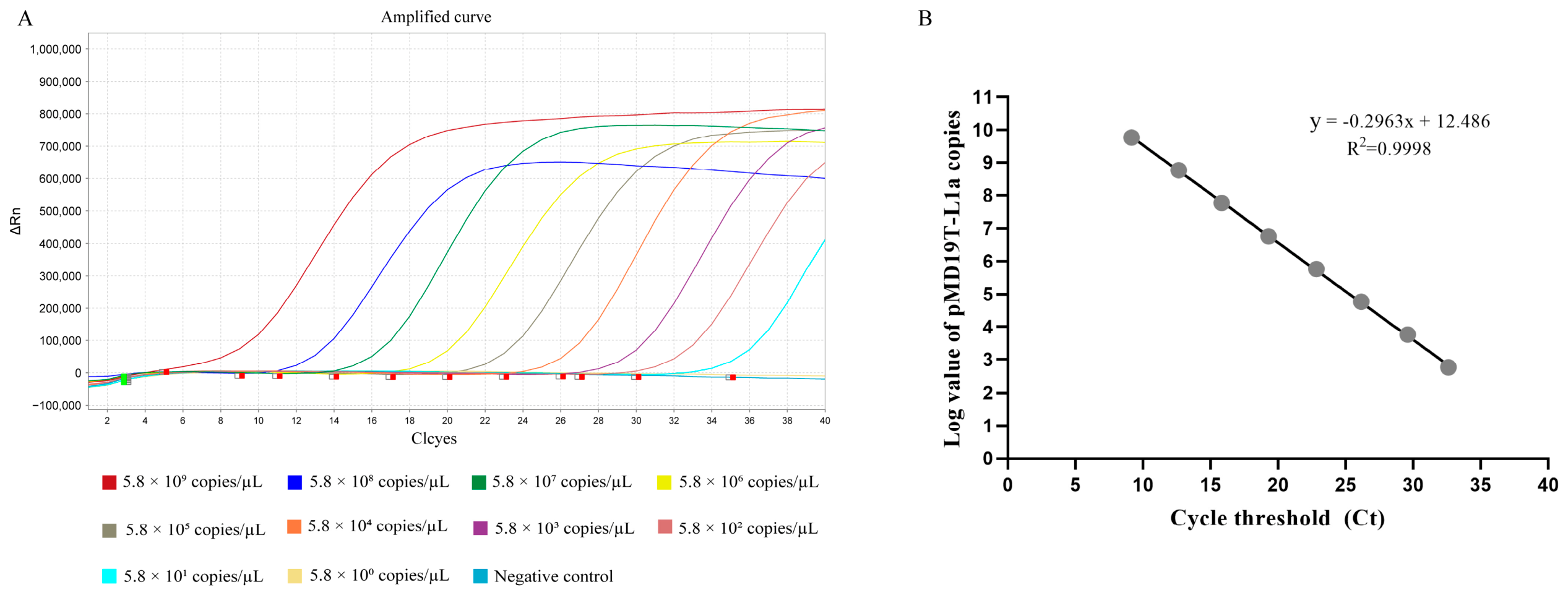
The initial concentration of the recombinant plasmid was 214.3 ng/µL. We prepared 10-fold serial dilutions and used them as standards for TaqMan probe-based qPCR. The assay produced typical S-shaped amplification curves over a range from 5.8 × 10¹⁰ to 5.8 × 101 copies/µL (Figure 1A). The Ct values increased as the template concentration decreased. Linear regression showed a strong linear relationship, with an R2 value of 0.9998 (Figure 1B). All intra-assay and inter-assay CVs were below 2% (Table 2). These results show that the assay has good repeatability and stability.

3.2. Sensitivity and Specificity of the TaqMan Probe-Based qPCR Assay

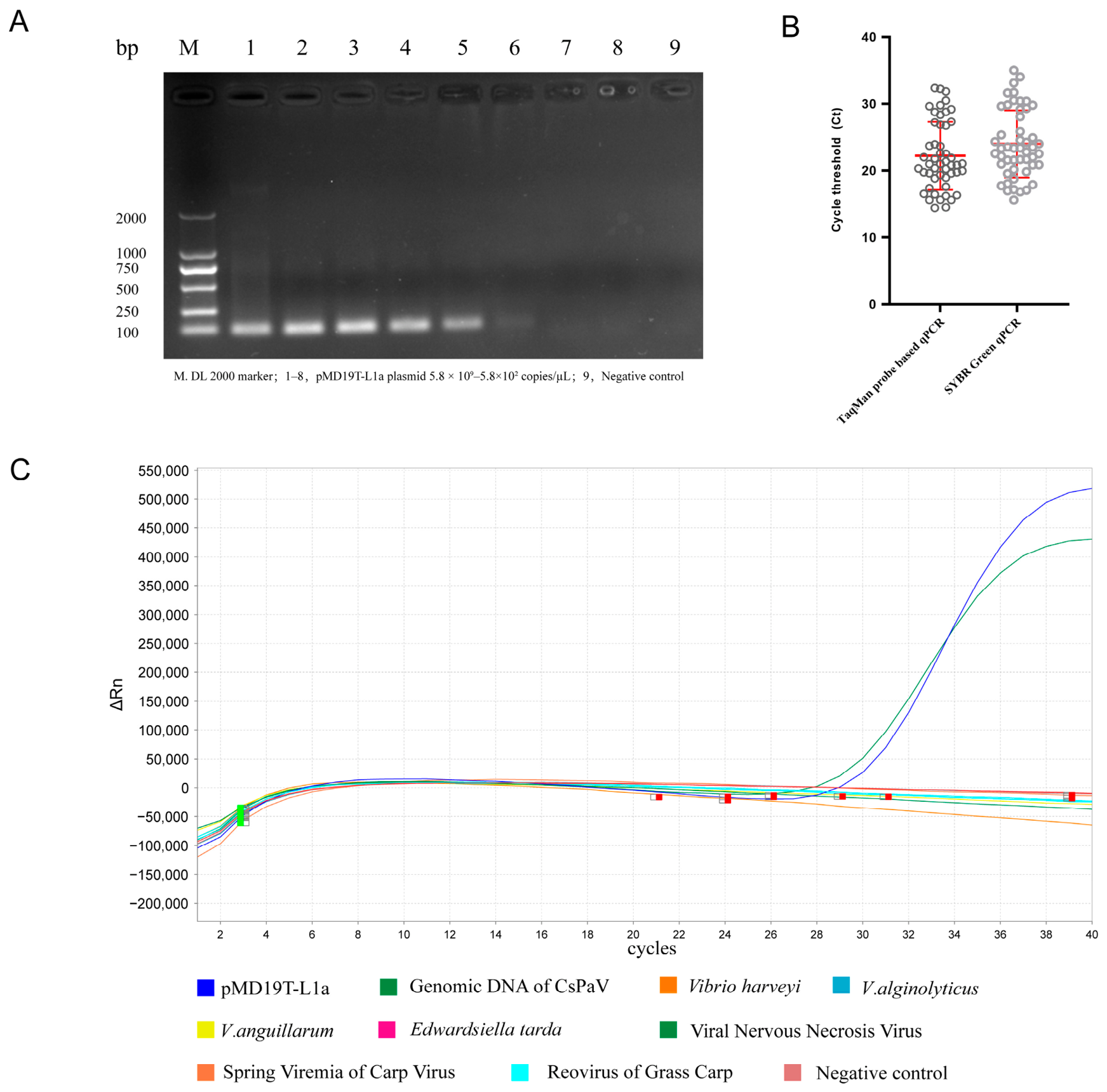
We prepared 10-fold serial dilutions of pMD19T-L1a and analyzed the amplification curves and Ct values at each concentration. The TaqMan probe-based qPCR assay detected as few as 5.8 × 101 copies/µL. This detection limit was 1000-fold lower than that of conventional PCR (Figure 2A). We also tested 50 spleen samples from Chinese tongue soles with both the TaqMan probe-based qPCR assay and the SYBR Green qPCR assay. Both assays produced the same positive rate. However, the TaqMan assay produced lower Ct values than the SYBR Green assay (Figure 2B). This result indicated greater analytical sensitivity under the conditions tested.
Only pMD19T-L1a and CsPaV genomic DNA produced typical amplification curves (Figure 2C). The other aquatic pathogens produced no specific amplification. These results show that the assay is specific for CsPaV under the conditions tested.

3.3. Tissue Distribution and Proliferation Kinetics of CsPaV

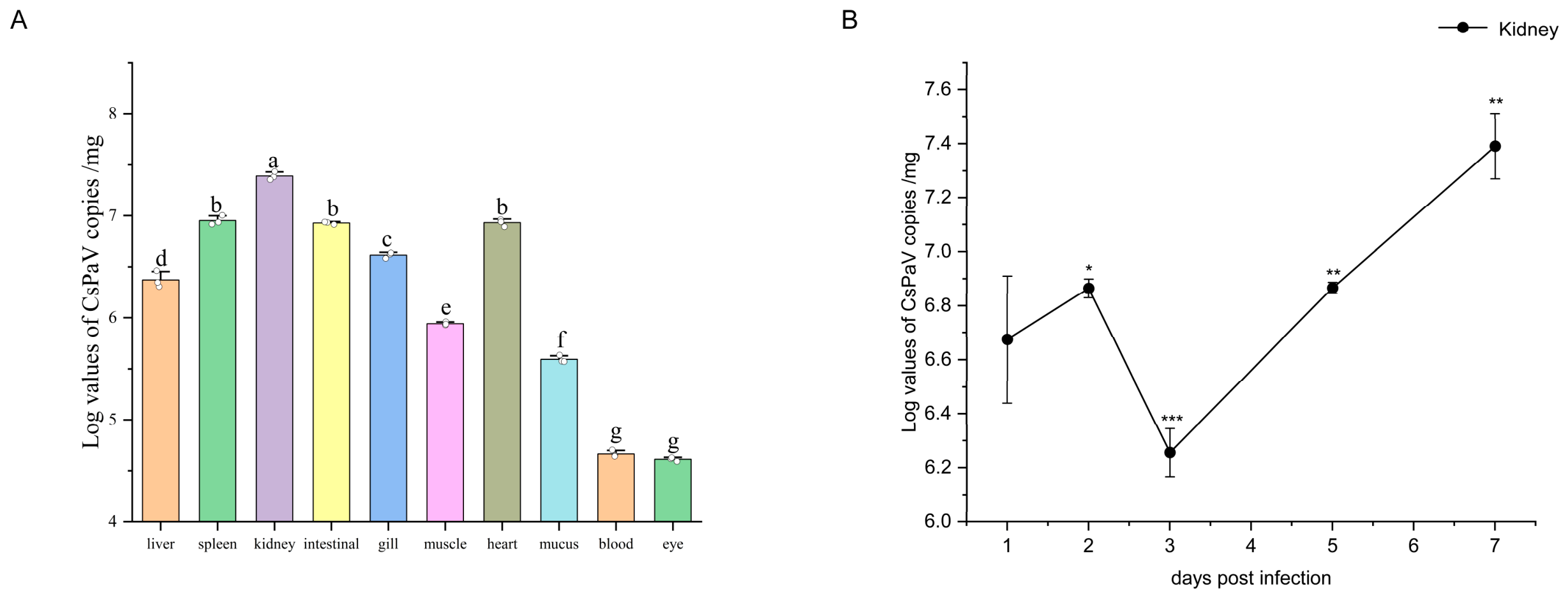
We used the established TaqMan probe-based qPCR assay to examine the tissue distribution and temporal changes in CsPaV DNA after experimental infection. The assay detected CsPaV DNA in all tissues examined. The kidney had the highest viral DNA load at 2.47 × 10⁷ copies/mg (Figure 3A). Viral DNA loads differed significantly among tissues (p < 0.05). Groups with the same lowercase letter did not differ significantly. The kidney viral DNA load changed over time. It decreased to 1.80 × 10⁶ copies/mg at 3 days post-infection and then increased to 2.47 × 10⁷ copies/mg at 7 days post-infection (Figure 3B).

3.4. Analysis of CsPaV Load in Fish with Different Disease Syndromes

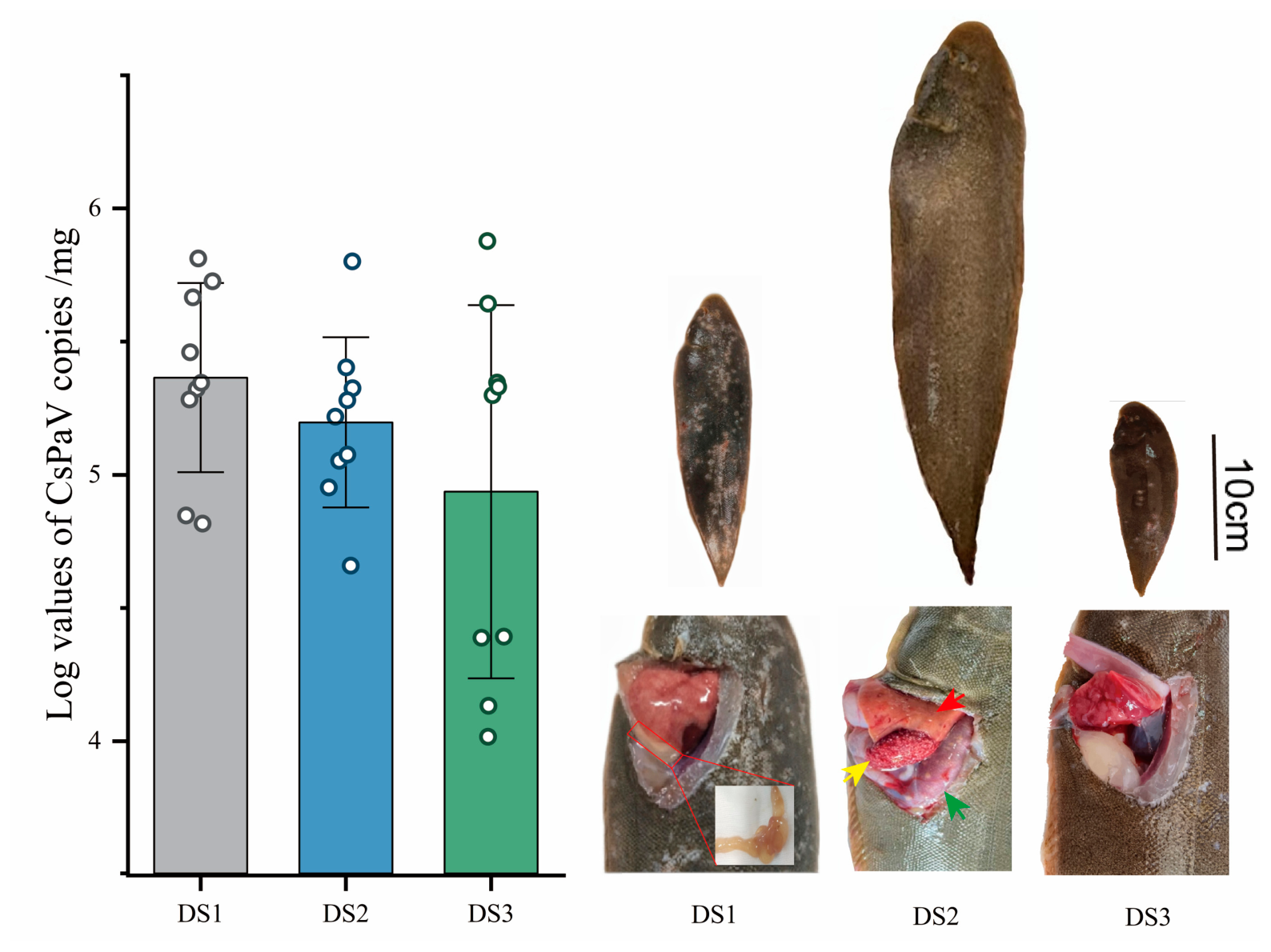
We observed three clinical syndromes in CsPaV-positive Chinese tongue soles of different body sizes. DS1 fish were 25–30 cm long and showed extensive scale loss, scattered skin ulcers, and intestinal fluid accumulation. DS2 fish were 38–45 cm long and showed abdominal swelling with splenic and renal nodules. DS3 fish were 15–20 cm long and showed skin ulcers without clear internal lesions (Figure 4). The mean CsPaV DNA loads were 2.31 × 10⁵ copies/mg in DS1, 1.57 × 10⁵ copies/mg in DS2, and 8.64 × 10⁴ copies/mg in DS3. DS1 had the highest mean value, followed by DS2 and DS3. However, the three groups did not differ significantly (Welch’s one-way ANOVA, F(2, 15.05) = 1.42, p = 0.272).

3.5. Molecular Epidemiological Analysis of CsPaV in Chinese Tongue Soles and Other Species

Between 2023 and 2026, we collected 176 Chinese tongue soles from Tianjin, Hebei Province, Shandong Province, Bohai Bay, and the Yellow Sea. The samples included juveniles about 5–8 cm long and adults longer than 15 cm. We tested kidney samples for CsPaV with the TaqMan probe-based qPCR assay. The overall positive rate was 80.68%. All CsPaV-positive Chinese tongue soles were adults (Figure 5A).
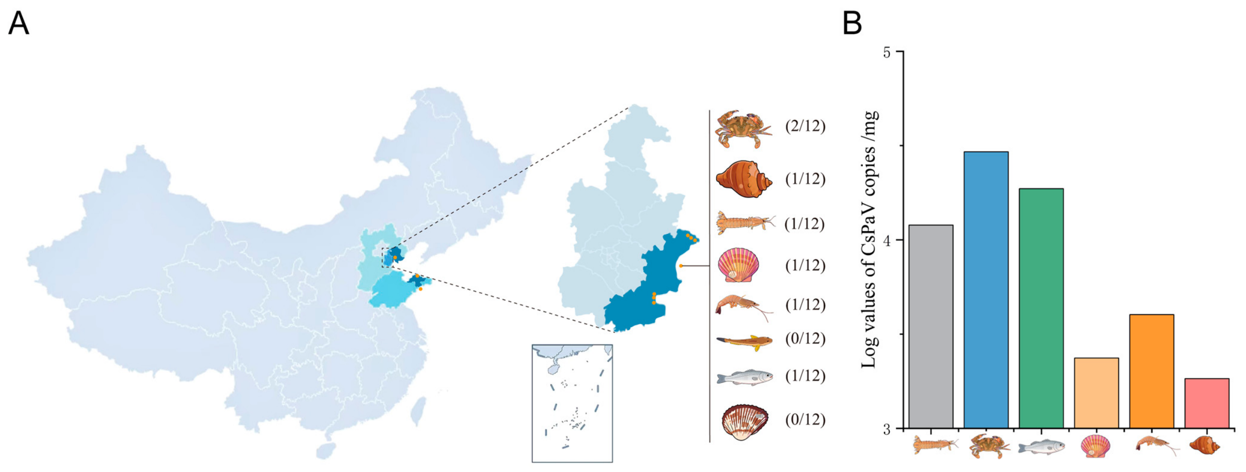
We screened 12 specimens from each of eight selected mollusk, crustacean, and fish species to examine the wider distribution of CsPaV DNA. The assay detected CsPaV DNA in six species. The positive results included Charybdis japonica (2/12, 16.7%), Rapana venosa (1/12, 8.3%), Litopenaeus vannamei (1/12, 8.3%), Chlamys farreri (1/12, 8.3%), Oratosquilla oratoria (1/12, 8.3%), and Lateolabrax japonicus (1/12, 8.3%) (Figure 5B). The assay did not detect CsPaV DNA in Scapharca subcrenata or Synechogobius hasta.

4. Discussion

Global aquaculture has grown rapidly and now provides a major part of the world’s food supply [22]. However, viral diseases in aquatic animals threaten the long-term development of this industry [23,24]. Emerging viruses are a particular concern. Researchers have recently reported several new viral diseases in fish. These diseases include scale drop disease in Asian sea bass caused by a megalocytivirus [25] and infections linked to previously unknown filoviruses, hantaviruses, and rhabdoviruses in European perch [26]. Other examples include koi sleepy disease caused by carp edema virus [27] and a possible new calicivirus genus detected in yellow catfish [28]. In 2024, our group first identified and described CsPaV in diseased Chinese tongue soles [1]. We proposed that CsPaV represents a new genus in the family Papillomaviridae [1]. Most emerging fish viruses lack commercial vaccines and specific antiviral treatments. Sensitive detection methods are therefore important for surveillance, early diagnosis, and timely disease control.
The major capsid protein L1 is produced at high levels and can assemble into virus-like particles that resemble native papillomavirus particles [29,30]. The L1 protein is abundant and strongly immunogenic. Its sequence also differs among virus types. These features have supported the use of L1 in papillomavirus detection and disease assessment [31,32]. The L1 gene is therefore a suitable target for molecular detection. In this study, we designed CsPaV L1-specific primers and a hydrolysis probe. We then established a sensitive and specific TaqMan probe-based qPCR assay.
Our assay uses a sequence-specific hydrolysis probe and closed-tube quantification, as do other TaqMan assays for aquatic viruses. Previous studies have shown that non-lethal samples can support repeated surveillance of aquatic viral infections [33,34]. Skin mucus may also serve as a non-lethal sample for CsPaV testing. Mucus swabs could allow repeated sampling without sacrificing valuable broodstock. However, skin mucus contained less CsPaV DNA than kidney tissue in this study. Paired-sample studies must therefore compare mucus and kidney testing before mucus swabs are used in the field. These studies should assess diagnostic sensitivity, agreement between sample types, and suitable decision limits. The CsPaV L1 assay had a wide linear range, a detection limit of 5.8 × 101 copies/µL, no cross-reactivity with the non-target pathogens tested, and low intra-assay and inter-assay CVs. However, these findings do not prove that the assay is clinically better than other methods. This study did not include direct comparisons between laboratories or a diagnostic-accuracy study.
Our previous study described tissue lesions associated with CsPaV infection [1]. However, the present viral DNA data cannot show whether tissue viral load is related to lesion severity. Future infection studies should combine repeated viral-load measurements with histopathology and in situ viral detection. These methods could show where CsPaV occurs in relation to tissue lesions. In the present study, we detected CsPaV DNA in the liver, spleen, kidney, intestine, muscle, heart, gill, skin mucus, blood, and eye. This result indicates a wide distribution of viral DNA. The kidney had the highest viral DNA load, whereas the eye had the lowest load. The kidney is a major blood-forming and immune organ in fish. The high DNA load may reflect viral accumulation or tissue preference. However, assays for viral RNA or antigen are needed to confirm active replication in the kidney. Other important fish viruses, including lymphocystis disease virus [35] and infectious salmon anemia virus [36], also target the kidney or use it as a main diagnostic tissue. Our results support kidney tissue as the preferred sample for clinical detection. Skin mucus had a lower viral DNA load, but all mucus samples tested positive. Skin mucus may therefore be useful for non-lethal sampling. Larger studies with repeated sampling must confirm this possibility.
CsPaV-positive Chinese tongue soles showed different clinical signs across body-size groups. However, the viral DNA loads did not differ significantly among the three syndrome groups. DS1 fish had the highest mean viral DNA load and showed scale loss, skin ulcers, and intestinal fluid accumulation. DS2 fish had an intermediate mean load and showed abdominal swelling, enlarged spleens and kidneys, and many white nodules. DS3 fish had the lowest mean load and showed mainly surface ulcers. These patterns may help guide future studies, but they do not show that body size caused the observed differences. Intestinal fluid accumulation may involve the intestinal lining or disturbed salt and water balance. Histopathology and in situ viral detection are needed to identify the affected cells. The lesions in DS2 resemble those reported in fish infected with Mycobacterium spp. [37] or red seabream iridovirus [38]. Researchers must exclude other infections before these lesions can be linked to CsPaV. Age-matched infection studies and immune-response measurements are also needed to test whether fish development affects disease outcome.
To investigate the potential environmental distribution and host range of CsPaV, we screened 12 specimens representing eight molluscans, other invertebrate, and fish species collected from Bohai Bay. CsPaV DNA was detected in one or two specimens from each of six species. Given the small sample size and the use of TaqMan probe-based qPCR alone, these detections should not be interpreted as evidence of productive infection or cross-class transmission; they may instead reflect transient exposure, environmental contamination, or mechanical carriage. Filter-feeding mollusks and other aquatic invertebrates may nevertheless serve as sentinels for the presence or enrichment of viral material in aquatic environments. Future studies should include larger, geographically and temporally stratified sample sets and should combine viral isolation, transcript-specific assays, and in situ hybridization to determine whether CsPaV enters and replicates in non-target hosts. Such evidence will be necessary to define the ecological reservoirs, transmission pathways, and aquaculture risks associated with CsPaV. This study has several limitations. The assay has not yet been comprehensively validated against genetically diverse CsPaV variants collected from different geographical regions and host species. Preliminary sequence analyses suggest that CsPaV may exhibit sequence variation among positive samples from different aquatic species, although these observations require further confirmation. Because mutations within the primer- or probe-binding regions could affect amplification efficiency and potentially lead to false-negative results, future studies should evaluate the conservation of these regions using larger, geographically representative sample sets and directly assess the effect of sequence variation on assay performance.
Farm monitoring programs should prioritize kidney tissue when they test dying or recently dead fish. Kidney tissue had the highest viral DNA load in this study. Routine monitoring could also include regular screening of healthy-looking broodstock and juveniles. Farm staff should promptly test fish that show scale loss, ulcers, abdominal swelling, or unusual mortality. Positive qPCR results in mollusks or crustaceans should be viewed as evidence of viral DNA in the environment or possible mechanical carriage. They should not yet be viewed as evidence that these animals are biological vectors. Confirmed positive results should lead to wider stock screening, movement control, equipment disinfection, and follow-up testing. Commercial farm studies are still needed to define action thresholds.
This study has several limitations. The infection experiment used only one dose, and the time-course analysis focused on the kidney. The data therefore cannot show a dose–response relationship or compare temporal patterns among different tissues. The assay measures CsPaV DNA, but it cannot separate inactive or remaining DNA from active viral replication. Viral RNA detection, antigen testing, in situ hybridization, or virus isolation is needed to confirm active infection. The assay has also not been fully tested against genetically diverse CsPaV variants from different regions and host species. Preliminary sequencing of positive samples from five aquatic species suggested sequence variation. However, these findings require further confirmation and are not presented as validated results. Changes in the primer- or probe-binding sites could reduce assay performance or cause false-negative results. Future studies should use larger and geographically diverse sample sets to test the conservation of these sites and the reliability of the assay.

5. Conclusions

In conclusion, we developed a sensitive and specific TaqMan probe-based qPCR assay that targets the CsPaV L1 gene. The assay detected as few as 5.8 × 101 copies/µL. It did not cross-react with the aquatic pathogens tested, and it showed good repeatability. The preliminary application identified kidney as the most suitable tissue among those examined. The assay also described temporal changes in kidney viral DNA after experimental infection. In addition, the assay supported screening of clinically diseased Chinese tongue soles and other aquatic species. The assay therefore provides a useful tool for CsPaV research and surveillance. However, the detection of viral DNA alone does not confirm active infection, host competence, or viral transmission. Further studies should include samples from different regions and genetic groups. These studies should also compare paired non-lethal samples and use methods that detect viral transcripts or locate viral material in tissues. Such work is needed before researchers can define standard diagnostic thresholds and farm-level disease-control procedures.

Author Contributions

Conceptualization, S.X.; methodology, S.X. and X.L. (Xinrui Liu); software, X.L. (Xinrui Liu) and M.X.; validation, X.L. (Xinrui Liu), S.Y., X.L. (Xiang Li), L.J. and Y.-X.L.; writing—original draft preparation, X.L. (Xinrui Liu); writing—review and editing, X.L. (Xinrui Liu) and S.X. All authors have read and agreed to the published version of the manuscript.

Funding

This research was funded by the Tianjin Science and Technology Program (24YDTPJC00330), Tianjin major project for high-quality species innovation (24ZXZYSN00020), and Innovation Team of Tianjin Marine Fisheries Research System (ITTMRS2026000-06).

Institutional Review Board Statement

The study was conducted in accordance with the Declaration of Helsinki and approved by the Ethics Committee of Tianjin Normal University (Approval date: 1 January 2024; Registration No. 2024041001).

Informed Consent Statement

Not applicable.

Data Availability Statement

The authors confirm that the data supporting the findings of this study are fully available within the article.

Acknowledgments

We thank Guo Meiling, Pang Yue, and Liu Yuru for providing samples for this study.

Conflicts of Interest

The authors declare no conflicts of interest. The funders had no role in the design of the study; in the collection, analyses, or interpretation of data; in the writing of the manuscript; or in the decision to publish the results.

References

  1. Xue, S.; Liu, X.; Liu, Y.; Lu, C.; Jia, L.; Yu, Y.; Liu, H.; Yang, S.; Zeng, Z.; Li, H.; et al. Determination and Characterization of Novel Papillomavirus and Parvovirus Associated with Mass Mortality of Chinese Tongue Sole (Cynoglossus semilaevis) in China. Viruses 2024, 16, 705. [Google Scholar] [CrossRef] [Scilit] [PubMed]
  2. Rector, A.; Van Ranst, M. Animal papillomaviruses. Virology 2013, 445, 213–223. [Google Scholar] [CrossRef] [Scilit] [PubMed]
  3. Jensen, J.E.; Becker, G.L.; Jackson, J.B.; Rysavy, M.B. Human Papillomavirus and Associated Cancers: A Review. Viruses 2024, 16, 680. [Google Scholar] [CrossRef] [Scilit] [PubMed]
  4. López-Bueno, A.; Mavian, C.; Labella, A.M.; Castro, D.; Borrego, J.J.; Alcami, A.; Alejo, A. Concurrence of Iridovirus, Polyomavirus, and a Unique Member of a New Group of Fish Papillomaviruses in Lymphocystis Disease-Affected Gilthead Sea Bream. J. Virol. 2016, 90, 8768–8779. [Google Scholar] [CrossRef] [Scilit] [PubMed]
  5. Tisza, M.J.; Pastrana, D.V.; Welch, N.L.; Stewart, B.; Peretti, A.; Starrett, G.J.; Pang, Y.Y.S.; Krishnamurthy, S.R.; Pesavento, P.A.; McDermott, D.H.; et al. Discovery of several thousand highly diverse circular DNA viruses. eLife 2020, 9, e51971. [Google Scholar] [CrossRef] [Scilit] [PubMed]
  6. Surján, A.; Fónagy, E.; Eszterbauer, E.; Harrach, B.; Doszpoly, A. Complete genome sequence of a novel fish papillomavirus detected in farmed wels catfish (Silurus glanis). Arch. Virol. 2021, 166, 2603–2606. [Google Scholar] [CrossRef] [Scilit] [PubMed]
  7. Kraberger, S.; Austin, C.; Farkas, K.; Desvignes, T.; Postlethwait, J.H.; Fontenele, R.S.; Schmidlin, K.; Bradley, R.W.; Warzybok, P.; Van Doorslaer, K.; et al. Discovery of novel fish papillomaviruses: From the Antarctic to the commercial fish market. Virology 2022, 565, 65–72. [Google Scholar] [CrossRef] [Scilit] [PubMed]
  8. Xi, Y.; Jiang, X.; Xie, X.; Zhao, M.; Zhang, H.; Qin, K.; Wang, X.; Liu, Y.; Yang, S.; Shen, Q.; et al. Viromics Reveals the High Diversity of Viruses from Fishes of the Tibet Highland. Microbiol. Spectr. 2023, 11, e0094623. [Google Scholar] [CrossRef] [Scilit] [PubMed]
  9. Blake, L.; Phillips-Savage, A.C.; Soto, E.; Oura, C.; Brown-Jordan, A.; Raines, C.D.; Buck, C.B.; Iwanowicz, L.R. Complete genome sequence of a novel papillomavirus in Siamese fighting fish (Betta splendens) from Trinidad and Tobago. Microbiol. Resour. Announc. 2024, 13, e0099923. [Google Scholar] [CrossRef] [Scilit] [PubMed]
  10. da Silva Fong, D.; Abrantes, J.; Moura, T.; Serra-Pereira, B.; Xavier, R.; Veríssimo, A.; Varsani, A.; Neves, F. Identification and characterization of a novel papillomavirus in thornback skate (Raja clavata). Microb. Genom. 2025, 11, 001541. [Google Scholar] [CrossRef] [Scilit] [PubMed]
  11. Van Doorslaer, K.; Chen, Z.; Bernard, H.-U.; Chan, P.K.S.; DeSalle, R.; Dillner, J.; Forslund, O.; Haga, T.; McBride, A.A.; Villa, L.L.; et al. ICTV Virus Taxonomy Profile: Papillomaviridae. J. Gen. Virol. 2018, 99, 989–990. [Google Scholar] [CrossRef] [Scilit] [PubMed]
  12. Willemsen, A.; Bravo, I.G. Origin and evolution of papillomavirus (onco) genes and genomes. Philos. Trans. R. Soc. B Biol. Sci. 2019, 374, 20180303. [Google Scholar] [CrossRef] [Scilit] [PubMed]
  13. Hu, H.; Zeng, W.; Wang, Y.; Wang, Q.; Bergmann, S.M.; Yin, J.; Li, Y.; Chen, X.; Gao, C.; Zhang, D.; et al. Development and application of a recombinant protein-based indirect ELISA for detection of anti-tilapia lake virus IgM in sera from tilapia. Aquaculture 2020, 520, 734756. [Google Scholar] [CrossRef] [Scilit]
  14. Ouyang, P.; Yang, R.; Chen, J.; Wang, K.; Geng, Y.; Lai, W.; Huang, X.; Chen, D.; Fang, J.; Chen, Z.; et al. First detection of carp edema virus in association with cyprinid herpesvirus 3 in cultured ornamental koi, Cyprinus carpio L., in China. Aquaculture 2018, 490, 162–168. [Google Scholar] [CrossRef] [Scilit]
  15. Vanhomwegen, J.; Kwasiborski, A.; Diop, A.; Boizeau, L.; Hoinard, D.; Vray, M.; Bercion, R.; Ndiaye, B.; Dublineau, A.; Michiyuki, S.; et al. Development and clinical validation of loop-mediated isothermal amplification (LAMP) assay to diagnose high HBV DNA levels in resource-limited settings. Clin. Microbiol. Infect. 2021, 27, 1858.e9–1858.e15. [Google Scholar] [CrossRef] [Scilit] [PubMed]
  16. Gias, E.; Johnston, C.; Keeling, S.; Spence, R.P.; McDonald, W.L. Development of real-time PCR assays for detection of megalocytiviruses in imported ornamental fish. J. Fish Dis. 2011, 34, 609–618. [Google Scholar] [CrossRef] [Scilit] [PubMed]
  17. Megarani, D.V.; Al-Hussinee, L.; Subramaniam, K.; Sriwanayos, P.; Imnoi, K.; Keleher, B.; Nicholson, P.; Surachetpong, W.; Tattiyapong, P.; Hick, P.; et al. Development of a TaqMan quantitative reverse transcription PCR assay to detect tilapia lake virus. Dis. Aquat. Organ. 2022, 152, 147–158. [Google Scholar] [CrossRef] [Scilit] [PubMed]
  18. Yamkasem, J.; Tattiyapong, P.; Surachetpong, W. Development and application of TaqMan probe-based quantitative PCR assays for the detection of tilapia parvovirus. J. Fish Dis. 2022, 45, 379–386. [Google Scholar] [CrossRef] [Scilit] [PubMed]
  19. Wang, H.; Gao, X.; Gao, Y.; Shang, X.; Li, J. Development of a TaqMan-Based Real-Time PCR Assay for the Specific Detection of Turbot Circovirus. J. Fish Dis. 2026, 49, e70084. [Google Scholar] [CrossRef] [Scilit] [PubMed]
  20. Waiyamitra, P.; Tattiyapong, P.; Sirikanchana, K.; Mongkolsuk, S.; Nicholson, P.; Surachetpong, W. A TaqMan RT-qPCR assay for tilapia Lake Virus (TiLV) detection in tilapia. Aquaculture 2018, 497, 184–188. [Google Scholar] [CrossRef] [Scilit]
  21. Perini, F.; Casabianca, A.; Battocchi, C.; Accoroni, S.; Totti, C.; Penna, A. New Approach Using the Real-Time PCR Method for Estimation of the Toxic Marine Dinoflagellate Ostreopsis cf. ovata in Marine Environment. PLoS ONE 2011, 6, e17699. [Google Scholar] [CrossRef] [Scilit] [PubMed]
  22. Nagarajan, D.; Chen, C.-W.; Ponnusamy, V.K.; Dong, C.-D.; Lee, D.-J.; Chang, J.-S. Sustainable aquaculture and seafood production using microalgal technology—A circular bioeconomy perspective. Chemosphere 2024, 366, 143502. [Google Scholar] [CrossRef] [Scilit] [PubMed]
  23. Kibenge, F.S.B. Emerging viruses in aquaculture. Curr. Opin. Virol. 2019, 34, 97–103. [Google Scholar] [CrossRef] [Scilit] [PubMed]
  24. Johny, T.K.; Swaminathan, T.R.; Sood, N.; Pradhan, P.K.; Lal, K.K. A panoptic review of techniques for finfish disease diagnosis: The status quo and future perspectives. J. Microbiol. Methods 2022, 196, 106477. [Google Scholar] [CrossRef] [Scilit] [PubMed]
  25. de Groof, A.; Guelen, L.; Deijs, M.; van der Wal, Y.; Miyata, M.; Ng, K.S.; van Grinsven, L.; Simmelink, B.; Biermann, Y.; Grisez, L.; et al. A Novel Virus Causes Scale Drop Disease in Lates calcarifer. PLoS Pathog. 2015, 11, e1005074. [Google Scholar] [CrossRef] [Scilit] [PubMed]
  26. Hierweger, M.M.; Koch, M.C.; Rupp, M.; Maes, P.; Di Paola, N.; Bruggmann, R.; Kuhn, J.H.; Schmidt-Posthaus, H.; Seuberlich, T. Novel Filoviruses, Hantavirus, and Rhabdovirus in Freshwater Fish, Switzerland, 2017. Emerg. Infect. Dis. 2021, 27, 3082–3091. [Google Scholar] [CrossRef] [Scilit] [PubMed]
  27. Kushala, K.B.; Nithin, M.S.; Girisha, S.K.; Dheeraj, S.B.; Sowndarya, N.S.; Puneeth, T.G.; Suresh, T.; Naveen Kumar, B.T.; Vinay, T.N. Fish immune responses to natural infection with carp edema virus (Koi sleepy disease): An emerging fish disease in India. Fish Shellfish Immunol. 2022, 130, 624–634. [Google Scholar] [CrossRef] [Scilit] [PubMed]
  28. Liu, W.; Xue, M.; Yang, T.; Li, Y.; Jiang, N.; Fan, Y.; Meng, Y.; Luo, X.; Zhou, Y.; Zeng, L. Characterization of a Novel RNA Virus Causing Massive Mortality in Yellow Catfish, Pelteobagrus fulvidraco, as an Emerging Genus in Caliciviridae (Picornavirales). Microbiol. Spectr. 2022, 10, e00624-22. [Google Scholar] [CrossRef] [Scilit] [PubMed]
  29. Kirnbauer, R.; Booy, F.; Cheng, N.; Lowy, D.R.; Schiller, J.T. Papillomavirus L1 major capsid protein self-assembles into virus-like particles that are highly immunogenic. Proc. Natl. Acad. Sci. USA 1992, 89, 12180–12184. [Google Scholar] [CrossRef] [Scilit] [PubMed]
  30. Buck, C.B.; Day, P.M.; Trus, B.L. The papillomavirus major capsid protein L1. Virology 2013, 445, 169–174. [Google Scholar] [CrossRef] [Scilit] [PubMed]
  31. Hu, H.; Zhao, J.; Yu, W.; Zhao, J.; Wang, Z.; Jin, L.; Yu, Y.; Han, L.; Wang, L.; Zhu, H.; et al. Human papillomavirus DNA, HPV L1 capsid protein and p16INK4a protein as markers to predict cervical lesion progression. Arch. Gynecol. Obstet. 2019, 299, 141–149. [Google Scholar] [CrossRef] [Scilit] [PubMed]
  32. Chaiwongkot, A.; Phanuphak, N.; Pankam, T.; Bhattarakosol, P. Human papillomavirus 16 L1 gene methylation as a potential biomarker for predicting anal intraepithelial neoplasia in men who have sex with men (MSM). PLoS ONE 2021, 16, e0256852. [Google Scholar] [CrossRef] [Scilit] [PubMed]
  33. Liamnimitr, P.; Thammatorn, W.; U-thoomporn, S.; Tattiyapong, P.; Surachetpong, W. Non-lethal sampling for Tilapia Lake Virus detection by RT-qPCR and cell culture. Aquaculture 2018, 486, 75–80. [Google Scholar] [CrossRef] [Scilit]
  34. Chiamkunakorn, C.; Machimbirike, V.I.; Senapin, S.; Khunrae, P.; Dong, H.T.; Rattanarojpong, T. Blood and liver biopsy for the non-destructive screening of tilapia lake virus. J. Fish Dis. 2019, 42, 1629–1636. [Google Scholar] [CrossRef] [Scilit] [PubMed]
  35. Valverde, E.J.; Borrego, J.J.; Sarasquete, M.C.; Ortiz-Delgado, J.B.; Castro, D. Target organs for lymphocystis disease virus replication in gilthead seabream (Sparus aurata). Vet. Res. 2017, 48, 21. [Google Scholar] [CrossRef] [Scilit] [PubMed]
  36. Moneke, E.; Groman, D.B.; Wright, G.M.; Stryhn, H.; Johnson, G.R.; Ikede, B.O.; Kibenge, F.S.B. Correlation of Virus Replication in Tissues with Histologic Lesions in Atlantic Salmon Experimentally Infected with Infectious Salmon Anemia Virus. Vet. Pathol. 2005, 42, 338–349. [Google Scholar] [CrossRef] [Scilit] [PubMed]
  37. Luo, Z.; Hao, S.; Bai, X.; Zhang, Z.; Ma, Y.; Feng, S.; Zhang, D. Identification and genomic analysis of Mycobacterium ulcerans ecovar Liflandii from the farmed Chinese tongue sole, Cynoglossus semilaevis Günther. Aquaculture 2022, 548, 737614. [Google Scholar] [CrossRef] [Scilit]
  38. Liu, X.; Chen, N.; Gao, X.; Zhang, Y.; Li, X.; Zhang, Y.; Bing, X.; Huang, H.; Zhang, X. The infection of red seabream iridovirus in mandarin fish (Siniperca chuatsi) and the host immune-related gene expression profiles. Fish Shellfish Immunol. 2018, 74, 474–484. [Google Scholar] [CrossRef] [Scilit] [PubMed]
Figure 1. Establishment of a TaqMan probe-based qPCR assay for CsPaV detection. (A) Serial dilutions of the pMD19T-L1a plasmid produced the amplification curves. (B) The standard curve shows the relationship between Ct values and the base-10 logarithm of the pMD19T-L1a plasmid copy number.
Viruses 18 00939 g001
Figure 2. Evaluation of the sensitivity and specificity of the TaqMan probe-based qPCR assay. (A) Serially diluted pMD19T-L1a plasmids were tested by conventional PCR. (B) The sensitivities of the TaqMan probe-based qPCR and SYBR Green qPCR assays were compared. (C) The analytical specificity of the TaqMan probe-based qPCR assay was evaluated.
Viruses 18 00939 g002
Figure 3. Tissue distribution and temporal dynamics of CsPaV after experimental infection in Chinese tongue soles. (A) The bars show CsPaV loads in tissues from experimentally infected fish. Different lowercase letters indicate significant differences among tissues. Bars with the same letter are not significantly different (p < 0.05). (B) The line shows temporal changes in CsPaV loads in kidney tissue after experimental infection. (Asterisks indicate statistically significant differences: * p < 0.05, ** p < 0.01, and *** p < 0.001.)
Viruses 18 00939 g003
Figure 4. CsPaV loads in Chinese tongue soles with different clinical syndromes. DS1 fish were 25–30 cm long and showed extensive scale loss, scattered skin ulcers, and intestinal fluid accumulation. DS2 fish were 38–45 cm long and showed abdominal swelling with splenic and renal nodules. The red arrow indicates a pale, swollen liver with nodules. The yellow arrow indicates a swollen spleen with nodules. The green arrow indicates a pale, swollen kidney with nodules. DS3 fish were 15–20 cm long and showed skin ulcers without clear internal lesions.
Viruses 18 00939 g004
Figure 5. Epidemiological findings and detection of CsPaV in field samples. (A) The map shows the prevalence of CsPaV in different sampling regions. (B) The graph shows CsPaV detection rates and viral DNA loads in other aquatic species.
Viruses 18 00939 g005
Table 1. Sampling information and CsPaV detection results for Chinese tongue soles and other aquatic species collected between 2023 and 2026.
Province/City/Sea Area of Sample CollectionCollection TimeSpeciesTarget TissuesCollection QuantityCsPaV-Positive RateBody Length
TianjinJanuary 2023Cynoglossus semilaevisL, S, K9100%15–20 cm; 40–45 cm
March 2023L, S, K35100%15–25 cm; 35 cm; 40–45 cm; 50–55 cm
April 2023L, S, K7100%15–20 cm
May 2023L, S, K10100%15–20 cm
July 2023L, S, K6100%35–45 cm
September 2023L, S, K5100%35–45 cm
September 2023L, S, K17100%15–20 cm; 35–45 cm
April 2024L, S, K9100%35–45 cm
April 2026L, S, K757%5–8 cm; 45–50 cm
Hebei/TangshanMay 2023L, S, K10100%15–20 cm
March 2025L, S, K3003–5 cm
Shandong/YantaiMay 2023L, S, K13100%10–15 cm
Bohai sea/117.59–118.00° E, 39.08–39.10° NOctober 2023L, S, K5100%15–25 cm
Yellow sea/Waters off Renjiatai, Qingdao (120.48° E, 36.097243° N)April 2024L, S, K13100%15–20 cm
Tianjin *December 2024Epinephelus sp.L, S, K10100%20–25 cm
September 2025Rapana venosaM128.3%8–10 cm
Charybdis JaponicaM1216.7%15–20 cm
Litopenaeus vannameiH128.3%15–20 cm
Chlamys farreriM128.3%8–10 cm
Scapharca subcrenataM1203–5 cm
Oratosquilla oratoriaM128.3%13–15 cm
Lateolabrax japonicusL128.3%18–23 cm
Synechogobius hastaL12015–18 cm
April 2026Epinephelus sp.L, S, K3015–20 cm
Note: * indicates specimens obtained from coastal seafood markets in Tianjin. All products sold at these markets originated from Bohai Bay. Abbreviations: L, liver; S, spleen; K, kidney; M, muscle; H, hepatopancreas.
Table 2. Intra-assay and inter-assay repeatability of the CsPaV TaqMan probe-based qPCR assay.
RepeatabilityPlasmid Concentration
(Copies/µL)
Ct Value
Repeat 1Repeat 2Repeat 3MeanSDCV (%)
Inter-group10812.712.612.59412.6310.060.47
10715.86615.79815.77915.8140.050.29
10619.19519.2719.41919.2940.110.59
10522.95522.94122.61522.8370.190.84
10426.12525.20926.09526.1430.060.23
Intra-group10812.63112.70212.5612.6310.070.56
10715.81415.96315.56215.780.21.29
10619.29418.73818.95418.9950.281.48
10522.83722.75622.68522.6850.20.87
10426.14325.93125.66525.7060.240.92
Disclaimer/Publisher’s Note: The statements, opinions and data contained in all publications are solely those of the individual author(s) and contributor(s) and not of MDPI and/or the editor(s). MDPI and/or the editor(s) disclaim responsibility for any injury to people or property resulting from any ideas, methods, instructions or products referred to in the content.