Detectability of Pathogenic RNA Virus Families in Different Body Sites: A Scoping Review

 ,
,  , , , and
Abstract
1. Introduction
2. Materials and Methods
2.1. Viruses and Specimen Types
- Blood specimens: whole blood, serum, plasma, and PBMC;
- Respiratory specimens: saliva, bronchoalveolar lavage (BAL), oropharyngeal swabs, nasopharyngeal swabs, nasal swabs, and oral swabs, sputum;
- Fecal specimens: stool and rectal swabs;
- Urine specimens: urine, urinary sediment, and urine supernatant;
- CNS specimens: cerebrospinal fluid (CSF).
2.2. Search Strategy and Selection Criteria
2.3. Eligibility Criteria
- Human cases with confirmed infection by a virus of interest.
- Viral nucleic acid testing (NAAT) performed.
- Clinical specimens of urine, blood, feces, cerebrospinal fluid, or respiratory material collected for diagnostic purposes.
- Duplicate patient data published elsewhere.
- Studies limited to post-mortem, asymptomatic screening, neonates, immunocompromised patients, pregnant persons, or antivirally treated cases prior to NAAT testing.
- Studies primarily comparing a novel non-NAAT assay against a standard NAAT.
- Reviews and conference abstracts.
2.4. Data Extraction
2.5. Synthesis of Evidence
3. Results

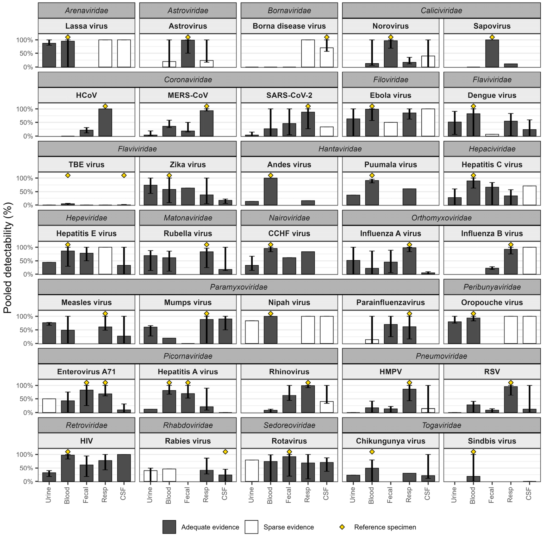
| Family | Virus | Urine | Blood | Respiratory | Cerebrospinal | Fecal | |||||||||||||||
|---|---|---|---|---|---|---|---|---|---|---|---|---|---|---|---|---|---|---|---|---|---|
| D | Range | Cd/Ct | References | D | Range | Cd/Ct | References | D | Range | Cd/Ct | References | D | Range | Cd/Ct | References | D | Range | Cd/Ct | References | ||
| Arenaviridae | Lassa virus | 80–100% | 7/8 | [55,56,57] | * | 0–100% | 39/41 | [55,56,57,58,59,60] | 100% | 2/2 | [57] | 100% | 2/2 | [58,60] | - | - | - | ||||
| Astroviridae | Human astrovirus | - | - | - | 0–100% | 1/5 | [19,61] | 17–100% | 6/25 | [16,61] | - | - | - | * | 50–100% | 101/102 | [16,19,61,62,63,64] | ||||
| Bornaviridae | Borna disease virus | 0% | 0/1 | [50] | 0% | 0/8 | [50,65] | 100% | 1/1 | [50] | * | 57–100% | 7/10 | [50,65,66] | 0% | 0/1 | [50] | ||||
| Caliciviridae | Norovirus | - | - | - | 6–100% | 50/388 | [67,68,69,70,71,72] | 12–35% | 66/384 | [16,73,74] | 0–100% | 2/5 | [67,68,72,75] | * | 69–100% | 794/819 | [16,67,68,69,70,71,72,73,74,75,76,77,78,79,80,81,82,83] | ||||
| Caliciviridae | Sapovirus | - | - | - | 0% | 0/10 | [19] | 12% | 5/43 | [16] | - | - | - | * | 100% | 148/148 | [16,19,84] | ||||
| Corona- viridae | HCoV | - | - | - | 0% | 0/58 | [17,53,54] | * | 100% | 775/775 | [17,18,53,54,85,86,87] | - | - | - | 11–31% | 19/89 | [18,53,54] | ||||
| Corona- viridae | MERS-CoV | 2–19% | 7/185 | [24,25] | 31–58% | 47/132 | [24,25] | * | 93–100% | 218/232 | [24,25] | - | - | - | 15–50% | 17/92 | [24,25] | ||||
| Corona- viridae | SARS-CoV-2 | 0–14% | 18/479 | [26,27,28,29,30,31,32,33,88,89,90,91,92,93,94] | 0–100% | 141/536 | [26,29,30,32,33,93,94,95,96,97,98,99,100,101,102,103] | * | 26–100% | 2613/2949 | [26,27,28,29,30,31,32,33,88,89,90,91,92,93,94,95,96,97,98,99,101,102,103,104,105,106,107,108,109,110,111,112,113,114,115,116,117,118,119,120,121] | 33% | 4/12 | [103] | 4–100% | 1040/2265 | [26,27,28,29,30,31,32,33,88,89,90,91,94,95,96,97,98,99,100,101,102,103,104,105,106,107,108,109,110,111,112,113,114,115,116,117,118,119,120,121,122,123,124,125] | ||||
| Filoviridae | Ebola virus | 0–100% | 14/22 | [34,35,126,127] | * | 57–100% | 810/820 | [34,35,126,127,128,129,130,131,132,133,134] | * | 62–100% | 53/62 | [34,35,127,132] | 100% | 2/2 | [34,129] | 50% | 2/4 | [35] | |||
| Flaviviridae | Dengue virus | 6–92% | 398/777 | [23,135,136,137,138,139,140,141,142,143,144,145] | * | 0–100% | 632–766 | [23,135,136,138,139,140,141,142,143,144,145,146,147,148,149] | 1–83% | 316–574 | [135,136,139,141,142,143,144,145] | 0–60% | 8/34 | [146,147,148,149] | 6% | 2/32 | [137] | ||||
| Flaviviridae | TBE virus | 0% | 0/11 | [150] | * | 0–6% | 13/281 | [37,150,151,152,153] | 0% | 0/15 | [150,153] | * | 0–2% | 1/220 | [151,152,153] | 0% | 0/2 | [37] | |||
| Flaviviridae | Zika virus | 43–100% | 676/912 | [23,38,154,155,156,157,158,159,160,161,162,163,164,165] | * | 9–100% | 643/1103 | [20,23,38,154,155,156,157,158,159,160,161,162,163,164,165,166] | 5–100% | 279/734 | [38,154,155,157,160,161,162,164] | 6–22% | 21/122 | [20,166] | 63% | 165/260 | [38] | ||||
| Hantaviridae | Andes virus | 13% | 8/60 | [167] | * | 100% | 131/131 | [167] | 16% | 21/131 | [167] | - | - | - | - | - | - | ||||
| Hantaviridae | Puumala virus | 37% | 25/67 | [168] | * | 82–96% | 91/100 | [168,169] | 61% | 20/33 | [169] | - | - | - | - | - | - | ||||
| Hepaci- viridae | Hepatitis C virus | 0–60% | 61/222 | [36,170,171,172,173,174] | * | 63–100% | 377/422 | [36,170,171,172,173,174,175,176,177,178,179,180] | 0–57% | 62/180 | [36,170,171,172,173,175,176] | 71% | 5/7 | [177] | 0–83% | 82/123 | [36,178,179,180] | ||||
| Hepeviridae | Hepatitis E virus | 44% | 4/9 | [181] | * | 30–100% | 128/149 | [181,182,183,184,185,186,187,188,189,190,191,192,193,194,195,196] | 100% | 8/8 | [192] | 0–100% | 6/18 | [182,183,185,188,189,190,191,194,195,196] | 50–100% | 84/108 | [184,185,186,187,189,193,194] | ||||
| Matona- viridae | Rubella virus | 14–87% | 349/506 | [197,198,199,200] | 11–85% | 362/594 | [197,199,200,201,202,203] | 25–96% | 533/642 | [197,199,200,201,202,203] | 14–100% | 4/22 | [21,203] | - | - | - | |||||
| Nairoviridae | CCHF virus | 15–67% | 22/66 | [39,204,205] | * | 83–100% | 63/66 | [39,204,205] | 83% | 15/18 | [39] | - | - | - | 61% | 11/18 | [39] | ||||
| Orthomyxoviridae | Influenza A virus | 0–100% | 30/58 | [40,41,42,51] | 0–86% | 25/112 | [40,41,42,51,206,207] | * | 83–100% | 160/163 | [40,41,42,51,206,207,208,209] | 0–9% | 1/20 | [51,206,209] | 6–89% | 40/88 | [40,41,42,51,208] | ||||
| Orthomyxoviridae | Influenza B virus | - | - | - | - | - | - | * | 75–100% | 177/192 | [18,210,211] | 100% | 2/2 | [212,213] | 18–28% | 30/132 | [18,210] | ||||
| Paramyxoviridae | Measles virus | * | 67–77% | 506/661 | [214,215,216] | 0–100% | 81/166 | [202,214,215,217,218] | * | 49–100% | 149/245 | [202,214,215,216,217,218,219] | 0–100% | 17/62 | [202,216,217,218,219,220] | - | - | - | |||
| Paramyxoviridae | Mumps virus | 28–65% | 136/226 | [221,222,223,224,225,226] | 20% | 23/116 | [222] | * | 0–100% | 363/412 | [221,222,223,224,225,226,227,228,229] | 50–100% | 173/192 | [21,221,223,227,228,229,230,231,232,233,234,235,236,237,238,239,240] | 0% | 0/1 | [229] | ||||
| Paramyxoviridae | Nipah virus | 83% | 5/6 | [241] | 100% | 19/19 | [242,243] | 100% | 7/7 | [242,244] | 100% | 1/1 | [242] | - | - | - | |||||
| Paramyxoviridae | Human parainfluenza virus | - | - | - | 0–100% | 1/7 | [17,245] | * | 17–100% | 8/13 | [17,18,245] | - | - | - | 25–100% | 7/10 | [18,246] | ||||
| Peribunyaviridae | Oropouche virus | 75–100% | 4/5 | [247,248] | * | 83–100% | 291/311 | [247,248,249,250,251,252,253,254,255] | 100% | 2/2 | [248] | 100% | 6/6 | [256,257] | - | - | - | ||||
| Phenuiviridae | Rift Valley fever virus | - | - | - | * | 79% | 49/62 | [258] | - | - | - | - | - | - | - | - | - | ||||
| Picobirnaviridae | Picobirnavirus | - | - | - | - | - | - | - | - | - | - | - | - | 100% | 51/51 | [259,260,261,262,263] | |||||
| Picornaviridae | Enterovirus A71 | 50% | 1/2 | [52] | 0–76% | 165/387 | [43,44,45,46,52,264] | * | 61–100% | 207/301 | [43,44,45,46,52,265,266,267] | 0–31% | 28/290 | [43,44,45,46,52,264,266,267] | * | 25–100% | 435/525 | [43,44,45,46,52,264,265,266,267] | |||
| Picornaviridae | Hepatitis A virus | 12% | 8/65 | [268] | * | 68–100% | 105/129 | [47,269,270,271,272] | 9–90% | 14/67 | [268,271] | 0% | 0/120 | [47] | * | 52–100% | 83/119 | [47,269,270,272] | |||
| Picornaviridae | Rhinovirus | - | - | - | 0–12% | 114/1369 | [17,273,274,275,276] | * | 91–100% | 1949/1961 | [17,18,273,274,275,276,277,278,279,280,281] | 33–100% | 4/10 | [280,282] | 44–100% | 186/295 | [18,280,281] | ||||
| Pneumoviridae | HMPV | 0% | 0/6 | [22] | 0–41% | 7/41 | [17,22,283] | * | 43–100% | 75/87 | [17,18,22,283,284,285,286,287] | 0–100% | 1/7 | [284,286,287] | 0–22% | 2/15 | [18,22] | ||||
| Pneumoviridae | RSV | 0% | 0/33 | [22] | 0–40% | 41/148 | [17,22,288,289] | * | 63–100% | 381/398 | [17,18,22,288,289,290,291,292,293,294,295] | 0–100% | 2/16 | [290,291,296] | 0–14% | 5/57 | [18,22] | ||||
| Retroviridae | HIV | 18–40% | 10/31 | [297,298] | * | 84–100% | 555/569 | [297,298,299,300,301,302,303,304,305,306,307,308,309] | 43–100% | 113/145 | [301,302,304,305,306,307] | 100% | 77/77 | [305,308] | 18–95% | 182/295 | [297,299,300,303,309] | ||||
| Rhabdoviridae | Rabies virus | 0–50% | 2/5 | [48,310] | 47% | 7/15 | [48] | 28–87% | 71/169 | [48,310,311,312,313,314,315] | * | 0–45% | 26/109 | [48,310,311,312,313,314,315] | - | - | - | ||||
| Sedoreoviridae | Rotavirus | 80% | 4/5 | [316] | 0–99% | 243/326 | [49,317,318,319,320,321,322,323] | 10–100% | 396/574 | [16,49,324] | 38–88% | 17/24 | [49,317,320] | * | 19–100% | 817/890 | [16,49,316,318,319,320,321,322,323,324] | ||||
| Spinareoviridae | Colorado tick fever virus | - | - | - | * | 100% | 11/11 | [325] | - | - | - | - | - | - | - | - | - | ||||
| Tobaniviridae | Torovirus | - | - | - | - | - | - | - | - | - | - | - | - | 100% | 18/18 | [326] | |||||
| Togaviridae | Chikungunya virus | 23% | 35/152 | [327] | * | 0–80% | 138/277 | [20,146,327,328,329] | 30% | 46/152 | [327] | 11–100% | 15/70 | [20,146,327,328,329] | - | - | - | ||||
| Togaviridae | Sindbis virus | - | - | - | * | 0–100% | 31/166 | [330,331,332,333,334] | - | - | - | 0% | 0/1 | [335] | - | - | - | ||||
4. Discussion
5. Conclusions
Supplementary Materials
Author Contributions
Funding
Institutional Review Board Statement
Informed Consent Statement
Data Availability Statement
Conflicts of Interest
Abbreviations
| NAAT | Nucleic acid amplification test |
| PBMC | Peripheral blood mononuclear cells |
Appendix A
| Category | Description |
|---|---|
| A | Above 5 patients AND Multiple specimen types of interest from the same patients AND Consecutive sampling over time from patients untreated with targeted antivirals |
| B | Above 5 patients AND Multiple specimen types of interest from the same patients OR Consecutive sampling over time from patients untreated with targeted antivirals |
| C | No sample size requirement. At least one specimen type of interest with or without consecutive sampling. |
References
- Cassedy, A.; Parle-McDermott, A.; O’Kennedy, R. Virus Detection: A Review of the Current and Emerging Molecular and Immunological Methods. Front. Mol. Biosci. 2021, 8, 637559. [Google Scholar] [CrossRef]
- Niedrig, M.; Patel, P.; El Wahed, A.A.; Schädler, R.; Yactayo, S. Find the Right Sample: A Study on the Versatility of Saliva and Urine Samples for the Diagnosis of Emerging Viruses. BMC Infect. Dis. 2018, 18, 707. [Google Scholar] [CrossRef]
- Hou, N.; Wang, K.; Zhang, H.; Bai, M.; Chen, H.; Song, W.; Jia, F.; Zhang, Y.; Han, S.; Xie, B. Comparison of Detection Rate of 16 Sampling Methods for Respiratory Viruses: A Bayesian Network Meta-Analysis of Clinical Data and Systematic Review. BMJ Glob. Health 2020, 5, 3053. [Google Scholar] [CrossRef] [PubMed]
- Shorten, R.J.; Brown, C.S.; Jacobs, M.; Rattenbury, S.; Simpson, A.J.; Mepham, S. Diagnostics in Ebola Virus Disease in Resource-Rich and Resource-Limited Settings. PLoS Negl. Trop. Dis. 2016, 10, e0004948. [Google Scholar] [CrossRef] [PubMed]
- Puhach, O.; Meyer, B.; Eckerle, I. SARS-CoV-2 Viral Load and Shedding Kinetics. Nat. Rev. Microbiol. 2023, 21, 147–161. [Google Scholar] [CrossRef] [PubMed]
- Cevik, M.; Tate, M.; Lloyd, O.; Maraolo, A.E.; Schafers, J.; Ho, A. SARS-CoV-2, SARS-CoV, and MERS-CoV Viral Load Dynamics, Duration of Viral Shedding, and Infectiousness: A Systematic Review and Meta-Analysis. Lancet Microbe 2021, 2, e13–e22. [Google Scholar] [CrossRef] [PubMed]
- Hagman, K.; Postigo, T.; Diez-Castro, D.; Ursing, J.; Bermejo-Martin, J.F.; de la Fuente, A.; Tedim, A.P. Prevalence and Clinical Relevance of Viraemia in Viral Respiratory Tract Infections: A Systematic Review. Lancet Microbe 2025, 6, 100967. [Google Scholar] [CrossRef] [PubMed]
- Woolhouse, M.E.J.; Howey, R.; Gaunt, E.; Reilly, L.; Chase-Topping, M.; Savill, N. Temporal Trends in the Discovery of Human Viruses. Proc. R. Soc. B Biol. Sci. 2008, 275, 2111–2115. [Google Scholar] [CrossRef] [PubMed]
- Lu, L.; Zhang, F.; Brierley, L.; Robertson, G.; Chase-Topping, M.; Lycett, S.; Woolhouse, M. Temporal Dynamics, Discovery, and Emergence of Human-Transmissible RNA Viruses. Mol. Biol. Evol. 2024, 41, msad272. [Google Scholar] [CrossRef] [PubMed]
- WHO. WHO R&D Blueprint for Epidemics; WHO: Geneva, Switzerland, 2024.
- Page, M.J.; McKenzie, J.E.; Bossuyt, P.M.; Boutron, I.; Hoffmann, T.C.; Mulrow, C.D.; Shamseer, L.; Tetzlaff, J.M.; Akl, E.A.; Brennan, S.E.; et al. The PRISMA 2020 Statement: An Updated Guideline for Reporting Systematic Reviews. BMJ 2021, 372, 71. [Google Scholar] [CrossRef] [PubMed]
- Tricco, A.C.; Lillie, E.; Zarin, W.; O’Brien, K.K.; Colquhoun, H.; Levac, D.; Moher, D.; Peters, M.D.J.; Horsley, T.; Weeks, L.; et al. PRISMA Extension for Scoping Reviews (PRISMA-ScR): Checklist and Explanation. Ann. Intern. Med. 2018, 169, 467–473. [Google Scholar] [CrossRef] [PubMed]
- Black, E.J.; Powell, C.S.; Dempsey, D.M.; Hendrickson, R.C.; Mims, L.R.; Lefkowitz, E.J. Virus Taxonomy: The Database of the International Committee on Taxonomy of Viruses. Nucleic Acids Res. 2025, 54, D776. [Google Scholar] [CrossRef] [PubMed]
- Lefkowitz, E.; Hendrickson, R.; International Committee on Taxonomy of Viruses. ICTV Master Species List (MSL); International Committee on Taxonomy of Viruses (ICTV): Birmingham, AL, USA, 2025. [Google Scholar] [CrossRef]
- Simmonds, P.; Butković, A.; Grove, J.; Mayne, R.; Mifsud, J.C.O.; Beer, M.; Bukh, J.; Drexler, J.F.; Kapoor, A.; Lohmann, V.; et al. Taxonomic Expansion and Reorganization of Flaviviridae. Nat. Microbiol. 2025, 10, 3026–3037. [Google Scholar] [CrossRef] [PubMed]
- Zhuo, R.; Parsons, B.D.; Lee, B.E.; Drews, S.J.; Chui, L.; Louie, M.; Crago, B.; Freedman, S.B.; Ali, S.; Pang, X. Identification of Enteric Viruses in Oral Swabs from Children with Acute Gastroenteritis. J. Mol. Diagn. 2018, 20, 56–62. [Google Scholar] [CrossRef] [PubMed]
- Proenca-Modena, J.L.; Valera, F.C.P.; Jacob, M.G.; Buzatto, G.P.; Saturno, T.H.; Lopes, L.; Souza, J.M.; Paula, F.E.; Silva, M.L.; Carenzi, L.R.; et al. High Rates of Detection of Respiratory Viruses in Tonsillar Tissues from Children with Chronic Adenotonsillar Disease. PLoS ONE 2012, 7, e42136. [Google Scholar] [CrossRef] [PubMed]
- Minodier, L.; Masse, S.; Capai, L.; Blanchon, T.; Ceccaldi, P.-E.; van der Werf, S.; Hanslik, T.; Charrel, R.; Falchi, A. Clinical and Virological Factors Associated with Gastrointestinal Symptoms in Patients with Acute Respiratory Infection: A Two-Year Prospective Study in General Practice Medicine. BMC Infect. Dis. 2017, 17, 729. [Google Scholar] [CrossRef] [PubMed]
- Portal, T.M.; Reymão, T.K.A.; Quinderé Neto, G.A.; Fiuza, M.K.D.C.; Teixeira, D.M.; Lima, I.C.G.; Sousa Júnior, E.C.; Bandeira, R.D.S.; De Deus, D.R.; Justino, M.C.A.; et al. Detection and Genotyping of Enteric Viruses in Hospitalized Children with Acute Gastroenteritis in Belém, Brazil: Occurrence of Adenovirus Viremia by Species F, Types 40/41. J. Med. Virol. 2019, 91, 378–384. [Google Scholar] [CrossRef] [PubMed]
- Brito Ferreira, M.L.; Militão de Albuquerque, M.D.F.P.; de Brito, C.A.A.; de Oliveira França, R.F.; Porto Moreira, Á.J.; de Morais Machado, M.Í.; da Paz Melo, R.; Medialdea-Carrera, R.; Dornelas Mesquita, S.; Lopes Santos, M.; et al. Neurological Disease in Adults with Zika and Chikungunya Virus Infection in Northeast Brazil: A Prospective Observational Study. Lancet Neurol. 2020, 19, 826–839. [Google Scholar] [CrossRef] [PubMed]
- Dashti, A.S.; Khalifeh, M.; Yousefifar, E.; Kadivar, M.R.; Jamalidoust, M.; Namayandeh, M.; Abdinia, B. The Prevalence of Viruses in the Cerebrospinal Fluid of Children with Aseptic Meningitis in Shiraz, Iran. Arch. Clin. Infect. Dis. 2020, 15, 1–6. [Google Scholar] [CrossRef]
- Von Linstow, M.-L.; Eugen-Olsen, J.; Koch, A.; Winther, T.N.; Westh, H.; Hogh, B. Excretion Patterns of Human Metapneumovirus and Respiratory Syncytial Virus among Young Children. Eur. J. Med. Res. 2006, 11, 329–335. [Google Scholar] [PubMed]
- Belaunzarán-Zamudio, P.F.; Rincón León, H.A.; Caballero Sosa, S.; Ruiz, E.; Nájera Cancino, J.G.; de La Rosa, P.R.; Guerrero Almeida, M.L.; Powers, J.H.; Beigel, J.H.; Hunsberger, S.; et al. Different Epidemiological Profiles in Patients with Zika and Dengue Infection in Tapachula, Chiapas in Mexico (2016–2018): An Observational, Prospective Cohort Study. BMC Infect. Dis. 2021, 21, 881. [Google Scholar] [CrossRef] [PubMed]
- Corman, V.M.; Albarrak, A.M.; Omrani, A.S.; Albarrak, M.M.; Farah, M.E.; Almasri, M.; Muth, D.; Sieberg, A.; Meyer, B.; Assiri, A.M.; et al. Viral Shedding and Antibody Response in 37 Patients with Middle East Respiratory Syndrome Coronavirus Infection. Clin. Infect. Dis. 2016, 62, 477–483. [Google Scholar] [CrossRef] [PubMed]
- Al-Abdely, H.M.; Midgley, C.M.; Alkhamis, A.M.; Abedi, G.R.; Lu, X.; Binder, A.M.; Alanazi, K.H.; Tamin, A.; Banjar, W.M.; Lester, S.; et al. Middle East Respiratory Syndrome Coronavirus Infection Dynamics and Antibody Responses among Clinically Diverse Patients, Saudi Arabia. Emerg. Infect. Dis. 2019, 25, 753–766. [Google Scholar] [CrossRef] [PubMed]
- Zheng, S.; Fan, J.; Yu, F.; Feng, B.; Lou, B.; Zou, Q.; Xie, G.; Lin, S.; Wang, R.; Yang, X.; et al. Viral Load Dynamics and Disease Severity in Patients Infected with SARS-CoV-2 in Zhejiang Province, China, January-March 2020: Retrospective Cohort Study. BMJ 2020, 369, m1443. [Google Scholar] [CrossRef] [PubMed]
- Young, B.E.; Ong, S.W.X.; Kalimuddin, S.; Low, J.G.; Tan, S.Y.; Loh, J.; Ng, O.-T.; Marimuthu, K.; Ang, L.W.; Mak, T.M.; et al. Epidemiologic Features and Clinical Course of Patients Infected with SARS-CoV-2 in Singapore. JAMA 2020, 323, 1488–1494. [Google Scholar] [CrossRef] [PubMed]
- Kim, J.-M.; Kim, H.M.; Lee, E.J.; Jo, H.J.; Yoon, Y.; Lee, N.-J.; Son, J.; Lee, Y.-J.; Kim, M.S.; Lee, Y.-P.; et al. Original Article Detection and Isolation of SARS-CoV-2 in Serum, Urine, and Stool Specimens of COVID-19 Patients from the Republic of Korea. Osong Public Health Res. Perspect. 2020, 11, 112–117. [Google Scholar] [CrossRef] [PubMed]
- Lescure, F.-X.; Bouadma, L.; Nguyen, D.; Parisey, M.; Wicky, P.-H.; Behillil, S.; Gaymard, A.; Bouscambert-Duchamp, M.; Donati, F.; Le Hingrat, Q.; et al. Clinical and Virological Data of the First Cases of COVID-19 in Europe: A Case Series. Lancet Infect. Dis. 2020, 20, 697–706. [Google Scholar] [CrossRef] [PubMed]
- Kujawski, S.A.; Wong, K.K.; Collins, J.P.; Epstein, L.; Killerby, M.E.; Midgley, C.M.; Abedi, G.R.; Ahmed, N.S.; Almendares, O.; Alvarez, F.N.; et al. Clinical and Virologic Characteristics of the First 12 Patients with Coronavirus Disease 2019 (COVID-19) in the United States. Nat. Med. 2020, 26, 861–868. [Google Scholar] [CrossRef] [PubMed]
- Cai, J.; Wang, X.; Zhao, J.; Ge, Y.; Xu, J.; Tian, H.; Chang, H.; Xia, A.; Wang, J.; Zhang, J.; et al. Comparison of Clinical and Epidemiological Characteristics of Asymptomatic and Symptomatic SARS-CoV-2 Infection in Children. Virol. Sin. 2020, 35, 803–810. [Google Scholar] [CrossRef] [PubMed]
- Yuan, C.; Wang, H.; Li, K.; Tang, A.; Dai, Y.; Wu, B.; Zhang, H.; Chen, J.; Liu, J.; Wu, W.; et al. SARS-CoV-2 Viral Shedding Characteristics and Potential Evidence for the Priority for Faecal Specimen Testing in Diagnosis. PLoS ONE 2021, 16, e0247367. [Google Scholar] [CrossRef] [PubMed]
- Joukar, F.; Yaghubi Kalurazi, T.; Khoshsorour, M.; Taramian, S.; Mahfoozi, L.; Balou, H.A.; Jafarinezhad, A.; Pourkazemi, A.; Hesni, E.; Asgharnezhad, M.; et al. Persistence of SARS-CoV-2 RNA in the Nasopharyngeal, Blood, Urine, and Stool Samples of Patients with COVID-19: A Hospital-Based Longitudinal Study. Virol. J. 2021, 18, 134. [Google Scholar] [CrossRef] [PubMed]
- Howlett, P.; Brown, C.; Helderman, T.; Brooks, T.; Lisk, D.; Deen, G.; Solbrig, M.; Lado, M. Ebola Virus Disease Complicated by Late-Onset Encephalitis and Polyarthritis, Sierra Leone. Emerg. Infect. Dis. 2016, 22, 150–152. [Google Scholar] [CrossRef] [PubMed]
- Bausch, D.G.; Towner, J.S.; Dowell, S.F.; Kaducu, F.; Lukwiya, M.; Sanchez, A.; Nichol, S.T.; Ksiazek, T.G.; Rollin, P.E. Assessment of the Risk of Ebola Virus Transmission from Bodily Fluids and Fomites. J. Infect. Dis. 2007, 196, S142–S147. [Google Scholar] [CrossRef] [PubMed]
- Hsu, H.H.; Wright, T.L.; Luba, D.; Martin, M.; Feinstone, S.M.; Garcia, G.; Greenberg, H.B. Failure to Detect Hepatitis C Virus Genome in Human Secretions with the Polymerase Chain Reaction. Hepatology 1991, 14, 763–767. [Google Scholar] [CrossRef] [PubMed]
- Veje, M.; Studahl, M.; Johansson, M.; Johansson, P.; Nolskog, P.; Bergström, T. Diagnosing Tick-Borne Encephalitis: A Re-Evaluation of Notified Cases. Eur. J. Clin. Microbiol. Infect. Dis. 2018, 37, 339–344. [Google Scholar] [CrossRef] [PubMed]
- Calvet, G.A.; Kara, E.O.; Bôtto-Menezes, C.H.A.; Castilho, M.d.C.; Franca, R.F.d.O.; Habib, N.; Neto, A.M.; Pereira, G.F.M.; Giozza, S.P.; Bermúdez, X.P.D.; et al. Detection and Persistence of Zika Virus in Body Fluids and Associated Factors: A Prospective Cohort Study. Sci. Rep. 2023, 13, 21557. [Google Scholar] [CrossRef] [PubMed]
- Yagci-Caglayik, D.; Kayaaslan, B.; Yapar, D.; Kocagul-Celikbas, A.; Ozkaya-Parlakay, A.; Emek, M.; Baykam, N.; Tezer, H.; Korukluoglu, G.; Ozkul, A. Monitoring Crimean-Congo Haemorrhagic Fever Virus RNA Shedding in Body Secretions and Serological Status in Hospitalised Patients, Turkey, 2015. Eurosurveillance 2020, 25, 1900284. [Google Scholar] [CrossRef] [PubMed]
- Zhu, Z.; Liu, Y.; Xu, L.; Guan, W.; Zhang, X.; Qi, T.; Shi, B.; Song, Z.; Liu, X.; Wan, Y.; et al. Extra-Pulmonary Viral Shedding in H7N9 Avian Influenza Patients. J. Clin. Virol. 2015, 69, 30–32. [Google Scholar] [CrossRef] [PubMed]
- To, K.K.W.; Chan, K.; Li, I.W.S.; Tsang, T.; Tse, H.; Chan, J.F.W.; Hung, I.F.N.; Lai, S.; Leung, C.; Kwan, Y.; et al. Viral Load in Patients Infected with Pandemic H1N1 2009 Influenza A Virus. J. Med. Virol. 2010, 82, 1–7. [Google Scholar] [CrossRef] [PubMed]
- Hu, Y.; Lu, S.; Song, Z.; Wang, W.; Hao, P.; Li, J.; Zhang, X.; Yen, H.-L.; Shi, B.; Li, T.; et al. Association between Adverse Clinical Outcome in Human Disease Caused by Novel Influenza A H7N9 Virus and Sustained Viral Shedding and Emergence of Antiviral Resistance. Lancet 2013, 381, 2273–2279. [Google Scholar] [CrossRef] [PubMed]
- Li, C.-C.; Yang, M.-Y.; Chen, R.-F.; Lin, T.-Y.; Tsao, K.-C.; Ning, H.-C.; Liu, H.-C.; Lin, S.-F.; Yeh, W.-T.; Chu, Y.-T.; et al. Clinical Manifestations and Laboratory Assessment in an Enterovirus 71 Outbreak in Southern Taiwan. Scand. J. Infect. Dis. 2002, 34, 104–109. [Google Scholar] [CrossRef] [PubMed]
- Tsai, J.-D.; Tsai, H.J.; Lin, T.-H.; Chang, Y.-Y.; Yang, S.-H.; Kuo, H.-T. Comparison of the Detection Rates of RT-PCR and Virus Culture Using a Combination of Specimens from Multiple Sites for Enterovirus-Associated Encephalomyelitis during Enterovirus 71 Epidemic. Jpn J. Infect. Dis. 2014, 67, 333–338. [Google Scholar] [CrossRef] [PubMed]
- Leon, K.E.; Schubert, R.D.; Casas-Alba, D.; Hawes, I.A.; Ramachandran, P.S.; Ramesh, A.; Pak, J.E.; Wu, W.; Cheung, C.K.; Crawford, E.D.; et al. Genomic and Serologic Characterization of Enterovirus A71 Brainstem Encephalitis. Neurol. Neuroimmunol. Neuroinflamm. 2020, 7, e703. [Google Scholar] [CrossRef] [PubMed]
- Zhou, Y.; Zhou, C.; Wang, K.; Qiu, Q.; Cheng, Y.; Li, Y.; Cui, P.; Liang, L.; Li, P.; Deng, X.; et al. Diagnostic Performance of Different Specimens in Detecting Enterovirus A71 in Children with Hand, Foot and Mouth Disease. Virol. Sin. 2023, 38, 268–275. [Google Scholar] [CrossRef] [PubMed]
- Joshi, M.S.; Tandale, B.V.; Gore, M.M.; Bhalla, S.; Gurav, Y.K.; Sapkal, G.N.; Kushwaha, K.P.; Mishra, A.C.; Chitambar, S.D. Evidence of Hepatitis A Virus Infection in the Patients with Acute Encephalitis Syndrome in Gorakhpur Region, North India. Acta Virol. 2018, 62, 208–213. [Google Scholar] [CrossRef] [PubMed]
- Li, H.; Liu, J.-J.; Ding, S.-J.; Cai, L.; Feng, Y.; Yu, P.-C.; Liu, S.-Q.; Lu, X.-X.; Tao, X.-Y.; Zhu, W.-Y. Human Rabies in China: Evidence-Based Suggestions for Improved Case Detection and Data Gathering. Infect. Dis. Poverty 2020, 9, 60. [Google Scholar] [CrossRef] [PubMed]
- Ushijima, H.; Xin, K.-Q.; Nishimura, S.; Morikawa, S.; Abe, T. Detection and Sequencing of Rotavirus VP7 Gene from Human Materials (Stools, Sera, Cerebrospinal Fluids, and Throat Swabs) by Reverse Transcription and PCR. J. Clin. Microbiol. 1994, 32, 2893–2897. [Google Scholar] [CrossRef] [PubMed]
- Grosse, L.; Lieftüchter, V.; Vollmuth, Y.; Hoffmann, F.; Olivieri, M.; Reiter, K.; Tacke, M.; Heinen, F.; Borggraefe, I.; Osterman, A.; et al. First Detected Geographical Cluster of BoDV-1 Encephalitis from Same Small Village in Two Children: Therapeutic Considerations and Epidemiological Implications. Infection 2023, 51, 1383–1398. [Google Scholar] [CrossRef] [PubMed]
- Yu, L.; Wang, Z.; Chen, Y.; Ding, W.; Jia, H.; Chan, J.F.-W.; To, K.K.-W.; Chen, H.; Yang, Y.; Liang, W.; et al. Clinical, Virological, and Histopathological Manifestations of Fatal Human Infections by Avian Influenza A(H7N9) Virus. Clin. Infect. Dis. 2013, 57, 1449–1457. [Google Scholar] [CrossRef] [PubMed]
- Pérez-Vélez, C.M.; Anderson, M.S.; Robinson, C.C.; McFarland, E.J.; Nix, W.A.; Pallansch, M.A.; Oberste, M.S.; Glodé, M.P. Outbreak of Neurologic Enterovirus Type 71 Disease: A Diagnostic Challenge. Clin. Infect. Dis. 2007, 45, 950–957. [Google Scholar] [CrossRef] [PubMed]
- Jevšnik, M.; Steyer, A.; Zrim, T.; Pokorn, M.; Mrvič, T.; Grosek, Š.; Strle, F.; Lusa, L.; Petrovec, M. Detection of Human Coronaviruses in Simultaneously Collected Stool Samples and Nasopharyngeal Swabs from Hospitalized Children with Acute Gastroenteritis. Virol. J. 2013, 10, 46. [Google Scholar] [CrossRef] [PubMed]
- Paloniemi, M.; Lappalainen, S.; Vesikari, T. Commonly Circulating Human Coronaviruses Do Not Have a Significant Role in the Etiology of Gastrointestinal Infections in Hospitalized Children. J. Clin. Virol. 2015, 62, 114–117. [Google Scholar] [CrossRef] [PubMed]
- Lunkenheimer, K.; Hufert, F.T.; Schmitz, H. Detection of Lassa Virus RNA in Specimens from Patients with Lassa Fever by Using the Polymerase Chain Reaction. J. Clin. Microbiol. 1990, 28, 2689–2692. [Google Scholar] [CrossRef] [PubMed]
- Choi, M.J.; Worku, S.; Knust, B.; Vang, A.; Lynfield, R.; Mount, M.R.; Objio, T.; Brown, S.; Griffith, J.; Hulbert, D.; et al. A Case of Lassa Fever Diagnosed at a Community Hospital-Minnesota 2014. Open Forum Infect. Dis. 2018, 5, ofy131. [Google Scholar] [CrossRef] [PubMed]
- Raabe, V.N.; Kann, G.; Ribner, B.S.; Andres, A.M.; Varkey, J.B.; Mehta, A.K.; Lyon, G.M.; Vanairsdale, S.; Faber, K.; Becker, S.; et al. Favipiravir and Ribavirin Treatment of Epidemiologically Linked Cases of Lassa Fever. Clin. Infect. Dis. 2017, 65, 855–859. [Google Scholar] [CrossRef] [PubMed]
- Okokhere, P.O.; Erameh, C.O.; Alikah, F.; Akhideno, P.E.; Iruolagbe, C.O.; Osazuwa, O.O.; Bankole, I.A.; Adomeh, D.I.; Odia, I.; Aire, C.O.; et al. Acute Lassa Virus Encephalitis with Lassa Virus in the Cerebrospinal Fluid but Absent in the Blood: A Case Report with a Positive Outcome. Case Rep. Neurol. 2018, 10, 159–164. [Google Scholar] [CrossRef] [PubMed]
- Demby, A.H.; Chamberlain, J.; Brown, D.W.G.; Clegg, C.S. Early Diagnosis of Lassa Fever by Reverse Transcription-PCR. J. Clin. Microbiol. 1994, 32, 2898–2903. [Google Scholar] [CrossRef] [PubMed]
- Vieth, S.; Drosten, C.; Lenz, O.; Vincent, M.; Omilabu, S.; Hass, M.; Becker-Ziaja, B.; ter Meulen, J.; Nichol, S.T.; Schmitz, H.; et al. RT-PCR Assay for Detection of Lassa Virus and Related Old World Arenaviruses Targeting the L Gene. Trans. R. Soc. Trop. Med. Hyg. 2007, 101, 1253–1264. [Google Scholar] [CrossRef] [PubMed]
- Cordey, S.; Zanella, M.-C.; Wagner, N.; Turin, L.; Kaiser, L. Novel Human Astroviruses in Pediatric Respiratory Samples: A One-Year Survey in a Swiss Tertiary Care Hospital. J. Med. Virol. 2018, 90, 1775–1778. [Google Scholar] [CrossRef] [PubMed]
- Mitchell, D.K.; Monroe, S.S.; Jiang, X.; Matson, D.O.; Glass, R.I.; Pickering, L.K. Virologic Features of an Astrovirus Diarrhea Outbreak in a Day Care Center Revealed by Reverse Transcriptase-Polymerase Chain Reaction. J. Infect. Dis. 1995, 172, 1437–1444. [Google Scholar] [CrossRef] [PubMed]
- Mitchell, D.K.; Van, R.; Morrow, A.L.; Monroe, S.S.; Glass, R.I.; Pickering, L.K. Outbreaks of Astrovirus Gastroenteritis in Day Care Centers. J. Pediatr. 1993, 123, 725–732. [Google Scholar] [CrossRef] [PubMed]
- Heinemann, M.; Strauchs, C.; Lütgehetmann, M.; Aepfelbacher, M.; Klupp, E.-M.; Owusu-Dabo, E.; Rolling, T.; Cramer, J.P.; Vinnemeier, C.D. Polymicrobial Enteric Infections in African Infants with Diarrhoea—Results from a Longitudinal Prospective Case–Control Study. Clin. Microbiol. Infect. 2021, 27, 1792–1798. [Google Scholar] [CrossRef] [PubMed]
- Niller, H.H.; Angstwurm, K.; Rubbenstroth, D.; Schlottau, K.; Ebinger, A.; Giese, S.; Wunderlich, S.; Banas, B.; Forth, L.F.; Hoffmann, D.; et al. Zoonotic Spillover Infections with Borna Disease Virus 1 Leading to Fatal Human Encephalitis, 1999–2019: An Epidemiological Investigation. Lancet Infect. Dis. 2020, 20, 467–477. [Google Scholar] [CrossRef] [PubMed]
- Bauswein, M.; Eid, E.; Eidenschink, L.; Schmidt, B.; Gessner, A.; Tappe, D.; Cadar, D.; Böhmer, M.M.; Jockel, L.; van Wickeren, N.; et al. Detection of Virus-Specific T Cells via ELISpot Corroborates Early Diagnosis in Human Borna Disease Virus 1 (BoDV-1) Encephalitis. Infection 2024, 52, 1663–1670. [Google Scholar] [CrossRef] [PubMed]
- Ito, S.; Takeshita, S.; Nezu, A.; Aihara, Y.; Usuku, S.; Noguchi, Y.; Yokota, S. Norovirus-Associated Encephalopathy. Pediatr. Infect. Dis. J. 2006, 25, 651–652. [Google Scholar] [CrossRef] [PubMed]
- Obinata, K.; Okumura, A.; Nakazawa, T.; Kamata, A.; Niizuma, T.; Kinoshita, K.; Shimizu, T. Norovirus Encephalopathy in A Previously Healthy Child. Pediatr. Infect. Dis. J. 2010, 29, 1057–1059. [Google Scholar] [CrossRef] [PubMed]
- Huhti, L.; Hemming-Harlo, M.; Vesikari, T. Norovirus Detection from Sera of Young Children with Acute Norovirus Gastroenteritis. J. Clin. Virol. 2016, 79, 6–9. [Google Scholar] [CrossRef] [PubMed]
- Reymão, T.K.A.; Fumian, T.M.; Justino, M.C.A.; Hernandez, J.M.; Bandeira, R.S.; Lucena, M.S.S.; Teixeira, D.M.; Farias, F.P.; Silva, L.D.; Linhares, A.C.; et al. Norovirus RNA in Serum Associated with Increased Fecal Viral Load in Children: Detection, Quantification and Molecular Analysis. PLoS ONE 2018, 13, e0199763. [Google Scholar] [CrossRef] [PubMed]
- Amexo, J.X.; Negoro, M.; Kuurdor, E.D.-M.; Lartey, B.L.; Sokejima, S.; Sugata, K.; Taniguchi, K. Molecular Epidemiology of Norovirus (NoV) Infection in Mie Prefecture: The Kinetics of Norovirus Antigenemia in Pediatric Patients. Viruses 2022, 14, 173. [Google Scholar] [CrossRef] [PubMed]
- Takanashi, S.; Hashira, S.; Matsunaga, T.; Yoshida, A.; Shiota, T.; Tung, P.G.; Khamrin, P.; Okitsu, S.; Mizuguchi, M.; Igarashi, T.; et al. Detection, Genetic Characterization, and Quantification of Norovirus RNA from Sera of Children with Gastroenteritis. J. Clin. Virol. 2009, 44, 161–163. [Google Scholar] [CrossRef] [PubMed]
- Dábilla, N.; Almeida, T.N.V.; Oliveira, A.C.R.; Kipnis, A.; Silva, T.N.; Fiaccadori, F.S.; de Sousa, T.T.; Cardoso, D.d.D.d.P.; Souza, M. Norovirus in Feces and Nasopharyngeal Swab of Children with and without Acute Gastroenteritis Symptoms: First Report of GI.5 in Brazil and GI.3 in Nasopharyngeal Swab. J. Clin. Virol. 2017, 87, 60–66. [Google Scholar] [CrossRef] [PubMed]
- Anfruns-Estrada, E.; Sabrià, A.; Fuentes, C.; Sabaté, S.; Razquin, E.; Cornejo, T.; Bartolomé, R.; Torner, N.; Izquierdo, C.; Soldevila, N.; et al. Detection of Norovirus in Saliva Samples from Acute Gastroenteritis Cases and Asymptomatic Subjects: Association with Age and Higher Shedding in Stool. Viruses 2020, 12, 1369. [Google Scholar] [CrossRef] [PubMed]
- Sánchez-Fauquier, A.; González-Galán, V.; Arroyo, S.; Rodà, D.; Pons, M.; García, J.-J. Norovirus-Associated Encephalitis in a Previously Healthy 2-Year-Old Girl. J. Pediatr. Infect. Dis. 2015, 34, 222–223. [Google Scholar] [CrossRef] [PubMed]
- Seitz, S.R.; Leon, J.S.; Schwab, K.J.; Lyon, G.M.; Dowd, M.; McDaniels, M.; Abdulhafid, G.; Fernandez, M.L.; Lindesmith, L.C.; Baric, R.S.; et al. Norovirus Infectivity in Humans and Persistence in Water. Appl. Environ. Microbiol. 2011, 77, 6884–6888. [Google Scholar] [CrossRef] [PubMed]
- El-Heneidy, A.; Grimwood, K.M.F.; Mihala, G.M.G.; Lambert, S.; Ware, R.S. Epidemiology of Norovirus in the First 2 Years of Life in an Australian Community-Based Birth Cohort. Pediatr. Infect. Dis. J. 2022, 41, 878–884. [Google Scholar] [CrossRef] [PubMed]
- Takanashi, S.; Wang, Q.; Chen, N.; Shen, Q.; Jung, K.; Zhang, Z.; Yokoyama, M.; Lindesmith, L.C.; Baric, R.S.; Saif, L.J. Characterization of Emerging GII.g/GII.12 Noroviruses from a Gastroenteritis Outbreak in the United States in 2010. J. Clin. Microbiol. 2011, 49, 3234–3244. [Google Scholar] [CrossRef] [PubMed]
- Frenck, R.; Bernstein, D.I.; Xia, M.; Huang, P.; Zhong, W.; Parker, S.; Dickey, M.; McNeal, M.; Jiang, X. Predicting Susceptibility to Norovirus GII.4 by Use of a Challenge Model Involving Humans. J. Infect. Dis. 2012, 206, 1386–1393. [Google Scholar] [CrossRef] [PubMed]
- Atmar, R.L.; Opekun, A.R.; Gilger, M.A.; Estes, M.K.; Crawford, S.E.; Neill, F.H.; Ramani, S.; Hill, H.; Ferreira, J.; Graham, D.Y. Determination of the 50% Human Infectious Dose for Norwalk Virus. J. Infect. Dis. 2014, 209, 1016–1022. [Google Scholar] [CrossRef] [PubMed]
- Gustavsson, L.; Skovbjerg, S.; Lindh, M.; Westin, J.; Andersson, L.-M. Low Serum Levels of CCL5 Are Associated with Longer Duration of Viral Shedding in Norovirus Infection. J. Clin. Virol. 2015, 69, 133–137. [Google Scholar] [CrossRef] [PubMed]
- Costantini, V.P.; Cooper, E.M.; Hardaker, H.L.; Lee, L.E.; Bierhoff, M.; Biggs, C.; Cieslak, P.R.; Hall, A.J.; Vinjé, J. Epidemiologic, Virologic, and Host Genetic Factors of Norovirus Outbreaks in Long-Term Care Facilities. Clin. Infect. Dis. 2016, 62, 1–10. [Google Scholar] [CrossRef] [PubMed]
- Rouphael, N.; Beck, A.; Kirby, A.E.; Liu, P.; Natrajan, M.S.; Lai, L.; Phadke, V.; Winston, J.; Raabe, V.; Collins, M.H.; et al. Dose-Response of a Norovirus GII.2 Controlled Human Challenge Model Inoculum. J. Infect. Dis. 2022, 226, 1771–1780. [Google Scholar] [CrossRef] [PubMed]
- El-Heneidy, A.; Ware, R.S.; Lambert, S.B.; Grimwood, K. Sapovirus Infections in an Australian Community-Based Healthy Birth Cohort during the First 2 Years of Life. Clin. Infect. Dis. 2023, 76, 1043–1049. [Google Scholar] [CrossRef] [PubMed]
- Pascalis, H.; Temmam, S.; Turpin, M.; Rollot, O.; Flahault, A.; Carrat, F.; de Lamballerie, X.; Gérardin, P.; Dellagi, K. Intense Co-Circulation of Non-Influenza Respiratory Viruses during the First Wave of Pandemic Influenza pH1N1/2009: A Cohort Study in Reunion Island. PLoS ONE 2012, 7, e44755. [Google Scholar] [CrossRef] [PubMed]
- Kiyuka, P.K.; Agoti, C.N.; Munywoki, P.K.; Njeru, R.; Bett, A.; Otieno, J.R.; Otieno, G.P.; Kamau, E.; Clark, T.G.; Van Der Hoek, L.; et al. Human Coronavirus NL63 Molecular Epidemiology and Evolutionary Patterns in Rural Coastal Kenya. J. Infect. Dis. 2018, 217, 1728–1739. [Google Scholar] [CrossRef] [PubMed]
- Grimwood, K.; Lambert, S.B.; Ware, R.S. Endemic Non-SARS-CoV-2 Human Coronaviruses in a Community-Based Australian Birth Cohort. Pediatrics 2020, 146, e2020009316. [Google Scholar] [CrossRef] [PubMed]
- Lo, I.L.; Lio, C.F.; Cheong, H.H.; Lei, C.I.; Cheong, T.H.; Zhong, X.; Tian, Y.; Sin, N.N. Evaluation of SARS-CoV-2 RNA Shedding in Clinical Specimens and Clinical Characteristics of 10 Patients with COVID-19 in Macau. Int. J. Biol. Sci. 2020, 16, 1698–1707. [Google Scholar] [CrossRef] [PubMed]
- A De Ioris, M.; Scarselli, A.; degli Atti, M.L.C.; Ravà, L.; Smarrazzo, A.; Concato, C.; Romani, L.; Scrocca, R.; Geremia, C.; Carletti, M.; et al. Dynamic Viral Severe Acute Respiratory Syndrome Coronavirus 2 RNA Shedding in Children: Preliminary Data and Clinical Consideration from a Italian Regional Center. J. Pediatr. Infect. Dis. Soc. 2020, 9, 366–369. [Google Scholar] [CrossRef] [PubMed]
- Chen, Y.; Chen, L.; Deng, Q.; Zhang, G.; Wu, K.; Ni, L.; Yang, Y.; Liu, B.; Wang, W.; Wei, C.; et al. The Presence of SARS-CoV-2 RNA in the Feces of COVID-19 Patients. J. Med. Virol. 2020, 92, 833–840. [Google Scholar] [CrossRef] [PubMed]
- Huang, J.-T.; Ran, R.-X.; Lv, Z.-H.; Feng, L.-N.; Ran, C.-Y.; Tong, Y.-Q.; Li, D.; Su, H.-W.; Zhu, C.-L.; Qiu, S.-L.; et al. Chronological Changes of Viral Shedding in Adult Inpatients with COVID-19 in Wuhan, China. Clin. Infect. Dis. 2020, 71, 2158–2166. [Google Scholar] [CrossRef] [PubMed]
- Liu, M.; Huang, H.; Bian, X.; Zheng, Z.; Li, N.; Sun, B.; Wu, J.-L. A Prospective Cohort Study of the Presence of SARS-CoV-2 in Clinical Samples from Multiple Bodily Sites: Implications for Transmission Routes of COVID-19. J. Bio-X Res. 2022, 5, 27–34. [Google Scholar] [CrossRef]
- Mumm, J.-N.; Ledderose, S.; Ostermann, A.; Rudelius, M.; Hellmuth, J.C.; Münchhoff, M.; Munker, D.; Scherer, C.; Volz, Y.; Ebner, B.; et al. Dynamics of Urinary and Respiratory Shedding of Severe Acute Respiratory Syndrome Virus 2 (SARS-CoV-2) RNA Excludes Urine as a Relevant Source of Viral Transmission. Infection 2022, 50, 635–642. [Google Scholar] [CrossRef] [PubMed]
- Chen, L.; Olson, L.B.; Naqvi, I.A.; Sullenger, B.A.; Que, L.G.; Denny, T.N.; Kraft, B.D. SARS-CoV-2 Viremia but Not Respiratory Viral Load Is Associated with Respiratory Complications in Patients with Severe COVID-19. BMC Pulm. Med. 2024, 24, 366. [Google Scholar] [CrossRef] [PubMed]
- Alidjinou, E.K.; Poissy, J.; Ouafi, M.; Caplan, M.; Benhalima, I.; Goutay, J.; Tinez, C.; Faure, K.; Chopin, M.-C.; Yelnik, C.; et al. Spatial and Temporal Virus Load Dynamics of Sars-Cov-2: A Single-Center Cohort Study. Diagnostics 2021, 11, 427. [Google Scholar] [CrossRef] [PubMed]
- Chen, G.-Q.; Luo, W.-T.; Zhao, C.-H.; Li, C.-N.; Hong, Z.-S.; Xia, J.-Y.; Liu, X. Comparison of Clinical Characteristics between Fecal/Perianal Swab Nucleic Acid-Positive and -Negative Patients with COVID-19. J. Infect. Dev. Ctries. 2020, 14, 847–852. [Google Scholar] [CrossRef] [PubMed]
- Eberhardt, K.A.; Meyer-Schwickerath, C.; Heger, E.; Knops, E.; Lehmann, C.; Rybniker, J.; Schommers, P.; Eichenauer, D.A.; Kurth, F.; Ramharter, M.; et al. RNAemia Corresponds to Disease Severity and Antibody Response in Hospitalized COVID-19 Patients. Viruses 2020, 12, 1045. [Google Scholar] [CrossRef] [PubMed]
- Li, H.; Ren, L.; Zhang, L.; Wang, Y.; Guo, L.; Wang, C.; Xiao, Y.; Rao, J.; Wang, X.; Liu, Y.; et al. High Anal Swab Viral Load Predisposes Adverse Clinical Outcomes in Severe COVID-19 Patients. Emerg. Microbes Infect. 2020, 9, 2707–2714. [Google Scholar] [CrossRef] [PubMed]
- Signorini, L.; Dolci, M.; Castelnuovo, N.; Crespi, L.; Incorvaia, B.; Bagnoli, P.; Parapini, S.; Basilico, N.; Galli, C.; Ambrogi, F.; et al. Longitudinal, Virological, and Serological Assessment of Hospitalized COVID-19 Patients. J. NeuroVirology 2022, 28, 113–122. [Google Scholar] [CrossRef] [PubMed]
- Ynga-Durand, M.; Maaß, H.; Milošević, M.; Krstanović, F.; Matešić, M.P.; Jonjić, S.; Protić, A.; Brizić, I.; Šustić, A.; Čičin-šain, L. SARS-CoV-2 Viral Load in the Pulmonary Compartment of Critically Ill COVID-19 Patients Correlates with Viral Serum Load and Fatal Outcomes. Viruses 2022, 14, 1292. [Google Scholar] [CrossRef] [PubMed]
- Yuan, C.; Zhu, H.; Yang, Y.; Cai, X.; Xiang, F.; Wu, H.; Yao, C.; Xiang, Y.; Xiao, H. Viral Loads in Throat and Anal Swabs in Children Infected with SARS-CoV-2. Emerg. Microbes Infect. 2020, 9, 1233–1237. [Google Scholar] [CrossRef] [PubMed]
- Zhang, J.; Li, K.; Zheng, L.; Ren, Z.; Song, T.; Yu, H.; Yang, Z.; Wang, L.; Jiang, L. Improving Detection Efficiency of SARS-CoV-2 Nucleic Acid Testing. Front. Cell. Infect. Microbiol. 2020, 10, 558472. [Google Scholar] [CrossRef] [PubMed]
- Zingaropoli, M.A.; Iannetta, M.; Piermatteo, L.; Pasculli, P.; Latronico, T.; Mazzuti, L.; Campogiani, L.; Duca, L.; Ferraguti, G.; De Michele, M.; et al. Neuro-Axonal Damage and Alteration of Blood–Brain Barrier Integrity in COVID-19 Patients. Cells 2022, 11, 2480. [Google Scholar] [CrossRef] [PubMed]
- Eijk, L.E.V.; Ertugrul, I.A.; Upasani, V.; Wold, K.I.; Vincenti-Gonzalez, M.F.; Veloo, A.C.M.; Bourgonje, A.R.; Pantano, D.; Gard, L.; de Boer, G.; et al. Temporal Dynamics and (Para)Clinical Factors Associated with (Long) Viral RNA Shedding in COVID-19 Nonhospitalized Individuals—The COVID-HOME Study. J. Med. Virol. 2024, 96, e70125. [Google Scholar] [CrossRef] [PubMed]
- Cairns, D.M.; Dulko, D.; Griffiths, J.K.; Golan, Y.; Cohen, T.; Trinquart, L.; Price, L.L.; Beaulac, K.R.; Selker, H.P. Efficacy of Niclosamide vs. Placebo in SARS-CoV-2 Respiratory Viral Clearance, Viral Shedding, and Duration of Symptoms among Patients with Mild to Moderate COVID-19: A Phase 2 Randomized Clinical Trial. JAMA Netw. Open 2022, 5, e2144942. [Google Scholar] [CrossRef] [PubMed]
- Díaz, L.A.; García-Salum, T.; Fuentes-López, E.; Reyes, D.; Ortiz, J.; Chahuan, J.; Levican, J.; Almonacid, L.I.; Valenzuela, G.H.; Serrano, E.; et al. High Prevalence of SARS-CoV-2 Detection and Prolonged Viral Shedding in Stools: A Systematic Review and Cohort Study. Gastroenterol. Hepatol. 2022, 45, 593–604. [Google Scholar] [CrossRef] [PubMed]
- Han, W.G.H.; Swart, A.; Marinović., A.B.; Eggink, D.; Reimerink, J.; Wijsman, L.A.; van der Veer, B.; Brink, S.v.D.; Brandt, A.-M.v.D.; van Tol, S.; et al. SARS-CoV-2 RNA and Antibody Dynamics in a Dutch Household Study with Dense Sampling Frame. Sci. Rep. 2022, 12, 7937. [Google Scholar] [CrossRef] [PubMed]
- Holm-Jacobsen, J.N.; Bundgaard-Nielsen, C.; Rold, L.S.; Jensen, A.-M.; Shakar, S.; Ludwig, M.; Kirk, K.F.; Donneborg, M.L.; Vonasek, J.H.; Pedersen, B.; et al. The Prevalence and Clinical Implications of Rectal SARS-CoV-2 Shedding in Danish COVID-19 Patients and the General Population. Front. Med. 2021, 8, 804804. [Google Scholar] [CrossRef] [PubMed]
- Jung, S.H.; Kim, S.W.; Lee, H.; Oh, J.H.; Lim, J. Serial Screening for SARS-CoV-2 in Rectal Swabs of Symptomatic COVID-19 Patients. J. Korean Med. Sci. 2021, 36, e301. [Google Scholar] [CrossRef] [PubMed]
- Khemiri, H.; Gdoura, M.; Ben Halima, S.; Krichen, H.; Cammà, C.; Lorusso, A.; Ancora, M.; Di Pasquale, A.; Cherni, A.; Touzi, H.; et al. SARS-CoV-2 Excretion Kinetics in Nasopharyngeal and Stool Samples from the Pediatric Population. Front. Med. 2023, 10, 1226207. [Google Scholar] [CrossRef] [PubMed]
- Lin, W.; Xie, Z.; Li, Y.; Li, L.; Wen, C.; Cao, Y.; Chen, X.; Ou, X.; Hu, F.; Li, F.; et al. Association between Detectable SARS-COV-2 RNA in Anal Swabs and Disease Severity in Patients with Coronavirus Disease 2019. J. Med. Virol. 2021, 93, 794–802. [Google Scholar] [CrossRef] [PubMed]
- Li, X.; Fang, C.; Yang, H.; Chen, M.; She, L.; Gong, Y.; Zeng, F.; Ye, J.; Tan, L.; Zhong, Q.; et al. Analysis of Viral Nucleic Acid and Cytokines in 12 Children with 2019-nCoV Infection. Chin. J. Microbiol. Immunol. 2020, 40, 574–578. [Google Scholar] [CrossRef]
- Park, S.-K.; Lee, C.-W.; Park, D.-I.; Woo, H.-Y.; Cheong, H.S.; Shin, H.C.; Ahn, K.; Kwon, M.-J.; Joo, E.-J. Detection of SARS-CoV-2 in Fecal Samples From Patients with Asymptomatic and Mild COVID-19 in Korea. Clin. Gastroenterol. Hepatol. 2021, 19, 1387–1394.e2. [Google Scholar] [CrossRef] [PubMed]
- Qiu, C.; Song, Z.; Wang, J.; Tian, C.; Liu, X.; Wu, T.; Li, W.; Zhang, S.; Lu, H. Saliva Specimen Complements Anal Swab in Assessing Patients with COVID-19 for Discharge from Hospital. Emerg. Microbes Infect. 2021, 10, 2090–2097. [Google Scholar] [CrossRef] [PubMed]
- Gonzalez, J.R.; Cardozo, D.P.; Corchuelo, S.; Zabaleta, G.; Alarcón, Z.; Sepulveda, M.T.H.; Donato, K.L.; Muñoz, C.F.; Diaz, D.A.A.; Perez, Y.G.T.; et al. Prolonged SARS-CoV-2 Nucleic Acid Conversion Time in Military Personnel Outbreaks with Presence of Specific IgG Antibodies. J. Med. Microbiol. 2022, 71, 001498. [Google Scholar] [CrossRef] [PubMed]
- Shi, D.; Wu, W.; Wang, Q.; Xu, K.; Xie, J.; Wu, J.; Lv, L.; Sheng, J.; Guo, J.; Wang, K.; et al. Clinical Characteristics and Factors Associated with Long-Term Viral Excretion in Patients with Severe Acute Respiratory Syndrome Coronavirus 2 Infection: A Single-Center 28-Day Study. J. Infect. Dis. 2020, 222, 910–918. [Google Scholar] [CrossRef] [PubMed]
- Wang, Q.-J.; Yao, Y.-Z.; Song, J.-S.; Wang, Q.; Xu, L.-Y.; Bao, Z.-J.; Mao, D.-W.; Zhou, J.-H.; Zhang, Z.-E.; Wang, Y.; et al. Kinetic Changes in Virology, Specific Antibody Response and Imaging during the Clinical Course of COVID-19: A Descriptive Study. BMC Infect. Dis. 2020, 20, 818. [Google Scholar] [CrossRef] [PubMed]
- Wang, Q.-X.; Huang, K.-C.; Qi, L.; Zeng, X.-H.; Zheng, S.-L. No Infectious Risk of COVID-19 Patients with Long-Term Fecal 2019-nCoV Nucleic Acid Positive. Eur. Rev. Med. Pharmacol. Sci. 2020, 24, 5772–5777. [Google Scholar] [CrossRef] [PubMed]
- Wu, W.; Shi, D.; Zhu, X.; Xie, J.; Xu, X.; Chen, Y.; Wu, J.; Li, L. Characteristics of COVID-19 Patients with SARS-CoV-2 Positivity in Feces. Front. Cell. Infect. Microbiol. 2022, 12, 853212. [Google Scholar] [CrossRef] [PubMed]
- Xu, Y.; Li, X.; Zhu, B.; Liang, H.; Fang, C.; Gong, Y.; Guo, Q.; Sun, X.; Zhao, D.; Shen, J.; et al. Characteristics of Pediatric SARS-CoV-2 Infection and Potential Evidence for Persistent Fecal Viral Shedding. Nat. Med. 2020, 26, 502–505. [Google Scholar] [CrossRef] [PubMed]
- Zhang, C.; Mu, J.; Zhang, D.; Li, J.; Wang, H.; Jin, Y.; Han, Y.; Li, H.; Yu, P.; Guo, R.; et al. Diverse Clinical Processes of 16 COVID-19 Cases Who Concentrated Infection in the Same Workplace in Beijing, China: An Observational Study. Medicine 2020, 99, e23800. [Google Scholar] [CrossRef] [PubMed]
- Wang, X.; Zheng, J.; Guo, L.; Yao, H.; Wang, L.; Xia, X.; Zhang, W. Fecal Viral Shedding in COVID-19 Patients: Clinical Significance, Viral Load Dynamics and Survival Analysis. Virus Res. 2020, 289, 198147. [Google Scholar] [CrossRef] [PubMed]
- Zhang, S.; Zhu, H.; Ye, H.; Hu, Y.; Zheng, N.; Huang, Z.; Xiong, Z.; Fu, L.; Cai, T. Risk Factors for Prolonged Virus Shedding of Respiratory Tract and Fecal in Adults with Severe Acute Respiratory Syndrome Coronavirus-2 Infection. J. Clin. Lab. Anal. 2021, 35, e23923. [Google Scholar] [CrossRef] [PubMed]
- Liu, H.; Wei, P.; Kappler, J.W.; Marrack, P.; Zhang, G. SARS-CoV-2 Variants of Concern and Variants of Interest Receptor Binding Domain Mutations and Virus Infectivity. Front. Immunol. 2022, 13, 825256. [Google Scholar] [CrossRef] [PubMed]
- Ribeiro, I.P.; Nascimento, L.G.D.; Tort, L.F.L.; Pereira, E.C.; Menezes, L.S.R.; Malta, F.C.; de Oliveira, B.C.E.P.D.; Rodrigues, J.P.; Manso, P.P.d.A.; Pelajo, M.; et al. Infectious SARS-CoV-2 Particles from Rectal Swab Samples from COVID-19 Patients in Brazil. Viruses 2023, 15, 1152. [Google Scholar] [CrossRef] [PubMed]
- Southern, T.R.; Racsa, L.D.; Albariño, C.G.; Fey, P.D.; Hinrichs, S.H.; Murphy, C.N.; Herrera, V.L.; Sambol, A.R.; Hill, C.E.; Ryan, E.L.; et al. Comparison of FilmArray and Quantitative Real-Time Reverse Transcriptase PCR for Detection of Zaire Ebolavirus from Contrived and Clinical Specimens. J. Clin. Microbiol. 2015, 53, 2956–2960. [Google Scholar] [CrossRef] [PubMed]
- Gay-Andrieu, F.; Magassouba, N.; Picot, V.; Phillips, C.L.; Peyrefitte, C.N.; Dacosta, B.; Doré, A.; Kourouma, F.; Ligeon-Ligeonnet, V.; Gauby, C.; et al. Clinical Evaluation of the BioFire FilmArray® BioThreat-E Test for the Diagnosis of Ebola Virus Disease in Guinea. J. Clin. Virol. 2017, 92, 20–24. [Google Scholar] [CrossRef] [PubMed]
- Lanini, S.; Portella, G.; Vairo, F.; Kobinger, G.P.; Pesenti, A.; Langer, M.; Kabia, S.; Brogiato, G.; Amone, J.; Castilletti, C.; et al. Blood Kinetics of Ebola Virus in Survivors and Nonsurvivors. J. Clin. Investig. 2015, 125, 4692–4698. [Google Scholar] [CrossRef] [PubMed]
- Sagui, E.; Janvier, F.; Baize, S.; Foissaud, V.; Koulibaly, F.; Savini, H.; Maugey, N.; Aletti, M.; Granier, H.; Carmoi, T. Severe Ebola Virus Infection with Encephalopathy: Evidence for Direct Virus Involvement. Clin. Infect. Dis. 2015, 61, 1627–1628. [Google Scholar] [CrossRef] [PubMed]
- Towner, J.S.; Rollin, P.E.; Bausch, D.G.; Sanchez, A.; Crary, S.M.; Vincent, M.; Lee, W.F.; Spiropoulou, C.F.; Ksiazek, T.G.; Lukwiya, M.; et al. Rapid Diagnosis of Ebola Hemorrhagic Fever by Reverse Transcription-PCR in an Outbreak Setting and Assessment of Patient Viral Load as a Predictor of Outcome. J. Virol. 2004, 78, 4330–4341. [Google Scholar] [CrossRef] [PubMed]
- Weller, S.A.; Bailey, D.; Matthews, S.; Lumley, S.; Sweed, A.; Ready, D.; Eltringham, G.; Richards, J.; Vipond, R.; Lukaszewski, R.; et al. Evaluation of the Biofire FilmArray BioThreat-E Test (v2.5) for Rapid Identification of Ebola Virus Disease in Heat-Treated Blood Samples Obtained in Sierra Leone and the United Kingdom. J. Clin. Microbiol. 2016, 54, 114–119. [Google Scholar] [CrossRef] [PubMed]
- Formenty, P.; Leroy, E.M.; Epelboin, A.; Libama, F.; Lenzi, M.; Sudeck, H.; Yaba, P.; Allaranger, Y.; Boumandlouki, P.; Nkounkou, V.B.; et al. Detection of Ebola Virus in Oral Fluid Specimens during Outbreaks of Ebola Virus Hemorrhagic Fever in the Republic of Congo. Clin. Infect. Dis. 2006, 42, 1521–1526. [Google Scholar] [CrossRef] [PubMed]
- Ji, Y.-J.; Duan, X.-Z.; Gao, X.-D.; Li, L.; Li, C.; Ji, D.; Li, W.-G.; Wang, L.-F.; Meng, Y.-H.; Yang, X.; et al. Clinical Presentations and Outcomes of Patients with Ebola Virus Disease in Freetown, Sierra Leone. Infect. Dis. Poverty 2016, 5, 61–70. [Google Scholar] [CrossRef] [PubMed]
- Kjaldgaard, L.; Claude, K.M.; Mukadi-Bamuleka, D.; Kitenge-Omasumbu, R.; Dixit, D.; Edidi-Atani, F.; Kuamfumu, M.M.; Bulabula-Penge, J.; Mambu-Mbika, F.; Tshiani-Mbaya, O.; et al. Virus Kinetics and Biochemical Derangements among Children with Ebolavirus Disease. eClinicalMedicine 2022, 53, 101638. [Google Scholar] [CrossRef] [PubMed]
- Humaidi, M.; Tien, W.P.; Yap, G.; Chua, C.R.; Ng, L.C. Non-Invasive Dengue Diagnostics—The Use of Saliva and Urine for Different Stages of the Illness. Diagnostics 2021, 11, 1345. [Google Scholar] [CrossRef] [PubMed]
- Zvereva, N.N.; Karan, L.S.; Sayfullin, R.F.; Grigoreva, Y.E.; Morozkin, E.S.; Pshenichcnaya, N.Y.; Smetanina, S.V.; Sayfullin, M.A. Detection of Dengue Virus RNA in Various Biological Fluids in Acute and Convalescent Phases. Infect. Dis. News Opin. Train. 2023, 12, 42–48. [Google Scholar] [CrossRef]
- Du, C.; Zeng, H.; Li, X.; Wu, C.; Huang, Y.; Yang, F.; Guo, C.; Xu, L.; Li, L.; Liang, Z.; et al. Assessing Wastewater Detection Limit for Dengue Virus via Urinary and Fecal Shedding Modeling. Water Res. X 2026, 31, 100548. [Google Scholar] [CrossRef]
- Hirayama, T.; Mizuno, Y.; Takeshita, N.; Kotaki, A.; Tajima, S.; Omatsu, T.; Sano, K.; Kurane, I.; Takasaki, T. Detection of Dengue Virus Genome in Urine by Real-Time Reverse Transcriptase PCR: A Laboratory Diagnostic Method Useful after Disappearance of the Genome in Serum. J. Clin. Microbiol. 2012, 50, 2047–2052. [Google Scholar] [CrossRef] [PubMed]
- Korhonen, E.M.; Huhtamo, E.; Virtala, A.-M.K.; Kantele, A.; Vapalahti, O. Approach to Non-Invasive Sampling in Dengue Diagnostics: Exploring Virus and NS1 Antigen Detection in Saliva and Urine of Travelers with Dengue. J. Clin. Virol. 2014, 61, 353–358. [Google Scholar] [CrossRef] [PubMed]
- Van den Bossche, D.; Cnops, L.; Van Esbroeck, M. Recovery of Dengue Virus from Urine Samples by Real-Time RT-PCR. Eur. J. Clin. Microbiol. Infect. Dis. 2015, 34, 1361–1367. [Google Scholar] [CrossRef] [PubMed]
- Barzon, L.; Gobbi, F.; Capelli, G.; Montarsi, F.; Martini, S.; Riccetti, S.; Sinigaglia, A.; Pacenti, M.; Pavan, G.; Rassu, M.; et al. Autochthonous Dengue Outbreak in Italy 2020: Clinical, Virological and Entomological Findings. J. Travel Med. 2021, 28, taab130. [Google Scholar] [CrossRef] [PubMed]
- Vita, S.; Lalle, E.; Caputi, P.; Faraglia, F.; D’ABramo, A.; Bordi, L.; De Carli, G.; Sberna, G.; Giancola, M.L.; Maffongelli, G.; et al. Dengue Fever as Autochthonous Infectious Disease in Italy: Epidemiological, Clinical and Virological Characteristics. Travel Med. Infect. Dis. 2024, 62, 102762. [Google Scholar] [CrossRef] [PubMed]
- Stauber, C.; Jacob-Nascimento, L.C.; Grosch, C.; Sousa, M.d.S.; Portilho, M.M.; Anjos, R.O.; Brinton, M.A.; Kitron, U.; Reis, M.G.; Ribeiro, G.S. Presence of Dengue Virus RNA in Urine and Oral Fluid of Laboratory-Confirmed Dengue Patients: Implications for Wastewater Surveillance. Braz. J. Infect. Dis. 2025, 29, 104484. [Google Scholar] [CrossRef] [PubMed]
- Matusali, G.; Manica, M.; D’ABramo, A.; Carletti, F.; Maffongelli, G.; Colavita, F.; Poletti, P.; Lalle, E.; Sberna, G.; Specchiarello, E.; et al. Dengue Virus Dynamic and Persistence in Body Fluids of Infected Patients in Italy, 2018–2023. J. Med. Virol. 2025, 97, e70322. [Google Scholar] [CrossRef] [PubMed]
- Andries, A.-C.; Duong, V.; Ly, S.; Cappelle, J.; Kim, K.S.; Try, P.L.; Ros, S.; Ong, S.; Huy, R.; Horwood, P.; et al. Value of Routine Dengue Diagnostic Tests in Urine and Saliva Specimens. PLoS Negl. Trop. Dis. 2015, 9, e0004100. [Google Scholar] [CrossRef] [PubMed]
- Mello, C.D.S.; Cabral-Castro, M.J.; Silva De Faria, L.C.; Peralta, J.M.; Puccioni-Sohler, M. Dengue and Chikungunya Infection in Neurologic Disorders from Endemic Areas in Brazil. Neurol. Clin. Pract. 2020, 10, 497–502. [Google Scholar] [CrossRef] [PubMed]
- Solomon, T.; Dung, N.M.; Vaughn, D.W.; Kneen, R.; Thao, L.T.T.; Raengsakulrach, B.; Loan, H.T.; Day, N.P.; Farrar, J.; Myint, K.S.; et al. Neurological Manifestations of Dengue Infection. Lancet 2000, 355, 1053–1059. [Google Scholar] [CrossRef] [PubMed]
- Tun, M.M.N.; Muthugala, R.; Nabeshima, T.; Soe, A.M.; Dumre, S.P.; Rajamanthri, L.; Jayawardana, D.; Attanayake, S.; Inoue, S.; Morita, K. Complete Genome Analysis and Characterization of Neurotropic Dengue Virus 2 Cosmopolitan Genotype Isolated from the Cerebrospinal Fluid of Encephalitis Patients. PLoS ONE 2020, 15, e0234508. [Google Scholar] [CrossRef] [PubMed]
- Mishra, V.K.; Khan, T.; Jeet, A.; Prakash, O.; Chandrakanta; Srivastava, A.K.; Singh, S.; Pathak, N.; Jain, A. Etiology of Meningoencephalitis in Children Aged Less than 5 Years. Indian J. Pathol. Microbiol. 2024, 67, 576–580. [Google Scholar] [CrossRef] [PubMed]
- Quarsten, H.; Andreassen, Å.K.; Paulsen, K.M.; Diekmann, M.J.; Eikeland, R.; Helleren, R.; Bergström, T.; Noraas, S.; Lorentzen, Å.R. No Detection of Tick-Borne Encephalitis Virus RNA in Blood, Urine or Saliva of Hospitalised Immunocompetent Tick-Borne Encephalitis Patients. PLoS ONE 2024, 19, e0305603. [Google Scholar] [CrossRef] [PubMed]
- Puchhammer-Stockl, E.; Kunz, C.; Mandl, C.W.; Heinz, F.X. Identification of Tick-Borne Encephalitis Virus Ribonucleic Acid in Tick Suspensions and in Clinical Specimens by a Reverse Transcription-Nested Polymerase Chain Reaction Assay. Clin. Diagn. Virol. 1995, 4, 321–326. [Google Scholar] [CrossRef] [PubMed]
- Saksida, A.; Duh, D.; Lotrič-Furlan, S.; Strle, F.; Petrovec, M.; Avšič-Županc, T. The Importance of Tick-Borne Encephalitis Virus RNA Detection for Early Differential Diagnosis of Tick-Borne Encephalitis. J. Clin. Virol. 2005, 33, 331–335. [Google Scholar] [CrossRef] [PubMed]
- Grygorczuk, S.; Dunaj-Małyszko, J.; Czupryna, P.; Sulik, A.; Toczyłowski, K.; Siemieniako-Werszko, A.; Żebrowska, A.; Pancewicz, S.; Moniuszko-Malinowska, A. The Detectability of the Viral RNA in Blood and Cerebrospinal Fluid of Patients with Tick-Borne Encephalitis. Int. J. Mol. Sci. 2022, 23, 9332. [Google Scholar] [CrossRef] [PubMed]
- Yoon, D.; Shin, S.H.; Jang, H.-C.; Kim, E.S.; Song, E.H.; Moon, S.M.; Shin, S.Y.; Choe, P.G.; Sung, J.-J.; Choi, E.H.; et al. Epidemiology and Clinical Characteristics of Zika Virus Infections Imported into Korea from March to October 2016. J. Korean Med. Sci. 2017, 32, 1440–1444. [Google Scholar] [CrossRef] [PubMed]
- Burger-Calderon, R.; Gonzalez, K.; Ojeda, S.; Zambrana, J.V.; Sanchez, N.; Cruz, C.C.; Laguna, H.S.; Bustos, F.; Plazaola, M.; Mercado, B.L.; et al. Zika Virus Infection in Nicaraguan Households. PLoS Negl. Trop. Dis. 2018, 12, e0006518. [Google Scholar] [CrossRef] [PubMed]
- George, K.S.; Sohi, I.S.; Dufort, E.M.; Dean, A.B.; White, J.L.; Limberger, R.; Sommer, J.N.; Ostrowski, S.; Wong, S.J.; Backenson, P.B.; et al. Zika Virus Testing Considerations: Lessons Learned from the First 80 Real-Time Reverse Transcription-PCR-Positive Cases Diagnosed in New York State. J. Clin. Microbiol. 2017, 55, 535–544. [Google Scholar] [CrossRef] [PubMed]
- Jeong, Y.E.; Cha, G.-W.; Cho, J.E.; Lee, E.J.; Jee, Y.; Lee, W.-J. Viral and Serological Kinetics in Zika Virus-Infected Patients in South Korea. Virol. J. 2017, 14, 70. [Google Scholar] [CrossRef] [PubMed]
- Rossini, G.; Gaibani, P.; Vocale, C.; Cagarelli, R.; Landini, M.P. Comparison of Zika Virus (ZIKV) RNA Detection in Plasma, Whole Blood and Urine—Case Series of Travel-Associated ZIKV Infection Imported to Italy, 2016. J. Infect. 2017, 75, 242–245. [Google Scholar] [CrossRef] [PubMed]
- Joguet, G.; Mansuy, J.-M.; Matusali, G.; Hamdi, S.; Walschaerts, M.; Pavili, L.; Guyomard, S.; Prisant, N.; Lamarre, P.; Dejucq-Rainsford, N.; et al. Effect of Acute Zika Virus Infection on Sperm and Virus Clearance in Body Fluids: A Prospective Observational Study. Lancet Infect. Dis. 2017, 17, 1200–1208. [Google Scholar] [CrossRef] [PubMed]
- Jia, H.; Zhang, M.; Chen, M.; Yang, Z.; Li, J.; Huang, G.; Guan, D.; Cen, X.; Zhang, L.; Feng, Q.; et al. Zika Virus Infection in Travelers Returning from Countries with Local Transmission, Guangdong, China, 2016. Travel Med. Infect. Dis. 2018, 21, 56–61. [Google Scholar] [CrossRef] [PubMed]
- Barzon, L.; Percivalle, E.; Pacenti, M.; Rovida, F.; Zavattoni, M.; Del Bravo, P.; Cattelan, A.M.; Palù, G.; Baldanti, F. Virus and Antibody Dynamics in Travelers with Acute Zika Virus Infection. Clin. Infect. Dis. 2018, 66, 1173–1180. [Google Scholar] [CrossRef] [PubMed]
- Paz-Bailey, G.; Rosenberg, E.S.; Doyle, K.; Munoz-Jordan, J.; Santiago, G.A.; Klein, L.; Perez-Padilla, J.; Medina, F.A.; Waterman, S.H.; Adams, L.E.; et al. Persistence of Zika Virus in Body Fluids-Final Report. N. Engl. J. Med. 2018, 379, 1234–1243. [Google Scholar] [CrossRef] [PubMed]
- Tozetto-Mendoza, T.R.; Avelino-Silva, V.I.; Fonseca, S.; Claro, I.M.; de Paula, A.V.; Levin, A.S.; Sabino, E.C.; Mendes-Correa, M.C.; Figueiredo, W.M.; Felix, A.C.; et al. Zika Virus Infection among Symptomatic Patients from Two Healthcare Centers in Sao Paulo State, Brazil: Prevalence, Clinical Characteristics, Viral Detection in Body Fluids and Serodynamics. Rev. do Inst. de Med. Trop. de Sao Paulo 2019, 61, e19. [Google Scholar] [CrossRef] [PubMed]
- Stone, M.; Bakkour, S.; Lanteri, M.C.; Brambilla, D.; Simmons, G.; Bruhn, R.; Kaidarova, Z.; Lee, T.-H.; Orlando Alsina, J.; Williamson, P.C.; et al. Zika Virus RNA and IgM Persistence in Blood Compartments and Body Fluids: A Prospective Observational Study. Lancet Infect. Dis. 2020, 20, 1446–1456. [Google Scholar] [CrossRef] [PubMed]
- Lustig, Y.; Koren, R.; Biber, A.; Zuckerman, N.; Mendelson, E.; Schwartz, E. Screening and Exclusion of Zika Virus Infection in Travellers by an NS1-Based ELISA and qRT-PCR. Clin. Microbiol. Infect. 2020, 26, 1687.e7–1687.e11. [Google Scholar] [CrossRef] [PubMed]
- Da Silva, I.R.F.; Frontera, J.A.; De Filippis, A.M.B.; Do Nascimento, O.J.M. Neurologic Complications Associated with the Zika Virus in Brazilian Adults. JAMA Neurol. 2017, 74, 1190–1198. [Google Scholar] [CrossRef] [PubMed]
- Ferrés, M.; Martínez-Valdebenito, C.; Henriquez, C.; Marco, C.; Angulo, J.; Barrera, A.; Palma, C.; Barriga Pinto, G.; Cuiza, A.; Ferreira, L.; et al. Viral Shedding and Viraemia of Andes Virus during Acute Hantavirus Infection: A Prospective Study. Lancet Infect. Dis. 2024, 24, 775–782. [Google Scholar] [CrossRef] [PubMed]
- Reynes, J.-M.; Schaeffer, L.; Papadopoulos, P.; Ait-Ahmed, M.; Siby-Diakite, D.; Ripaux-Lefèvre, M.; Buivan, T.-P.; Lechat, S.; Vray, M.; Galempoix, J.-M.; et al. Molecular Detection of Orthohantavirus Puumalaense in Plasma and Urine Samples from Hospitalized Patients Presenting with a Serologically Confirmed Acute Hantavirus Infection in France. J. Clin. Microbiol. 2023, 61, e0037223. [Google Scholar] [CrossRef] [PubMed]
- Pettersson, L.; Rasmuson, J.; Andersson, C.; Ahlm, C.; Evander, M. Hantavirus-Specific IgA in Saliva and Viral Antigen in the Parotid Gland in Patients with Hemorrhagic Fever with Renal Syndrome. J. Med. Virol. 2011, 83, 864–870. [Google Scholar] [CrossRef] [PubMed]
- Shafique, M.; Ahmad, N.; Awan, F.R.; Mustafa, T.; Ullah, M.; Qureshi, J.A. Investigating the Concurrent Presence of HCV in Serum, Oral Fluid and Urine Samples from Chronic HCV Patients in Faisalabad, Pakistan. Arch. Virol. 2009, 154, 1523–1527. [Google Scholar] [CrossRef] [PubMed]
- Liou, T.; Chang, T.; Young, K.; Lin, X.; Lin, C.; Wu, H. Detection of HCV RNA in Saliva, Urine, Seminal Fluid, and Ascites. J. Med. Virol. 1992, 37, 197–202. [Google Scholar] [CrossRef] [PubMed]
- Numata, N.; Ohori, H.; Hayakawa, Y.; Saitoh, Y.; Tsunoda, A.; Kanno, A. Demonstration of Hepatitis C Virus Genome in Saliva and Urine of Patients with Type C Hepatitis: Usefulness of the Single Round Polymerase Chain Reaction Method for Detection of the HCV Genome. J. Med. Virol. 1993, 41, 120–128. [Google Scholar] [CrossRef] [PubMed]
- Caldwell, S.H.; Sue, M.; Bowden, J.H.; Dickson, R.C.; Driscoll, C.J.; Yeaton, P.; Stevenson, W.C.; Ishitani, M.B.; McCullough, C.S.; Pruett, T.L.; et al. Hepatitis C Virus in Body Fluids after Liver Transplantation. Liver Transplant. Surg. 1996, 2, 124–129. [Google Scholar] [CrossRef] [PubMed]
- Lu, T.; Han, Y.; Zhang, R.; Zhang, K.; Lin, G.; Li, J. Quantitative Detection of Hepatitis C Virus RNA in Urine of Patients with Chronic Hepatitis C Using a Novel Real-time PCR Assay. J. Med. Virol. 2019, 91, 115–123. [Google Scholar] [CrossRef] [PubMed]
- Roy, K.M.; Bagg, J.; Bird, G.L.A.; Spence, E.; Follett, E.A.; Mills, P.R.; Lau, J.Y.N. Serological and Salivary Markers Compared with Biochemical Markers for Monitoring Interferon Treatment for Hepatitis C Virus Infection. J. Med. Virol. 1995, 47, 429–434. [Google Scholar] [CrossRef] [PubMed]
- Kage, M.; Ogasawara, S.; Kosai, K.-I.; Nakashima, E.; Shimamatsu, K.; Kojiro, M.; Kimura, A.; Fujisawa, T.; Matsukuma, Y.; Ito, Y.; et al. Hepatitis C Virus RNA Present in Saliva but Absent in Breast-Milk of the Hepatitis C Carrier Mother. J. Gastroenterol. Hepatol. 1997, 12, 518–521. [Google Scholar] [CrossRef] [PubMed]
- Maggi, F.; Giorgi, M.; Fornai, C.; Morrica, A.; Vatteroni, M.L.; Pistello, M.; Siciliano, G.; Nuccorini, A.; Bendinelli, M. Detection and Quasispecies Analysis of Hepatitis C Virus in the Cerebrospinal Fluid of Infected Patients. J. Neurovirol. 1999, 5, 319–323. [Google Scholar] [CrossRef] [PubMed]
- Beld, M.; Sentjens, R.; Rebers, S.; Weel, J.; Wertheim-Van Dillen, P.; Sol, C.; Boom, R. Detection and Quantitation of Hepatitis C Virus RNA in Feces of Chronically Infected Individuals. J. Clin. Microbiol. 2000, 38, 3442–3444. [Google Scholar] [CrossRef] [PubMed]
- Heidrich, B.; Steinmann, E.; Plumeier, I.; Kirschner, J.; Sollik, L.; Ziegert, S.; Engelmann, M.; Lehmann, P.; Manns, M.P.; Pieper, D.H.; et al. Frequent Detection of HCV RNA and HCVcoreAg in Stool of Patients with Chronic Hepatitis C. J. Clin. Virol. 2016, 80, 1–7. [Google Scholar] [CrossRef] [PubMed]
- Monrroy, H.; Angulo, J.; Pino, K.; Labbé, P.; Miquel, J.F.; López-Lastra, M.; Soza, A. Detection of High Biliary and Fecal Viral Loads in Patients with Chronic Hepatitis C Virus Infection. Gastroenterol. Hepatol. 2017, 40, 339–347. [Google Scholar] [CrossRef] [PubMed]
- Geng, Y.; Zhao, C.; Huang, W.; Harrison, T.J.; Zhang, H.; Geng, K.; Wang, Y. Detection and Assessment of Infectivity of Hepatitis E Virus in Urine. J. Hepatol. 2016, 64, 37–43. [Google Scholar] [CrossRef] [PubMed]
- Moisset, X.; Vitello, N.; Bicilli, E.; Courtin, R.; Ferrier, A.; Taithe, F.; Lahaye, C.; Hssain, A.A.; Garrouste, C.; Pierre, C. Severe Bilateral Amyotrophic Neuralgia Associated with Major Dysphagia Secondary to Acute Hepatitis E. F1000Research 2013, 2, 259. [Google Scholar] [CrossRef] [PubMed]
- Silva, M.; Wicki, B.; Tsouni, P.; Cunningham, S.; Doerig, C.; Zanetti, G.; Aubert, V.; Sahli, R.; Moradpour, D.; Kuntzer, T. Hepatitis E Virus Infection as a Direct Cause of Neuralgic Amyotrophy. Muscle Nerve 2016, 54, 325–327. [Google Scholar] [CrossRef] [PubMed]
- Kamar, N.; Bendall, R.P.; Peron, J.M.; Cintas, P.; Prudhomme, L.; Mansuy, J.M.; Rostaing, L.; Keane, F.; Ijaz, S.; Izopet, J.; et al. Hepatitis E Virus and Neurologic Disorders. Emerg. Infect. Dis. 2011, 17, 173–179. [Google Scholar] [CrossRef] [PubMed]
- Aggarwal, R.; Kini, D.; Sofat, S.; Naik, S.R.; Krawczynski, K. Duration of Viraemia and Faecal Viral Excretion in Acute Hepatitis E. Lancet 2000, 356, 1081–1082. [Google Scholar] [CrossRef] [PubMed]
- Takahashi, M.; Tanaka, T.; Azuma, M.; Kusano, E.; Aikawa, T.; Shibayama, T.; Yazaki, Y.; Mizuo, H.; Inoue, J.; Okamoto, H. Prolonged Fecal Shedding of Hepatitis E Virus (HEV) during Sporadic Acute Hepatitis E: Evaluation of Infectivity of HEV in Fecal Specimens in a Cell Culture System. J. Clin. Microbiol. 2007, 45, 3671–3679. [Google Scholar] [CrossRef] [PubMed]
- Chandra, N.S.; Sharma, A.; Malhotra, B.; Rai, R.R. Dynamics of HEV Viremia, Fecal Shedding and Its Relationship with Transaminases and Antibody Response in Patients with Sporadic Acute Hepatitis E. Virol. J. 2010, 7, 213. [Google Scholar] [CrossRef] [PubMed]
- Comont, T.; Bonnet, D.; Sigur, N.; Gerdelat, A.; Legrand-Abravanel, F.; Kamar, N.; Alric, L. Acute hepatitisE Infection Associated with Guillain-Barré Syndrome in an Immunocompetent Patient. La Rev. de Médecine Interne 2014, 35, 333–336. [Google Scholar] [CrossRef] [PubMed]
- Van Den Berg, B.; Van Der Eijk, A.A.; Pas, S.D.; Hunter, J.G.; Madden, R.G.; Tio-Gillen, A.P.; Dalton, H.R.; Jacobs, B.C. Guillain-Barré Syndrome Associated with Preceding Hepatitis E Virus Infection. Neurology 2014, 82, 491–497. [Google Scholar] [CrossRef] [PubMed]
- Velay, A.; Kack-Kack, W.; Abravanel, F.; Lhomme, S.; Leyendecker, P.; Kremer, L.; Chamouard, P.; Izopet, J.; Fafi-Kremer, S.; Barth, H. Parsonage-Turner Syndrome Due to Autochthonous Acute Genotype 3f Hepatitis E Virus Infection in a Nonimmunocompromised 55-Year-Old Patient. J. Neurovirol. 2017, 23, 615–620. [Google Scholar] [CrossRef] [PubMed]
- Fritz, M.; Berger, B.; Schemmerer, M.; Endres, D.; Wenzel, J.J.; Stich, O.; Panning, M. Pathological Cerebrospinal Fluid Findings in Patients with Neuralgic Amyotrophy and Acute Hepatitis e Virus Infection. J. Infect. Dis. 2018, 217, 1897–1901. [Google Scholar] [CrossRef] [PubMed]
- Rivero-Juarez, A.; Frias, M.; Lopez-Lopez, P.; Martinez-Peinado, A.; Risalde, M.Á.; Brieva, T.; Machuca, I.; Camacho, Á.; García-Bocanegra, I.; Gomez-Villamandos, J.C.; et al. Detection of Hepatitis E Virus RNA in Saliva for Diagnosis of Acute Infection. Zoonoses Public Health 2018, 65, 584–588. [Google Scholar] [CrossRef] [PubMed]
- Goel, A.; Padmaprakash, K.V.; Benjamin, M.; Katiyar, H.; Aggarwal, R. Temporal Profile of HEV RNA Concentration in Blood and Stool from Patients with Acute Uncomplicated Hepatitis E in a Region with Genotype 1 Predominance. J. Viral Hepat. 2020, 27, 631–637. [Google Scholar] [CrossRef] [PubMed]
- Rahmig, J.; Grey, A.; Berning, M.; Schaefer, J.; Lesser, M.; Reichmann, H.; Puetz, V.; Barlinn, K.; Siepmann, T. Disseminated Inflammation of the Central Nervous System Associated with Acute Hepatitis E: A Case Report. BMC Neurol. 2020, 20, 391. [Google Scholar] [CrossRef] [PubMed]
- Fritz-Weltin, M.; Isenmann, N.; Frommherz, E.; Niedermeier, L.; Csernalabics, B.; Boettler, T.; Neumann-Haefelin, C.; Endres, D.; Panning, M.; Berger, B. Acute CNS Infections—Expanding the Spectrum of Neurological Manifestations of Hepatitis E Virus? J. Neurol. Sci. 2021, 423, 117387. [Google Scholar] [CrossRef] [PubMed]
- Hafkesbrink, M.; Schemmerer, M.; Wenzel, J.J.; Isenmann, S. Acute Hepatitis E Virus Infection Presenting as Meningo-Encephalitis. Infection 2024, 53, 475–479. [Google Scholar] [CrossRef] [PubMed]
- Uchino, K.; Miyoshi, T.; Mori, Y.; Komase, K.; Okayama, F.; Shibata, Y.; Yoshida, H.; Numata, T.; Takeda, M.; Tanaka, T. Comparison of Virological and Serological Methods for Laboratory Confirmation of Rubella. J. Clin. Virol. 2020, 123, 104257. [Google Scholar] [CrossRef] [PubMed]
- Mosquera, M.D.M.; Sanz, J.C.; Echevarría, J.E.; Herranz, N.; Fernández, M.; De Ory, F. Diagnostic Performance of Specific IgM Detection and Genomic Amplification in Rubella. Enferm. Infecc. Y Microbiol. Clin. 2006, 24, 251–253. [Google Scholar] [CrossRef] [PubMed]
- Singh, M.P.; Kumar, A.; Gautam, N.; Khurana, J.; Gupta, M.; Ratho, R.K. Rubella Outbreak in the Union Territory of Chandigarh, North India. J. Med. Virol. 2015, 87, 344–349. [Google Scholar] [CrossRef] [PubMed]
- Kanbayashi, D.; Kurata, T.; Kaida, A.; Kubo, H.; Yamamoto, S.P.; Egawa, K.; Hirai, Y.; Okada, K.; Kaida, Y.; Ikemori, R.; et al. Shedding of Rubella Virus in Postsymptomatic Individuals; Viral RNA Load Is a Potential Indicator to Estimate Candidate Patients Excreting Infectious Rubella Virus. J. Clin. Virol. 2023, 160, 105377. [Google Scholar] [CrossRef] [PubMed]
- Domonova, E.A.; OIu, S.; Kuevda, D.A.; Larichev, V.F.; Safonova, A.P.; Burchik, M.A.; Butenko, G.M.; Shipulin, G.A. Detection of Rubella Virus RNA in Clinical Material by Real Time Polymerase Chain Reaction Method. Zh. Mikrobiol. Epidemiol. Immunobiol. 2012, 89, 60–67. [Google Scholar]
- del Mar Mosquera, M.; de Ory, F.; Moreno, M.; Echevarría, J.E. Simultaneous Detection of Measles Virus, Rubella Virus, and Parvovirus B19 by Using Multiplex PCR. J. Clin. Microbiol. 2002, 40, 111–116. [Google Scholar] [CrossRef] [PubMed][Green Version]
- Messedi, E.; Fki-Berrajah, L.; Gargouri, S.; Chouikha, A.; Chaari, A.; Bouaziz, M.; Jallouli, H.; Yahia, A.B.; Hammami, A.; Triki, H.; et al. Clinical Epidemiological and Molecular Aspects of Rubella Outbreak with High Number of Neurological Cases, Tunisia 2011–2012. J. Clin. Virol. 2014, 61, 248–254. [Google Scholar] [CrossRef] [PubMed]
- Thomas, S.; Thomson, G.; Dowall, S.; Bruce, C.; Cook, N.; Easterbrook, L.; O’Donoghue, L.; Summers, S.; Ajazaj, L.; Hewson, R.; et al. Review of Crimean Congo Hemorrhagic Fever Infection in Kosova in 2008 and 2009: Prolonged Viremias and Virus Detected in Urine by PCR. Vector-Borne Zoonotic Dis. 2012, 12, 800–804. [Google Scholar] [CrossRef] [PubMed]
- Sahay, R.R.; Shete, A.M.; Yadav, P.D.; Patil, S.; Majumdar, T.; Jain, R.; Nyayanit, D.A.; Kaushal, H.; Panjwani, S.J.; Upadhyay, K.J.; et al. Sequential Determination of Viral Load, Humoral Responses and Phylogenetic Analysis in Fatal and Non-Fatal Cases of Crimean-Congo Hemorrhagic Fever Patients from Gujarat, India, 2019. PLoS Negl. Trop. Dis. 2021, 15, e0009718. [Google Scholar] [CrossRef] [PubMed]
- Ito, Y.; Ichiyama, T.; Kimura, H.; Shibata, M.; Ishiwada, N.; Kuroki, H.; Furukawa, S.; Morishima, T. Detection of Influenza Virus RNA by Reverse Transcription-PCR and Proinflammatory Cytokines in Influenza-Virus-Associated Encephalopathy. J. Med. Virol. 1999, 58, 420–425. [Google Scholar] [CrossRef]
- Kaiser, L.; Fritz, R.S.; Straus, S.E.; Gubareva, L.; Hayden, F.G. Symptom Pathogenesis during Acute Influenza: Interleukin-6 and Other Cytokine Responses. J. Med. Virol. 2001, 64, 262–268. [Google Scholar] [CrossRef] [PubMed]
- Suryaprasad, A.; Morgan, O.W.; Peebles, P.; Warner, A.; Kerin, T.K.; Esona, M.D.; Bowen, M.D.; Sessions, W.; Xu, X.; Cromeans, T.; et al. Virus Detection and Duration of Illness among Patients with 2009 Pandemic Influenza a (H1N1) Virus Infection in Texas. Clin. Infect. Dis. 2011, 52, S109–S115. [Google Scholar] [CrossRef] [PubMed][Green Version]
- Omari, I.; Breuer, O.; Kerem, E.; Berger, I. Neurological Complications and Pandemic Influenza A (H1N1) Virus Infection. Acta Paediatr. Int. J. Paediatr. 2011, 100, e12–e16. [Google Scholar] [CrossRef] [PubMed]
- Dilantika, C.; Sedyaningsih, E.R.; Kasper, M.R.; Agtini, M.; Listiyaningsih, E.; Uyeki, T.M.; Burgess, T.H.; Blair, P.J.; Putnam, S.D. Influenza Virus Infection among Pediatric Patients Reporting Diarrhea and Influenza-like Illness. BMC Infect. Dis. 2010, 10, 3. [Google Scholar] [CrossRef] [PubMed]
- Ng, S.; Ni, M.Y.; Fang, V.J.; Ip, D.K.M.; Chan, K.-H.; Leung, G.M.; Peiris, J.S.M.; Cowling, B.J. Characteristics of Vaccine Failures in a Randomized Placebo-Controlled Trial of Inactivated Influenza Vaccine in Children. Pediatr. Infect. Dis. J. 2014, 33, e63–e66. [Google Scholar] [CrossRef] [PubMed]
- McCullers, J.A.; Facchini, S.; Chesney, P.J.; Webster, R.G. Influenza B Virus Encephalitis. Clin. Infect. Dis. 1999, 28, 898–900. [Google Scholar] [CrossRef] [PubMed]
- Hayase, Y.; Tobita, K. Probable Post-Influenza Cerebellitis. Intern. Med. 1997, 36, 747–749. [Google Scholar] [CrossRef] [PubMed][Green Version]
- Riddell, M.A.; Chibo, D.; Kelly, H.A.; Catton, M.G.; Birch, C.J. Investigation of Optimal Specimen Type and Sampling Time for Detection of Measles Virus RNA during a Measles Epidemic. J. Clin. Microbiol. 2001, 39, 375–376. [Google Scholar] [CrossRef] [PubMed]
- Permar, S.R.; Moss, W.J.; Ryon, J.J.; Monze, M.; Cutts, F.; Quinn, T.C.; Griffin, D.E. Prolonged Measles Virus Shedding in Human Immunodeficiency Virus-Infected Children, Detected by Reverse Transcriptase-Polymerase Chain Reaction. J. Infect. Dis. 2001, 183, 532–538. [Google Scholar] [CrossRef] [PubMed]
- Magurano, F.; Baggieri, M.; Fortuna, C.; Bella, A.; Filia, A.; Rota, M.C.; Benedetti, E.; Bucci, P.; Marchi, A.; Nicoletti, L. Measles Elimination in Italy: Data from Laboratory Activity, 2011–2013. J. Clin. Virol. 2015, 64, 34–39. [Google Scholar] [CrossRef] [PubMed]
- Matsuzono, Y.; Narita, M.; Ishiguro, N.; Togashi, T. Detection of Measles Virus from Clinical Samples Using the Polymerase Chain Reaction. Arch. Pediatr. Adolesc. Med. 1994, 148, 289–293. [Google Scholar] [CrossRef] [PubMed]
- Nakayama, T.; Mori, T.; Yamaguchi, S.; Sonoda, S.; Asamura, S.; Yamashita, R.; Takeuchi, Y.; Urano, T. Detection of Measles Virus Genome Directly from Clinical Samples by Reverse Transcriptase-Polymerase Chain Reaction and Genetic Variability. Virus Res. 1995, 35, 1–16. [Google Scholar] [CrossRef] [PubMed]
- Fox, A.; Hung, T.M.; Wertheim, H.; Hoa, L.N.M.; Vincent, A.; Lang, B.; Waters, P.; Ha, N.H.; Trung, N.V.; Farrar, J.; et al. Acute Measles Encephalitis in Partially Vaccinated Adults. PLoS ONE 2013, 8, e71671. [Google Scholar] [CrossRef] [PubMed]
- Yong, Y.K.; Chong, H.T.; Wong, K.T.; Tan, C.T.; Devi, S. Aetiology of Viral Central Nervous System Infection, a Malaysian Study. Neurol. Asia 2008, 13, 65–71. [Google Scholar]
- Tan, K.E.; Anderson, M.; Krajden, M.; Petric, M.; Mak, A.; Naus, M. Mumps Virus Detection during an Outbreak in a Highly Unvaccinated Population in British Columbia. Can. J. Public Health 2011, 102, 47–50. [Google Scholar] [CrossRef] [PubMed]
- Ribas, M.d.L.A.; Tejero, Y.; Valcarcel, M.; Galindo, M.; Cordero, Y.; Sausy, A.; Muller, C.P.; Hübschen, J.M. Mumps Epidemiology in Cuba between 2004 and 2015. Arch. Virol. 2018, 163, 3059–3064. [Google Scholar] [CrossRef] [PubMed]
- Krause, C.H.; Eastick, K.; Ogilvie, M.M. Real-Time PCR for Mumps Diagnosis on Clinical Specimens—Comparison with Results of Conventional Methods of Virus Detection and Nested PCR. J. Clin. Virol. 2006, 37, 184–189. [Google Scholar] [CrossRef] [PubMed]
- Hatchette, T.F.; Davidson, R.; Clay, S.; Pettipas, J.; LeBlanc, J.; Sarwal, S.; Smieja, M.; Forward, K.R. Laboratory Diagnosis of Mumps in a Partially Immunized Population: The Nova Scotia Experience. Can. J. Infect. Dis. Med. Microbiol. 2009, 20, e157–e162. [Google Scholar] [CrossRef] [PubMed]
- Gouma, S.; Hahné, S.J.M.; Gijselaar, D.B.; Koopmans, M.P.G.; van Binnendijk, R.S. Severity of Mumps Disease Is Related to MMR Vaccination Status and Viral Shedding. Vaccine 2016, 34, 1868–1873. [Google Scholar] [CrossRef] [PubMed]
- Nunn, A.; Masud, S.; Krajden, M.; Naus, M.; Jassem, A.N. Diagnostic Yield of Laboratory Methods and Value of Viral Genotyping during an Outbreak of Mumps in a Partially Vaccinated Population in British Columbia, Canada. J. Clin. Microbiol. 2018, 56, e01954-17. [Google Scholar] [CrossRef] [PubMed]
- Uchida, K.; Shinohara, M.; Shimada, S.-I.; Segawa, Y.; Doi, R.; Gotoh, A.; Hondo, R. Rapid and Sensitive Detection of Mumps Virus RNA Directly from Clinical Samples by Real-Time PCR. J. Med. Virol. 2005, 75, 470–474. [Google Scholar] [CrossRef] [PubMed]
- Yoshida, N.; Fujino, M.; Ota, Y.; Notomi, T.; Nakayama, T. Simple Differentiation Method of Mumps Hoshino Vaccine Strain from Wild Strains by Reverse Transcription Loop-Mediated Isothermal Amplification (RT-LAMP). Vaccine 2007, 25, 1281–1286. [Google Scholar] [CrossRef] [PubMed]
- Khalili, M.; Hajiabadi, H.R.; Akbari, M.; Esfahani, B.N.; Saleh, R.; Moghim, S. Viral Aetiology of Acute Central Nervous System Infections in Children, Iran. J. Med. Microbiol. 2022, 71, 001602. [Google Scholar] [CrossRef] [PubMed]
- Valassina, M.; Meacci, F.; Valensin, P.E.; Cusi, M.G. Detection of Neurotropic Viruses Circulating in Tuscany: The Incisive Role of Toscana Virus. J. Med. Virol. 2000, 60, 86–90. [Google Scholar] [CrossRef]
- Poggio, G.P.; Rodriguez, C.; Cisterna, D.; Freire, M.C.; Cello, J. Nested PCR for Rapid Detection of Mumps Virus in Cerebrospinal Fluid from Patients with Neurological Diseases. J. Clin. Microbiol. 2000, 38, 274–278. [Google Scholar] [CrossRef] [PubMed]
- Carr, M.J.; Moss, E.; Waters, A.; Dean, J.; Jin, L.; Coughlan, S.; Connell, J.; Hall, W.W.; Hassan, J. Molecular Epidemiological Evaluation of the Recent Resurgence in Mumps Virus Infections in Ireland. J. Clin. Microbiol. 2010, 48, 3288–3294. [Google Scholar] [CrossRef] [PubMed][Green Version]
- Hosseininasab, A.; Alborzi, A.; Ziyaeyan, M.; Jamalidoust, M.; Moeini, M.; Pouladfar, G.; Abbasian, A.; Kadivar, M.R. Viral Etiology of Aseptic Meningitis among Children in Southern Iran. J. Med. Virol. 2011, 83, 884–888. [Google Scholar] [CrossRef] [PubMed]
- Mallewa, M.; Vallely, P.; Faragher, B.; Banda, D.; Klapper, P.; Mukaka, M.; Khofi, H.; Pensulo, P.; Taylor, T.; Molyneux, M.; et al. Viral CNS Infections in Children from a Malaria-Endemic Area of Malawi: A Prospective Cohort Study. Lancet Glob. Health 2013, 1, E153–E160. [Google Scholar] [CrossRef] [PubMed]
- Bárcena-Panero, A.; De Ory, F.; Castellanos, A.; Echevarría, J.E.; Joan, A.C.; Domínguez, A.; García-Comas, L.; Godoy, P.; Guisasola, E.; Masa, J.; et al. Mumps-Associated Meningitis and Encephalitis in Patients with No Suspected Mumps Infection. Diagn. Microbiol. Infect. Dis. 2014, 79, 171–173. [Google Scholar] [CrossRef] [PubMed]
- Cilla, G.; Montes, M.; Zapico, M.S.; Piñeiro, L.; Satrustegi, M.; Pérez-Yarza, E.G.; Pérez-Trallero, E. Genetic Characterization of Historical Epidemic Mumps Viruses in Northern Spain, 1987–1990. Infect. Genet. Evol. 2014, 28, 5–10. [Google Scholar] [CrossRef] [PubMed]
- Rasti, M.; Makvandi, M.; Neisi, N.; Azaran, A.; Rastegarvand, N.; Khalafkhany, D.; Jahangirnezhad, E.; Teimoori, A.; Hadian, M.; Shabani, A.; et al. Three Cases of Mumps Virus and Enterovirus Coinfection in Children with Enteroviral Meningitis. Medicine 2016, 95, e5610. [Google Scholar] [CrossRef] [PubMed]
- Taherkhani, R.; Farshadpour, F. Molecular Identification and Clinical Characteristics of Mumps Virus and Human Herpesviruses Associated with Aseptic Meningitis in South of Iran. VirusDisease 2021, 32, 666–673. [Google Scholar] [CrossRef] [PubMed]
- Safavi, M.; Taghipour, S.; Vasei, M.; Eshaghi, H.; Haghi Ashtiani, M.T. Evaluation of Human Bocavirus as Well as Other Well-Known Viral Etiologic Agents by PCR in the Cerebrospinal Fluid of Children with Clinical Impression of Viral Meningoencephalitis Referred to the Children’s Medical Center in Tehran, Iran, from 2019 to 2020. J. Med. Virol. 2022, 94, 4944–4949. [Google Scholar] [CrossRef] [PubMed]
- Li, W.; Feng, Y.; Zhong, H.; Jiang, M.; Zhang, J.; Lin, S.; Chen, N.; He, S.; Zhang, K.; Fu, S.; et al. Incongruence between Confirmed and Suspected Clinical Cases of Japanese Encephalitis Virus Infection. Front. Cell. Infect. Microbiol. 2024, 14, 1302314. [Google Scholar] [CrossRef] [PubMed]
- Chadha, M.S.; Comer, J.A.; Lowe, L.; Rota, P.A.; Rollin, P.E.; Bellini, W.J.; Ksiazek, T.G.; Mishra, A.C. Nipah Virus-Associated Encephalitis Outbreak, Siliguri, India. Emerg. Infect. Dis. 2006, 12, 235–240. [Google Scholar] [CrossRef] [PubMed]
- Yadav, P.D.; Sahay, R.R.; Balakrishnan, A.; Mohandas, S.; Radhakrishnan, C.; Gokhale, M.D.; Balasubramanian, R.; Abraham, P.; Gupta, N.; Sugunan, A.P.; et al. Nipah Virus Outbreak in Kerala State, India Amidst of COVID-19 Pandemic. Front. Public Health 2022, 10, 818545. [Google Scholar] [CrossRef] [PubMed]
- Arunkumar, G.; Chandni, R.; Mourya, D.T.; Singh, S.K.; Sadanandan, R.; Sudan, P.; Bhargava, B. Outbreak Investigation of Nipah Virus Disease in Kerala, India, 2018. J. Infect. Dis. 2019, 219, 1867–1878. [Google Scholar] [CrossRef] [PubMed]
- Hassan, Z.; Sazzad, H.M.; Luby, S.P.; Sturm-Ramirez, K.; Bhuiyan, M.U.; Rahman, M.Z.; Islam, M.; Ströher, U.; Sultana, S.; Kafi, M.A.H.; et al. Nipah Virus Contamination of Hospital Surfaces during Outbreaks, Bangladesh, 2013–2014. Emerg. Infect. Dis. 2018, 24, 15–21. [Google Scholar] [CrossRef] [PubMed]
- Kalimuddin, S.; Sessions, O.M.; Hou, Y.; Ooi, E.E.; Sim, D.; Cumaraswamy, S.; Tan, T.E.; Lai, S.H.; Low, C.Y. Successful Clearance of Human Parainfluenza Virus Type 2 Viraemia with Intravenous Ribavirin and Immunoglobulin in a Patient with Acute Myocarditis. J. Clin. Virol. 2013, 56, 37–40. [Google Scholar] [CrossRef] [PubMed]
- Shen, L.; Yan, H.; Li, W.; Tian, Y.; Lin, C.; Liu, B.; Wang, Y.; Jia, L.; Zhang, D.; Yang, P.; et al. Occurrence of Respiratory Viruses among Outpatients with Diarrhea in Beijing, China, 2019–2020. Front. Microbiol. 2023, 13, 1073980. [Google Scholar] [CrossRef] [PubMed]
- Pomari, E.; Matucci, A.; Accordini, S.; Mantovani, R.P.; Gianesini, N.; Mori, A.; Castilletti, C. ddPCR for the Detection and Absolute Quantification of Oropouche Virus. Viruses 2024, 16, 1426. [Google Scholar] [CrossRef] [PubMed]
- Fonseca, L.M.d.S.; Carvalho, R.H.; Bandeira, A.C.; Sardi, S.I.; Campos, G.S. Oropouche Virus Detection in Febrile Patients’ Saliva and Urine Samples in Salvador, Bahia, Brazil. Jpn. J. Infect. Dis. 2020, 73, 164–165. [Google Scholar] [CrossRef] [PubMed]
- Cardoso, B.F.; Serra, O.P.; Heinen, L.B.D.S.; Zuchi, N.; de Souza, V.C.; Naveca, F.G.; Dos Santos, M.A.M.; Slhessarenko, R.D. Detection of Oropouche Virus Segment S in Patients and inCulex Quinquefasciatus in the State of Mato Grosso, Brazil. Mem. Inst. Oswaldo Cruz 2015, 110, 745–754. [Google Scholar] [CrossRef] [PubMed]
- Martins-Luna, J.; del Valle-Mendoza, J.; Silva-Caso, W.; Sandoval, I.; del Valle, L.J.; Palomares-Reyes, C.; Carrillo-Ng, H.; Peña-Tuesta, I.; Aguilar-Luis, M.A. Oropouche Infection a Neglected Arbovirus in Patients with Acute Febrile Illness from the Peruvian Coast. BMC Res. Notes 2020, 13, 67. [Google Scholar] [CrossRef] [PubMed]
- Weidmann, M.; Rudaz, V.; Nunes, M.R.T.; Vasconcelos, P.F.C.; Hufert, F.T. Rapid Detection of Human Pathogenic Orthobunyaviruses. J. Clin. Microbiol. 2003, 41, 3299–3305. [Google Scholar] [CrossRef] [PubMed]
- Alva-Urcia, C.; Aguilar-Luis, M.A.; Palomares-Reyes, C.; Silva-Caso, W.; Suarez-Ognio, L.; Weilg, P.; Manrique, C.; Vasquez-Achaya, F.; Del Valle, L.J.; Del Valle-Mendoza, J. Emerging and Reemerging Arboviruses: A New Threat in Eastern Peru. PLoS ONE 2017, 12, e0187897. [Google Scholar] [CrossRef] [PubMed]
- Wise, E.L.; Pullan, S.T.; Márquez, S.; Paz, V.; Mosquera, J.D.; Zapata, S.; Jackson, S.K.; Fejer, G.; Trueba, G.; Logue, C.H. Isolation of Oropouche Virus from Febrile Patient, Ecuador. Emerg. Infect. Dis. 2018, 24, 935–937. [Google Scholar] [CrossRef] [PubMed]
- de Souza Costa, M.C.; Siqueira Maia, L.M.; Costa de Souza, V.; Gonzaga, A.M.; Correa de Azevedo, V.; Ramos Martins, L.; Chavez Pavoni, J.H.; Gomes Naveca, F.; Dezengrini Slhessarenko, R. Arbovirus Investigation in Patients from Mato Grosso during Zika and Chikungunya Virus Introdution in Brazil, 2015–2016. Acta Trop. 2019, 190, 395–402. [Google Scholar] [CrossRef] [PubMed]
- Ciuoderis, K.A.; Berg, M.G.; Perez, L.J.; Hadji, A.; Perez-Restrepo, L.S.; Aristizabal, L.C.; Forberg, K.; Yamaguchi, J.; Cardona, A.; Weiss, S.; et al. Oropouche Virus as an Emerging Cause of Acute Febrile Illness in Colombia. Emerg. Microbes Infect. 2022, 11, 2645–2657. [Google Scholar] [CrossRef] [PubMed]
- Bastos, M.S.; Lessa, N.; Naveca, F.G.; Monte, R.L.; Braga, W.S.; Figueiredo, L.T.M.; Ramasawmy, R.; Mourão, M.P.G. Detection of Herpesvirus, Enterovirus, and Arbovirus Infection in Patients with Suspected Central Nervous System Viral Infection in the Western Brazilian Amazon. J. Med. Virol. 2014, 86, 1522–1527. [Google Scholar] [CrossRef] [PubMed]
- Bastos, M.D.S.; Figueiredo, L.T.M.; Naveca, F.G.; Monte, R.L.; Lessa, N.; De Figueiredo, R.M.P.; Gimaque, J.B.D.L.; João, G.P.; Ramasawmy, R.; Mourão, M.P.G. Short Report: Identification of Oropouche Orthobunyavirus in the Cerebrospinal Fluid of Three Patients in the Amazonas, Brazil. Am. J. Trop. Med. Hyg. 2012, 86, 732–735. [Google Scholar] [CrossRef] [PubMed]
- Bird, B.H.; Bawiec, D.A.; Ksiazek, T.G.; Shoemaker, T.R.; Nichol, S.T. Highly Sensitive and Broadly Reactive Quantitative Reverse Transcription-PCR Assay for High-Throughput Detection of Rift Valley Fever Virus. J. Clin. Microbiol. 2007, 45, 3506–3513. [Google Scholar] [CrossRef] [PubMed]
- Rosen, B.I.; Fangc, Z.Y.; Glass, R.I.; Monroe, S.S. Cloning of Human Picobirnavirus Genomic Segments and Development of an RT-PCR Detection Assay. Virology 2000, 277, 316–329. [Google Scholar] [CrossRef] [PubMed]
- Bhattacharya, R.; Sahoo, G.C.; Nayak, M.K.; Rajendran, K.; Dutta, P.; Mitra, U.; Bhattacharya, M.K.; Naik, T.N.; Bhattacharya, S.K.; Krishnan, T. Detection of Genogroup I and II Human Picobirnaviruses Showing Small Genomic RNA Profile Causing Acute Watery Diarrhoea among Children in Kolkata, India. Infect. Genet. Evol. 2007, 7, 229–238. [Google Scholar] [CrossRef] [PubMed]
- Ganesh, B.; Nataraju, S.M.; Rajendran, K.; Ramamurthy, T.; Kanungo, S.; Manna, B.; Nagashima, S.; Sur, D.; Kobayashi, N.; Krishnan, T. Detection of Closely Related Picobirnaviruses among Diarrhoeic Children in Kolkata: Evidence of Zoonoses? Infect. Genet. Evol. 2010, 10, 511–516. [Google Scholar] [CrossRef] [PubMed]
- Ganesh, B.; Nagashima, S.; Ghosh, S.; Nataraju, S.M.; Rajendran, K.; Manna, B.; Ramamurthy, T.; Niyogi, S.K.; Kanungo, S.; Sur, D.; et al. Detection and Molecular Characterization of Multiple Strains of Picobirnavirus Causing Mixed Infection in a Diarrhoeic Child: Emergence of Prototype Genogroup Ii-like Strain in Kolkata, India. Int. J. Mol. Epidemiol. Genet. 2011, 2, 61–72. [Google Scholar] [PubMed]
- Kapusinszky, B.; Minor, P.; Delwart, E. Nearly Constant Shedding of Diverse Enteric Viruses by Two Healthy Infants. J. Clin. Microbiol. 2012, 50, 3427–3434. [Google Scholar] [CrossRef] [PubMed]
- Wang, Y.; Zou, G.; Xia, A.; Wang, X.; Cai, J.; Gao, Q.; Yuan, S.; He, G.; Zhang, S.; Zeng, M.; et al. Enterovirus 71 Infection in Children with Hand, Foot, and Mouth Disease in Shanghai, China: Epidemiology, Clinical Feature and Diagnosis. Virol. J. 2015, 12, 83. [Google Scholar] [CrossRef] [PubMed]
- Han, J.; Ma, X.-J.; Wan, J.-F.; Liu, Y.-H.; Han, Y.-L.; Chen, C.; Tian, C.; Gao, C.; Wang, M.; Dong, X.-P. Long Persistence of EV71 Specific Nucleotides in Respiratory and Feces Samples of the Patients with Hand-Foot-Mouth Disease after Recovery. BMC Infect. Dis. 2010, 10, 178. [Google Scholar] [CrossRef] [PubMed]
- Casas-Alba, D.; De Sevilla, M.F.; Valero-Rello, A.; Fortuny, C.; García-García, J.-J.; Ortez, C.; Muchart, J.; Armangué, T.; Jordan, I.; Luaces, C.; et al. Outbreak of Brainstem Encephalitis Associated with Enterovirus-A71 in Catalonia, Spain (2016): A Clinical Observational Study in a Children’s Reference Centre in Catalonia. Clin. Microbiol. Infect. 2017, 23, 874–881. [Google Scholar] [CrossRef] [PubMed]
- Taravilla, C.N.; Pérez-Sebastián, I.; Salido, A.G.; Serrano, C.V.; Extremera, V.C.; Rodríguez, A.D.; Marín, L.L.; Sanz, M.A.; Traba, O.M.S.; González, A.S. Enterovirus A71 Infection and Neurologic Disease, Madrid, Spain, 2016. Emerg. Infect. Dis. 2019, 25, 25–32. [Google Scholar] [CrossRef] [PubMed]
- Joshi, M.S.; Bhalla, S.; Kalrao, V.R.; Dhongade, R.K.; Chitambar, S.D. Exploring the Concurrent Presence of Hepatitis A Virus Genome in Serum, Stool, Saliva, and Urine Samples of Hepatitis A Patients. Diagn. Microbiol. Infect. Dis. 2014, 78, 379–382. [Google Scholar] [CrossRef] [PubMed]
- Munné, M.S.; Velasco, M.C.C.; Moreiro, R.; Vladimirsky, S.; Otegui, L.; Castro, R.; Brajterman, L.; Soto, S.; Mutti, J.; Nucifora, S.; et al. Duration of Viremia and Fecal Shedding of the Virus in Hepatitis A Infected Children. Acta Gastroenterol. Latinoam. 2006, 36, 182–189. [Google Scholar] [PubMed]
- Tjon, G.M.S.; Coutinho, R.A.; Van Den Hoek, A.; Esman, S.; Wijkmans, C.J.; Hoebe, C.J.P.A.; Wolters, B.; Swaan, C.; Geskus, R.B.; Dukers, N.; et al. High and Persistent Excretion of Hepatitis A Virus in Immunocompetent Patients. J. Med. Virol. 2006, 78, 1398–1405. [Google Scholar] [CrossRef] [PubMed][Green Version]
- Leon, L.A.A.; De Almeida, A.J.; De Paula, V.S.; Tourinho, R.S.; Villela, D.A.M.; Gaspar, A.M.C.; Lewis-Ximenez, L.L.; Pinto, M.A. Longitudinal Study of Hepatitis A Infection by Saliva Sampling: The Kinetics of HAV Markers in Saliva Revealed the Application of Saliva Tests for Hepatitis A Study. PLoS ONE 2015, 10, e0145454. [Google Scholar] [CrossRef] [PubMed]
- Ishizaka, A.; Koga, M.; Mizutani, T.; Lim, L.A.; Adachi, E.; Ikeuchi, K.; Ueda, R.; Aoyagi, H.; Tanaka, S.; Kiyono, H.; et al. Prolonged Gut Dysbiosis and Fecal Excretion of Hepatitis a Virus in Patients Infected with Human Immunodeficiency Virus. Viruses 2021, 13, 2101. [Google Scholar] [CrossRef] [PubMed]
- Xatzipsalti, M.; Kyrana, S.; Tsolia, M.; Psarras, S.; Bossios, A.; Laza-Stanca, V.; Johnston, S.L.; Papadopoulos, N.G. Rhinovirus Viremia in Children with Respiratory Infections. Am. J. Respir. Crit. Care Med. 2005, 172, 1037–1040. [Google Scholar] [CrossRef] [PubMed]
- Fuji, N.; Suzuki, A.; Lupisan, S.; Sombrero, L.; Galang, H.; Kamigaki, T.; Tamaki, R.; Saito, M.; Aniceto, R.; Olveda, R.; et al. Detection of Human Rhinovirus C Viral Genome in Blood among Children with Severe Respiratory Infections in the Philippines. PLoS ONE 2011, 6, e27247. [Google Scholar] [CrossRef] [PubMed]
- Lu, X.; Schneider, E.; Jain, S.; Bramley, A.M.; Hymas, W.; Stockmann, C.; Ampofo, K.; Arnold, S.R.; Williams, D.J.; Self, W.H.; et al. Rhinovirus Viremia in Patients Hospitalized with Community-Acquired Pneumonia. J. Infect. Dis. 2017, 216, 1104–1111. [Google Scholar] [CrossRef] [PubMed]
- Baillie, V.L.; Moore, D.P.; Mathunjwa, A.; Morailane, P.; Simões, E.A.F.; Madhi, S.A. A Prospective Case-Control Study on the Association of Rhinovirus Nasopharyngeal Viral Load and Viremia in South African Children Hospitalized with Severe Pneumonia. J. Clin. Virol. 2020, 125, 104288. [Google Scholar] [CrossRef] [PubMed]
- Kling, S.; Donninger, H.; Williams, Z.; Vermeulen, J.; Weinberg, E.; Latiff, K.; Ghildyal, R.; Bardin, P. Persistence of Rhinovirus RNA after Asthma Exacerbation in Children. Clin. Exp. Allergy 2005, 35, 672–678. [Google Scholar] [CrossRef] [PubMed]
- Wood, L.G.; Powell, H.; Grissell, T.V.; Davies, B.; Shafren, D.R.; Whitehead, B.F.; Hensley, M.J.; Gibson, P.G. Persistence of Rhinovirus RNA and IP-10 Gene Expression after Acute Asthma. Respirology 2011, 16, 291–299. [Google Scholar] [CrossRef] [PubMed]
- Loeffelholz, M.J.; Trujillo, R.; Pyles, R.B.; Miller, A.L.; Alvarez-Fernandez, P.; Pong, D.L.; Chonmaitree, T. Duration of Rhinovirus Shedding in the Upper Respiratory Tract in the First Year of Life. Pediatrics 2014, 134, 1144–1150. [Google Scholar] [CrossRef] [PubMed]
- Harvala, H.; McIntyre, C.L.; McLeish, N.J.; Kondracka, J.; Palmer, J.; Molyneaux, P.; Gunson, R.; Bennett, S.; Templeton, K.; Simmonds, P. High Detection Frequency and Viral Loads of Human Rhinovirus Species A to C in Fecal Samples; Diagnostic and Clinical Implications. J. Med. Virol. 2012, 84, 536–542. [Google Scholar] [CrossRef] [PubMed]
- Savolainen-Kopra, C.; Simonen-Tikka, M.-L.; Klemola, P.; Blomqvist, S.; Suomenrinne, S.; Näntö-Salonen, K.; Simell, O.; Roivainen, M. Human Rhinoviruses in INDIS-Study Material-Evidence for Recovery of Viable Rhinovirus from Fecal Specimens. J. Med. Virol. 2013, 85, 1466–1472. [Google Scholar] [CrossRef] [PubMed]
- Capaul, S.E.; Gorgievski-Hrisoho, M. Detection of Enterovirus RNA in Cerebrospinal Fluid (CSF) Using NucliSens EasyQ Enterovirus Assay. J. Clin. Virol. 2005, 32, 236–240. [Google Scholar] [CrossRef] [PubMed]
- Maggi, F.; Pifferi, M.; Vatteroni, M.; Fornai, C.; Tempestini, E.; Anzilotti, S.; Lanini, L.; Andreoli, E.; Ragazzo, V.; Pistello, M.; et al. Human Metapneumovirus Associated with Respiratory Tract Infections in a 3-Year Study of Nasal Swabs from Infants in Italy. J. Clin. Microbiol. 2003, 41, 2987–2991. [Google Scholar] [CrossRef] [PubMed]
- Fernández, I.S.; Polo, M.R.; Muñoz-Almagro, C.; Carretero, L.M.; Muñoz, A.R.; Roura, R.C.; Dueñas, B.P. Human Metapneumovirus in the Cerebrospinal Fluid of a Patient with Acute Encephalitis. Arch. Neurol. 2012, 69, 649–652. [Google Scholar] [CrossRef] [PubMed]
- Ebihara, T.; Endo, R.; Kikuta, H.; Ishiguro, N.; Ishiko, H.; Hara, M.; Takahashi, Y.; Kobayashi, K. Human Metapneumovirus Infection in Japanese Children. J. Clin. Microbiol. 2004, 42, 126–132. [Google Scholar] [CrossRef] [PubMed]
- Arnold, J.C.; Singh, K.K.; Milder, E.; Spector, S.A.; Sawyer, M.H.; Gavali, S.; Glaser, C. Human Metapneumovirus Associated with Central Nervous System Infection in Children. Pediatr. Infect. Dis. J. 2009, 28, 1057–1060. [Google Scholar] [CrossRef] [PubMed]
- Niizuma, T.; Okumura, A.; Kinoshita, K.; Shimizu, T. Acute Encephalopathy Associated with Human Metapneumovirus Infection. Jpn J. Infect. Dis. 2014, 67, 213–215. [Google Scholar] [CrossRef] [PubMed]
- Yui, I.; Hoshi, A.; Shigeta, Y.; Takami, T.; Nakayama, T. Detection of Human Respiratory Syncytial Virus Sequences in Peripheral Blood Mononuclear Cells. J. Med. Virol. 2003, 70, 481–489. [Google Scholar] [CrossRef] [PubMed]
- Kokturk, N.; Bozdayi, G.; Yilmaz, S.; Doğan, B.; Gulbahar, O.; Rota, S.; Tatlicioglu, T. Detection of Adenovirus and Respiratory Syncytial Virus in Patients with Chronic Obstructive Pulmonary Disease: Exacerbation versus Stable Condition. Mol. Med. Rep. 2015, 12, 3039–3046. [Google Scholar] [CrossRef] [PubMed]
- Sugimoto, M.; Morichi, S.; Kashiwagi, Y.; Suzuki, S.; Nishimata, S.; Yamanaka, G.; Sawada, A.; Kawashima, H. A Case of Respiratory Syncytial Virus-Associated Encephalopathy in Which the Virus Was Detected in Cerebrospinal Fluid and Intratracheal Aspiration despite Negative Rapid Test Results. J. Infect. Chemother. 2020, 26, 393–396. [Google Scholar] [CrossRef] [PubMed]
- Zlateva, K.T.; Van Ranst, M. Detection of Subgroup B Respiratory Syncytial Virus in the Cerebrospinal Fluid of a Patient with Respiratory Syncytial Virus Pneumonia. Pediatr. Infect. Dis. J. 2004, 23, 1065–1066. [Google Scholar] [CrossRef] [PubMed]
- Lee, F.E.-H.; Walsh, E.E.; Falsey, A.R. The Effect of Steroid Use in Hospitalized Adults with Respiratory Syncytial Virus-Related Illness. Chest 2011, 140, 1156–1161. [Google Scholar] [CrossRef] [PubMed]
- Cunningham, S.; Piedra, P.A.; Martinon-Torres, F.; Szymanski, H.; Brackeva, B.; Dombrecht, E.; Detalle, L.; Fleurinck, C.; Verhulst, S.; Matthijs, I.; et al. Nebulised ALX-0171 for Respiratory Syncytial Virus Lower Respiratory Tract Infection in Hospitalised Children: A Double-Blind, Randomised, Placebo-Controlled, Phase 2b Trial. Lancet Respir. Med. 2021, 9, 21–32. [Google Scholar] [CrossRef] [PubMed]
- Lui, G.; Wong, C.K.; Chan, M.; Chong, K.C.; Wong, R.; Chu, I.; Zhang, M.; Li, T.; Hui, D.S.C.; Lee, N.; et al. Host Inflammatory Response Is the Major Marker of Severe Respiratory Syncytial Virus Infection in Older Adults. J. Infect. 2021, 83, 686–692. [Google Scholar] [CrossRef] [PubMed]
- Esposito, S.; Scarselli, E.; Lelii, M.; Scala, A.; Vitelli, A.; Capone, S.; Fornili, M.; Biganzoli, E.; Orenti, A.; Nicosia, A.; et al. Antibody Response to Respiratory Syncytial Virus Infection in Children <18 Months Old. Hum. Vaccines Immunother. 2016, 12, 1700. [Google Scholar] [CrossRef] [PubMed]
- Erez, D.L.; Yarden-Bilavsky, H.; Mendelson, E.; Yuhas, Y.; Ashkenazi, S.; Nahum, E.; Berent, E.; Hindiyeh, M.; Bilavsky, E. Apnea Induced by Respiratory Syncytial Virus Infection Is Not Associated with Viral Invasion of the Central Nervous System. Pediatr. Infect. Dis. J. 2014, 33, 880–881. [Google Scholar] [CrossRef] [PubMed]
- Chakrabarti, A.K.; Caruso, L.; Ding, M.; Shen, C.; Buchanan, W.; Gupta, P.; Rinaldo, C.R.; Chen, Y. Detection of HIV-1 RNA/DNA and CD4 mRNA in Feces and Urine from Chronic HIV-1 Infected Subjects with and without Anti-Retroviral Therapy. AIDS Res. Ther. 2009, 6, 20. [Google Scholar] [CrossRef] [PubMed]
- Mzingwane, M.L.; Hunt, G.; Lassauniere, R.; Kalimashe, M.; Bongwe, A.; Ledwaba, J.; Chaisson, R.E.; Martinson, N.; Richter, K.; Bowyer, S.M.; et al. Detection and Molecular Characterization of Urinary Tract HIV-1 Populations. Ann. Clin. Microbiol. Antimicrob. 2019, 18, 27. [Google Scholar] [CrossRef] [PubMed]
- Kiviat, N.B.; Critchlow, C.W.; Hawes, S.E.; Kuypers, J.; Surawicz, C.; Goldbaum, G.; van Burik, J.; Lampinen, T.; Holmes, K.K. Determinants of Human Immunodeficiency Virus DNA and RNA Shedding in the Anal-Rectal Canal of Homosexual Men. J. Infect. Dis. 1998, 177, 571–578. [Google Scholar] [CrossRef] [PubMed]
- Zuckerman, R.A.; Whittington, W.L.H.; Celum, C.L.; Collis, T.K.; Lucchetti, A.J.; Sanchez, J.L.; Hughes, J.P.; Coombs, R.W. Higher Concentration of HIV RNA in Rectal Mucosa Secretions than in Blood and Seminal Plasma, among Men Who Have Sex with Men, Independent of Antiretroviral Therapy. J. Infect. Dis. 2004, 190, 156–161. [Google Scholar] [CrossRef] [PubMed]
- Yapijakis, C.; Panis, V.; Koufaliotis, N.; Yfanti, G.; Karachalios, S.; Roumeliotou, A.; Mantzavinos, Z. Immunological and Molecular Detection of Human Immunodeficiency Virus in Saliva, and Comparison with Blood Testing. Eur. J. Oral Sci. 2006, 114, 175–179. [Google Scholar] [CrossRef] [PubMed]
- Wahl, S.M.; Redford, M.; Christensen, S.; Mack, W.; Cohn, J.; Janoff, E.N.; Mestecky, J.; Jenson, H.B.; Navazesh, M.; Cohen, M.; et al. Systemic and Mucosal Differences in HIV Burden, Immune, and Therapeutic Responses. J. Acquir. Immune Defic. Syndr. 2011, 56, 401–411. [Google Scholar] [CrossRef] [PubMed]
- Fernández-González, M.; García, J.A.; Padilla, S.; García-Abellán, J.; Agulló, V.; Gutiérrez, F.; Masiá, M. Rectal and Seminal HIV-1 RNA Decay towards Virological Suppression in Infected MSM Initiating Dolutegravir/Abacavir/Lamivudine. J. Antimicrob. Chemother. 2020, 75, 668–674. [Google Scholar] [CrossRef] [PubMed]
- Liuzzi, G.; Chirianni, A.; Clementi, M.; Bagnarelli, P.; Valenza, A.; Cataldo, P.T.; Piazza, M. Analysis of HIV-1 Load in Blood, Semen and Saliva: Evidence for Different Viral Compartments in a Cross-Sectional and Longitudinal Study. AIDS 1996, 10, F51–F56. [Google Scholar] [CrossRef] [PubMed]
- Pilcher, C.D.; Shugars, D.C.; Fiscus, S.A.; Miller, W.C.; Menezes, P.; Giner, J.; Dean, B.; Robertson, K.; Hart, C.E.; Lennox, J.L.; et al. HIV in Body Fluids during Primary HIV Infection: Implications for Pathogenesis, Treatment and Public Health. AIDS 2001, 15, 837–845. [Google Scholar] [CrossRef] [PubMed]
- Yagyu, F.; Ikeda, Y.; Ariyoshi, K.; Sugiura, W.; Wongkhomthong, S.-A.; Masuda, M.; Ushijima, H. Differentiation of Subtypes B and E of Human Immunodeficiency Virus Type 1 by Polymerase Chain Reaction Using Novel Env Gene Primers. J. Virol. Methods 2002, 101, 11–20. [Google Scholar] [CrossRef] [PubMed]
- Twigg, H.L., III; Weiden, M.; Valentine, F.; Schnizlein-Bick, C.T.; Bassett, R.; Zheng, L.; Wheat, J.; Day, R.B.; Rominger, H.; Collman, R.G.; et al. Effect of Highly Active Antiretroviral Therapy on Viral Burden in the Lungs of HIV-Infected Subjects. J. Infect. Dis. 2008, 197, 109–116. [Google Scholar] [CrossRef] [PubMed]
- Sturdevant, C.B.; Joseph, S.B.; Schnell, G.; Price, R.W.; Swanstrom, R.; Spudich, S. Compartmentalized Replication of R5 T Cell-Tropic HIV-1 in the Central Nervous System Early in the Course of Infection. PLoS Pathog. 2015, 11, e1004720. [Google Scholar] [CrossRef] [PubMed]
- Phanuphak, N.; Pattanachaiwit, S.; Pankam, T.; Teeratakulpisarn, N.; Chamnan, P.; Pathipvanich, P.; Thongpaen, S.; Nonenoy, S.; Jantarapakde, J.; Pengnonyang, S.; et al. Sexually Transmitted Infections and HIV RNA Levels in Blood and Anogenital Compartments among Thai Men Who Have Sex with Men before and after Antiretroviral Therapy: Implication for Treatment as Prevention Programme. J. Int. AIDS Soc. 2018, 21, e25186. [Google Scholar] [CrossRef] [PubMed]
- Wacharapluesadee, S.; Hemachudha, T. Urine Samples for Rabies RNA Detection in the Diagnosis of Rabies in Humans. Clin. Infect. Dis. 2002, 34, 874–875. [Google Scholar] [CrossRef] [PubMed]
- Mani, R.S.; Madhusudana, S.N.; Mahadevan, A.; Reddy, V.; Belludi, A.Y.; Shankar, S.K. Utility of Real-Time Taqman PCR for Antemortem and Postmortem Diagnosis of Human Rabies. J. Med. Virol. 2014, 86, 1804–1812. [Google Scholar] [CrossRef] [PubMed]
- Crepin, P.; Audry, L.; Rotivel, Y.; Gacoin, A.; Caroff, C.; Bourhy, H. Intravitam Diagnosis of Human Rabies by PCR Using Saliva and Cerebrospinal Fluid. J. Clin. Microbiol. 1998, 36, 1117–1121. [Google Scholar] [CrossRef] [PubMed]
- Susilawathi, N.M.; Darwinata, A.E.; Dwija, I.B.N.P.; Budayanti, N.S.; Wirasandhi, G.A.K.; Subrata, K.; Susilarini, N.K.; Sudewi, R.A.A.; Wignall, F.S.; Mahardika, G.N.K. Epidemiological and Clinical Features of Human Rabies Cases in Bali 2008–2010. BMC Infect. Dis. 2012, 12, 81. [Google Scholar] [CrossRef] [PubMed]
- Mani, R.S.; Anand, A.M.; Madhusudana, S.N. Human Rabies in India: An Audit from a Rabies Diagnostic Laboratory. Trop. Med. Int. Health 2016, 21, 556–563. [Google Scholar] [CrossRef] [PubMed]
- Damodar, T.; Mani, R.S.; Prathyusha, P.V. Utility of Rabies Neutralizing Antibody Detection in Cerebrospinal Fluid and Serum for Ante-Mortem Diagnosis of Human Rabies. PLoS Negl. Trop. Dis. 2019, 13, e0007128. [Google Scholar] [CrossRef] [PubMed]
- Yokoyama, T.; Sugimoto, N.; Taniguchi, K.; Komoto, S.; Yuno, T.; Ohta, K.; Hashimoto, H.; Seno, A.; Ashida, A.; Fujieda, M.; et al. Molecular and Immunohistochemical Detection of Rotavirus in Urinary Sediment Cells of Children with Rotavirus Gastroenteritis. Clin. Microbiol. Infect. 2011, 17, 1190–1193. [Google Scholar] [CrossRef] [PubMed]
- Nakagomi, T.; Nakagomi, O. Rotavirus Antigenemia in Children with Encephalopathy Accompanied by Rotavirus Gastroenteritis. Arch. Virol. 2005, 150, 1927–1931. [Google Scholar] [CrossRef] [PubMed]
- Ray, P.; Fenaux, M.; Sharma, S.; Malik, J.; Subodh, S.; Bhatnagar, S.; Greenberg, H.; Glass, R.I.; Gentsch, J.; Bhan, M.K. Quantitative Evaluation of Rotaviral Antigenemia in Children with Acute Rotaviral Diarrhea. J. Infect. Dis. 2006, 194, 588–593. [Google Scholar] [CrossRef] [PubMed]
- Huang, X.L.; Chen, J.; Yu, Y.P.; Chen, L.Q.; Li, Z.Y.; Zhao, Z.Y. Viraemia and Extraintestinal Involvement after Rotavirus Infection. Zhejiang Da Xue Xue Bao Yi Xue Ban 2006, 35, 69–75. [Google Scholar] [CrossRef] [PubMed]
- Nishimura, S.; Ushijima, H.; Shiraishi, H.; Kanazawa, C.; Abe, T.; Kaneko, K.; Fukuyama, Y. Detection of Rotavirus in Cerebrospinal Fluid and Blood of Patients with Convulsions and Gastroenteritis by Means of the Reverse Transcription Polymerase Chain Reaction. Brain Dev. 1993, 15, 457–459. [Google Scholar] [CrossRef] [PubMed]
- Chitambar, S.D.; Tatte, V.S.; Dhongde, R.; Kalrao, V. High Frequency of Rotavirus Viremia in Children with Acute Gastroenteritis: Discordance of Strains Detected in Stool and Sera. J. Med. Virol. 2008, 80, 2169–2176. [Google Scholar] [CrossRef] [PubMed]
- Hemming, M.; Huhti, L.; Räsänen, S.; Salminen, M.; Vesikari, T. Rotavirus Antigenemia in Children Is Associated with More Severe Clinical Manifestations of Acute Gastroenteritis. Pediatr. Infect. Dis. J. 2014, 33, 366–371. [Google Scholar] [CrossRef] [PubMed]
- Justino, M.C.A.; Campos, E.A.; Mascarenhas, J.D.P.; Soares, L.S.; Guerra, S.D.F.S.; Furlaneto, I.P.; Pavão, M.J.C.; Maciel, T.S.; Farias, F.P.; Bezerra, O.M.; et al. Rotavirus Antigenemia as a Common Event among Children Hospitalised for Severe, Acute Gastroenteritis in Belém, Northern Brazil. BMC Pediatr. 2019, 19, 193. [Google Scholar] [CrossRef] [PubMed]
- Satter, S.M.; Katz, E.; Hossain, M.E.; Fariha, F.; Talha, M.; Smart, S.L.; Bowen, M.D.; Rahman, M.; Parashar, U.D.; Cortese, M.M. Detection of Rotavirus in Respiratory Specimens From Bangladeshi Children Aged <2 Years Hospitalized for Acute Gastroenteritis. J. Infect. Dis. 2024, 229, 457–461. [Google Scholar] [CrossRef] [PubMed]
- Lambert, A.J.; Kosoy, O.; Velez, J.O.; Russell, B.J.; Lanciotti, R.S. Detection of Colorado Tick Fever Viral RNA in Acute Human Serum Samples by a Quantitative Real-Time RT-PCR Assay. J. Virol. Methods 2007, 140, 43–48. [Google Scholar] [CrossRef] [PubMed]
- Duckmanton, L.; Luan, B.; Devenish, J.; Tellier, R.; Petric, M. Characterization of Torovirus from Human Fecal Specimens. Virology 1997, 239, 158–168. [Google Scholar] [CrossRef] [PubMed][Green Version]
- Martins, E.B.; Silva, M.F.B.; Tassinari, W.S.; de Bruycker-Nogueira, F.; Moraes, I.C.V.; Rodrigues, C.D.S.; Santos, C.C.; Sampaio, S.A.; Pina-Costa, A.; Fabri, A.A.; et al. Detection of Chikungunya Virus in Bodily Fluids: The INOVACHIK Cohort Study. PLoS Negl. Trop. Dis. 2022, 16, e0010242. [Google Scholar] [CrossRef] [PubMed]
- de Almeida, P.N.G.; Alves, J.C.; Santos, C.N.O.; Magalhães, L.S.; da Cunha, W.S.; Macêdo, P.J.O.M.; dos Santos, C.A.; Litwinski, V.V.C.; Teixeira, M.M.; Altmann, D.M.; et al. Detection of Chikungunya Virus RNA in Cerebrospinal Fluid of Patients with Severe Neurological Disorders. SN Compr. Clin. Med. 2023, 5, 50. [Google Scholar] [CrossRef]
- Sampaio, M.P.D.S.; do Rosário, M.S.; Martins, L.C.; Trindade, L.V.L.; Francisco, M.V.L.D.O.; Costa, B.G.G.; Vasconcelos, G.A.; Lima, I.A.B.; Macêdo, Y.S.F.; Carvalho, F.M.L.; et al. Detection of Encephalitis-Causing Viruses Reveals Predominance of Chikungunya Virus in the State of Bahia, Brazil. Int. J. Infect. Dis. 2024, 145, 107090. [Google Scholar] [CrossRef] [PubMed]
- Horling, J.; Vene, S.; Franzen, C.; Niklasson, B. Detection of Ockelbo Virus RNA in Skin Biopsies by Polymerase Chain Reaction. J. Clin. Microbiol. 1993, 31, 2004–2009. [Google Scholar] [CrossRef] [PubMed]
- Kurkela, S.; Manni, T.; Myllynen, J.; Vaheri, A.; Vapalahti, O. Clinical and Laboratory Manifestations of Sindbis Virus Infection: Prospective Study, Finland, 2002–2003. J. Infect. Dis. 2005, 191, 1820–1829. [Google Scholar] [CrossRef] [PubMed]
- Sane, J.; Kurkela, S.; Levanov, L.; Nikkari, S.; Vaheri, A.; Vapalahti, O. Development and Evaluation of a Real-Time RT-PCR Assay for Sindbis Virus Detection. J. Virol. Methods 2012, 179, 185–188. [Google Scholar] [CrossRef] [PubMed]
- Mohamed, N.; Magzoub, M.; Mohamed, R.E.H.; Aleanizy, F.S.; Alqahtani, F.Y.; Nour, B.Y.M.; Alkarsany, M.M.S. Prevalence and Identification of Arthropod-Transmitted Viruses in Kassala State, Eastern Sudan. Libyan J. Med. 2019, 14, 1564511. [Google Scholar] [CrossRef] [PubMed]
- Nikiforova, M.A.; Kuznetsova, N.A.; Shchetinin, A.M.; Butenko, A.M.; Kozlova, A.A.; Larichev, V.P.; Vakalova, E.V.; Azarian, A.R.; Rubalsky, O.V.; Bashkina, O.A.; et al. Arboviruses in the Astrakhan Region of Russia for 2018 Season: The Development of Multiplex PCR Assays and Analysis of Mosquitoes, Ticks, and Human Blood Sera. Infect. Genet. Evol. 2021, 88, 104711. [Google Scholar] [CrossRef] [PubMed]
- Castellot, A.; Camacho, J.; Fernández-García, M.D.; Tarragó, D. Shotgun Metagenomics to Investigate Unknown Viral Etiologies of Pediatric Meningoencephalitis. PLoS ONE 2023, 18, e0296036. [Google Scholar] [CrossRef] [PubMed]
- Lowry, S.A.; Wolfe, M.K.; Boehm, A.B. Respiratory Virus Concentrations in Human Excretions That Contribute to Wastewater: A Systematic Review and Meta-Analysis. J. Water Health 2023, 21, 831–848. [Google Scholar] [CrossRef] [PubMed]
- Tembo, J.; Simulundu, E.; Changula, K.; Handley, D.; Gilbert, M.; Chilufya, M.; Asogun, D.; Ansumana, R.; Kapata, N.; Ntoumi, F.; et al. Recent Advances in the Development and Evaluation of Molecular Diagnostics for Ebola Virus Disease. Expert Rev. Mol. Diagn. 2019, 19, 325–340. [Google Scholar] [CrossRef] [PubMed]
- Garbuglia, A.R.; Lapa, D.; Pauciullo, S.; Raoul, H.; Pannetier, D. Nipah Virus: An Overview of the Current Status of Diagnostics and Their Role in Preparedness in Endemic Countries. Viruses 2023, 15, 2062. [Google Scholar] [CrossRef] [PubMed]
- Klingelhöfer, D.; Braun, M.; Naser, C.A.; Brüggmann, D.; Groneberg, D.A. Emerging Nipah Virus with Pandemic Potential and High Mortality Rates: Is the Scientific Community Learning from Former Pandemics? Rev. Med. Virol. 2025, 35, e70028. [Google Scholar] [CrossRef] [PubMed]
- Rodriguez, L.L.; De Roo, A.; Guimard, Y.; Trappier, S.G.; Sanchez, A.; Bressler, D.; Williams, A.J.; Rowe, A.K.; Bertolli, J.; Khan, A.S.; et al. Persistence and Genetic Stability of Ebola Virus during the Outbreak in Kikwit, Democratic Republic of the Congo, 1995. J. Infect. Dis. 1999, 179, S170–S176. [Google Scholar] [CrossRef] [PubMed]
- Keita, A.K.; Vidal, N.; Toure, A.; Kalifa Diallo, M.S.; N’Fally, M.; Baize, S.; Mateo, M.; Raoul, H.; Mely, S.; Subtil, F.; et al. A 40-Month Follow-Up of Ebola Virus Disease Survivors in Guinea (PostEbogui) Reveals Long-Term Detection of Ebola Viral Ribonucleic Acid in Semen and Breast Milk. Open Forum Infect. Dis. 2019, 6, ofz482. [Google Scholar] [CrossRef] [PubMed]
- Worku, D.A. Tick-Borne Encephalitis (TBE): From Tick to Pathology. J. Clin. Med. 2023, 12, 6859. [Google Scholar] [CrossRef] [PubMed]
- Taba, P.; Schmutzhard, E.; Forsberg, P.; Lutsar, I.; Ljøstad, U.; Mygland, Å.; Levchenko, I.; Strle, F.; Steiner, I. EAN Consensus Review on Prevention, Diagnosis and Management of Tick-Borne Encephalitis. Eur. J. Neurol. 2017, 24, 1214-e61. [Google Scholar] [CrossRef] [PubMed]
- Branche, A.R.; Falsey, A.R. Parainfluenza Virus Infection. Semin. Respir. Crit. Care Med. 2016, 37, 538–554. [Google Scholar] [CrossRef] [PubMed]
- Wang, X.; Li, Y.; Deloria-Knoll, M.; Madhi, S.A.; Cohen, C.; Arguelles, V.L.; Basnet, S.; Bassat, Q.; Brooks, W.A.; Echavarria, M.; et al. Global Burden of Acute Lower Respiratory Infection Associated with Human Parainfluenza Virus in Children Younger than 5 Years for 2018: A Systematic Review and Meta-Analysis. Lancet Glob. Health 2021, 9, e1077–e1087. [Google Scholar] [CrossRef] [PubMed]
- Rafeek, R.A.M.; Divarathna, M.V.M.; Noordeen, F. A Review on Disease Burden and Epidemiology of Childhood Parainfluenza Virus Infections in Asian Countries. Rev. Med. Virol. 2021, 31, e2164. [Google Scholar] [CrossRef] [PubMed]
- Lipkin, W.I.; Briese, T.; Hornig, M. Borna Disease Virus—Fact and Fantasy. Virus Res. 2011, 162, 162–172. [Google Scholar] [CrossRef] [PubMed]
- Hornig, M.; Briese, T.; Licinio, J.; Khabbaz, R.F.; Altshuler, L.L.; Potkin, S.G.; Schwemmle, M.; Siemetzki, U.; Mintz, J.; Honkavuori, K.; et al. Absence of Evidence for Bornavirus Infection in Schizophrenia, Bipolar Disorder and Major Depressive Disorder. Mol. Psychiatry 2012, 17, 486–493. [Google Scholar] [CrossRef] [PubMed]
- Chen, S.J.; Rai, C.I.; Wang, S.C.; Chen, Y.C. Infection and Prevention of Rabies Viruses. Microorganisms 2025, 13, 380. [Google Scholar] [CrossRef] [PubMed]
- Kim, A.E.; Brandstetter, E.; Wilcox, N.; Heimonen, J.; Graham, C.; Han, P.D.; Starita, L.M.; McCulloch, D.J.; Casto, A.M.; Nickerson, D.A.; et al. Evaluating Specimen Quality and Results from a Community-Wide, Home-Based Respiratory Surveillance Study. J. Clin. Microbiol. 2021, 59, e02934-20. [Google Scholar] [CrossRef] [PubMed]
- Nakamura, S.; Yang, C.S.; Sakon, N.; Ueda, M.; Tougan, T.; Yamashita, A.; Goto, N.; Takahashi, K.; Yasunaga, T.; Ikuta, K.; et al. Direct Metagenomic Detection of Viral Pathogens in Nasal and Fecal Specimens Using an Unbiased High-Throughput Sequencing Approach. PLoS ONE 2009, 4, e4219. [Google Scholar] [CrossRef] [PubMed]
- Chan, P.; Spudich, S. HIV Compartmentalization in the CNS and Its Impact in Treatment Outcomes and Cure Strategies. Curr. HIV/AIDS Rep. 2022, 19, 207–216. [Google Scholar] [CrossRef] [PubMed]
- Edén, A.; Fuchs, D.; Hagberg, L.; Nilsson, S.; Spudich, S.; Svennerholm, B.; Price, R.W.; Gisslén, M. HIV-1 Viral Escape in Cerebrospinal Fluid of Subjects on Suppressive Antiretroviral Treatment. J. Infect. Dis. 2010, 202, 1819–1825. [Google Scholar] [CrossRef] [PubMed]
- Berger, A.; Groh, A.M.; Diaz, D.; Canchola, J.A.; Toptan, T.; Ciesek, S.; Jarem, D.; Kuchta, A.L.; Moonsamy, P.; Vehreschild, M.J.G.T. Temporal Dynamics of SARS-CoV-2 Detection in Household Contacts: Divergences Between Time to First Positive Test, Symptom Onset, and Maximum Viral Load. Infect. Dis. Ther. 2025, 14, 2605–2617. [Google Scholar] [CrossRef] [PubMed]
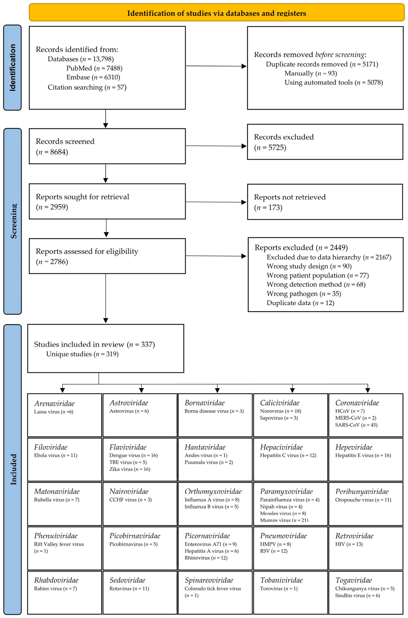

Disclaimer/Publisher’s Note: The statements, opinions and data contained in all publications are solely those of the individual author(s) and contributor(s) and not of MDPI and/or the editor(s). MDPI and/or the editor(s) disclaim responsibility for any injury to people or property resulting from any ideas, methods, instructions or products referred to in the content. |
© 2026 by the authors. Licensee MDPI, Basel, Switzerland. This article is an open access article distributed under the terms and conditions of the Creative Commons Attribution (CC BY) license.
Share and Cite
Ilsby, C.S.; Kristensen, T.L.; Bagge, K.; Bukh, J.; Lisby, J.G.; Schneider, U.V. Detectability of Pathogenic RNA Virus Families in Different Body Sites: A Scoping Review. Viruses 2026, 18, 743. https://doi.org/10.3390/v18070743
Ilsby CS, Kristensen TL, Bagge K, Bukh J, Lisby JG, Schneider UV. Detectability of Pathogenic RNA Virus Families in Different Body Sites: A Scoping Review. Viruses. 2026; 18(7):743. https://doi.org/10.3390/v18070743
Chicago/Turabian StyleIlsby, Christian Schaadt, Thomas Leineweber Kristensen, Kristian Bagge, Jens Bukh, Jan Gorm Lisby, and Uffe Vest Schneider. 2026. "Detectability of Pathogenic RNA Virus Families in Different Body Sites: A Scoping Review" Viruses 18, no. 7: 743. https://doi.org/10.3390/v18070743
APA StyleIlsby, C. S., Kristensen, T. L., Bagge, K., Bukh, J., Lisby, J. G., & Schneider, U. V. (2026). Detectability of Pathogenic RNA Virus Families in Different Body Sites: A Scoping Review. Viruses, 18(7), 743. https://doi.org/10.3390/v18070743