Article

The Role of HBx Mutations in Chronic Hepatitis B with Acute Exacerbation

1
Department of Infectious Diseases, East Campus, Renmin Hospital of Wuhan University, Wuhan 430223, China
2
State Key Laboratory of Virology, Department of Medical Microbiology, School of Basic Medical Sciences, Wuhan University, Wuhan 430071, China
3
Department of Fever Clinic, East Campus, Renmin Hospital of Wuhan University, Wuhan 430223, China
4
Hubei Province Key Laboratory of Allergy & Immunology, Wuhan University, Wuhan 430071, China
*
Authors to whom correspondence should be addressed.
These authors contributed equally to this work.
Viruses 2025, 17(9), 1223; https://doi.org/10.3390/v17091223
Submission received: 29 April 2025 / Revised: 31 August 2025 / Accepted: 3 September 2025 / Published: 7 September 2025

Abstract

Hepatitis B virus (HBV) infection remains a significant global health burden, primarily due to its chronic complications, including acute exacerbation, cirrhosis, hepatocellular carcinoma (HCC), and related sequelae. Acute exacerbation of chronic hepatitis B (CHB-AE) is common and often represents the earliest clinical manifestation. The Hepatitis B virus X protein (HBx) (17-kDa) is not only essential for viral replication but also plays a role in the development of HCC. To investigate the role of HBx mutation in CHB-AE progression, we enrolled 33 hospitalized CHB-AE patients and 31 patients with HBV-related liver failure (controls) from mainland China between January 2017 and June 2018. Single mutation 36 of HBx was significantly more prevalent in CHB-AE patients (p < 0.05), whereas Joint Mutation 1 was more frequent in HBV-related liver failure patients (p < 0.05). HBx mutations, including Single mutation 36 and Joint Mutations 2 and 3, were significantly associated with high HBV DNA levels (p < 0.05), while Joint mutation 1 predominated in the low HBV DNA group (p < 0.01). Age-stratified analysis showed that Single mutation 36 and Joint Mutation 2 were more common in younger patients (<35 years old) (p < 0.05), whereas Joint mutation 1 was more frequent in older age (≥35 years old) (p < 0.05). Moreover, antiviral therapy markedly reduced the prevalence of Joint mutation 1 from 82.98% in treatment-naïve patients to 29.41% in treatment-experienced patients (p < 0.0001). These findings suggest that specific HBx mutations are associated with viral replication levels, disease progression, and patient demographics. Such mutations may serve as molecular markers for disease severity and potential therapeutic targets in both CHB-AE and HBV-related liver failure.

1. Introduction

Hepatitis B Virus (HBV) infection continues to pose a major global health challenge. According to the World Health Organization (WHO), an estimated 254 million people were living with chronic hepatitis B infection in 2022, with 1.2 million new infections annually. In the same year, HBV-related complications such as cirrhosis and hepatocellular carcinoma (HCC) led to approximately 1.1 million deaths worldwide [1]. Current antiviral therapies, including pegylated interferon and nucleos(t)ide analogues (NAs), can slow disease progression and reduce the risk of cirrhosis and HCC, but they rarely achieve complete HBV eradication or hepatitis B surface antigen (HBsAg) clearance [2]. Chronic HBV infection (CHB) is a dynamic condition marked by complex interactions between the virus, hepatocytes, and the host immune system [3]. A characteristic clinical event is acute exacerbation (CHB-AE), which occurs in 10–30% of patients annually [4,5]. In HBV endemic regions, CHB-AE is common and often represents the earliest clinical manifestation of the disease [6]. It arises from immune clearance phase flares or viral reactivation in inactive or resolved carriers, potentially leading to severe liver injury [6,7]. Clinically, CHB-AE can manifest as elevated alanine aminotransferase (ALT), jaundice, and hepatic decompensation, with severe cases carrying a high mortality risk [8]. According to the 2015 Asian Pacific Association for the Study of the Liver (APASL) guideline, CHB-AE is defined as an intermittent ALT elevation > 5× the upper limit of normal and/or > 2× the baseline level [9]. The high burden of HBV infection and the challenge in managing CHB-AE emphasize the urgent need for better prognostic markers and therapeutic strategies.
The HBV genome is a partially double-stranded relaxed circular DNA (rcDNA) composed of four open reading frames (ORFs): preS1/S2/S, pre-core/core, polymerase, and X. These ORFs encode viral proteins, including the hepatitis B surface (HBs) antigens (LHBs, MHBs, and SHBs), hepatitis B e antigen (HBe), hepatitis B core antigen (HBc), DNA polymerase (DNA Pol), and Hepatitis B virus X protein (HBx) [10]. HBx, a 154–amino acid non-structural protein (~17 kDa) encoded by the X-ORF, is essential for HBV replication and modulation of host cellular functions [11,12,13]. Structurally, the N-terminal third (residues 1–50) of HBx functions as a negative regulator, while the C-terminal domain acts as a transcriptional activator [14]. In the nucleus, HBx interacts with covalently closed circular DNA (cccDNA) [15] and the transcriptional machinery to promote viral gene expression [16,17,18]. In the cytoplasm, it modulates signaling pathways that affect cell survival, metabolism, and proliferation [19,20]. These pleiotropic activities link HBx not only to HBV replication but also to hepatocarcinogenesis [21,22,23,24]. Due to the error-prone reverse transcription process in HBV replication, the viral genome—especially HBx—is prone to mutations, including point mutations, insertions, and deletions [25]. Such genetic variations can alter viral replication capacity, disease progression, and responses to antiviral therapies, occasionally leading to drug resistance [26].
However, the role of HBx mutations in CHB-AE remains poorly characterized. Clarifying this relationship could improve our understanding of HBV pathogenesis, help identify molecular markers of disease severity, and inform the development of more effective therapeutic strategies.

2. Materials and Methods

2.1. Sample Collection

A total of 64 serum samples were collected from patients at Renmin Hospital of Wuhan University between January 2017 and June 2018.

2.2. Exclusion Criteria

Patients were excluded from the study if they met any of the following conditions:
Positive for HIV antibody;
Positive for HAV antibody;
Positive for HCV antibody;
Positive for HDV antibody;
Negative or undetectable HBV DNA levels in serum.

2.3. Serologic Testing

HBV serological markers were determined by using the chemiluminescent microparticle immunoassay (CMIA) technique with an Architect-i2000 automatic analyzer (Abbott Laboratories, Chicago, IL, USA). Commercially available kits from Abbott Laboratories were used for testing. The analytical threshold of anti-HBs was set at 10 mIU/mL. Serum samples with HBsAg concentrations greater than 25,000 IU/mL were diluted according to the Manual Dilution Procedure. ALT levels were measured using an ADVIA automatic biochemical analyzer (Siemens, Munich, Germany).

2.4. HBV DNA Quantification

Blood samples were stored at −70 °C until processing. HBV DNA was extracted from 600 μL of serum and quantified using a commercially available real-time fluorescence quantitative kit (FOSUN DIAGNOSTICS, Shanghai, China) with a lower detection threshold of 15 IU/mL.

2.5. HBx Gene Amplification

The extracted HBV DNA was used as a template for polymerase chain reaction (PCR) to amplify the HBx gene. The primer sequences were:
  • Forward primer: 5′-ATGGCTGCTAGGCTGTGCTGCCAAC-3′.
  • Reverse primer: 5′-TTAGGCAGAGGTGAAAAAGTTGCAT-3′.
The PCR conditions were as follows:
(1)
Initial denaturation at 95 °C for 5 min.
(2)
35 cycles of denaturation at 94 °C for 30 s, annealing at 55 °C for 30 s, and extension at 72 °C for 30 s.
(3)
Final extension at 72 °C for 10 min.
The amplified products were separated on a 1.5% agarose gel prepared in 1× TAE buffer containing GelRed nucleic acid stain. Electrophoresis was performed at 100 V for 40 min, and bands were visualized under ultraviolet light.

2.6. HBx Mutation Analysis

The PCR products of the HBx gene were sequenced, and the resulting sequences were aligned with the HBV reference sequence (X01587.1) [27] available in the NCBI database using the BLAST+ 2.17.0 tool (https://blast.ncbi.nlm.nih.gov/Blast.cgi) accessed on 26 July 2025. Mutations were identified through this comparative analysis.

2.7. Statistical Analysis

Statistical analyses were performed using SPSS software (Version 20, IBM Corp., Armonk, NY, USA). Categorical variables were analyzed using the chi-square test, while continuous variables were compared using Student’s t-test. A p-value of < 0.05 was considered statistically significant.

3. Results

3.1. Baseline Characteristics of Patients

3.1.1. Study Cohort Characteristics

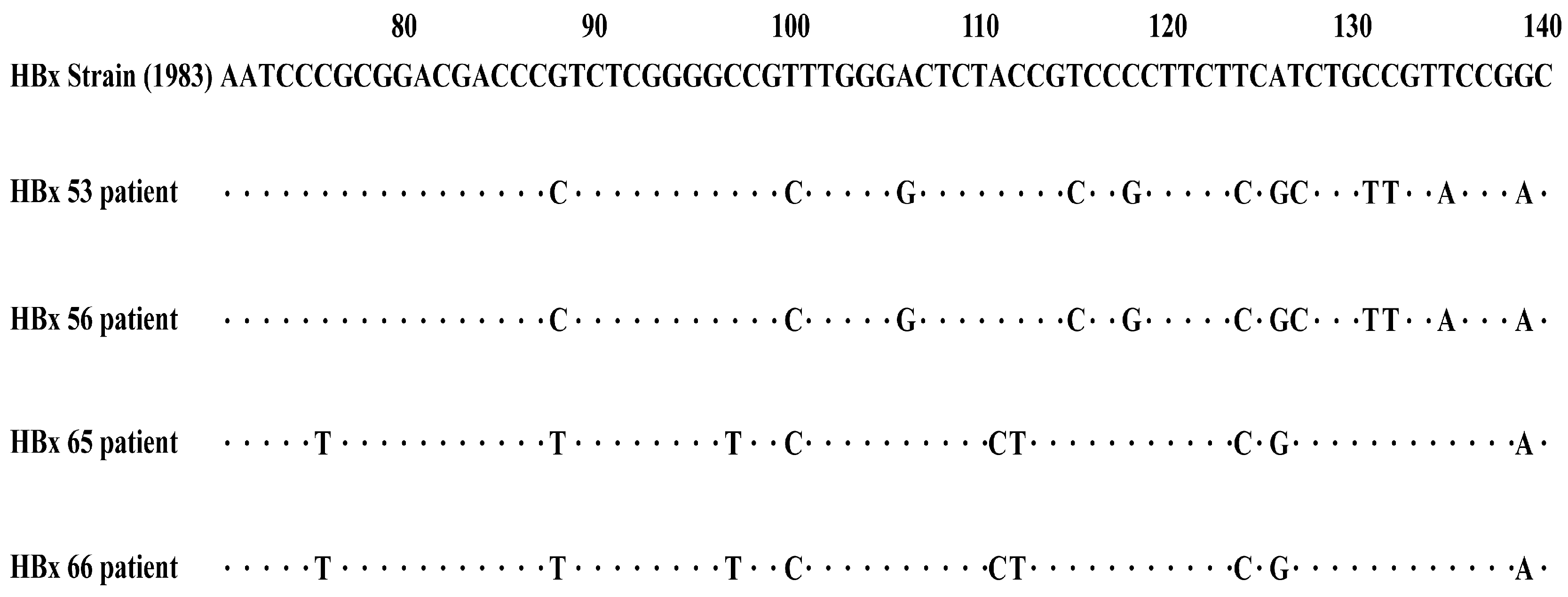
Between January 2017 to June 2018, serum samples were collected from hospitalized patients at Renmin Hospital of Wuhan University. The study enrolled 33 patients with CHB-AE and 31 patients with HBV-related liver failure. The cohort consisted of 54 males and 10 females, with a mean age of 43.64 ± 12.85 years. Among liver failure cases, 25 had acute-on-chronic liver failure (ACLF), 5 had chronic liver failure, and 1 had acute liver failure. Diagnoses were made according to the “The guideline of prevention and treatment for chronic hepatitis B (2015 version)” [28] and the “Diagnostic and treatment guidelines for liver failure (2012 version)” [29]. The full-length HBx gene was successfully amplified from all samples, with an expected fragment size of approximately 465 bp (Figure 1). HBx Mutations were detected in all patients (64/64), with multiple mutation types observed at different loci (Figure 2).

3.1.2. Baseline Comparison Between CHB-AE and HBV-Related Liver Failure

There were no significant differences between the two groups in gender distribution (CHB-AE: 78.79% male vs. liver failure: 90.32% male, p = 0.3546) or age (CHB-AE: 57.58% ≥ 35 years vs. liver failure: 70.97% ≥ 35 years, p = 0.2645).
However, CHB-AE patients had significantly higher HBV DNA levels (≥log 5 copies/mL: 87.88% vs. 64.52%, p = 0.0275) and higher HBeAg positivity (63.64% vs. 32.26%, p = 0.0121) (Table 1).

3.2. The HBx Mutations and the Severity of CHB

Single mutation 36 (T36A/S): More frequent in CHB-AE than liver failure patients (39.39% vs. 16.13%, p = 0.0386).
Joint Mutation 1 (R26C + P33S + P38S): More frequent in liver failure (80.65%) than CHB-AE (57.58%, p = 0.0466).
Joint Mutation 2 (S39P + P40A + S43P + A44L/V/F + Q87R/W/K/G): Detected in 36.36% (12/33) of CHB-AE vs. 16.13% (5/31) of liver failure patients, without statistical significance (p = 0.0670).
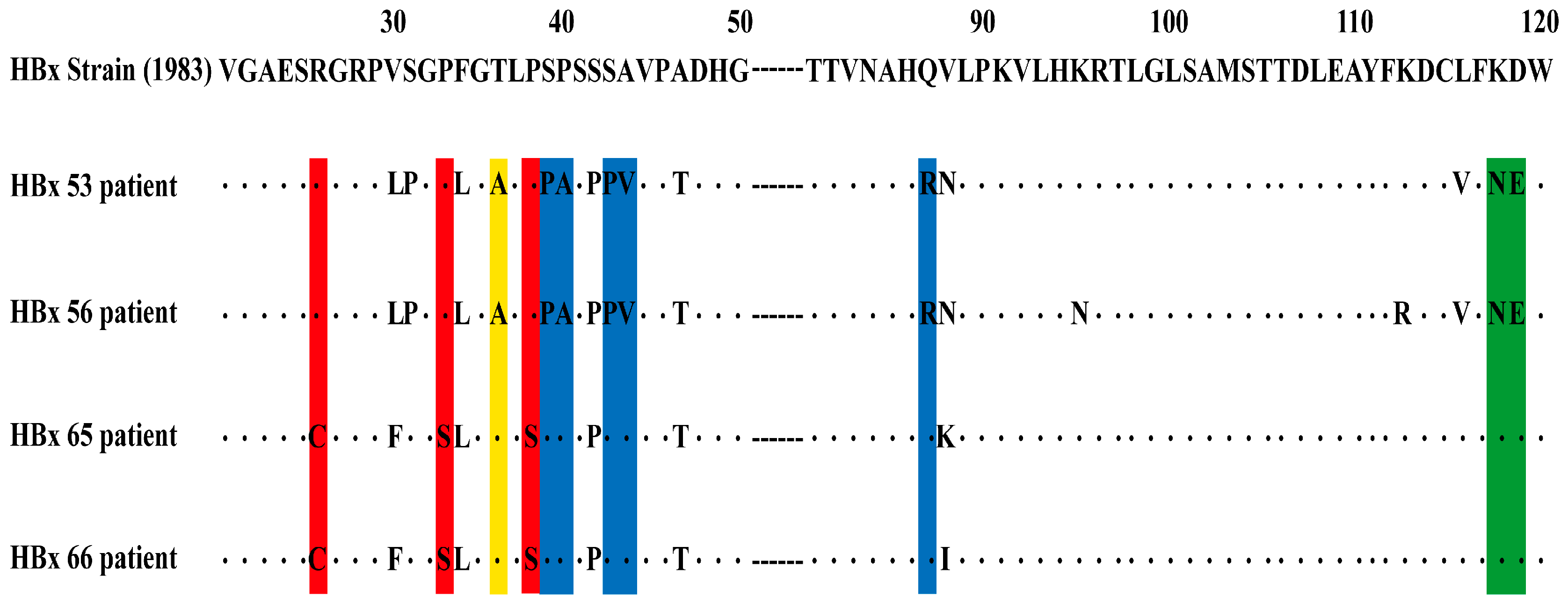
Joint Mutation 3 (K118N/T + D119E): No significant difference between two groups (33.33% vs. 16.13%, p = 0.1122) (Figure 3, Table 2).
Overall, the distribution of HBx mutations varies with the severity of CHB. Single mutation T36A/S was more prevalent in CHB-AE patients, whereas Joint Mutation 1 (R26C + P33S + P38S) occurred more frequently in liver failure cases. Joint Mutation 2 showed a higher occurrence in CHB-AE patients, but the difference was not statistically significant, while Joint Mutation 3 showed no significant difference between the two groups. These findings suggest that certain specific mutation patterns may be associated with different stages of disease progression, though further studies are needed for confirmation.

3.3. HBx Mutations and HBV DNA Viral Load

Joint Mutation 1 (R26C + P33S + P38S): More common in low viral load patients (100% vs. 59.18%, p = 0.0077). Joint Mutation 2 (S39P + P40A + S43P + A44L/V/F + Q87R/W/K/G): Present only in high viral load patients (34.69% vs. 0%, p = 0.0199). Joint Mutation 3 (K118N/T + D119E): More frequent in high viral load patients (32.65% vs. 0%, p = 0.0268).
Single Mutation 36 (T36A/S): Detected only in high viral load patients (≥log 5: 36.73% vs. < log 5: 0%, p = 0.0147).
Overall, Single Mutation 36, Joint Mutation 2, and Joint Mutation 3 were significantly associated with high HBV DNA levels (p < 0.05), whereas Joint Mutation 1 HBx was linked to low HBV DNA levels (p < 0.01) (Table 3).

3.4. The HBx Mutations and Patients’ Age

Single mutation 36 (T36A/S): More frequent in younger patients (<35 years: 43.48% vs. ≥ 35 years: 19.51%, p = 0.0407).
Joint mutation 1 (R26C + P33S + P38S): More common in older patients (≥35 years: 78.05% vs. < 35 years: 52.17%, p = 0.0321).
Joint mutation 2 (S39P + P40A + S43P + A44L/V/F + Q87R/W/K/G): More prevalent in younger patients (43.48% vs. 17.07%, p = 0.0217).
Joint mutation 3 (K118N/T + D119E): No significant age-related difference (p = 0.0505).
Age-stratified analysis revealed that Single mutation 36 and Joint Mutation 2 were significantly more prevalent in patients < 35 years old (p < 0.05), while Joint Mutation 1 was more common in those aged 35 years and older (p < 0.05). Joint Mutation 3 showed no significant age association (Table 4).

3.5. HBx Mutations and Antiviral Treatment History

All HBx mutation frequencies were lower in treatment-experienced patients, but only Joint mutation 1 showed a significant reduction (treatment-naïve: 82.98% vs. treatment-experienced: 29.41%, p < 0.0001) (Table 5).

3.6. Risk Factors Related to Prognosis

Gender, age, HBV DNA level, HBeAg, HBx mutations and antiviral treatment were not significantly associated with prognosis (p > 0.05) (Supplementary Table S1).

4. Discussion

HBx is a multifunctional 17-kDa protein essential for HBV replication and pathogenesis [30]. Mutations in HBx influence disease progression through several mechanisms:
(1)
Oncogenesis and Disease Progression: Certain variants (e.g., I127T, V131I, and F132Y/I/R) are associated with progression from chronic liver disease to HCC [31,32].
(2)
Transcriptional regulation: HBx modulates both viral and host gene expression, impacting cell cycle, proliferation, apoptosis, and immune signaling [33,34].
(3)
Immune Modulation: HBx enhances pro-inflammatory cytokine expression, contributing to persistent hepatic inflammation and HCC risk [35].
(4)
Genomic Instability: HBx promotes chromosomal instability, a hallmark of tumorigenesis [36].
(5)
Fibrosis and Cirrhosis: Certain HBx mutations are linked to cirrhosis onset [37,38].
These findings underscore the multifaceted roles of HBx mutations in promoting liver disease progression, liver failure, cirrhosis, and HCC. In this study, we explored the specific association between HBx mutations and the progression of CHB-AE and HBV-related liver failure.
Our study revealed a striking contrast between Single Mutation 36 and Joint Mutation 1. Single mutation 36 was significantly more prevalent in CHB-AE (p = 0.0386), whereas Joint Mutation 1 was more common in HBV-related liver failure (p = 0.0466), suggesting divergent roles in disease progression.
The relationship between HBx mutations and viral replication observed in our study aligns with the known role of HBx in enhancing cccDNA transcription and supporting high-level replication [39,40,41,42]. Mutations such as Single mutation 36, Joint Mutation 2, and Joint 3 were enriched in high viral load group, consistent with an active replication phenotype. In contrast, the predominance of Joint mutation 1 in low viral load patients suggests it may emerge during viral suppression or disease decompensation, potentially reflecting a late-stage adaptation.
Single Mutation 36 was found to be associated with higher HBV DNA levels and younger age (<35 years), whereas Joint Mutation 1 correlated with lower HBV DNA levels and older age (≥35 years). This pattern may be explained by the fact that younger individuals are more likely to be in immune tolerance or immune clearance phases, characterized by high viral loads and positive HBeAg status. In contrast, Joint Mutation 1 predominated in older individuals with lower viral loads.
These findings are consistent with previous reports suggesting that HBx mutations tend to accumulate with age [43]. Yuen et al. further proposed that increased viral diversity is associated with older age, accompanied by lower levels of HBV-DNA, HBsAg and HBeAg [44]. Collectively, these observations emphasize the importance of considering age as a potential confounder factor when assessing the relationship between HBx mutation and HCC risk [45].
Interestingly, Joint Mutation 1 appeared to be mutually exclusive with other mutations, suggesting that it may arise through distinct evolutionary or functional pathways. This exclusivity highlights the need for further studies to clarify its biological significance and potential role in disease progression.
Antiviral therapy also influenced mutation prevalence. The frequency of Joint Mutation 1 was markedly reduced in treatment-experienced patients compared to treatment-naïve patients (29.41% vs. 82.98%, p < 0.0001), suggesting that sustained viral suppression may limit its emergence. Previous studies have shown that HBx mutations can develop during lamivudine or entecavir therapy, sometimes contributing to drug resistance [12,46]. However, in our cohort, other mutations did not differ significantly between treatment groups, indicating that some HBx variants may arise independently of antiviral exposure.
We also observed significantly higher HBV DNA levels and HBeAg positivity in CHB-AE compared to liver failure patients, supporting the notion that active replication and immune clearance are characteristic of CHB-AE, whereas lower viral loads may reflect advanced disease or impaired immune control. This aligns with studies linking high viral loads to increased HCC risk, even under antiviral therapy [47,48,49,50,51].
Limitations of this study include the relatively small sample size and the lack of longitudinal follow-up, which restricts conclusions about causality and mutation dynamics over time. Larger, prospective studies are needed to confirm these associations and elucidate the molecular mechanisms by which specific HBx mutations influence disease phenotype.

5. Conclusions

We identified several distinct HBx mutation patterns—Single Mutation 36, Joint Mutations 1, 2, and 3—with opposing associations for Single Mutation 36 and Joint Mutation 1. Single Mutation 36 may serve as a biomarker for CHB-AE, while Joint Mutation 1 may indicate HBV-related liver failure. These findings provide new insights into HBV pathogenesis and suggest that HBx mutations could serve as useful biomarkers or therapeutic targets in managing chronic HBV infection.

Supplementary Materials

The following supporting information can be downloaded at: https://www.mdpi.com/article/10.3390/v17091223/s1, Table S1: The risk factors related to prognosis.

Author Contributions

Conceptualization, X.C. and F.Z.; formal analysis, P.Z.; funding acquisition, F.Z.; investigation, X.C. and J.S.; methodology, Y.T., Y.Z., T.L. and Y.L.; project administration, F.Z.; supervision, X.C. and F.Z.; writing—original draft, X.C., J.S. and P.Z.; writing—review & editing, X.C. and F.Z. All authors have read and agreed to the published version of the manuscript.

Funding

This work was supported by the National Natural Science Foundation of China (Fan Zhu, No. 82272321).

Institutional Review Board Statement

The study was approved by the Ethical Committee of Wuhan University in 2013 (Project No. 13015), and written informed consent was obtained from all participants.

Informed Consent Statement

Informed consent was obtained from all subjects involved in the study. Written informed consent was obtained from the patient(s) to publish this paper.

Data Availability Statement

All data are available from the corresponding author upon request.

Conflicts of Interest

The authors declare no conflicts of interest.

Abbreviations

The following abbreviations are used in this manuscript:
ACLFAcute-on-chronic liver failure
ALTAlanine aminotransferase
cccDNACovalently closed circular DNA
CHB-AEAcute exacerbation of chronic hepatitis B
HBVHepatitis B virus
HBxHepatitis B virus X protein
HCCHepatocellular carcinoma
NAsNucleos(t)ide analogues
ORFsOpen reading frames

References

  1. WHO. Hepatitis B. 2021. Available online: https://www.who.int/news-room/fact-sheets/detail/hepatitis-b (accessed on 9 April 2024).
  2. Jeng, W.J.; Papatheodoridis, G.V.; Lok, A.S.F. Hepatitis B. Lancet 2023, 401, 1039–1052. [Google Scholar] [CrossRef]
  3. Chang, M.L.; Liaw, Y.F. Hepatitis B Flare in Hepatitis B e Antigen-Negative Patients: A Complicated Cascade of Innate and Adaptive Immune Responses. Int. J. Mol. Sci. 2022, 23, 1552. [Google Scholar] [CrossRef]
  4. Tsai, W.L.; Sun, W.C.; Cheng, J.S. Chronic Hepatitis B with Spontaneous Severe Acute Exacerbation. Int. J. Mol. Sci. 2015, 16, 28126–28145. [Google Scholar] [CrossRef]
  5. Liaw, Y.F. Acute exacerbation and superinfection in patients with chronic viral hepatitis. J. Formos. Med. Assoc. 1995, 94, 521–528. [Google Scholar]
  6. Lall, S.; Agarwala, P.; Kumar, G.; Sharma, M.K.; Gupta, E. The dilemma of differentiating between acute hepatitis B and chronic hepatitis B with acute exacerbation: Is quantitative serology the answer? Clin. Mol. Hepatol. 2020, 26, 187–195. [Google Scholar] [CrossRef]
  7. Chang, M.L.; Liaw, Y.F. Hepatitis B flares in chronic hepatitis B: Pathogenesis, natural course, and management. J. Hepatol. 2014, 61, 1407–1417. [Google Scholar] [CrossRef] [PubMed]
  8. Wong, V.W.S.; Chan, H.L.Y. Severe acute exacerbation of chronic hepatitis B: A unique presentation of a common disease. J. Gastroenterol. Hepatol. 2009, 24, 1179–1186. [Google Scholar] [CrossRef] [PubMed]
  9. Sarin, S.K.; Kumar, M.; Lau, G.K.; Abbas, Z.; Chan, H.L.; Chen, C.J.; Chen, D.S.; Chen, H.L.; Chen, P.J.; Chien, R.N.; et al. Asian-Pacific clinical practice guidelines on the management of hepatitis B: A 2015 update. Hepatol. Int. 2016, 10, 1–98. [Google Scholar] [CrossRef]
  10. Jiang, Y.; Han, Q.J.; Zhao, H.J.; Zhao, J. The Mechanisms of HBV-Induced Hepatocellular Carcinoma. J. Hepatocell. Carcinoma 2021, 8, 435–450. [Google Scholar] [CrossRef] [PubMed]
  11. Nassal, M. HBV cccDNA: Viral persistence reservoir and key obstacle for a cure of chronic hepatitis B. Gut 2015, 64, 1972–1984. [Google Scholar] [CrossRef]
  12. Zhou, B.; He, W.M.; Hou, J.L. HBx mutations emerged during antiviral therapy: A new face of a multifaceted HBV protein? Hepatol. Int. 2020, 14, 944–946. [Google Scholar] [CrossRef]
  13. Tang, H.; Oishi, N.; Kaneko, S.; Murakami, S. Molecular functions and biological roles of hepatitis B virus x protein. Cancer Sci. 2006, 97, 977–983. [Google Scholar] [CrossRef]
  14. Hernández, S.; Venegas, M.; Brahm, J.; Villanueva, R.A. The viral transactivator HBx protein exhibits a high potential for regulation via phosphorylation through an evolutionarily conserved mechanism. Infect. Agent. Cancer 2012, 7, 27. [Google Scholar] [CrossRef]
  15. Belloni, L.; Pollicino, T.; Nicola, F.D.; Guerrieri, F.; Raffa, G.; Fanciulli, M.; Raimondo, G.; Levrero, M. Nuclear HBx binds the HBV minichromosome and modifies the epigenetic regulation of cccDNA function. Proc. Natl. Acad. Sci. USA 2009, 106, 19975–19979. [Google Scholar] [CrossRef]
  16. Bouchard, M.J.; Schneider, R.J. The enigmatic X gene of hepatitis B virus. J. Virol. 2004, 78, 12725–12734. [Google Scholar] [CrossRef] [PubMed]
  17. Benhenda, S.; Cougot, D.; Buendia, M.A.; Neuveut, C. Hepatitis B virus X protein molecular functions and its role in virus life cycle and pathogenesis. Adv. Cancer Res. 2009, 103, 75–109. [Google Scholar] [PubMed]
  18. Schollmeier, A.; Glitscher, M.; Hildt, E. Relevance of HBx for Hepatitis B Virus-Associated Pathogenesis. Int. J. Mol. Sci. 2023, 24, 4964. [Google Scholar] [CrossRef]
  19. Ma, J.; Sun, T.; Park, S.; Shen, G.; Liu, J. The role of hepatitis B virus X protein is related to its differential intracellular localization. Acta Biochim. Biophys. Sin. 2011, 43, 583–588. [Google Scholar] [CrossRef]
  20. Slagle, B.L.; Bouchard, M.J. Role of HBx in hepatitis B virus persistence and its therapeutic implications. Curr. Opin. Virol. 2018, 30, 32–38. [Google Scholar] [CrossRef] [PubMed]
  21. Yeh, C.T.; Shen, C.H.; Tai, D.I.; Chu, C.M.; Liaw, Y.F. Identification and characterization of a prevalent hepatitis B virus X protein mutant in Taiwanese patients with hepatocellular carcinoma. Oncogene 2000, 19, 5213–5220. [Google Scholar] [CrossRef]
  22. Chen, G.G.; Li, M.Y.; Ho, R.L.K.; Chak, E.C.W.; Lau, W.Y.; Lai, P.B.S. Identification of hepatitis B virus X gene mutation in Hong Kong patients with hepatocellular carcinoma. J. Clin. Virol. 2005, 34, 7–12. [Google Scholar] [CrossRef]
  23. Salarnia, F.; Besharat, S.; Zhand, S.; Javid, N.; Khodabakhshi, B.; Moradi, A. Mutations in Hepatitis-B X-Gene Region: Chronic Hepatitis-B versus Cirrhosis. J. Clin. Diagn. Res. 2017, 11, OC31–OC34. [Google Scholar] [CrossRef]
  24. Chaturvedi, V.K.; Singh, A.; Dubey, S.K.; Hetta, H.F.; John, J.; Singh, M.P. Molecular mechanistic insight of hepatitis B virus mediated hepatocellular carcinoma. Microb. Pathog. 2019, 128, 184–194. [Google Scholar] [CrossRef]
  25. Rajoriya, N.; Combet, C.; Zoulim, F.; Janssen, H.L.A. How viral genetic variants and genotypes influence disease and treatment outcome of chronic hepatitis B. Time for an individualised approach? J. Hepatol. 2017, 67, 1281–1297. [Google Scholar] [CrossRef]
  26. Akrami, H.; Monjezi, M.R.; Ilbeigi, S.; Amiri, F.; Fattahi, M.R. The association between hepatitis B Virus mutations and the risk of liver disease and hepatocellular carcinoma. Curr. Mol. Med. 2022, 22, 514–523. [Google Scholar] [CrossRef]
  27. Fujiyama, A.; Miyanohara, A.; Nozaki, C.; Yoneyama, T.; Ohtomo, N.; Matsubara, K. Cloning and structural analyses of hepatitis B virus DNAs, subtype adr. Nucleic Acids Res. 1983, 11, 4601–4610. [Google Scholar] [CrossRef]
  28. Hou, J.; Wang, G.; Wang, F.; Cheng, J.; Ren, H.; Zhuang, H.; Sun, J.; Li, L.; Li, J.; Meng, Q.; et al. Guideline of Prevention and Treatment for Chronic Hepatitis B (2015 Update). J. Clin. Transl. Hepatol. 2017, 5, 297–318. [Google Scholar] [CrossRef]
  29. Liver Failure and Artificial Liver Group; Chinese Society of Infectious Diseases; Chinese Medical Association; Severe Liver Diseases and Artificial Liver Group; Chinese Society of Hepatology; Chinese Medical Association. Diagnostic and treatment guidelines for liver failure (2012 version). Zhonghua Gan Zang Bing Za Zhi 2013, 21, 177–183. [Google Scholar]
  30. Bouchard, M.J.; Wang, L.; Schneider, R.J. Activation of focal adhesion kinase by hepatitis B virus HBx protein: Multiple functions in viral replication. J. Virol. 2006, 80, 4406–4414. [Google Scholar] [CrossRef] [PubMed]
  31. Al-Qahtani, A.A.; Al-Anazi, M.R.; Nazir, N.; Ghai, R.; Abdo, A.A.; Sanai, F.M.; Al-Hamoudi, W.K.; Alswat, K.A.; Al-Ashgar, H.I.; Khan, M.Q.; et al. Hepatitis B virus (HBV) X gene mutations and their association with liver disease progression in HBV-infected patients. Oncotarget 2017, 8, 105115–105125. [Google Scholar] [CrossRef] [PubMed]
  32. Luber, B.; Lauer, U.; Weiss, L.; Hohne, M.; Hofschneider, P.H.; Kekule, A.S. The hepatitis B virus transactivator HBx causes elevation of diacylglycerol and activation of protein kinase C. Res. Virol. 1993, 144, 311–321. [Google Scholar] [CrossRef]
  33. Ma, M.R.; Yi, L.; Pei, Y.F.; Zhang, Q.M.; Tong, C.; Zhao, M.Y.; Chen, Y.H.; Zhu, J.H.; Zhang, W.G.; Yao, F.; et al. USP26 as a hepatitis B virus-induced deubiquitinase primes hepatocellular carcinogenesis by epigenetic remodeling. Nat. Commun. 2024, 15, 7856. [Google Scholar] [CrossRef]
  34. Damme, E.V.; Vanhove, J.; Severyn, B.; Verschueren, L.; Pauwels, F. The Hepatitis B Virus Interactome: A Comprehensive Overview. Front. Microbiol. 2021, 12, 724877. [Google Scholar] [CrossRef]
  35. He, T.; Zhang, N.; Wang, L.; Wan, B.S.; Wang, X.Q.; Zhang, L. GPR43 regulates HBV X protein (HBx)-induced inflammatory response in human LO2 hepatocytes. Biomed. Pharmacother. 2020, 123, 109737. [Google Scholar] [CrossRef]
  36. Zhang, D.; Guo, S.C.; Schrodi, S.J. Mechanisms of DNA Methylation in Virus-Host Interaction in Hepatitis B Infection: Pathogenesis and Oncogenetic Properties. Int. J. Mol. Sci. 2021, 22, 9858. [Google Scholar] [CrossRef] [PubMed]
  37. Rizzo, G.E.M.; Cabibbo, G.; Craxì, A. Hepatitis B Virus-Associated Hepatocellular Carcinoma. Viruses 2022, 14, 986. [Google Scholar] [CrossRef]
  38. Waris, G.; Huh, K.W.; Siddiqui, A. Mitochondrially associated hepatitis B virus X protein constitutively activates transcription factors STAT-3 and NF-kappa B via oxidative stress. Mol. Cell. Biol. 2001, 21, 7721–7730. [Google Scholar] [CrossRef]
  39. Lucifora, J.; Arzberger, S.; Durantel, D.; Belloni, L.; Strubin, M.; Levrero, M.; Zoulim, F.; Hantz, O.; Protzer, U. Hepatitis B Virus X protein is essential to initiate and maintain virus replication after infection. J. Hepatol. 2011, 55, 996–1003. [Google Scholar] [CrossRef]
  40. Tsuge, M.; Hiraga, N.; Akiyama, R.; Tanaka, S.; Matsushita, M.; Mitsui, F.; Abe, H.; Kitamura, S.; Hatakeyama, T.; Kimura, T.; et al. HBx protein is indispensable for development of viraemia in human hepatocyte chimeric mice. J. Gen. Virol. 2010, 91, 1854–1864. [Google Scholar] [CrossRef] [PubMed]
  41. Chen, H.S.; Kaneko, S.; Girones, R.; Anderson, R.W.; Hornbuckle, W.E.; Tennant, B.C.; Cote, P.J.; Gerin, J.L.; Purcell, R.H.; Miller, R.H. The woodchuck hepatitis virus X gene is important for establishment of virus infection in woodchucks. J. Virol. 1993, 67, 1218–1226. [Google Scholar] [CrossRef] [PubMed]
  42. Zoulim, F.; Saputelli, J.; Seeger, C. Woodchuck hepatitis virus X protein is required for viral infection in vivo. J. Virol. 1994, 68, 2026–2030. [Google Scholar] [CrossRef]
  43. Yang, H.I.; Yeh, S.H.; Chen, P.J.; Iloeje, U.H.; Jen, C.L.; Su, J.; Wang, L.Y.; Lu, S.N.; You, S.L.; Chen, D.S.; et al. REVEAL-HBV Study Group: Associations between hepatitis B virus genotype and mutations and the risk of hepatocellular carcinoma. J. Nat Cancer Inst. 2008, 100, 1134–1143. [Google Scholar] [CrossRef]
  44. Yuen, L.; Revill, P.A.; Rosenberg, G.; Wagner, J.; Littlejohn, M.; Bayliss, J.; Jackson, K.; Tan, S.K.; Gaggar, A.; Kitrinos, K.; et al. HBV variants are common in the ‘immune-tolerant’ phase of chronic hepatitis B. J. Viral. Hepat. 2020, 27, 1061–1070. [Google Scholar] [CrossRef] [PubMed]
  45. Wang, Y.L.; Zeng, L.I.; Chen, W.Q. HBV X gene point mutations are associated with the risk of hepatocellular carcinoma: A systematic review and meta-analysis. Mol. Clin. Oncol. 2016, 4, 1045–1051. [Google Scholar] [CrossRef] [PubMed]
  46. Lin, C.L.; Chien, R.N.; Chu, Y.D.; Liang, H.K.; Huang, Y.H.; Ke, P.Y.; Lin, K.H.; Lin, Y.H.; Yeh, C.T. Hepatitis B virus X gene mutants emerge during antiviral therapy and increase cccDNA levels to compensate for replication suppression. Hepatol. Int. 2020, 14, 973–984. [Google Scholar] [CrossRef]
  47. Tang, H.; Delgermaa, L.; Huang, F.; Oishi, N.; Liu, L.; He, F.; Zhao, L.S.; Murakami, S. The transcriptional transactivation function of HBx protein is important for its augmentation role in hepatitis B virus replication. J. Virol. 2005, 79, 5548–5556. [Google Scholar] [CrossRef]
  48. Choi, W.M.; Yip, T.C.F.; Kim, W.R.; Yee, L.J.; Rooney, C.B.; Curteis, T.; Clark, L.J.; Jafry, Z.; Chen, C.H.; Chen, C.Y.; et al. Chronic hepatitis B baseline viral load and on-treatment liver cancer risk: A multinational cohort study of HBeAg-positive patients. Hepatology 2024, 80, 428–439. [Google Scholar] [CrossRef] [PubMed]
  49. Chinese Society of Hepatology; Chinese Medical Association. Expert opinion on expanding anti-HBV treatment for chronic hepatitis B. Zhonghua Gan Zang Bing Za Zhi 2022, 30, 131–136. [Google Scholar]
  50. You, H.; Wang, F.S.; Li, T.S.; Xu, X.Y.; Sun, Y.M.; Nan, Y.M.; Wang, G.Q.; Hou, J.L.; Duan, Z.P.; Wei, L.; et al. Guidelines for the Prevention and Treatment of Chronic Hepatitis B (version 2022). J. Clin. Transl. Hepatol. 2023, 11, 1425–1442. [Google Scholar] [CrossRef]
  51. Lok, J.; Dusheiko, G. Editorial: Reassessing antiviral treatment criteria for chronic hepatitis B. Hepatology 2023, 78, 1332–1333. [Google Scholar] [CrossRef]
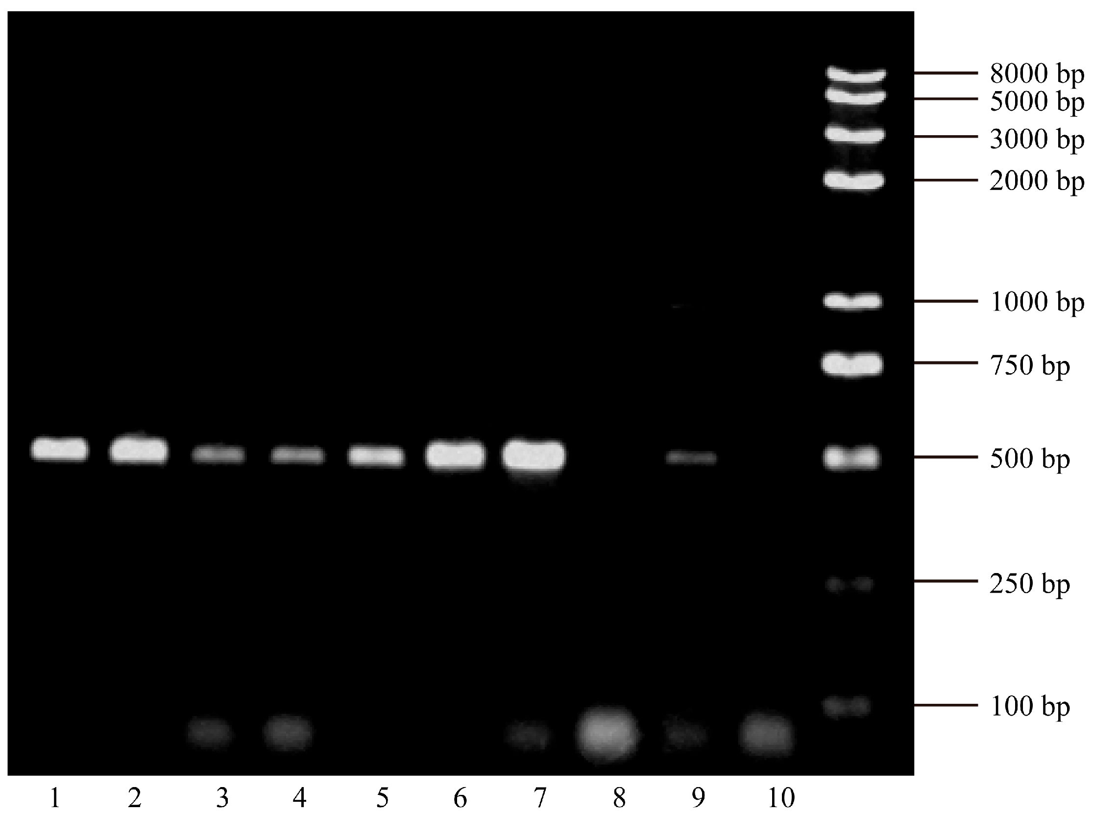
Figure 1. Electrophoretic analysis of HBx gene PCR products on an agarose gel. CHB-AE: Lane 1 (HBx 66 patient), 5 (HBx 57 patient), 7 (HBx 53 patient), 8 (HBx 49 patient) and 10 (HBx 22 patient); ACLF: Lane 2 (HBx 65 patient), 3 (HBx 62 patient), 4 (HBx 61 patient) and 9 (HBx 21 patient); Chronic liver failure: Lane 6 (HBx 56 patient).
Viruses 17 01223 g001
Figure 2. The HBx gene nucleotide sequence alignment of patients with CHB-AE and HBV-related liver failure.
Viruses 17 01223 g002
Figure 3. The HBx gene amino acid sequence alignment patients with CHB-AE and HBV-related liver failure. HBx53: patient 53#, CHB-AE, with Single mutation 36 (marked as yellow), joint 2 (marked as blue), and joint 3 (marked as green) mutations of HBx. HBx56: patient 56#, Chronic Liver Failure, with Single mutation 36, joint 2, and joint 3 mutations of HBx. HBx65: patient 65#, ACLF, with joint 1 (marked as red) mutation of HBx. HBx66: patient 66#, CHB-AE, with joint 1 mutation of HBx.
Viruses 17 01223 g003
Table 1. The baseline characteristics of patients with CHB-AE and HBV-related liver failure.
Chronic Hepatitis B with Acute Exacerbation
(n = 33) (%)
HBV-Related
Liver Failure
(n = 31) (%)
p-Value
GenderMale26 (78.79)28 (90.32)0.3546
Female7 (21.21)3 (9.68)
Age
(years)
≥3519 (57.58)22 (70.97)0.2645
<3514 (42.42)9 (29.03)
HBV DNA
Log level
(copies/mL)
≥529 (87.88)20 (64.52)0.0275
<54 (12.12)11 (35.48)
HBeAg
(0–1 COI)
positive21 (63.64)10 (32.26)0.0121
negative12 (36.36)21 (67.74)
Table 2. The HBx mutations and the severity of CHB.
HBx
Mutations
Chronic Hepatitis B with Acute Exacerbation
(n = 33) (%)
HBV-Related
Liver Failure
(n = 31) (%)
p-Value
Single Mutation 36
(T36A/S)
13 (39.39)5 (16.13)0.0386
Joint Mutation 1
(R26C + P33S + P38S)
19 (57.58)25 (80.65)0.0466
Joint Mutation 2
(S39P + P40A + S43P + A44L/V/F + Q87R/W/K/G)
12 (36.36)5 (16.13)0.0670
Joint Mutation 3
(K118N/T + D119E)
11 (33.33)5 (16.13)0.1122
Table 3. The HBxAg mutations and HBV DNA viral load.
HBx
Mutations
High HBV DNA
Log Level ≥ 5
(n = 49) (%)
Low HBV DNA Log Level < 5
(n = 15) (%)
p-Value
Single Mutation 36
(T36A/S)
18 (36.73)0 (0)0.0147
Joint Mutation 1
(R26C + P33S + P38S)
29 (59.18)15 (100)0.0077
Joint Mutation 2
(S39P + P40A + S43P + A44L/V/F + Q87R/W/K/G)
17 (34.69)0 (0)0.0199
Joint Mutation 3
(K118N/T + D119E)
16 (32.65)0 (0)0.0268
Table 4. The HBx mutations and patients’ age.
HBx
Mutations
Age ≥ 35
(n = 41) (%)
Age < 35
(n = 23) (%)
p-Value
Single Mutation 36
(T36A/S)
8 (19.51)10 (43.48)0.0407
Joint Mutation 1
(R26C + P33S + P38S)
32 (78.05)12 (52.17)0.0321
Joint Mutation 2
(S39P + P40A + S43P + A44L/V/F + Q87R/W/K/G)
7 (17.07)10 (43.48)0.0217
Joint Mutation 3
(K118N/T + D119E)
7 (17.07)9 (39.13%)0.0505
Table 5. The HBx mutations and antiviral treatment history.
HBx
Mutations
Treatment-Naïve
(n = 47) (%)
Treatment-Experienced
(n = 17) (%)
p-Value
Single Mutation 36
(T36A/S)
14 (29.79)4 (23.53)0.8595
Joint Mutation 1
(R26C + P33S + P38S)
39 (82.98)5 (29.41)<0.0001
Joint Mutation 2
(S39P + P40A + S43P + A44L/V/F + Q87R/W/K/G)
14 (29.79)3 (17.65)0.5152
Joint Mutation 3
(K118N/T + D119E)
13 (27.66)3 (17.65)0.6240
Disclaimer/Publisher’s Note: The statements, opinions and data contained in all publications are solely those of the individual author(s) and contributor(s) and not of MDPI and/or the editor(s). MDPI and/or the editor(s) disclaim responsibility for any injury to people or property resulting from any ideas, methods, instructions or products referred to in the content.