Review

Epitranscriptomic Regulation of Hepatitis B Virus by RNA 5-Methylcytosine: Functions, Mechanisms, and Therapeutic Potential

1
School of Pharmacy, Hubei University of Chinese Medicine, Wuhan 430065, China
2
Center for Emerging Infectious Diseases, Wuhan Institute of Virology, Chinese Academy of Sciences, Wuhan 430071, China
3
Hubei JiangXia Laboratory, Wuhan 430200, China
*
Authors to whom correspondence should be addressed.
Viruses 2025, 17(9), 1159; https://doi.org/10.3390/v17091159
Submission received: 6 July 2025 / Revised: 20 August 2025 / Accepted: 22 August 2025 / Published: 24 August 2025
(This article belongs to the Special Issue Epigenetic Modifications in Viral Infections, Volume II)

Abstract

Hepatitis B virus (HBV) remains a major global health challenge, with over 296 million people chronically infected worldwide. Despite the availability of antiviral therapies, a functional cure is rarely achieved, highlighting the need for novel therapeutic strategies. RNA 5-methylcytosine (m5C) is a pivotal epitranscriptomic mark implicated in RNA stability, transport, and translation. Emerging evidence shows that m5C is conserved within HBV RNA and plays critical roles in the viral life cycle. This review provides a comprehensive overview of the molecular mechanisms governing m5C deposition and recognition, summarizes recent advances in m5C biology, and highlights the emerging role of epitranscriptomic m5C regulation in HBV infection. We discuss the identification of HBV-specific m5C sites, the functions of key regulatory enzymes, and their interplay in viral RNA stabilization and evasion of innate immune responses. Interplay between m5C and other RNA modifications—particularly N6-methyladenosine (m6A)—is examined alongside virus-specific m5C regulation in EV71, HIV, HCV, EBV, and SARS-CoV-2. Potential links between m5C dysregulation and HBV-induced hepatocarcinogenesis are outlined, and emerging therapeutic strategies targeting the m5C machinery are highlighted. Together, these insights position the epitranscriptomic landscape as a promising avenue for innovative antiviral strategies.

1. Introduction

Hepatitis B virus (HBV) causes chronic infections in over 290 million individuals worldwide and remains a leading cause of liver cirrhosis and hepatocellular carcinoma (HCC) [1]. Despite the availability of effective vaccines and antiviral agents, complete viral eradication remains elusive due to the persistence of viral RNA species and the stable reservoir of infected hepatocytes [1]. HBV is an enveloped, hepatotropic virus with a partially double-stranded, circular genome of approximately 3.2 kb [2]. Its replication relies on reverse transcription of a pregenomic RNA (pgRNA) intermediate, ultimately generating covalently closed circular DNA (cccDNA), which serves as a stable transcriptional template and underlies viral persistence [3].
In recent years, post-transcriptional regulation of viral and host RNAs via epitranscriptomic modifications has emerged as a crucial layer of host–virus interaction. To date, more than 180 distinct chemical modifications have been identified on RNA [4], with the most extensively studied including N6-methyladenosine (m6A), 5-methylcytosine (m5C), pseudouridine (Ψ), N7-methylguanosine (m7G), N1-methyladenosine (m1A), 2′-O-Methylation (Nm), and N4-acetylcytidine (ac4C) [5]. These modifications play important roles in shaping both host cell and viral processes during infection (Table 1). Among these, m5C—methylation at the fifth carbon of cytosine—is second only to m6A in terms of research prominence. It has been implicated in diverse RNA regulatory processes, including RNA stability [6,7], translation [8], selective cleavage, and nuclear export [9].
Despite growing interest in RNA modifications during viral infections, the role of m5C in HBV infection remains poorly defined. The molecular mechanisms governing m5C deposition and its functional consequences in the context of HBV replication, immune evasion, and pathogenesis have yet to be fully elucidated. This represents a critical gap in our understanding of the HBV life cycle and its interplay with host RNA metabolism. Addressing this knowledge gap may uncover novel therapeutic targets and enhance our comprehension of the epitranscriptomic dimension of virus–host interactions.

2. RNA m5C: Distribution and Regulatory Mechanisms

m5C is a widespread and conserved modification found across multiple RNA species, including messenger RNAs (mRNAs), transfer RNAs (tRNAs), ribosomal RNAs (rRNAs), and other noncoding RNAs (ncRNAs) [38]. Over 10,000 potential m5C modification sites have been detected throughout the human transcriptome [39]. In eukaryotic cells, m5C sites are enriched in untranslated regions, coding sequences, and near splice sites, with differential distribution between nuclear and cytoplasmic transcripts [40]. As an important form of post-transcriptional modification of RNA, m5C widely affects gene expression and biological processes at multiple levels.

2.1. m5C Writers

Methyltransferases (writers) transfer a methyl group from S-adenosyl-L-methionine (SAM) to the C5 position of cytosine to form 5-methylcytosine [41]. The human genome encodes at least eight catalytically active m5C writers, comprising DNA methyltransferases 2 (DNMT2, also known as TRDMT1) and members of the NOP2/Sun (NSUN) family (NSUN1–7). Unlike canonical DNMTs (DNMT1, DNMT3A, and DNMT3B), DNMT2 primarily targets RNA, methylating cytosine 38 of specific tRNAs [42], and has also been implicated in mRNA methylation [43,44]. The NSUN family members exhibit substrate and subcellular specificity: NSUN1 (NOP2) and NSUN5 methylate rRNAs in the nucleolus, NSUN3 and NSUN4 operate in mitochondria to modify mitochondrial tRNAs (mt-tRNAs) and mt-rRNAs, and NSUN6 targets cytoplasmic tRNAs [45], whereas NSUN2—predominantly localized in the nucleus but also present in the cytoplasm—is responsible for m5C modification on a broad spectrum of substrates, including rRNAs [46], tRNAs [47], mRNAs [48], mt-RNAs [49], and tsRNAs [50]. Notably, several m5C writers—NSUN1, NSUN2, NSUN5, and DNMT2—have been shown to methylate viral RNAs [51], highlighting conserved host–virus enzymatic machinery that shapes the epitranscriptomic landscape during infection. This convergence suggests that viruses may co-opt host RNA modification systems to fine-tune their gene expression, enhance replication efficiency, and evade host immune surveillance (Figure 1).

2.2. m5C Erasers

The removal of 5-methylcytosine (m5C) marks on RNA is catalyzed by specific demethylases (erasers), including ten-eleven translocation (TET) family dioxygenases (TET1–3) and the Fe(II)/α-ketoglutarate-dependent enzyme ALKBH1 [52]. The TET enzymes oxidize m5C to 5-hydroxymethylcytosine (hm5C), a key intermediate in active demethylation. Among them, TET2 has been identified as the first RNA m5C demethylase, with a particularly important role in regulating dynamic m5C turnover on mRNAs during hematopoietic differentiation and cellular stress responses [53,54,55]. In contrast, ALKBH1 exhibits substrate specificity for tRNAs, where it demethylates m5C through a stepwise oxidation to hm5C and subsequently to 5-formylcytosine (f5C) [56]. The resulting f5C can pair with adenine, thereby reshaping RNA secondary structure [57]. ALKBH1 activity is critical for maintaining tRNA structure and decoding fidelity, particularly under metabolic or oxidative stress conditions. Together, TETs and ALKBH1 confer reversibility to m5C modifications, enabling cells to dynamically remodel the epitranscriptomic landscape in response to environmental cues, developmental signals, or pathogenic stress. Their involvement in viral RNA regulation is only beginning to be explored, raising the possibility that viruses may exploit these host erasers to modulate RNA fate and immune recognition (Figure 1).

2.3. m5C Readers

Reader proteins specifically recognize m5C-modified RNAs and regulate their functional outcomes by controlling RNA export, stability, translational efficiency, and subcellular localization. In eukaryotic cells, the currently identified m5C readers include ALYREF, YBX1, YBX2, YBX3, and FMRP (Fragile X Mental Retardation Protein) [52,58,59,60]. Among them, ALYREF recognizes m5C via lysine 171 (K171) and promotes the nuclear export of modified mRNAs [61]. Its recruitment is initiated at the 5′ end of the transcript via CBP80, followed by association with the 3′ end through interactions with PABPN1 and further stabilization via the 3′ end processing factor CstF64. [62]. YBX1 binds m5C through the tryptophan 65 (W65) within its cold-shock domain, stabilizing transcripts in the cytoplasm [58]. YBX2 similarly engages m5C via W100 of the cold-shock domain to facilitate liquid–liquid phase separation in germ cells [58]. Zhang et al. demonstrated that NSUN6-mediated m5C sites recruit both YBX1 and YBX3, thereby increasing mRNA stability [60]. FMRP has also been implicated in the m5C pathway through its interaction with TET1, where it facilitates R-loop demethylation and indirectly links m5C dynamics to DNA damage repair [59]. In addition, emerging evidence suggests that other RNA-binding proteins, such as SRSF2 and LIN28B, may act as m5C readers, expanding the functional repertoire of this modification to include alternative splicing regulation, translational control, and developmental timing [63,64] (Figure 1). These findings underscore the diverse and context-dependent interpretation of m5C marks by specialized reader proteins.

3. Biological Functions of m5C Methylation

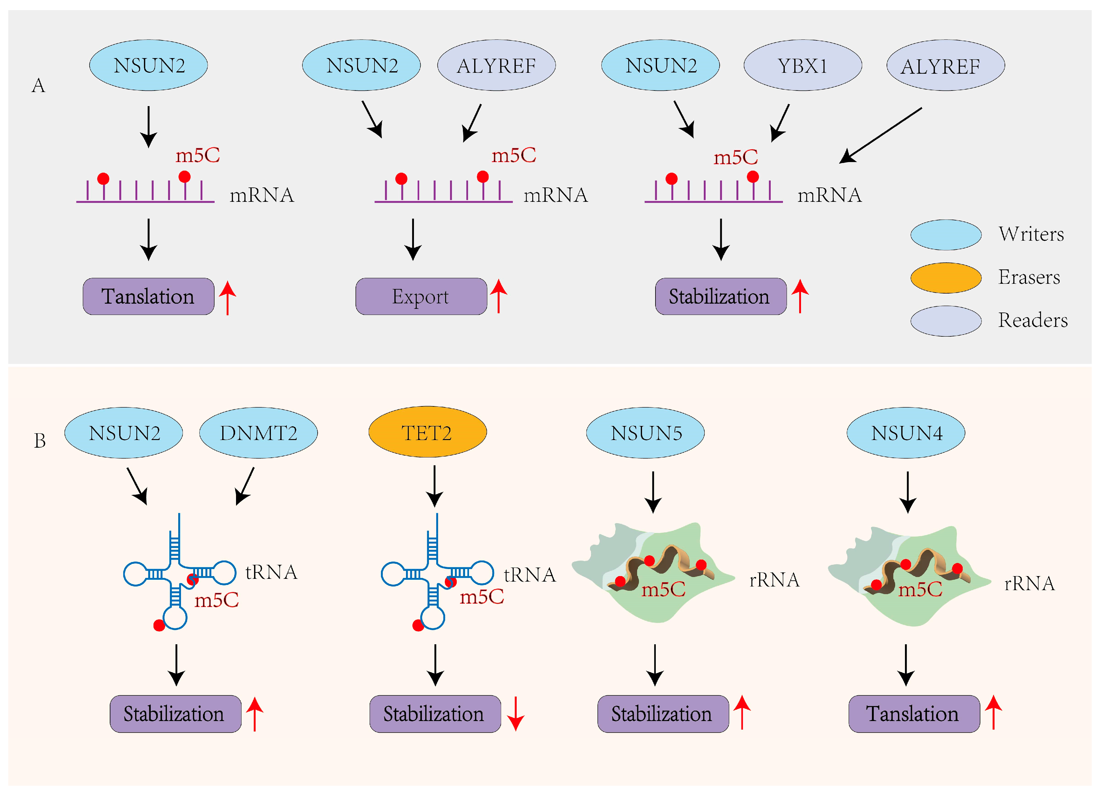
The biological roles of m5C are increasingly recognized in both coding and ncRNAs. Like m6A, m5C modifications regulate RNA metabolism by modulating mRNA translation, stability, and nuclear export, as well as maintaining the structural integrity and function of tRNAs and rRNAs [65].

3.1. m5C in mRNAs

m5C primarily affects mRNA fate through three mechanisms: translation, nuclear export, and stability. Modifications within coding sequences are generally associated with reduced translation efficiency, while those in 3′ UTRs typically enhance translation [66,67]. NSUN2-catalyzed m5C modifications are recognized by ALYREF, which facilitates mRNA export from the nucleus to the cytoplasm [61,68]. Additionally, m5C enhances mRNA stability by promoting interactions with proteins like YBX1, contributing to oncogenic gene expression in cancers such as bladder and cervical cancer [6,69]. Two other parallel studies in humans and zebrafish have also shown that YBX1 regulates m5C-dependent mRNA stability [6,70]. Moreover, ALYREF influences the stability and splicing of mRNAs by binding to the m5C-modified sites on transcripts such as PKM2 [71], RABL6, and TK1 [72] (Figure 2A). In cancer contexts, ALYREF enhances BIRC5 mRNA stability via m5C, driving ovarian tumor progression [73], and upregulates NOTCH1 in an m5C-dependent manner to activate NOTCH signaling and facilitate nasopharyngeal carcinoma metastasis [74].

3.2. m5C in ncRNAs

m5C modifications in ncRNAs, especially in tRNAs and rRNAs, play key roles in RNA stability and translational control [52,54] (Figure 2B). In tRNAs, m5C is mainly found in the variable region and anticodon loop, where it stabilizes the RNA structure and prevents degradation [75]. In humans, mice, and plants, m5C methylation mediated by TRM4/NSUN2 or DNMT2 protects specific tRNAs such as tRNAAsp-GTC and tRNAGly-GCC from oxidative cleavage or exonuclease activity [66,76]. TET2 has also been shown to modulate tRNA function by oxidizing m5C, linking m5C dynamics to translational regulation [54]. In rRNA, NSUN5 modifies 25S rRNA at C2278, and its loss under oxidative stress disrupts local folding and ribosome stability [77]. NSUN4 methylates 12S mitochondrial rRNA, supporting mitoribosome assembly and function [78]. Furthermore, m5C sites have been identified in lncRNAs, where they are thought to influence RNA processing and stability, protein translation, and RNA–protein interactions, though their precise functions remain under investigation [79,80].

4. Detection Methods of RNA m5C Methylation

Although m5C was first identified in RNA as early as the 1970s [81,82], the limited sensitivity and resolution of early technologies restricted its detection, particularly in structured or low-abundance RNAs, thereby impeding transcriptome-wide profiling and precise site-specific characterization. In recent years, rapid advances in high-throughput sequencing, mass spectrometry, and chemical biology have enabled more precise and comprehensive detection of RNA m5C modifications. Current techniques vary in terms of resolution, specificity, throughput, and RNA class compatibility, and often require integrative approaches to yield accurate biological insights.

4.1. RNA Bisulfite Sequencing (RNA-BisSeq)

RNA-BisSeq, originally developed for DNA methylation analysis [83], was later adapted to RNA [84] and remains one of the few methods capable of mapping m5C at single-nucleotide resolution. This approach relies on bisulfite treatment, which deaminates unmethylated cytosines into uracil while preserving m5C intact. Subsequent reverse transcription and sequencing then enable precise mapping of methylated sites (Figure 3A). Despite its specificity, RNA-BisSeq has key limitations: it requires large amounts of high-quality RNA, is sensitive to degradation due to harsh chemical conditions, and is less reliable in structured or low-abundance transcripts [85]. Furthermore, incomplete conversion and mapping ambiguities can generate false positives or negatives, especially in regions with strong secondary structures.

4.2. Immunoprecipitation-Based Techniques

Antibody-based methods such as MeRIP-seq enrich for m5C-modified fragments and offer transcriptome-wide coverage [86] (Figure 3B). However, this method suffers from low resolution and is dependent on antibody specificity and affinity, limiting its ability to precisely localize individual m5C sites [86]. A more refined chemical approach, 5-azacytidine-mediated RNA immunoprecipitation (5-aza-IP), takes advantage of the covalent trapping of methyltransferase–RNA complexes using 5-azacytidine, enabling detection of enzyme-specific m5C sites, such as those deposited by NSUN2 or DNMT2 [87] (Figure 3C). Nevertheless, the cytotoxicity and nonspecific incorporation of 5-aza analogs pose significant challenges. An alternative approach, miCLIP (methylation individual-nucleotide resolution crosslinking and immunoprecipitation), exploits catalytically dead mutants of RNA cytosine methyltransferases (RCMTs) that form an irreversible covalent bond with their m5C substrates. Following partial RNA fragmentation, these RCMT–RNA adducts are enriched by anti-RCMT immunoprecipitation, and library preparation captures the characteristic reverse-transcription stops or misincorporations at the crosslinked site. Sequencing and alignment thus yield single-nucleotide resolution maps of m5C positions [88] (Figure 3D). While highly specific, miCLIP is technically complex and often requires overexpression systems, which may introduce artifacts.

4.3. LC-MS/MS and Nanopore Sequencing

LC-MS/MS (Liquid Chromatography–Tandem Mass Spectrometry) enables quantitative detection of global m5C levels across RNA populations by separating and detecting modified nucleosides (Figure 3E) but lacks positional information and cannot distinguish between RNA species [85]. To address this, third-generation sequencing platforms, such as single-molecule real-time (SMRT) sequencing and nanopore-based direct RNA sequencing, have emerged as powerful tools for native RNA modification detection [89]. These technologies can detect changes in electrical signals or pulse kinetics induced by nucleotide modifications in real time, without the need for chemical conversion or amplification (Figure 3F). Notably, nanopore sequencing enables full-length RNA molecule detection and isoform-specific modification profiling. Although current limitations include elevated error rates, underdeveloped modification-calling algorithms, and challenges in discriminating similar epitranscriptomic marks, this technology provides distinct advantages—particularly when integrated with conventional biochemical approaches in comprehensive analytical pipelines.

5. Molecular Mechanisms of HBV-Related HCC

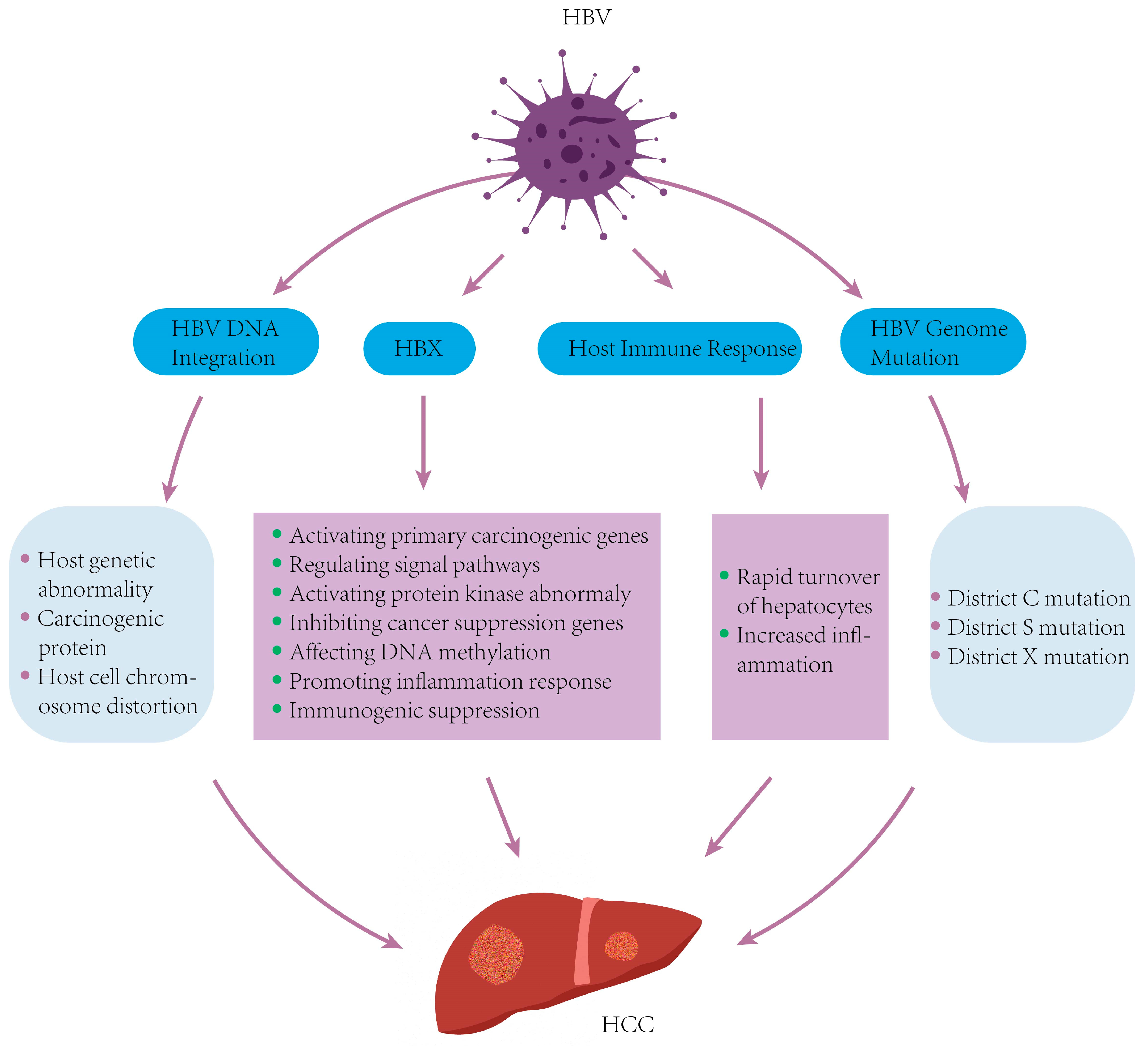
Hepatocellular carcinoma (HCC) is the most frequent primary liver cancer, with chronic HBV infection as a principal etiological factor [90]. HBV induces hepatocarcinogenesis through complex mechanisms involving both direct viral oncogenic effects and virus-mediated alterations of host cell processes [74]. Central to this process are the persistence of covalently cccDNA, integration of HBV DNA into the host genome, accumulation of viral mutations, and the pleiotropic activities of viral proteins—particularly the hepatitis B virus X protein (HBx) [91]. In addition, growing evidence highlights the contribution of epigenetic dysregulation, including DNA methylation and histone modifications, as well as immune evasion and chronic inflammation, in HBV-driven hepatocarcinogenesis [8,9,92].

5.1. HBV Replication Cycle

HBV enters host hepatocytes via sodium taurocholate co-transporting polypeptide (NTCP)-mediated endocytosis [93]. Upon entry, the nucleocapsid is released into the cytoplasm and subsequently transported into the nucleus, where it releases the relaxed circular DNA (rcDNA). The host DNA repair machinery then converts rcDNA into episomal covalently cccDNA, which serves as the transcriptional template for viral RNAs, including pgRNA (encoding core protein and viral polymerase) and subgenomic RNAs (encoding envelope proteins and HBx) [94]. The pgRNA–polymerase complex is encapsidated, followed by reverse transcription to synthesize rcDNA. Mature nucleocapsids either recycle to the nucleus to replenish the cccDNA pool or acquire envelope proteins (HBsAg) in the endoplasmic reticulum to form complete virions for secretion. Notably, the persistence of cccDNA and the oncogenic activity of HBx protein constitute key mechanisms underlying HBV-related HCC pathogenesis [95] (Figure 4).

5.2. HBV DNA Integration into the Host Genome

Integration of HBV DNA into the host genome is a hallmark of chronic infection and is detected in up to 80–90% of HBV-related HCC cases [95]. This process is primarily mediated by host DNA double-strand break repair pathways, including non-homologous end joining (NHEJ) and microhomology-mediated end joining (MMEJ), and typically occurs shortly after infection or during hepatocyte proliferation, when genomic instability and DNA damage are more prevalent [96]. Integrated HBV DNA tends to insert into vulnerable genomic regions, such as CpG islands, telomeric zones, and cancer-associated loci, such as TERT, MLL4, and CCNE1 [97]. These events can lead to insertional mutagenesis, activation of proto-oncogenes, disruption of tumor suppressor genes, and induction of chromosomal instability. In addition to altering gene expression, integrated HBV DNA can generate truncated viral transcripts and chimeric fusion proteins with oncogenic potential [98]. Importantly, integrated HBV DNA is replication-defective and does not contribute to the production of infectious virions, but it continues to express viral proteins—particularly HBx and HBsAg—that can chronically stimulate the immune system, promote immune escape, and contribute to tumor microenvironment remodeling [99]. Such changes may act independently or in synergy to promote tumorigenesis. Current antiviral therapies fail to eliminate integrated HBV DNA or cccDNA, making these elements persistent drivers of liver disease. Therefore, understanding the mechanisms and consequences of HBV DNA integration is essential for guiding efforts to prevent or delay disease progression (Figure 4).

5.3. HBV Genomic Mutations

HBV is a DNA virus with a high mutation rate due to the lack of proofreading activity by its reverse transcriptase. The viral genome includes four open reading frames (ORFs)—S, P, C, and X [100], with mutations in the C, S, and X regions linked to HCC, while those in the P region often relate to antiviral resistance [101]. For instance, the G1896A mutation in the pre-C region generates a premature stop codon in the hepatitis B e antigen (HBeAg) ORF, reducing its expression without hindering viral replication. This leads to persistent infection, exacerbating liver injury and promoting cancer progression [102]. Mutations in the pre-S region can cause the intracellular accumulation of surface proteins in the endoplasmic reticulum (ER), inducing ER stress, oxidative stress, and DNA damage response pathways, which ultimately drive tumor formation. Point mutations, deletions, or insertions in the pre-S sequences further increase HCC risk [103]. Additionally, HBx, encoded by the X region, plays a crucial role in hepatocyte transformation [104]. Mutations like T1753V and A1762T/G1764A in the core promoter region are frequently associated with HCC and may promote tumor progression through altered regulation of genes such as TGF-β1 [105,106] (Figure 4).

5.4. Abnormal Expression of the HBx Gene and Its Encoded Protein

The HBx gene, one of four overlapping ORFs in the HBV genome, encodes the multifunctional HBx protein, a key regulator in HBV pathogenesis [107]. Certain mutations—particularly K130M and V131I—are associated with a 4- to 5-fold increased risk of HCC [108]. HBx can interact directly with host transcription factors and activate intracellular signaling cascades [107]. It promotes HCC development by activating proto-oncogenes such as RAS, C-FOS, and C-MYC, driving cell proliferation and differentiation [109]. HBx also modulates pathways like JAK-STAT, Notch, and MAPK, enhancing cancer progression [110,111,112]. Additionally, HBx contributes to tumorigenesis by inhibiting tumor suppressors like p53 [113], altering DNA methylation [114], inducing inflammation [115], and suppressing host immune responses [105] (Figure 4).

6. The Role of m5C in the HBV Life Cycle

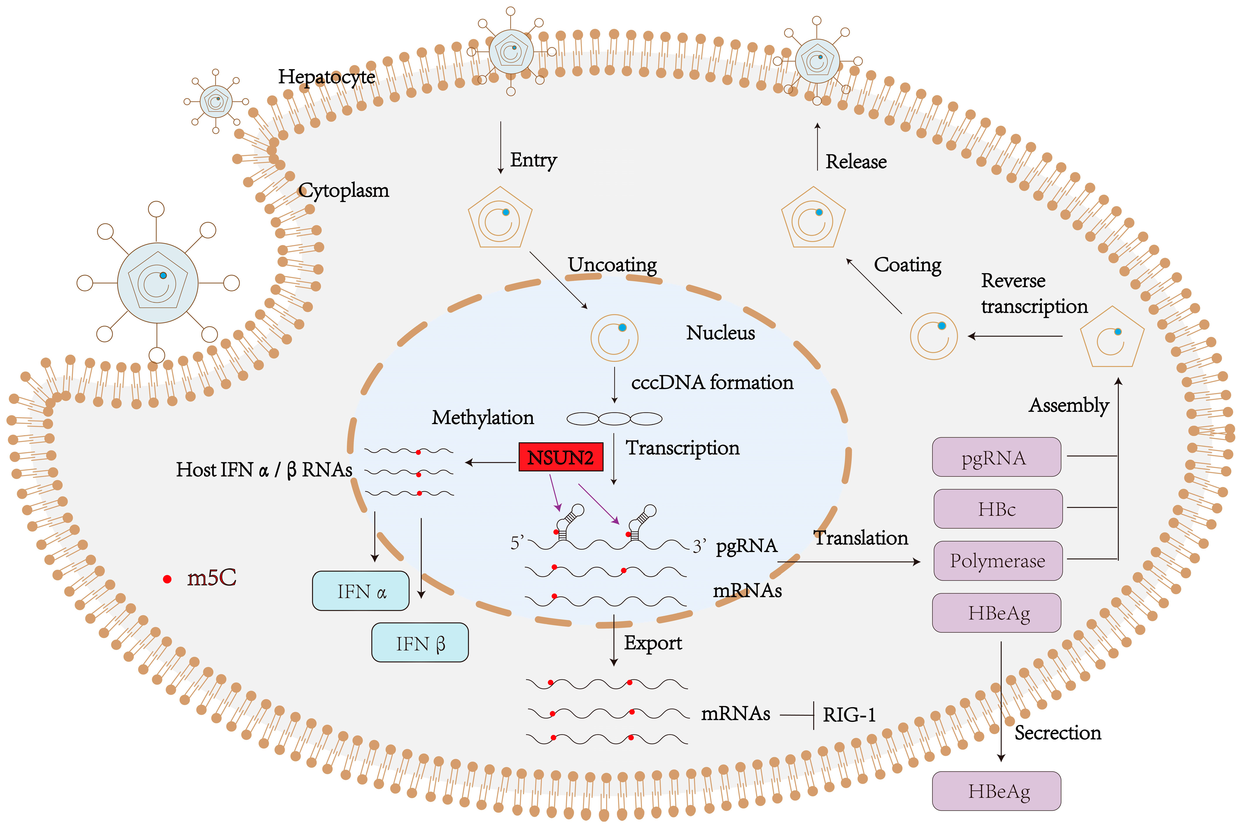
Epitranscriptomic modifications regulate critical phases of viral infection. In HBV, the pgRNA functions dually as the reverse transcription template and as the bicistronic mRNA encoding core and polymerase proteins. While m6A modifications on pgRNA are known to enhance encapsidation, reverse transcription, and immune evasion [116,117,118], m5C has emerged as another pivotal regulator of HBV replication and host interactions.

6.1. m5C Enhances pgRNA Stability and Encapsidation

m5C modification plays a key role in maintaining the stability of HBV pgRNA, the essential intermediate responsible for both protein translation and the reverse transcription of the viral genome. High-resolution bisulfite sequencing has identified multiple m5C-modified cytosines within the pgRNA, particularly at C131 in the ε element and C2017 in the 3′ UTR [119]. These sites are evolutionarily conserved and are catalyzed by NSUN2 [119]. Loss of NSUN2, via genetic knockdown or knockout, significantly diminishes HBV RNA levels, viral DNA synthesis, and secretion of HBeAg. Furthermore, site-directed mutagenesis of C131 or C2017 to uracil impairs RNA stability, underscoring the functional importance of these modifications [119]. Mechanistically, m5C appears to enhance the association between pgRNA and viral core proteins, facilitating efficient encapsidation [120]. Stability assessments further show that m5C-containing pgRNAs exhibit a longer half-life compared to unmethylated variants, supporting the role of m5C in protecting viral RNA from degradation (Figure 5).

6.2. m5C in Element (ε) Regulates HBV Reverse Transcription

Reverse transcription of pgRNA into HBV DNA is initiated at the ε stem-loop structure located at the 5′ end of the pgRNA. This structure is required for viral polymerase recognition and serves as the initiation site for (-)-strand DNA synthesis. Several cytosines in ε are subject to m5C modification in an NSUN2-dependent manner. Disruption of ε methylation, either by mutating the cytosines or NSUN2 depletion, abolishes reverse transcription, leading to the formation of non-infectious empty capsids. Reintroducing m5C via NSUN2 restoration rescues DNA synthesis and mature virion production, demonstrating that m5C marks act as essential molecular cues for effective reverse transcription and virion maturation [120]. Moreover, m5C marks in ε are functionally complementary to m6A marks in the same region [120], suggesting that a complex epitranscriptomic network regulates the RNA structural landscape to orchestrate viral life-cycle events (Figure 5).

6.3. m5C Modulates Innate Immune Recognition and Viral Evasion

Unlike many DNA viruses, HBV primarily triggers host innate immunity through RNA-sensing mechanisms rather than DNA-sensing pathways. This is largely because hepatocytes express low levels of key DNA-sensing factors, such as the stimulator of interferon genes (STING), rendering RNA pattern recognition receptors—particularly retinoic acid-inducible gene I (RIG-I)—the principal sensors of HBV infection [121]. Our recent studies have revealed that m5C methylation of HBV RNA plays a critical role not only in promoting viral gene expression but also in helping HBV evade host innate immunity [122]. Specifically, we identified a conserved m5C site at nucleotide 1291 on the HBV transcript, which serves dual functions in regulating viral mRNA export and interfering with immune sensing. Mechanistically, m5C at nt1291 facilitates the recruitment of the nuclear export adaptor ALYREF, thereby promoting the efficient cytoplasmic export of HBV mRNA. This enhances the translation of viral proteins, notably HBx, and contributes to productive viral replication. Loss of this methylation mark—either through NSUN2 knockdown or site-directed mutation—disrupts ALYREF binding, impairs HBx translation, and reduces viral propagation. More importantly, m5C at nt1291 also plays an essential role in antagonizing the host RNA sensor RIG-I. We demonstrated that methylation at this site inhibits the binding of RIG-I to HBV RNA, thereby preventing the downstream activation of the mitochondrial antiviral-signaling protein (MAVS) pathway and blocking interferon-β (IFN-β) production. In the absence of m5C, RIG-I recognition is restored, leading to a significant upregulation of IFN-β and reactivation of the host antiviral response. In addition to modifying its own RNA, HBV also actively modulates host m5C epitranscriptomic machinery. We found that HBV infection downregulates the m5C methyltransferase NSUN2 in hepatocytes, leading to reduced m5C methylation on host interferon-related host mRNAs such as interferon beta 1 (IFNB1). This reduction compromises the stability and expression of these transcripts, thereby attenuating type I interferon production and facilitating viral persistence. Collectively, these findings reveal a dual role for NSUN2-mediated m5C methylation in HBV pathogenesis: enhancing viral mRNA export and translation while simultaneously dampening host antiviral signaling. Through this epitranscriptomic strategy, HBV coordinates the manipulation of both viral and host transcriptomes, highlighting m5C as a promising therapeutic target in chronic hepatitis B (Figure 5).

7. m5C Methylation in HBV-Related HCC and Associated Liver Diseases

m5C plays a critical role in the initiation and progression of various tumors. Transcriptome-wide analyses reveal distinct m5C patterns in HCC versus adjacent non-tumor liver, suggesting a malignancy-specific epitranscriptomic signature [123,124]. One study revealed that the m5C reader ALYREF modulates methylation of key transcripts to promote HCC progression [125]. Subsequent research showed that ALYREF binds m5C sites on EGFR mRNA, enhancing transcript stability and activating downstream STAT3 signaling to further drive tumor growth [126]. Notably, Sun et al. reported that NSUN2 deposits m5C on the lncRNA H19, increasing its stability; m5C-modified H19 then recruits the oncoprotein G3BP1, leading to MYC accumulation and poor differentiation in HBV-related HCC [127].
Beyond cancer, m5C also modulates other HBV-related liver pathologies. In fibrogenesis, YBX1 knockout in hepatic progenitor cells reduces hepatic stellate cell activation and mitigates liver fibrosis in mice [128], suggesting that m5C regulators may influence HBV-associated fibrotic processes. To date, however, there are no direct studies reporting m5C regulation in HBV-related hepatitis, cirrhosis, or liver failure. Nevertheless, evidence from other liver disease contexts highlights broader roles of m5C. For instance, TET2 modulates alcohol-induced liver injury by regulating Srebp1 mRNA stability [129], and ALYREF cooperates with NSUN2 to promote m5C-dependent export and translation of CDKN1A mRNA during adipogenesis in non-alcoholic fatty liver disease [130]. These findings, while not directly HBV-related, provide valuable clues suggesting that epitranscriptomic dysregulation of m5C may broadly contribute to liver disease progression and thus warrants further investigation in HBV-associated settings.
Currently, the role of RNA m5C modification in HCC is becoming increasingly well-defined, making methylation-related proteins promising therapeutic targets for HCC treatment. Sorafenib, a targeted therapy for advanced HCC, has limited efficacy due to drug resistance. However, combining sorafenib with NSUN1 siRNA significantly enhances HCC sensitivity to sorafenib [131]. Qu et al. identified that targeting the ALKBH5 surface represents a promising therapeutic strategy for HBV-induced HCC [132]. As our understanding of m5C regulators in HBV-driven liver disease deepens, targeting writer, eraser, or reader proteins may yield novel antiviral and anti-tumor strategies.

8. Crosstalk Between m5C and Other Modifications in the HBV Life Cycle

RNA modifications often coexist on the same transcript and cooperate to form an intricate “epitranscriptomic code.” In HBV, the most abundant internal modification, m6A, is enriched at the ε packaging signal and within the HBx ORF, where it stabilizes pgRNA, enhances encapsidation, and suppresses RIG-I-mediated immune sensing via YTHDF readers; additionally, m6A modification of host PTEN mRNA promotes HBx-mediated immune evasion and hepatocarcinogenesis [133]. Beyond m6A, other modifications also contribute to viral RNA regulation: ac4C boosts internal ribosome entry site (IRES)-driven translation in enteroviruses, while Ψ stabilizes RNA secondary structures and improves translational efficiency in various viral contexts.
In the case of HBV, it remains unclear whether m6A, ac4C, and Ψ co-localize with m5C on the same transcripts, or whether they exert synergistic or antagonistic effects. Elucidating such interactions requires high-resolution mapping of RNA modifications. Emerging technologies such as nanopore direct RNA sequencing and crosslinking-based methods like miCLIP offer promising tools to uncover combinatorial modification patterns. These approaches could help delineate how m5C cooperates or competes with other modifications to regulate viral RNA stability, nuclear export, immune evasion, and replication. Hepatitis D virus (HDV), a satellite virus of HBV that significantly exacerbates liver disease pathogenesis, is known to harbor epitranscriptomic modifications such as m6A and A-to-I RNA editing. However, whether HDV RNA contains m5C modifications remains unexplored, presenting an important avenue for future research [134,135].

9. Comparative Insights: m5C in Other RNA and DNA Viruses

m5C is a widespread and conserved modification found across diverse RNA viruses, with virus-specific functions and regulatory contexts. In enterovirus 71 (EV71), NSUN2-mediated m5C installation within the 5′ UTR enhances RNA stability and facilitates IRES-dependent translation [11]. In human immunodeficiency virus type 1 (HIV-1), the genomic RNA is heavily methylated (approximately 11–14 m5C-modified sites), which promotes the export of unspliced transcripts, ribosome engagement, and alternative splicing regulation [136]. For severe acute respiratory syndrome coronavirus 2 (SARS-CoV-2), m5C marks are distributed across the viral genome in an NSUN2-dependent manner; NSUN2 knockout leads to global hypomethylation of viral RNA, resulting in increased RNA stability, elevated viral loads, and aggravated lung pathology in infected mice [137]. MLV RNAs bear an exceptionally high level of m5C epitranscriptomic modifications, and downregulation of the m5C writer NSUN2 inhibits MLV replication [138]. In hepatitis C virus (HCV), m5C modification within the NS5A coding region enhances RNA stability and replication efficiency, with YBX1 acting as a critical reader protein that mediates these effects [139]. Additionally, a growing body of evidence suggests that other RNA viruses—including Dengue virus, Zika virus, respiratory syncytial virus (RSV), and vesicular stomatitis virus (VSV)—also exploit NSUN2-dependent m5C modifications to modulate viral replication and evade host immune responses, although the specific methylation sites and interacting proteins vary across viruses [140,141]. Collectively, these findings highlight both the evolutionary conservation and the functional divergence of m5C regulation in RNA viruses. While NSUN2 is a common methyltransferase utilized across pathogens, the biological consequences of m5C—ranging from enhanced translation and RNA stability to immune modulation and RNA processing—are highly virus- and context-specific. These comparative insights further underscore the distinct and multifaceted role of m5C in HBV pathogenesis and its potential as a therapeutic target (Table 2).

10. Clinical and Therapeutic Perspectives

10.1. Genotype- and Host-Dependent Variation in m5C

HBV comprises at least ten major genotypes (A–J), which differ in their geographic prevalence, disease progression, and treatment response. It remains to be determined whether differential HBV genotypes harbor distinct m5C landscapes or NSUN2 affinities. Genotype-specific pgRNA secondary structures may influence the accessibility of m5C writers or readers, potentially affecting viral replication efficiency and immune recognition. Likewise, vaccine non-responders may possess altered epitranscriptomic profiles that compromise viral antigen expression, while antiviral-resistant strains may harbor polymerase mutations near m5C sites, potentially interfering with methylation or disrupting reader recruitment. Comparative methylome profiling across diverse HBV genotypes and patient cohorts will be essential to elucidate these genotype- and host-specific variations and their implications for disease progression and therapeutic response.

10.2. m5C in HBV-Driven Hepatocarcinogenesis

Dysregulation of the m5C machinery has been implicated in hepatocarcinogenesis. In HCC, NSUN2 is frequently overexpressed and associated with poor prognosis, enhanced cellular proliferation, and impaired antiviral defense [144]. As an m5C reader, ALYREF stabilizes oncogenic transcripts—such as EGFP—thereby promoting downstream signaling pathways like STAT3, which are known to drive tumor progression [126]. Moreover, persistent NSUN2-mediated methylation of host immune-related mRNAs, such as TREX2 and IRF3, may undermine DNA sensing and interferon responses, creating an immunosuppressive microenvironment that favors malignant transformation [140]. In the setting of chronic HBV infection, sustained m5C activity may therefore facilitate malignant transformation by both subverting innate immunity and promoting proliferative signaling.

10.3. Targeting the m5C Machinery for Therapy

Given the dual roles of m5C in viral persistence and tumorigenesis, the RNA methylation machinery presents an attractive therapeutic target. Small-molecule inhibitors of NSUN2, or agents that disrupt ALYREF–RNA interactions, may destabilize HBV pgRNA and restore immune sensing. Conversely, enhancing the activity of demethylases such as TET2 might promote the removal of m5C from viral transcripts, accelerating their degradation. Cutting-edge technologies—such as dCas13-based epitranscriptomic editors—could be deployed to selectively erase m5C marks at specific HBV RNA loci. Finally, combining m5C-targeted strategies with standard-of-care nucleoside analogs or immunotherapies may yield synergistic effects, suppressing both viral replication and HCC progression. Nonetheless, several challenges must be addressed—chief among them the need for high specificity toward viral versus host transcripts, adequate delivery across hepatocyte membranes, and minimal off-target effects. Future work should focus on developing selective m5C modulators and validating their efficacy in relevant in vivo models.

11. Conclusions and Outlook

RNA m5C has emerged as a pivotal epitranscriptomic mark that governs RNA stability, processing, localization, and immune recognition in both eukaryotic cells and a growing array of RNA viruses. In the context of HBV, m5C—catalyzed primarily by the methyltransferase NSUN2—plays a multifaceted role in viral replication and host–pathogen interactions. Specific m5C modifications at conserved loci on the pgRNA, including C131, multiple residues within the ε encapsidation signal, C2017 in the 3′ UTR, and nt1291, contribute to enhanced RNA stability, efficient reverse transcription, and evasion of innate immune sensing. These effects are mediated in part through the recruitment of m5C readers such as ALYREF, which facilitate viral mRNA export and translational efficiency. Intriguingly, HBV also actively suppresses NSUN2 expression in infected hepatocytes, leading to reduced m5C methylation on host interferon-related transcripts such as IFNB1, thereby dampening type I interferon responses and promoting viral persistence.
Despite these advances, several challenges and opportunities remain. First, the interplay among m5C writers, erasers, and readers has yet to be mapped comprehensively in HBV and host transcripts; elucidating these regulatory networks and how they interact with other RNA modifications, such as m6A, ac4C, and Ψ, is essential for decoding the “epitranscriptomic code” that governs viral RNA fate. Second, genotype-dependent differences in HBV RNA structure may influence m5C deposition and function—a question that demands comparative methylome analyses across HBV genotypes and clinical cohorts. Third, translating mechanistic insights into therapies will require specific, cell-permeable inhibitors of NSUN2 or ALYREF, as well as tools for targeted epitranscriptomic editing (e.g., dCas13 fusions). Ultimately, validating the role of m5C in HBV pathogenesis will require robust in vivo models—including humanized liver mice and patient-derived organoids—alongside longitudinal patient cohort studies that link epitranscriptomic patterns to disease progression, treatment outcomes, and HCC risk.
In summary, m5C functions as both a facilitator of HBV replication and a suppressor of host immune defenses, placing it at the crossroads of viral persistence and oncogenesis. As our understanding of epitranscriptomic regulation deepens, m5C is poised to become not only a biomarker of HBV disease activity but also a novel therapeutic target. A multidisciplinary approach—integrating RNA biology, virology, immunology, and drug development—will be required to unlock the full potential of m5C-based interventions in chronic hepatitis B and HBV-related liver cancer.

Author Contributions

Conceptualization, X.Z. (Xuliu Zhou), W.G., F.Z. and H.H.; writing and editing, X.Z. (Xuliu Zhou), Y.H., X.Z. (Xueyan Zhang), W.G., F.Z. and H.H.; funding acquisition, W.G., F.Z. and H.H. All authors have read and agreed to the published version of the manuscript.

Funding

This work was supported by the Natural Science Foundation of Hubei Province [2024AFB1065, 2025AFB976], the Hubei Jiangxia Laboratory Biosafety Key R&D Project [JXBS013], the Hubei Province Young Science and Technology Talent Morning Light Lift Project [202415], the National Key Research and Development Program [2024YFC2309400], and the Strategic Priority Research Program of the Chinese Academy of Sciences [XDB0490000].

Institutional Review Board Statement

Not applicable.

Informed Consent Statement

Not applicable.

Data Availability Statement

No new data were created or analyzed in this study. Data sharing is not applicable to this article.

Conflicts of Interest

The authors declare no conflicts of interest.

References

  1. Yardeni, D.; Ghany, M.G. Review article: Hepatitis B-current and emerging therapies. Aliment. Pharmacol. Ther. 2022, 55, 805–819. [Google Scholar] [CrossRef] [PubMed]
  2. Li, H.; Yan, L.; Shi, Y.; Lv, D.; Shang, J.; Bai, L.; Tang, H. Hepatitis B Virus Infection: Overview. Adv. Exp. Med. Biol. 2020, 1179, 1–16. [Google Scholar] [CrossRef] [PubMed]
  3. Hu, J.; Protzer, U.; Siddiqui, A. Revisiting Hepatitis B Virus: Challenges of Curative Therapies. J. Virol. 2019, 93, e01032-19. [Google Scholar] [CrossRef] [PubMed]
  4. Boccaletto, P.; Stefaniak, F.; Ray, A.; Cappannini, A.; Mukherjee, S.; Purta, E.; Kurkowska, M.; Shirvanizadeh, N.; Destefanis, E.; Groza, P.; et al. MODOMICS: A database of RNA modification pathways. Nucleic Acids Res. 2022, 50, D231–D235. [Google Scholar] [CrossRef] [PubMed]
  5. Roundtree, I.A.; Evans, M.E.; Pan, T.; He, C. Dynamic RNA Modifications in Gene Expression Regulation. Cell 2017, 169, 1187–1200. [Google Scholar] [CrossRef]
  6. Chen, X.; Li, A.; Sun, B.F.; Yang, Y.; Han, Y.N.; Yuan, X.; Chen, R.X.; Wei, W.S.; Liu, Y.; Gao, C.C.; et al. 5-methylcytosine promotes pathogenesis of bladder cancer through stabilizing mRNAs. Nat. Cell Biol. 2019, 21, 978–990. [Google Scholar] [CrossRef]
  7. Chen, Z.; Huang, L.; Wang, K.; Zhang, L.; Zhong, X.; Yan, Z.; Liu, B.; Zhu, P. rtcisE2F promotes the self-renewal and metastasis of liver tumor-initiating cells via N6-methyladenosine-dependent E2F3/E2F6 mRNA stability. Sci. China Life Sci. 2022, 65, 1840–1854. [Google Scholar] [CrossRef]
  8. Zheng, Z.H.; Zhang, G.L.; Jiang, R.F.; Hong, Y.Q.; Zhang, Q.Y.; He, J.P.; Liu, X.R.; Yang, Z.S.; Yang, L.; Jiang, X.; et al. METTL3 is essential for normal progesterone signaling during embryo implantation via m6A-mediated translation control of progesterone receptor. Proc. Natl. Acad. Sci. USA 2023, 120, e2214684120. [Google Scholar] [CrossRef]
  9. Qiao, Y.; Sun, Q.; Chen, X.; He, L.; Wang, D.; Su, R.; Xue, Y.; Sun, H.; Wang, H. Nuclear m6A reader YTHDC1 promotes muscle stem cell activation/proliferation by regulating mRNA splicing and nuclear export. eLife 2023, 12, e82703. [Google Scholar] [CrossRef]
  10. Li, J.; Zhang, L. The emerging role of m5C modification in viral infection. Virology 2025, 610, 110606. [Google Scholar] [CrossRef]
  11. Liu, L.; Chen, Z.; Zhang, K.; Hao, H.; Ma, L.; Liu, H.; Yu, B.; Ding, S.; Zhang, X.; Zhu, M.; et al. NSUN2 mediates distinct pathways to regulate enterovirus 71 replication. Virol. Sin. 2024, 39, 574–586. [Google Scholar] [CrossRef]
  12. Zhang, Y.; Zhang, L.S.; Dai, Q.; Chen, P.; Lu, M.; Kairis, E.L.; Murugaiah, V.; Xu, J.; Shukla, R.K.; Liang, X.; et al. 5-methylcytosine (m5C) RNA modification controls the innate immune response to virus infection by regulating type I interferons. Proc. Natl. Acad. Sci. USA 2022, 119, e2123338119. [Google Scholar] [CrossRef] [PubMed]
  13. Ye, L.; Yao, X.; Xu, B.; Chen, W.; Lou, H.; Tong, X.; Fang, S.; Zou, R.; Hu, Y.; Wang, Z.; et al. RNA epigenetic modifications in ovarian cancer: The changes, chances, and challenges. Wiley Interdiscip. Rev. RNA 2023, 14, e1784. [Google Scholar] [CrossRef] [PubMed]
  14. Winans, S.; Beemon, K. m5C Goes Viral. Cell Host Microbe 2019, 26, 154–155. [Google Scholar] [CrossRef]
  15. Zhang, X.; Peng, Q.; Wang, L. N6-methyladenosine modification-a key player in viral infection. Cell. Mol. Biol. Lett. 2023, 28, 78. [Google Scholar] [CrossRef]
  16. Zhu, M.; Huang, F.; Sun, H.; Liu, K.; Chen, Z.; Yu, B.; Hao, H.; Liu, H.; Ding, S.; Zhang, X.; et al. Characterization of ACTN4 as a novel antiviral target against SARS-CoV-2. Signal Transduct. Target. Ther. 2024, 9, 243. [Google Scholar] [CrossRef]
  17. Meyer, K.D.; Jaffrey, S.R. The dynamic epitranscriptome: N6-methyladenosine and gene expression control. Nat. Rev. Mol. Cell Biol. 2014, 15, 313–326. [Google Scholar] [CrossRef]
  18. Meng, Y.; Shu, Z.; Wang, X.; Hong, L.; Wang, B.; Jiang, J.; He, K.; Cao, Q.; Shi, F.; Wang, H.; et al. Hepatitis B Virus-Mediated m6A Demethylation Increases Hepatocellular Carcinoma Stemness and Immune Escape. Mol. Cancer Res. 2024, 22, 642–655. [Google Scholar] [CrossRef] [PubMed]
  19. Li, S.; Mason, C.E. The pivotal regulatory landscape of RNA modifications. Annu. Rev. Genom. Hum. Genet. 2014, 15, 127–150. [Google Scholar] [CrossRef]
  20. Zhang, X.; Qin, S.; Huang, F.; Liu, H.; Wang, J.; Chen, Z.; Hao, H.; Ding, S.; Liu, L.; Yu, B.; et al. N4-acetylcytidine coordinates with NP1 and CPSF5 to facilitate alternative RNA processing during the replication of minute virus of canines. Nucleic Acids Res. 2025, 53, gkaf229. [Google Scholar] [CrossRef]
  21. Schiffers, S.; Oberdoerffer, S. ac4C: A fragile modification with stabilizing functions in RNA metabolism. RNA 2024, 30, 583–594. [Google Scholar] [CrossRef]
  22. Hao, H.; Liu, W.; Miao, Y.; Ma, L.; Yu, B.; Liu, L.; Yang, C.; Zhang, K.; Chen, Z.; Yang, J.; et al. N4-acetylcytidine regulates the replication and pathogenicity of enterovirus 71. Nucleic Acids Res. 2022, 50, 9339–9354. [Google Scholar] [CrossRef] [PubMed]
  23. Zheng, Q.; Gan, H.; Yang, F.; Yao, Y.; Hao, F.; Hong, L.; Jin, L. Cytoplasmic m1A reader YTHDF3 inhibits trophoblast invasion by downregulation of m1A-methylated IGF1R. Cell Discov. 2020, 6, 12. [Google Scholar] [CrossRef]
  24. Yan, Q.; Zhou, J.; Wang, Z.; Ding, X.; Ma, X.; Li, W.; Jia, X.; Gao, S.J.; Lu, C. NAT10-dependent N4-acetylcytidine modification mediates PAN RNA stability, KSHV reactivation, and IFI16-related inflammasome activation. Nat. Commun. 2023, 14, 6327. [Google Scholar] [CrossRef]
  25. Tsai, K.; Jaguva Vasudevan, A.A.; Martinez Campos, C.; Emery, A.; Swanstrom, R.; Cullen, B.R. Acetylation of Cytidine Residues Boosts HIV-1 Gene Expression by Increasing Viral RNA Stability. Cell Host Microbe 2020, 28, 306–312.e306. [Google Scholar] [CrossRef]
  26. Pereira-Montecinos, C.; Valiente-Echeverría, F.; Soto-Rifo, R. Epitranscriptomic regulation of viral replication. Biochim. Biophys. Acta Gene Regul. Mech. 2017, 1860, 460–471. [Google Scholar] [CrossRef]
  27. Levi, O.; Arava, Y.S. Pseudouridine-mediated translation control of mRNA by methionine aminoacyl tRNA synthetase. Nucleic Acids Res. 2021, 49, 432–443. [Google Scholar] [CrossRef]
  28. Carlile, T.M.; Martinez, N.M.; Schaening, C.; Su, A.; Bell, T.A.; Zinshteyn, B.; Gilbert, W.V. mRNA structure determines modification by pseudouridine synthase 1. Nat. Chem. Biol. 2019, 15, 966–974. [Google Scholar] [CrossRef] [PubMed]
  29. Martinez, N.M.; Su, A.; Burns, M.C.; Nussbacher, J.K.; Schaening, C.; Sathe, S.; Yeo, G.W.; Gilbert, W.V. Pseudouridine synthases modify human pre-mRNA co-transcriptionally and affect pre-mRNA processing. Mol. Cell 2022, 82, 645–659.e649. [Google Scholar] [CrossRef]
  30. Li, S.; Feng, T.; Liu, Y.; Yang, Q.; Song, A.; Wang, S.; Xie, J.; Zhang, J.; Yuan, B.; Sun, Z. m1A inhibition fuels oncolytic virus-elicited antitumor immunity via downregulating MYC/PD-L1 signaling. Int. J. Oral Sci. 2024, 16, 36. [Google Scholar] [CrossRef] [PubMed]
  31. Filipowicz, W. Functions of the 5′-terminal m7G cap in eukaryotic mRNA. FEBS Lett. 1978, 96, 1–11. [Google Scholar] [CrossRef]
  32. Kiriakidou, M.; Tan, G.S.; Lamprinaki, S.; De Planell-Saguer, M.; Nelson, P.T.; Mourelatos, Z. An mRNA m7G cap binding-like motif within human Ago2 represses translation. Cell 2007, 129, 1141–1151. [Google Scholar] [CrossRef] [PubMed]
  33. Ma, J.; Han, H.; Huang, Y.; Yang, C.; Zheng, S.; Cai, T.; Bi, J.; Huang, X.; Liu, R.; Huang, L.; et al. METTL1/WDR4-mediated m7G tRNA modifications and m7G codon usage promote mRNA translation and lung cancer progression. Mol. Ther. 2021, 29, 3422–3435. [Google Scholar] [CrossRef] [PubMed]
  34. Létoquart, J.; Huvelle, E.; Wacheul, L.; Bourgeois, G.; Zorbas, C.; Graille, M.; Heurgué-Hamard, V.; Lafontaine, D.L. Structural and functional studies of Bud23-Trm112 reveal 18S rRNA N7-G1575 methylation occurs on late 40S precursor ribosomes. Proc. Natl. Acad. Sci. USA 2014, 111, E5518–E5526. [Google Scholar] [CrossRef]
  35. Werner, M.; Purta, E.; Kaminska, K.H.; Cymerman, I.A.; Campbell, D.A.; Mittra, B.; Zamudio, J.R.; Sturm, N.R.; Jaworski, J.; Bujnicki, J.M. 2′-O-ribose methylation of cap2 in human: Function and evolution in a horizontally mobile family. Nucleic Acids Res. 2011, 39, 4756–4768. [Google Scholar] [CrossRef] [PubMed]
  36. Belanger, F.; Stepinski, J.; Darzynkiewicz, E.; Pelletier, J. Characterization of hMTr1, a human Cap1 2′-O-ribose methyltransferase. J. Biol. Chem. 2010, 285, 33037–33044. [Google Scholar] [CrossRef]
  37. Drazkowska, K.; Tomecki, R.; Warminski, M.; Baran, N.; Cysewski, D.; Depaix, A.; Kasprzyk, R.; Kowalska, J.; Jemielity, J.; Sikorski, P.J. 2′-O-Methylation of the second transcribed nucleotide within the mRNA 5′ cap impacts the protein production level in a cell-specific manner and contributes to RNA immune evasion. Nucleic Acids Res. 2022, 50, 9051–9071. [Google Scholar] [CrossRef]
  38. Lu, Y.; Yang, L.; Feng, Q.; Liu, Y.; Sun, X.; Liu, D.; Qiao, L.; Liu, Z. RNA 5-Methylcytosine Modification: Regulatory Molecules, Biological Functions, and Human Diseases. Genom. Proteom. Bioinform. 2024, 22, qzae063. [Google Scholar] [CrossRef]
  39. Bourgeois, G.; Ney, M.; Gaspar, I.; Aigueperse, C.; Schaefer, M.; Kellner, S.; Helm, M.; Motorin, Y. Eukaryotic rRNA Modification by Yeast 5-Methylcytosine-Methyltransferases and Human Proliferation-Associated Antigen p120. PLoS ONE 2015, 10, e0133321. [Google Scholar] [CrossRef]
  40. Xu, Y.; Qi, S.; Zhang, G.; Liu, D.; Xu, D.; Qin, T.; Cheng, Q.; Kang, H.; Hu, B.; Huang, Z. One-pot ligation of multiple mRNA fragments on dsDNA splint advancing regional modification and translation. Nucleic Acids Res. 2025, 53, gkae1280. [Google Scholar] [CrossRef]
  41. Trixl, L.; Lusser, A. The dynamic RNA modification 5-methylcytosine and its emerging role as an epitranscriptomic mark. Wiley Interdiscip. reviews. RNA 2019, 10, e1510. [Google Scholar] [CrossRef]
  42. Goll, M.G.; Kirpekar, F.; Maggert, K.A.; Yoder, J.A.; Hsieh, C.L.; Zhang, X.; Golic, K.G.; Jacobsen, S.E.; Bestor, T.H. Methylation of tRNA Asp by the DNA methyltransferase homolog Dnmt2. Science 2006, 311, 395–398. [Google Scholar] [CrossRef] [PubMed]
  43. Chen, H.; Yang, H.; Zhu, X.; Yadav, T.; Ouyang, J.; Truesdell, S.S.; Tan, J.; Wang, Y.; Duan, M.; Wei, L.; et al. m5C modification of mRNA serves a DNA damage code to promote homologous recombination. Nat. Commun. 2020, 11, 2834. [Google Scholar] [CrossRef] [PubMed]
  44. Xue, S.; Xu, H.; Sun, Z.; Shen, H.; Chen, S.; Ouyang, J.; Zhou, Q.; Hu, X.; Cui, H. Depletion of TRDMT1 affects 5-methylcytosine modification of mRNA and inhibits HEK293 cell proliferation and migration. Biochem. Biophys. Res. Commun. 2019, 520, 60–66. [Google Scholar] [CrossRef] [PubMed]
  45. Li, J.; Li, H.; Long, T.; Dong, H.; Wang, E.D.; Liu, R.J. Archaeal NSUN6 catalyzes m5C72 modification on a wide-range of specific tRNAs. Nucleic Acids Res. 2019, 47, 2041–2055. [Google Scholar] [CrossRef]
  46. Squires, J.E.; Patel, H.R.; Nousch, M.; Sibbritt, T.; Humphreys, D.T.; Parker, B.J.; Suter, C.M.; Preiss, T. Widespread occurrence of 5-methylcytosine in human coding and non-coding RNA. Nucleic Acids Res. 2012, 40, 5023–5033. [Google Scholar] [CrossRef]
  47. Gonskikh, Y.; Tirrito, C.; Bommisetti, P.; Mendoza-Figueroa, M.S.; Stoute, J.; Kim, J.; Wang, Q.; Song, Y.; Liu, K.F. Spatial regulation of NSUN2-mediated tRNA m5C installation in cognitive function. Nucleic Acids Res. 2025, 53, gkae1169. [Google Scholar] [CrossRef]
  48. Dominissini, D.; Rechavi, G. 5-methylcytosine mediates nuclear export of mRNA. Cell Res. 2017, 27, 717–719. [Google Scholar] [CrossRef]
  49. Shinoda, S.; Kitagawa, S.; Nakagawa, S.; Wei, F.Y.; Tomizawa, K.; Araki, K.; Araki, M.; Suzuki, T.; Suzuki, T. Mammalian NSUN2 introduces 5-methylcytidines into mitochondrial tRNAs. Nucleic Acids Res. 2019, 47, 8734–8745. [Google Scholar] [CrossRef] [PubMed]
  50. Li, P.; Ying, S.; Zou, Y.; Wang, X.; Zhang, R.; Huang, C.; Dai, M.; Xu, K.; Feng, G.; Li, X.; et al. NSun2-Mediated tsRNAs Alleviate Liver Fibrosis via FAK Dephosphorylation. Cell Prolif. 2025, e70058. [Google Scholar] [CrossRef]
  51. Li, M.; Tao, Z.; Zhao, Y.; Li, L.; Zheng, J.; Li, Z.; Chen, X. 5-methylcytosine RNA methyltransferases and their potential roles in cancer. J. Transl. Med. 2022, 20, 214. [Google Scholar] [CrossRef]
  52. Chen, Y.S.; Yang, W.L.; Zhao, Y.L.; Yang, Y.G. Dynamic transcriptomic m5C and its regulatory role in RNA processing. Wiley Interdiscip. Rev. RNA 2021, 12, e1639. [Google Scholar] [CrossRef]
  53. Shen, Q.; Zhang, Q.; Shi, Y.; Shi, Q.; Jiang, Y.; Gu, Y.; Li, Z.; Li, X.; Zhao, K.; Wang, C.; et al. Tet2 promotes pathogen infection-induced myelopoiesis through mRNA oxidation. Nature 2018, 554, 123–127. [Google Scholar] [CrossRef]
  54. Shen, H.; Ontiveros, R.J.; Owens, M.C.; Liu, M.Y.; Ghanty, U.; Kohli, R.M.; Liu, K.F. TET-mediated 5-methylcytosine oxidation in tRNA promotes translation. J. Biol. Chem. 2021, 296, 100087. [Google Scholar] [CrossRef] [PubMed]
  55. Li, Y.; Xue, M.; Deng, X.; Dong, L.; Nguyen, L.X.T.; Ren, L.; Han, L.; Li, C.; Xue, J.; Zhao, Z.; et al. TET2-mediated mRNA demethylation regulates leukemia stem cell homing and self-renewal. Cell Stem Cell 2023, 30, 1072–1090.e1010. [Google Scholar] [CrossRef] [PubMed]
  56. Tan, L.; Shi, Y.G. Tet family proteins and 5-hydroxymethylcytosine in development and disease. Development 2012, 139, 1895–1902. [Google Scholar] [CrossRef]
  57. Wang, X.; Jin, X.-Y.; Cheng, L. Recent Advance in the Study on 5-Formylcytosine (f5C) RNA Modification. Isr. J. Chem. 2024, 64, e202300178. [Google Scholar] [CrossRef]
  58. Wang, X.; Wang, M.; Dai, X.; Han, X.; Zhou, Y.; Lai, W.; Zhang, L.; Yang, Y.; Chen, Y.; Wang, H.; et al. RNA 5-methylcytosine regulates YBX2-dependent liquid-liquid phase separation. Fundam. Res. 2022, 2, 48–55. [Google Scholar] [CrossRef]
  59. Yang, H.; Wang, Y.; Xiang, Y.; Yadav, T.; Ouyang, J.; Phoon, L.; Zhu, X.; Shi, Y.; Zou, L.; Lan, L. FMRP promotes transcription-coupled homologous recombination via facilitating TET1-mediated m5C RNA modification demethylation. Proc. Natl. Acad. Sci. USA 2022, 119, e2116251119. [Google Scholar] [CrossRef] [PubMed]
  60. Zhang, Y.Y.; Li, C.T.; Zhou, Y.J.; Li, H.; Li, J.; Xiong, Q.P.; Zhou, W.; Huang, W.; Zhang, Q.C.; Xiang, Y.; et al. A cohort of mRNAs undergo high-stoichiometry NSUN6-mediated site-specific m5C modification. Nat. Commun. 2025, 16, 6119. [Google Scholar] [CrossRef]
  61. Yang, X.; Yang, Y.; Sun, B.F.; Chen, Y.S.; Xu, J.W.; Lai, W.Y.; Li, A.; Wang, X.; Bhattarai, D.P.; Xiao, W.; et al. 5-methylcytosine promotes mRNA export—NSUN2 as the methyltransferase and ALYREF as an m5C reader. Cell Res. 2017, 27, 606–625. [Google Scholar] [CrossRef] [PubMed]
  62. Shi, M.; Zhang, H.; Wu, X.; He, Z.; Wang, L.; Yin, S.; Tian, B.; Li, G.; Cheng, H. ALYREF mainly binds to the 5′ and the 3′ regions of the mRNA in vivo. Nucleic Acids Res. 2017, 45, 9640–9653. [Google Scholar] [CrossRef]
  63. Su, J.; Wu, G.; Ye, Y.; Zhang, J.; Zeng, L.; Huang, X.; Zheng, Y.; Bai, R.; Zhuang, L.; Li, M.; et al. NSUN2-mediated RNA 5-methylcytosine promotes esophageal squamous cell carcinoma progression via LIN28B-dependent GRB2 mRNA stabilization. Oncogene 2021, 40, 5814–5828. [Google Scholar] [CrossRef]
  64. Ma, H.L.; Bizet, M.; Soares Da Costa, C.; Murisier, F.; de Bony, E.J.; Wang, M.K.; Yoshimi, A.; Lin, K.T.; Riching, K.M.; Wang, X.; et al. SRSF2 plays an unexpected role as reader of m5C on mRNA, linking epitranscriptomics to cancer. Mol. Cell 2023, 83, 4239–4254.e4210. [Google Scholar] [CrossRef] [PubMed]
  65. Song, H.; Zhang, J.; Liu, B.; Xu, J.; Cai, B.; Yang, H.; Straube, J.; Yu, X.; Ma, T. Biological roles of RNA m5C modification and its implications in Cancer immunotherapy. Biomark. Res. 2022, 10, 15. [Google Scholar] [CrossRef]
  66. Huang, T.; Chen, W.; Liu, J.; Gu, N.; Zhang, R. Genome-wide identification of mRNA 5-methylcytosine in mammals. Nat. Struct. Mol. Biol. 2019, 26, 380–388. [Google Scholar] [CrossRef]
  67. Schumann, U.; Zhang, H.N.; Sibbritt, T.; Pan, A.; Horvath, A.; Gross, S.; Clark, S.J.; Yang, L.; Preiss, T. Multiple links between 5-methylcytosine content of mRNA and translation. BMC Biol. 2020, 18, 40. [Google Scholar] [CrossRef]
  68. Liu, Y.; Yang, Y.; Wu, R.; Gao, C.C.; Liao, X.; Han, X.; Zeng, B.; Huang, C.; Luo, Y.; Liu, Y.; et al. mRNA m5C inhibits adipogenesis and promotes myogenesis by respectively facilitating YBX2 and SMO mRNA export in ALYREF-m5C manner. Cell. Mol. Life Sci. 2022, 79, 481. [Google Scholar] [CrossRef]
  69. Chen, Y.; Zuo, X.; Wei, Q.; Xu, J.; Liu, X.; Liu, S.; Wang, H.; Luo, Q.; Wang, Y.; Yang, Y.; et al. Upregulation of LRRC8A by m5C modification-mediated mRNA stability suppresses apoptosis and facilitates tumorigenesis in cervical cancer. Int. J. Biol. Sci. 2023, 19, 691–704. [Google Scholar] [CrossRef]
  70. Yang, Y.; Wang, L.; Han, X.; Yang, W.L.; Zhang, M.; Ma, H.L.; Sun, B.F.; Li, A.; Xia, J.; Chen, J.; et al. RNA 5-Methylcytosine Facilitates the Maternal-to-Zygotic Transition by Preventing Maternal mRNA Decay. Mol. Cell 2019, 75, 1188–1202.e1111. [Google Scholar] [CrossRef]
  71. Wang, J.Z.; Zhu, W.; Han, J.; Yang, X.; Zhou, R.; Lu, H.C.; Yu, H.; Yuan, W.B.; Li, P.C.; Tao, J.; et al. The role of the HIF-1α/ALYREF/PKM2 axis in glycolysis and tumorigenesis of bladder cancer. Cancer Commun. 2021, 41, 560–575. [Google Scholar] [CrossRef]
  72. Wang, N.; Chen, R.X.; Deng, M.H.; Wei, W.S.; Zhou, Z.H.; Ning, K.; Li, Y.H.; Li, X.D.; Ye, Y.L.; Wen, J.H.; et al. m5C-dependent cross-regulation between nuclear reader ALYREF and writer NSUN2 promotes urothelial bladder cancer malignancy through facilitating RABL6/TK1 mRNAs splicing and stabilization. Cell Death Dis. 2023, 14, 139. [Google Scholar] [CrossRef]
  73. Navarro-Torné, A.; Anderson, A.; Panwar, K.; Ghys, E.; Benninghoff, B.; Weynants, V.; Beddows, S.; Checchi, M. Corrigendum to How has post-implementation surveillance of high-coverage vaccination with HPV16/18-AS04 vaccine in England added to evidence about its cross-protective effects? Vaccine 2024, 42, 126215, Erratum in Vaccine 2025, 47, 126701. [Google Scholar] [CrossRef] [PubMed]
  74. Jin, Y.; Yao, J.; Fu, J.; Huang, Q.; Luo, Y.; You, Y.; Zhang, W.; Zhong, Q.; Xia, T.; Xia, L. ALYREF promotes the metastasis of nasopharyngeal carcinoma by increasing the stability of NOTCH1 mRNA. Cell Death Dis. 2024, 15, 578. [Google Scholar] [CrossRef] [PubMed]
  75. Chen, Y.; Sierzputowska-Gracz, H.; Guenther, R.; Everett, K.; Agris, P.F. 5-Methylcytidine is required for cooperative binding of Mg2+ and a conformational transition at the anticodon stem-loop of yeast phenylalanine tRNA. Biochemistry 1993, 32, 10249–10253. [Google Scholar] [CrossRef]
  76. Yang, L.; Perrera, V.; Saplaoura, E.; Apelt, F.; Bahin, M.; Kramdi, A.; Olas, J.; Mueller-Roeber, B.; Sokolowska, E.; Zhang, W.; et al. m5C Methylation Guides Systemic Transport of Messenger RNA over Graft Junctions in Plants. Curr. Biol. 2019, 29, 2465–2476.e2465. [Google Scholar] [CrossRef]
  77. Burgess, A.L.; David, R.; Searle, I.R. Conservation of tRNA and rRNA 5-methylcytosine in the kingdom Plantae. BMC Plant Biol. 2015, 15, 199. [Google Scholar] [CrossRef] [PubMed]
  78. Metodiev, M.D.; Spåhr, H.; Loguercio Polosa, P.; Meharg, C.; Becker, C.; Altmueller, J.; Habermann, B.; Larsson, N.G.; Ruzzenente, B. NSUN4 is a dual function mitochondrial protein required for both methylation of 12S rRNA and coordination of mitoribosomal assembly. PLoS Genet. 2014, 10, e1004110. [Google Scholar] [CrossRef] [PubMed]
  79. Amort, T.; Rieder, D.; Wille, A.; Khokhlova-Cubberley, D.; Riml, C.; Trixl, L.; Jia, X.Y.; Micura, R.; Lusser, A. Distinct 5-methylcytosine profiles in poly(A) RNA from mouse embryonic stem cells and brain. Genome Biol. 2017, 18, 1. [Google Scholar] [CrossRef] [PubMed]
  80. David, R.; Burgess, A.; Parker, B.; Li, J.; Pulsford, K.; Sibbritt, T.; Preiss, T.; Searle, I.R. Transcriptome-Wide Mapping of RNA 5-Methylcytosine in Arabidopsis mRNAs and Noncoding RNAs. Plant Cell 2017, 29, 445–460. [Google Scholar] [CrossRef]
  81. Dubin, D.T.; Taylor, R.H. The methylation state of poly A-containing messenger RNA from cultured hamster cells. Nucleic Acids Res. 1975, 2, 1653–1668. [Google Scholar] [CrossRef]
  82. Iwanami, Y.; Brown, G.M. Methylated bases of ribosomal ribonucleic acid from HeLa cells. Arch. Biochem. Biophys. 1968, 126, 8–15. [Google Scholar] [CrossRef] [PubMed]
  83. Frommer, M.; McDonald, L.E.; Millar, D.S.; Collis, C.M.; Watt, F.; Grigg, G.W.; Molloy, P.L.; Paul, C.L. A genomic sequencing protocol that yields a positive display of 5-methylcytosine residues in individual DNA strands. Proc. Natl. Acad. Sci. USA 1992, 89, 1827–1831. [Google Scholar] [CrossRef] [PubMed]
  84. Gu, W.; Hurto, R.L.; Hopper, A.K.; Grayhack, E.J.; Phizicky, E.M. Depletion of Saccharomyces cerevisiae tRNA(His) guanylyltransferase Thg1p leads to uncharged tRNAHis with additional m5C. Mol. Cell. Biol. 2005, 25, 8191–8201. [Google Scholar] [CrossRef]
  85. Guo, G.; Pan, K.; Fang, S.; Ye, L.; Tong, X.; Wang, Z.; Xue, X.; Zhang, H. Advances in mRNA 5-methylcytosine modifications: Detection, effectors, biological functions, and clinical relevance. Mol. Ther. Nucleic Acids 2021, 26, 575–593. [Google Scholar] [CrossRef]
  86. Zhang, L.S.; Dai, Q.; He, C. Base-Resolution Sequencing Methods for Whole-Transcriptome Quantification of mRNA Modifications. Acc. Chem. Res. 2024, 57, 47–58. [Google Scholar] [CrossRef] [PubMed]
  87. Khoddami, V.; Cairns, B.R. Identification of direct targets and modified bases of RNA cytosine methyltransferases. Nat. Biotechnol. 2013, 31, 458–464. [Google Scholar] [CrossRef]
  88. George, H.; Ule, J.; Hussain, S. Illustrating the Epitranscriptome at Nucleotide Resolution Using Methylation-iCLIP (miCLIP). Methods Mol. Biol. 2017, 1562, 91–106. [Google Scholar] [CrossRef]
  89. Zhao, L.; Zhang, H.; Kohnen, M.V.; Prasad, K.; Gu, L.; Reddy, A.S.N. Analysis of Transcriptome and Epitranscriptome in Plants Using PacBio Iso-Seq and Nanopore-Based Direct RNA Sequencing. Front. Genet. 2019, 10, 253. [Google Scholar] [CrossRef]
  90. Chan, S.L.; Wong, V.W.; Qin, S.; Chan, H.L. Infection and Cancer: The Case of Hepatitis B. J. Clin. Oncol. 2016, 34, 83–90. [Google Scholar] [CrossRef]
  91. Rizzo, G.E.M.; Cabibbo, G.; Craxì, A. Hepatitis B Virus-Associated Hepatocellular Carcinoma. Viruses 2022, 14, 986. [Google Scholar] [CrossRef]
  92. D’Souza, S.; Lau, K.C.; Coffin, C.S.; Patel, T.R. Molecular mechanisms of viral hepatitis induced hepatocellular carcinoma. World J. Gastroenterol. 2020, 26, 5759–5783. [Google Scholar] [CrossRef]
  93. Herrscher, C.; Roingeard, P.; Blanchard, E. Hepatitis B Virus Entry into Cells. Cells 2020, 9, 1486. [Google Scholar] [CrossRef]
  94. Tsukuda, S.; Watashi, K. Hepatitis B virus biology and life cycle. Antivir. Res. 2020, 182, 104925. [Google Scholar] [CrossRef] [PubMed]
  95. Pollicino, T.; Caminiti, G. HBV-Integration Studies in the Clinic: Role in the Natural History of Infection. Viruses 2021, 13, 368. [Google Scholar] [CrossRef]
  96. Mason, W.S.; Liu, C.; Aldrich, C.E.; Litwin, S.; Yeh, M.M. Clonal expansion of normal-appearing human hepatocytes during chronic hepatitis B virus infection. J. Virol. 2010, 84, 8308–8315. [Google Scholar] [CrossRef]
  97. Péneau, C.; Imbeaud, S.; La Bella, T.; Hirsch, T.Z.; Caruso, S.; Calderaro, J.; Paradis, V.; Blanc, J.F.; Letouzé, E.; Nault, J.C.; et al. Hepatitis B virus integrations promote local and distant oncogenic driver alterations in hepatocellular carcinoma. Gut 2022, 71, 616–626. [Google Scholar] [CrossRef]
  98. Wang, J.; Zindy, F.; Chenivesse, X.; Lamas, E.; Henglein, B.; Bréchot, C. Modification of cyclin A expression by hepatitis B virus DNA integration in a hepatocellular carcinoma. Oncogene 1992, 7, 1653–1656. [Google Scholar]
  99. Zoulim, F.; Chen, P.J.; Dandri, M.; Kennedy, P.T.; Seeger, C. Hepatitis B virus DNA integration: Implications for diagnostics, therapy, and outcome. J. Hepatol. 2024, 81, 1087–1099. [Google Scholar] [CrossRef] [PubMed]
  100. Hossain, M.G.; Mahmud, M.M.; Nazir, K.H.M.N.H.; Ueda, K. PreS1 Mutations Alter the Large HBsAg Antigenicity of a Hepatitis B Virus Strain Isolated in Bangladesh. Int. J. Mol. Sci. 2020, 21, 546. [Google Scholar] [CrossRef] [PubMed]
  101. Chevaliez, S.; Rodriguez, C.; Poiteau, L.; Soulier, A.; Donati, F.; Darty-Mercier, M.; Pioche, C.; Leroy, V.; Brodard, V.; Zoulim, F.; et al. Primary resistance of hepatitis B virus to nucleoside and nucleotide analogues. J. Viral Hepat. 2019, 26, 278–286. [Google Scholar] [CrossRef] [PubMed]
  102. Podlaha, O.; Gane, E.; Brunetto, M.; Fung, S.; Chuang, W.L.; Pan, C.Q.; Jiang, Z.; Liu, Y.; Bhardwaj, N.; Mukherjee, P.; et al. Large-scale viral genome analysis identifies novel clinical associations between hepatitis B virus and chronically infected patients. Sci. Rep. 2019, 9, 10529. [Google Scholar] [CrossRef]
  103. Lin, Y.-T.; Jeng, L.-B.; Chan, W.-L.; Su, I.-J.; Teng, C.-F. Hepatitis B Virus Pre-S Gene Deletions and Pre-S Deleted Proteins: Clinical and Molecular Implications in Hepatocellular Carcinoma. Viruses 2021, 13, 862. [Google Scholar] [CrossRef]
  104. Panasiuk, Y.V.; Vlasenko, N.V.; Churilova, N.S.; Klushkina, V.V.; Dubodelov, D.V.; Kudryavtseva, E.N.; Korabelnikova, M.I.; Rodionova, Z.S.; Semenenko, T.A.; Kuzin, S.N.; et al. Modern views on the role of X gene of the hepatitis B virus (Hepadnaviridae: Orthohepadnavirus: Hepatitis B virus) in the pathogenesis of the infection it causes. Probl. Virol. 2022, 67, 7–17. [Google Scholar] [CrossRef]
  105. Ren, F.; Li, W.; Xiang, A.; Wang, L.; Li, M.; Guo, Y. Distribution and difference of APOBEC-induced mutations in the TpCpW context of HBV DNA between HCC and non-HCC. J. Med. Virol. 2020, 92, 53–61. [Google Scholar] [CrossRef]
  106. Yan, W.; Rao, D.; Fan, F.; Liang, H.; Zhang, Z.; Dong, H. Hepatitis B virus X protein and TGF-β: Partners in the carcinogenic journey of hepatocellular carcinoma. Front. Oncol. 2024, 14, 1407434. [Google Scholar] [CrossRef] [PubMed]
  107. Salerno, D.; Chiodo, L.; Alfano, V.; Floriot, O.; Cottone, G.; Paturel, A.; Pallocca, M.; Plissonnier, M.L.; Jeddari, S.; Belloni, L.; et al. Hepatitis B protein HBx binds the DLEU2 lncRNA to sustain cccDNA and host cancer-related gene transcription. Gut 2020, 69, 2016–2024. [Google Scholar] [CrossRef]
  108. An, P.; Xu, J.; Yu, Y.; Winkler, C.A. Host and Viral Genetic Variation in HBV-Related Hepatocellular Carcinoma. Front. Genet. 2018, 9, 261. [Google Scholar] [CrossRef] [PubMed]
  109. Heukers, R.; Altintas, I.; Raghoenath, S.; De Zan, E.; Pepermans, R.; Roovers, R.C.; Haselberg, R.; Hennink, W.E.; Schiffelers, R.M.; Kok, R.J.; et al. Targeting hepatocyte growth factor receptor (Met) positive tumor cells using internalizing nanobody-decorated albumin nanoparticles. Biomaterials 2014, 35, 601–610. [Google Scholar] [CrossRef]
  110. Grohmann, M.; Wiede, F.; Dodd, G.T.; Gurzov, E.N.; Ooi, G.J.; Butt, T.; Rasmiena, A.A.; Kaur, S.; Gulati, T.; Goh, P.K.; et al. Obesity Drives STAT-1-Dependent NASH and STAT-3-Dependent HCC. Cell 2018, 175, 1289–1306.e1220. [Google Scholar] [CrossRef]
  111. Zhou, S.J.; Deng, Y.L.; Liang, H.F.; Jaoude, J.C.; Liu, F.Y. Hepatitis B virus X protein promotes CREB-mediated activation of miR-3188 and Notch signaling in hepatocellular carcinoma. Cell Death Differ. 2017, 24, 1577–1587. [Google Scholar] [CrossRef]
  112. Tu, W.; Gong, J.; Tian, D.; Wang, Z. Hepatitis B Virus X Protein Induces SATB1 Expression Through Activation of ERK and p38MAPK Pathways to Suppress Anoikis. Dig. Dis. Sci. 2019, 64, 3203–3214. [Google Scholar] [CrossRef]
  113. Yang, W.Y.; Rao, P.S.; Luo, Y.C.; Lin, H.K.; Huang, S.H.; Yang, J.M.; Yuh, C.H. Omics-based Investigation of Diet-induced Obesity Synergized with HBx, Src, and p53 Mutation Accelerating Hepatocarcinogenesis in Zebrafish Model. Cancers 2019, 11, 1899. [Google Scholar] [CrossRef]
  114. Wei, X.; Xiang, T.; Ren, G.; Tan, C.; Liu, R.; Xu, X.; Wu, Z. miR-101 is down-regulated by the hepatitis B virus x protein and induces aberrant DNA methylation by targeting DNA methyltransferase 3A. Cell Signal. 2013, 25, 439–446. [Google Scholar] [CrossRef]
  115. Ji, L.; Lin, Z.; Wan, Z.; Xia, S.; Jiang, S.; Cen, D.; Cai, L.; Xu, J.; Cai, X. miR-486-3p mediates hepatocellular carcinoma sorafenib resistance by targeting FGFR4 and EGFR. Cell Death Dis. 2020, 11, 250. [Google Scholar] [CrossRef]
  116. Imam, H.; Kim, G.W.; Mir, S.A.; Khan, M.; Siddiqui, A. Interferon-stimulated gene 20 (ISG20) selectively degrades N6-methyladenosine modified Hepatitis B Virus transcripts. PLoS Pathog. 2020, 16, e1008338. [Google Scholar] [CrossRef]
  117. Kim, G.W.; Siddiqui, A. Hepatitis B Virus X Protein Expression Is Tightly Regulated by N6-Methyladenosine Modification of Its mRNA. J. Virol. 2022, 96, e0165521. [Google Scholar] [CrossRef]
  118. Kim, G.W.; Imam, H.; Khan, M.; Siddiqui, A. N6-Methyladenosine modification of hepatitis B and C viral RNAs attenuates host innate immunity via RIG-I signaling. J. Biol. Chem. 2020, 295, 13123–13133. [Google Scholar] [CrossRef] [PubMed]
  119. Feng, J.; Xu, T.; He, M.; Li, J.; Yao, P.; Ma, C.; Yang, S.; Xu, Z.; Yan, K.; Chen, X.; et al. NSUN2-mediated m5C modification of HBV RNA positively regulates HBV replication. PLoS Pathog. 2023, 19, e1011808. [Google Scholar] [CrossRef] [PubMed]
  120. Su, P.A.; Chang, C.H.; Yen, S.B.; Wu, H.Y.; Tung, W.J.; Hu, Y.P.; Chen, Y.I.; Lin, M.H.; Shih, C.; Chen, P.J.; et al. Epitranscriptomic cytidine methylation of the hepatitis B viral RNA is essential for viral reverse transcription and particle production. Proc. Natl. Acad. Sci. USA 2024, 121, e2400378121. [Google Scholar] [CrossRef] [PubMed]
  121. Sato, S.; Li, K.; Kameyama, T.; Hayashi, T.; Ishida, Y.; Murakami, S.; Watanabe, T.; Iijima, S.; Sakurai, Y.; Watashi, K.; et al. The RNA sensor RIG-I dually functions as an innate sensor and direct antiviral factor for hepatitis B virus. Immunity 2015, 42, 123–132. [Google Scholar] [CrossRef]
  122. Ding, S.; Liu, H.; Liu, L.; Ma, L.; Chen, Z.; Zhu, M.; Liu, L.; Zhang, X.; Hao, H.; Zuo, L.; et al. Epigenetic addition of m5C to HBV transcripts promotes viral replication and evasion of innate antiviral responses. Cell Death Dis. 2024, 15, 39. [Google Scholar] [CrossRef] [PubMed]
  123. Zhang, Q.; Zhu, B.; Yang, H.; Li, F.; Qu, Y.; Lu, L.; Zhang, Q. Exploration of YBX1 role in the prognostic value and immune characteristics by single-cell and bulk sequencing analysis for liver hepatocellular carcinoma. J. Gene Med. 2024, 26, e3680. [Google Scholar] [CrossRef]
  124. Zhang, Q.; Zheng, Q.; Yu, X.; He, Y.; Guo, W. Overview of distinct 5-methylcytosine profiles of messenger RNA in human hepatocellular carcinoma and paired adjacent non-tumor tissues. J. Transl. Med. 2020, 18, 245. [Google Scholar] [CrossRef]
  125. Xue, C.; Gu, X.; Zheng, Q.; Shi, Q.; Yuan, X.; Su, Y.; Jia, J.; Jiang, J.; Lu, J.; Li, L. ALYREF mediates RNA m5C modification to promote hepatocellular carcinoma progression. Signal Transduct. Target. Ther. 2023, 8, 130. [Google Scholar] [CrossRef]
  126. Nulali, J.; Zhang, K.; Long, M.; Wan, Y.; Liu, Y.; Zhang, Q.; Yang, L.; Hao, J.; Yang, L.; Song, H. ALYREF-mediated RNA 5-Methylcytosine modification Promotes Hepatocellular Carcinoma Progression Via Stabilizing EGFR mRNA and pSTAT3 activation. Int. J. Biol. Sci. 2024, 20, 331–346. [Google Scholar] [CrossRef]
  127. Sun, Z.; Xue, S.; Zhang, M.; Xu, H.; Hu, X.; Chen, S.; Liu, Y.; Guo, M.; Cui, H. Aberrant NSUN2-mediated m5C modification of H19 lncRNA is associated with poor differentiation of hepatocellular carcinoma. Oncogene 2020, 39, 6906–6919. [Google Scholar] [CrossRef]
  128. Li, B.; Li, F.; Gu, T.; Guo, Y.; Shen, B.; Xu, X.; Shen, Z.; Chen, L.; Zhang, Q.; Dong, H.; et al. Specific knockdown of Y-box binding protein 1 in hepatic progenitor cells inhibits proliferation and alleviates liver fibrosis. Eur. J. Pharmacol. 2022, 921, 174866. [Google Scholar] [CrossRef] [PubMed]
  129. Li, Q.; Pan, Y.; Zhang, J.; Hu, B.; Qin, D.; Liu, S.; Chen, N.; Zhang, L. TET2 regulation of alcoholic fatty liver via Srebp1 mRNA in paraspeckles. iScience 2024, 27, 109278. [Google Scholar] [CrossRef] [PubMed]
  130. Liu, Y.; Zhao, Y.; Wu, R.; Chen, Y.; Chen, W.; Liu, Y.; Luo, Y.; Huang, C.; Zeng, B.; Liao, X.; et al. mRNA m5C controls adipogenesis by promoting CDKN1A mRNA export and translation. RNA Biol. 2021, 18, 711–721. [Google Scholar] [CrossRef]
  131. Zhang, H.; Zhai, X.; Liu, Y.; Xia, Z.; Xia, T.; Du, G.; Zhou, H.; Franziska Strohmer, D.; Bazhin, A.V.; Li, Z.; et al. NOP2-mediated m5C Modification of c-Myc in an EIF3A-Dependent Manner to Reprogram Glucose Metabolism and Promote Hepatocellular Carcinoma Progression. Research 2023, 6, 0184. [Google Scholar] [CrossRef]
  132. Qu, S.; Jin, L.; Huang, H.; Lin, J.; Gao, W.; Zeng, Z. A positive-feedback loop between HBx and ALKBH5 promotes hepatocellular carcinogenesis. BMC Cancer 2021, 21, 686. [Google Scholar] [CrossRef]
  133. Kim, G.W.; Imam, H.; Khan, M.; Mir, S.A.; Kim, S.J.; Yoon, S.K.; Hur, W.; Siddiqui, A. HBV-Induced Increased N6 Methyladenosine Modification of PTEN RNA Affects Innate Immunity and Contributes to HCC. Hepatology 2021, 73, 533–547. [Google Scholar] [CrossRef] [PubMed]
  134. Kim, G.W.; Moon, J.S.; Gudima, S.O.; Siddiqui, A. N6-Methyladenine Modification of Hepatitis Delta Virus Regulates Its Virion Assembly by Recruiting YTHDF1. J. Virol. 2022, 96, e0112422. [Google Scholar] [CrossRef] [PubMed]
  135. Wei, T.; Li, J.; Lei, X.; Lin, R.; Wu, Q.; Zhang, Z.; Shuai, S.; Tian, R. Multimodal CRISPR screens uncover DDX39B as a global repressor of A-to-I RNA editing. Cell Rep. 2025, 44, 116009. [Google Scholar] [CrossRef]
  136. Courtney, D.G.; Tsai, K.; Bogerd, H.P.; Kennedy, E.M.; Law, B.A.; Emery, A.; Swanstrom, R.; Holley, C.L.; Cullen, B.R. Epitranscriptomic Addition of m5C to HIV-1 Transcripts Regulates Viral Gene Expression. Cell Host Microbe 2019, 26, 217–227. [Google Scholar] [CrossRef]
  137. Wang, H.; Feng, J.; Fu, Z.; Xu, T.; Liu, J.; Yang, S.; Li, Y.; Deng, J.; Zhang, Y.; Guo, M.; et al. Epitranscriptomic m5C methylation of SARS-CoV-2 RNA regulates viral replication and the virulence of progeny viruses in the new infection. Sci. Adv. 2024, 10, eadn9519. [Google Scholar] [CrossRef]
  138. Courtney, D.G.; Chalem, A.; Bogerd, H.P.; Law, B.A.; Kennedy, E.M.; Holley, C.L.; Cullen, B.R. Extensive Epitranscriptomic Methylation of A and C Residues on Murine Leukemia Virus Transcripts Enhances Viral Gene Expression. mBio 2019, 10, e01209–e01219. [Google Scholar] [CrossRef]
  139. Li, Z.L.; Xie, Y.; Xie, Y.; Chen, H.; Zhou, X.; Liu, M.; Zhang, X.L. HCV 5-Methylcytosine Enhances Viral RNA Replication through Interaction with m5C Reader YBX1. ACS Chem. Biol. 2024, 19, 1648–1660. [Google Scholar] [CrossRef] [PubMed]
  140. Wang, H.; Feng, J.; Zeng, C.; Liu, J.; Fu, Z.; Wang, D.; Wang, Y.; Zhang, L.; Li, J.; Jiang, A.; et al. NSUN2-mediated M5c methylation of IRF3 mRNA negatively regulates type I interferon responses during various viral infections. Emerg. Microbes Infect. 2023, 12, 2178238. [Google Scholar] [CrossRef]
  141. Li, Z.L.; Xie, Y.; Wang, Y.; Wang, J.; Zhou, X.; Zhang, X.L. NSUN2-mediated HCV RNA m5C Methylation Facilitates Viral RNA Stability and Replication. Genom. Proteom. Bioinform. 2025, 23, qzaf008. [Google Scholar] [CrossRef]
  142. Courtney, D.G. Post-Transcriptional Regulation of Viral RNA through Epitranscriptional Modification. Cells 2021, 10, 1129. [Google Scholar] [CrossRef] [PubMed]
  143. Henry, B.A.; Kanarek, J.P.; Kotter, A.; Helm, M.; Lee, N. 5-methylcytosine modification of an Epstein-Barr virus noncoding RNA decreases its stability. RNA 2020, 26, 1038–1048. [Google Scholar] [CrossRef] [PubMed]
  144. Song, D.; An, K.; Zhai, W.; Feng, L.; Xu, Y.; Sun, R.; Wang, Y.; Yang, Y.G.; Kan, Q.; Tian, X. NSUN2-mediated mRNA m5C Modification Regulates the Progression of Hepatocellular Carcinoma. Genom. Proteom. Bioinform. 2023, 21, 823–833. [Google Scholar] [CrossRef] [PubMed]
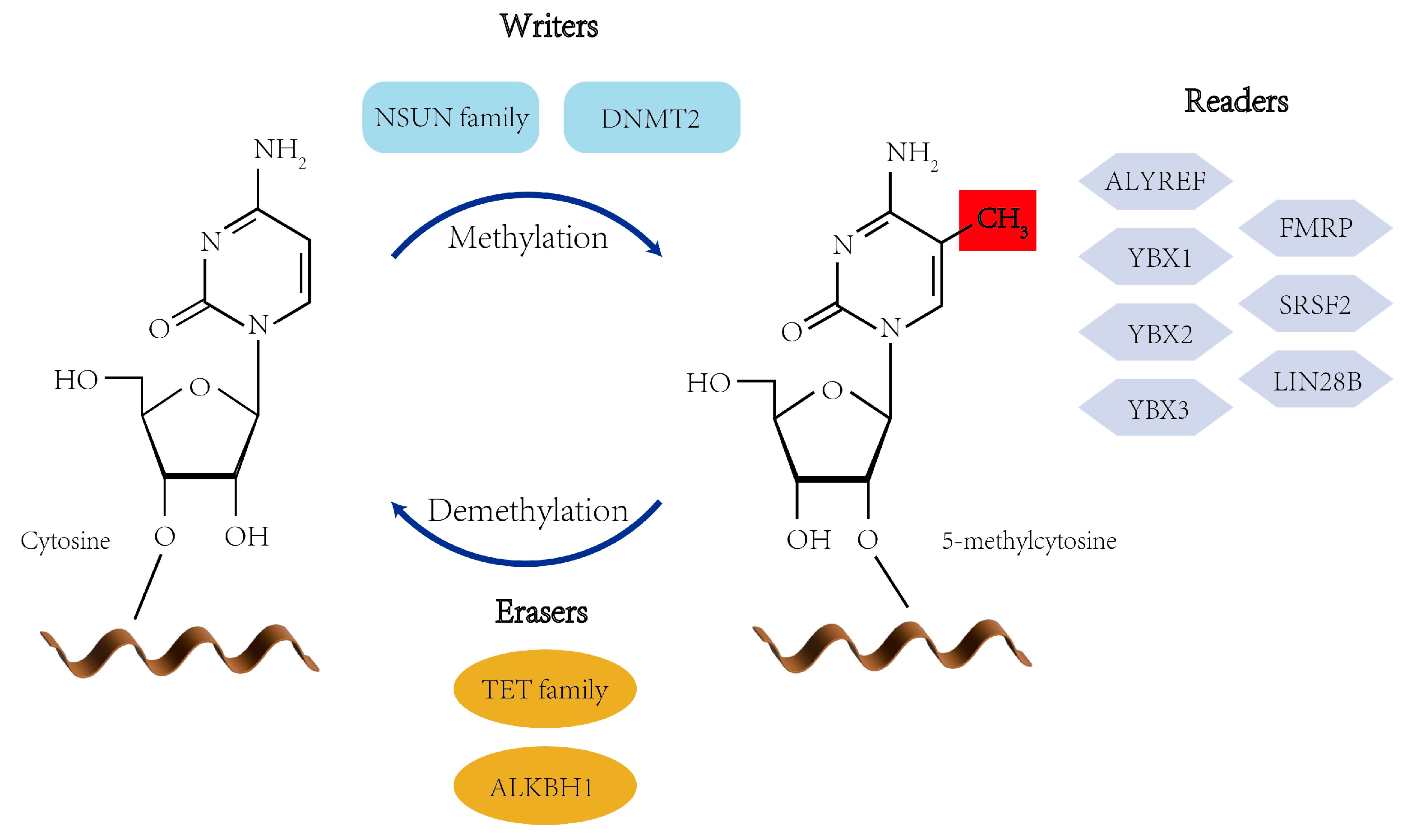
Figure 1. Writer, eraser, and reader proteins of RNA m5C modification. Writers of RNA m5C modification include NSUN family and DNMT2, erasers of RNA m5C modification include TET family and ALKBH1, and readers of RNA m5C modification include YBX1-3, ALYREF, FMRP, SRSF2, and LIN28B.
Viruses 17 01159 g001
Figure 2. Biological function of m5C methylation modification. (A) m5C modification regulates mRNA translation, nucleation, and stability. (B) m5C modification enhances ncRNA stability and translation.
Viruses 17 01159 g002
Figure 3. Detection methods of RNA m5C methylation. The detection methods for RNA m5C methylation mainly include RNA-BisSeq (A), MeRIP-seq (B), 5-aza-seq (C), miCLIP-seq (D), LC-MS/MS (E) and nanopore sequencing (F).
Viruses 17 01159 g003
Figure 4. Mechanisms of HBV-induced HCC formation. The molecular mechanisms of HBV-related HCC mainly include four aspects: HBV DNA integrates into the host genome, the rapid turnover of hepatocytes and increased inflammation caused by the host immune response, HBV genomic mutations, and abnormal expressions of HBx.
Viruses 17 01159 g004
Figure 5. The role of m5C in the HBV life cycle. NSUN2-mediated m5C methylation on pgRNA enhances its stability and promotes specific recognition by the viral core protein (HBc), facilitating efficient nucleocapsid assembly. This modification serves three key functions: (1) it strengthens pgRNA-HBc interactions through structural remodeling of the 5′ ε stem-loop, (2) enhances reverse transcription efficiency by maintaining optimal RNA conformation for polymerase binding, and (3) mediates immune evasion through dual mechanisms—impairing RIG-I recognition of viral RNA while suppressing HBc-induced inflammatory responses. The NSUN2-dependent m5C modification thus coordinates multiple stages of HBV replication while simultaneously counteracting host antiviral defenses.
Viruses 17 01159 g005
Table 1. Summary of major RNA modifications in viral infections.
ModificationsRNA SubstratesTypical LocationKey Roles in Viral InfectionsReferences
m5CmRNA, tRNA, rRNA, viral RNACDS/UTRRNA stability, translation efficiency, immune evasion, nuclear export, and splicing [10,11,12,13,14]
m6AmRNA, lncRNA, circRNA, rRNA, viral RNA3′ UTR, near stop codonRNA stability, translation efficiency, immune evasion, nuclear export, splicing, and secondary structure[15,16,17,18,19]
ac4CmRNA, tRNA, rRNA, viral RNACoding regionRNA stability, translation efficiency, splicing, and secondary structure[15,20,21,22,23,24,25]
ΨtRNA, rRNA, viral RNAStructural regionsRNA stability, translation efficiency, and pre-mRNA processing[26,27,28,29]
m1AmRNA, tRNA, viral RNA5′ UTR and coding regionsRNA stability and splicing[22,23,30]
m7GmRNA, rRNA, viral RNA5′ cap, internal sitesRNA stability, translation efficiency, and ribosome biogenesis[31,32,33,34]
NmrRNA, snRNA, mRNA, viral RNAsRibose 2′-hydroxylRNA stability, translation efficiency, and pre-mRNA processing[35,36,37]
Table 2. m5C in other viruses.
VirusWriterNotable m5C SitesKey FunctionsReferences
HIV-1NSUN2Gag-pol overlap, splice sitesExport, translation, splicing regulation[136,142]
HCVNSUN2C7525 (NS5A)RNA stability, replication, release[141]
EV71NSUN25′ UTR nt 584, CDS nt 1460IRES-mediated translation, RNA stability[11]
SARS-CoV-2NSUN23′ UTR, ORF dynamicsRNA turnover, replication regulation[140]
EBVNSUN2EBER1 C145ncRNA stability modulation[143]
MLVNSUN25′ UTR, 3′ UTRGenome packaging, RNA stability, immune evasion[138]
Disclaimer/Publisher’s Note: The statements, opinions and data contained in all publications are solely those of the individual author(s) and contributor(s) and not of MDPI and/or the editor(s). MDPI and/or the editor(s) disclaim responsibility for any injury to people or property resulting from any ideas, methods, instructions or products referred to in the content.