Article

Genomic and Biological Characterization of a Novel Proteus mirabilis Phage with Anti-Biofilm Activity

1
College of Animal Science and Technology, Shihezi University, Shihezi 832003, China
2
College of Food and Bioengineering, Henan University of Science and Technology, Luoyang 471023, China
*
Authors to whom correspondence should be addressed.
These authors contributed equally to this work.
Viruses 2025, 17(11), 1419; https://doi.org/10.3390/v17111419 (registering DOI)
Submission received: 1 October 2025 / Revised: 17 October 2025 / Accepted: 22 October 2025 / Published: 25 October 2025
(This article belongs to the Section Bacterial Viruses)

Abstract

The emergence of multidrug-resistant (MDR) Proteus mirabilis poses a significant threat in porcine farming and public health, highlighting the need for alternative biocontrol agents. This study aimed to isolate and characterize a lytic bacteriophage with therapeutic potential against MDR P. mirabilis. Using the clinical MDR P. mirabilis strain Pm 07 as host, a bacteriophage, vB_Pmc_P-07 (P-07), was successfully isolated from fecal and sewage samples via an enrichment protocol. Phage P-07 forms plaques surrounded by a distinct translucent “halo,” suggesting the production of depolymerase. It achieved high titers of up to 1.40 × 10⁸ PFU/mL and exhibited a narrow host range, high stability across a broad range of temperatures (40–60 °C) and pH (4–12), as well as considerable anti-biofilm activity. An optimal multiplicity of infection (MOI) of 0.001 was determined. Whole-genome sequencing revealed a linear double-stranded DNA genome of 58,582 bp with a GC content of 46.91%, encoding 63 open reading frames. Crucially, no virulence or antibiotic resistance genes were detected, supporting its safety profile. Phylogenetic analysis classified P-07 within the Casjensviridae family, closely related to phages PM87 and pPM01. These findings indicate that phage P-07 is a novel, safe, and effective lytic phage with strong potential as a biocontrol agent against biofilm-forming MDR P. mirabilis in swine.

1. Introduction

Proteus mirabilis is a Gram-negative, rod-shaped bacterium widely distributed in natural environments [1]. As an opportunistic pathogen, it causes infections in the digestive, respiratory, and urinary tracts of both humans and animals, with rising incidence rates in recent years [2,3].
In swine farming, P. mirabilis represents a growing concern. While often presenting with subtle clinical manifestations, its ability to form biofilms and serve as a reservoir for multidrug resistance genes poses serious challenges to farm management and animal health [4,5]. Moreover, the zoonotic potential of this pathogen is underscored by its detection in retail meat products, including pork. Strains isolated from meat sources show remarkable genetic similarity and virulence gene overlap with human clinical isolates, suggesting a plausible foodborne transmission route [6,7].
The global spread of multidrug-resistant (MDR) bacteria, causing an estimated 700,000 deaths annually, has been recognized by the WHO as a critical public health threat. This urgency has renewed interest in bacteriophage therapy as a promising alternative to conventional antibiotics. Phages offer unique advantages including high specificity, self-replication capability, and a favorable safety profile [8,9]. Their potential in livestock production is increasingly acknowledged, with documented efficacy against pathogens like Escherichia coli and Salmonella in poultry and swine [10,11].
Nevertheless, specific research on phage therapy targeting P. mirabilis in agricultural settings remains limited. To address the knowledge gap, this study aimed to isolate and characterize a lytic bacteriophage specific to porcine-originated MDR P. mirabilis, investigating its biological characteristics, genomic features, and anti-biofilm potential to develop a foundation for phage-based control strategies in swine farming.

2. Materials and Methods

2.1. Bacterial Strains and Sample Collection

MDR P. mirabilis strain Pm 07 and other tested strains (41 strains of P. mirabilis, 18 strains of E. coli, 3 strains of Klebsiella pneumoniae, and two strains of Staphylococcus aureus) were isolated from Xinjiang Uygur Autonomous Region of China and identified by the Animal Infectious Disease Laboratory at Shihezi University, and their detailed information is provided in Supplementary Table S1. The three primary MDR strains investigated in this study—Pm 07, Pm 08, and Pm 13—were all confirmed as ESBL-producing P. mirabilis. Pm 07 exhibited resistance to 18 antibiotics, including cefalexin (CA), penicillin (PG), amoxicillin (AM), enrofloxacin (ENR), ciprofloxacin (CIP), norfloxacin (NF), trimethoprim-sulfamethoxazole (SXT), tetracycline (TC), doxycycline (DOX), gentamicin (GN), kanamycin (KAN), tobramycin (TOB), florfenicol (FFC), chloramphenicol (CL), polymyxin B (PB), erythromycin (ERY), vancomycin (VAN), and imipenem (IMP). Pm 08 was resistant to 19 antibiotics, including cefotaxime (CTX), cefalexin (CA), penicillin (PG), amoxicillin (AM), enrofloxacin (ENR), ciprofloxacin (CIP), norfloxacin (NF), trimethoprim-sulfamethoxazole (SXT), tetracycline (TC), doxycycline (DOX), gentamicin (GN), kanamycin (KAN), tobramycin (TOB), florfenicol (FFC), chloramphenicol (CL), polymyxin B (PB), erythromycin (ERY), vancomycin (VAN), and imipenem (IMP). Strain Pm 13 showed resistance to 17 antibiotics, including ceftazidime (CAZ), cefalexin (CA), penicillin (PG), amoxicillin (AM), enrofloxacin (ENR), ciprofloxacin (CIP), norfloxacin (NF), trimethoprim-sulfamethoxazole (SXT), tetracycline (TC), doxycycline (DOX), kanamycin (KAN), tobramycin (TOB), florfenicol (FFC), chloramphenicol (CL), polymyxin B (PB), erythromycin (ERY), vancomycin (VAN), and imipenem (IMP). All antibiotic susceptibility testing was performed by the Kirby-Bauer disk diffusion method on Mueller-Hinton agar, with results interpreted according to the Clinical and Laboratory Standards Institute (CLSI) guidelines. In addition, the crystal violet staining assay confirmed varying biofilm-forming capacities among the isolates: strain Pm 07 exhibited strong biofilm formation with an OD590 value > 4 × the negative control (DNC), strain Pm 08 demonstrated moderate capacity (2× DNC < OD590 ≤ 4× DNC), and strain Pm 13 showed weak capacity (DNC < OD590 ≤ 2× DNC). (data not yet published).
Feces and wastewater were collected from large-scale pig farms in the Shihezi region of Xinjiang and underwent subsequent phage isolation.

2.2. Enrichment of Bacteriophages

Bacteriophage enrichment was performed using a modified protocol based on the method by Jamalludeen et al. [12]. Briefly, SM buffer (50 mM Tris-HCl [pH 7.5], 100 mM NaCl, 8 mM MgSO4, and 0.01% gelatin) was added to mixed fecal-sewage samples, followed by static sedimentation at 4 °C for 24–48 h. The supernatant was then collected via centrifugation (2910× g, 10 min, 4 °C) and filtered through 0.45 μm membranes for storage at 4 °C. Concurrently, Pm 07 was inoculated into LB broth and cultured with shaking until the logarithmic growth phase was reached (37 °C). For phage amplification, 40 mL of filtered fecal sample was mixed with 200 μL of fresh Pm 07 culture in an equal volume of 2× LB broth, and the mixture was incubated with shaking (37 °C, 180 r/min) for 12–16 h. Finally, the culture was centrifuged (16,760× g, 15 min, 4 °C), and the supernatant was passed through a 0.22 μm sterile membrane to remove bacterial debris and obtain the purified bacteriophage stock solution.

2.3. Isolation and Purification of Bacteriophage

The presence of lytic bacteriophages in the crude phage solution was detected using the spot assay [13]. Briefly, 200 μL log period P. mirabilis culture was spread evenly onto LB solid medium using a sterile cotton swab. After the bacterial suspension was absorbed, 10 μL of the crude phage solution was spotted onto the plate. The plates were air-dried completely, inverted, and incubated at 37 °C for 12–16 h to observe plaque formation.
Phage purification was subsequently carried out using the double-layer agar method [14]. Individual plaques were aseptically picked and transferred into SM buffer, followed by incubation overnight at 4 °C. The suspension was then centrifuged at 11,640× g for 10 min and sterilized by filtration through a 0.22 μm membrane. For phage amplification, 100 μL of host bacterial suspension and 100 μL of phage solution were mixed in LB broth and incubated with shaking (180 r/min, 37 °C) for 6–8 h. The culture was centrifuged again (11,640× g, 10 min), and the supernatant was collected. Ten-fold serial dilutions of the supernatant were prepared. From each dilution, 100 µL was mixed thoroughly with 100 µL of host bacteria, allowed to adsorb statically, mixed with molten soft agar, and poured onto LB agar plates. After solidification, the plates were incubated at 37 °C. This purification procedure was repeated 3 to 5 times until plaques with uniform morphology and size were obtained.

2.4. Concentration and Electron Microscopy Observation of Bacteriophage

Bacteriophage particles were concentrated using the polyethylene glycol (PEG8000) precipitation method [15]. Briefly, a 200 mL phage lysate was initially prepared. The supernatant was collected by centrifugation (7450× g, 15 min, 4 °C) and sterilized via membrane filtration. To remove residual host nucleic acids, DNase I and RNase A were added to the filtrate at a final concentration of 1 μg/mL each, followed by incubation at 37 °C for 1 h. Subsequently, NaCl was added to a final concentration of 1 M, and the mixture was incubated on ice for 2 h. After centrifugation (16,760× g, 20 min, 4 °C), the pellet was discarded. PEG8000 was added to the supernatant to a final concentration of 10% (w/v), dissolved completely by gentle stirring, and the solution was incubated on ice for another hour. The mixture was then centrifuged (11,640× g, 10 min, 4 °C) again. The resulting pellet was resuspended in 1 mL of SM buffer and incubated at 4 °C for 1 h. An equal volume of chloroform was added for extraction. Following centrifugation (1050× g) at 4 °C for 15 min, the aqueous phase was carefully collected as the concentrated phage preparation.
For electron microscopy, the concentrated phage solution (50 µL) was applied onto copper grids (with carbon support film) and allowed to adsorb for 15 min. Negative staining was performed using 2% (w/v) phosphotungstic acid for 5 min. Excess stain was removed with filter paper, and the grids were irradiated with infrared light for 30 min. Samples were visualized using an HT7700 transmission electron microscope (Hitachi High-Technologies Corporation, Tokyo, Japan).

2.5. Lytic Spectrum Profiling

The host range of the isolated bacteriophage was determined against a panel of bacterial strains, including 41 P. mirabilis, 18 E. coli, 3 K. pneumoniae, and 2 S. aureus isolates (excluding the host strain), using a spot assay. Briefly, 200 μL of fresh logarithmic-phase bacterial culture was spread evenly onto LB agar plates. After the surface dried, 5 μL of phage suspension was spotted onto pre-designated areas. The plates were then incubated overnight at 37 °C. A clear lytic zone observed at the spot location, contrasted with confluent bacterial growth in the surrounding control area, was interpreted as a positive lytic activity against the corresponding strain.

2.6. Optimal Multiplicity of Infection (MOI) Determination

The optimal multiplicity of infection (MOI) was determined as previously described [16,17]. Briefly, mixtures containing equal volumes of host bacterial suspension and phage solutions at different MOIs (0.00001, 0.0001, 0.001, 0.01, 0.1, 1, 10, 100) were prepared in 800 μL of LB broth and mixed thoroughly. After 4 h of incubation with shaking (180 r/min, 37 °C), the cultures were centrifuged (11,640× g, 10 min, 4 °C). The supernatants were filtered through 0.22 μm membranes, and phage titers were determined using the double-layer agar method. All assays were performed in triplicate, and the results are presented as the mean values.

2.7. One-Step Growth Curve Analysis

The one-step growth curve of the phage was analyzed according to the method described by Pajunen et al. [18]. Briefly, the phage and its host bacteria were mixed at the optimal multiplicity of infection (MOI) and incubated at 37 °C for 10 min to allow adsorption. The mixture was then centrifuged (16,760× g, 10 min) to remove unadsorbed phages. The pellet was washed twice with phosphate-buffered saline (PBS), resuspended in 50 mL of fresh LB broth, and incubated at 37 °C with shaking (180 r/min). Samples (1 mL each) were collected at predetermined time points over a period of 180 min, immediately centrifuged (16,760× g, 2 min), and the supernatant was subjected to phage titration using the double-layer agar plaque assay. All titrations were performed in triplicate, and the phage titers are expressed as the mean values.

2.8. Thermal Stability Assessment

The thermal stability of the phage was evaluated by incubating 2 mL aliquots of the phage suspension in water baths at temperatures ranging from 40 °C to 80 °C (40 °C, 50 °C, 60 °C, 70 °C, 80 °C). At 20, 40, and 60 min intervals, 200 μL samples were withdrawn and immediately centrifuged (16,760× g, 5 min, 4 °C) to remove any debris. The phage titer in the supernatant was subsequently determined using the double-layer agar plaque assay [19]. The experiment was performed in triplicate, and the results are expressed as the mean ± standard deviation.

2.9. pH Tolerance Evaluation

The pH tolerance of the phage was assessed by exposing the viral particles to a broad range of pH conditions. Aliquots (100 μL) of the phage suspension were mixed with 900 μL of LB broth pre-adjusted to different pH values (ranging from 1 to 14). The mixtures were incubated statically at 37 °C for 2 h. Subsequently, the samples were centrifuged (16,760× g, 5 min, 4 °C) to pellet any potential debris or aggregates, and the surviving phage titer in the supernatant was determined using the double-layer agar plaque assay. The experiment was conducted in triplicate, and the results are presented as the mean ± standard deviation.

2.10. Bacteriophage-Mediated Anti-Biofilm Activity Analysis

The anti-biofilm activity of the bacteriophage against P. mirabilis was evaluated using a crystal violet staining assay. Prior to the phage treatment, the biofilm formation dynamics of the host strain P. mirabilis 07 were characterized. The bacteria were inoculated and cultured as described below, and the biofilm biomass was quantified at 4, 8, 12, 24, 36, and 48 h to establish the growth kinetics. This preliminary study confirmed that a mature biofilm was formed at 24 h, which was consequently selected as the time point for phage application in the subsequent anti-biofilm assay.
For the main assay, host bacteria were inoculated (1:100) into fresh liquid medium and incubated at 37 °C with shaking at 180 r/min until the OD600 reached the logarithmic phase. A 1 mL aliquot of the culture was transferred to each well of a 24-well plate and incubated statically at 37 °C for 24 h to allow for the development of mature biofilms. After careful removal of the planktonic culture, the wells were gently rinsed three times with PBS to remove non-adherent cells. The established biofilms were then treated with 1 mL of phage suspensions at concentrations of 5.6 × 10⁸ PFU/mL or 5.6 × 10⁷ PFU/mL, using LB broth as a negative control. Following static incubation at 37 °C for 2, 4, and 6 h post-phage addition, the wells were washed again three times with PBS. The remaining adherent biofilm biomass was quantified by crystal violet staining, and the absorbance was measured at 590 nm. Results are shown as the mean ± SD.

2.11. Genomic DNA Extraction, Sequencing, and Bioinformatics Analysis

Bacteriophage genomic DNA was extracted using the standard phenol-chloroform method. Briefly, a purified high-titer phage lysate was treated with DNase I and RNase A (1 µg/mL each) at 37 °C for 30 min to remove residual host nucleic acids. Subsequently, EDTA (pH 8.0), Proteinase K, and SDS were added to final concentrations of 20 mM, 50 µg/mL, and 0.5% (w/v), respectively, followed by incubation at 56 °C for 1 h to digest the capsid proteins. Nucleic acids were extracted with an equal volume of phenol-chloroform-isoamyl alcohol (25:24:1) and precipitated with isopropanol. The DNA pellet was washed with 75% ethanol, air-dried, and finally resuspended in nuclease-free water.
The purified DNA underwent Illumina sequencing at Huitong Biotechnology Co., Ltd., Hangzhou (Zhejiang, China). Genome assembly employed Newbler software (Version:2.9). Protein-coding genes were predicted using GeneMarkS. Antibiotic resistance genes were identified via CARD (http://card.mcmaster.ca/, accessed on 16 February 2025), while virulence factors were detected using the Center for Genomic Epidemiology platform (http://www.genomicepidemiology.org/services/, accessed on 16 February 2025). Circular genome maps were generated with CGView Server (https://js.cgview.ca/, accessed on 16 February 2025).
The terminase large subunit—a signature phage gene critical for phylogenetic analysis [20]—underwent BLAST (https://blast.ncbi.nlm.nih.gov/Blast.cgi, accessed on 16 February 2025) alignment on NCBI. Phylogenetic trees were constructed using MEGA 11.0.
The observation of translucent halos surrounding the plaques of phage P-07 suggested the potential production of depolymerase, which was subsequently investigated through genomic analysis. Following gene annotation, phylogenetic analysis of the phage tail fiber protein was performed using MEGA 11.0 to elucidate its evolutionary relationships. Furthermore, the tertiary structure of the tail fiber protein was predicted using the Phyre2 online server (http://www.sbg.bio.ic.ac.uk/phyre2, accessed on 9 September 2025).

2.12. Data Analysis

All statistical analyses utilized GraphPad Prism 9.0 (GraphPad Software, Inc., San Diego, CA, USA). Significance was determined by multiple t-tests. Differences were considered statistically significant at p < 0.05 and highly significant at p < 0.01. Error bars denote the standard deviation (SD) of means.

3. Results

3.1. Isolation, Purification, and Electron Microscopy Morphology

A lytic bacteriophage infecting Proteus mirabilis was isolated from fecal-wastewater samples and designated vB_Pmc_P-07 (abbreviated as P-07) in accordance with the guidelines of the International Committee on Taxonomy of Viruses (ICTV). Plaque assay showed that P-07 formed clear plaques surrounded by distinct translucent halos (Figure 1A). Transmission electron microscopy (TEM) imaging at 80 kV revealed that P-07 has an icosahedral head and a short tail, with approximate dimensions of 60 nm in head diameter and 12 nm in tail length (Figure 1B).

3.2. Lytic Spectrum Determination

Bacteriophage P-07 exhibited strict specificity, lysing only P. mirabilis and displaying no cross-species lytic activity against E. coli, K. pneumoniae, or S. aureus. When tested against a panel of 42 P. mirabilis isolates (including the host strain), the lysis rate was 21.42% (see Supplementary Table S1).

3.3. Optimal Multiplicity of Infection (MOI) and One-Step Growth Curve of P-07

The optimal multiplicity of infection (MOI) for phage P-07 was determined to be 0.001, yielding a peak titer of 9.66 × 10⁹ PFU/mL (Figure 2A). The one-step growth curve revealed a latent period of approximately 30 min, followed by a burst phase. The phage titer increased rapidly during the exponential rise period (30–60 min) and then more gradually until reaching a plateau at 100 min post-infection. The final burst size was calculated to be 474.99 PFU/cell, with a plateau titer of 8.830 log PFU/mL (Figure 2B).

3.4. Thermal and pH Stability of Phage P-07

Phage P-07 demonstrated notable thermal stability, maintaining stable titers at temperatures ranging from 40 to 60 °C for up to 40 min, with only a marginal decrease observed after 60 min of incubation (Figure 3A). However, at 70 °C, the phage titer declined rapidly after 20 min and was completely inactivated by 40 min. At 80 °C, the phage was completely inactivated after 20 min of incubation, with no plaques detected.
The phage also exhibited high tolerance to a broad pH range. High activity was retained within pH 5 to 11, while complete inactivation occurred under strongly acidic (pH ≤ 4) or strongly alkaline (pH ≥ 12) conditions (Figure 3B). Collectively, these findings indicate that P-07 possesses robust stability under various environmental conditions.

3.5. Biofilm Clearance Efficacy Against P. Mirabilis 07

P. mirabilis 07 exhibited a typical biofilm growth dynamic, characterized by a steady increase in biomass from 4 to 24 h, peaking at 24 h. Subsequently, the biofilm entered a decline phase, resulting in a reduction in total biomass (Supplementary Figure S1).
Bacteriophage P-07 demonstrated significant anti-biofilm activity against P. mirabilis 07 in a concentration and time dependent manner. After 2 h of treatment, both low-concentration (0.603 ± 0.031) and high-concentration (0.533 ± 0.022) groups showed significantly reduced OD590 values compared to the control group (0.814 ± 0.063) (p < 0.05). This inhibitory effect was maintained at 4 h, with OD590 values remaining significantly lower in both treatment groups (low-concentration: 0.564 ± 0.040; high-concentration: 0.518 ± 0.029) versus control (0.823 ± 0.131) (p < 0.05). By 6 h, the anti-biofilm effect became more pronounced, with both concentration groups (low-concentration: 0.469 ± 0.017; high-concentration: 0.436 ± 0.015) showing a highly significant reduction compared to the control (0.805 ± 0.094) (p < 0.01) (Figure 4A).

3.6. Strain-Dependent Anti-Biofilm Activity

Bacteriophage P-07 exhibited distinct anti-biofilm efficacy against preformed biofilms of P. mirabilis strains with varying biofilm-forming capacities. When treating 24 h mature biofilms formed by the moderate biofilm-forming strain P. mirabilis 08, the high-concentration P-07 significantly reduced OD590 values (0.603 ± 0.012) after 2 h compared to control (0.765 ± 0.043) (p < 0.05). At 4 h post-treatment, both low-concentration (0.573 ± 0.020) and high-concentration (0.529 ± 0.019) groups showed significant inhibition versus control (0.753 ± 0.032) (p < 0.05). After 6 h, while the low-concentration group (0.552 ± 0.016) demonstrated a significant reduction (p < 0.05), the high-concentration group (0.477 ± 0.015) showed highly significant efficacy compared to control (0.749 ± 0.016) (p < 0.01, Figure 4B).
For 24 h, mature biofilms formed by the weak biofilm-forming strain P. mirabilis 13; significant anti-biofilm effects were only observed after 6 h of treatment, where the high-concentration group (0.535 ± 0.011) exhibited significantly lower OD590 values than the control (0.662 ± 0.012) (p < 0.05, Figure 4C).

3.7. Differential Anti-Biofilm Efficacy Across Strains with Varying Biofilm-Forming Capacities

The anti-biofilm efficacy of P-07 demonstrated clear stratification corresponding to the biofilm-forming capacity of the bacterial strains. At low concentration, 2 h treatment reduced OD590 values by 0.210, 0.076, and 0.035 in 24 h mature biofilms formed by P. mirabilis 07 (strong biofilm-forming), P. mirabilis 08 (moderate biofilm-forming), and P. mirabilis 13 (weak biofilm-forming), respectively, compared to their controls. These reductions increased in a time-dependent manner, reaching 0.258, 0.180, and 0.054 after 4 h, and 0.336, 0.197, and 0.080 at 6 h. Throughout all time points, low-concentration P-07 showed significantly stronger clearance efficacy against mature biofilms of P. mirabilis 07 compared to those of both P. mirabilis 08 and P. mirabilis 13 (p < 0.05).
The high-concentration group demonstrated enhanced biofilm clearance against mature biofilms: 2 h treatment decreased OD590 values by 0.280, 0.161, and 0.051 for 24 h biofilms of P. mirabilis 07, 08, and 13, respectively. The reductions increased to 0.304, 0.225, and 0.090 at 4 h, and reached 0.367, 0.272, and 0.128 at 6 h. Similarly, high-concentration P-07 consistently exhibited significantly superior clearance efficacy against mature biofilms of P. mirabilis 07 compared to those of both P. mirabilis 08 and P. mirabilis 13 at all time points (p < 0.05).

3.8. Whole-Genome Analysis of Phage P-07

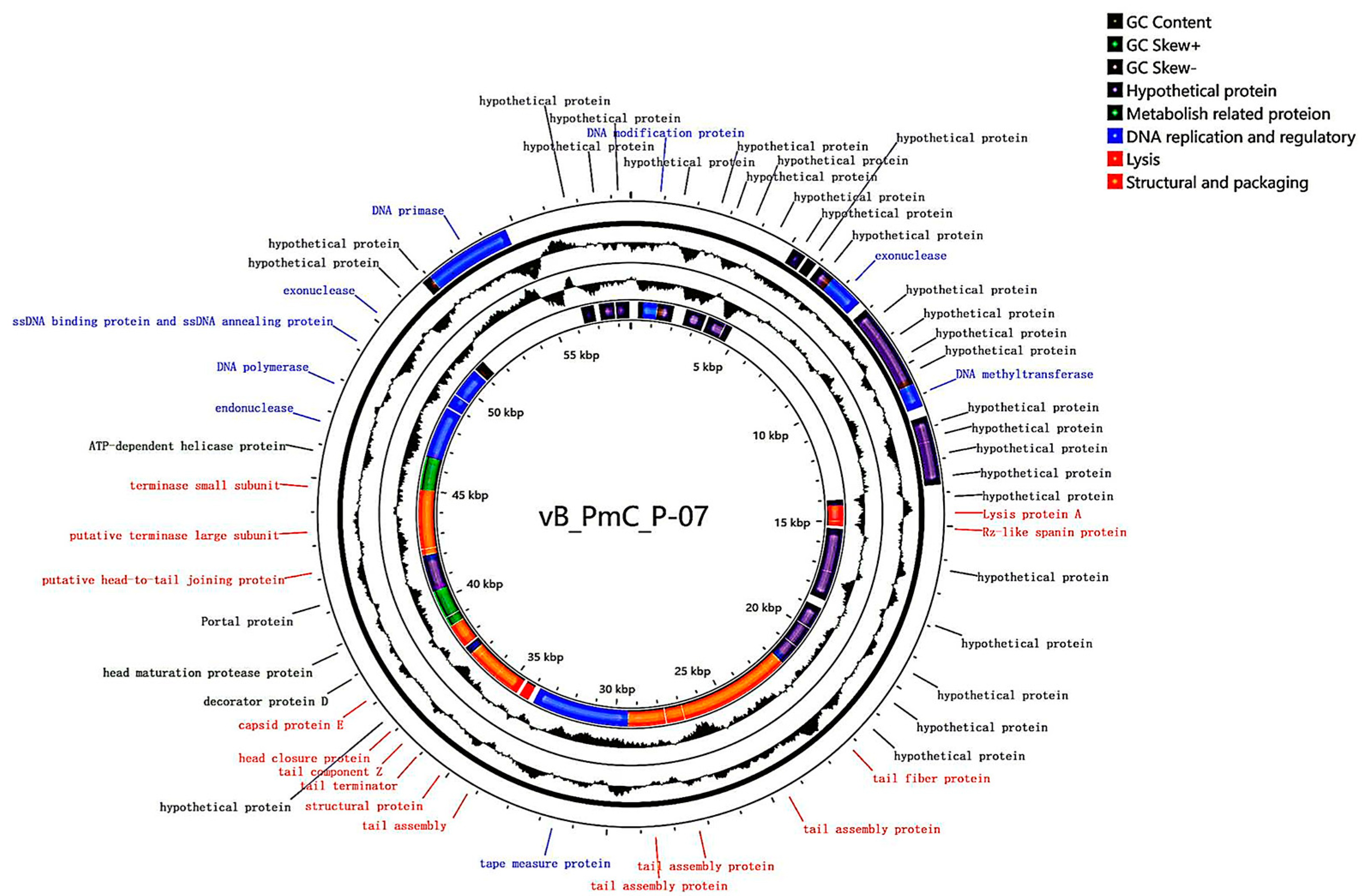
Whole-genome sequencing of phage P-07 yielded a complete double-stranded DNA genome of 58,582 bp with a GC content of 46.91%. Nucleotide composition analysis revealed the following distribution: 25.09% adenine (A), 28.01% thymine (T), 24.55% cytosine (C), and 22.36% guanine (G). The genome sequence has been deposited in GenBank under accession number PV817856.
Gene prediction and annotation identified 63 open reading frames (ORFs), including 15 transcribed in the forward direction and 48 in the reverse direction. Functional categorization revealed 30 ORFs encoding putative proteins, which were classified into four distinct modules: structural/packaging (orange), DNA replication/regulation (blue), auxiliary metabolic (green), and lysis (red). Notable functional assignments included: ORF24 (endolysin) in the lysis module; ORF46 (head maturation protease) in the metabolic module; ORF53 (DNA polymerase), ORF58 (primase), ORF2 (DNA modifier), ORF18 (methyltransferase), and ORF13/52/55 (exonucleases) in the DNA replication module; along with ORF25 (Rz), ORF39 (structural protein), ORF50/49 (terminase), ORF31 (tail fiber), ORF40 (tail terminator), ORF42 (head closure protein), ORF44 (capsid), ORF45 (decoration protein), ORF48 (head-tail connector), and ORF32-36/38 (tail tube proteins) in the structural/packaging module (Figure 5, Table 1).
Screening for antibiotic resistance genes and virulence factors using the CARD database and the Center for Genomic Epidemiology platform, respectively, revealed no identifiable resistance genes or virulence factors in the P-07 genome.
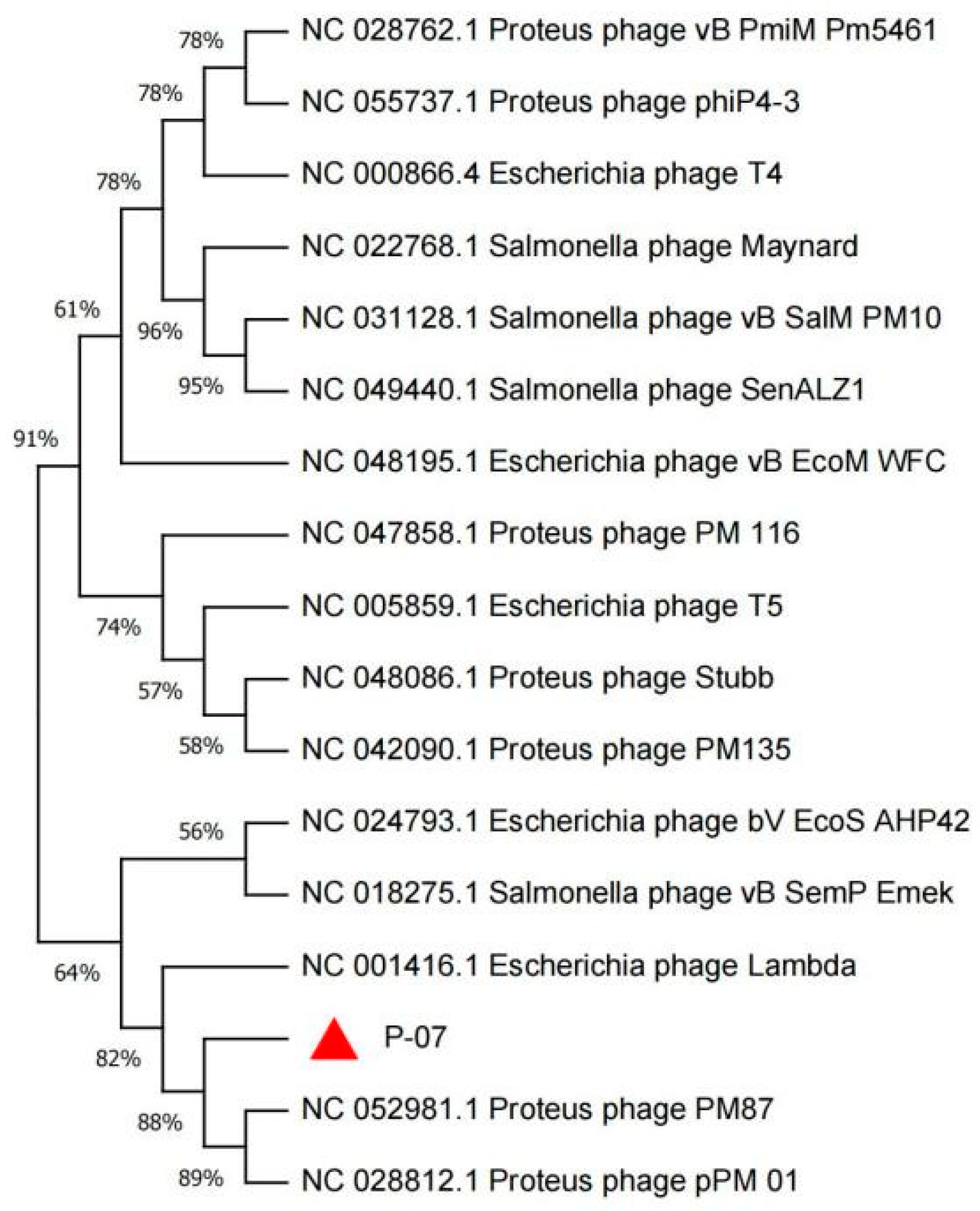
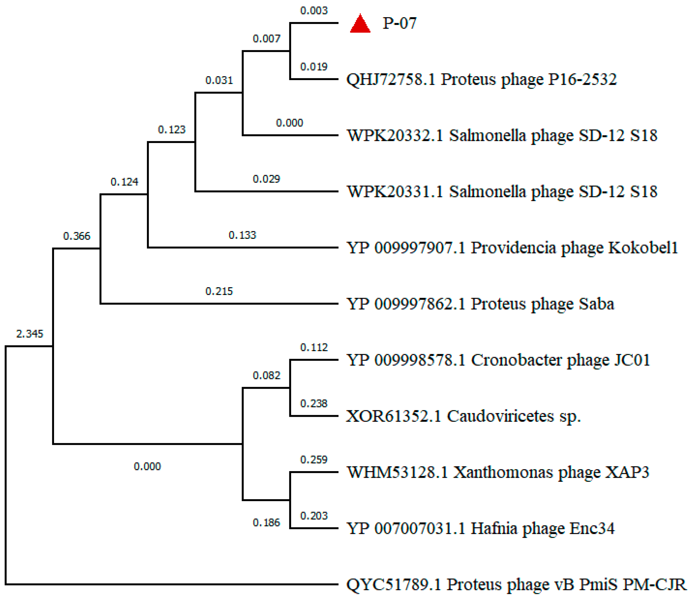
Phylogenetic analysis based on the terminase large subunit sequence positioned P-07 within the class Caudoviricetes, family Casjensviridae, and genus Lavrentievirus. The terminase large subunit showed 88% amino acid identity to those of P. mirabilis phages PM87 and pPM01, confirming its close evolutionary relationship with these known phages (Figure 6).
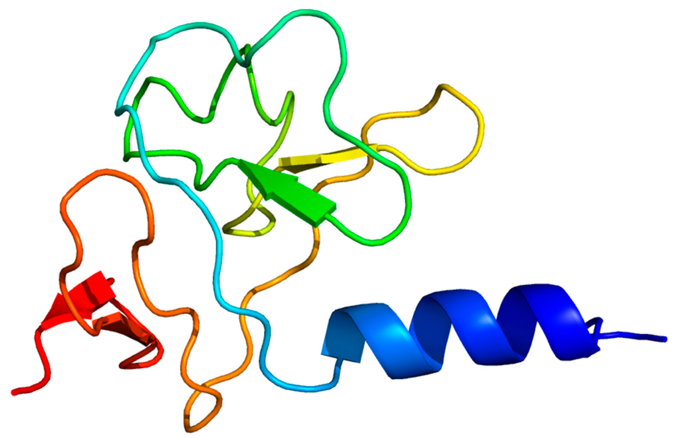
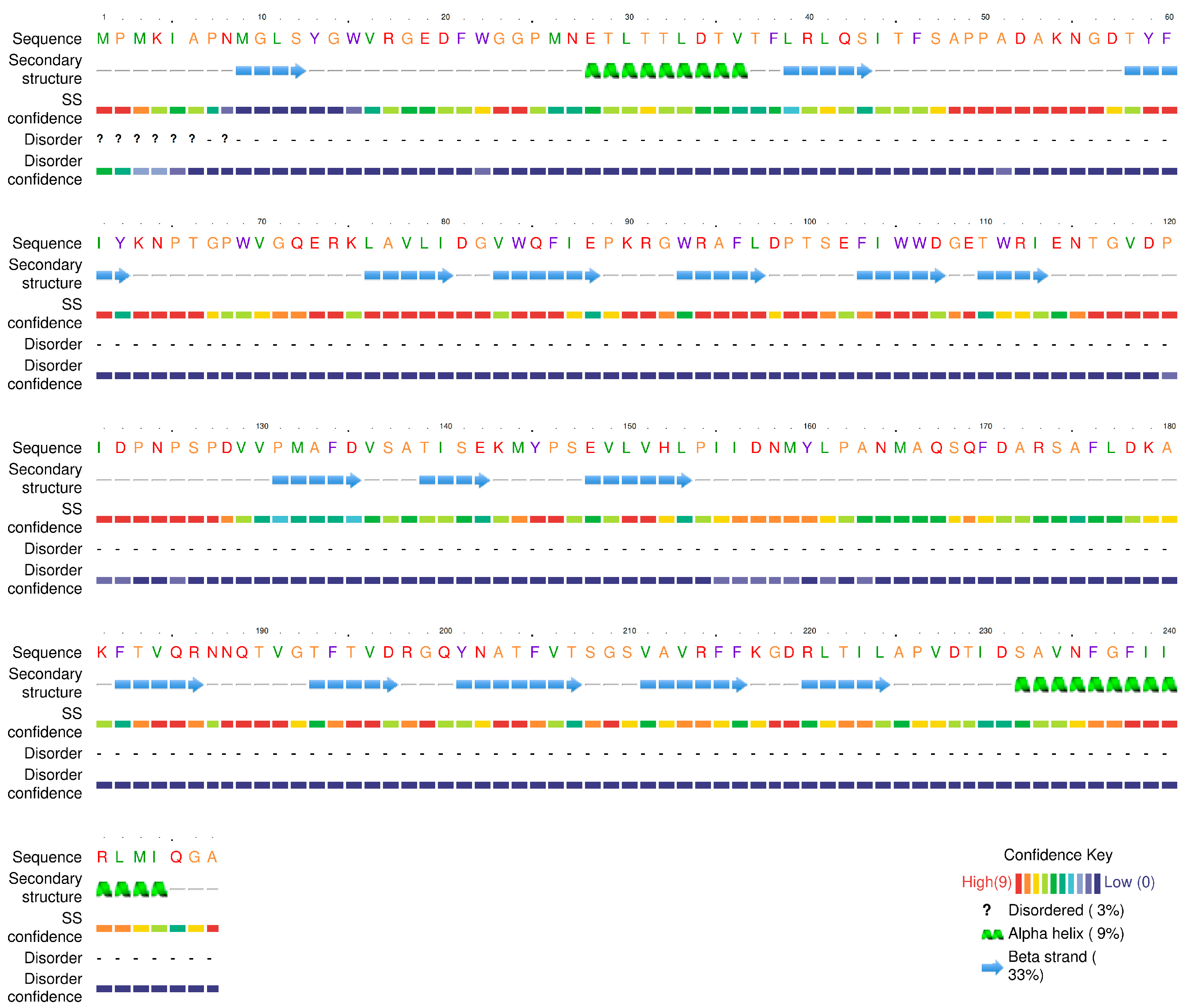
In-depth bioinformatic analysis was performed on ORF31, annotated as a tail fiber protein. Its tertiary structure was predicted using the Phyre2 online server. The prediction revealed two key functional domains: a C-terminal region (residues 178–225) exhibiting limited sequence coverage (19%) with lyase enzyme templates (c6p1yB_, c7a8yD_), and an N-terminal region (residues 22–115) showing significant structural homology (37% coverage) to the baseplate-tail complex of a marine siphophage (template c8gtcS_). The model dimensions for this baseplate-binding region are 38.817 Å (X) × 37.000 Å (Y) × 30.111 Å (Z) (Figure 7). Secondary structure composition analysis indicated a predominance of β-sheets (33%) over α-helices (9%), consistent with typical structural protein features (Figure 8). These findings suggest ORF31 is a multifunctional protein, with its N-terminal domain facilitating phage adsorption through baseplate integration and its C-terminal domain potentially enabling enzymatic degradation of host polysaccharides.
Building upon these structural predictions, we propose that ORF31 represents a “tail fiber-depolymerase fusion functional module,” a genomic feature commonly employed by phages to enhance biofilm penetration. This configuration ensures the synchronous transcription and assembly of the tail fiber with its associated enzymatic activity, thereby maximizing infection efficiency. The presence of an aspartate-rich domain within ORF31 is particularly significant, as specific aspartate residues have been experimentally demonstrated to serve as essential catalytic residues in polysaccharide-degrading enzymes; for instance, site-directed mutagenesis of key aspartates (Asp227 and Asp342) in an isomalto-dextranase completely abolished its hydrolytic activity [21]. This, coupled with the established role of aspartate in forming catalytically competent active sites through mechanisms such as hydrogen bonding and transition-state stabilization in glycoside hydrolases [22], provides strong genetic and mechanistic evidence that ORF31 encodes a depolymerase, equipping phage P-07 with the ability to degrade biofilm matrices.
Phylogenetic analysis of the tail fiber protein, a key determinant of host recognition often associated with depolymerase activity, was conducted using MEGA 11. As shown in Figure 9, P-07 shares the closest evolutionary relationship with Proteus phage P16-2532 (GenBank accession: QHJ72758), with a coverage of 71% and a high amino acid identity of 98.29%, strongly supporting their common classification within the genus Lavrentievirus. In contrast, P-07 is distantly related to Gorganvirus phage vB_PmiS_PM-CJR (GenBank accession: QYC51789), which is also reported to possess depolymerase activity. This disparity indicates that the genetic determinants for depolymerase production can be found across distinct phage genera with divergent evolutionary backgrounds (Figure 9).

4. Discussion

Antibiotic misuse has rendered multidrug-resistant (MDR) bacteria a global public health threat [23]. As an opportunistic pathogen, P. mirabilis causes diarrhea, vomiting, and urinary tract infections, with MDR strains posing severe challenges. Bacteriophages represent promising candidates for controlling MDR pathogens and biofilm infections. In this study, we isolated a lytic phage, designated vB_Pmc_P-07 (P-07), from fecal-wastewater samples, which belongs to the Caudoviricetes class. The phage formed plaques surrounded by distinct translucent halos—a phenotypic indicator of potential depolymerase production, which is a feature highly relevant for combating biofilm-associated infections. Guided by this observation, we conducted genomic and structural analyses that revealed a C-terminal domain (residues 178–225) in the tail fiber protein (ORF31) with structural homology to lyase enzymes, supporting its putative depolymerase function. This molecular evidence, combined with the halo phenotype, indicates that P-07 may encode enzymatic machinery capable of degrading bacterial exopolysaccharides—a crucial ability for penetrating and disrupting biofilms. However, this functional attribution remains speculative and warrants further validation through studies such as the heterologous expression of ORF31 and enzymatic assays using exopolysaccharide substrates.
Characterizing phage biology is essential for developing therapeutic strategies against zoonotic infections. P-07 exhibited a narrow host range, lysing only P. mirabilis without cross-species activity against E. coli, K. pneumoniae, or S. aureus. This finding is consistent with Pan et al. [24] and reflects receptor-binding specificity. Although its host range is limited, this phage enriches the Proteus phage repository and holds clinical potential. One-step growth analysis revealed advantageous reproductive replication kinetics: a latent period of 30 min and a burst size of 474.99 PFU/cell, outperforming phage PmP19 (40 min, 352 PFU/cell) [25]. The shorter latent period indicates a quicker onset of lytic activity, which is therapeutically beneficial for rapid bacterial clearance, while the larger burst size implies higher replicative efficiency, potentially leading to more robust and sustained antibacterial action. Furthermore, P-07 demonstrated notable environmental stability, retaining high viability at 40–60 °C and pH 4.0–12.0, which is comparable to the resilience reported for phage vB_PmM_S, vB_PmM_W, vB_PmM_X [26].
The formidable resistance of P. mirabilis biofilms, which can elevate antibiotic tolerance up to 1000-fold compared to planktonic cells [27], highlights the need for alternative therapeutic strategies. In this context, phage-based approaches have shown considerable promise. For instance, Mirzaei et al. [28] documented that a phage cocktail could effectively suppress P. mirabilis colonization on urinary catheters, while Soontarach et al. [29] reported concentration-dependent biofilm inhibition by phage LysAB1245 against Pseudomonas aeruginosa and Staphylococcus aureus. Consistent with these reports, our results confirm the anti-biofilm capacity of P-07 against porcine-originated MDR P. mirabilis, showing significant biomass reduction across multiple time points and phage concentrations. Removal efficacy increased over time, with the strongest effect at 6 h post-treatment—a pattern indicative of cumulative lytic and enzymatic degradation. These findings align with previous work by Esmael et al. [21] and support the potential of P-07 as a candidate for treating biofilm-associated infections.
Genomic analysis of P-07 (58,582 bp, 46.91% GC content, 63 ORFs) expands Proteus phage databases. Phylogenetically classified under Casjensviridae family and Lavrentievirus genus, it resembles phage vB_PmiP_pPm05, though with a shorter genome, and shares similarities with RS1pmA (42,112 bp, 41.10% GC) [8]. Notably, P-07 encodes only one lysis protein (ORF24) versus four in RS1pmA. Functional annotation categorized proteins into four modules: genes related to metabolism, genes related to lysis, structural and packaging genes, and genes related to DNA replication and regulation. A terminase complex ensured precise genome encapsidation [30]. Phylogenetic analysis of the terminase large subunit indicated that P-07 is most closely related to P. mirabilis phages PM87 and pPM01.
Tail fiber protein phylogeny offers evolutionary insights into the distribution of depolymerase function. While P-07 shares the closest relationship with Proteus phage P16-2532, it is distantly related to Gorganvirus phage vB_PmiS_PM-CJR, which also possesses depolymerase activity. This evolutionary divergence indicates independent acquisition of depolymerase capability across distinct phage genera, highlighting convergent evolution of this therapeutically valuable trait.
Critically, no virulence or antibiotic resistance genes were detected, substantially reducing risks in therapeutic development. Compared to traditional antibiotics, P-07 represents a safer clinical alternative without resistance concerns—a vital advancement against the MDR crisis.

Supplementary Materials

The following supporting information can be downloaded at: https://www.mdpi.com/article/10.3390/v17111419/s1, Table S1: Host range of phage vB_Pmc_P-07 (P-07); Figure S1: Biofilm formation dynamics of Proteus mirabilis 07.

Author Contributions

Conceptualization, Y.Q. and Y.L. (Yan Liang); methodology, Y.L. (Yan Liang); software, N.L. and S.W. validation, Y.L. (Yanfang Li); formal analysis, Y.L. (Yuwan Li); investigation, Y.L. (Yan Liang) and N.L.; resources, Y.L. (Yanfang Li); data curation, Y.L. (Yuwan Li); writing—original draft preparation, N.L.; writing—review and editing, Y.Q.; visualization, N.L.; supervision, Y.Q.; project administration, Y.Q.; funding acquisition, Y.Q. All authors have read and agreed to the published version of the manuscript.

Funding

This research was funded by the Natural Science Support Program Project of the Xinjiang Production and Construction Corps (2024DA007), the Shihezi University Technology Transfer and Promotion Program (CGZH202309). The APC was funded by the Natural Science Support Program Project of the Xinjiang Production and Construction Corps (2024DA007).

Institutional Review Board Statement

Not applicable.

Informed Consent Statement

Not applicable.

Data Availability Statement

Data supporting the findings are presented in the article.

Acknowledgments

The authors thank the College of Animal Science and Technology for facilitating the study and the aforementioned funding bodies for their financial support.

Conflicts of Interest

The authors declare no conflicts of interest.

Abbreviations

The following abbreviations are used in this manuscript:
MDRMultidrug-Resistant
ESBLExtended-Spectrum Beta-Lactamase
PFUPlaque Forming Units
MOIMultiplicity of Infection
CARDComprehensive Antibiotic Resistance Database

References

  1. Adeolu, M.; Alnajar, S.; Naushad, S.; Gupta, R.S. Genome-based phylogeny and taxonomy of the ‘Enterobacteriales’: Proposal for Enterobacterales ord. nov. divided into the families Enterobacteriaceae, Erwiniaceae fam. nov., Pectobacteriaceae fam. nov., Yersiniaceae fam. nov., Hafniaceae fam. nov., Morganellaceae fam. nov., and Budviciaceae fam. nov. Int. J. Syst. Evol. Microbiol. 2016, 66, 5575–5599. [Google Scholar] [CrossRef]
  2. Colantonio, M.A.; Hartzell, M.; Shannon, B. Proteus mirabilis: An Unlikely Device-related Infection. J. Community Hosp. Intern. Med. Perspect. 2025, 15, 84–88. [Google Scholar] [CrossRef]
  3. Sun, Y.D.; Wen, S.S.; Zhao, L.L.; Xia, Q.Q.; Pan, Y.; Liu, H.H.; Wei, C.W.; Chen, H.Y.; Ge, J.W.; Wang, H.B. Association among biofilm formation, virulence gene expression, and antibiotic resistance in Proteus mirabilis isolates from diarrhetic animals in Northeast China. BMC Vet. Res. 2020, 16, 176. [Google Scholar] [CrossRef] [PubMed]
  4. Qu, X.; Zhou, J.; Huang, H.; Wang, W.; Xiao, Y.; Tang, B.; Liu, H.; Xu, C.; Xiao, X. Genomic Investigation of Proteus mirabilis Isolates Recovered from Pig Farms in Zhejiang Province, China. Front. Microbiol. 2022, 13, 952982. [Google Scholar] [CrossRef] [PubMed]
  5. Keeratikunakorn, K.; Kaewchomphunuch, T.; Kaeoket, K.; Ngamwongsatit, N. Antimicrobial activity of cell free supernatants from probiotics inhibits against pathogenic bacteria isolated from fresh boar semen. Sci. Rep. 2023, 13, 5995. [Google Scholar] [CrossRef]
  6. Liu, X.L.; Wu, S.Y.; Yu, Z. Zoonotic Risks of Proteus mirabilis: Detection, Pathogenicity, and Antibiotic Resistance in Animals and Animal-Derived Foods. Microorganisms 2025, 13, 2060. [Google Scholar] [CrossRef] [PubMed]
  7. Chinnam, B.K.; Nelapati, S.; Tumati, S.R.; Bobbadi, S.; Chaitanya Peddada, V.; Bodempudi, B. Detection of β-Lactamase-Producing Proteus mirabilis Strains of Animal Origin in Andhra Pradesh, India and Their Genetic Diversity. J. Food Prot. 2021, 84, 1374–1379. [Google Scholar] [CrossRef]
  8. Alves, D.R.; Nzakizwanayo, J.; Dedi, C.; Olympiou, C.; Hanin, A.; Kot, W.; Hansen, L.; Lametschs, R.; Gahan, C.G.M.; Schellenberger, P.; et al. Genomic and Ecogenomic Characterization of Proteus mirabilis Bacteriophages. Front. Microbiol. 2019, 10, 1783. [Google Scholar] [CrossRef]
  9. Royer, S.; Morais, A.P.; da Fonseca Batistão, D.W. Phage therapy as strategy to face post-antibiotic era: A guide to beginners and experts. Arch. Microbiol. 2021, 203, 1271–1279. [Google Scholar] [CrossRef]
  10. Chen, J.; Han, J.; Yang, Z.; Zhou, W.; He, Y.; Chen, X.; Li, X.; Zou, T.; You, J. Bacteriophages as Potential Anti-Pathogenic Agents for Intestinal Health of Weaned Piglets in the Post-Antibiotic Era: An Updated Review. Animals 2025, 15, 1713. [Google Scholar] [CrossRef]
  11. Soffritti, I.; D’Accolti, M.; Bini, F.; Mazziga, E.; Volta, A.; Bisi, M.; Mazzacane, S.; De Cesare, A.; Indio, V.; Manfreda, G.; et al. Harnessing probiotics and bacteriophages to fight Salmonella and limit the use of antibiotics in broilers: A study in commercial conditions. Poult. Sci. 2025, 104, 105595. [Google Scholar] [CrossRef]
  12. Jamalludeen, N.; Johnson, R.P.; Friendship, R.T.; Kropinski, A.M.; Lingohr, E.J.; Gyles, C.L. Isolation and characterization of nine bacteriophages that lyse O149 enterotoxigenic Escherichia coli. Vet. Microbiol. 2007, 124, 47–57. [Google Scholar] [CrossRef]
  13. Chang, H.C.; Chen, C.R.; Lin, J.W.; Shen, G.H.; Chang, K.M.; Tseng, Y.H.; Weng, S.F. Isolation and characterization of novel giant Stenotrophomonas maltophilia phage phiSMA5. Appl. Environ. Microbiol. 2005, 71, 1387–1393. [Google Scholar] [CrossRef]
  14. Cai, R.P.; Wang, Z.J.; Wang, G.; Zhang, H.; Cheng, M.J.; Gu, Z.M.; Ji, Y.L.; Xi, H.Y.; Wang, X.W.; Xue, Y.B.; et al. Biological properties and genomics analysis of vB_KpnS_GH-K3, a Klebsiella phage with a putative depolymerase-like protein. Virus Genes 2019, 55, 696–706. [Google Scholar] [CrossRef]
  15. Wan, S.; Li, N.; Habib, S.; Zheng, P.; Li, Y.; Liang, Y.; Qu, Y. Biological Characteristics and Whole-Genome Analysis of a Porcine E. coli Phage. Vet. Sci. 2025, 12, 57. [Google Scholar] [CrossRef]
  16. Li, F.; Xing, S.; Fu, K.; Zhao, S.P.; Liu, J.F.; Tong, Y.G.; Zhou, L.J. Genomic and biological characterization of the Vibrio alginolyticus-infecting “Podoviridae” bacteriophage, vB_ValP_IME271. Virus Genes 2019, 55, 218–226. [Google Scholar] [CrossRef] [PubMed]
  17. Hou, G.; Zhou, H.; Yu, X.; Li, N.; Li, Y.; Qu, Y.; Li, Y.; Liang, Y.; Yan, D.; Yang, S.; et al. Biological characteristics and whole genome analysis of an ovine-derived multidrug-resistant Klebsiella pneumoniae phage. China Anim. Husb. Vet. Med. 2024, 51, 2047–2057. (In Chinese) [Google Scholar]
  18. Pajunen, M.; Kiljunen, S.; Skurnik, M. Bacteriophage phiYeO3-12, specific for Yersinia enterocolitica serotype O:3, is related to coliphages T3 and T7. J. Bacteriol. 2000, 182, 5114–5120. [Google Scholar] [CrossRef] [PubMed]
  19. Yang, R.Y. Safety Evaluation of a Phage Cocktail for Bovine Mastitis. Master’s Thesis, Shihezi University, Shihezi, China, 2022. (In Chinese). [Google Scholar]
  20. Shahin, K.; Bouzari, M.; Wang, R. Isolation, characterization and genomic analysis of a novel lytic bacteriophage vB_SsoS-ISF002 infecting Shigella sonnei and Shigella flexneri. J. Med. Microbiol. 2018, 67, 376–386. [Google Scholar] [CrossRef]
  21. Tochihara, T.; Sasaki, K.; Araki, O.; Morimoto, N.; Watanabe, K.; Hatada, Y.; Ito, S.; Ito, H.; Matsui, H. Site-directed mutagenesis establishes aspartic acids-227 and -342 as essential for enzyme activity in an isomalto-dextranase from Arthrobacter globiformis. Biotechnol. Lett. 2004, 26, 659–664. [Google Scholar] [CrossRef] [PubMed]
  22. Joshi, M.D.; Sidhu, G.; Pot, I.; Brayer, G.D.; Withers, S.G.; McIntosh, L.P. Hydrogen bonding and catalysis: A novel explanation for how a single amino acid substitution can change the pH optimum of a glycosidase. J. Mol. Biol. 2000, 299, 255–279. [Google Scholar] [CrossRef] [PubMed]
  23. Esmael, A.; Abo-Elmaaty, S.A.; Khafaga, E.M.; Abdelrahman, S.; Hassan, M.G. Efficacy of three lytic bacteriophages for eradicating biofilms of multidrug-resistant Proteus mirabilis. Arch. Virol. 2021, 166, 3311–3322. [Google Scholar] [CrossRef]
  24. Pan, S.Y.; Cen, Q.B.; Wang, H.Y.; Kong, X.Y.; Pang, S.Y.; Gao, D.Y.; Song, J. Analysis of biological characteristics of a multidrug-resistant Proteus mirabilis phage and its antibacterial effect on chicken breast. Food Sci. 2025, 46, 89–97. [Google Scholar]
  25. Han, S.; Li, S.; Hu, G.; Lei, M.T.; Li, C.H.; Xiao, L.C.; Li, S.Q. Characterization and genomic analysis phage PmP19, a novel Proteus mirabilis phage with a broad host range. Arch. Virol. 2024, 169, 221. [Google Scholar] [CrossRef]
  26. Song, J.; Xu, R.Y.; Jin, Y.Q.; Chen, L.; Wang, Y.H.; Ruan, H.R.; Zheng, J.S.; Wu, R. Isolation, identification of canine-derived Proteus mirabilis phage and its effect on biofilm. Acta Vet. Zootech. Sin. 2020, 51, 2785–2793. [Google Scholar]
  27. Mah, T.-F. Biofilm-specific antibiotic resistance. Future Microbiol. 2012, 7, 1061–1072. [Google Scholar] [CrossRef]
  28. Mirzaei, A.; Wagemans, J.; Nasr, E.B.; Lavigne, R.; Moghim, S. A Phage Cocktail to Control Surface Colonization by Proteus mirabilis in Catheter-Associated Urinary Tract Infections. Microbiol. Spectr. 2022, 10, e0209222. [Google Scholar] [CrossRef]
  29. Soontarach, R.; Srimanote, P.; Voravuthikunchai, S.P.; Chusri, S. Antibacterial and Anti-Biofilm Efficacy of Endolysin LysAB1245 against a Panel of Important Pathogens. Pharmaceuticals 2024, 17, 155. [Google Scholar] [CrossRef]
  30. Baquero, D.P.; Liu, Y.; Wang, F.; Egelman, E.H.; Prangishvili, D.; Krupovic, M. Structure and assembly of archaeal viruses. Adv. Virus Res. 2020, 108, 127–164. [Google Scholar]
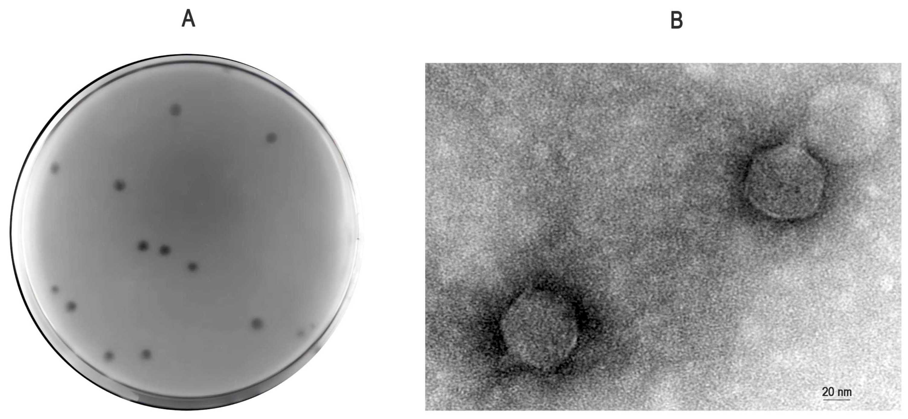
Figure 1. Morphological characteristics of bacteriophage vB_Pmc_P-07 (P-07). (A) Plaque morphology observed on a lawn of Proteus mirabilis. Clear plaques surrounded by translucent halos are visible; (B) Transmission electron micrograph of an individual P-07 virion negatively stained with 2% phosphotungstic acid (imaging performed at 80 kV). The phage particle exhibits an icosahedral head and a short tail. Scale bar, 20 nm.
Viruses 17 01419 g001
Figure 2. Replication characteristics of bacteriophage vB_Pmc_P-07 (P-07). (A) Determination of the optimal multiplicity of infection (MOI). The phage titer was measured after infection at different MOIs, with the highest yield observed at an MOI of 0.001; (B) One-step growth curve of P-07. The phage exhibited a latent period of approximately 30 min, followed by a rapid rise phase and a final plateau at 100 min, resulting in a burst size of 474.99 PFU/cell. Error bars represent the standard deviation from triplicate experiments.
Viruses 17 01419 g002
Figure 3. Environmental stability of bacteriophage vB_Pmc_P-07 (P-07). (A) Thermal stability. Phage viability was assessed after incubation at various temperatures (40–80 °C) for up to 60 min. P-07 maintained stability at 40–60 °C but was rapidly inactivated at 70 °C; (B) pH tolerance. Phage activity was evaluated after exposure to a broad pH range (pH 1–14) for 2 h. P-07 retained high infectivity within the pH 5–11 range, while being inactivated under strongly acidic (pH ≤ 4) or alkaline (pH ≥ 12) conditions.
Viruses 17 01419 g003
Figure 4. Concentration and time dependent anti-biofilm activity of bacteriophage P-07 against preformed P. mirabilis biofilms. (A) Anti-biofilm efficacy against 24 h mature biofilms formed by the strong biofilm-forming strain P. mirabilis 07. Biofilms were treated with low- (5.6 × 10⁷ PFU/mL) or high-concentration (5.6 × 10⁸ PFU/mL) phage suspensions for 2, 4, and 6 h. Both concentrations significantly reduced biofilm biomass compared to the control at all time points (* p < 0.05; ** p < 0.01); (B) Anti-biofilm efficacy against 24 h mature biofilms formed by the moderate biofilm-forming strain P. mirabilis 08. Significant reductions were observed at all time points, with enhanced inhibition at 6 h (* p < 0.05; ** p < 0.01); (C) Anti-biofilm efficacy against 24 h mature biofilms formed by the weak biofilm-forming strain P. mirabilis 13. Significant reduction was only observed in the high-concentration group after 6 h of treatment (* p < 0.05).
Viruses 17 01419 g004
Figure 5. Circular genome map of bacteriophage vB_Pmc_P-07 (P-07). The map depicts the complete double-stranded DNA genome (58,582 bp). Open Reading Frames (ORFs) on the forward and reverse strands, color-coded by functional module (structural/packaging: orange; DNA replication/regulation: blue; lysis: red; auxiliary metabolic: green).
Viruses 17 01419 g005
Figure 6. Phylogenetic analysis of the terminase large subunit of bacteriophage P-07. The maximum-likelihood tree was constructed based on amino acid sequence alignment, confirming the evolutionary position of P-07 (indicated by a red triangle) within the class Caudoviricetes, family Casjensviridae, and genus Lavrentievirus. Bootstrap values (≥70%) are shown at branch nodes.
Viruses 17 01419 g006
Figure 7. Tertiary structure prediction and domain architecture of phage P-07 tail fiber protein. The tertiary structure model of the ORF31 tail fiber protein is presented, coloured in a rainbow spectrum from the N-terminus (blue) to the C-terminus (red). Phyre2 modeling identified critical functional domains in ORF31, including a structural N-terminal region (residues 22–115; 38.817 × 37.000 × 30.111 Å) homologous to baseplate complexes and a C-terminal region with lyase homology. Secondary structure composition (9% α-helix, 33% β-sheet) supports its multifunctional role in phage infection.
Viruses 17 01419 g007
Figure 8. Secondary structure composition and functional implications of the tail fiber protein (ORF31). Analysis of the predicted structure reveals a distinct secondary structure profile dominated by β-sheets (33%) with minimal α-helical content (9%). This composition is consistent with the protein’s dual functional role, providing both structural stability for baseplate integration and flexibility for enzymatic activity in the C-terminal domain.
Viruses 17 01419 g008
Figure 9. Phylogenetic analysis and evolutionary relationships of the tail fiber protein. The maximum-likelihood tree was constructed based on amino acid sequence alignment, illustrating the phylogenetic placement of P-07 ORF31 (indicated by a red triangle) among related phage tail fiber proteins.
Viruses 17 01419 g009
Table 1. Genomic features and functional annotation of phage vB_Pmc_P-07.
ORFSTARTSTOPFunctional ModuleFunctionIdentity/%E ValueAccession Number
4640,45239,175Metabolism-related proteinhead maturation protease protein99.76%0YP_009199670.1
5146,54945,026ATP-dependent helicase protein99.80%0UGO49911.1
21183545DNA replication and regulatoryDNA modification 99.53%2.00 × 10−153UGO51540.1
1365977685exonuclease100.00%0YP_009997974.1
1810,60511,342DNA methyltransferase99.59%0UGO36038.1
3733,75629,425tape measure 99.79%0UVD32353.1
5246,79646,509endonuclease78.02%4.00 × 10−47YP_009997885.1
5348,82946,796DNA polymerase99.11%0UNI72738.1
5449,49948,894binding and annealing 98.99%1.00 × 10−139YP_009998014.1
5550,85949,531exonuclease97.74%0YP_010187223.1
5852,03954,714DNA primase99.78%0UNI72672.1
2414,96114,257Lysislysis protein A100.00%1.00 × 10−170UGO51513.1
2515,28614,972Structural and packagingRz-like spanin 100.00%4.00 × 10⁻³⁴YP_009199647.1
3122,62221,879tail fiber 99.60%2.00 × 10−179YP_009199654.1
3226,44322,622tail assembly 99.29%0UGO48774.1
3326,64526,436tail assembly 81.54%5.00 × 10⁻³¹YP_009997909.1
3426,88126,645tail assembly 82.05%1.00 × 10−39YP_009997910.1
3527,70926,894tail assembly 82.29%5.00 × 10−171YP_009997911.1
3629,41927,731tail assembly 99.29%0YP_009997997.1
3834,44933,997tail assembly 100.00%8.00 × 10−102UGO51498.1
3935,77534,588structural 99.75%0UGO49923.1
4036,29135,779tail terminator99.41%9.00 × 10−119YP_009199664.1
4136,89836,281tail component Z99.51%1.00 × 10−144WOZ54555.1
4237,25436,901head closure 100.00%2.00 × 10−80UGO36011.1
4438,73237,665capsid E99.72%0UGO49918.1
4539,16138,745decorator protein D99.28%2.00 × 10−94YP_009199669.1
4842,36742,092head-to-tail joining 97.80%2.00 × 10−59UGO51620.1
4944,44642,380terminase large subunit100.00%0UGO51619.1
5045,02344,439terminase small subunit99.48%2.00 × 10−141UXY92233.1
1548327Hypothetical proteinhypothetical protein98.63%2.00 × 10−46WP_196727143.1
319271187hypothetical protein99.59%7.00 × 10−177WOZ54581.1
434032834hypothetical protein99.47%6.00 × 10−135WOZ54583.1
539103509hypothetical protein100.00%2.00 × 10−94QHJ72730.1
640793891hypothetical protein100.00%3.00 × 10−38UGO49958.1
743114069hypothetical protein97.50%2.00 × 10−47EKV7661800.1
846494323hypothetical protein100.00%5.00 × 10−72YP_009199627.1
951195550hypothetical protein99.30%4.00 × 10−97WP_407818269.1
1056325943hypothetical protein100.00%9.00 × 10−68WP_318768141.1
1159496269hypothetical protein100.00%7.00 × 10−72WP_196727151.1
1262636610hypothetical protein100.00%3.00 × 10−75WP_196727152.1
1479558773hypothetical protein100.00%0WOZ54596.1
1587709537hypothetical protein99.61%0WOZ54597.1
1695349980hypothetical protein100.00%8.00 × 10−107YP_009998137.1
17997310,605hypothetical protein99.52%3.00 × 10−153WOZ54599.1
1911,64412,177hypothetical protein100.00%4.00 × 10−129WP_196727159.1
2012,17012,421hypothetical protein97.59%3.00 × 10−52UNI72704.1
2112,42313,292hypothetical protein100.00%0WP_196727160.1
2213,28913,753hypothetical protein97.40%1.00 × 10−108EKV7661814.1
2314,25714,003hypothetical protein98.81%2.00 × 10−51YP_009199645.1
2617,28215,297hypothetical protein96.52%0WP_318768180.1
2718,92617,286hypothetical protein91.74%0WOZ54610.1
2819,95218,936hypothetical protein99.41%0YP_009199651.1
2920,94819,965hypothetical protein100.00%0YP_009199652.1
3021,87920,959hypothetical protein99.35%0UNI72715.1
4337,60137,254hypothetical protein98.26%3.00 × 10−73UXY92226.1
5651,40250,920hypothetical protein96.88%1.00 × 10−103UGO51612.1
5751,75552,039hypothetical protein79.79%2.00 × 10−48YP_009997934.1
5956,36256,123hypothetical protein97.47%2.00 × 10−45UGO51678.1
6056,93756,359hypothetical protein98.44%9.00 × 10−140YP_009199616.1
6157,14656,934hypothetical protein98.57%4.00 × 10−41UGO49967.1
6257,84857,159hypothetical protein97.38%9.00 × 10−164WOZ54577.1
6358,50957,901hypothetical protein93.56%4.00 × 10−136UGO51542.1
Disclaimer/Publisher’s Note: The statements, opinions and data contained in all publications are solely those of the individual author(s) and contributor(s) and not of MDPI and/or the editor(s). MDPI and/or the editor(s) disclaim responsibility for any injury to people or property resulting from any ideas, methods, instructions or products referred to in the content.