Article

Cargo and Biological Properties of Extracellular Vesicles Released from Human Adenovirus Type 4-Infected Lung Epithelial Cells

1
Lovelace Biomedical Research Institute, Albuquerque, NM 87108, USA
2
IDeA National Resource for Quantitative Proteomics, Little Rock, AR 72205, USA
3
Department of Pathology, Microbiology and Immunology, New York Medical College, Valhalla, NY 10595, USA
*
Author to whom correspondence should be addressed.
Viruses 2025, 17(10), 1300; https://doi.org/10.3390/v17101300
Submission received: 19 August 2025 / Revised: 19 September 2025 / Accepted: 20 September 2025 / Published: 25 September 2025
(This article belongs to the Special Issue Epidemiology, Pathogenesis and Immunity of Adenovirus)

Abstract

Extracellular vesicles (EVs) are rapidly gaining recognition as critical mediators of inter-cellular communication during viral infections. To contribute to fill the gap in knowledge regarding the role of EVs in adenovirus infection, we used human adenovirus type 4 of species Mastadenovirus exoticum (HAdV-E4), a prevalent respiratory and ocular pathogen, and characterized the cargo and biological properties of EVs released by HAdV-E4-infected A549 lung epithelial cells at a pre-lytic stage of infection. Using immunocapture-based isolation and multi-omics approaches, we found that infection profoundly alters the EV uploaded proteome and small non-coding RNA repertoire. Mass spectrometry identified 268 proteins unique to EVs purified from infected cells (AdV-EVs), with enrichment in pathways supporting vesicle trafficking and viral protein translation, and importantly also a few virus-encoded proteins. A small RNA transcriptome analysis showed differential uploading in AdV-EVs of various small non-coding RNAs, including snoRNAs, as well as the presence of virus associated RNAs I and II. Notably, AdV-EVs contained viral genomic DNA and could initiate productive infection upon delivery to naïve cells in the absence of detectable viral particles. Our data suggest that EVs released during the HAdV-E4 infection may serve as vehicles for non-lytic viral dissemination and highlight their possible role in intra-host dissemination

1. Introduction

Extracellular vesicles (EVs) are a heterogeneous class of particles of various sizes delimited by a lipidic bilayer that are released by virtually all cell types and can be generated either in the cell internal compartments by the multivesicular body (exosomes), or by budding off the cell surface (ectosomes or microparticles) [1]. During their biogenesis, EVs acquire a variety of molecules that include membrane and cytosolic proteins, all classes of RNAs, and DNA [2,3]. The specific EV composition varies based on the type of originating cell and can change depending on the cell response to external stimuli [4]. A fundamental property of EVs is the ability to transfer their cargo to neighboring or distant cells and thus affect their biological functions. EVs have gained recognition as important mediators of intercellular communication and play significant roles in several diseases, including infectious diseases [5]. In the context of viral infections, there is growing evidence that a few viruses can use EVs as shuttles to transfer viral components, genetic material, or even complete virions, which in turns favors virus dissemination and increases the virus ability to evade the immune system [6]. Studies have shown that several non-enveloped viruses can take advantage of different EV biogenetic processes to acquire an envelope and be released non-lytically to the extracellular space. In particular, the picornaviruses hepatitis A virus and enterovirus A71 exploit the endosomal sorting complexes required for transporting ESCRT-mediated exosome biogenesis pathway [7,8]. Coxsackievirus B3 and rhesus rotavirus can be shed through microvesicles, originating from the plasma membrane of infected cells [9,10]. Interestingly, poliovirus, another picornavirus, can be encapsulated either in microvesicles or in autophagosome-derived vesicles for non-lytic exit [11].
Human adenoviruses (HAdVs) are non-enveloped, double-stranded DNA viruses in the genus Mastadenovirus of the Adenoviridae family and are grouped into seven species (HAdV-A through G). HAdVs are causative agents of respiratory diseases of variable severity (species B, C, and E), conjunctivitis (species B, D, and E), and gastroenteritis (species F and G) [12,13]. While most infections are self-limited and associated with mild clinical presentations in immunocompetent hosts, they can be life-threatening in individuals with a compromised immune system, such as patients with primary immunodeficiencies and transplant recipients. In the latter, the detected infections are most frequently the result of viral reactivation from latency and often become systemic affecting various organ systems [14]. At present, there are no approved antivirals specific for adenovirus, and the only available vaccine targets types E4 and B7. The vaccine is licensed for use exclusively in military recruits entering basic training and other military personnel at high risk for the HAdV infection [15].
Only a few studies have already provided insights into the possible involvement of EVs in the adenovirus infection. These studies, however, were carried out with a non-replicative HAdV-B3 [16] or with oncolytic HAdV-C5 based vectors [17,18,19,20,21], leaving opportunity to examine the characteristics and role of EVs in the adenovirus infection using more pathophysiologically relevant experimental systems. In this study, we show that the HAdV-E4 infection of lung epithelial cells elicits profound changes in the protein and RNA cargo composition of EVs isolated from infected cell supernatants at a pre-lytic time post infection. Furthermore, we found that EV-associated HAdV-E4 DNA can be transferred to naïve cells and initiate a productive infection.

2. Materials and Methods

2.1. Cell Culture

A549 human type II alveolar epithelial cells (ATCC CL-185) were grown in the minimum essential medium (MEM; ThermoFisher Scientific, Waltham, MA, USA) supplemented with 8% heat-inactivated newborn calf serum (HI-NBCS; Rocky Mountain Biologicals, Missoula, MT, USA), 1.5 g/L sodium bicarbonate (MilliporeSigma, Burlington, MA, USA), 2 mM L-glutamine, 100 U/mL penicillin, 100 µg/mL streptomycin), and 25 mM HEPES (all from ThermoFisher Scientific). For EV isolation, A549 cells were propagated in roller bottles in the RPMI 1640 medium (ThermoFisher Scientific) containing 10% HI-NBCS, 1.05 g/L sodium bicarbonate, 25 mM HEPES, 100 U/mL penicillin, and 100 µg/mL streptomycin, at 37 °C.

2.2. Virus Propagation and Stock Production

The prototype-like vaccine strain CL68578 of human adenovirus E4 (HAdV-E4) used in our studies was derived from the pVQ WT number 11183 genomic clone obtained from Advanced Bioscience Laboratories (ABL), Inc. (Rockville, MD, USA) as previously described [22] by transfection of A549 cells in the culture. Stocks were produced at passage 4–5 and their infectious titers determined by plaque assay as described [22].

2.3. Viral Infection for EV Isolation

A549 cell monolayers in roller bottles were infected with HAdV-E4 at a multiplicity of infection of 10 PFUs/cell in Hanks Balanced Salt Solution (HBSS; ThermoFisher Scientific). After 2 h absorption, cells were rinsed three times with PBS and replenished with MEM containing 1% HI-NBCS EV-depleted by centrifugation at 120,000× g for 16 h at 4 °C in an Optima XL 100 ultracentrifuge equipped with a SW 32 Ti rotor (Beckman Coulter, Indianapolis, IN, USA), 1.05 g/L sodium bicarbonate, 25 mM HEPES, 100 units/mL penicillin, and 100 µg/mL streptomycin. At 24 h post-infection, the conditioned medium from mock-infected or HAdV-E4-infected cells was harvested and centrifuged at 300× g for 10′ at 4 °C, then at 10,000× g for 30′ at 4 °C. The resulting clarified medium was concentrated by ultrafiltration with a 100 KDa cutoff regenerated cellulose membrane in a Centricon unit (MilliporeSigma), at 4 °C, until reduced to a volume of ~2 mL. EVs were isolated from the concentrated medium with EV isolation kits Pan (human), or CD81 human (Cat # 130-111-572 and 130-110-914, Miltenyi Biotec, Auburn, CA, USA), according to the manufacturer’s instructions. The purified EVs were either processed immediately, or stored at −80 °C for further analysis.

2.4. Western Blot

EV proteins were obtained by lysis with RIPA buffer containing protease inhibitors (MilliporeSigma) and quantified with Micro BCA protein assay kit (ThermoFisher Scientific). Equal amounts of protein extracts (5 μg) were separated on a 4–12% Bolt Bis-Tris Plus Mini Protein Gel (ThermoFisher Scientific), subsequently blotted onto a 0.2 µm PVDF membrane (ThermoFisher Scientific) and then blocked with 5% BSA (MilliporeSigma) at 37 °C for 1 h. Incubation with primary antibodies anti-huCD9 (mouse mAb, clone MM2/57, cat #CBL 162, MilliporeSigma), anti-huCD63 (Cat# 556019, BD Pharmingen, Franklin Lakes, NJ, USA), anti-CD81 (mouse mAb cat # SC-166029, Santa Cruz Biotechnology, Dallas, TX, USA), anti-Alix (rabbit mAb E6P9B cat #92880 Cell Signaling Technology, Danvers, MA, USA), and anti GRP94 (rabbit mAb D6X2Q cat# 20292, Cell Signaling Technology, Danvers, MA, USA) was carried out at 4 °C overnight. Following incubation with appropriate HRP-conjugated secondary antibodies for 1 h at room temperature, the immunoreactive proteins were visualized using SuperSignal West Femto Maximum Sensitivity Substrate (ThermoFisher Scientific). Images were acquired with a ChemiDoc MP Imaging System (Bio-Rad, Hercules, CA, USA) and analyzed with the Image Lab Software 6.1.0 (Bio-Rad).

2.5. Transmission Electron Microscopy (TEM)

The TEM was performed at the Electron Microscopy core facility at the University of New Mexico (UNM). Aliquots of 10 μL of EV preparations were fixed with 1% glutaraldehyde overnight at 4 °C and transferred to UNM for negative staining with uranyl acetate, and imaging in an HT7700 microscope (Hitachi, Shaumburg, IL, USA) equipped with an XR16M 16-megapixel digital camera (AMT, Woburn, MA, USA). Ten to fifteen images were acquired from each grid at various magnifications.

2.6. Nanoparticle Tracking Analysis (NTA)

The NTA was performed by Alpha Nano Tech, LLC (Research Triangle Park, NC, USA). Samples were fixed with 1% paraformaldehyde before the analysis to inactivate any potentially infectious particles. Samples were incubated with 50 µg/mL of the lipophilic membrane dye CMDR (ThermoFisher Scientific) for 30 min in the dark at room temperature, diluted with high purity water, and analyzed in a Zetaview Quatt instrument (Particle Metrix, Ammersee, Germany) in both scatter and fluorescent mode.

2.7. Flow Cytometry Analysis of EV Surface Markers

The characterization of EV surface markers was carried out on EVs isolated with the CD81 EV isolation kit (Miltenyi Biotec) using the MACSPlex Exosome kit (Miltenyi Biotec), according to the manufacturer’s instructions. This bead-based flow cytometry assay includes a cocktail of 37 various fluorescently labeled bead populations (capture beads), each coated with a specific antibody binding the respective surface epitopes. The isolated EVs were incubated overnight with the capture beads, followed by staining with a cocktail of APC-conjugated anti-CD9 and anti-CD63 antibodies provided with the kit for 1 h at room temperature. The fluorescence intensity of each bead population reflects the relative abundance of the corresponding surface marker. Cytofluorimetric analyses were performed on a BD FACSCelesta flow cytometer equipped with the BD FACSDiva Software V 9.0 (BD Biosciences, Franklin Lakes, NJ, USA). The data were normalized on the average of the CD9 and CD63 capture beads median fluorescence intensities for each sample analyzed. The protein-protein interaction network analysis was performed with STRING (https://string-db.org accessed on 14 December 2023).

2.8. Proteome Profiling

EV proteins were obtained by lysis with RIPA buffer containing protease inhibitors (MilliporeSigma). For the immunoprecipitation experiments, 5 µg of EV lysates were incubated with 10 µL of heat inactivated anti-HAdV-E4 rabbit antiserum (S-1001, a generous gift from the Viral and Rickettsial Disease Laboratory, California Department of Health Services) overnight at 4 °C. Immunocomplexes were recovered using MagnaBind protein G magnetic beads (ThermoFisher Scientific). At IDeA National Resource for Quantitative Proteomics, the total protein from each sample was reduced, alkylated, and digested using single-pot, solid-phase-enhanced sample preparation [23] with sequencing grade modified porcine trypsin (Promega, Fitchburg, WI, USA). Tryptic peptides were then separated by reverse phase XSelect CSH C18 2.5 μm resin (Waters, Milford, MA, USA) on an in-line 150 × 0.075 mm column using an UltiMate 3000 RSLCnano system (ThermoFisher Scientific). Peptides were eluted with a mixture of 0.1% formic acid, 0.5% acetonitrile (buffer A) and 0.1% formic acid, 99.9% acetonitrile (buffer B), using a 60 min gradient from the 98:2 to 65:35 buffer A:B ratio. The eluted peptides were ionized by electrospray (2.2 kV), followed by a mass spectrometric analysis on an Orbitrap Exploris 480 mass spectrometer (ThermoFisher Scientific). To assemble a chromatogram library, six gas-phase fractions were acquired on the Orbitrap Exploris with 4 m/z DIA spectra (4 m/z precursor isolation windows at 30,000 resolution, normalized AGC target 100%, maximum inject time 66 ms) using a staggered window pattern from narrow mass ranges using optimized window placements. Precursor spectra were acquired after each DIA duty cycle, spanning the m/z range of the gas-phase fraction (i.e., 496–602 m/z, 60,000 resolution, normalized AGC target 100%, maximum injection time 50 ms). For wide-window acquisitions, the Orbitrap Exploris was configured to acquire a precursor scan (385–1015 m/z, 60,000 resolution, normalized AGC target 100%, maximum injection time 50 ms) followed by 50 × 12 m/z DIA spectra (12 m/z precursor isolation windows at 15,000 resolution, normalized AGC target 100%, maximum injection time 33 ms) using a staggered window pattern with optimized window placements. Following data acquisition, data were searched using Spectronaut (Biognosys AG, Zurich, Switzerland) v 18.5 against the UniProt Homo sapiens plus human adenovirus E serotype 4 database (April 2023) using the directDIA method with an identification precursor and protein q-value cutoff of 1%, generate decoys set to true, the protein inference workflow set to maxLFQ, inference algorithm set to IDPicker, quantity level set to MS2, cross-run normalization set to false, and the protein grouping quantification set to median peptide and precursor quantity. Protein MS2 intensity values were assessed for quality using ProteiNorm [24]. The data were normalized using the VSN [25] and analyzed using proteoDA to perform a statistical analysis using Linear Models for Microarray Data (limma) with empirical Bayes (eBayes) smoothing to the standard errors [26,27]. Proteins with an FDR adjusted p-value < 0.05 and a fold change > 2 were considered significant. Gene ontology (GO) and network analysis were performed using ClueGO v. 2.5.8 and Cytoscape v. 3.9.1. The EV protein databases ExoCarta (http://www.exocarta.org accessed on 29 January 2024) [28] and Vesiclepedia (http://microvesicles.org accessed on 29 January 2024) [29] were used as references for analyzing the identified proteins.

2.9. Characterization of Small RNA Cargo

Total RNA was isolated from EVs with TRIzol LS reagent (ThermoFisher Scientific) and miRNeasy Micro Kit (Qiagen, Germantown, MD, USA). The residual DNA was removed by in-column treatment with RNase-Free DNase Set (Qiagen). Sample quality was assessed by High Sensitivity RNA Tapestation (Agilent Technologies Inc., Santa Clara, CA, USA) and yield quantified by the Qubit RNA HS assay (ThermoFisher Scientific). Sequencing services were contracted from CD Genomics (Shirley, NY, USA). Library preparation was performed with SMARTer Small RNA (Takara Bio USA Inc., San Jose, CA, USA) following manufacturer’s instructions. Samples were pooled and sequenced on an Illumina Novaseq S4 sequencer for 150 bp read length in paired-end mode. Reads were mapped to the reference genomes by HISAT2, and htseq-count Software v2.0.3 was used for quantification. Annotation sources for the small RNAs were miRBase v. 22 for miRNAs, piRNABank, piRBase and piRNACluster for piRNAs, and GENCODE release 27 for snRNAs and snoRNAs. The RNAInter database, a curated repository of experimental RNA interactions [30], was used to identify potential interactors for snoRNAs. Only interactions with a confidence score ≥ 0.25 were considered. The human adenovirus E, complete genome (GenBank accession #NC_003266.2) was used for virus associated (VA) RNA identification. The DESeq2 Software v1.40.2 was used for differential abundance analysis. GO and network analysis were performed using Cytoscape v. 3.9.1 and ClueGO v. 2.5.8. For validating the VA RNA detection, 10 ng of total RNA isolated by the method described above were reverse transcribed with SuperScript III First-Strand Synthesis System and random primers (ThermoFisher Scientific) according to the manufacturer’s instructions. cDNA was amplified using PowerUP SYBR Green Master Mix (ThermoFisher Scientific) with the following primers and concentrations: VA RNAI FW 5′-CTAAGCGAACGGGTTGGGCTG-3′, 400 nM, RV 5′-CCAGTACCACGTTAGCTGCGG-3′, 200 nM; VA RNAII FW 5′-AGAATCGCCAGGGTTGCGTTG-3′, 400 nM, RV 5′-TTGGAAACGACGGGGCAGC-3′, 400 nM; ACTB (β-actin) FW 5′-TCCTCCTGAGCGCAAGTACTC-3′, 200 nM, RV 5′-CGGACTCGTCATACTCCTGCT-3′, 200 nM. The amplification was carried out in a QuantStudio 5 Real-Time PCR System (Thermo Fisher Scientific) and the following cycling conditions: 95.0 °C for 10′ followed by 40 cycles of 95.0 °C for 15″ and 60.0 °C for 1′. The data analysis was performed with the QuantStudio Design & Analysis Software v1.5.2 (Thermo Fisher Scientific).

2.10. Infectivity and Neutralization Assays

The infectivity of EV preparations was assessed by a plaque assay in 12 well-plates. The infectious EV titers were expressed as plaque forming units (PFUs)/mL. Neutralization assays were carried out using heat-inactivated reference rabbit antiserum S-1001 to HAdV-E4 (prototype strain RI-67), a generous gift from Dr. Shigeo Yagi, California Department of Public Health. Two-fold serial dilutions of serum were incubated with approximately 20–30 PFUs of purified HAdV-E4 or 20–30 PFUs of purified EVs for 1 h at 37 °C and then neutralization mixes were delivered onto A549 cell monolayers in triplicate. After adsorption for 1 h at 37 °C, monolayers were overlaid with 0.55% low melt agarose in the MEM.

2.11. Triton, Proteinase K and DNase Treatments of EV Preparations

EVs were lysed in the presence of 1% Triton X-100 (Bio-Rad) on ice for 10′, where indicated. EVs were then incubated with 10 mg/mL proteinase K (Bioline, Taunton, MA, USA) for 2 h at 37 °C. Proteinase K activity was stopped by addition of 1 mM PMSF (MilliporeSigma), followed (or not) by digestion with 10 U of DNase I (ThermoFisher Scientific) for 30′ at 37 °C. The treated samples were processed for the qPCR and plaque assay as described below.

2.12. Quantitative PCR (qPCR) Detection of Viral DNA

DNA was extracted from EV preparations derived from infected cells with DNeasy Blood and Tissue Kit (Qiagen), according to manufacturer’s instructions. A total of 9 µL of isolated DNA were used per each reaction. The DNA amplification was carried out with TaqMan Fast Advanced Master Mix (Thermo Fisher Scientific) in the presence of 250 nM probe and 900 nM of each primer in 20 µL of reaction mix, using a QuantStudio 5 Real-Time PCR System (ThermoFisher Scientific). Cycling conditions were as follows: 95.0 °C for 20″ followed by 40 cycles of 95.0 °C for 1″ and 60.0 °C for 20″. Probe and primers were modified from [31], and obtained from Integrated DNA Technologies (Coralville, IA, USA) as follows: FW 5′-GCCCCAGTGGTCTTACATGCACATC-3′; RV 5′-GCCACCGTGGGGTTTCTAAACTT-3′; probe 5′-/56-FAM/TGCACCAGACCCGGGCTCAGGTACTCCGA/36-TAMSp/-3′. DNA isolated from purified HAdV-E4 viral particles was quantified with the Qubit DNA HS assay (ThermoFisher Scientific) and used to generate a standard curve, ranging from 1 × 100 to 1 × 10⁹ genome copies per reaction. The lower detection limit of the developed assay was 200 genome copies per reaction. All the reactions were carried out in duplicate. The data analysis was performed with the QuantStudio Design & Analysis Software v1.5.2 (ThermoFisher Scientific).

3. Results

3.1. Characterization of EVs from Mock- and HAdV-E4-Infected A549 Cell Supernatants

The population of EVs released from the A549 cells in the early non-lytic stages of HAdV-E4 infection was characterized using several approaches. EVs were isolated from the supernatant of mock-infected (Ctr-EVs) or HAdV-E4-infected (AdV-EVs) A549 cell monolayers at 24 h post-infection (hpi), when a clear and wide-spread cytopathic effect was visible, but before cell lysis and consequent release of viral progeny. As shown in Figure 1A, the cells were still attached and viable. To minimize the likelihood of presence of contaminating viral particles in the EV preparations, we opted for an immunocapture-based EV isolation method. In this method, EVs are isolated from the cell culture supernatants by using magnetic beads conjugated to antibodies against EV markers tetraspanins CD9, CD63, and CD81 [32]. The purity, size distribution, and concentration of the isolated EVs was assessed by western blot, transmission electron microscopy (TEM), and the nanoparticle tracking analysis (NTA) to obtain basic information on the preparations following the latest recommendations of the International Society for Extracellular Vesicles (ISEV) [1]. As shown in Figure 1B, both Ctr-EVs and AdV-EVs preps exhibited a marked enrichment of EV markers CD63, CD9, CD81 and Alix compared to their respective cells of origin. Absence of the endoplasmic reticulum marker GRP94 in EV lysates indicated negligible source cellular compartment contamination. The analysis of TEM images from Ctr-EVs and AdV-EVs revealed the presence of rounded particles with a diameter ranging from 50 to 150 nm and, importantly, absence of contaminating viral particles in EV preps purified from HAdV-E4-infected cell supernatants. (Figure 1C). The ability to visualize and discriminate the HAdV particles from EVs was verified by examining a sample of purified HAdV-E4 particles, which displayed the characteristic icosahedral shape and a diameter of about 70 nm (Figure 1D). To better characterize the EV size distribution and measure particle concentrations, both Ctr-EVs and AdV-EVs were subjected to the NTA. To specifically detect signals from the EVs and exclude from the analysis the magnetic beads to which they were attached as a result of the immunomagnetic purification procedure, EV membranes were labelled with the lipophilic fluorescent dye CMDR and signals were acquired in both scatter and fluorescent mode. A control performed with magnetic beads alone demonstrated absence of fluorescent signal after CMDR labelling (Figure 1E). Ctr-EVs and AdV-EVs showed a similar size distribution profile, with a peak at around 170 nm, and the number of particles obtained in the two preparations was also similar (Table 1 and Figure 1E). The detected particle size was slightly higher than expected, but this can be explained by the fact that each magnetic bead can capture multiple EVs, as readily visible in the TEM images.

3.2. Characterization of the AdV-EVs and Ctr-EVs Surface Markers

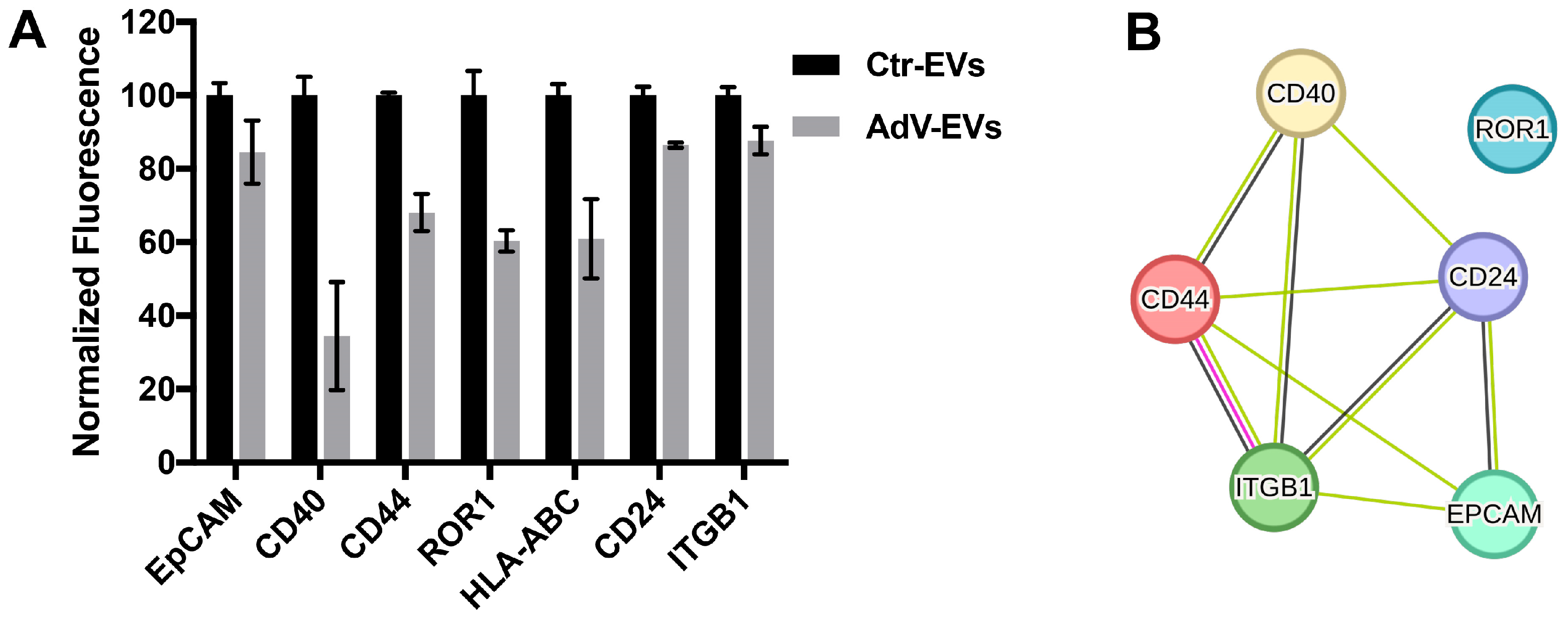
To better characterize the protein composition of the EV membranes, Ctr-EVs and AdV-EVs were screened for the presence of 37 different proteins by flow cytometry. Consistent with their epithelial origin, both EV populations expressed the epithelial marker EpCAM (CD326). The other proteins detected were CD40, CD44, ROR1, HLA-ABC, CD24 and ITGB1 (Integrin beta-1, also known as CD29), which were less abundant in AdV-EVs compared to Ctr-EVs (Figure 2A). A functional enrichment analysis conducted by interrogating the STRING protein interaction database [33] indicated that EpCAM, CD40, CD44, CD24 and ITGB1 are part of a functional protein association network (enrichment p-value = 1.37 × 10⁻⁵), which includes a direct interaction between CD44 and ITGB1 (Figure 2B). Interestingly, CD40, a member of the TNF-receptor superfamily that mediates humoral and adaptive immune responses in the lung [34], was the surface protein with the lowest abundance in AdV-EVs compared to Ctr-EVs.

3.3. Proteomics Profiling of AdV-EVs and Ctr-EVs

To determine whether the HAdV-E4 infection induces alterations in the protein composition of EVs secreted by A549 cells, the matched biological replicates of AdV-EVs (n = 5) and Ctr-EVs (n = 5) were isolated as described above and analyzed by data-independent acquisition mass spectrometry (DIA-MS). Proteins with an average spectral count > 2 for each group were considered. A total of 1,837 proteins were identified in Ctr-EVs and 2083 in AdV-EVs. Of these, 1,815 proteins (86.2%) were in common between Ctr-EVs and AdV-EVs; 22 proteins (1.0%) were exclusively present in Ctr-EVs, while 268 (12.7%) were exclusively present in AdV-EVs (Figure 3A). The proteins commonly found in EVs were detected in both the Ctr-EVs and AdV-EVs datasets. Among the proteins identified in both Ctr-EVs and AdV-EVs, 23 were present only in the ExoCarta list, 23 were present only in the Vesiclepedia list, and 67 proteins were present in both the ExoCarta and Vesiclepedia lists (Figure 3B). To obtain more specific information regarding the protein composition of EVs derived from the HAdV-E4-infected A549 cells, we performed a gene ontology (GO) and a network analysis on the 268 proteins that were exclusive to AdV-EVs. Significantly enriched GO terms were grouped in an annotation network based on the presence of similar or common proteins. The resulting network was composed of four major components, in which the most representative terms were endosomal transport, vesicle targeting, vesicle organization, vesicle fusion, and cytoplasmic translation (Figure 3C). Since these proteins were not detected in Ctr-EVs, their presence in AdV-EVs may be indicative of increased vesicle trafficking and viral protein translation in the originating cells as a consequence of the ongoing viral infection. To compare the relative protein abundance between AdV-EVs and Ctr-EVs, the normalized exclusive intensities were used for differential analysis.
A total of 326 proteins were differentially abundant (|log2 fold change| > 1, p-value < 0.05). Of these, 107 were more abundant in AdV-EVs and 219 were less abundant in AdV-EVs compared to Ctr-EVs, as shown in the volcano plot in Figure 4A. A hierarchical clustering of the differentially abundant proteins was generated and the corresponding heatmap shows that Ctr-EVs and AdV-EVs cluster separately (Figure 4B). The top ten proteins with increased or decreased abundance in AdV-EVs compared to Ctr-EVs are shown in Table 2.
The differentially abundant proteins were further investigated by the GO analysis. The most enriched biological processes for the proteins with increased abundance in AdV-EVs included the GO terms representative of protein translation in response to viral infections, cytoskeletal remodeling, interactions with the extracellular matrix, and mTOR and ErbB2 signaling pathways (Figure 4C). Interestingly, adenovirus proteins E4-ORF1 and E4-ORF4 can activate mTOR signaling to promote protein translation and thus facilitate viral replication [35], and E1A can affect ErbB2 expression and inhibit cell proliferation [36]. The proteins with decreased abundance in AdV-EVs showed enrichment for terms related to the Toll-like receptor signaling, tumor necrosis factor (TNF), interleukin-mediated inflammatory response, and activation of the complement pathway, which are all components of the innate antiviral immune response [37,38] (Figure 4D). The differential abundance analysis confirmed that CD40, which showed reduced surface expression in AdV-EVs (Figure 2), was less abundant in AdV-EVs in terms of total proteins and contributed to the TNFR/NF-kB-related pathways illustrated in Figure 4D. The proteomics dataset was also mined for the presence of HAdV-E4-encoded proteins. A set of eight adenoviral proteins were detected in AdV-EVs. Of these, six are structural components of the virion and two, E2A-DBP (DNA Binding Protein) and L4-100K, are non-structural proteins (Figure 5). The DBP plays a critical role in the AdV life cycle controlling viral DNA replication and transcription, and the mRNA stability [39]. L4-100K, also known as shutoff protein, blocks the translation of host proteins, while promoting the translation of viral proteins [40].

3.4. Small RNA Content of AdV-EVs and Ctr-EVs

EVs can carry a spectrum of RNA molecules that encompass protein-coding RNAs and non-coding RNAs. Among the non-coding RNAs found in EVs, the small RNA class includes microRNAs (miRNAs), small nucleolar RNA (snoRNAs), small nuclear RNAs (snRNAs), piwi-interacting RNAs (piRNAs), transfer RNA (tRNAs), and ribosomal RNAs (rRNAs) [2]. To characterize the small RNA cargo of EVs released during the early non-lytic stages of the HAdV-E4 infection, we performed small RNA sequencing of matched Ctr-EVs (n = 3) and AdV-EVs (n = 3) collected at 24 hpi. The data analysis identified a total of 813 and 806 unique transcripts with average counts ≥ 2 in Ctr-EVs and AdV-EVs, respectively. The most represented class of small RNAs in both Ctr-EVs and AdV-EVs was piRNAs, followed by snoRNAs, snRNAs and miRNAs (Table 3).
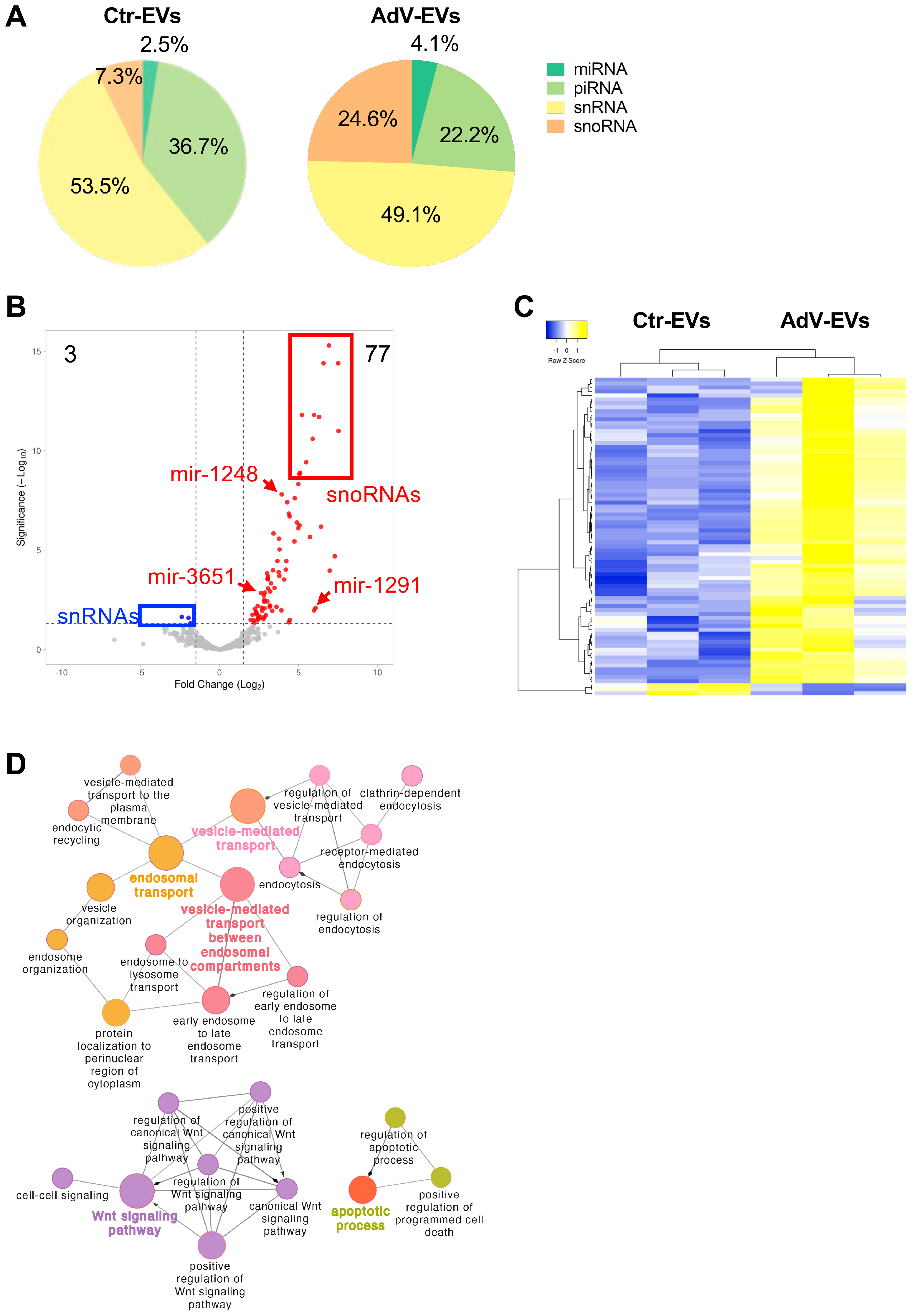
The relative abundance of each small RNA class in terms of mapped reads was also analyzed (Figure 6A). The snRNAs were the most abundant class in both Ctr-EVs and AdV-EVs, accounting for 53.5% and 49.1% of total mapped reads, respectively. The piRNAs, which were the second most abundant class in Ctr-EVs, showed a drastic reduction in AdV-EVs, while miRNAs and especially snoRNAs showed increased abundance in AdV-EVs. The differential abundance analysis performed comparing AdV-EVs versus Ctr-EVs (|log2 fold change| > 1, adjusted p-value < 0.05), confirmed the general increased transcript presence in the cargo of AdV-EVs, especially for the snoRNAs, and identified 70 snoRNAs, 4 piRNAs, and 3 miRNA precursors as upregulated, while three snRNAs were downregulated (Table 4 and Figure 6B). Table 5 shows the 10 most upregulated and the three down-regulated small non-coding transcripts in AdV-EVs. The hierarchical clustering heatmap generated with the differentially uploaded transcripts shows that Ctr-EVs and AdV-EVs cluster separately (Figure 6C). Because of the growing evidence of a role of certain classes of snoRNAs in viral infections [41], the RNAInter database was interrogated to identify potential interactors for the 70 upregulated snoRNAs, focusing in particular on RNA binding proteins (RBPs) as possible effectors. Among the total 376 predicted interactors, 40 were classified as RBPs. The two top interactors were METTL3 and FMR1, showing potential binding to 30 and 29 different snoRNAs, respectively. Interestingly, METTL3 is a cellular m6A writer enzyme recently shown to control the efficiency of HAdV-5 late gene transcript splicing [42], and the FMR1 homolog FXR1 is a cellular RBP recently described as a novel m6A reader that regulates AdV late mRNA transcript stability [43].
Among the 40 mature miRNAs identified in the dataset, four were exclusively detected in AdV-EVs, namely let-7f-5p, miR-93-5p, miR-7704 and miR-7706. To obtain predictive insights about the possible effect of EV-mediated delivery of these miRNAs to recipient naïve cells, we interrogated three different databases of miRNA-target interactions: TargetScan, microT-CDS and miRDB. The predicted target genes that were present in all three databases (intersection) were used for subsequent analyses. The resulting gene list was further filtered by using a publicly available expression dataset from The Genotype-Tissue Expression (GTEx) Project (https://www.gtexportal.org accessed on 4 February 2025) consisting of 515 human lung tissue samples. From this dataset, only genes that had a median expression above a threshold of three transcript per million (TPM) were considered. The final gene list comprised 979 unique target genes, of which 388 were predicted targets of let-7f-5p, 626 of miR-93-5p, 5 of miR-7004, 15 of miR-7006. To identify biological pathways that could be potentially inhibited by the four miRNAs when delivered to recipient cells, the predicted target genes were analyzed by the GO and the results visualized in an interaction network (Figure 6D). The analysis showed enrichment for terms related to endosomal transport, vesicle trafficking, Wnt signaling pathway and apoptotic process. It has been shown that the Wnt signaling pathway is dysregulated during viral infections [44], while suppression of apoptosis has also been associated with adenovirus infection [45].
Altogether, our data indicate that the HAdV-E4 infection alters the small ncRNA composition of EVs released by the infected cells. Like HAdVs of other species, HAdV-E4 encodes two small non-coding RNAs, known as virus associated (VA) RNAI and VA RNAII, which are transcribed by RNA Pol III, and are not translated into protein. The VA RNAs are proviral factors that modulate innate host cell defenses and interfere with cellular processes such as nuclear RNA export, protein synthesis and miRNA biogenesis [46,47]. To investigate whether EVs released by AdV-infected cells carry VA RNAs, the reads obtained from the transcriptomics data were also mapped to the HAdV-E4 genome (GenBank accession #NC_003266.2). Both VA RNAI and VA RNAII were identified in the small RNA cargo of AdV-EVs, with the VA RNAI content being more abundant than that of VA RNAII (Figure 7A). The observed difference in abundance mirrors the relative VA RNAI/VA RNAII abundance reported for the HAdV-C-infected cells [46]. The RNA-Seq data for VA RNAI/VA RNAII were subsequently validated by RT-qPCR (Figure 7B).

3.5. Characterization of EV Biological Properties

We showed that AdV-EVs carry viral proteins and viral RNAs. Next, we tested AdV-EVs for presence of viral genomic DNA by qPCR and evaluated their infectivity by plaque assay in the A549 cells. As shown in Figure 8A, several plaques were visible in infected wells indicating that purified EVs from the AdV-infected cells carry and transduce the replication competent HAdV-E4 genomes to the naïve recipient cells successfully. To gain more insights about the localization of EV-associated viral DNA, we subjected AdV-EVs to a series of treatments with DNase I, proteinase K, Triton X-100, and all possible combinations of these and evaluated their infectivity by plaque assay. We observed a significant reduction in the number of plaques after proteinase K treatment in all treatment combinations. The DNase treatment alone was not effective in reducing the number of plaques. Interestingly, the treatment with the detergent did not abolish plaque formation but resulted in the formation of plaques of smaller size (Figure 8B). The effect of the treatments on the structural integrity of the EVs was also evaluated by TEM. The treatment with DNase I, proteinase K or the combination of the two did not result in any apparent change. In contrast, and as expected, the exposure to detergent Triton X-100 lysed all vesicular structures (Figure 8C). To further evaluate the impact of the various treatments, total DNA was extracted from each treated sample, to determine genome copy concentration by qPCR (Figure 8D). The reduction of viral DNA load was observed only in samples treated with proteinase K and DNase I, or with Triton X-100, Proteinase K and DNase I. Taking into consideration that a full-length HAdV genome is needed for a productive infection, these data suggest that the viral genome is carried in detergent-resistant DNA–protein complexes associated with EVs, and that proteins present on the surface of the EVs are required for efficient uptake by target cells and subsequent traffic to the nucleus.

3.6. Neutralization Assays

As part of the characterization of the documented infectivity of AdV-EV preps, we carried out plaque reduction neutralization assays using HAdV-E4 virus as a reference for comparison. Interestingly, as shown in Table 6, the rabbit hyperimmune serum S-1001 raised against HAdV-E4 effectively neutralized EV infectivity, completely preventing plaque formation at dilutions ranging from 1:16 to 1:256 and neutralizing ~70% of EV infectivity at dilution 1:2048. The virus, on the other hand, was completely neutralized by serum dilution 1:1024, and ~70% neutralized by serum dilution 1:8192.
To identify possible EV-associated targets of neutralizing antibodies, the protein extracts from Ctr-EVs and AdV-EVs were subjected to immunoprecipitation with the anti-HAdV-E4 rabbit serum and processed for proteomics profiling by mass spectrometry. These experiments confirmed the presence of the AdV proteins in AdV-EVs (penton, hexon, pre-hexon-linking protein IIIa and hexon-interlacing protein pIX), and also identified 123 proteins that were present in both Ctr-EVs and AdV-EVs. A GO annotation analysis focused on cellular component (CC) ontology was performed. The results revealed that 23 of the identified proteins are plasma membrane-associated (Table 7) and thus potential targets of neutralizing antibodies.

4. Discussion

The present study describes the cargo composition of small EVs secreted by the HAdV-E4-infected cells at 24 h post infection prior to the lytic release of progeny virions and identifies a potential role for these vesicles in non-lytic cell-to-cell dissemination of infection.
Altogether, the results of the conducted transcriptomics and proteomics analyses indicate that the HAdV-E4 infection greatly affects the content of EVs released by infected cells. While the identified differentially uploaded proteome and small non-coding RNA cargo likely reflect changes in the infected cells occurring in response to infection, they also reveal specific HAdV strategies to hijack EV biogenesis and exploit cargo sorting pathways to upload selected viral-encoded molecules into small EVs increasing the virus’ replicative potential [6]. In support of this hypothesis, we observed dysregulation of proteins involved in the vesicle formation process and in endosomal transport (Figure 3C), and the presence in AdV-EVs of miRNAs predicted to be involved in such processes (Figure 6D), although the precise role of let-7f-5p, miR-93-5p, miR-7704 and miR-7706 in adenovirus infection has not been elucidated to the present.
In agreement with the data reported by Saari and colleagues from studies conducted using oncolytic adenovirus vector Ad5/3-D24-GMCSF [17,48], we identified our AdV-EV population purified using an immunocapture approach as infectious and carrying HAdV-E4 genomic DNA, as well as several virus-encoded proteins. As part of their studies characterizing the role of exosomes in the removal of harmful DNA, Takahashi and colleagues also reported the detection of viral genomic DNA in exosomes secreted from fetal lung fibroblast TIG-3 cells infected with an adenovirus expressing GFP [49]. Consistent with the recent observations of Brachtlova et al. [18], our RNAseq efforts allowed the identification in the cargo of AdV-EVs of the unique viral small non-coding RNAs VA RNA I and VA RNA II.
In addition to identifying viral components, our proteomic and transcriptomic analyses reveal systematic changes in the EV cargo that suggest functional consequences for cell communication and immune modulation during infection. The enrichment in ribosomal proteins and translation-associated factors (e.g., RPL4, RPL7, RPS6) in AdV-EVs may favor protein synthesis in recipient cells and thereby support viral gene expression. In contrast, the decreased abundance of proteins involved in innate immune signaling (such as CD40 and TRAF2) and components of Toll-like receptor and complement pathways suggests a down-modulation of antiviral responses. Together with the selective uploading of viral VA RNAs and several snoRNAs, known to interfere with RNA processing and PKR activation, these findings indicate that AdV-EVs might exert proviral effects by creating a cellular environment more permissive for viral replication. Similar shifts in the EV cargo have been reported in other virus–EV systems as follows: the AdV-B3 infection altered EV composition in line with modulation of immune signaling [16], and related phenomena have also been observed in the HBV and poliovirus infections, where EVs were found to carry replication-supporting proteins and immune regulators [50,51]. In this broader context, the changes detected in AdV-EVs fit with a general strategy whereby some viruses exploit EV biogenesis to facilitate spread and attenuate host antiviral defenses.
We specifically designed our studies to focus on the EV population released by AdV-infected cells before cell lysis to avoid contamination of our preparations with progeny AdV virions. In the absence of detectable viral particles in our EV preparations by the TEM, we attributed infectivity to efficient uptake and nuclear delivery of capsid-free viral genomes by EVs. Although we cannot rule out the existence of residual viral particles below the TEM sensitivity, the high plaque-forming activity observed in our assays cannot reasonably be explained by such occasional virions. The detergent/protease/nuclease treatments further show that infectivity is resistant to membrane solubilization but abolished when DNA and associated proteins are degraded, consistent with detergent-resistant DNA–protein complexes being the principal infectious entities in our EV preparations. The integrity of the infected cells at the early time post infection chosen for our harvest (Figure 1) and the size of our purified EV population rule out the presence of the large cytotoxic vesicles encapsulating mature virions described by Ran and colleagues in their vesicle preparations derived from the A549 cells infected with oncolytic Ad5 hTERTp-E1A at 48 h post infection when most cells were detached from the culture dishes [19]. While virions of other virus families have been detected inside small EVs [11,52], there is currently no evidence that the AdV virions can become encapsulated in small (<200 nm) EVs. However, it has become evident that cells infected by a variety of virus families can secrete EVs containing viral genetic material [53,54], disclosing a potential role for EVs in non-lytic viral egress and dissemination [11]. The presence of infectious DNA–protein complexes in EV preparations raises the question of how viral genomes are incorporated into vesicles and subsequently delivered to the nucleus of recipient cells. Considering the canonical replicative cycle of adenovirus, one possibility is that viral genomes bound to viral proteins exit the nucleus either through nuclear envelope disruption, leakage into the cytoplasm, or capture by autophagic vesicles, and subsequently enter the multivesicular bodies through ESCRT-dependent sorting. These complexes may enter recipient cells by EV endocytosis or membrane fusion. Once internalized, DNA associated with proteins that carry nuclear localization signals such as the DNA binding protein DBP and the viral core protein pIVa2, could be transported into the nucleus without an intact capsid. Although these mechanisms are speculative, our finding that infectivity is abolished by combined DNase and protease treatment supports the idea that EVs transmit viral DNA in the form of protein–DNA assemblies rather than complete virions. Further work will be needed to better describe the molecular composition of these complexes and the precise route by which they reach the nucleus.
Here we showed, for a replication-competent HAdV type of medical importance, that tetraspanin CD9-, CD63- and CD81-positive small EVs released by infected lung epithelial cells at a pre-lytic early stage during infection carry the viral genome, select structural and non-structural viral proteins and VA RNAs providing a vehicle for protected cell-to-cell dissemination of the HAdV infection. Collectively, our data and those of previous studies indicate that AdVs have developed strategies to exploit cellular sorting mechanisms for proteins [55,56], harmful DNA [49], and miRNAs [57] into small EVs. Given their multiple proviral functions including the inhibition of host innate immune responses through binding of PKR and suppression of the host miRNA biogenesis [47], the presence of VA RNAs inside AdV-EVs appears advantageous to the functional efficiency of these EVs as early vehicles for infection dissemination. This is also the case with the DBP, a non-structural protein encoded in the E2A transcriptional unit that plays key roles in viral DNA replication, transcription, and viral gene expression [39], and with non-structural viral shutoff protein L4-100K, which favors translation of viral proteins at the expenses of host proteins [40].
Our characterization of the small non-coding RNA and protein cargo provides a foundation of reference data for future studies investigating the conservation of the identified viral-encoded cargo across members of the seven species comprising HAdV types, and for necessary studies designed to further understand the involvement of EVs during adenovirus infection and their potential contribution to the expansion of cellular/tissue tropism in vivo.
Finally, our intriguing findings of neutralization activity of rabbit polyclonal anti HAdV-4 serum against the AdV-EV infectivity are consistent with the observations of Saari and colleagues using rabbit polyclonal anti-HAdV-5 antibody on EVs purified from Ad5/3-D24-GMCSF-infected cells [17]. The presence of neutralizing antibodies in both rabbit sera is likely the result of the use of infected cell lysates–instead of purified virions–for the immunization of rabbits. Future work addressing these questions must include appropriate control sera raised against other serotypes or uninfected cell lysates. However, these unexpected observations disclose the existence of potential targets for passive immune therapeutic intervention that warrant further investigation.

Author Contributions

Conceptualization, A.N., C.O. and A.E.K.; methodology, A.N. and A.E.K.; visualization, A.N.; validation, A.N.; formal analysis, A.N., S.B. and A.E.K.; writing—original draft preparation, A.N., S.B. and A.E.K.; writing—review and editing, A.N., S.B., C.O. and A.E.K.; supervision, A.E.K. All authors have read and agreed to the published version of the manuscript.

Funding

This research received intramural funding from Lovelace Biomedical Research Institute. The IDeA National Resource for Quantitative Proteomics is supported by NIH grant R24GM137786; Chioma Okeoma received funding from the National Institutes of Health grants R01DA042348-01, R01DA050169 (to CMO), R21/R33DA053643, and S10OD 036364.

Institutional Review Board Statement

Not applicable.

Informed Consent Statement

Not applicable.

Data Availability Statement

The raw proteomics and small RNAseq data supporting the conclusions of this article will be made available by the authors upon request.

Acknowledgments

The authors wish to thank Susan Fort at Lovelace for technical assistance with EV preparation and the WB analysis; Tamara Howard at the Electron Microscopy Facility, The University of New Mexico Health Sciences Center for sample processing for the TEM; and Sam Mackintosh, IDeA National Resource for Quantitative Proteomics for general support and advice with mass spectrometry. This project was supported by Lovelace Biomedical Research Institute intramural support to A.E.K.

Conflicts of Interest

The authors declare no conflicts of interest.

Abbreviations

The following abbreviations are used in this manuscript:
HAdVHuman adenovirus
EVExtracellular vesicle
NTANanoparticle Tracking Analysis
TEMTransmission Electron Microscopy

References

  1. Welsh, J.A.; Goberdhan, D.C.I.; O’Driscoll, L.; Buzas, E.I.; Blenkiron, C.; Bussolati, B.; Cai, H.; Di Vizio, D.; Driedonks, T.A.P.; Erdbrügger, U.; et al. Minimal information for studies of extracellular vesicles (MISEV2023): From basic to advanced approaches. J. Extracell. Vesicles 2024, 13, e12404. [Google Scholar] [CrossRef]
  2. Kim, K.M.; Abdelmohsen, K.; Mustapic, M.; Kapogiannis, D.; Gorospe, M. RNA in extracellular vesicles. Wiley Interdiscip. Rev. RNA 2017, 8, 10.1002/wrna.1413. [Google Scholar] [CrossRef]
  3. Jeppesen, D.K.; Fenix, A.M.; Franklin, J.L.; Higginbotham, J.N.; Zhang, Q.; Zimmerman, L.J.; Liebler, D.C.; Ping, J.; Liu, Q.; Evans, R.; et al. Reassessment of Exosome Composition. Cell 2019, 177, 428–445.e18. [Google Scholar] [CrossRef] [PubMed]
  4. Dixson, A.C.; Dawson, T.R.; Di Vizio, D.; Weaver, A.M. Context-specific regulation of extracellular vesicle biogenesis and cargo selection. Nat. Rev. Mol. Cell Biol. 2023, 24, 454–476. [Google Scholar] [CrossRef]
  5. Amin, S.; Massoumi, H.; Tewari, D.; Roy, A.; Chaudhuri, M.; Jazayerli, C.; Krishan, A.; Singh, M.; Soleimani, M.; Karaca, E.E.; et al. Cell Type-Specific Extracellular Vesicles and Their Impact on Health and Disease. Int. J. Mol. Sci. 2024, 25, 2730. [Google Scholar] [CrossRef]
  6. Mardi, N.; Haiaty, S.; Rahbarghazi, R.; Mobarak, H.; Milani, M.; Zarebkohan, A.; Nouri, M. Exosomal transmission of viruses, a two-edged biological sword. Cell Commun. Signal 2023, 21, 19. [Google Scholar] [CrossRef]
  7. Feng, Z.; Hensley, L.; McKnight, K.L.; Hu, F.; Madden, V.; Ping, L.; Jeong, S.-H.; Walker, C.; Lanford, R.E.; Lemon, S.M. A pathogenic picornavirus acquires an envelope by hijacking cellular membranes. Nature 2013, 496, 367–371. [Google Scholar] [CrossRef]
  8. Huang, H.-I.; Lin, J.-Y.; Chiang, H.-C.; Huang, P.-N.; Lin, Q.-D.; Shih, S.-R. Exosomes Facilitate Transmission of Enterovirus A71 From Human Intestinal Epithelial Cells. J. Infect. Dis. 2020, 222, 456–469. [Google Scholar] [CrossRef]
  9. Robinson, S.M.; Tsueng, G.; Sin, J.; Mangale, V.; Rahawi, S.; McIntyre, L.L.; Williams, W.; Kha, N.; Cruz, C.; Hancock, B.M.; et al. Coxsackievirus B exits the host cell in shed microvesicles displaying autophagosomal markers. PLoS Pathog. 2014, 10, e1004045. [Google Scholar] [CrossRef]
  10. Iša, P.; Pérez-Delgado, A.; Quevedo, I.R.; López, S.; Arias, C.F. Rotaviruses Associate with Distinct Types of Extracellular Vesicles. Viruses 2020, 12, 763. [Google Scholar] [CrossRef]
  11. Yang, J.E.; Rossignol, E.D.; Chang, D.; Zaia, J.; Forrester, I.; Raja, K.; Winbigler, H.; Nicastro, D.; Jackson, W.T.; Bullitt, E. Complexity and ultrastructure of infectious extracellular vesicles from cells infected by non-enveloped virus. Sci. Rep. 2020, 10, 7939. [Google Scholar] [CrossRef] [PubMed]
  12. Lynch, J.P., 3rd; Kajon, A.E. Adenovirus: Epidemiology, Global Spread of Novel Types, and Approach to Treatment. Semin. Respir. Crit. Care Med. 2021, 42, 800–821. [Google Scholar] [CrossRef] [PubMed]
  13. Kajon, A.E. Adenovirus infections: New insights for the clinical laboratory. J. Clin. Microbiol. 2024, 62, e0083622. [Google Scholar] [CrossRef]
  14. Lion, T. Adenovirus persistence, reactivation, and clinical management. FEBS Lett. 2019, 593, 3571–3582. [Google Scholar] [CrossRef]
  15. MacNeil, K.M.; Dodge, M.J.; Evans, A.M.; Tessier, T.M.; Weinberg, J.B.; Mymryk, J.S. Adenoviruses in medicine: Innocuous pathogen, predator, or partner. Trends Mol. Med. 2023, 29, 4–19. [Google Scholar] [CrossRef]
  16. Ipinmoroti, A.O.; Crenshaw, B.J.; Pandit, R.; Kumar, S.; Sims, B.; Matthews, Q.L. Human Adenovirus Serotype 3 Infection Modulates the Biogenesis and Composition of Lung Cell-Derived Extracellular Vesicles. J. Immunol. Res. 2021, 2021, 2958394. [Google Scholar] [CrossRef]
  17. Saari, H.; Turunen, T.; Lõhmus, A.; Turunen, M.; Jalasvuori, M.; Butcher, S.J.; Ylä-Herttuala, S.; Viitala, T.; Cerullo, V.; Siljander, P.R.M.; et al. Extracellular vesicles provide a capsid-free vector for oncolytic adenoviral DNA delivery. J. Extracell. Vesicles 2020, 9, 1747206. [Google Scholar] [CrossRef]
  18. Brachtlova, T.; Li, J.; van der Meulen-Muileman, I.H.; Sluiter, F.; von Meijenfeldt, W.; Witte, I.; Massaar, S.; van den Oever, R.; de Vrij, J.; van Beusechem, V.W. Quantitative Virus-Associated RNA Detection to Monitor Oncolytic Adenovirus Replication. Int. J. Mol. Sci. 2024, 25, 6551. [Google Scholar] [CrossRef]
  19. Ran, L.; Tan, X.; Li, Y.; Zhang, H.; Ma, R.; Ji, T.; Dong, W.; Tong, T.; Liu, Y.; Chen, D.; et al. Delivery of oncolytic adenovirus into the nucleus of tumorigenic cells by tumor microparticles for virotherapy. Biomaterials 2016, 89, 56–66. [Google Scholar] [CrossRef]
  20. Garofalo, M.; Saari, H.; Somersalo, P.; Crescenti, D.; Kuryk, L.; Aksela, L.; Capasso, C.; Madetoja, M.; Koskinen, K.; Oksanen, T.; et al. Antitumor effect of oncolytic virus and paclitaxel encapsulated in extracellular vesicles for lung cancer treatment. J. Control. Release 2018, 283, 223–234. [Google Scholar] [CrossRef]
  21. Garofalo, M.; Villa, A.; Rizzi, N.; Kuryk, L.; Rinner, B.; Cerullo, V.; Yliperttula, M.; Mazzaferro, V.; Ciana, P. Extracellular vesicles enhance the targeted delivery of immunogenic oncolytic adenovirus and paclitaxel in immunocompetent mice. J. Control. Release 2019, 294, 165–175. [Google Scholar] [CrossRef]
  22. Bair, C.R.; Zhang, W.; Gonzalez, G.; Kamali, A.; Stylos, D.; Blanco, J.C.G.; Kajon, A.E. Human Adenovirus Type 4 Comprises Two Major Phylogroups with Distinct Replicative Fitness and Virulence Phenotypes. J. Virol. 2022, 96, e01090-21. [Google Scholar] [CrossRef]
  23. Hughes, C.S.; Moggridge, S.; Müller, T.; Sorensen, P.H.; Morin, G.B.; Krijgsveld, J. Single-pot, solid-phase-enhanced sample preparation for proteomics experiments. Nat. Protoc. 2019, 14, 68–85. [Google Scholar] [CrossRef]
  24. Graw, S.; Tang, J.; Zafar, M.K.; Byrd, A.K.; Bolden, C.; Peterson, E.C.; Byrum, S.D. proteiNorm—A User-Friendly Tool for Normalization and Analysis of TMT and Label-Free Protein Quantification. ACS Omega 2020, 5, 25625–25633. [Google Scholar] [CrossRef] [PubMed]
  25. Huber, W.; von Heydebreck, A.; Sultmann, H.; Poustka, A.; Vingron, M. Variance stabilization applied to microarray data calibration and to the quantification of differential expression. Bioinformatics 2002, 18 (Suppl. S1), S96–S104. [Google Scholar] [CrossRef] [PubMed]
  26. Thurman, T.J.; Washam, C.L.; Alkam, D.; Bird, J.T.; Gies, A.; Dhusia, K.; Robeson, M.S.; Byrum, S.D. proteoDA: A package for quantitative proteomics. J. Open Source Softw. 2023, 8, 5184. [Google Scholar] [CrossRef]
  27. Ritchie, M.E.; Phipson, B.; Wu, D.; Hu, Y.; Law, C.W.; Shi, W.; Smyth, G.K. limma powers differential expression analyses for RNA-sequencing and microarray studies. Nucleic Acids Res. 2015, 43, e47. [Google Scholar] [CrossRef]
  28. Keerthikumar, S.; Chisanga, D.; Ariyaratne, D.; Al Saffar, H.; Anand, S.; Zhao, K.; Samuel, M.; Pathan, M.; Jois, M.; Chilamkurti, N.; et al. ExoCarta: A Web-Based Compendium of Exosomal Cargo. J. Mol. Biol. 2016, 428, 688–692. [Google Scholar] [CrossRef]
  29. Chitti, S.V.; Gummadi, S.; Kang, T.; Shahi, S.; Marzan, A.L.; Nedeva, C.; Sanwlani, R.; Bramich, K.; Stewart, S.; Petrovska, M.; et al. Vesiclepedia 2024: An extracellular vesicles and extracellular particles repository. Nucleic Acids Res. 2023, 52, D1694–D1698. [Google Scholar] [CrossRef]
  30. Kang, J.; Tang, Q.; He, J.; Li, L.; Yang, N.; Yu, S.; Wang, M.; Zhang, Y.; Lin, J.; Cui, T.; et al. RNAInter v4.0: RNA interactome repository with redefined confidence scoring system and improved accessibility. Nucleic Acids Res. 2022, 50, D326–D332. [Google Scholar] [CrossRef]
  31. Heim, A.; Ebnet, C.; Harste, G.; Pring-Åkerblom, P. Rapid and quantitative detection of human adenovirus DNA by real-time PCR. J. Med. Virol. 2003, 70, 228–239. [Google Scholar] [CrossRef]
  32. Rubinstein, E.; Thery, C.; Zimmermann, P. Tetraspanins affect membrane structures and the trafficking of molecular partners: What impact on extracellular vesicles? Biochem. Soc. Trans. 2025, 53, 371–382. [Google Scholar] [CrossRef]
  33. Szklarczyk, D.; Kirsch, R.; Koutrouli, M.; Nastou, K.; Mehryary, F.; Hachilif, R.; Gable, A.L.; Fang, T.; Doncheva, N.T.; Pyysalo, S.; et al. The STRING database in 2023: Protein-protein association networks and functional enrichment analyses for any sequenced genome of interest. Nucleic Acids Res. 2023, 51, D638–D646. [Google Scholar] [CrossRef]
  34. Kawabe, T.; Matsushima, M.; Hashimoto, N.; Imaizumi, K.; Hasegawa, Y. CD40/CD40 ligand interactions in immune responses and pulmonary immunity. Nagoya J. Med. Sci. 2011, 73, 69–78. [Google Scholar] [PubMed]
  35. O’Shea, C.C.; Choi, S.; McCormick, F.; Stokoe, D. Adenovirus overrides cellular checkpoints for protein translation. Cell Cycle 2005, 4, 883–888. [Google Scholar] [CrossRef] [PubMed]
  36. Jung, B.K.; Kim, Y.J.; Hong, J.; Chang, H.G.; Yoon, A.R.; Yun, C.O. ErbB3-Targeting Oncolytic Adenovirus Causes Potent Tumor Suppression by Induction of Apoptosis in Cancer Cells. Int. J. Mol. Sci. 2022, 23, 7127. [Google Scholar] [CrossRef] [PubMed]
  37. Albarnaz, J.D.; Weekes, M.P. Proteomic analysis of antiviral innate immunity. Curr. Opin. Virol. 2023, 58, 101291. [Google Scholar] [CrossRef]
  38. Mellors, J.; Tipton, T.; Longet, S.; Carroll, M. Viral Evasion of the Complement System and Its Importance for Vaccines and Therapeutics. Front. Immunol. 2020, 11, 1450. [Google Scholar] [CrossRef]
  39. Bertzbach, L.D.; Seddar, L.; von Stromberg, K.; Ip, W.H.; Dobner, T.; Hidalgo, P. The adenovirus DNA-binding protein DBP. J. Virol. 2024, 98, e0188523. [Google Scholar] [CrossRef]
  40. Weitzman, M.D.; Ornelles, D.A. Inactivating intracellular antiviral responses during adenovirus infection. Oncogene 2005, 24, 7686–7696. [Google Scholar] [CrossRef]
  41. Murray, J.L.; Sheng, J.; Rubin, D.H. A role for H/ACA and C/D small nucleolar RNAs in viral replication. Mol. Biotechnol. 2014, 56, 429–437. [Google Scholar] [CrossRef]
  42. Price, A.M.; Hayer, K.E.; McIntyre, A.B.R.; Gokhale, N.S.; Abebe, J.S.; Della Fera, A.N.; Mason, C.E.; Horner, S.M.; Wilson, A.C.; Depledge, D.P.; et al. Direct RNA sequencing reveals m(6)A modifications on adenovirus RNA are necessary for efficient splicing. Nat. Commun. 2020, 11, 6016. [Google Scholar] [CrossRef]
  43. Hajikhezri, Z.; Kaira, Y.; Schubert, E.; Darweesh, M.; Svensson, C.; Akusjarvi, G.; Punga, T. Fragile X-Related Protein FXR1 Controls Human Adenovirus Capsid mRNA Metabolism. J. Virol. 2023, 97, e0153922. [Google Scholar] [CrossRef]
  44. van Zuylen, W.J.; Rawlinson, W.D.; Ford, C.E. The Wnt pathway: A key network in cell signalling dysregulated by viruses. Rev. Med. Virol. 2016, 26, 340–355. [Google Scholar] [CrossRef]
  45. Zhao, H.; Granberg, F.; Pettersson, U. How adenovirus strives to control cellular gene expression. Virology 2007, 363, 357–375. [Google Scholar] [CrossRef] [PubMed]
  46. Punga, T.; Darweesh, M.; Akusjärvi, G. Synthesis, Structure, and Function of Human Adenovirus Small Non-Coding RNAs. Viruses 2020, 12, 1182. [Google Scholar] [CrossRef] [PubMed]
  47. Vachon, V.K.; Conn, G.L. Adenovirus VA RNA: An essential pro-viral non-coding RNA. Virus Res. 2016, 212, 39–52. [Google Scholar] [CrossRef]
  48. Koski, A.; Kangasniemi, L.; Escutenaire, S.; Pesonen, S.; Cerullo, V.; Diaconu, I.; Nokisalmi, P.; Raki, M.; Rajecki, M.; Guse, K.; et al. Treatment of cancer patients with a serotype 5/3 chimeric oncolytic adenovirus expressing GMCSF. Mol. Ther. 2010, 18, 1874–1884. [Google Scholar] [CrossRef]
  49. Takahashi, A.; Okada, R.; Nagao, K.; Kawamata, Y.; Hanyu, A.; Yoshimoto, S.; Takasugi, M.; Watanabe, S.; Kanemaki, M.T.; Obuse, C.; et al. Exosomes maintain cellular homeostasis by excreting harmful DNA from cells. Nat. Commun. 2017, 8, 15287. [Google Scholar] [CrossRef]
  50. Zhao, X.; Wu, Y.; Duan, J.; Ma, Y.; Shen, Z.; Wei, L.; Cui, X.; Zhang, J.; Xie, Y.; Liu, J. Quantitative proteomic analysis of exosome protein content changes induced by hepatitis B virus in Huh-7 cells using SILAC labeling and LC-MS/MS. J. Proteome Res. 2014, 13, 5391–5402. [Google Scholar]
  51. Oberholster, L.; Mathias, A.; Perriot, S.; Blaser, E.; Canales, M.; Jones, S.; Culebras, L.; Gimenez, M.; Kaynor, G.C.; Sapozhnik, A.; et al. Comprehensive proteomic analysis of JC polyomavirus-infected human astrocytes and their extracellular vesicles. Microbiol. Spectr. 2023, 11, e0275123. [Google Scholar] [CrossRef] [PubMed]
  52. Wu, Q.; Glitscher, M.; Tonnemacher, S.; Schollmeier, A.; Raupach, J.; Zahn, T.; Eberle, R.; Krijnse-Locker, J.; Basic, M.; Hildt, E. Presence of Intact Hepatitis B Virions in Exosomes. Cell Mol. Gastroenterol. Hepatol. 2023, 15, 237–259. [Google Scholar] [CrossRef] [PubMed]
  53. Chatterjee, S.; Kordbacheh, R.; Sin, J. Extracellular Vesicles: A Novel Mode of Viral Propagation Exploited by Enveloped and Non-Enveloped Viruses. Microorganisms 2024, 12, 274. [Google Scholar] [CrossRef] [PubMed]
  54. Kumar, A.; Kodidela, S.; Tadrous, E.; Cory, T.J.; Walker, C.M.; Smith, A.M.; Mukherjee, A.; Kumar, S. Extracellular Vesicles in Viral Replication and Pathogenesis and Their Potential Role in Therapeutic Intervention. Viruses 2020, 12, 887. [Google Scholar] [CrossRef]
  55. Chen, Y.; Zhao, Y.; Yin, Y.; Jia, X.; Mao, L. Mechanism of cargo sorting into small extracellular vesicles. Bioengineered 2021, 12, 8186–8201. [Google Scholar] [CrossRef]
  56. Waury, K.; Gogishvili, D.; Nieuwland, R.; Chatterjee, M.; Teunissen, C.E.; Abeln, S. Proteome encoded determinants of protein sorting into extracellular vesicles. J. Extracell. Biol. 2024, 3, e120. [Google Scholar] [CrossRef]
  57. Groot, M.; Lee, H. Sorting Mechanisms for MicroRNAs into Extracellular Vesicles and Their Associated Diseases. Cells 2020, 9, 1044. [Google Scholar] [CrossRef]
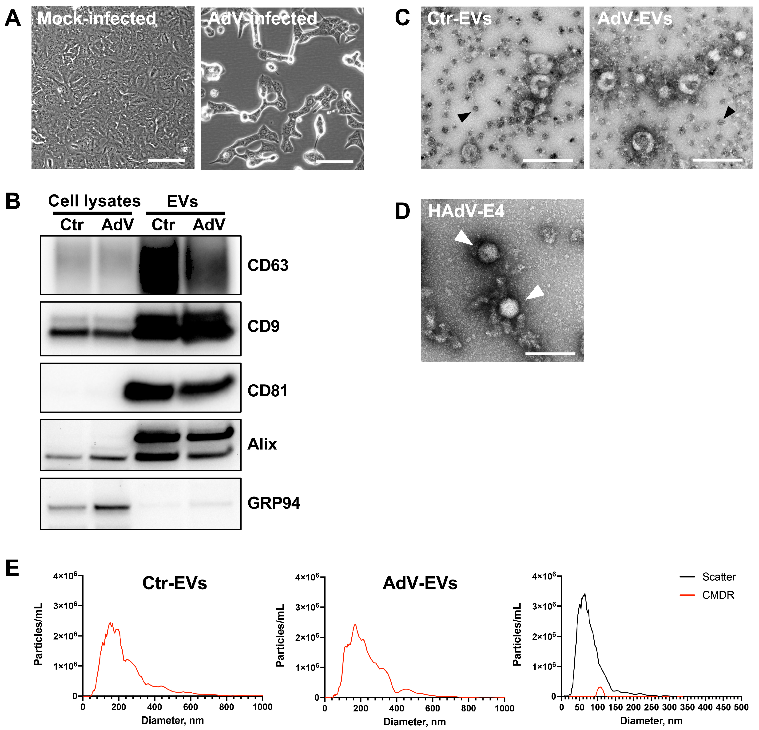
Figure 1. Characterization of the EV populations isolated from the mock- and AdV-infected A549 cells. (A) Phase contrast images of the A549 cells showing the magnitude of cytopathic effect at 24 h post-infection. Cells were infected at MOI = 10. Scale bars = 100 µm. (B) The representative Western blot analysis showing enrichment of the EV markers CD63, CD9, CD81, Alix in EVs-Ctr and EVs-AdV, compared to their cells of origin (cell lysates). The ER marker GRP94 was used to assess the presence of non-EV contaminants. Images are representative of three different experiments. (C) The TEM images of EVs isolated by magnetic immunocapture form the supernatants of mock-infected cells (Ctr-EVs) or HAdV-E4-infected (AdV-EVs) A549 cells. The magnetic beads are recognizable by their opaque appearance and uniform size of 50 nm (black arrows). Scale bars = 200 nm. (D) The TEM image of the purified HAdV-E4 particles (white arrows). Scale bar = 200 nm. (E) The fluorescent nanoparticle tracking analysis of Ctr-EVs (left panel), AdV-EVs (middle panel), and magnetic beads alone (right panel).
Viruses 17 01300 g001
Figure 2. Characterization of the surface markers of AdV-EVs and Ctr-EVs. (A) The MACSPlex flow cytometry detection of surface markers. The data represent ratios of normalized median fluorescent intensity (MFI) in AdV-EVs over Ctr-EVs. The mean values from of two independent experiments ± SEM are shown. (B) The STRING protein–protein interaction network analysis of the proteins shown in (A). The yellow lines represent text mining evidence, the black lines represent co-expression evidence, and the purple line represents experimental evidence.
Viruses 17 01300 g002
Figure 3. Proteome profiling of AdV-EVs and Ctr-EV. (A) Two-way Venn diagram of proteins identified in Ctr-EVs and AdV-EVs with spectral counts ≥ 2 in 5 biological replicates. 22 and 268 proteins were unique to Ctr-EVs and AdV-EVs, respectively. (B) 4-way Venn diagram showing the intersection between proteins in (A) and the top-100 EV proteins included in the Exocarta and Vesiclepedia repositories. (C) GO and network analysis of proteins found exclusively in AdV-EVs. Nodes represent GO terms with adjusted p-value < 0.05 after Benjamini-Hochberg correction. Ontology sources were Biological Process, Reactome Pathways and Immune System.
Viruses 17 01300 g003
Figure 4. The functional analysis of differentially abundant proteins in AdV-EVs versus Ctr-EVs. (A) A volcano plot of differentially abundant proteins in AdV-EVs versus Ctr-EVs. A total of 107 proteins were more abundant in AdV-EVs (log2FC > 1, p-value < 0.05), and 219 proteins were less abundant in AdV-EVs (log2FC < 1, p-value < 0.05). (B) The hierarchical clustering heatmap of differentially abundant proteins. The data from five biological replicates are shown. The yellow color indicates increased abundance, while the blue color indicates decreased abundance. (C,D) The GO and network analysis of more abundant (C) and less abundant (D) proteins in AdV-EVs. The nodes represent the GO terms with adjusted p-value < 0.05 after the Benjamini-Hochberg correction. Ontology sources were Biological Process, Reactome Pathways, and Immune System.
Viruses 17 01300 g004
Figure 5. Detection of adenovirus proteins in EVs-AdV. The hierarchical clustering heatmap of six structural and two non-structural HAdV-E4 proteins identified in AdV-EVs. The data from five biological replicates are shown. The yellow color indicates increased abundance, while the blue color indicates decreased abundance.
Viruses 17 01300 g005
Figure 6. Analysis of the small RNA transcriptome of Ctr-EVs and AdV-EVs. (A) The relative transcript abundance for each class of small RNAs in Ctr-EVs and AdV-EVs. (B) The volcano plot of differentially uploaded small RNAs in AdV-EVs versus Ctr-EVs. A total of 77 small RNAs were up-regulated in AdV-EVs (log2FC > 1, adjusted p-value < 0.05), and three small RNAs were down-regulated in AdV-EVs-(log2FC < 1, adjusted p-value < 0.05). (C) The hierarchical clustering heatmap of differentially uploaded small RNAs. The Data from three biological replicates are shown. The yellow color indicates increased presence, while the blue color indicates decreased presence. (D) The GO and network analysis of mature miRNAs exclusively found in AdV-EVs. The nodes represent GO terms with adjusted p-value < 0.05 after the Benjamini-Hochberg correction. Ontology sources were Biological Process, Reactome Pathways, and Immune System.
Viruses 17 01300 g006
Figure 7. Detection of VA-RNAs in AdV-EVs. (A) The normalized read counts that aligned to the VA RNAI and VA RNAII sequences of the HAdV-E4 genome. The individual data points of three biological replicates are shown. (B) The RT-qPCR expression data of VA RNA I and VA RNA II in Ctr-EVs and AdV-EVs. The data were normalized on ACTB expression and reported as -ddCt relative to EVs-Ctr. The mean ± SEM from two biological replicates is shown.
Viruses 17 01300 g007
Figure 8. Characterization of EV-associated infectivity. (A) The plaque assay in the A549 cells showing the infectivity of AdV-EVs. (B) The plaque assay performed with AdV-EV preparations subjected to the following treatments: 1, untreated; 2, DNase I; 3, proteinase K; 4, Triton X-100; 5, Triton X-100/proteinase K; 6, Triton X-100/DNase; 7, proteinase K/DNase I; 8, Triton X-100/proteinase K/DNase I. (C) The TEM images of AdV-EVs treated as in (B). Scale bars = 100 nm. (D) The genome copy number quantification by qPCR of AdV-EVs treated as indicated above. The bars represent the ratio of genome copy number/mL of treated AdV-EVs over genome copy number/mL of untreated AdV-EVs. The data are shown as mean of three biological replicates ± SEM.
Viruses 17 01300 g008
Table 1. Vesicle size distribution and concentration of purified Ctr- EVs and AdV- EVs.
Median Size ± SDConcentration (Particles/mL ± SD)
Ctr-EVs176 ± 18 nm1.45 × 1011 ± 5.00 × 10⁹
AdV-EVs165 ± 5 nm1.17 × 1011 ± 9.75 × 10⁹
Table 2. Top ten proteins with increased or decreased abundance in AdV-EVs compared to Ctr-EVs.
Increased Abundance Decreased Abundance
Protein Namelog2 Fold ChangeProtein Namelog2 Fold Change
FUS2.58SLITRK6−3.36
SLC20A12.49UBB;UBC−3.10
RPS62.32TRAF2−2.87
RPL72.29KRT19−2.86
RPL152.25PDGFA−2.84
COMT2.20DKK1−2.80
RPLP02.19CALML5−2.78
DTNB2.16KRT6B−2.72
RPL42.11RSPO3−2.70
KLHL132.04IFT81−2.70
Table 3. Number of unique small non-coding RNA transcripts in Ctr- and Adv-EVs grouped by class.
ClassCtr-EVsAdV-EVs
miRNA (precursors)1013
miRNA (mature)4028
piRNA375378
snRNA124107
snoRNA264280
Total unique transcripts813806
Table 4. Number of small non-coding RNA transcripts with differential relative abundance in AdV-EVs grouped by class.
ClassHigher
Abundance
Lower
Abundance
miRNA(p)30
piRNA40
snRNA03
snoRNA700
Total773
Table 5. The top transcripts with higher abundance and lower abundance in AdV- EVs compared Ctr-EVs.
Up-RegulatedDown-Regulated
Gene IDlog2 Fold ChangeGene IDlog2 Fold Change
SNORA417.53RNU6ATAC2P−2.39
SNORA74A7.51RNU6ATAC−1.97
SNORA227.30RNVU1-1−1.84
SNORA22C6.97
SNORA506.92
SNORA50C6.58
SNORA50A6.43
SNORA786.30
hsa-mir-12916.08
SNORA22B5.99
Table 6. Comparative neutralization of AdV-EV and HAdV-E4 infectivity by rabbit polyclonal anti HAdV-E4 serum.
Serum DilutionPercent Plaque Reduction
AdV-EVsHAdV-E4
1:256100100
1:51298100
1:102490100
1:20487396
1:4196Not tested90
1:8192Not tested67
Table 7. The plasma membrane-associated proteins identified by immunoprecipitation of protein extracts of AdV- EVs with rabbit anti-HAdV4 hyperimmune serum and mass spectrometry. The GO annotation analysis of cellular component terms.
Gene IDDescription
ABCA1Phospholipid-transporting ATPase ABCA1
ANXA2Annexin A2
APMAPAdipocyte plasma membrane-associated protein
APOA1Apolipoprotein A-I
ATP2B1Plasma membrane calcium-transporting ATPase 1
CD81CD81 antigen
CTNND1Catenin delta-1
DSPDesmoplakin
GNB2Guanine nucleotide-binding protein G(I)/G(S)/G(T) subunit beta-2
HPHaptoglobin
IGFBP3Insulin-like growth factor-binding protein 3
ITGA3Integrin alpha-3
ITGB1Integrin beta-1
JUPJunction plakoglobin
KRT77Keratin, type II cytoskeletal 1b
MYO1BUnconventional myosin-Ib
MYO1CUnconventional myosin-Ic
PKP1Plakophilin-1
PLEKHA7Pleckstrin homology domain-containing family A member 7
PLGPlasminogen
SLC12A2Solute carrier family 12 member 2
SLC3A2Amino acid transporter heavy chain SLC3A2
SMOC1SPARC-related modular calcium-binding protein 1
STOMStomatin
TIMP3Metalloproteinase inhibitor 3
Disclaimer/Publisher’s Note: The statements, opinions and data contained in all publications are solely those of the individual author(s) and contributor(s) and not of MDPI and/or the editor(s). MDPI and/or the editor(s) disclaim responsibility for any injury to people or property resulting from any ideas, methods, instructions or products referred to in the content.