Article

The Evolution of Highly Pathogenic Avian Influenza A (H5) in Poultry in Nigeria, 2021–2022

by
Clement Meseko
1,*,†,
Adelaide Milani
2,†,
Bitrus Inuwa
1,
Chinonyerem Chinyere
1,
Ismaila Shittu
1,
James Ahmed
1,
Edoardo Giussani
2,
Elisa Palumbo
2,
Bianca Zecchin
2,
Francesco Bonfante
2,
Silvia Maniero
2,
Angélique Angot
3,
Mamadou Niang
4,
Alice Fusaro
2,
Federica Gobbo
2,
Calogero Terregino
2,
Taiwo Olasoju
5,
Isabella Monne
2 and
Maryam Muhammad
1
1
Regional Laboratory for Animal Influenza & Transboundary Diseases, National Veterinary Research Institute (NVRI), Vom 930101, Nigeria
2
Istituto Zooprofilattico Sperimentale delle Venezie (IZSVe), Division of Comparative Biomedical Sciences (BSBIO), 35128 Padova, Italy
3
Animal Health Service (NSAH), Food and Agriculture Organization of the United Nations (FAO-UN), 00198 Rome, Italy
4
Regional Office for Africa (RAF), Emergency Centre for Transboundary Animal Diseases (ECTAD), Food and Agriculture Organization of the United Nations (FAO-UN), Accra 00233, Ghana
5
Federal Department of Veterinary and Pest Control Services (FDV&PCS), Federal Ministry of Agriculture and Rural Development (FMARD), Abuja 900108, Nigeria
*
Author to whom correspondence should be addressed.
These authors contributed equally to this work.
Viruses 2023, 15(6), 1387; https://doi.org/10.3390/v15061387
Submission received: 12 May 2023 / Revised: 9 June 2023 / Accepted: 14 June 2023 / Published: 17 June 2023
(This article belongs to the Special Issue Drivers of Evolution of Animal RNA Viruses, Volume II)

Abstract

:
In 2021, amidst the COVID-19 pandemic and global food insecurity, the Nigerian poultry sector was exposed to the highly pathogenic avian influenza (HPAI) virus and its economic challenges. Between 2021 and 2022, HPAI caused 467 outbreaks reported in 31 of the 37 administrative regions in Nigeria. In this study, we characterized the genomes of 97 influenza A viruses of the subtypes H5N1, H5N2, and H5N8, which were identified in different agro-ecological zones and farms during the 2021–2022 epidemic. The phylogenetic analysis of the HA genes showed a widespread distribution of the H5Nx clade 2.3.4.4b and similarity with the HPAI H5Nx viruses that have been detected in Europe since late 2020. The topology of the phylogenetic trees indicated the occurrence of several independent introductions of the virus into the country, followed by a regional evolution of the virus that was most probably linked to its persistent circulation in West African territories. Additional evidence of the evolutionary potential of the HPAI viruses circulating in this region is the identification in this study of a putative H5N1/H9N2 reassortant virus in a mixed-species commercial poultry farm. Our data confirm Nigeria as a crucial hotspot for HPAI virus introduction from the Eurasian territories and reveal a dynamic pattern of avian influenza virus evolution within the Nigerian poultry population.

1. Introduction

Poultry operations in Nigeria faced many challenges amidst the Coronavirus disease (COVID-19) pandemic, including global food insecurity and economic downturn [1]. Though the problems associated with highly pathogenic avian influenza (HPAI) in Nigeria and many countries predate the COVID-19 pandemic, persistent outbreaks, morbidity, and mortality in chickens and other birds have continued unabated and worsened the economic and public health impacts of these problems. The fear that HPAI, a zoonotic disease, could be a progenitor of another pandemic is rife and driven by ecological, behavioural, and socioeconomic changes, as well as the ability of this virus to rapidly evolve through both genetic reassortment and the acquisition of mutations [2,3].
The HPAI phenotype is caused by the H5 and H7 subtypes of the influenza A virus (IAV) of the Orthomyxoviridae family; this virus occurs naturally in wild waterfowls as a low pathogenic avian influenza (LPAI) virus and can mutate into HPAI [4] when transmitted to poultry. Influenza A viruses (IAV) are divided into subtypes based on their combinations of the two surface glycoproteins HA and NA; in birds, 16 HA and 9 NA types have been identified [5].
Over the last two decades, the world has witnessed multiple intercontinental epidemic waves of the H5Nx subtype of HPAI from Asia and Europe to Africa [6] as a consequence of the spread of viruses descended from the H5N1 virus A/goose/Guangdong/1/1996 (Gs/GD), which was first detected in China in 1996. Since their emergence, these viruses have dramatically expanded their geographical distribution, spreading to distinct continents and remaining well entrenched in a number of countries. They have been acquiring a broader host range, with a constantly expanding list of free-living wild bird species, which can potentially be affected by the virus, with spill-over events in mammals being increasingly reported [7,8].
Since 2006, multiple Gs/GD/96 H5Nx lineages have been introduced in Nigeria, which have had a negative impact on poultry productivity and food security and threatened public health. The first introduction of the Gs/Gd lineage of the HPAI clade 2.2 to Nigeria happened during the harmattan (winter) season of 2006 [9,10]. Between 2006 and 2008, when the epidemic occurred, millions of poultry birds either died or were culled to contain the further spread of the disease and the economic losses of farmers [11]. These control efforts paid off and the epizootic was declared over by a self-declaration of disease-free status at the World Organization for Animal Health (WOAH, formerly OIE) General Assembly in 2013 (https://www.woah.org/app/uploads/2022/06/eng-archive-2000-may-20222.pdf, accessed on 15 March 2023) [12].
Not long afterwards, a more devastating resurgence of another strain of HPAI caused by clade 2.3.2.1c was witnessed in 2015/2016 [13,14]. Thereafter, a series of re-introductions of various subtypes of HPAI into the Nigerian agro-ecological space, including the subtype H5N8, became more frequent [15]. H5N6 was detected in live bird markets (LBMs) in 2019 [16]. Adding to this plethora of outbreaks and detections of H5Nx in Nigeria, the low pathogenic avian influenza (LPAI) subtype, H9N2, has been added to the mix and is frequently and widely isolated in poultry, especially at LBMs [17].
Apparently, Nigeria has emerged as a regional hotspot of HPAI in sub-Saharan Africa [6] and accounts for a significant proportion of the cases reported to the WOAH from the African region over the last two decades (https://www.woah.org/en/disease/avian-influenza/#ui-id-2, accessed on 15 March 2023). More intriguing is the diversity of HPAI and LPAI that has been detected in the Nigerian agro-ecological space since 2006. HPAI of the H5N2 subtype has previously been detected in healthy wild waterfowl in Nigeria [18]. By 2011, Snoeck et al. [19] again reported the detection of reassortant LPAI H5N2 viruses in African wild birds sampled in the Hadejia-Nguru wetlands of northern Nigeria. Similarly, in 2013, Coker et al. [12] recovered genetically similar strains of H5N2 in commercial waterfowls at an LBM in southwestern Nigeria. These previous H5N2 strains were limited in their spread, as subsequent surveillance activities did not detect the H5N2, even in LBMs [16,17].
The agro-ecological source of the introduction of AIV into Nigeria is not fully understood and has been poorly investigated [10]. We can merely theorize based on the available epidemiological and genetic data that AIV is most likely introduced seasonally to Nigeria through activities and contact with migratory waterfowls from Eurasia [20]. Nigeria remains a country at risk of the continuous new introduction of AIV, with chances that an endemic situation may result in the co-circulation, co-infection, and emergence of possible new reassortant viruses.
Since early January 2021, new outbreaks of HPAI H5Nx have caused multiple outbreaks across the country and been reported in 30 out of the 37 administrative territories (Pers Comm: Federal Department of Veterinary and Pest Control Services), resulting in 467 positive cases as of December 2022. The transmission of these new HPAIVs in poultry flocks, where the H9N2 subtype is also entrenched in the poultry population, is a matter of great concern given the proclivity for the reassortment of AIV [17]. Using a wide collection of samples gathered throughout the 2021 and 2022 epidemic events in Nigeria, we investigated the genetic diversity of the AIVs circulating across the Nigerian states and provided evidence of the emergence of a putative H5N1/H9N2 reassortant in a mixed-species commercial poultry farm.

2. Materials and Methods

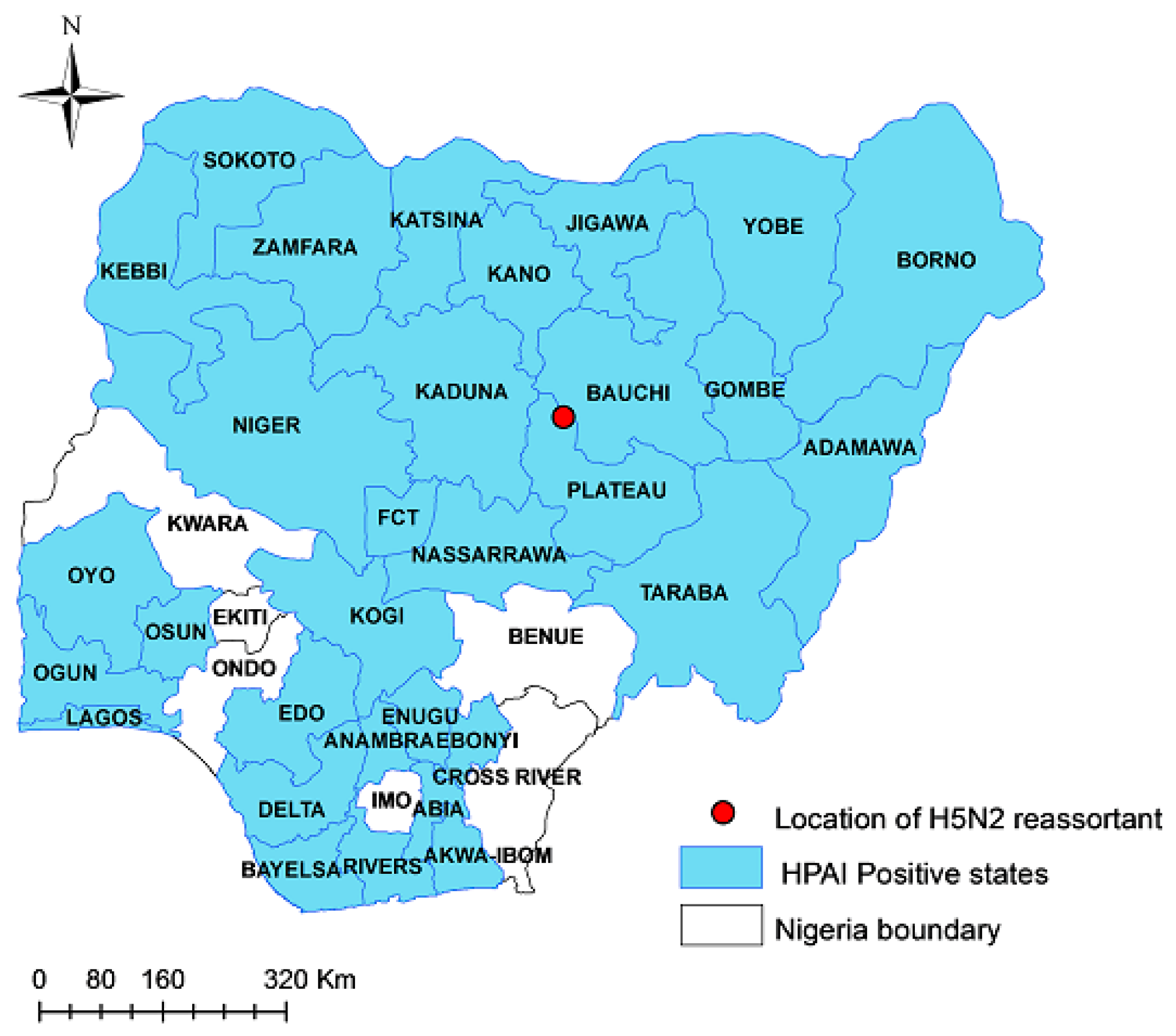
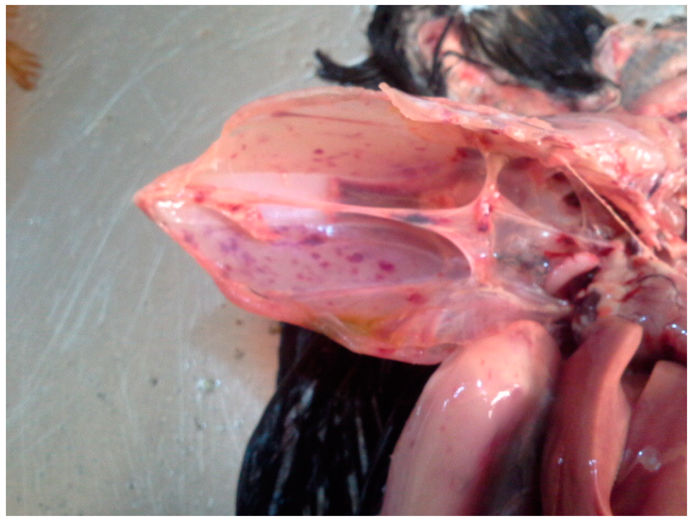
2.1. Sample Collection and Necropsy

Diagnostic outbreak investigations were carried out following a suspicion of HPAI outbreaks in Nigeria. From previous experience, poultry farmers are sensitive to abnormal mortality patterns and are quick to raise the index of suspicion by contacting the nearest private and state veterinarians. The attending veterinarians’ initial observations, with respect to clinical manifestations, were documented. Fresh carcasses were selected and shipped under cold-chain to the Regional Laboratory for Animal Influenza, National Veterinary Research Institute (NVRI) in Vom, for diagnosis and necropsy. Parenchymatous organs and tissues were harvested from the carcasses, pooling several tissues from the same flocks and species for further processing.

2.2. Virus Identification and Isolation

The issues were homogenized and supernatant fluid was collected for a nucleic acid extraction using the Qiagen Viral RNA kit (Qiagen, Hilden, Germany), following the manufacturer’s instructions. The total RNA was extracted from a pool of combined tissues and first screened for the IAV matrix gene (M-gene) and H5, H7, H9, and Nx IAV subtypes via an RT-qPCR using the QuantiTect Multiplex (Qiagen, Hilden, Germany) and OneStep RT-PCR (Qiagen, Hilden, Germany) kits, as previously described [21,22,23,24,25]. Influenza-A-positive samples were isolated and typed according to the OIE Terrestrial Manual (https://www.woah.org/fileadmin/Home/eng/Animal_Health_in_the_World/docs/pdf/2.03.04_AI.pdf, accessed on 15 March 2023).
To ascertain the presence of an H5N2 reassortant virus in the samples potentially co-infected with H9N2 and HPAI H5N1 viruses, homogenate supernatants were subjected in parallel to cloning through multiple rounds of limiting dilution in embryonated chicken eggs and plaque purification in MDCK cells. Briefly, for the limiting dilution cloning in the eggs, the original sample was serially diluted in a phosphate-buffered solution with antibiotics and incubated for 2 h at room temperature with chicken polyclonal antiserum raised against a G1 lineage H9N2 virus before its inoculation in the eggs. To plaque purify the virus, the homogenate supernatant was serially diluted in cell culture medium and plated onto the MDCK cells for 1 h at 37 °C in 5% CO2. After the removal of the inoculum, a 0.9% agar overlay was added and the plates were incubated for 72 h. The allantoic fluid and several plaques were collected from the eggs/cells infected with the highest dilution and underwent further diagnostic screening and NGS analyses.

2.3. Genomic Sequencing and Phylogenetic Analysis

Ninety-seven HPAI-H5-positive clinical specimens were selected based on their epidemiological locations, species, and time of outbreak and sent to the EURL, WOAH, and FAO reference laboratories for avian influenza (AI) in Italy (Istituto Zooprofilattico Sperimentale delle Venezie), where they underwent sequencing and genetic data analyses.
A target RT-PCR approach was used to amplify the influenza A virus whole genomes, as previously described [6]. The sequencing libraries were obtained using the Nextera XT DNA Sample preparation kit (Illumina, San Diego, CA, USA) and sequenced on an MiSeq instrument (San Diego, CA, USA) using the 2 × 250 bp PairedEnd [PE] mode.
The raw sequencing reads produced by the MiSeq instrument were cleaned with Trimmomatic v0.32 [26] with a minimum quality of 20. Illumina Nextera XT adapter sequences were clipped from the reads using scythe v0.991 (https://github.com/vsbuffalo/scythe, accessed on 15 March 2023) and amplification primers were removed using sickle v1.33 (https://github.com/najoshi/sickle, accessed on 15 March 2023). Reads that were unpaired or shorter than 80 bp were discarded. The cleaned reads were aligned against a reference genome using the MEM algorithm from BWA v0.7.1229. Picard tools v2.1.0 (http://broadinstitute.github.io/picard/, accessed on 15 March 2023) and GATK v3.530-32 [27] were used to improve the alignment quality, correct potential errors, and recalibrate the base quality score. LoFreq v2.1.233 [28] was used to call the single-nucleotide polymorphisms (SNPs).
The consensus sequence was created with an in-house script. The consensus sequences were aligned using the MAFFT v7 online server (https://mafft.cbrc.jp/alignment/server/, accessed on 15 March 2023) [29] and compared with the sequences of the most related virus strains available in GISAID. Maximum likelihood phylogenetic trees of each gene segment were obtained using IQTREE v1.6.6 (https://github.com/iqtree/iqtree1, accessed on 15 March 2023) and their robustness was determined via an ultrafast bootstrap resampling analysis of 1000 replications [30,31]. The phylogenetic trees were visualized using the FigTree v1.4.4 software (http://tree.bio.ed.ac.uk/software/figtree/, accessed on 15 March 2023).
The FluSurver tool (https://flusurver.bii.a-star.edu.sg/, accessed on 15 March 2023) was used to identify the amino acid mutations in the influenza A virus proteins, which could affect biological functions such as virulence, host adaptation, and drug resistance.

3. Results

3.1. Virus Identification and Pathological Findings

Depression, somnolence, drooling fluid from the mouth, diarrhoea, hock sitting, inappetence, a drop in egg production, and a sudden/high mortality were the most frequent clinical signs raising the suspicion of an HPAI infection. The laboratory investigations applied to the samples collected from the farms reporting HPAI suspicions allowed for the confirmation of 467 outbreaks of HPAI H5Nx across 31 administrative regions in Nigeria from January 2021 to December 2022 (Figure 1).
More specifically, 454 A(H5N1), 12 A(H5N8), and 1 A(H5N2) outbreaks were reported from the total of the 467 outbreaks reported in the country. It is of note that the unique A (H5N2) subtype was identified in a tissue sample collected from a mixed-species poultry flock of 20,792 birds (8271 ducks and 12,521 layer chickens) in Toro LGA of Bauchi State, northeastern Nigeria (Longitude:10.056813 and Latitude:09.0685465) (Figure 1), where a sudden increase in mortality was registered (2000 birds within 24 h).
The HPAI H5N1 and H5N8 outbreaks were reported in a mixture of backyard, semi-intensive, and intensive commercial operations with mixed species, mainly including chickens (broiler, layers, and growers) and ducks (Supplementary Table S1). Necropsy revealed gross lesions that were typical of HPAI, including diffuse and ecchymotic haemorrhages, which are evidence of capillary damage. The congestion observed in the carcasses of commercial chickens was subtle and did not involve the multiple organ damage that is known to accompany HPAI. The gross lesions observed among the peacocks, geese, and broilers/indigenous chickens were classical and included cyanoses of combs, beaks, and wattles, subcutaneous haemorrhages on the shank, hock joints, and breast muscles, and ecchymotic haemorrhages in the proventriculus and ventriculus. In addition, there was hepatic congestion with a friable texture and streaks of peripheral pallor, petechial haemorrhages in the thigh and breast muscles, enlarged and congested spleens, severe peritonitis and the adhesion of visceral organs, haemorrhagic enteritis, and severe haemorrhagic tracheitis, as well as haemorrhages in the ceca and cecal tonsils. These major pathological presentations are shown in Figure 2.

3.2. Phylogenetic Analyses

The whole genome sequences of ninety viruses (eleven H5N8, seventy-eight H5N1, and one H5N2 virus) and partial genomes of five H5N1 and two H5N8 viruses were obtained (Table S1).
The phylogenetic analysis of the HA genes confirmed that all the HPAI H5N1, H5N2, and H5N8 Nigerian viruses belonged to clade 2.3.4.4b. The HPAI H5N1 viruses belonged to the H5N1 A/Eurasian_Wigeon/Netherlands/1/2020-like genotype, while the HPAI H5N8 viruses belonged to the H5N8 A/duck/Chelyabinsk/1207-1/2020-like genotype. [32] The H5N2 virus is a new reassortant virus that has never been identified in Europe. An in-depth analysis of the HA phylogeny revealed the existence of two major genetic groups in Nigeria, namely 1 and 2 (Figure 3). Group 1 comprised the H5N1 (n = 83) and H5N2 (n = 1) Nigerian strains, as well as the H5N1 viruses collected in Europe (2020–2021) and other West African countries (2020–2022). In particular, the Nigerian H5N1 viruses formed different genetic clusters within Group 1, suggesting the occurrence of multiple virus introductions at the beginning of 2021, followed by a local spread. Only one of these clusters (Group 1-A), which included most of the characterized H5N1 viruses (58/83), persistently circulated in the country up to 2022. Group 2 comprised only Nigerian viruses of the H5N8 subtype, which showed the highest identity with the H5N8 viruses identified in central Asia and Europe between October 2020 and January 2021, suggesting separate virus incursions, likely through different routes. It is also of note that the H5N8 subtype was detected in the country only for few months after its introduction at the beginning of 2021 and has not been seen extensively circulating in other West African countries.
The tree topology of N1 (Supplementary Figure S1), N8 (Supplementary Figure S2), and the other six internal gene segments confirms a genetic clustering similar to that shown for the haemagglutinin gene for the H5N1 and H5N8 viruses, respectively. Differently, the complete genome analyses of the H5N2 virus indicated that it emerged from reassortment events between the H5N1 and low pathogenic H9N2 viruses circulating in the poultry in Nigeria. Specifically, six genes (PB2, PB1, PA, HA, NP, and NS) are highly related to the Nigerian H5N1 viruses, while two genes (NA and M) show the highest identity with the H9N2 viruses of G1 lineage identified in Nigeria between 2019 and 2021 (Figure 1, Figures S3–S9) [17]. As for the endemic circulation of the H9N2 subtype in Nigeria and weak positivity detected by the real-time RT-PCR for the H9 subtype [25], an ultradeep sequencing approach was applied to characterize the population diversity within the H5N2-positive sample and exclude an H5N1/H9N2 coinfection. No reads ascribable to the N1 subtype were identified, suggesting the existence of an H5N2/H9N2 coinfection. Therefore, clonal purification by limiting the dilution in the embryonated eggs and MDCK cells of the isolate obtained from the original clinical material was performed and the clone was genetically characterized. Genetic analyses of the eight gene segments confirmed the presence of an H5N2 reassortant subtype.

3.3. Genetic Characterization

The molecular assessment of the consensus sequences of all the Nigerian viruses revealed the presence of several mutations that are likely associated with an increased zoonotic potential or adaptation to poultry. A detailed analysis of the HA gene showed that, in all the viruses except one (A/Avian/Nigeria/VRD-21-019_21RS744-61/2021), the mutation S137A (H3 numbering) and mutation S158N occurred, whereas the mutation T160A, which causes the loss of glycosylation at site 158, was present in all the H5N8 strains and in eighteen H5N1 viruses. All these mutations have been demonstrated to cause increased alpha2,6-SA binding [33,34] and, in particular substitutions, T160A can contribute to acquiring a dual receptor-binding property [35]. Glycosylation on hemagglutinin proteins needs particular attention, considering its role in the viral properties involved in virus attachment to target cell receptors, virulence, receptor-binding specificity, and host adaptation [36].
For the NA protein, most of the H5N1 Nigerian viruses (seventy-three out of eighty-two) presented a twenty-two amino acid deletion in the NA stalk region. It has been shown that this feature can increase the virulence of a virus [37] and is a marker of virus adaptation from wild aquatic birds to poultry [38,39,40].
The investigation of the polymerase complex protein revealed that the PB2 protein of the A/chicken/Nigeria/743A_22VIR3286-80/2021 virus possessed the mutation D701N, which has been recognized as an important mammalian adaptive marker related to increased replication and virulence in mammals [41,42,43,44,45]. Furthermore, the H5N1 virus A/Avian/Nigeria/271PT_22VIR3286-71/2021 presented K482R, which has been reported to increase the epolymerase activity in mammalian cell line [46].
Eighty-two out of eighty-three viruses showed the Y52H mutation in their NP protein, which seems to confer a resistance to the butyrophilin subfamily 3 member A3 (BTN3A3), a potent avian influenza virus inhibitor in humans [47]. All the viruses investigated in our study presented N30D and T215A mutations in the Matrix protein (M1), which have been proven to affect the pathogenicity of H5N1 viruses in mammals [48,49]. Mutations detected in the Matrix-2 (M2) protein and associated with amantadine resistance have been found in three viruses; in detail, the S31N mutation [50,51] in H5N2 A/chicken/Nigeria/743A_22VIR3286-800/2021 and V27I [52] in the H5N1 A/Avian/Nigeria/741_22VIR3286-29/2021 and A/Chicken/Nigeria/751_22VIR3286-35/2022 viruses.
All the viruses showed the P42S mutation in the NS1 protein, which has been described as increasing virulence in mice and also demonstrated to be critical for the H5N1 virus to antagonize the interferon induction [53]. Furthermore, the L103F and I106M mutations, which can increase virulence in mice, were detected in all the viruses analysed here [54].
All the H5N1 and H5N2 viruses showed the N205S mutation in NS1 and T48A mutation in NS2, which cause decreased antiviral responses in hosts and correspond to N200S and T47A in the study previously published by Imai et al. [55] (Table 1). Further mutations supposed to be host specificity markers according to statistical inferences are shown in Table 1.

4. Discussion

Nigeria has been identified as a regional hotspot for HPAI since 2006 [6,9]. How the maiden outbreak was introduced into the agro-ecological space is debatable, but its introduction through migratory waterfowls has been widely claimed [6,8,14].
In the current study, the clade 2.3.4.4b H5N1, H5N2, and H5N8 AIVs were confirmed in commercial poultry farms across Nigeria, where there is a concurrent endemic circulation of H9N2. Longitudinal outbreak investigations over the last decade have revealed the frequent seeding and reassortments of HPAI H5Nx from external sources to poultry in Nigeria [14].
In the past, the AIV subtype H5N2 has been detected in Nigeria under different agro-ecological situations and conditions, but none of these involved chickens in commercial poultry farms [12,18,19]. A molecular and phylogenetic analysis of these earlier H5N2 strains that were detected in Nigeria showed some degree of relationship, especially in their ecology and origin in relation to waterfowls from Europe [20]. None of these viral strains have become established in local poultry or other bird populations, despite the sustained active and passive surveillance for AIV in Nigeria [17].
In the last 10 years, other subtypes and clades of AIV, including the H5N1, H5N8, H5N6, and H9N2 subtypes detected in Nigeria, have mostly been the consequence of direct introduction through migratory waterfowls, except for the H9N2 subtype, which was most probably introduced through the poultry trade [6]. However, in the present study, apart from re-introductions of H5Nx at different times, there appears to be evidence of a genetic reassortment between the circulating H5N1 and H9N2 viruses. This is not surprising, as Asian countries, as well as Egypt, have had experiences similar to Nigeria, where the introduction and spread of HPAI H5Nx resulted in an inter-subtype reassortment with H9N2 viruses endemically circulating in local poultry [60,61]. However, the identification of a new reassortant H5N1/H9N2 avian influenza virus in Nigeria suggests that the emergence of reassortant highly pathogenic H5N1/H9N2 avian influenza viruses has recently become a more frequent event in West Africa. Indeed, another H5N1/H9N2 virus containing the H9N2 PA gene was reported in Burkina Faso in late 2021 [62].
Genomic and virological evidence suggests that the reassortment and emergence of the H5N2 virus in Nigeria may have taken place in Nigerian agro-ecology. Indeed, the combination of H5N1 and H9N2, where the HA and NA segments are donated by the circulating H5N1 and H9N2 parent viruses to the new progeny, appears to be unique to Nigeria. An interesting dimension of this reassortment event is also the co-existence of an entire H9N2 subtype in the same cocktail of the sample where the reassortment genes were detected, providing further proof that the N2 was donated from the H9N2 progenitor and that the emergence of the reassortant strain was most probably identified at its source. As no further outbreaks caused by this reassortant strain have been identified in the region so far, it may be suggested that the early detection of this virus prevented its further spread. However, additional experimental studies are needed to exclude that this lack of viral dissemination was the consequence of a reduced viral fitness of the emerged H5N2 virus. This also lends credence to the necessity of implementing active surveillance and virus searches in the region in order to detect these and other circulating IAV subtypes for improved control of the disease.
Analyses of the phylogenetic topologies revealed that the sequences found in Nigeria were dispersed throughout the trees, forming multiple genetic clusters, indicating the occurrence of several independent introductions of the virus into the country that occurred in previous epidemic waves [6,15]. Two main clusters can be identified, both finding their potential precursors in viruses identified in Europe and central Asia: a cluster provisionally named here as Group 1 includes most of the Nigerian H5N1 viruses and viruses circulating in Niger, Benin, Mali, Burkina Faso, and Ghana from June 2021 to January 2022, and one genetic group formed by all the Nigerian H5N8 viruses (Group 2) characterized in this study. The high genetic similarity existing between the Nigerian viruses identified within Groups 1 and 2, respectively, indicates that lateral transmission events most likely happened, determining a high number of secondary cases in the poultry sector after the virus introduction. Despite the sources of this introduction being far from conclusively identified, the phylogenies indicate that the country has been experiencing the co-circulation of viruses, most probably newly introduced from Europe through wild birds, since late 2020. Since then, these viruses have been then evolving in Western African areas. Indeed, what is distinct from previous epidemic seasons is that most of the 2021–2022 H5N1 viruses characterized here are highly related to the HPAI H5N1 viruses identified in West Africa since early 2021, suggesting a persistent circulation of the viruses in the region.
The whole genome analyses revealed the presence of molecular mutations in the HPAI H5N1 and H5N2 viruses, which could be associated with an increased zoonotic potential. Particularly interesting was the presence of the mammalian adaptive marker D701N found in the PB2 protein of the H5N2 virus A/chicken/Nigeria/743A_22VIR3286-80/2021; it was experimentally shown by Yang et al. [63] that, in a mouse-adapted H9N2 virus, this mutation conferred a higher virulence and more efficient viral replication in mouse lungs and livers. Furthermore, the H5N1 virus A/avian/Nigeria/271PT_22VIR3286-71/2021 isolated in Kano presents K482R, which has been reported to increase the polymerase activity in mammalian cell lines [46]. All the H5N1s described here present the S137A mutation (H3 numbering) (position 149 from the initial methionine), which has been demonstrated to cause increased alpha2,6-SA binding and has been already described as characterizing the HA gene of all the H5 viruses identified in Europe since early 2021 [7,33]. Furthermore, the deglycosylation at site 158N in the HA protein, caused by the substitution of T160A, can contribute to acquiring a dual receptor-binding property and could represent an important evolutionary molecular marker within clade 2.3.4.4 H5Nx [35].

5. Conclusions

This study confirms Nigeria as a hotspot for the virus introduction of HPAI H5Nx viruses. Despite the lack of efficient surveillance plans for wild birds across the western African region, the results of our investigations suggest that HPAI H5Nx viruses were introduced from Eurasian territories, most likely via migratory birds. The occurrence of lateral spread transmission events between distinct poultry farms indicates a biosecurity breach in the poultry system in the country, although a regional spread through wild bird movements cannot be excluded. The relatedness between the Nigerian viruses and those identified in other western African countries suggests the possible existence of inter-border livestock movement and poultry trade in these territories.
The emergence of a further putative reassortant virus in West Africa, with the presence of a mutation with zoonotic potential (D701N), added to the identification of other H5N1/H9N2 reassortants in Burkina Faso during late 2021 [62], raising concerns about the risk that western African countries may be a new source of emerging avian influenza viruses with increased zoonotic potential. Indeed, the role of the H9N2 subtype as a main donor of viral genes that result in zoonotic infections is well documented [64]. The diversified biosecurity measures applied across the country, the sales of poultry through poorly regulated live markets, and the limited financial resources available to implement the appropriate HPAI virus surveillance and control measures are all features that make the eradication of the diseases in this area an extremely challenging goal, besides increasing the chance for the virus to evolve as expected. Given the pattern of introduction and spread of IAV in Nigeria, the importance of the pre-emptive bio-surveillance of wild migratory birds in major wetlands and live bird markets, combined with stringent biosecurity measures, cannot be overemphasised. Future discussion on additional control measure such as vaccination may be an appropriate tool in some circumstances, but their use may sometimes have negative impacts. Therefore, the government must first carry out risk/benefit assessments of the situation. The economic and public health threat of the endemicity of AIV in Nigeria, as portended in this study, may have far-reaching consequences for sub-Saharan Africa and the rest of the world.

Supplementary Materials

The following supporting information can be downloaded at: https://www.mdpi.com/article/10.3390/v15061387/s1, Table S1: Nigerian influenza A viruses sequenced in this study, Table S2: Acknowledgment table of the authors, originating and submitting laboratories of the sequences from GISAID’s EpiFlu™ Database on which this research is based in part. Figure S1: Maximum likelihood phylogenetic tree of the NA (N1) gene; Figure S2: Maximum likelihood phylogenetic tree of the NA (N8) gene; Figure S3: Maximum likelihood phylogenetic tree of the NA (N2) gene; Figure S4: Maximum likelihood phylogenetic tree of the PB2 gene; Figure S5: Maximum likelihood phylogenetic tree of the PB1 gene; Figure S6: Maximum likelihood phylogenetic tree of the PA gene; Figure S7: Maximum likelihood phylogenetic tree of the NP gene; Figure S8: Maximum likelihood phylogenetic tree of the M gene; Figure S9: Maximum likelihood phylogenetic tree of the NS gene. Figure S10: Maximum likelihood phylogenetic tree of the HA gene with representative viruses detected in Asia, Europe and America.

Author Contributions

C.M., I.M., A.M. I.S. and M.M.—involved in study design, conceptualization, laboratory analysis, manuscript writing. B.I. and C.C.—involved in lab analysis, manuscript writing. J.A.—involved in sampling, lab analyses, manuscript writing. B.Z., F.B., A.F., C.T., F.G., E.G. and E.P.—involved in laboratory analysis, manuscript writing. S.M.—involved in analyses, manuscript writing. A.A. and M.N.—involved in manuscript writing. T.O.—involved in sampling and data analysis. All authors have read and agreed to the published version of the manuscript.

Funding

Partial support for this work was provided by the United Nations Food and Agriculture Organization (UN-FAO) with funding from USAID under the OSRO/GLO/507/USA project within the “Global Health Security Agenda” (GHSA) program. The work was partially funded by the Italian Ministry of Health—Ricerca Corrente IZSVE IZS VE 10/19 RC “Sequenziamento di terza generazione: sviluppo di strategie innovative per l’identificazione e la caratterizzazione di virus prioritari per il settore avicolo”. The funders had no role in study design, data collection and analysis, decision to publish, or preparation of the manuscript.

Institutional Review Board Statement

Not applicable.

Informed Consent Statement

Not applicable.

Data Availability Statement

The consensus sequences of the viruses analyzed in this study were submitted to GISAID’s EpiFlu™ Database.

Acknowledgments

We acknowledge Christianah Ronke Odita for constructing the outbreak map. We appreciate State and Federal Surveillance Officers of the Ministry of Agriculture for outbreak samples collection. We would also like to thank Morgane Gourlaouen (FAO-UN) for her expert advice and technical support.

Conflicts of Interest

The authors declare no conflict of interest.

References

  1. Omotayo, A.O.; Omotoso, A.B.; Daud, S.A.; Omotayo, O.P.; Adeniyi, B.A. Rising Food Prices and Farming Households Food Insecurity during the COVID-19 Pandemic: Policy Implications from SouthWest Nigeria. Agriculture 2022, 12, 363. [Google Scholar] [CrossRef]
  2. Morse, S.S.; Mazet, J.A.K.; Woolhouse, M.; Parrish, C.R.; Carroll, D.; Karesh, W.B.; Zambrana-Torrelio, C.; Lipkin, W.I.; Daszak, P. Prediction and Prevention of the next Pandemic Zoonosis. Lancet 2012, 380, 1956–1965. [Google Scholar] [CrossRef]
  3. Lutz, M.M.; Dunagan, M.M.; Kurebayashi, Y.; Takimoto, T. Key Role of the Influenza A Virus PA Gene Segment in the Emergence of Pandemic Viruses. Viruses 2020, 12, 365. [Google Scholar] [CrossRef] [Green Version]
  4. de Bruin, A.C.M.; Funk, M.; Spronken, M.I.; Gultyaev, A.P.; Fouchier, R.A.M.; Richard, M. Hemagglutinin Subtype Specificity and Mechanisms of Highly Pathogenic Avian Influenza Virus Genesis. Viruses 2022, 14, 1566. [Google Scholar] [CrossRef]
  5. Webster, R.G.; Bean, W.J.; Gorman, O.T.; Chambers, T.M.; Kawaoka, Y. Evolution and Ecology of Influenza A Viruses. Microbiol. Rev. 1992, 56, 152–179. [Google Scholar] [CrossRef] [PubMed]
  6. Fusaro, A.; Zecchin, B.; Vrancken, B.; Abolnik, C.; Ademun, R.; Alassane, A.; Arafa, A.; Awuni, J.A.; Couacy-Hymann, E.; Coulibaly, M.B.; et al. Disentangling the Role of Africa in the Global Spread of H5 Highly Pathogenic Avian Influenza. Nat. Commun. 2019, 10, 5310. [Google Scholar] [CrossRef] [Green Version]
  7. Adlhoch, C.; Fusaro, A.; Gonzales, J.L.; Kuiken, T.; Marangon, S.; Niqueux, É.; Staubach, C.; Terregino, C.; Guajardo, I.M.; Chuzhakina, K.; et al. Avian Influenza Overview June–September 2022. EFSA J. Eur. Food Saf. Auth. 2022, 20, e07597. [Google Scholar] [CrossRef]
  8. Meseko, C.; Globig, A.; Ijomanta, J.; Joannis, T.; Nwosuh, C.; Shamaki, D.; Harder, T.; Hoffman, D.; Pohlmann, A.; Beer, M.; et al. Evidence of Exposure of Domestic Pigs to Highly Pathogenic Avian Influenza H5N1 in Nigeria. Sci. Rep. 2018, 8, 5900. [Google Scholar] [CrossRef] [Green Version]
  9. Joannis, T.M.; Meseko, C.A.; Oladokun, A.T.; Ularamu, H.G.; Egbuji, A.N.; Solomon, P.; Nyam, D.C.; Gado, D.A.; Luka, P.; Ogedengbe, M.E.; et al. Serologic and Virologic Surveillance of Avian Influenza in Nigeria, 2006–2007. Eurosurveillance 2008, 13, 19007. [Google Scholar] [CrossRef]
  10. Fusaro, A.; Joannis, T.; Monne, I.; Salviato, A.; Yakubu, B.; Meseko, C.; Oladokun, T.; Fassina, S.; Capua, I.; Cattoli, G. Introduction into Nigeria of a Distinct Genotype of Avian Influenza Virus (H5N1). Emerg. Infect. Dis. 2009, 15, 445–447. [Google Scholar] [CrossRef] [PubMed]
  11. Oladokun, A.T.; Meseko, C.A.; Ighodalo, E.; John, B.; Ekong, P.S. Effect of Intervention on the Control of Highly Pathogenic Avian Influenza in Nigeria. Pan. Afr. Med. J. 2012, 13, 14. [Google Scholar]
  12. Coker, T.; Meseko, C.; Odaibo, G.; Olaleye, D. Circulation of the Low Pathogenic Avian Influenza Subtype H5N2 Virus in Ducks at a Live Bird Market in Ibadan, Nigeria. Infect. Dis. Poverty 2014, 3, 38. [Google Scholar] [CrossRef] [Green Version]
  13. Monne, I.; Meseko, C.; Joannis, T.; Shittu, I.; Ahmed, M.; Tassoni, L.; Fusaro, A.; Cattoli, G. Highly Pathogenic Avian Influenza A(H5N1) Virus in Poultry, Nigeria, 2015. Emerg. Infect. Dis. 2015, 21, 1275–1277. [Google Scholar] [CrossRef]
  14. Tassoni, L.; Fusaro, A.; Milani, A.; Lemey, P.; Awuni, J.A.; Sedor, V.B.; Dogbey, O.; Commey, A.N.O.; Meseko, C.; Joannis, T.; et al. Genetically Different Highly Pathogenic Avian Influenza A(H5N1) Viruses in West Africa, 2015. Emerg. Infect. Dis. 2016, 22, 2132–2136. [Google Scholar] [CrossRef] [Green Version]
  15. Laleye, A.T.; Bianco, A.; Shittu, I.; Sulaiman, L.; Fusaro, A.; Inuwa, B.; Oyetunde, J.; Zecchin, B.; Bakam, J.; Pastori, A.; et al. Genetic Characterization of Highly Pathogenic Avian Influenza H5Nx Clade 2.3.4.4b Reveals Independent Introductions in Nigeria. Transbound. Emerg. Dis. 2022, 69, 423–433. [Google Scholar] [CrossRef] [PubMed]
  16. Shittu, I.; Bianco, A.; Gado, D.; Mkpuma, N.; Sulaiman, L.; Laleye, A.; Gobbo, F.; Bortolami, A.; Bonfante, F.; Vakuru, C.; et al. First Detection of Highly Pathogenic H5N6 Avian Influenza Virus on the African Continent. Emerg. Microbes Infect. 2020, 9, 886–888. [Google Scholar] [CrossRef] [PubMed] [Green Version]
  17. Sulaiman, L.; Shittu, I.; Fusaro, A.; Inuwa, B.; Zecchin, B.; Gado, D.; Schivo, A.; Bianco, A.; Laleye, A.; Gobbo, F.; et al. Live Bird Markets in Nigeria: A Potential Reservoir for H9N2 Avian Influenza Viruses. Viruses 2021, 13, 1445. [Google Scholar] [CrossRef] [PubMed]
  18. Gaidet, N.; Cattoli, G.; Hammoumi, S.; Newman, S.H.; Hagemeijer, W.; Takekawa, J.Y.; Cappelle, J.; Dodman, T.; Joannis, T.; Gil, P.; et al. Evidence of Infection by H5N2 Highly Pathogenic Avian Influenza Viruses in Healthy Wild Waterfowl. PLoS Pathog. 2008, 4, e1000127. [Google Scholar] [CrossRef] [Green Version]
  19. Snoeck, C.J.; Adeyanju, A.T.; de Landtsheer, S.; Ottosson, U.; Manu, S.; Hagemeijer, W.; Mundkur, T.; Muller, C.P. Reassortant Low-Pathogenic Avian Influenza H5N2 Viruses in African Wild Birds. J. Gen. Virol. 2011, 92, 1172–1183. [Google Scholar] [CrossRef]
  20. Meseko, C.A.; Ehizibolo, D.O.; Vakuru, C. Migratory Waterfowls from Europe as Potential Source of Highly Pathogenic Avian Influenza Infection to Nigeria Poultry. Niger. Vet. J. 2018, 39, 1. [Google Scholar] [CrossRef]
  21. Spackman, E.; Senne, D.A.; Myers, T.J.; Bulaga, L.L.; Garber, L.P.; Perdue, M.L.; Lohman, K.; Daum, L.T.; Suarez, D.L. Development of a Real-Time Reverse Transcriptase PCR Assay for Type A Influenza Virus and the Avian H5 and H7 Hemagglutinin Subtypes. J. Clin. Microbiol. 2002, 40, 3256–3260. [Google Scholar] [CrossRef] [Green Version]
  22. Slomka, M.J.; Pavlidis, T.; Banks, J.; Shell, W.; McNally, A.; Essen, S.; Brown, I.H. Validated H5 Eurasian Real-Time Reverse Transcriptase-Polymerase Chain Reaction and Its Application in H5N1 Outbreaks in 2005–2006. Avian Dis. 2007, 51, 373–377. [Google Scholar] [CrossRef]
  23. Monne, I.; Ormelli, S.; Salviato, A.; De Battisti, C.; Bettini, F.; Salomoni, A.; Drago, A.; Zecchin, B.; Capua, I.; Cattoli, G. Development and Validation of a One-Step Real-Time PCR Assay for Simultaneous Detection of Subtype H5, H7, and H9 Avian Influenza Viruses. J. Clin. Microbiol. 2008, 46, 1769–1773. [Google Scholar] [CrossRef] [PubMed] [Green Version]
  24. Hoffmann, B.; Hoffmann, D.; Henritzi, D.; Beer, M.; Harder, T.C. Riems Influenza a Typing Array (RITA): An RT-QPCR-Based Low Density Array for Subtyping Avian and Mammalian Influenza a Viruses. Sci. Rep. 2016, 6, 27211. [Google Scholar] [CrossRef] [Green Version]
  25. Panzarin, V.; Marciano, S.; Fortin, A.; Brian, I.; D’amico, V.; Gobbo, F.; Bonfante, F.; Palumbo, E.; Sakoda, Y.; Le, K.T.; et al. Redesign and Validation of a Real-Time RT-PCR to Improve Surveillance for Avian Influenza Viruses of the H9 Subtype. Viruses 2022, 14, 1263. [Google Scholar] [CrossRef]
  26. Bolger, A.M.; Lohse, M.; Usadel, B. Trimmomatic: A Flexible Trimmer for Illumina Sequence Data. Bioinformatics 2014, 30, 2114–2120. [Google Scholar] [CrossRef] [PubMed] [Green Version]
  27. Van der Auwera, G.A.; Carneiro, M.O.; Hartl, C.; Poplin, R.; Del Angel, G.; Levy-Moonshine, A.; Jordan, T.; Shakir, K.; Roazen, D.; Thibault, J.; et al. From FastQ Data to High Confidence Variant Calls: The Genome Analysis Toolkit Best Practices Pipeline. Curr. Protoc. Bioinform. 2013, 43, 11.10.1–11.10.33. [Google Scholar] [CrossRef] [Green Version]
  28. Wilm, A.; Aw, P.P.K.; Bertrand, D.; Yeo, G.H.T.; Ong, S.H.; Wong, C.H.; Khor, C.C.; Petric, R.; Hibberd, M.L.; Nagarajan, N. LoFreq: A Sequence-Quality Aware, Ultra-Sensitive Variant Caller for Uncovering Cell-Population Heterogeneity from High-Throughput Sequencing Datasets. Nucleic Acids Res. 2012, 40, 11189–11201. [Google Scholar] [CrossRef] [PubMed] [Green Version]
  29. Katoh, K.; Standley, D.M. MAFFT Multiple Sequence Alignment Software Version 7: Improvements in Performance and Usability. Mol. Biol. Evol. 2013, 30, 772–780. [Google Scholar] [CrossRef] [Green Version]
  30. Nguyen, L.T.; Schmidt, H.A.; Von Haeseler, A.; Minh, B.Q. IQ-TREE: A Fast and Effective Stochastic Algorithm for Estimating Maximum-Likelihood Phylogenies. Mol. Biol. Evol. 2015, 32, 268–274. [Google Scholar] [CrossRef]
  31. Hoang, D.T.; Chernomor, O.; Von Haeseler, A.; Minh, B.Q.; Vinh, L.S. UFBoot2: Improving the Ultrafast Bootstrap Approximation. Mol. Biol. Evol. 2018, 35, 518–522. [Google Scholar] [CrossRef]
  32. European Food Safety Authority; European Centre for Disease Prevention and Control; European Union Reference Laboratory for Avian Influenza; Adlhoch, C.; Fusaro, A.; Gonzales, J.L.; Kuiken, T.; Marangon, S.; Mirinaviciute, G.; Niqueux, É.; et al. Avian Influenza Overview December 2022–March 2023. EFSA J. 2023, 21, e07917. [Google Scholar] [CrossRef]
  33. Yang, Z.-Y.; Wei, C.-J.; Kong, W.-P.; Wu, L.; Xu, L.; Smith, D.F.; Nabel, G.J. Immunization by Avian H5 Influenza Hemagglutinin Mutants with Altered Receptor Binding Specificity. Science 2007, 317, 825–828. [Google Scholar] [CrossRef] [PubMed] [Green Version]
  34. Suttie, A.; Deng, Y.M.; Greenhill, A.R.; Dussart, P.; Horwood, P.F.; Karlsson, E.A. Inventory of Molecular Markers Affecting Biological Characteristics of Avian Influenza A Viruses. Virus Genes 2019, 55, 739–768. [Google Scholar] [CrossRef] [PubMed] [Green Version]
  35. Gao, R.; Gu, M.; Liu, K.; Li, Q.; Li, J.; Shi, L.; Li, X.; Wang, X.; Hu, J.; Liu, X.; et al. T160A Mutation-Induced Deglycosylation at Site 158 in Hemagglutinin Is a Critical Determinant of the Dual Receptor Binding Properties of Clade 2.3.4.4 H5NX Subtype Avian Influenza Viruses. Vet. Microbiol. 2018, 217, 158–166. [Google Scholar] [CrossRef]
  36. Sun, H.; Deng, G.; Sun, H.; Song, J.; Zhang, W.; Li, H.; Wei, X.; Li, F.; Zhang, X.; Liu, J.; et al. N-Linked Glycosylation Enhances Hemagglutinin Stability in Avian H5N6 Influenza Virus to Promote Adaptation in Mammals. PNAS Nexus 2022, 1, pgac085. [Google Scholar] [CrossRef] [PubMed]
  37. Munier, S.; Larcher, T.; Cormier-Aline, F.; Soubieux, D.; Su, B.; Guigand, L.; Labrosse, B.; Cherel, Y.; Quéré, P.; Marc, D.; et al. A Genetically Engineered Waterfowl Influenza Virus with a Deletion in the Stalk of the Neuraminidase Has Increased Virulence for Chickens. J. Virol. 2010, 84, 940–952. [Google Scholar] [CrossRef] [Green Version]
  38. Banks, J.; Speidel, E.S.; Moore, E.; Plowright, L.; Piccirillo, A.; Capua, I.; Cordioli, P.; Fioretti, A.; Alexander, D.J. Changes in the Haemagglutinin and the Neuraminidase Genes Prior to the Emergence of Highly Pathogenic H7N1 Avian Influenza Viruses in Italy. Arch. Virol. 2001, 146, 963–973. [Google Scholar] [CrossRef]
  39. Croville, G.; Soubies, S.M.; Barbieri, J.; Klopp, C.; Mariette, J.; Bouchez, O.; Camus-Bouclainville, C.; Guérin, J.L. Field Monitoring of Avian Influenza Viruses: Whole-Genome Sequencing and Tracking of Neuraminidase Evolution Using 454 Pyrosequencing. J. Clin. Microbiol. 2012, 50, 2881–2887. [Google Scholar] [CrossRef] [Green Version]
  40. Li, J.; Dohna, H.Z.; Cardona, C.J.; Miller, J.; Carpenter, T.E. Emergence and Genetic Variation of Neuraminidase Stalk Deletions in Avian Influenza Viruses. PLoS ONE 2011, 6, e14722. [Google Scholar] [CrossRef]
  41. Gao, Y.; Zhang, Y.; Shinya, K.; Deng, G.; Jiang, Y.; Li, Z.; Guan, Y.; Tian, G.; Li, Y.; Shi, J.; et al. Identification of Amino Acids in HA and PB2 Critical for the Transmission of H5N1 Avian Influenza Viruses in a Mammalian Host. PLoS Pathog. 2009, 5, e1000709. [Google Scholar] [CrossRef] [PubMed] [Green Version]
  42. Le, Q.M.; Sakai-Tagawa, Y.; Ozawa, M.; Ito, M.; Kawaoka, Y. Selection of H5N1 Influenza Virus PB2 during Replication in Humans. J. Virol. 2009, 83, 5278–5281. [Google Scholar] [CrossRef] [PubMed] [Green Version]
  43. Li, Z.; Chen, H.; Jiao, P.; Deng, G.; Tian, G.; Li, Y.; Hoffmann, E.; Webster, R.G.; Matsuoka, Y.; Yu, K. Molecular Basis of Replication of Duck H5N1 Influenza Viruses in a Mammalian Mouse Model. J. Virol. 2005, 79, 12058–12064. [Google Scholar] [CrossRef] [Green Version]
  44. Steel, J.; Lowen, A.C.; Mubareka, S.; Palese, P. Transmission of Influenza Virus in a Mammalian Host Is Increased by PB2 Amino Acids 627K or 627E/701N. PLoS Pathog. 2009, 5, e1000252. [Google Scholar] [CrossRef] [PubMed] [Green Version]
  45. Taft, A.S.; Ozawa, M.; Fitch, A.; Depasse, J.V.; Halfmann, P.J.; Hill-Batorski, L.; Hatta, M.; Friedrich, T.C.; Lopes, T.J.S.; Maher, E.A.; et al. Identification of Mammalian-Adapting Mutations in the Polymerase Complex of an Avian H5N1 Influenza Virus. Nat. Commun. 2015, 6, 7491. [Google Scholar] [CrossRef] [PubMed] [Green Version]
  46. Yamayoshi, S.; Kiso, M.; Yasuhara, A.; Ito, M.; Shu, Y.; Kawaoka, Y. Enhanced Replication of Highly Pathogenic Influenza A(H7N9) Virus in Humans. Emerg. Infect. Dis. 2018, 24, 746–750. [Google Scholar] [CrossRef] [Green Version]
  47. Pinto, R.M.; Bakshi, S.; Lytras, S.; Zakaria, M.K.; Swingler, S.; Worrell, J.C.; Herder, V.; Varjak, M.; Cameron-Ruiz, N.; Rodriguez, M.C.; et al. Zoonotic Avian Influenza Viruses Evade Human BTN3A3 Restriction. bioRxiv 2022. [Google Scholar] [CrossRef]
  48. Fan, S.; Deng, G.; Song, J.; Tian, G.; Suo, Y.; Jiang, Y.; Guan, Y.; Bu, Z.; Kawaoka, Y.; Chen, H. Two Amino Acid Residues in the Matrix Protein M1 Contribute to the Virulence Difference of H5N1 Avian Influenza Viruses in Mice. Virology 2009, 384, 28–32. [Google Scholar] [CrossRef] [Green Version]
  49. Guo, J.; Chen, J.; Li, Y.; Li, Y.; Deng, G.; Shi, J.; Liu, L.; Chen, H.; Li, X. SUMOylation of Matrix Protein M1 and Filamentous Morphology Collectively Contribute to the Replication and Virulence of Highly Pathogenic H5N1 Avian Influenza Viruses in Mammals. J. Virol. 2022, 96, e0163021. [Google Scholar] [CrossRef]
  50. Cheung, C.L.; Rayner, J.M.; Smith, G.J.D.; Wang, P.; Naipospos, T.S.P.; Zhang, J.; Yuen, K.Y.; Webster, R.G.; Peiris, J.S.M.; Guan, Y.; et al. Distribution of Amantadine-Resistant H5N1 Avian Influenza Variants in Asia. J. Infect. Dis. 2006, 193, 1626–1629. [Google Scholar] [CrossRef] [Green Version]
  51. Lan, Y.; Zhang, Y.; Dong, L.; Wang, D.; Huang, W.; Xin, L.; Yang, L.; Zhao, X.; Li, Z.; Wang, W.; et al. A Comprehensive Surveillance of Adamantane Resistance among Human Influenza A Virus Isolated from Mainland China between 1956 and 2009. Antivir. Ther. 2010, 15, 853–859. [Google Scholar] [CrossRef] [PubMed]
  52. Lee, J.; Young, J.S.; Jeung, H.P.; Lee, J.H.; Yun, H.B.; Song, M.S.; Oh, T.K.; Han, H.S.; Pascua, P.N.Q.; Choi, Y.K. Emergence of Amantadine-Resistant H3N2 Avian Influenza A Virus in South Korea. J. Clin. Microbiol. 2008, 46, 3788–3790. [Google Scholar] [CrossRef] [Green Version]
  53. Jiao, P.; Tian, G.; Li, Y.; Deng, G.; Jiang, Y.; Liu, C.; Liu, W.; Bu, Z.; Kawaoka, Y.; Chen, H. A Single-Amino-Acid Substitution in the NS1 Protein Changes the Pathogenicity of H5N1 Avian Influenza Viruses in Mice. J. Virol. 2008, 82, 1146–1154. [Google Scholar] [CrossRef] [PubMed] [Green Version]
  54. Kuo, R.-L.; Krug, R.M. Influenza a Virus Polymerase Is an Integral Component of the CPSF30-NS1A Protein Complex in Infected Cells. J. Virol. 2009, 83, 1611–1616. [Google Scholar] [CrossRef] [Green Version]
  55. Imai, H.; Shinya, K.; Takano, R.; Kiso, M.; Muramoto, Y.; Sakabe, S.; Murakami, S.; Ito, M.; Yamada, S.; thi Quynh Le, M.; et al. The HA and NS Genes of Human H5N1 Influenza A Virus Contribute to High Virulence in Ferrets. PLoS Pathog. 2010, 6, e1001106. [Google Scholar] [CrossRef] [Green Version]
  56. Miotto, O.; Heiny, A.T.; Tan, T.W.; Thomas, J.T.; Brusic, V. Identification of Human-to-Human Transmissibility Factors in PB2 Proteins of Influenza A by Large-Scale Mutual Information Analysis. BMC Bioinform. 2008, 9, S18. [Google Scholar] [CrossRef] [Green Version]
  57. Finkelstein, D.B.; Mukatira, S.; Mehta, P.K.; Obenauer, J.C.; Su, X.; Webster, R.G.; Naeve, C.W. Persistent Host Markers in Pandemic and H5N1 Influenza Viruses. J. Virol. 2007, 81, 10292–10299. [Google Scholar] [CrossRef] [PubMed] [Green Version]
  58. Allen, J.E.; Gardner, S.N.; Vitalis, E.A.; Slezak, T.R. Conserved Amino Acid Markers from Past Influenza Pandemic Strains. BMC Microbiol. 2009, 9, 77. [Google Scholar] [CrossRef] [PubMed] [Green Version]
  59. Heikkinen, L.S.; Kazlauskas, A.; Melén, K.; Wagner, R.; Ziegler, T.; Julkunen, I.; Saksela, K. Avian and 1918 Spanish Influenza a Virus NS1 Proteins Bind to Crk/CrkL Src Homology 3 Domains to Activate Host Cell Signaling. J. Biol. Chem. 2008, 283, 5719–5727. [Google Scholar] [CrossRef] [PubMed] [Green Version]
  60. Hassan, K.E.; Saad, N.; Abozeid, H.H.; Shany, S.; El-Kady, M.F.; Arafa, A.; EL-Sawah, A.A.A.; Pfaff, F.; Hafez, H.M.; Beer, M.; et al. Genotyping and Reassortment Analysis of Highly Pathogenic Avian Influenza Viruses H5N8 and H5N2 from Egypt Reveals Successive Annual Replacement of Genotypes. Infect. Genet. Evol. 2020, 84, 104375. [Google Scholar] [CrossRef]
  61. Chen, J.; Xu, L.; Liu, T.; Xie, S.; Li, K.; Li, X.; Zhang, M.; Wu, Y.; Wang, X.; Wang, J.; et al. Novel Reassortant Avian Influenza A(H5N6) Virus, China, 2021. Emerg. Infect. Dis. 2022, 28, 1701–1707. [Google Scholar] [CrossRef] [PubMed]
  62. Ouoba, L.B.; Habibata-Zerbo, L.; Zecchin, B.; Barbierato, G.; Hamidou-Ouandaogo, S.; Palumbo, E.; Giussani, E.; Bortolami, A.; Niang, M.; Traore-Kam, A.; et al. Emergence of a Reassortant 2.3.4.4b Highly Pathogenic H5N1 Avian Influenza Virus Containing H9N2 PA Gene in Burkina Faso, West Africa, in 2021. Viruses 2022, 14, 1901. [Google Scholar] [CrossRef] [PubMed]
  63. Yang, F.; Zhang, X.; Liu, F.; Yao, H.; Wu, N.; Wu, H. Rapid Emergence of a PB2 D701N Substitution during Adaptation of an H9N2 Avian Influenza Virus in Mice. Arch. Virol. 2022, 167, 2299–2303. [Google Scholar] [CrossRef] [PubMed]
  64. Peacock, T.P.; James, J.; Sealy, J.E.; Iqbal, M. A Global Perspective on H9N2 Avian Influenza Virus. Viruses 2019, 11, 620. [Google Scholar] [CrossRef] [PubMed] [Green Version]
Figure 1. Map of Nigeria showing location and distribution of HPAI in Nigeria, 2021–2022.
Viruses 15 01387 g001
Figure 2. Haemorrhages of organs and tissues caused by HPAI in Nigeria, 2021–2022.
Viruses 15 01387 g002
Figure 3. Phylogenetic tree of the nucleotide sequence of the HA gene obtained with the Maximum Likelihood method by using IQ-TREE v.1.6.12. H5Nx Nigerian viruses analysed in this study are in bold. The orange box (Group-1) includes the 2021-2022 H5N1 (black) and the reassortant H5N2 (red) Nigerian viruses. The light blue box (Group-2) includes the H5N8 Nigerian viruses. The Ultra-fast bootstrap values higher than 80% are indicated next to the nodes. The ovals next to the tree show the genetic composition of the three different genotypes (H5N1, H5N8, and H5N2) characterized in Nigeria between 2021 and 2022. Horizontal bars within each oval represent the 8 gene segments, coloured according to their origin (orange: H5N1; light blue: H5N8; and red: H9N2).
Viruses 15 01387 g003
Table 1. Amino acid markers associated with specific phenotypic effects found in the genome of HPAI H5Nx viruses from Nigeria sequenced in the current study.
ProteinGenetic MarkerPhenotypic EffectVirus NameReferences
PB2T105VMay be associated with human adaptation (statistical analysis)A/chicken/Nigeria/648_22VIR3286-5/2021[56]
D701NMammalian adaptive markerA/chicken/Nigeria/743A_22VIR3286-80/2021 (H5N2)[41,42,43,44,45]
K482RIncrease polymerase activity in mammalian cell lineA/avian/Nigeria/271PT_22VIR3286-71/2021 [46]
PAD55NHost specificity marker through statistical methods (D in avian, N in human)A/guinea_fowl/Nigeria/VRD-21-169_21VIR7423-21/2021[57]
S409NHost specificity marker through statistical methods (S in avian, N in human)A/chicken/Nigeria/040_22VIR3286-50/2022[57]
HAPLREKRRKR/
GLF
HPAI cleavage siteAll virusesNot available
S137A Increased α2-6 sialic acid virus bindingAll viruses except for A/avian/Nigeria/VRD-21-019_21RS744-61/2021[33]
S158NIncreased α2-6 sialic acid virus bindingAll viruses [34]
T160AIncreased α2-6 sialic acid virus bindingIn all the H5N8 strains, and in eighteen H5N1 viruses[34,35]
NA44-65 del in the stalk regionEnhanced virulence, marker of viral adaptation from wild bird to poultryIn 73 out of 82 H5N1 viruses[37,38,39,40]
NPY52HConfer resistance to BTN3A3 (butyrophilin subfamily 3 member A3) inhibitorIn 82 out of 83 H5N1 viruses[47]
M1N30D, T215A Increased virulence in miceAll viruses[48,49]
M2V27IAssociated with amantadine resistanceA/avian/Nigeria/741_22VIR3286-29/2021 A/chicken/Nigeria/751_22VIR3286-35/2022[52]
S31NAssociated with amantadine resistanceA/chicken/Nigeria/743A_22VIR3286-80/2021 (H5N2)[50,51]
NS1P42SIncrease virulence in mice and could be critical to antagonize the interferon inductionAll viruses[53]
P87SHost specificity marker through statistical methods (S in human, P in avian)All viruses with the exception of A/chicken/Nigeria/725_22VIR3286-22/2021 A/avian/Nigeria/115_22VIR3286-61/2022[58]
L103F Increase virulence in miceAll viruses[54]
I106MIncrease virulence in miceAll viruses[54]
K217EDecrease replicative or pathogenic potential of the virusA/chicken/Nigeria/157_22VIR3286-68/2022[59]
N205SDecreased antiviral response in hostAll H5N1 viruses[55]
NS2T48ADecreased antiviral response in hostAll H5N1 viruses[55]
Disclaimer/Publisher’s Note: The statements, opinions and data contained in all publications are solely those of the individual author(s) and contributor(s) and not of MDPI and/or the editor(s). MDPI and/or the editor(s) disclaim responsibility for any injury to people or property resulting from any ideas, methods, instructions or products referred to in the content.

