Article

Whole-Genome Analysis Reveals That Bacteriophages Promote Environmental Adaptation of Staphylococcus aureus via Gene Exchange, Acquisition, and Loss

1
College of Food Science and Engineering, Yangzhou University, Yangzhou 225127, China
2
College of Veterinary Medicine, Yangzhou University, Yangzhou 225001, China
*
Author to whom correspondence should be addressed.
Viruses 2022, 14(6), 1199; https://doi.org/10.3390/v14061199
Submission received: 25 April 2022 / Revised: 28 May 2022 / Accepted: 30 May 2022 / Published: 31 May 2022
(This article belongs to the Special Issue Bacteriophage Bioinformatics)

Abstract

:
The study of bacteriophages is experiencing a resurgence owing to their antibacterial efficacy, lack of side effects, and low production cost. Nonetheless, the interactions between Staphylococcus aureus bacteriophages and their hosts remain unexplored. In this study, whole-genome sequences of 188 S. aureus bacteriophages—20 Podoviridae, 56 Herelleviridae, and 112 Siphoviridae—were obtained from the National Center for Biotechnology Information (NCBI, USA) genome database. A phylogenetic tree was constructed to estimate their genetic relatedness using single-nucleotide polymorphism analysis. Comparative analysis was performed to investigate the structural diversity and ortholog groups in the subdividing clusters. Mosaic structures and gene content were compared in relation to phylogeny. Phylogenetic analysis revealed that the bacteriophages could be distinguished into three lineages (I–III), including nine subdividing clusters and seven singletons. The subdividing clusters shared similar mosaic structures and core ortholog clusters, including the genes involved in bacteriophage morphogenesis and DNA packaging. Notably, several functional modules of bacteriophages 187 and 2368A shared more than 95% nucleotide sequence identity with prophages in the S. aureus strain RJ1267 and the Staphylococcus pseudintermedius strain SP_11306_4, whereas other modules exhibited little nucleotide sequence similarity. Moreover, the cluster phages shared similar types of holins, lysins, and DNA packaging genes and harbored diverse genes associated with DNA replication and virulence. The data suggested that the genetic diversity of S. aureus bacteriophages was likely due to gene replacement, acquisition, and loss among staphylococcal phages, which may have crossed species barriers. Moreover, frequent module exchanges likely occurred exclusively among the subdividing cluster phages. We hypothesize that during evolution, the S. aureus phages enhanced their DNA replication in host cells and the adaptive environment of their host.

1. Introduction

Bacteriophages (phages) are natural viral predators of bacteria that have been used therapeutically for over a century [1]. The increasing prevalence of antimicrobial resistance is leading to the resurgence of phage therapy [2]. Bacteriophages can replicate exponentially in the presence of susceptible bacteria and can kill the target bacteria irrespective of their antimicrobial resistance status [3]. Phages offer several advantages over antibiotics: (i) target specificity, which protects the microbiota of the host; (ii) the capacity to multiply at the site of infection; and (iii) low production costs [4]. Furthermore, phages and their proteins have other applications as vaccine adjuvants, vaccine nanocarriers, and anti-biofilm agents, as well as in bacterial biosensing, gene transfer, drug and therapeutic gene therapy, surface disinfection, bacteriophage display, and food bio-preservation [5]. The various applications of bacteriophages emphasize why studying the interactions between phages and their hosts in natural environments is necessary to evaluate the safety, efficiency, and threats of phage therapy.
Phages play an important role in bacterial evolution, as phage genetic material accounts for approximately 20% of some bacterial genomes [6]. Phage evolution is driven by horizontal gene transfer with other phages and host genomes, resulting in genetic diversity and mosaic genome architecture [7]. Nonetheless, evolutionary relationships between phages differ according to the host, as genome mosaicism varies depending on the host, lifestyle, and genetic constitution of phages [8]. Staphylococcus aureus is a highly pathogenic bacterium that can cause illnesses ranging from minor skin infections to life-threatening diseases, such as pneumonia, toxic shock syndrome, and sepsis, in both humans and domestic animals [9]. Multidrug-resistant and methicillin-resistant S. aureus (MRSA) strains are frequently detected in clinical and livestock-associated environments and food chains owing to their phenotypic plasticity and adaptability [9,10]. Recently, several phages were established to be safe and effective in treating severe S. aureus infections [11,12]. S. aureus phage diversity at the nucleotide, structural, and genomic levels is vital to elucidating any possible universal patterns in viral evolutionary relationships [13].
As of September 2021, 192 complete S. aureus phage genome sequences have been recorded in the reference sequence database of GenBank, compared to approximately 14,000 S. aureus genome assemblies. Based on the description in the NCBI genome database and genomic analysis in this study, the 192 phages of the S. aureus hosts belong mainly to five families: Podoviridae (n = 20), Herelleviridae (n = 56), Siphoviridae (n = 112), Myoviridae (n = 1), and other unclassified (n = 3) phages. A study in 2012 analyzed the genomes of 85 staphylococcal phages and indicated extensive mosaicism, with genes organized into functional modules that are frequently exchanged between phages [14]. A study in 2019 compared 205 staphylococcal genomes and identified staphylococcal viral genetic diversity and gene flux patterns within and across different phage groups [13]. Our study investigated the 188 genome sequences of S. aureus phages belonging to the three main families (Podoviridae, Herelleviridae, and Siphoviridae) to (i) provide a comprehensive assessment of structural diversity, (ii) understand phage evolutionary strategy according to interaction with its host, and (iii) understand the crucial role of phage infection in host adaptation.

2. Materials and Methods

2.1. Collection of Viral Metadata

In September 2021, a total of 192 S. aureus phage genomic sequences were obtained from the ‘Genome Information by Organism’ section of the National Center for Biotechnology Information (NCBI, Bethesda, MD, USA) genome database in the FASTA format. The 188 genomic sequences belonging to the Podoviridae, Herelleviridae, and Siphoviridae families were then entered into three bioinformatics tools (FGENESB [15], Glimmer v3.02 [16], and GeneMarkS [17]) to predict open reading frames (ORFs). ORFs were annotated using the ‘NCBI Prokaryotic Genome Automatic Annotation Pipeline’ [18], ‘Clusters of Orthologous Groups’ [19], ‘InterProScan’ [20], and ‘eggNOG’ functions [21]. Genes encoding tRNAs were screened using tRNAScan-SE [22].

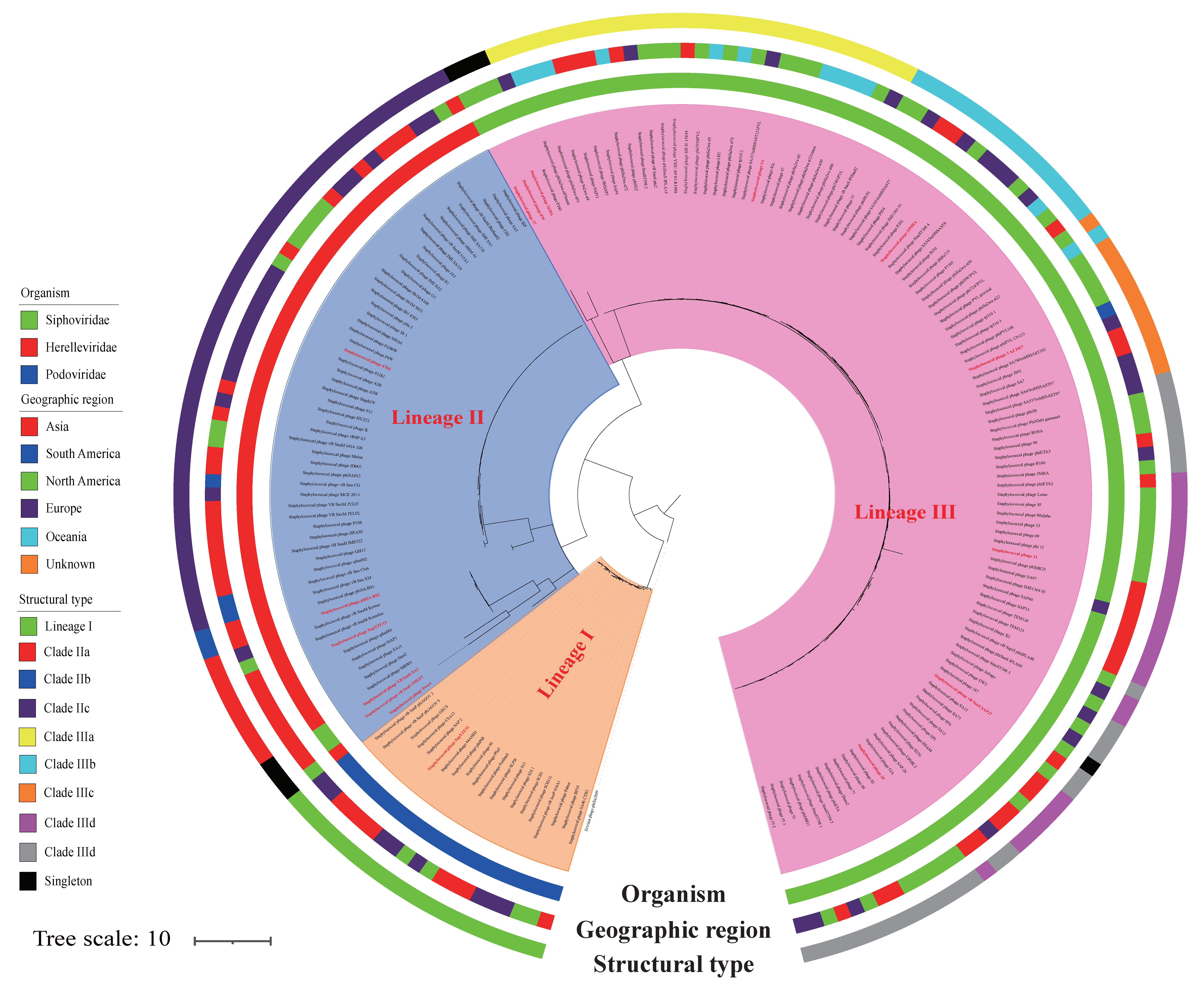
2.2. Phylogenetic Analysis

A phylogenetic tree was constructed, based on single-nucleotide polymorphisms (SNPs) using the kSNP3 software package v3.0 (https://sourceforge.net/projects/ksnp/files/, accessed on 20 April 2022), as described previously [23], to assess the evolutionary relationship of genome sequences among the 189 published bacteriophages (Table S1). The k-mer size was set to 15, and the optimum size was estimated using Kchooser software [23]. The 189 sequences comprised four datasets, encompassing the genomes of 20 Podoviridae, 56 Herelleviridae, 112 Siphoviridae phages, and 1 Erwinia phage, phiEa2809, as the outgroup. The phylogenetic tree was rooted using the outgroup and annotated using iTOL [24].

2.3. Orthogroup Clustering

Based on the phylogenetic tree, the mosaic structure of the S. aureus phages was aligned using progressive MAUVE [25]. Based on their structural similarity, S. aureus phages were classified into nine main clades and seven singletons. The protein sequences of the nine main lineages of S. aureus phages were used for orthogroup clustering, as described previously [26]. The sequences of these 9 main clades comprised 20 Podoviridae phages in lineage I, 8 Herelleviridae phages in clade IIa, 2 Herelleviridae phages in clade IIb, 45 Herelleviridae phages in clade IIc, 29 Siphoviridae phages in clade IIIa, 16 Siphoviridae phages in clade IIIb, 11 Siphoviridae phages in clade IIIc, 22 Siphoviridae phages in clade IIId, and 28 Siphoviridae phages in clade IIIe.

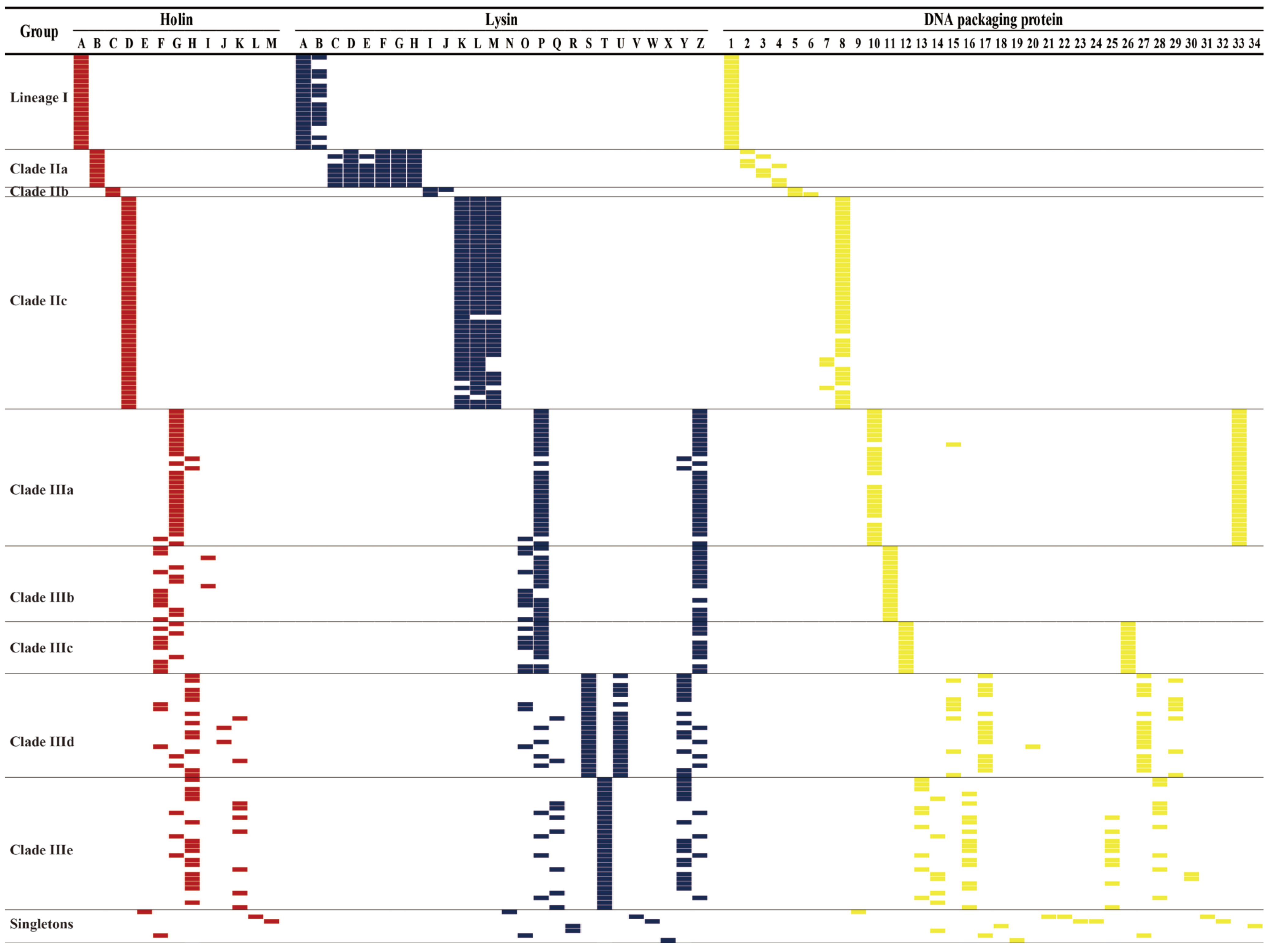
2.4. Analysis and Comparison of Holins, Lysins, DNA Packaging Proteins, Antimicrobial Resistance, Transposase, and Virulence Genes

A subset of 99 genes associated with DNA replication (n = 13), host cell lysis (n = 39), DNA packaging (n = 33), lysogeny (n = 6), virulence (n = 7), and antimicrobial resistance (n = 1) among the 188 S. aureus phages was analyzed based on their amino acid sequence identity, with a cut-off of 80%. The 13 DNA replication-associated genes included genes encoding DNA synthesis proteins, DNA-binding proteins, DNA polymerase, DNA primase/helicase, DNA helicase, DNA primase, DNA modification proteins, DNA methylase, DNA repair, DNA sliding clump inhibitor, RNA ligase, RNA polymerase, and the type III restriction enzyme. The genes associated with host cell lysis included 13 holin and 26 lysin genes. The genes associated with DNA packaging proteins comprised 23 genes encoding the large packaging subunits and 10 genes encoding the small packaging subunits. The six genes associated with lysogeny were those encoding recombinase (rec), transposase (tnp), integrase (int), repressor, anti-repressor, and Clp protease (clp). The seven virulence genes comprised the virulence E family protein (VirE), Panton-Valentine leukocidin (pvl), dUTP pyrophosphatase (dut), complement inhibitor sciderin (scn), staphylokinase (sak), beta hemolysin (hlb), and gamma hemolysin (hlg). Only one antimicrobial resistance gene encoding beta-lactamase (bla) was found in the 188 genome sequences. These genes were assembled and aligned with the 188 genomes using a BLASTx search, as described elsewhere [9].

2.5. Statistical Analyses

The SPSS software (version 19) was used for statistical analyses. Pearson’s chi-square test (two-tailed) was performed to analyze the differences in the distribution of genes associated with DNA metabolism, host cell lysis, DNA packaging, lysogeny, virulence, and antimicrobial resistance among the subdividing clusters.

3. Results and Discussion

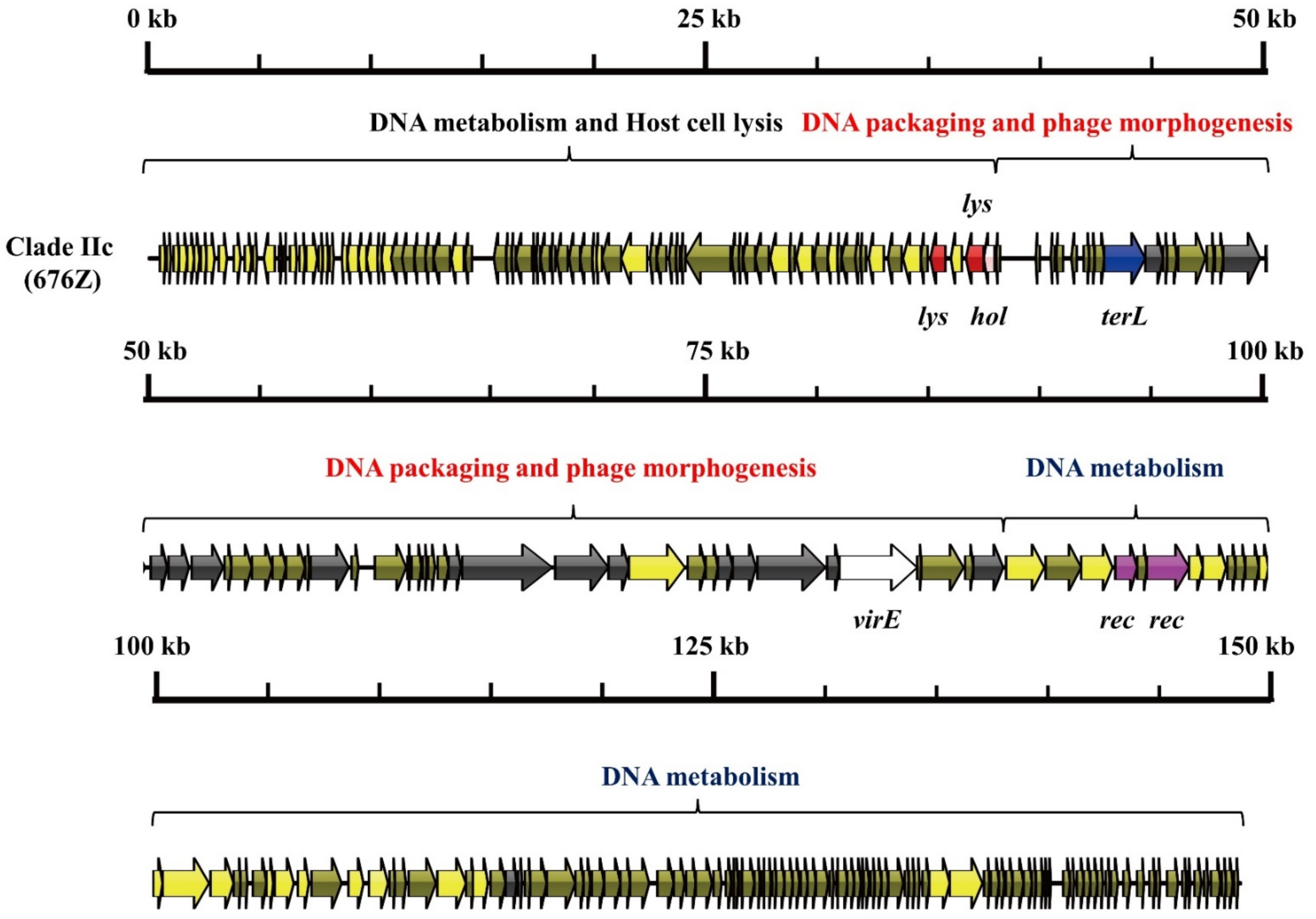
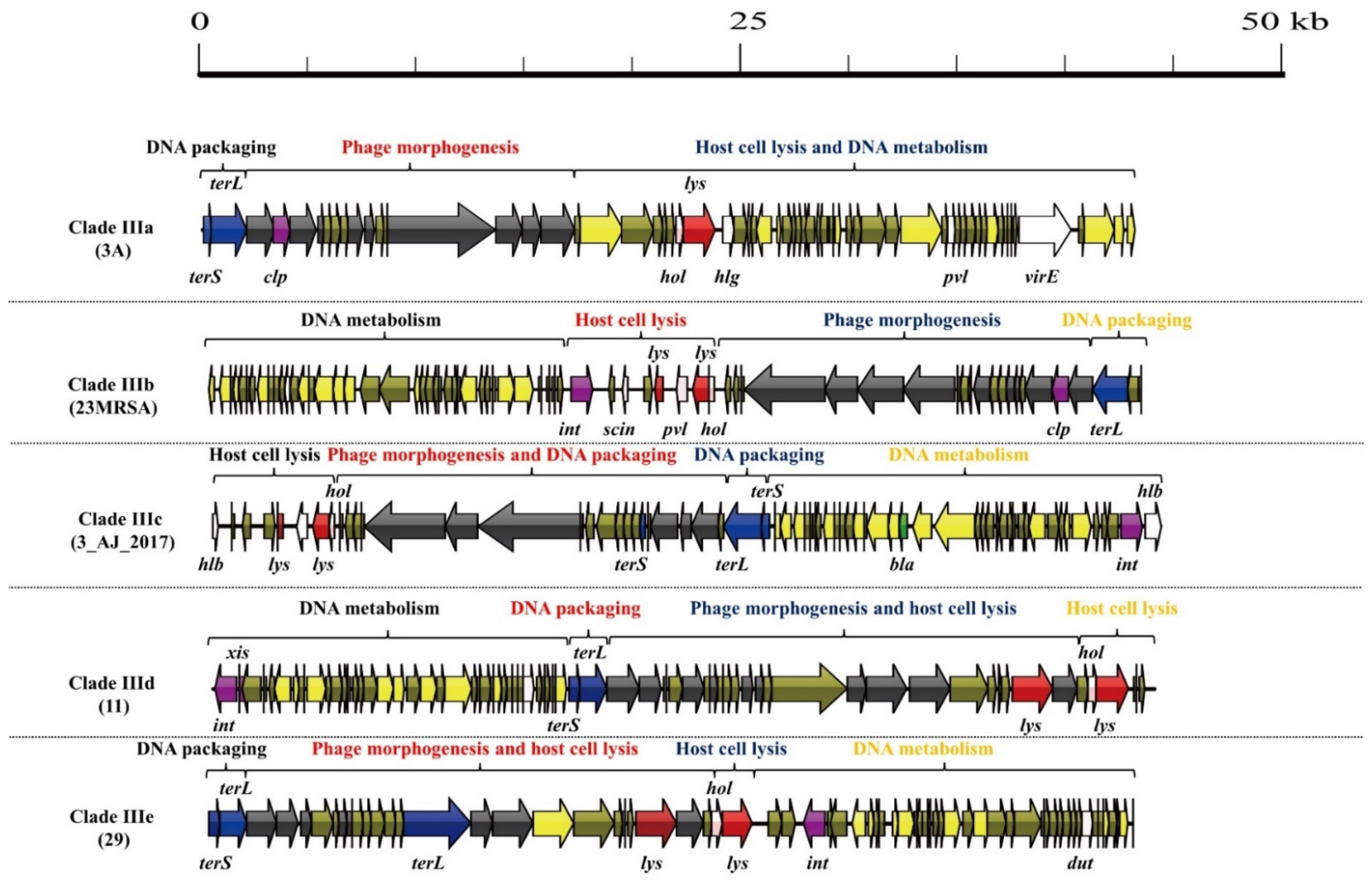
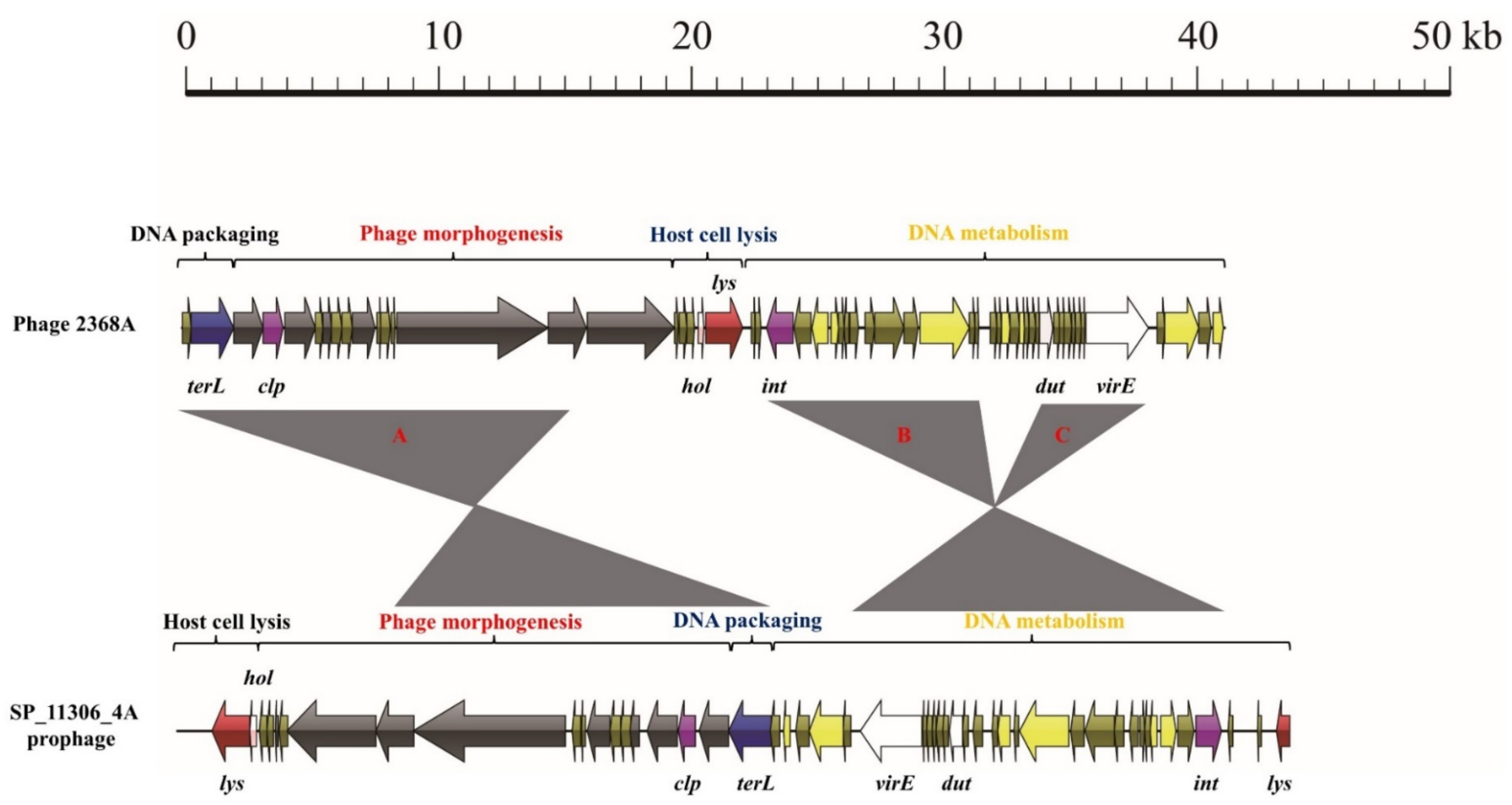
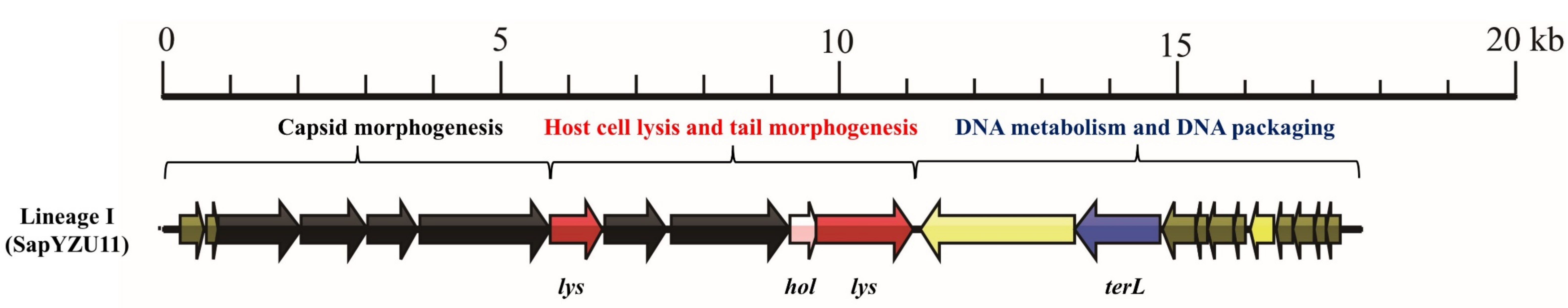
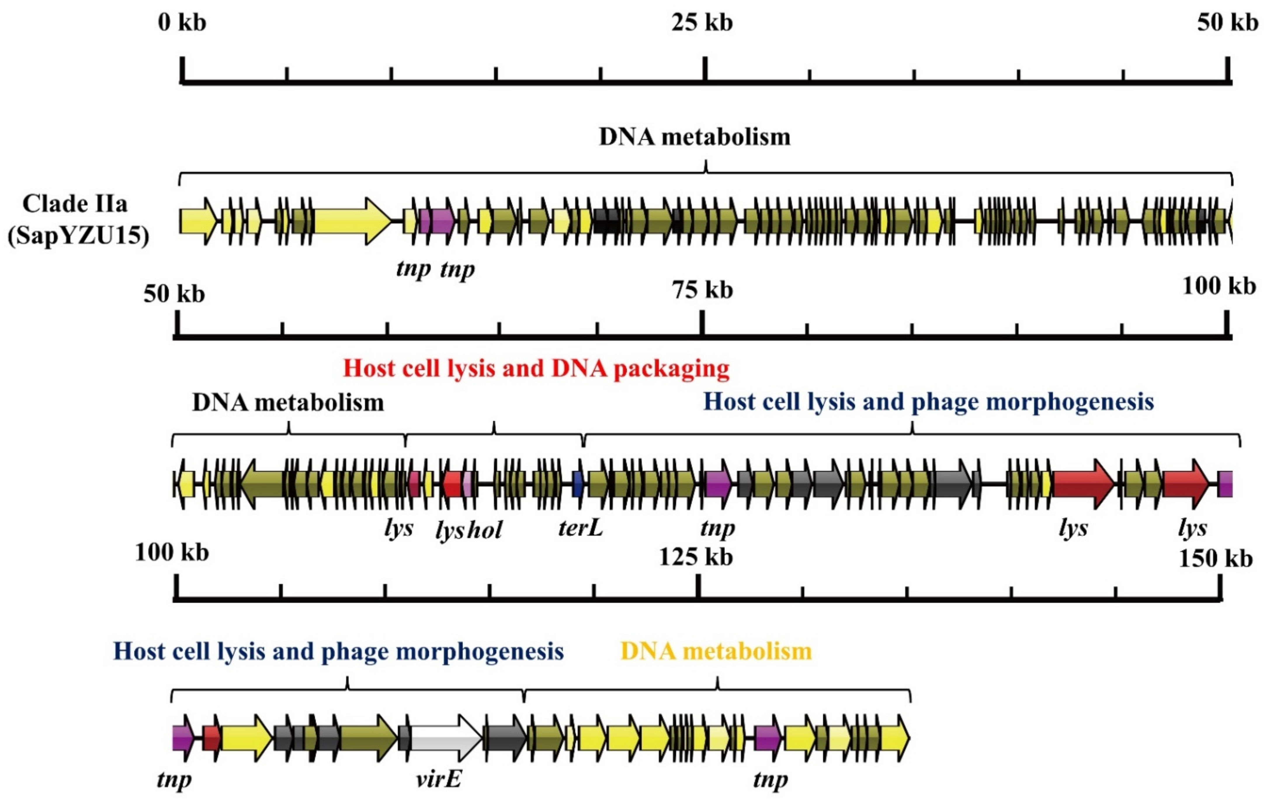
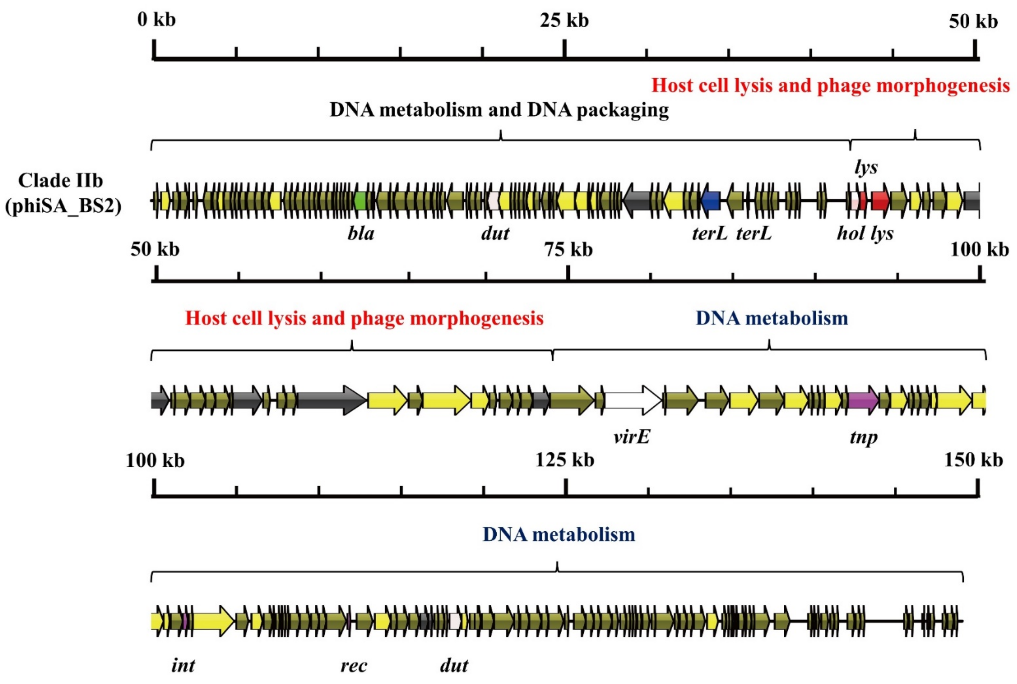
3.1. S. aureus Phages in Subdividing Clusters Exhibit Similar Mosaic Structures

Phylogenetic analysis based on the 18,125 SNPs (from the 188 phage genome sequences) and the Erwinia phage phiEa2809 sequence revealed three major genetic lineages (I–III; Figure 1). Lineage I consisted of 20 Podoviridae phages, and the genome sequences of these 20 phages were 16.7–18.2-kilobase-long sequences (44AHJD and phiP68, respectively). These phages included 18–22 ORFs (Portland and SapYZU11, respectively) and contained no tRNA genes (Table S1). The guanine-cytosine content (G+C%) of the Podoviridae phages varied from 28.8% to 29.6% (SapYZU11 and 44AHJD, respectively). Lineage II comprised 3 singletons (Herelleviridae phage ‘Twort’, Siphoviridae phage ‘VB_SauS_SA2’, and Siphoviridae phage ‘vB_SauS_IMEP5’) and 3 clades (IIa–IIc). The 56 Herelleviridae phage genomes were 127.2–151.6-kilobase-long sequences (Sb_1 and vB_SauM_0414_108, respectively), including 179–247 ORFs (Twort and vB_SauM_0414_108, respectively) and containing no more than 5 tRNA genes (vB_Sau_CG). The G+C% content of the Herelleviridae phages varied from 29.7% to 30.8% (phiSA_BS2 and vB_Sau_S24, respectively). Lineage III consisted of 110 Siphoviridae phages, which were classified into 4 singletons (2638A, EW, 37, and 187) and 5 clades (IIIa–IIIe). These genomes were 34.7–89.1-kilobase-long sequences (SA7 and VB_SauS_SA2, respectively), including 51–131 ORFs (SA7 and VB_SauS_SA2, respectively), and containing only 1 tRNA gene (VB_SauS_SA2). The G+C% content of the Siphoviridae phages varied from 29.0% to 36.9% (DW2 and 2638A, respectively). These results were consistent with a previous study that computed a distance matrix of mostly S. aureus-infecting phages (n = 85), based on shared gene content [14]. Collinear and MAUVE analyses (Figures S1–S9) revealed that phages in the same clade share similar mosaic structures and are isolated from distinct geographic origins. These results indicate that clade members share a common core gene pool that can easily be transmitted among geographic regions.
To understand the genetic diversity of S. aureus phages, we compared the mosaic structure and genetic content across phylogenetic groups, and 16 mosaic structures were found among the S. aureus phages (Table 1). Phages typically consist of four main functional modules (DNA metabolism, DNA packaging, phage morphogenesis, and host cell lysis) and other important functional genes related to lysogeny, virulence, and antimicrobial resistance. In lineage I (Figure 2), the capsid morphogenesis module was a 5755-base-pair-long structure and contained six ORFs, including genes encoding major head proteins, upper collar proteins, lower collar proteins, minor structural proteins, and two hypothetical proteins. The host cell lysis and tail morphogenesis module spanned the region from orf7 to orf11 and harbored genes encoding 2 lysins, 1 holin, and 2 tail fibers. The DNA metabolism and packaging module consisted of 11 ORFs, including genes that encode DNA polymerase, DNA packaging proteins, and DNA-binding proteins.
As shown in Figure 3, Figure 4 and Figure 5, three mosaic structures were found in clades IIa–IIc. Notably, the major difference between these clades and other S. aureus phages was the abundance of genes associated with DNA metabolism. Clade IIa contained two modules associated with DNA metabolism. The first DNA metabolism module was composed of 102 ORFs, which contained seven genes encoding DNA metabolism-related proteins, including DNA synthesis proteins, DNA polymerase I, DNA repair recombinase, and DNA-binding proteins. The second DNA metabolism module consisted of 44 ORFs, which harbored six genes encoding DNA metabolism-related proteins, including RNA polymerase, DNA helicase, a type III restriction enzyme, DNA methylase, DNA repair exonuclease, and DNA primase. Clade IIb contained two modules associated with DNA metabolism, which harbored eight genes encoding type III restriction enzymes, DNA helicase, DNA primase/helicase, DNA synthesis proteins, DNA polymerase I, and DNA modification proteins. Furthermore, clade IIc contained two modules associated with DNA metabolism, which contained 10 genes encoding RNA ligase, a type III restriction enzyme, DNA helicase, DNA primase, DNA synthesis, DNA polymerase, RNA polymerase, and a DNA sliding clump inhibitor.
As shown in Figure 6, five mosaic structures were found in clades IIIa–IIIe. Notably, the major difference between these clades and other S. aureus phages was the abundance of genes associated with lysogeny and virulence. Clade IIIa contained two genes encoding lysogeny proteins (Clp protease and repressor) and four virulence genes (hlg, pvl, dut, and virE). Clade IIIb contained three genes encoding lysogeny proteins (integrase, anti-repressor protein, and Clp protease) and four virulence genes (dut, pvl, scn, and sak). Clade IIIc contained three genes encoding lysogeny proteins (integrase, anti-repressor protein, and Clp protease) and three virulence genes (hlb, sak, and dut). Clade IIId contained four genes encoding lysogeny proteins (integrase, excisionase, repressor, and anti-repressor) and one virulence gene (dut). Clade IIIe contained two genes encoding lysogeny proteins (integrase and anti-repressor protein) and one virulence gene (dut).
Our phylogenetic analysis revealed nine main mosaic structures of S. aureus, indicating the structural diversity and high genetic mosaicism of S. aureus phages. These results were consistent with those of previous studies [13,14]. Although the genomes of lineage III phages displayed obvious functional modules, those of lineage I and lineage II were hybridized. A previous study indicated that genome mosaicism varies depending on the host, lifestyle, and genetic constitution of the phages [7]. The two modules of genes associated with DNA metabolism in clades IIa–IIc accelerated the synthesis of phage macromolecules and, hence, increased phage production. Moreover, the integrase and C repressor coding regions identified in clades IIIa–IIIc exhibited extensive diversity, which is consistent with the results of a study indicating that S. aureus integrase diversity has a minimum of 38% nucleotide identity [27]. These results revealed the distinct genetic features of S. aureus phages, suggesting diverse interactions between phages and their hosts. Although phage classification has historically been based on characteristics such as genome type (ssDNA, ssRNA, dsDNA, or dsRNA), viral morphology, and host range, it is currently undergoing a major overhaul, primarily using genome-based methods [8]. Therefore, our comprehensive exploration of structural diversity has modernized the classification of S. aureus phages.

3.2. S. aureus Phages in Subdividing Clusters Shared Similar Ortholog Clusters

To explore the core genome of S. aureus phages, ortholog clusters were analyzed in the subdividing clusters (Table S2). BLASTx revealed 34 orthogroups and 16 ORFs in lineage I. These 16 ORFs comprised 6 genes associated with phage morphogenesis and 1 gene associated with DNA packaging. Clade IIa consisted of 8 Herelleviridae phages and 222 orthogroups. A total of 137 ORFs were found in clade IIa, which contained 8 genes associated with phage morphogenesis and 1 gene associated with DNA packaging. Clade IIb consisted of 2 Herelleviridae phages and 229 orthogroups. A total of 202 ORFs were observed in both phages, including 6 genes associated with phage morphogenesis and 1 gene associated with DNA packaging. Clade IIc contained 437 orthogroups. A total of 97 ORFs were observed, including 16 genes associated with phage morphogenesis and 1 gene associated with DNA packaging.
In clade IIIa, 29 Siphoviridae phages contained 177 orthogroups. A total of 27 ORFs were observed, including 7 genes associated with phage morphogenesis and 2 genes associated with DNA packaging. Clade IIIb contained 161 orthogroups and 23 ORFs, including 8 genes associated with phage morphogenesis and 1 gene associated with DNA packaging. Clade IIIc consisted of 11 Siphoviridae phages and 128 orthogroups. A total of 27 ORFs were observed, including 6 genes associated with phage morphogenesis and 2 genes associated with DNA packaging. In clade IIId, the 22 Siphoviridae phages contained 161 orthogroups and 32 ORFs, including 10 genes associated with phage morphogenesis and 2 genes associated with DNA packaging. Clade IIIe phages contained 218 orthogroups and 23 ORFs, including 6 genes associated with phage morphogenesis and 1 gene associated with DNA packaging.
Despite the genetic and structural diversity of this species, it is notable that the cluster members share common ortholog groups. A previous study analyzed the genome sequence of 205 staphylococci phages and found that the genomes have mosaic architectures and that individual genes with common ancestors are positioned in distinct genomic contexts in different clusters [13]. Consistently, our study revealed that each cluster yielded a pan-genome size of 34–437 genes and shared 16–22 genes in the core genome. The absence of core ortholog groups in all the S. aureus phages indicates the frequent exchange, acquisition, and loss of genetic material. Nonetheless, genes associated with phage morphogenesis and DNA packaging were observed in each ortholog group of the subdividing clusters. Phage genomic diversity is difficult to establish because of the absence of a conserved genetic marker and a large number of phages in the biosphere [8]. However, genes associated with phage morphogenesis and DNA packaging may be genetic markers for subdividing cluster phages. A DNA packaging protein that assembles a motor complex may effectively pump DNA into tailed phage procapsids and accelerate phage assembly [28,29]. The disruption of DNA packaging genes completely abolished phage DNA packing events, suggesting that these genes play a prominent role in the transfer of S. aureus phages [30]. Therefore, the conserved DNA packaging gene indicates a similar DNA packaging mechanism in the subdividing cluster phages. However, the present study was limited to the complete phage genomes deposited in GenBank, and an updated genetic analysis is thus necessary to provide accurate genetic markers for phage classification and identification.

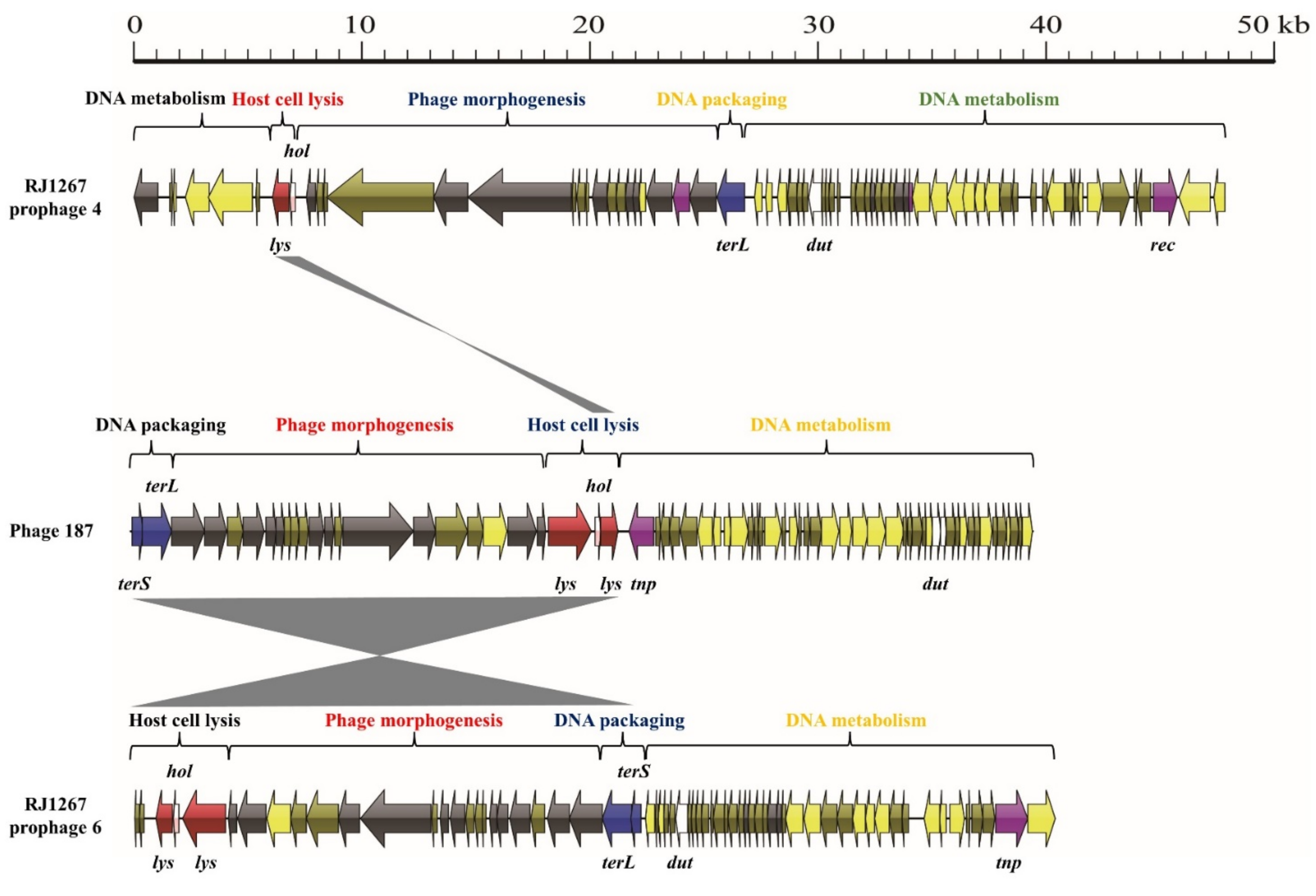
3.3. Exchange of Functional Modules and the Insertion/Deletion of Small DNA Segments Promote the Evolution of S. aureus Phages

To further understand the interaction between S. aureus phages and their hosts, the mosaic structures of singleton phages 187 and 2638A were analyzed. The phage-187 genome comprised four functional modules, as mentioned previously (Figure 7). The DNA packaging module was an 1818-base-pair-long structure and harbored 2 genes encoding the small and large terminase subunits. This region shared 98.0% nucleotide sequence identity with prophage 6 in the S. aureus strain RJ1267 (CP047321). The phage morphogenesis module was an 18,416-base-pair-long structure and contained 13 genes involved in phage morphogenesis and 1 lysin gene. This region shared 98.9% nucleotide sequence identity with that of RJ1267. The host cell lysis module was a 1021-base-pair-long structure and harbored one lysin and one holin gene. Notably, this module shared 99.3% and 99.0% nucleotide sequence identity with that of RJ1267. However, the DNA metabolism module of phage-187 shared little nucleotide sequence identity with the DNA metabolism module of strain RJ1267. This module was an 18,216-base-pair-long structure and contained 42 ORFs, including genes involved in DNA metabolism, lysogen, virulence, and the toxin-antitoxin system.
The phage-2638A genome was also composed of four functional modules (Figure 8). The DNA packaging module was a 2057-base-pair-long structure and harbored one gene encoding the large terminase subunit. This region shared 98.0% nucleotide sequence identity with prophage 3 from the Staphylococcus pseudintermedius strain SP_11306_4A (CP065919). The phage morphogenesis module was an 18,253-base-pair-long structure and contained six genes encoding phage morphogenesis and one clp gene. Region A in this module was a 13,064-base-pair-long structure and shared 99.7% nucleotide sequence identity with that of strain SP_11306_4A. However, the remaining region shared less than 90% nucleotide sequence identity with that of SP_11306_4A. The host cell lysis module of phage-2638A was a 1711-base-pair-long structure and harbored one lysin and one holin gene. The DNA metabolism module of phage-2638A was a 19,061-base-pair-long structure and contained 35 ORFs, including genes involved in DNA polymerase, integrase, and virulence. Regions B and C in this module were 6415- and 3510-base-pair-long structures and shared 96.2% and 96.9% nucleotide sequence identity with those of SP_11306_4A, respectively.
Phages 187 and 2638A, isolated from S. aureus strains in Canada and the United States, respectively, shared little nucleotide sequence identity with the genome sequences in the NCBI database. However, the DNA packaging, phage morphogenesis, and host cell lysis modules of phage-187 shared a high nucleotide sequence identity with a prophage in the S. aureus strain RJ1267, which was isolated from a sputum sample in Shanghai, China. These results suggest that phage-187 and prophage 6 in the S. aureus strain RJ1267 probably shared a common ancestor, which subsequently underwent an exchange of DNA metabolism module. Consistently, the DNA packaging module of phage-2368A was similar to that of prophage in S. pseudintermedius strain SP_11306_4, which was isolated from a canine skin sample in the US. However, the host cell lysis module shared little nucleotide sequence identity with SP_11306_4. These results reveal that the exchange of functional modules among staphylococcal phages may cross the species barriers, which is consistent with the results of a study indicating that the gene exchange between staphylococcal phages may cross the species barriers because they coexist in a common host [31]. Moreover, small DNA segment insertion/deletion events were observed in the DNA metabolism module and phage morphogenesis module of 2368A, which is consistent with previous findings that the transduction of phiSaBov was accompanied by the mobilization of the genomic islands vSaα, vSaβ, and vSaγ [30,32]. Our study indicates that the genetic diversity of S. aureus phages is likely due to the exchange of functional modules and the insertion/deletion of small DNA segments, which may cross species barriers. Therefore, gene exchange, acquisition, and loss resulting from the exchange of functional modules and the insertion/deletion of small DNA segments promote the evolution of S. aureus phages. Future research should, however, elucidate the exact mechanism of gene exchange between S. aureus and its hosts.

3.4. S. aureus Phages Enhance Phage DNA Replication in the Host Cells and the Environment Adaptivity of Its Host

To further assess the diversity of S. aureus phages and clusters, we explored the gene content associated with the six functions mentioned. As shown in Figure 9, holin A, lysins A and B, and DNA packaging protein 1 were only observed in lineage I (p < 0.001). Holin B, lysins C–H, and DNA packaging proteins 2–4 were only observed in clade IIa. Holin C, lysins I–J, and DNA packaging proteins 5–6 were only observed in clade IIb. Holin D, lysins K–M, and DNA packaging proteins 7–8 were only observed in clade IIc. Holins F–M, lysins O–Z, the large DNA packaging subunits J–W, and the small packaging subunits A–J were only observed in lineage III. Notably, the cluster members share similar types of holins, lysins, and DNA packaging proteins, as well as the mosaic structure and gene content, indicating at least three possibilities: (i) phages in the same cluster shared a common ancestor and spread among distinct continents along with their host, (ii) the module exchange occurred independently of host cell lysis and DNA packaging modules, or (iii) frequent module exchange occurred exclusively among cluster members. Considering the widespread and high structural similarity of cluster phages, we propose that frequent module exchange occurred exclusively among cluster members.
As shown in Table 2, genes encoding DNA polymerase were more frequently detected in lineages I, IIa, IIb, IIIa, and IIIb (50%, 75.0%, 50.0%, 65.5%, and 50.0%, respectively) than in clades IIc (42.2%), IIIc (36.4%), IIId (18.2%), and IIIe (32.1%) (p < 0.05). Other DNA replication genes (DNA synthesis, DNA primase/helicase, DNA helicase, DNA primase, DNA sliding clump inhibitor, RNA polymerase, and type III restriction enzyme) were more frequently detected in clades IIa (50.0%, 62.5%, 62.5%, 62.5%, 62.5%, 37.5%, 62.5%, and 62.5%, respectively), IIb (50.0%, 50.0%, 100.0%, 50.0%, 50.0%, 50.0%, and 50.0%, respectively), and IIc (37.8%, 48.9%, 57.8%, 46.7%, 40.0%, 46.7%, and 48.9%, respectively) than in lineage I (20.0%, 20.0%, 20.0%, 20.0%, 10%, 20.0%, and 25.0%, respectively, p > 0.05). However, the gene encoding the DNA-binding protein was detected more frequently in lineage I (75.0%) and clade IIIc (54.5%) than in clade IIIb (31.3%) (p > 0.05).
DNA replication is driven by multiple enzymes, including DNA helicase, which separates double-stranded template DNA; RNA polymerase, which synthesizes an RNA primer; DNA synthesis protein, which initiates Okazaki fragment synthesis; and DNA polymerase, which synthesizes leading and lagging daughter strands [33,34,35]. Therefore, the prevalence of DNA replication genes in S. aureus phages enhances phage DNA replication in host cells. It was surprising to observe the abundance of genes encoding type III DNA restriction and modification enzymes in S. aureus phages, which is inconsistent with previous results that 28.1% of Acinetobacter phages encoded type II restriction–modification systems [36]. Type III DNA restriction and modification enzymes are responsible for host-specific barriers and protect bacterial cells against bacteriophage infections [37]. Therefore, the presence of modified nucleosides in phage genomes may protect host cells against other bacteriophage infections.
No lysogeny-associated genes were found in lineage I (Table 2). However, genes encoding recombinase and transposase were exclusively found in lineages IIa, IIb, and IIc (p < 0.001). Genes encoding integrase, repressor, and anti-repressor were predominantly detected in clades IIIa–IIIe. This result indicated that integration systems varied based on the subdividing clusters, which is inconsistent with the results of a study revealing no obvious link between the types of integrase, host species, or subclusters [13].
In terms of virulence and antimicrobial resistance genes, pvl was more prevalent in phages belonging to clades IIb (50.0%), IIIb (68.8%), IIIc (63.6%), and IIId (50.0%) than in those belonging to clades I (10.0%), IIa (25.0%), IIc (33.3%), IIIa (34.5%), and IIIe (39.3%) (p < 0.01). However, dut, encoding dUTPase, was more frequently detected in clades IIb (50.0%), IIIc (36.4%), and IIId (36.4%) than in clades I (5.0%), IIa (12.5%), IIc (20.0%), IIIa (24.1%), IIIb (12.5%), and IIIe (28.6%) (p < 0.05). Conversely, scn was found in only 12 phages, encompassing clades IIc (4.4%), IIIa (3.4%), IIIb (6.3%), IIIc (27.3%), and IIIe (10.7%), and in two singleton phages. Other virulence genes, including virE (44.7%), sak (5.3%), hlb (1.1%), and hlg (1.1%), were also found in the S. aureus phages. Notably, 16 S. aureus phages (8.5%) contained bla-encoding beta-lactamase.

4. Conclusions

The abundance of virulence-determinant genes in the phage genomes was consistent with the results of a previous study [13]. Panton-Valentine leukocidin is a cytotoxin that induces pore formation in leukocyte cell membrane receptors, which leads to a higher pathogenic potential and the recurrence of community-associated MRSA [38]. The dUTPase enzyme is essential for DNA integrity and viability in many prokaryotic and eukaryotic organisms, as it controls the transfer of virulence genes via a proto-oncogenic G protein-like mechanism [39]. Furthermore, sciderin is an important protein associated with host defense that interferes with the activation of the human complement system [40]. Additionally, staphylokinase is a fibrinolytic agent that plays an important role in dissolving blood clots on fibrin surfaces [41]. β-Haemolysin acts as a hemolytic in sheep, contributes to biofilm formation in rabbit endocarditis models, and enhances the ability of S. aureus to colonize murine skin [42]. The abundance of these virulence genes suggests that the evolutionary model of S. aureus phages promotes host pathogenicity. β-Lactamase, which hydrolyses the β-lactam ring, is the primary resistance mechanism of antibacterial activity against β-lactam antibiotics caused by their extensive use [43]. Our results also indicated that S. aureus phage evolution contributes to the adaptive environment of its host.
In conclusion, our study provides insight into the interaction between S. aureus phages and their hosts by exploring their genomic, structural, and genetic diversity. Our analysis suggests that the genes associated with phage morphogenesis and DNA packaging are conserved in the subdividing clusters, despite the mosaic structural diversity of S. aureus phages. The genetic diversity of S. aureus phages is likely due to gene exchange, acquisition, and loss resulting from the exchange of functional modules and the insertion/deletion of small DNA segments among staphylococcal phages, which may cross species barriers. Moreover, module exchange probably occurred exclusively among the subdividing cluster phages. Through these evolutionary strategies, S. aureus phages enhance phage DNA replication in host cells and contribute to the adaptive environment of their host.

Supplementary Materials

The following supporting information can be downloaded at: https://www.mdpi.com/article/10.3390/v14061199/s1, Figure S1: MARVE analysis of genomes of 20 Podoviridae phages in lineage I. Figure S2: MARVE analysis of genomes of 8 Herelleviridae phages in clade IIa (lineage II). Figure S3: MARVE analysis of genomes of 2 Herelleviridae phages in clade IIb (lineage II). Figure S4: MARVE analysis of genomes of 46 Herelleviridae phages in clade IIe (lineage II). Figure S5: MARVE analysis of genomes of 29 Siphoviridae phages in clade IIIa (lineage III). Figure S6: MARVE analysis of genomes of 16 Siphoviridae phages in clade IIIb (lineage III). Figure S7: MARVE analysis of genomes of 11 Siphoviridae phages in clade IIIc (lineage III). Figure S8: MARVE analysis of genomes of 22 Siphoviridae phages in clade IIId (lineage III). Figure S9: MARVE analysis of genomes of 28 Siphoviridae phages in clade IIIe (lineage III). Table S1: Metadata for 189 phages publicly available on NCBI. The 189 phages comprise four data sets including the genomes of 20 Podoviridae phages, 56 Herelleviridae phages, 112 Siphoviridae phages, and 1 Erwinia phage phiEa2809 as the outgroup. Table S2: Orthogroup clusters of lineage I, clades IIa–IIc, and clades IIIa–IIIe phages.

Author Contributions

Study conception and design: Z.Y. and W.Z.; acquisition of data: W.Z., H.W. and Y.L.; analysis and interpretation of data: W.Z., Y.G. and X.Z.; drafting of the manuscript: W.Z., L.Y. and Z.Y.; critical revision: Z.Y. and G.Z. All authors have read and agreed to the published version of the manuscript.

Funding

This research was financially supported by the National Natural Science Foundation of China (grant number: 32102100), the Jiangsu Key Research & Development Program (grant number: BE2019436-5), the Yangzhou University Food Quality and Safety Talents Project (grant number: YZUJX2020-A3), and the Yangzhou City Talents Project.

Institutional Review Board Statement

Not applicable.

Informed Consent Statement

Not applicable.

Data Availability Statement

The study results’ data are included in the article or Supplementary Materials. Further specific information regarding the dataset analyzed during the study can be obtained from the corresponding author on reasonable request.

Conflicts of Interest

The authors have no conflict of interest to declare.

References

  1. Fernandez, L.; Rodriguez, A.; Garcia, P. Phage or foe: An insight into the impact of viral predation on microbial communities. ISME J. 2018, 12, 1171–1179. [Google Scholar] [CrossRef] [PubMed] [Green Version]
  2. Watts, G. Phage therapy: Revival of the bygone antimicrobial. Lancet 2017, 390, 2539–2540. [Google Scholar] [CrossRef]
  3. Kortright, K.E.; Chan, B.K.; Koff, J.L.; Turner, P.E. Phage therapy: A renewed approach to combat antibiotic-resistant bacteria. Cell Host Microbe 2019, 25, 219–232. [Google Scholar] [CrossRef] [PubMed] [Green Version]
  4. Monteiro, R.; Pires, D.P.; Costa, A.R.; Azeredo, J. Phage therapy: Going temperate? Trends Microbiol. 2019, 27, 368–378. [Google Scholar] [CrossRef] [PubMed] [Green Version]
  5. Rehman, S.; Ali, Z.; Khan, M.; Bostan, N.; Naseem, S. The dawn of phage therapy. Rev. Med. Virol. 2019, 29, e2041. [Google Scholar] [CrossRef] [PubMed]
  6. Chan, B.K.; Abedon, S.T. Bacteriophages and their enzymes in biofilm control. Curr. Pharm. Des. 2015, 21, 85–99. [Google Scholar] [CrossRef]
  7. Mavrich, T.N.; Hatfull, G.F. Bacteriophage evolution differs by host, lifestyle and genome. Nat. Microbiol. 2017, 2, 17112. [Google Scholar] [CrossRef] [Green Version]
  8. Dion, M.B.; Oechslin, F.; Moineau, S. Phage diversity, genomics and phylogeny. Nat. Rev. Microbiol. 2020, 18, 125–138. [Google Scholar] [CrossRef]
  9. Zhou, W.; Li, X.; Osmundson, T.; Shi, L.; Ren, J.; Yan, H. WGS analysis of ST9-MRSA-XII isolates from live pigs in China provides insights into transmission among porcine, human and bovine hosts. J. Antimicrob. Chemother. 2018, 73, 2652–2661. [Google Scholar] [CrossRef] [Green Version]
  10. Zhou, W.; Li, X.; Shi, L.; Wang, H.H.; Yan, H. Novel SCCmec type XII methicillin-resistant Staphylococcus aureus isolates identified from a swine production and processing chain. Vet. Microbiol. 2018, 225, 105–113. [Google Scholar] [CrossRef]
  11. Petrovic, F.A.; Lin, R.; Ho, J.; Maddocks, S.; Ben, Z.N.; Iredell, J.R. Safety of bacteriophage therapy in severe Staphylococcus aureus infection. Nat. Microbiol. 2020, 5, 465–472. [Google Scholar] [CrossRef]
  12. Speck, P.; Smithyman, A. Safety and efficacy of phage therapy via the intravenous route. FEMS Microbiol. Lett. 2016, 363, fnv242. [Google Scholar] [CrossRef]
  13. Oliveira, H.; Sampaio, M.; Melo, L.; Dias, O.; Pope, W.H.; Hatfull, G.F.; Azeredo, J. Staphylococci phages display vast genomic diversity and evolutionary relationships. BMC Genom. 2019, 20, 357. [Google Scholar] [CrossRef] [Green Version]
  14. Deghorain, M.; Van Melderen, L. The Staphylococci phages family: An overview. Viruses 2012, 4, 3316–3335. [Google Scholar] [CrossRef] [Green Version]
  15. Liu, Z.; Liu, S.; Yao, J.; Bao, L.; Zhang, J.; Li, Y.; Jiang, C.; Sun, L.; Wang, R.; Zhang, Y.; et al. The channel catfish genome sequence provides insights into the evolution of scale formation in teleosts. Nat. Commun. 2016, 7, 11757. [Google Scholar] [CrossRef]
  16. Delcher, A.L.; Bratke, K.A.; Powers, E.C.; Salzberg, S.L. Identifying bacterial genes and endosymbiont DNA with Glimmer. Bioinformatics 2007, 23, 673–679. [Google Scholar] [CrossRef]
  17. Borodovsky, M.; Lomsadze, A. Gene identification in prokaryotic genomes, phages, metagenomes, and EST sequences with GeneMarkS suite. Curr. Protoc. Microbiol. 2014, 32, 1–17. [Google Scholar] [CrossRef]
  18. Tatusova, T.; Dicuccio, M.; Badretdin, A.; Chetvernin, V.; Nawrocki, E.P.; Zaslavsky, L.; Lomsadze, A.; Pruitt, K.D.; Borodovsky, M.; Ostell, J. NCBI prokaryotic genome annotation pipeline. Nucleic Acids Res. 2016, 44, 6614–6624. [Google Scholar] [CrossRef]
  19. Galperin, M.Y.; Kristensen, D.M.; Makarova, K.S.; Wolf, Y.I.; Koonin, E.V. Microbial genome analysis: The COG approach. Brief. Bioinform. 2019, 20, 1063–1070. [Google Scholar] [CrossRef]
  20. Jones, P.; Binns, D.; Chang, H.Y.; Fraser, M.; Li, W.; Mcanulla, C.; Mcwilliam, H.; Maslen, J.; Mitchell, A.; Nuka, G.; et al. InterProScan 5: Genome-scale protein function classification. Bioinformatics 2014, 30, 1236–1240. [Google Scholar] [CrossRef] [Green Version]
  21. Cantalapiedra, C.P.; Hernandez-Plaza, A.; Letunic, I.; Bork, P.; Huerta-Cepas, J. EggNOG-mapper v2: Functional annotation, orthology assignments, and domain prediction at the metagenomic scale. Mol. Biol. Evol. 2021, 38, 5825–5829. [Google Scholar] [CrossRef]
  22. Chan, P.P.; Lowe, T.M. TRNAscan-SE: Searching for tRNA genes in genomic sequences. Methods Mol. Biol. 2019, 1962, 1–14. [Google Scholar]
  23. Gardner, S.N.; Slezak, T.; Hall, B.G. KSNP3.0: SNP detection and phylogenetic analysis of genomes without genome alignment or reference genome. Bioinformatics 2015, 31, 2877–2878. [Google Scholar] [CrossRef] [Green Version]
  24. Letunic, I.; Bork, P. Interactive Tree of Life (iTOL) v5: An online tool for phylogenetic tree display and annotation. Nucleic Acids Res. 2021, 49, W293–W296. [Google Scholar] [CrossRef]
  25. Darling, A.E.; Mau, B.; Perna, N.T. ProgressiveMauve: Multiple genome alignment with gene gain, loss and rearrangement. PLoS ONE 2010, 5, e11147. [Google Scholar] [CrossRef] [Green Version]
  26. Emms, D.M.; Kelly, S. OrthoFinder: Phylogenetic orthology inference for comparative genomics. Genome Biol. 2019, 20, 238. [Google Scholar] [CrossRef] [Green Version]
  27. Goerke, C.; Pantucek, R.; Holtfreter, S.; Schulte, B.; Zink, M.; Grumann, D.; Broker, B.M.; Doskar, J.; Wolz, C. Diversity of prophages in dominant Staphylococcus aureus clonal lineages. J. Bacteriol. 2009, 191, 3462–3468. [Google Scholar] [CrossRef] [Green Version]
  28. Wangchuk, J.; Prakash, P.; Bhaumik, P.; Kondabagil, K. Bacteriophage N4 large terminase: Expression, purification and X-ray crystallographic analysis. Acta Crystallogr. Sect. F Struct. Biol. Commun. 2018, 74, 198–204. [Google Scholar] [CrossRef]
  29. Yang, Y.; Yang, P.; Wang, N.; Chen, Z.; Su, D.; Zhou, Z.H.; Rao, Z.; Wang, X. Architecture of the herpesvirus genome-packaging complex and implications for DNA translocation. Protein Cell 2020, 11, 339–351. [Google Scholar] [CrossRef] [PubMed] [Green Version]
  30. Moon, B.Y.; Park, J.Y.; Robinson, D.A.; Thomas, J.C.; Park, Y.H.; Thornton, J.A.; Seo, K.S. Mobilization of genomic islands of Staphylococcus aureus by temperate bacteriophage. PLoS ONE 2016, 11, e151409. [Google Scholar] [CrossRef]
  31. Deghorain, M.; Bobay, L.M.; Smeesters, P.R.; Bousbata, S.; Vermeersch, M.; Perez-Morga, D.; Dreze, P.A.; Rocha, E.P.; Touchon, M.; Van Melderen, L. Characterization of novel phages isolated in coagulase-negative staphylococci reveals evolutionary relationships with Staphylococcus aureus phages. J. Bacteriol. 2012, 194, 5829–5839. [Google Scholar] [CrossRef] [PubMed] [Green Version]
  32. Moon, B.Y.; Park, J.Y.; Hwang, S.Y.; Robinson, D.A.; Thomas, J.C.; Fitzgerald, J.R.; Park, Y.H.; Seo, K.S. Phage-mediated horizontal transfer of a Staphylococcus aureus virulence-associated genomic island. Sci. Rep. 2015, 5, 9784. [Google Scholar] [CrossRef] [PubMed] [Green Version]
  33. Benkovic, S.J.; Valentine, A.M.; Salinas, F. Replisome-mediated DNA replication. Annu. Rev. Biochem. 2001, 70, 181–208. [Google Scholar] [CrossRef] [PubMed] [Green Version]
  34. Lee, J.; Chastain, P.N.; Griffith, J.D.; Richardson, C.C. Lagging strand synthesis in coordinated DNA synthesis by bacteriophage t7 replication proteins. J. Mol. Biol. 2002, 316, 19–34. [Google Scholar] [CrossRef]
  35. Stano, N.M.; Jeong, Y.J.; Donmez, I.; Tummalapalli, P.; Levin, M.K.; Patel, S.S. DNA synthesis provides the driving force to accelerate DNA unwinding by a helicase. Nature 2005, 435, 370–373. [Google Scholar] [CrossRef]
  36. Oliveira, H.; Domingues, R.; Evans, B.; Sutton, J.M.; Adriaenssens, E.M.; Turner, D. Genomic diversity of bacteriophages infecting the genus acinetobacter. Viruses 2022, 14, 181. [Google Scholar] [CrossRef]
  37. Rao, D.N.; Dryden, D.T.; Bheemanaik, S. Type III restriction-modification enzymes: A historical perspective. Nucleic Acids Res. 2014, 42, 45–55. [Google Scholar] [CrossRef] [Green Version]
  38. Darboe, S.; Dobreniecki, S.; Jarju, S.; Jallow, M.; Mohammed, N.I.; Wathuo, M.; Ceesay, B.; Tweed, S.; Basu, R.R.; Okomo, U.; et al. Prevalence of Panton-Valentine leukocidin (PVL) and antimicrobial resistance in community-acquired clinical Staphylococcus aureus in an urban gambian hospital: A 11-Year period retrospective pilot study. Front. Cell. Infect. Microbiol. 2019, 9, 170. [Google Scholar] [CrossRef] [Green Version]
  39. Tormo-Mas, M.A.; Donderis, J.; Garcia-Caballer, M.; Alt, A.; Mir-Sanchis, I.; Marina, A.; Penades, J.R. Phage dUTPases control transfer of virulence genes by a proto-oncogenic G protein-like mechanism. Mol. Cell 2013, 49, 947–958. [Google Scholar] [CrossRef] [Green Version]
  40. De Jong, N.; Vrieling, M.; Garcia, B.L.; Koop, G.; Brettmann, M.; Aerts, P.C.; Ruyken, M.; van Strijp, J.; Holmes, M.; Harrison, E.M.; et al. Identification of a staphylococcal complement inhibitor with broad host specificity in equid Staphylococcus aureus strains. J. Biol. Chem. 2018, 293, 4468–4477. [Google Scholar] [CrossRef] [Green Version]
  41. Bhando, T.; Singh, S.; Hade, M.D.; Kaur, J.; Dikshit, K.L. Integration of VEK-30 peptide enhances fibrinolytic properties of staphylokinase. Biotechnol. Appl. Biochem. 2021, 68, 213–220. [Google Scholar] [CrossRef]
  42. Jung, P.; Abdelbary, M.M.; Kraushaar, B.; Fetsch, A.; Geisel, J.; Herrmann, M.; Witte, W.; Cuny, C.; Bischoff, M. Impact of bacteriophage Saint3 carriage on the immune evasion capacity and hemolytic potential of Staphylococcus aureus CC398. Vet. Microbiol. 2017, 200, 46–51. [Google Scholar] [CrossRef]
  43. Tooke, C.L.; Hinchliffe, P.; Bragginton, E.C.; Colenso, C.K.; Hirvonen, V.H.A.; Takebayashi, Y.; Spencer, J. Β-Lactamases and β-lactamase inhibitors in the 21st century. J. Mol. Biol. 2019, 431, 3472–3500. [Google Scholar] [CrossRef]
Figure 1. Phylogeny of 188 S. aureus phages and the Erwinia phage, phiEa2809, based on 681,666 single-nucleotide polymorphisms. The inner ring is colored according to the organism; the middle ring, according to the geographic region; and the outer ring, according to the structural type.
Viruses 14 01199 g001
Figure 2. Mosaic structure of the lineage I phage SapYZU11. Functional modules are annotated with different colors. ORFs are shown as arrows, indicating the transcription direction, and the colors of the arrows represent different fragments. Gene color code: virulence determinants, white; holin gene, pink; lysin gene, red; genes associated with lysogeny, purple; bla, green; DNA packaging genes, blue; genes associated with DNA metabolism, yellow; and genes encoding hypothetical proteins, brown.
Viruses 14 01199 g002
Figure 3. Mosaic structure of the clade IIa phage, SapYZU15.
Viruses 14 01199 g003
Figure 4. Mosaic structure of the clade IIb phage, phiSA_BS2.
Viruses 14 01199 g004
Figure 5. Mosaic structure of the clade IIc phage, 676Z.
Viruses 14 01199 g005
Figure 6. Comparative structural analysis of clades IIIa–IIIe.
Viruses 14 01199 g006
Figure 7. Comparative structural analysis of phage 187 against prophages 4 and 6 of the S. aureus isolate, RJ1267. Areas shaded in gray represent regions with >95% nucleotide sequence identity.
Viruses 14 01199 g007
Figure 8. Comparative structural analysis of phage 2368A against a prophage of Staphylococcal pseudintermedius strain SP_11306_4. Areas shaded in gray represent regions with >95% nucleotide sequence identity.
Viruses 14 01199 g008
Figure 9. Heat map showing the distribution of holin, lysin, and DNA packaging genes in S. aureus phages. The pattern of gene presence (colored blocks) or absence (white) is shown.
Viruses 14 01199 g009
Table 1. Summary of the gene content of functional modules present in the S. aureus phages.
GroupsFamilyPhage Morphogenesis Host Cell LysisDNA MetabolismDNA PackagingLysogenyVirulenceAntimicrobial ResitanceRepresent Phage
Lineage IPodoviridaemajor head protein (1), upper collar protein (1), lower collar protein (1), minor structural protein (1), and tail fibers (2)lysin (2) and holin (1)DNA polymerase (1) and DNA binding protein (1)DNA packaging protein (1)---SapYZU11 (MW864250)
Clade IIaHerelleviridaemajor capsid protein (2), capsid protein (1), portal protein (2), tail protein (2), microtubule-associated protein (1), tail sheath protein (1), and baseplate J-like protein (1)lysin (5) and holin (1)DNA synthesis (3), DNA polymerase I (2), DNA repair recombinase (1), DNA-binding protein (1), RNA polymerase (1), DNA helicase (1), Type III restriction enzyme (1), DNA methylase (1), DNA repair exonuclease (1) and DNA primase (1)DNA packaging protein (1)transposase (5)virulence-associated E family protein (1)-SapYZU15 (MW864252)
Clade IIbHerelleviridaeportal protein (1), major capsid protein (1), major tail sheath (1), tail tape measure protein (1), baseplate J-like protein (1), and major tail protein (2)lysin (5) and holin (1)Type III restriction enzyme (1), DNA helicase (1), DNA primase/helicase (1), DNA synthesis (2), DNA polymerase I (2) and DNA modification protein (1)DNA packaging protein (2)recombinase (1)dUTP pyrophosphatase (2) and virulence-associated E family protein (1)beta-lactamase (1)phiSA_BS2 (MH028956.1)
Clade IIcHerelleviridaehead protein (1), portal protein (1), prohead protease (1), major capsid protein (1), tail sheath protein (1), tail morphogenetic protein (8), and Baseplate J-like protein (1)lysin (2) and holin (1)RNA ligase (1), Type III restriction enzyme (1), DNA helicase (1), DNA primase (1), DNA synthesis (2), DNA polymerase (2), RNA polymerase (1) and DNA sliding clump inhibitor (1)DNA packaging protein (1)recombinase (2)--676Z (JX080302.2)
Clade IIIaSiphoviridaeportal protein (1), major capsid protein (1), head-tail connector protein (1), tail protein (3), and tail tape measure protein (2) lysin (1) and holin (1)DNA polymerase (1)DNA packaging protein (2)Clp protease (1) and repressor (1)gamma-hemolysin (1), PVL (1), dUTP pyrophosphatase (1) and virulence-associated E family protein (1)-3A (NC_007053.1)
Clade IIIbSiphoviridaeminor structural protein (1), tail protein (2), tape measure protein (1), major tail protein (1), head-tail adaptor protein (2), major capsid protein (1), and portal protein (1)lysin (2) and holin (1)DNA-binding protein (1) and DNA polymerase III (1)DNA packaging protein (1)integrase (1), anti-repressor protein (1) and Clp protease (1)dUTP pyrophosphatase (1), PVL (1), complement inhibitor SCIN-A (1) and staphylokinase (1)-23MRA (NC_028775.1)
Clade IIIcSiphoviridaeminor structural protein (1), tail length tape measure protein (1), tape measure protein (1), major tail protein (1), capsid protein (1), prohead protease (1), and portal protein (1)lysin (2) and holin (1)DNA-binding protein (1) and DNA repair protein (1)DNA packaging protein (3)anti-repressor protein (1), repressor (1) and integrase (1)beta-hemolysin (2), staphylokinase (1), and dUTP pyrophosphatase (1) beta-lactamase (1)3_AJ_2017 (KX232515.1)
Clade IIIdSiphoviridaeportal protein (1), minor capsid protein (1), head protein (1), head-tail connector protein (1), tail protein (3), tail assembly chaperone (1), minor structural protein (1), and baseplate upper protein (1)lysin (2) and holin (1)DNA-binding protein (1) and DNA helicase (1) DNA packaging protein (2)integrase (1), excisionase (1), Repressor (1) and anti-repressor (1)dUTP pyrophosphatase (1) -11 (NC_004615.1)
Clade IIIeSiphoviridaeportal protein (1), minor head protein (1), scaffolding protein (1), head-tail connector protein (1), head closure protein (1), tail protein (2), and baseplate upper protein (1)lysin (2) and holin (1)DNA-binding protein (1) and DNA helicase (1)DNA packaging protein (3)integrase (1) and anti-repressor protein (1)dUTP pyrophosphatase (1) -29 (NC_007061.1)
Lineage II singletonHerelleviridaeportal protein (1), prohead protease (1), major capsid protein (1), tail sheath protein (1), tail tube protein (1), tail tape measure protein (1), baseplate J-like protein (1), and tail morphogenetic protein (1)lysin (2) and holin (1)RNA polymerase (1), Type III restriction enzyme (1), DNA helicase (1), DNA primase (1), DNA synthesis (3), DNA polymerase I (1) and DNA-binding protein (1)DNA packaging protein (2)recombination exonuclease (2) and transposase (1)virulence-associated E family protein (1)-Twort (NC_007021.1)
Lineage II singletonSiphoviridaeportal protein (1), head-tail adaptor protein (1), major capsid protein (1), major tail protein (1) and tail length tape measure protein (1)lysin (2) and holin (1)RNA ligase (1), DNA-binding protein (1), DNA primase (1), DNA helicase (1), DNA synthesis (2) and DNA polymerase (1)DNA packaging protein (4)integrase (1)--VB_SauS_SA2 (MH356730.1)
Lineage II singletonSiphoviridaetail fiber protein (3), tail tape measure protein, major tail protein (4), capsid protein (1), prohead protease (1), and portal protein (1)lysin (1) and holin (1)DNA-binding protein (1) and DNA helicase (1)DNA packaging protein (3)-dUTP pyrophosphatase (1)-vB_SauS_IMEP5 (KX156762.1)
Lineage III singletonSiphoviridaeportal protein (1), capsid protein (1), tail protein (2), tape measure protein (1), and minor structural protein (1)lysin (1)DNA polymerase (1)DNA packaging protein (1)integrase (1) and Clp protease (1)dUTP pyrophosphatase (1) and virulence-associated E family protein (1)2638A (NC_007051.1)
Lineage III singletonSiphoviridaeportal protein (1), head morphogenesis protein (1), scaffolding protein (1), major head protein (1), head-tail connector protein (1), head closure protein (1), major tail protein (1), tail protein (2), and baseplate upper protein (2)lysin (2) and holin (1)DNA-binding protein (2) and DNA helicase (1)DNA packaging protein (2)integrase (1)dUTP pyrophosphatase (1)-EW (NC_007056.1)
Lineage III singletonSiphoviridaeportal protein (1), capsid protein (1), head-tail connector protein (1), tail tube protein (1), tail protein (1), tail tape measure protein (1), tail fiber protein (2), and minor structural protein (1)lysin (1) and holin (1)DNA-binding protein (1) and DNA helicase (1)DNA packaging protein (2)integrase (1)PVL (1), dUTP pyrophosphatase (1) and virulence-associated protein E family protein (2)-vB_SauS_fPfSau02 (MK348510.1)
Lineage III singletonSiphoviridaeportal protein (1), minor capsid protein (1), capsid and scaffold protein (1), capsid protein (1), tail protein (4), head-tail connector protein (1), major tail protein (1), tail assembly chaperone (1), and baseplate upper protein (1)lysin (2) and holin (1)DNA-binding protein (1)DNA packaging protein (2)integrase (1) and anti-repressor (1)PVL (2) and dUTP pyrophosphatase (1)-187 (NC_007047.1)
Table 2. Prevalence rates of genes associated with DNA metabolism, lysogeny, virulence, and antimicrobial resistance among S. aureus phages in the subdividing clusters. NA, not applicable.
FunctionProteinsNumber (%) of Positive Bacteriophagesp-Value
Lineage I (n = 20)Clade IIa (n = 8)Clade IIb (n = 2)Clade IIc (n = 45)Clade IIIa (n = 29)Clade IIIb (n = 16)Clade IIIc (n = 11)Clade IIId (n = 22)Clade IIIe (n = 28)Singletons (n = 7)Total
(n = 188)
DNA metabolismDNA synthesis4(20.0)4(50.0)1(50.0)17(37.8)7(24.1)2(12.5)3(27.3)6(27.3)6(21.4)1(14.3)51(27.1)NA
DNA binding15(75.0)4(50.0)1(50.0)16(35.6)10(34.5)5(31.3)6(54.5)9(40.9)13(46.4)3(42.9)82(43.6)NA
DNA polymerase10(50.0)6(75.0)1(50.0)19(42.2)19(65.5)8(50.0)4(36.4)4(18.2)9(32.1)6(85.7)86(45.7)0.020
DNA primase/helicase4(20.0)5(62.5)1(50.0)22(48.9)9(31.0)3(18.8)3(27.3)7(31.8)7(25.0)2(28.6)63(33.5)NA
DNA helicase4(20.0)5(62.5)2(1.0)26(57.8)9(31.0)4(25.0)3(27.3)12(54.5)9(32.1)3(42.9)77(41.0)NA
DNA primase4(20.0)5(62.5)1(50.0)21(46.7)9(31.0)3(18.8)3(27.3)7(31.8)7(25.0)2(28.6)62(33.0)NA
DNA modification0(0)0(0)0(0)1(2.2)0(0)0(0)0(0)0(0)0(0)0(0)1(0.5)NA
DNA methylase0(0)1(12.5)0(0)0(0)0(0)1(6.3)0(0)0(0)0(0)0(0)2(1.1)NA
DNA repair3(15.0)2(25.0)0(0)4(8.9)1(3.4)1(6.3)0(0)2(9.1)3(10.7)0(0)16(8.5)NA
DNA sliding clump inhibitor2(10.0)3(37.5)1(50.0)18(40.0)8(27.6)2(12.5)3(27.3)5(22.7)6(21.4)2(28.6)50(26.6)NA
RNA ligase2(10.0)3(37.5)0(0)10(22.2)6(20.7)1(6.3)1(9.1)1(4.5)4(14.3)1(14.3)29(15.4)NA
RNA polymerase4(20.0)5(62.5)1(50.0)21(46.7)9(31.0)3(18.8)3(27.3)7(31.8)7(25.0)2(28.6)62(33.0)NA
Type III restriction enzyme5(25.0)5(62.5)1(50.0)22(48.9)9(31.0)3(18.8)3(27.3)7(31.8)7(25.0)2(28.6)64(34.0)NA
LysogenyRecombinase0(0)0(0)2(1.0)45(1.0)0(0)0(0)0(0)0(0)0(0)1(14.3)48(25.5)<0.001
Transposase0(0)7(87.5)0(0)1(2.2)0(0)0(0)0(0)0(0)0(0)1(14.3)9(4.8)<0.001
Integrase0(0)0(0)0(0)0(0)11(37.9)9(56.3)6(54.5)5(22.7)8(28.6)5(71.4)44(23.4)<0.001
Repressor0(0)0(0)0(0)0(0)4(13.8)3(18.8)5(45.5)10(45.5)3(10.7)1(14.3)26(13.8)<0.001
Anti-repressor0(0)0(0)0(0)0(0)2(6.9)13(81.3)10(90.9)21(95.5)17(60.7)1(14.3)64(34.0)<0.001
Clp protease0(0)0(0)0(0)0(0)29(1.0)13(81.3)0(0)0(0)0(0)1(14.3)43(22.9)NA
VirulenceVirulence E family protein4(20.0)6(75.0)1(50.0)25(55.6)15(51.7)8(50.0)5(45.5)10(45.5)9(32.1)4(57.1)87(46.3)NA
Panton-Valentine leukocidin2(10.0)2(25.0)1(50.0)15(33.3)10(34.4)11(68.8)7(63.4)11(50.0)11(39.3)5(71.4)75(39.9)0.005
dUTP pyrophosphatase0(0)1(12.5)1(50.0)9(20.0)7(24.1)2(12.5)4(36.4)8(36.4)8(28.6)2(28.6)43(22.9)0.025
Complement inhibitor sciderin0(0)0(0)0(0)2(4.4)1(3.4)1(6.3)3(27.3)0(0)3(10.7)2(28.6)12(6.4)0.079
Staphylokinase0(0)0(0)0(0)2(4.4)1(3.4)1(6.3)3(27.3)1(4.5)1(3.6)1(14.3)10(5.3)NA
beta hemolysin0(0)0(0)0(0)0(0)0(0)0(0)1(9.1)0(0)1(3.6)0(0)2(1.1)NA
gamma hemolysin0(0)0(0)0(0)0(0)1(3.4)1(6.3)0(0)0(0)0(0)0(0)2(1.1)NA
ARGbeta-lactamase1(5.0)1(12.5)0(0)2(4.4)0(0)1(6.3)4(36.4)1(4.5)4(14.3)2(28.6)13(8.5)NA
Publisher’s Note: MDPI stays neutral with regard to jurisdictional claims in published maps and institutional affiliations.

