Review

The Role of the Flavivirus Replicase in Viral Diversity and Adaptation

by
Haley S. Caldwell
1,2,
Janice D. Pata
2,3 and
Alexander T. Ciota
1,2,*
1
The Arbovirus Laboratory, Wadsworth Center, New York State Department of Health, Slingerlands, NY 12159, USA
2
Department of Biomedical Sciences, State University of New York at Albany, School of Public Health, Rensselaer, NY 12144, USA
3
Wadsworth Center, New York State Department of Health, Albany, NY 12208, USA
*
Author to whom correspondence should be addressed.
Viruses 2022, 14(5), 1076; https://doi.org/10.3390/v14051076
Submission received: 7 April 2022 / Revised: 3 May 2022 / Accepted: 6 May 2022 / Published: 17 May 2022
(This article belongs to the Special Issue RNA Viruses: Structure, Adaptation, and Evolution)

Abstract

:
Flaviviruses include several emerging and re-emerging arboviruses which cause millions of infections each year. Although relatively well-studied, much remains unknown regarding the mechanisms and means by which these viruses readily alternate and adapt to different hosts and environments. Here, we review a subset of the different aspects of flaviviral biology which impact host switching and viral fitness. These include the mechanism of replication and structural biology of the NS3 and NS5 proteins, which reproduce the viral genome; rates of mutation resulting from this replication and the role of mutational frequency in viral fitness; and the theory of quasispecies evolution and how it contributes to our understanding of genetic and phenotypic plasticity.

1. Introduction

Flaviviruses, which cause severe morbidity and mortality in humans around the world, are single-stranded positive sense RNA viruses typically vectored by arthropods including mosquitoes and ticks. Flaviviruses are found on every major continent except Antarctica, and infection in humans results in a range of clinical outcomes including debilitating neurological or hemorrhagic complications. Many of these pathogens are emerging or re-emerging and have resulted in significant epidemics in recent years. West Nile virus (WNV) emerged in 1999 in the Western Hemisphere, causing an epidemic resulting in massive avian fatalities and encephalitis in humans. Due to large populations of competent Culex spp. mosquitoes and passerine birds, West Nile has since become endemic in the USA resulting in an estimated 7 million human infections [1,2,3]. Similarly, Zika virus (ZIKV) has recently emerged, causing epidemics in Micronesia in 2007, French Polynesia in 2013–2014, and ultimately in the Western Hemisphere beginning in 2016. ZIKV caused an estimated 440,000–1,300,000 infections in the Western Hemisphere alone. These recent epidemics of ZIKV demonstrated additional complications not previously associated with infection, including Guillain–Barré syndrome in adults and congenital Zika syndrome in infants [4]. Dengue virus (DENV) is already endemic in areas containing more than a quarter of the world’s population and results in an estimated 400 million infections per year. As with the majority of flaviviruses, most infections (~80%) remain asymptomatic or subclinical with 20% resulting in illness and 5% progressing to severe disease which, for DENV, can include hemorrhaging or shock [5,6]. Yellow fever virus (YFV), although vaccine preventable, is also re-emerging and can cause a hemorrhagic fever which can progress to kidney failure. Prior to 2016 there were relatively fewer cases recorded worldwide, but cases began to spike thereafter at highly irregular intervals particularly in Africa. Although there is a paucity of data on yellow fever cases, currently there is an estimated incidence of 10,350 per 100,000 people [7]. The reemergence of YFV is due to waning immunity, vaccine shortages, invasion of vectors to urbanized areas, and increasing urbanization [8]. ZIKV, DENV, and YFV differ in transmission cycles from WNV, with Aedes spp. Serving as the principal vectors and humans capable of acting as amplifying hosts [4,6]. Although these flaviviruses continue to reemerge and spread, there are currently no approved antiviral drug treatments. Additional vaccine preventable flaviviruses, like Japanese encephalitis virus (JEV), continue to cause periodic outbreaks and significant disease burdens [9].
Although the differences between invertebrate and vertebrate infection have yet to be fully elucidated, the generic processes of infection and replication are relatively well characterized for flaviviruses. Entry is facilitated via the envelope protein (E) which exists as a series of dimers together with the viral membrane protein (M) on the surface of the nearly icosahedral virion [10,11]. Within the virion is the viral genome which is complexed with the capsid protein (C). The E protein recognizes one or more cell surface proteins including but not limited to heparin or other glycosaminoglycans as attachment factors and binds to a host receptor which has not yet been defined for all flaviviruses [12]. Other implicated attachment factors include DC-SIGN, L-SIGN, TIM, and TAM, and heat shock proteins [13,14,15,16,17,18]. There is a paucity of data regarding host-specific receptors in both vertebrate and invertebrate hosts and much of the current work has focused on DENV. Entry occurs primarily via clathrin-mediated endocytosis as noted in WNV, DENV, and ZIKV [9,10,11,12,13,14,15,16,17,18,19,20,21]. This method of internalization requires the E protein to undergo significant conformational changes to bring the viral and endosomal membranes in close proximity with membrane fusion occurring in a pH-dependent manner. The linear viral genome is then released from the endosome, where it is directly translated into a polyprotein with a single open reading frame, with its 5′ type 1 cap aiding in ribosomal initiation [22,23]. The polyprotein is processed by both cellular and viral proteases into individual structural and non-structural proteins [24]. These new proteins co-opt the remaining viral genomes and begin asymmetric replication, with the negative-sense RNA genome acting as template for more positive-sense RNA genomes [25]. Resultant genomes are capped. Assembly and replication occur within the cytoplasm, where viral genomes assemble with E, C, and prM proteins and bud into the ER lumen [26,27,28]. The particle is subsequently acidified, allowing furin to cleave the prM protein to prevent viral fusion during egress [29].
The replication complex for flaviviruses has been extensively studied, however far less is understood regarding the replication-independent roles of the nonstructural proteins. The 7 nonstructural (NS) proteins, including NS1, NS2A, NS2B, NS3, NS4A, NS4B, and NS5, form a membrane-bound replication complex in invaginations within the ER (Figure 1). This is believed to protect assembly and replication processes from the host cell. NS1 anchors the complex to the lumen of the ER, while NS2A serves as a scaffolding protein as does NS4A. While the role of NS4B is less clear, it has been found to interact with NS3 and is essential for replication [30,31,32,33,34]. NS2A and NS4A have also been implicated in replication [31,32]. NS3, its cofactor NS2B, and NS5 form the core of the replication complex with NS5 directly replicating viral RNA and capping it while NS3 unwinds dsRNA intermediates. The activities of both of these enzymes are critical in viral replication and their mechanism of action has been well characterized [30,32]. Here, we review what is known about the role of each of these primary replication complex proteins in flavivirus fitness, virulence and transmission, and identify how further insight into the mechanisms of host-specific replication and evolution could inform novel therapeutics and concepts in virus adaptation.

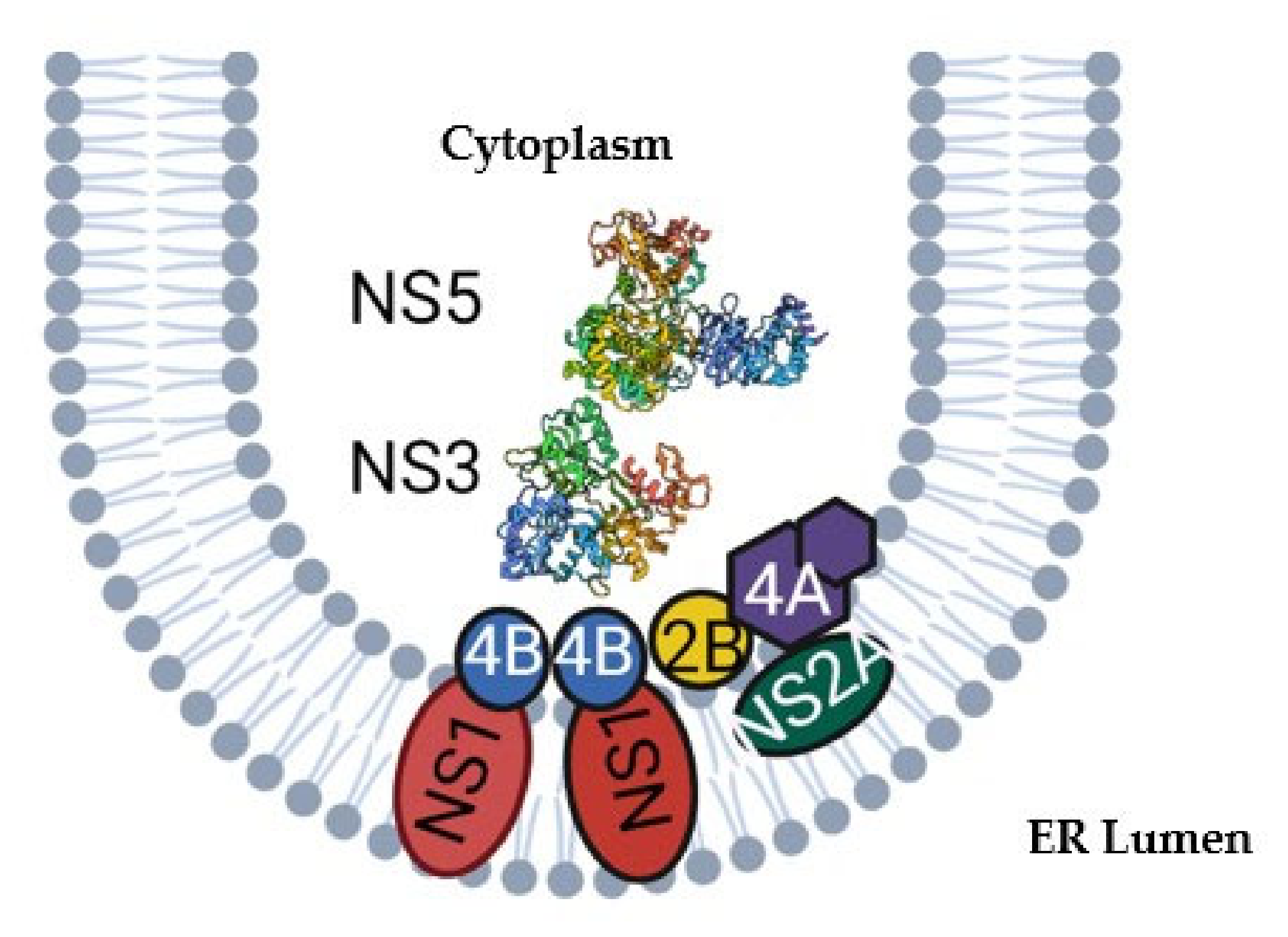
2. Structure and Function of NS3 and NS5

The NS5 is the largest flaviviral protein and is a natural fusion of a methyltransferase (MTase) and an RNA-dependent RNA polymerase (RdRP) (Figure 2A). The C-terminal domain includes the RdRP while the N-terminal includes the MTase domain, identified by the AdoMet-dependent MTase motif as well as a guanylyltransferase, indicated by a GTP-binding site and previously characterized capping mechanisms [37,38]. Methyltransferase activity has been confirmed using a recombinant full length NS5 and truncated NS5 containing only the MTase, and via incorporation of radiolabeled methyl groups [39,40,41]. RdRP activity was ascribed to this domain with similar methodology, utilizing a recombinant NS5 fusion protein with glutathione S-transferase with RdRP activity and performing a radiolabel assay to determine incorporation of the label in the newly synthesized RNA [42]. Guanylyltransferase activity via the detection of the GMP-enzyme intermediate when GTP is used as a substrate, the GMP moiety can be transferred to the diphosphate end of the RNA transcript [43]. NS5 is a primer-independent RdRP, meaning it is able to initiate replication de novo. This initiation is carried out using the priming loop, an extension of the thumb domain. The initial step is binding of two nucleotides, including a purine, at the priming site, as well as binding of the template RNA, which is mediated by genome cyclization and occurs due to complementary sequences in the 3′ and 5′ UTRs Figure 2B [44]. The RdRP recognizes a conserved stem loop region within the 5′ UTR which acts as a promoter [45]. The priming loop serves to act as a platform to stabilize these initial steps. Two divalent cations bind to the active site for which binding sites are highly conserved in RdRPs. Catalysis results in the generation of a pyrophosphate, a dinucleotide and translocation. The remaining dinucleotide can be elongated via NTP hydrolysis [46,47].
The NS5 RdRP shares a highly similar structure to other polymerases with the domains resembling a cupped right hand [51,52]. Within the three structural domains: fingers, palm, and thumb, there are seven conserved motifs (A-G) (Figure 2A-C) [37,53,54]. This hand can adopt an open and closed conformation, mediated by movements of motifs F and G [52]. The open conformation exposes the active site during initiation and elongation, while the closed conformation clasps around the incoming nucleotide to facilitate addition [46]. The active site consists of three conserved aspartates located in motifs A and D which are critical for catalysis. Motif B contacts the template and incoming rNTP and has a highly flexible loop which may be involved in translocation [55]. Motif D acts as scaffold for the palm domain and is important during conformation changes. Motif E aids in NTP binding (Figure 2C) [54,56].
The flaviviral NS3 protein contains a C-terminal helicase (NS3h) and an N-terminal serine protease. The helicase belongs to superfamily 2 [57] which is represented by the well-characterized hepatitis C virus (HCV) NS3 [58]. Although not a flavivirus, HCV is a member of the Flaviviridae family and the hepacivirus genus and shares many similarities with members of the flavivirus genus (including WNV, DENV, YFV, ZIKV). The minimal qualification for a superfamily 2 helicase is ATP-dependent directional translocation [57,58]. For flaviviruses, helicases are directional, translocating in the 3′–5′ direction along the tracking RNA during unwinding and can traverse breaks in the phosphodiester backbone [59,60]. NS3h is composed of three subdomains known as 1, 2, and 3 or alternately, N, C, and 3. Subdomains 1 and 2 are conserved across all SF2 helicases where they contain the characteristic Rec-A-like folds of the helicase core and form a deep groove housing the nucleotide binding pocket, in motif 1, and the RNA binding sites [57,58,60]. Motif II binds Mg2+ cations required for ATP catalysis [61]. Despite its stated role in unwinding dsRNA during replication, NS3h has been found to translocate and bind to both DNA and RNA, possibly due to the extensive contacts to the phosphodiester backbone of the tracking strand which stabilizes unwinding [62,63,64]. These contacts are not DNA or RNA specific and are mediated instead via water molecules or directly via subdomains 1 and 2 and motifs 1a, TxGx, 4 and 5 [58,59]. However, when NS2B and NS3 are bound together, DNA unwinding is repressed [65]. This difference in selectivity may reflect the different function required in different aspects of the viral life cycle, with NS2B-NS3 being required during polyprotein processing and NS3/NS5 complex being required in ER invaginations to aid in replication [66,67]. Motif VI is involved in altering the conformation of NS3h to closed conformation of NS3 where the substrate is gripped [58,67]. NS3h activity is powered by ATP binding, which is coordinated via motifs I, II. Binding weakens nucleic acid interaction via domains Ia, TxGx, 4, and 5, allowing translocation and unwinding. Hydrolysis of ATP via motifs I, II, and VI triggers rebinding to the nucleic acid in a forward motion while motif III coordinates these processes [57,58] acting as a linker between motifs I and II [68].
In addition to its helicase and NTPase activities, NS3h acts a triphosphatase on positive sense RNA genomes to begin the coordinated process of capping at the 5′ end [69]. The triphosphatase removes a single phosphate group from the backbone of the positive-sense RNA strand, resulting in a diphosphate [39]. At this point, the NS5 methyltransferase (MTase) located at the N terminus of NS5, dephosphorylates GTP into a GMP moiety and transfers it to the 5′ dephosphorylated end of the positive sense RNA to be capped via its guanylyltransferase activity [70]. The core of the MTase consists of beta strands alternating with alpha helices, and contains a SAM binding pocket, RNA binding site, GTP pocket, and K-D-K-E motif at the active site (Figure 2D) [41,71]. The MTase domain also contains two other distinctive enzymatic functions: a guanine-N7-methyltransferase and an RNA 2′O-methyltransferase at the N-terminus [39,41]. Both processes utilize S-adenosyl-l-methionine (AdoMet) as a methyl donor, converting it into S-adenosyl-l-homocysteine. These methyl groups are transferred sequentially onto the N7 atom of the cap guanine and the 2′-hydroxyl group of the ribose moiety of that same RNA nucleotide (Figure 2D) [39,41,67,70]. This results in a type 1 cap structure that is identical to host mRNA, allowing for efficient translation by host machinery and increased stability as well as immune evasion [72,73,74,75]. Mutations that impair MTase activity decrease or abolish replication, indicating the importance of capping and the MTase in viral replication [41].
The role of NS3 in host-specific adaptation has been examined for WNV, with the two most prominent examples being the naturally occurring substitution T249P and the laboratory-derived S411A. T249A is a well-studied mutation that was positively selected when WNV first arrived in North America in 1999 and has been linked to increased virulence in American crows, although this phenotype is not consistent across avian species [76,77]. Mechanistic studies of this mutant NS3 revealed increased ATP activity but similar helicase activity, which failed to explain the altered phenotype of strains with this substitution. Temperature studies indicated increased viral load in avian fever conditions, and further mechanistic studies using altered temperatures may help to elucidate how mechanistic differences reflect phenotypic differences [77]. Experiments with hyperactive Kunjin virus containing the S411A NS3 mutation resulted in decreased viral infection of cells, increased mortality and dissemination in mosquitoes, as well as decreased infection. These results in part can be explained by the hyperactive helicase activity caused by the mutation, which could increase cytopathogenicity and decreased mosquito survival [78].
Although the chemical processes of flaviviral replication and capping are well characterized, the coordination of each of these steps remains elusive. Flavivirus NS proteins clearly interact via the replication complex and the NS3 and NS5 enzymes via direct contact [79], yet the precise role of each domain and protein in replication is unknown. While the MTase has been extensively studied in RNA capping, its role in RNA replication is unclear. However, deletion of the MTase domain significantly decreases viral RNA synthesis, suggesting other roles for the MTase in replication [52,80]. How the replicated RNA is passed between the NS3h, RdRP and MTase for complete genome replication is largely unknown. While crystal structures for full length NS3 and NS5 have been solved for many flaviviruses [35,36,51,52,81], none have been solved for NS3 bound to NS5. The orientation of the MTase relative to RdRP also differs between crystal structures. Several models have been posited for how RNA is replicated and capped. Crystallization of the JEV NS5 revealed a small and flexible linker region, which connects the two domains of NS5, as well as a MTase entry site on the opposite side of the NS5 dsRNA exit path [51]. A highly conserved three amino acid sequence (GTR) across flaviviruses is found adjacent to the variable linker. This sequence could serve as a pivot point, allowing for flexible movement of the MTase to different orientations, including one in which the entry site of the MTase is perfectly aligned with the RdRP exit [51]. Subsequent studies demonstrated the importance of both the GTR sequence and the linker. Mutagenesis of the GTR region in DENV severely repressed viral replication [82]. DENV2 NS5 containing either JEV or ZIKV linkers completely abolished viral replication, potentially due to altered flexibility hindering interactions with either the RdRP or NS3 [83]. Later work also supported the idea of the GTR region acting as a hinge; studies resolved a DENV NS5 structure with an altered MTase orientation. Upon aligning the JEV NS5 (PDB 4K6M) to their own structure (PDB 5CCV), Klema et al. found using DynDom that significant bending of the linker region using GTR as a pivot was required to perfectly align the structures [48]. Interestingly, they also crystallized DENV NS5 in a dimer formation where the methyltransferase entry site and RdRP exit are located on the same side and propose that this may be how flaviviral RNA replication and capping is coordinated [48,70]. Although progress has been made in unraveling the journey of a flavivirus RNA during replication, more studies are required to precisely define how the RNA is processed and how the initial step of capping is performed by NS3.

3. Fidelity

RNA viruses, including flaviviruses, are notorious for having highly error-prone genome replication relative to bacteria and eukaryotes whose genomes are replicated by DNA polymerases rather than RNA polymerases. The reason behind this discrepancy stems largely from the lack of proofreading displayed by all RNA viruses, which lack exonucleases able to excise mispaired bases (except for coronaviruses). The result is a mutation rate of approximately 10−4 mutations per nucleotide copied, or approximately 1 mutation per genome replicated for a typical 11kb flavivirus [84,85,86]. These high mutation rates generate many diverse viral genomes within the course of a single infection, due to the high rate of viral replication that leads to significant viral loads [84]. This diverse array of genomes is often referred to as quasispecies. The composition and breadth of quasispecies (addressed in detail below) have a significant impact on viral fitness and adaptability when new selective pressures are introduced [87,88,89,90,91,92].
Fidelity refers to the accuracy of replication enzymes in faithfully copying genomes, RNA viruses have relatively low fidelity compared to other organisms. This is both beneficial in terms of the generation of diversity and problematic in that these viruses are theorized to be maintained close to error catastrophe, where the accumulation of deleterious genomes due to high mutation rates leads to extinction [93,94]. This notion has been shown experimentally through the use of mutagens, including ribavirin, favipiravir, and 5′fluorouracil (5-FU) in a variety of RNA viruses including flaviviruses [95,96,97]. Higher concentrations of ribavirin have been shown to result in: (1) Reduced levels of viable virus, (2) reduced specific infectivity, and (3) increased mutation frequency, which correlated with loss of viable virus and lethal mutagenesis. WNV has been driven to lethal mutagenesis by favipiravir when used in vitro, although the precise mechanism of action of favipiravir remains unknown [98]. Thus, error catastrophe can be induced via loss of replication fidelity [97,99].
Perturbation of viral fidelity can thus be a useful tool in studying viral evolution and the importance of viral diversity in fitness and adaptation. Ribavirin has been used extensively to experimentally isolate high-fidelity mutants of viruses including poliovirus, St. Louis encephalitis virus (SLEV), WNV, Coxsackievirus B3, Venezuelan equine encephalitis virus (VEEV), and chikungunya virus (CHIKV) [87,91,100,101,102,103,104]. Interestingly, many of the fidelity variants generated in this manner contained structural changes far from the active site [91,101,104], suggesting that remote sites can greatly impact RdRP fidelity through indirect effects on catalysis. For instance, poliovirus G64S, a high-fidelity substitution isolated through passage in ribavirin, was shown to increase fidelity by stabilizing the triphosphate in a catalytically competent conformation [101,105].
Several fidelity mutants of the picornaviruses have since been identified either by passaging virus in the presence of a mutagen or via structural prediction. While picornaviruses are structurally and biochemically distinct from flaviviruses, these studies provided the basis for our understanding of the phenotypic consequences of altered fidelity for RNA viruses. Foot-and-mouth disease virus fidelity mutants W237F (high fidelity) and W237I (low fidelity) and W237L (low fidelity) demonstrate universally decreased virulence and mortality within mice but not within cell culture, similar to previous studies [106]. This result is likely due to cell culture being poorly representative of host environments [75]. Replication fidelity has been measured by genome sequencing or biochemical assays. Specifically, single nucleotide discrimination of CTP vs 2′dCTP with the high fidelity mutant W237F demonstrated the highest discrimination factor while the low fidelity mutants had the lowest [107]. Low and high-fidelity mutants have also been generated for Coxsackievirus B3, which were characterized using similar assays, as well as single nucleotide incorporation assays which detect correct incorporation via fluorescence. Nine low fidelity mutations were identified which, similar to previous picornavirus fidelity mutants, were attenuated in vivo but not in vitro [102]. Additionally, the lowest fidelity mutants generated were not viable and either did not grow or had new attenuating mutations, reinforcing the notion of error catastrophe. Further studies identified high fidelity mutations including F364Y, which again demonstrated in vivo attenuation [103]. As such, fidelity mutants are largely attenuated in vivo, potentially due to the lack of diversity generated by high fidelity mutants and the proximity to error catastrophe in low fidelity mutants.
The impact of altered fidelity has also been evaluated in arboviruses, including CHIKV, VEEV, SLEV, and WNV, which unlike picornaviruses are multi-host and thus experience unique transmission bottlenecks and additional selective constraints. Initial studies identified fidelity variants in CHIKV following passage in 5-FU [100,108]. The mutation C483Y was shown to be associated with a high-fidelity phenotype determined via sequencing because in vitro biochemical nucleotide incorporation assays do not exist for alphaviruses [100]. Unlike the picornaviral fidelity variants, C483Y, located in the RdRP, demonstrated no attenuation in vitro, although in competition the WT grew significantly better in insect cell lines. This result was reflected in vivo, with significantly lower viral loads in the bodies and legs of infected mosquitoes. Similarly, during infection of a mammalian host, significant attenuation in viremia and tissue tropism of C483Y was identified. Low fidelity mutants were also generated via substitution at the 483 position [108]. Unsurprisingly, these variants produced more defective genomes than WT and were also attenuated in mammalian hosts in each tissue type (muscle, blood, brain, and liver). Unlike the high-fidelity mutant, low fidelity mutants were significantly attenuated in mosquito cell culture when one-step growth kinetics were measured. In mosquito hosts, reversion of the low fidelity mutants occurred, consistent with attenuation driving selection against this phenotype. While increased viral diversity could be preferable within these hosts due to selective pressure from the mosquito RNA interference response (RNAi), which is based on sequence similarity [109], there is clearly a limit to this benefit. Further studies on alphavirus fidelity with VEEV identified a low fidelity variant following passaging in the presence of 5-FU. Unlike previous studies on CHIKV, the low fidelity VEEV variant was more virulent than the WT and showed no attenuation in tissue tropism. As different electroporation pools (used to inject mutant virus constructs into host cells, allowing them to replicate and serve as a stock of the mutant virus in question) generated from the low fidelity mutant were shown to have significantly different impacts on virulence, this study highlighted the importance of the individual composition of quasispecies and the tremendous impact they can exert on viral fitness [110].
The impact of fidelity on flavivirus fitness and host switching has also been examined for WNV and SLEV. WNV fidelity mutants were generated with selection for ribavirin resistance and included a high-fidelity mutant containing V793I and G806R NS5 substitutions located in the thumb subdomain of the RdRP, and a low fidelity mutant, with a T248I substitution in the methyltransferase. The location of these mutations offers unique insight into the role of remote interactions in regulating flavivirus fidelity. The WNV RdRP mutations demonstrate the influence of the priming loop on polymerase fidelity and the low fidelity mutant demonstrated, for the first time, that structural alteration of the flavivirus methyltransferase can have a significant impact on polymerase fidelity. These fidelity phenotypes were confirmed via in vitro primer extension assay, which examines the ability of NS5 to incorporate each of the nucleotides accurately during elongation. These studies confirmed deep-sequencing data demonstrating the fidelity can be mispair specific. The fidelity differences were most pronounced for A:C misincorporation, suggesting specific structural perturbations could be associated with unique mutational biases and evolutionary trajectories. In vitro cell culture studies revealed no attenuation in vertebrate cells, but attenuation in mosquito cells as was seen with CHIKV fidelity mutants. Vector competence of Culex spp. mosquitoes for WNV fidelity mutants was shown to be significancy impaired, with highly attenuated infectivity and no evidence of virus transmission [104]. Studies with the SLEV high fidelity mutant, attributed to a E416K RdRP substitution, additionally support the previous results demonstrating attenuation in mosquito but not mammalian cells. Interestingly, this attenuation was limited to mosquito cells with an intact RNAi response, providing further evidence that a limited capacity for RNAi evasion likely contributes to the decreased fitness of high-fidelity variants in mosquitoes [91]. RdRP fidelity also greatly impacts viral evolution and viability during host switching in vitro. During 20 sequential passages alternating from mosquito to avian cells, only the wildtype survived all attempted passages and maintained a higher viral load, demonstrating that wildtype fidelity is fine-tuned for host switching and perturbing fidelity likely decreases the capacity for long-term maintenance with host alteration [92].
Despite the utility of fidelity mutants in probing the relationships between viral diversity and host adaptations, significant issues plague our understanding of these interactions, which stem both from different methodologies used to define fidelity and conflicting results in unique experimental systems and hosts. Early methods for defining fidelity consisted of sequencing small regions of the genome to examine mutation frequency, which fail to examine diversity across the entirety of the genome and is limited by the uncertain influence of evolutionary pressures and process errors of sequencing platforms. Biochemical assays are more reliable in directly measuring activity and misincorporation by the RdRP but are not available for all viral species. Further, how the context of the host cell environment and the fully intact replication complex could influence polymerase fidelity is not well-defined. Additionally, different biochemical methodologies are used to define different viruses with some utilizing nucleotide incorporation assays and others nucleotide discrimination assays, making attributing differences in phenotype to specific structural variations difficult. Indeed, different results in terms of phenotypic attenuation have even been obtained for the same mutant [87,101,110], highlighting the complexity of accurately defining these relationships. For an excellent review on the benefits and challenges with studies of fidelity variants see Kautz and Forrester 2018 [106].

4. Quasispecies and Host Adaptation

Underlying these fidelity studies is the question of how viral diversity impacts viral fitness, adaptation, and evolution. Viral infections, particularly by RNA viruses, result in a diverse cloud of genomes generated by polymerase misincorporation during replication [111]. This diverse population is referred to as a quasispecies, a notion first described in the 1970s in reference to early life as evolving from “replicons” [112]. These replicons were diverse and stemmed from a master sequence, the replicon with the highest fitness or replicative capacity, which is bound to its closely related neighbors by the inevitability of mutation. Thus, quasispecies theory posits that individual mutants are not the subject of selection but rather the population as a whole [112,113]. This is in contrast to Darwinian notions of evolution, were selection acts on individuals within a population. It also argues for the notion of “survival of the flattest” whereby viral evolution favors mutating toward areas of sequence space where fitness is relatively flat (not large valleys or peaks) so that the quasispecies swarm retains fitness in the face of mutation, frequent bottlenecks, and fluctuating selective pressures [113].
From this initial framework stems several related theories regarding quasispecies behavior, including Muller’s ratchet, the effect of genetic bottlenecks on fitness, and error catastrophe. Genetic bottlenecks within and between hosts are predicted to decrease the fitness of quasispecies due to the loss of diversity and the likelihood that any individual variant will have deleterious mutations. Serial plaque transfers of individual variants to cells have supported the notion that genetic bottlenecks decrease fitness, which is known in population genetics as Muller’s ratchet [114]. Muller’s ratchet predicts that small population sizes of asexual organisms with a high mutation rate will accumulate deleterious mutations in an irreversible manner in the absence of outside sources of diversity. However, if larger populations of quasispecies infect a new selective environment or host, adaptive selection will occur. These ideas have been supported by a number of in vitro passage studies [115,116,117,118]. Linked to this theory is the idea of error catastrophe, the theoretical threshold of replication fidelity below which viruses cannot produce sufficient viable genomes due to the accumulation of deleterious mutations [93,119,120]. Numerous antiviral strategies have utilized mutagenic compounds as treatments for viruses like hepatitis C virus (HCV) [121], a Hepacivirus in the family Flaviviridae, and human immunodeficiency virus (HIV) [122]. Lethal mutagenesis has also been shown to be a viable mechanism to decrease flavivirus fitness [98,123], which is further supported by reports of attenuation of low fidelity variants as discussed above.
Quasispecies exist as an individual unit of selection and work cooperatively or competitively to collectively constitute the fitness of a virus. This notion has been shown experimentally, where the fitness of individual genomes within a quasispecies have been found to be distinct from the viral swarm [90] and neuroinvasive pathogenesis has been rescued to wildtype levels by the addition of diverse quasispecies [88]. Maintenance of quasispecies diversity allows for the existence of variants uniquely fit to a variety of environments [117,124].
Quasispecies are host-dependent [115], which makes intuitive sense given the highly unique selective pressures which exists between different hosts. The trade-off hypothesis suggests that alternating replication in two taxonomically divergent hosts (vertebrates and not vertebrates) with distinct selective environments can explain the relatively low rates of evolution of arboviruses [125]. Indeed, many host-specific mutations result in a fitness trade-off in divergent hosts, yet broadly adaptive mutations have also been noted in experimental systems. Adaptation of WNV following sequential passage of transmitted virus via mosquitoes was associated with substitutions in the WNV replicase without decreased viremia in an avian model, yet comparative studies with SLEV demonstrate that the extent of adaptive trade-offs may be species-specific, even among closely related viruses [126,127]. Additional studies of WNV adaptation utilizing serial or alternate passage in vivo in chicks or mosquitoes also demonstrated host-specific fitness, yet mosquito-passaged WNV results have been variable, demonstrating that adaptive tradeoffs are not inevitable [128]. The relatively lower diversity of resulting quasispecies after passaging in chicks compared to mosquitoes suggests stronger purifying selection in avian hosts for WNV and SLEV. This notion has been supported via sequencing of naturally occurring WNV [129,130] and experimentally via infection of susceptible bird populations [131]. Although whole genome sequencing of naturally occurring WNV in avian organs demonstrated higher levels of diversity than naturally infected mosquitoes [132]. The increased diversity in mosquitoes may be attributed to density-dependent selection resulting from RNAi evasion [133,134]. Interestingly, higher levels of intrahost diversity in the vector were not identified in the tick-borne Powassan virus, highlighting how selective pressures on flavivirus quasispecies are both host and virus-specific [135].
The existence of quasispecies does not preclude continued evolution of the master sequence. Natural evolution of WNV from the ancestral NY99 genotype to the WN02 genotype, characterized by a single consensus substitution in the envelope gene, V159A, was shown to be driven by superior fitness in mosquito hosts [136]. WNV continues to experience adaptive evolution, as ancestral WN02 strains have begun to be displaced by newer strains including WNV NY10 [137]. Such evolutionary events are not limited to WNV; ZIKV has recently emerged in part due to mutation A188V in NS1, which increases infectivity in Aedes aegypti, and S17N in the prM, which likely contributes to fitness and virulence in neural progenitor cells [138,139].

5. Conclusions

Although progress has been made in understanding the role of quasispecies and individual mutations in host adaptation, much remains unclear. The most accurate sequencing technologies utilize read lengths that make minority haplotype reconstruction difficult, which impedes the capacity to study mutations in appropriate genomic contexts. In addition, many studies to date have utilized experimental host systems and experimentally derived mutations to study adaptation, which are unlikely to fully reflect the selective pressure of natural systems. Mechanistic explanations for many of the phenotypes attributed to specific mutations is also largely lacking. The nature of quasispecies makes it difficult to study the influence of individual mutations with variable frequencies. Rather, reverse genetics approaches are typically used to generate a given mutant as the master sequence and adaptation is studied in this context. The fact that arboviruses are almost exclusively error-prone RNA viruses can likely be explained by evolutionary selection and the perpetual diversification of flaviviruses is arguably among the most important attributes driving the global success of these pathogens. Our knowledge of the process of flaviviral replication, the organization and function of the flaviviral replication complex and its role in host-specific adaptation has greatly increased. Future studies should aim to more accurately define the role of individual proteins of the replication complex in polymerase function and fidelity during replication in distinct cell types, utilize improved sequence and single cell technologies to expand our understanding of quasispecies structures, and focus on phenotypic studies in natural hosts.

Author Contributions

Conceptualization, H.S.C. and A.T.C. Investigation, H.S.C. Visualization, H.S.C. and J.D.P. Resources and Funding, A.T.C. Writing – original draft H.S.C. Writing, review and editing, H.S.C., A.T.C. and J.D.P. Supervision, A.T.C. All authors have read and agreed to the published version of the manuscript.

Funding

National Institute of Health R21 AI146856, National Institute of Health R01 AI1680907 and National Institute of Health NIH R01 GM080573.

Institutional Review Board Statement

Not applicable.

Informed Consent Statement

Not applicable.

Data Availability Statement

Not applicable.

Conflicts of Interest

The authors declare no conflict of interest.

References

  1. Kilpatrick, A.M. Globalization, land use, and the invasion of West Nile virus. Science 2011, 334, 323–327. [Google Scholar] [CrossRef] [Green Version]
  2. Kramer, L.D.; Styer, L.M.; Ebel, G.D. A Global Perspective on the Epidemiology of West Nile Virus. Annu. Rev. Entomol. 2007, 53, 61–81. [Google Scholar] [CrossRef] [PubMed] [Green Version]
  3. Ronca, S.E.; Ruff, J.C.; Murray, K.O. A 20-year historical review of West Nile virus since its initial emergence in North America: Has West Nile virus become a neglected tropical disease? PLoS Negl. Trop. Dis. 2021, 15, e0009190. [Google Scholar] [CrossRef] [PubMed]
  4. Didier, M.; Gubler, D.J. Zika Virus. Clin. Microbiol. Rev. 2016, 29, 487–524. [Google Scholar] [CrossRef] [Green Version]
  5. Centers for Disease Control and Prevention (CDC); Brunette, G.W.; Nemhauser, J.B. CDC Yellow Book 2020 Health Information for International Travel: Health Information for International Travel; Oxford University Press: Oxford, UK, 2019. [Google Scholar] [CrossRef]
  6. Gubler, D.J. Dengue and Dengue Hemorrhagic Fever. Clin. Microbiol. Rev. 1998, 11, 480–496. [Google Scholar] [CrossRef] [PubMed] [Green Version]
  7. Nwaiwu, A.U.; Musekiwa, A.; Tamuzi, J.L.; Sambala, E.Z.; Nyasulu, P.S. The incidence and mortality of yellow fever in Africa: A systematic review and meta-analysis. BMC Infect. Dis. 2021, 21, 1089. [Google Scholar] [CrossRef]
  8. Gardner, C.L.; Ryman, K.D. Yellow Fever: A Reemerging Threat. Clin. Lab. Med. 2010, 30, 237–260. [Google Scholar] [CrossRef] [PubMed] [Green Version]
  9. Erlanger, T.E.; Weiss, S.; Keiser, J.; Utzinger, J.; Wiedenmayer, K. Past, present, and future of Japanese encephalitis. Emerg. Infect. Dis. 2009, 15, 1–7. [Google Scholar] [CrossRef]
  10. Therkelsen, M.D.; Klose, T.; Vago, F.; Jiang, W.; Rossmann, M.G.; Kuhn, R.J. Flaviviruses have imperfect icosahedral symmetry. Proc. Natl. Acad. Sci. USA 2018, 115, 11608. [Google Scholar] [CrossRef] [Green Version]
  11. Barrows, N.J.; Campos, R.K.; Liao, K.C.; Prasanth, K.; Soto-Acosta, R.; Yeh, S.; Schott-Lerner, G.; Pompon, J.; Sessions, O.; Bradrick, S.; et al. Biochemistry and Molecular Biology of Flaviviruses. Chem. Rev. 2018, 118, 4448–4482. [Google Scholar] [CrossRef]
  12. Chen, Y.; Maguire, T.; Hileman, R.E.; Fromm, J.; Esko, J.; Lindhardt, R.; Marks, R. Dengue virus infectivity depends on envelope protein binding to target cell heparan sulfate. Nat. Med. 1997, 3, 866–871. [Google Scholar] [CrossRef]
  13. Pujhari, S.; Brustolin, M.; Macias, V.M.; Nissly, R.; Nomura, M.; Kuchipudi, S.; Rasgon, J. Heat shock protein 70 (Hsp70) mediates Zika virus entry, replication, and egress from host cells. Emerg. Microbes Infect. 2019, 8, 8–16. [Google Scholar] [CrossRef] [Green Version]
  14. Reyes-Del Valle, J.; Chávez-Salinas, S.; Medina, F.; del Angel, R.M. Heat shock protein 90 and heat shock protein 70 are components of dengue virus receptor complex in human cells. J. Virol. 2005, 79, 4557–4567. [Google Scholar] [CrossRef] [Green Version]
  15. Dejnirattisai, W.; Webb, A.I.; Chan, V.; Jumainsong, A.; Davidson, A.; Mongolsapaya, J.; Screaton, G. Lectin Switching During Dengue Virus Infection. J. Infect. Dis. 2011, 203, 1775–1783. [Google Scholar] [CrossRef] [Green Version]
  16. Meertens, L.; Carnec, X.; Lecoin, M.P.; Ramdasi, R.; Guivel-Benhassine, F.; Lew, E.; Lemke, G.; Schwartz, O.; Amara, A. The TIM and TAM families of phosphatidylserine receptors mediate dengue virus entry. Cell Host Microbe. 2012, 12, 544–557. [Google Scholar] [CrossRef] [Green Version]
  17. Navarro-Sanchez, E.; Altmeyer, R.; Amara, A.; Schwartz, O.; Fieschi, F.; Virelizier, J.; Arenzana-Seisdedos, F.; Desprès, P. Dendritic-cell-specific ICAM3-grabbing non-integrin is essential for the productive infection of human dendritic cells by mosquito-cell-derived dengue viruses. EMBO Rep. 2003, 4, 723–728. [Google Scholar] [CrossRef]
  18. Tassaneetrithep, B.; Burgess, T.H.; Granelli-Piperno, A.; Trumpfheller, C.; Finke, J.; Sun, W.; Eller, M.; Pattanapanyasat, K.; Sarasombath, S.; Birx, D.; et al. DC-SIGN (CD209) Mediates Dengue Virus Infection of Human Dendritic Cells. J. Exp. Med. 2003, 197, 823–829. [Google Scholar] [CrossRef] [Green Version]
  19. Chu, J.J.H.; Ng, M.L. Infectious entry of West Nile virus occurs through a clathrin-mediated endocytic pathway. J. Virol. 2004, 78, 10543–10555. [Google Scholar] [CrossRef] [Green Version]
  20. Hackett, B.A.; Cherry, S. Flavivirus internalization is regulated by a size-dependent endocytic pathway. Proc. Natl. Acad. Sci. USA 2018, 115, 4246–4251. [Google Scholar] [CrossRef] [Green Version]
  21. Acosta, E.G.; Castilla, V.; Damonte, E.B. Functional entry of dengue virus into Aedes albopictus mosquito cells is dependent on clathrin-mediated endocytosis. J. Gen. Virol. 2008, 89, 474–484. [Google Scholar] [CrossRef]
  22. Chiu, W.W.; Kinney, R.M.; Dreher, T.W. Control of translation by the 5′- and 3′-terminal regions of the dengue virus genome. J. Virol. 2005, 79, 8303–8315. [Google Scholar] [CrossRef] [Green Version]
  23. Sanford, T.J.; Mears, H.; Fajardo, T.; Locker, N.; Sweeney, T.R. Circularization of flavivirus genomic RNA inhibits de novo translation initiation. Nucleic Acids Res. 2019, 47, 9789–9802. [Google Scholar] [CrossRef] [PubMed] [Green Version]
  24. Ruiz-Linares, A.; Cahour, A.; Després, P.; Girard, M.; Bouloy, M. Processing of yellow fever virus polyprotein: Role of cellular proteases in maturation of the structural proteins. J. Virol. 1989, 63, 4199–4209. [Google Scholar] [CrossRef] [PubMed] [Green Version]
  25. Garcia-Blanco, M.A.; Vasudevan, S.G.; Bradrick, S.S.; Nicchitta, C. Flavivirus RNA transactions from viral entry to genome replication. Antivir. Res. 2016, 134, 244–249. [Google Scholar] [CrossRef] [PubMed]
  26. Reid, D.W.; Campos, R.K.; Child, J.R.; Zheng, T.; Chan, K.; Bradrick, S.; Vasudevan, S.; Garcia-Blanco, M.; Nicchitta, C. Dengue Virus Selectively Annexes Endoplasmic Reticulum-Associated Translation Machinery as a Strategy for Co-opting Host Cell Protein Synthesis. J. Virol. 2018, 92, e01766-17. [Google Scholar] [CrossRef] [PubMed] [Green Version]
  27. Den Boon, J.A.; Ahlquist, P. Organelle-Like Membrane Compartmentalization of Positive-Strand RNA Virus Replication Factories. Annu. Rev. Microbiol. 2010, 64, 241–256. [Google Scholar] [CrossRef] [PubMed]
  28. Welsch, S.; Miller, S.; Romero-Brey, I.; Merz, A.; Bleck, C.; Walther, P.; Fuller, S.; Antony, C.; Krijnse-Locker, J.; Bartenschlager, R. Composition and three-dimensional architecture of the dengue virus replication and assembly sites. Cell Host Microbe. 2009, 5, 365–375. [Google Scholar] [CrossRef] [Green Version]
  29. Oliveira, E.R.A.; de Alencastro, R.B.; Horta, B.A.C. New insights into flavivirus biology: The influence of pH over interactions between prM and E proteins. J. Comput. Aided Mol. Des. 2017, 31, 1009–1019. [Google Scholar] [CrossRef]
  30. Lindenbach, B.D.; Rice, C.M. Genetic interaction of flavivirus nonstructural proteins NS1 and NS4A as a determinant of replicase function. J. Virol. 1999, 73, 4611–4621. [Google Scholar] [CrossRef] [Green Version]
  31. Miller, S.; Sparacio, S.; Bartenschlager, R. Subcellular Localization and Membrane Topology of the Dengue Virus Type 2 Non-structural Protein 4B. J. Biol. Chem. 2006, 281, 8854–8863. [Google Scholar] [CrossRef] [Green Version]
  32. Mackenzie, J.M.; Khromykh, A.A.; Jones, M.K.; Westaway, E.G. Subcellular Localization and Some Biochemical Properties of the Flavivirus Kunjin Nonstructural Proteins NS2A and NS4A. Virology 1998, 245, 203–215. [Google Scholar] [CrossRef]
  33. Xie, X.; Gayen, S.; Kang, C.; Yuan, Z.; Shi, P.Y. Membrane topology and function of dengue virus NS2A protein. J. Virol. 2013, 87, 4609–4622. [Google Scholar] [CrossRef] [Green Version]
  34. Mackenzie, J.M.; Jones, M.K.; Young, P.R. Immunolocalization of the Dengue Virus Nonstructural Glycoprotein NS1 Suggests a Role in Viral RNA Replication. Virology 1996, 220, 232–240. [Google Scholar] [CrossRef] [Green Version]
  35. Upadhyay, A.K.; Cyr, M.; Longenecker, K.; Tripathi, R.; Sun, C.; Kempf, D.J. Crystal structure of full-length Zika virus NS5 protein reveals a conformation similar to Japanese encephalitis virus NS5. Acta Crystallogr. Sect. F 2017, 73, 116–122. [Google Scholar] [CrossRef] [Green Version]
  36. Tian, H.; Ji, X.; Yang, X.; Xie, W.; Yang, K.; Chen, C.; Wu, C.; Chi, H.; Mu, Z.; Wang, Z.; et al. The crystal structure of Zika virus helicase: Basis for antiviral drug design. Protein Cell 2016, 7, 450–454. [Google Scholar] [CrossRef] [Green Version]
  37. Koonin, E. The phylogeny of RNA-dependent RNA polymerases of positive-strand RNA viruses. J. Gen. Virol. 1991, 72, 2197–2206. [Google Scholar] [CrossRef]
  38. Koonin, E. Computer-assisted identification of a putative methyltransferase domain in NS5 protein of flaviviruses and 2 protein of reovirus. J. Gen. Virol. 1993, 74, 733–740. [Google Scholar] [CrossRef]
  39. Issur, M.; Geiss, B.J.; Bougie, I.; Picard-Jean, F.; Despins, S.; Mayette, J.; Hobdey, S.; Bisaillon, M. The flavivirus NS5 protein is a true RNA guanylyltransferase that catalyzes a two-step reaction to form the RNA cap structure. RNA 2009, 15, 2340–2350. [Google Scholar] [CrossRef] [Green Version]
  40. Egloff, M.P.; Benarroch, D.; Selisko, B.; Romette, J.L.; Canard, B. An RNA cap (nucleoside-2′-O-)-methyltransferase in the flavivirus RNA polymerase NS5: Crystal structure and functional characterization. EMBO J. 2002, 21, 2757–2768. [Google Scholar] [CrossRef] [Green Version]
  41. Ray, D.; Shah, A.; Tilgner, M.; Guo, Y.; Zhao, Y.; Dong, H.; Deas, T.; Zhou, Y.; Li, H.; Shi, P. West Nile virus 5′-cap structure is formed by sequential guanine N-7 and ribose 2′-O methylations by nonstructural protein 5. J. Virol. 2006, 80, 8362–8370. [Google Scholar] [CrossRef] [Green Version]
  42. Tan, B.H.; Fu, J.; Sugrue, R.J.; Yap, E.H.; Chan, Y.C.; Tan, Y.H. Recombinant Dengue Type 1 Virus NS5 Protein Expressed inEscherichia coliExhibits RNA-Dependent RNA Polymerase Activity. Virology 1996, 216, 317–325. [Google Scholar] [CrossRef] [PubMed]
  43. Ahola, T.; Kääriäinen, L. Reaction in alphavirus mRNA capping: Formation of a covalent complex of nonstructural protein nsP1 with 7-methyl-GMP. Proc. Natl. Acad. Sci. USA 1995, 92, 507–511. [Google Scholar] [CrossRef] [PubMed] [Green Version]
  44. You, S.; Falgout, B.; Markoff, L.; Padmanabhan, R. In vitro RNA Synthesis from Exogenous Dengue Viral RNA Templates Requires Long Range Interactions between 5′- and 3′-Terminal Regions That Influence RNA Structure. J. Biol. Chem. 2001, 276, 15581–15591. [Google Scholar] [CrossRef] [PubMed] [Green Version]
  45. Westaway, E.G.; Mackenzie, J.M.; Khromykh, A.A. Kunjin RNA replication and applications of Kunjin replicons. In Advances in Virus Research; Academic Press: Cambridge, MA, USA, 2003; Volume 59, pp. 99–140. [Google Scholar] [CrossRef]
  46. Bhatia, S.; Narayanan, N.; Nagpal, S.; Nair, D.T. Antiviral therapeutics directed against RNA dependent RNA polymerases from positive-sense viruses. Mol. Asp. Med. 2021, 81, 101005. [Google Scholar] [CrossRef]
  47. Ng, K.K.S.; Arnold, J.J.; Cameron, C.E. Structure-function relationships among RNA-dependent RNA polymerases. Curr Top. Microbiol. Immunol. 2008, 320, 137–156. [Google Scholar] [CrossRef] [Green Version]
  48. Klema, V.J.; Ye, M.; Hindupur, A.; Teramoto, T.; Gottipati, K.; Padmanabhan, R.; Choi, K. Dengue Virus Nonstructural Protein 5 (NS5) Assembles into a Dimer with a Unique Methyltransferase and Polymerase Interface. PLoS Pathog. 2016, 12, e1005451. [Google Scholar] [CrossRef] [Green Version]
  49. Peng, G.; Peersen, O.B. Structural basis for active site closure by the poliovirus RNA-dependent RNA polymerase. Proc. Natl. Acad. Sci. USA 2010, 107, 22505–22510. [Google Scholar] [CrossRef] [Green Version]
  50. Yap, L.J.; Luo, D.; Chung, K.Y.; Lim, S.P.; Bodenreider, C.; Noble, C.; Shi, P.Y.; Lescar, J.; Lescar, J. Crystal Structure of the Dengue Virus Methyltransferase Bound to a 5′-Capped Octameric RNA. PLoS ONE 2010, 5, e12836. [Google Scholar] [CrossRef]
  51. Lu, G.; Gong, P. Crystal Structure of the Full-Length Japanese Encephalitis Virus NS5 Reveals a Conserved Methyltransferase-Polymerase Interface. PLoS Pathog. 2013, 9, e1003549. [Google Scholar] [CrossRef] [Green Version]
  52. Zhao, B.; Yi, G.; Du, F.; Chuang, Y.; Vaughan, R.; Sankaran, B.; Kao, C.; Li, P. Structure and function of the Zika virus full-length NS5 protein. Nat. Commun. 2017, 8, 14762. [Google Scholar] [CrossRef] [Green Version]
  53. Hansen, J.L.; Long, A.M.; Schultz, S.C. Structure of the RNA-dependent RNA polymerase of poliovirus. Structure 1997, 5, 1109–1122. [Google Scholar] [CrossRef] [Green Version]
  54. Bruenn, J.A. A structural and primary sequence comparison of the viral RNA-dependent RNA polymerases. Nucleic Acids Res. 2003, 31, 1821–1829. [Google Scholar] [CrossRef]
  55. Selisko, B.; Papageorgiou, N.; Ferron, F.; Canard, B. Structural and Functional Basis of the Fidelity of Nucleotide Selection by Flavivirus RNA-Dependent RNA Polymerases. Viruses 2018, 10, 59. [Google Scholar] [CrossRef] [Green Version]
  56. Lohmann, V.; Körner, F.; Herian, U.; Bartenschlager, R. Biochemical properties of hepatitis C virus NS5B RNA-dependent RNA polymerase and identification of amino acid sequence motifs essential for enzymatic activity. J. Virol. 1997, 71, 8416–8428. [Google Scholar] [CrossRef] [Green Version]
  57. Fairman-Williams, M.E.; Guenther, U.P.; Jankowsky, E. SF1 and SF2 helicases: Family matters. Curr. Opin. Struct. Biol. 2010, 20, 313–324. [Google Scholar] [CrossRef] [Green Version]
  58. Singleton, M.R.; Dillingham, M.S.; Wigley, D.B. Structure and Mechanism of Helicases and Nucleic Acid Translocases. Annu. Rev. Biochem. 2007, 76, 23–50. [Google Scholar] [CrossRef]
  59. Luo, D.; Xu, T.; Watson, R.P.; Scherer-Becker, D.; Sampath, A.; Jahnke, W.; Yeong, S.; Wang, C.; Lim, S.; Strongin, A.; et al. Insights into RNA unwinding and ATP hydrolysis by the flavivirus NS3 protein. EMBO J. 2008, 27, 3209–3219. [Google Scholar] [CrossRef] [Green Version]
  60. Kawaoka, J.; Jankowsky, E.; Pyle, A.M. Backbone tracking by the SF2 helicase NPH-II. Nat. Struct. Mol. Biol. 2004, 11, 526–530. [Google Scholar] [CrossRef]
  61. Benarroch, D.; Egloff, M.P.; Mulard, L.; Guerreiro, C.; Romette, J.L.; Canard, B. A Structural Basis for the Inhibition of the NS5 Dengue Virus mRNA 2′-O-Methyltransferase Domain by Ribavirin 5′-Triphosphate. J. Biol. Chem. 2004, 279, 35638–35643. [Google Scholar] [CrossRef] [Green Version]
  62. Tackett, A.J.; Wei, L.; Cameron, C.E.; Raney, K.D. Unwinding of nucleic acids by HCV NS3 helicase is sensitive to the structure of the duplex. Nucleic Acids Res. 2001, 29, 565–572. [Google Scholar] [CrossRef] [Green Version]
  63. Taylor, S.D.; Solem, A.; Kawaoka, J.; Pyle, A.M. The NPH-II Helicase Displays Efficient DNA˙RNA Helicase Activity and a Pronounced Purine Sequence Bias. J. Biol. Chem. 2010, 285, 11692–11703. [Google Scholar] [CrossRef] [PubMed] [Green Version]
  64. Guenther, U.P.; Handoko, L.; Laggerbauer, B.; Jablonka, S.; Chari, A.; Alzheimer, M.; Ohmer, J.; Plöttner, O.; Gehring, N.; Sickmann, A.; et al. IGHMBP2 is a ribosome-associated helicase inactive in the neuromuscular disorder distal SMA type 1 (DSMA1). Hum. Mol. Genet. 2009, 18, 1288–1300. [Google Scholar] [CrossRef] [PubMed] [Green Version]
  65. Chernov, A.; Shiryaev, S.A.; Aleshin, A.E.; Ratnikov, B.; Smith, J.; Liddington, R.; Strongin, A. The two-component NS2B-NS3 proteinase represses DNA unwinding activity of the West Nile virus NS3 helicase. J. Biol. Chem. 2008, 283, 17270–17278. [Google Scholar] [CrossRef] [PubMed] [Green Version]
  66. Amberg, S.M.; Rice, C.M. Mutagenesis of the NS2B-NS3-mediated cleavage site in the flavivirus capsid protein demonstrates a requirement for coordinated processing. J. Virol. 1999, 73, 8083–8094. [Google Scholar] [CrossRef] [Green Version]
  67. Bollati, M.; Alvarez, K.; Assenberg, R.; Baronti, C.; Canard, B.; Cook, S.; Coutard, B.; Decroly, E.; de Lamballerie, X.; Gould, E.; et al. Structure and functionality in flavivirus NS-proteins: Perspectives for drug design. Antivir. Res. 2010, 87, 125–148. [Google Scholar] [CrossRef]
  68. Wu, J.; Bera, A.K.; Kuhn, R.J.; Smith, J.L. Structure of the Flavivirus helicase: Implications for catalytic activity, protein interactions, and proteolytic processing. J. Virol. 2005, 79, 10268–10277. [Google Scholar] [CrossRef] [Green Version]
  69. Xu, T.; Sampath, A.; Chao, A.; Wen, D.; Nanao, M.; Chene, P.; Vasudevan, S.; Lescar, J. Structure of the Dengue virus helicase/nucleoside triphosphatase catalytic domain at a resolution of 2.4 A. J. Virol. 2005, 79, 10278–10288. [Google Scholar] [CrossRef] [Green Version]
  70. Klema, V.J.; Padmanabhan, R.; Choi, K.H. Flaviviral replication complex: Coordination between RNA synthesis and 5′-RNA capping. Viruses 2015, 7, 4640–4656. [Google Scholar] [CrossRef]
  71. Martin, J.L.; McMillan, F.M. SAM (dependent) I AM: The S-adenosylmethionine-dependent methyltransferase fold. Curr. Opin. Struct. Biol. 2002, 12, 783–793. [Google Scholar] [CrossRef]
  72. Decroly, E.; Ferron, F.; Lescar, J.; Canard, B. Conventional and unconventional mechanisms for capping viral mRNA. Nat. Rev. Microbiol. 2011, 10, 51–65. [Google Scholar] [CrossRef]
  73. Cleaves, G.R.; Dubin, D.T. Methylation status of intracellular dengue type 2 40 S RNA. Virology 1979, 96, 159–165. [Google Scholar] [CrossRef]
  74. Ramanathan, A.; Robb, G.B.; Chan, S.H. mRNA capping: Biological functions and applications. Nucleic Acids Res. 2016, 44, 7511–7526. [Google Scholar] [CrossRef] [PubMed]
  75. Brinton, M.A. Host Factors Involved in West Nile Virus Replication. Ann. N. Y. Acad. Sci. 2001, 951, 207–219. [Google Scholar] [CrossRef]
  76. Brault, A.C.; Huang, C.Y.H.; Langevin, S.A.; Kinney, R.; Bowen, R.; Ramey, W.; Panella, N.; Holmes, E.; Powers, A.; Miller, B. A single positively selected West Nile viral mutation confers increased virogenesis in American crows. Nat. Genet. 2007, 39, 1162–1166. [Google Scholar] [CrossRef] [PubMed]
  77. Langevin, S.A.; Bowen, R.A.; Reisen, W.K.; Andrade, C.; Ramey, W.; Maharaj, P.; Anishchenko, M.; Kenney, J.; Duggal, N.; Romo, H.; et al. Host Competence and Helicase Activity Differences Exhibited by West Nile Viral Variants Expressing NS3-249 Amino Acid Polymorphisms. PLoS ONE 2014, 9, e100802. [Google Scholar] [CrossRef] [PubMed]
  78. Du Pont, K.E.; Davidson, R.B.; McCullagh, M.; Geiss, B.J. Motif V regulates energy transduction between the flavivirus NS3 ATPase and RNA-binding cleft. J. Biol. Chem. 2020, 295, 1551–1564. [Google Scholar] [CrossRef] [PubMed]
  79. Kapoor, M.; Zhang, L.; Ramachandra, M.; Kusukawa, J.; Ebner, K.E.; Padmanabhan, R. Association between NS3 and NS5 Proteins of Dengue Virus Type 2 in the Putative RNA Replicase Is Linked to Differential Phosphorylation of NS5. J. Biol. Chem. 1995, 270, 19100–19106. [Google Scholar] [CrossRef] [PubMed] [Green Version]
  80. Wu, J.; Lu, G.; Zhang, B.; Gong, P. Perturbation in the conserved methyltransferase-polymerase interface of flavivirus NS5 differentially affects polymerase initiation and elongation. J. Virol. 2015, 89, 249–261. [Google Scholar] [CrossRef] [Green Version]
  81. Zhao, Y.; Soh, T.S.; Zheng, J.; Chan, K.; Phoo, W.; Lee, C.; Tay, M.; Swaminathan, K.; Cornvik, T.; Lim, S.; et al. A Crystal Structure of the Dengue Virus NS5 Protein Reveals a Novel Inter-domain Interface Essential for Protein Flexibility and Virus Replication. PLoS Pathog. 2015, 11, e1004682. [Google Scholar] [CrossRef]
  82. Li, X.D.; Shan, C.; Deng, C.L.; Ye, H.; Shi, P.; Yuan, Z.; Gong, P.; Zhang, B. The Interface between Methyltransferase and Polymerase of NS5 Is Essential for Flavivirus Replication. PLoS Negl. Trop. Dis. 2014, 8, e2891. [Google Scholar] [CrossRef]
  83. El Sahili, A.; Soh, T.S.; Schiltz, J.; Gharbi-Ayachi, A.; Seh, C.; Shi, P.; Lim, S.; Lescar, J. NS5 from Dengue Virus Serotype 2 Can Adopt a Conformation Analogous to That of Its Zika Virus and Japanese Encephalitis Virus Homologues. J. Virol. 2019, 94, e01294-19. [Google Scholar] [CrossRef]
  84. Drake, J.W. The Distribution of Rates of Spontaneous Mutation over Viruses, Prokaryotes, and Eukaryotes. Ann. N. Y. Acad. Sci. 1999, 870, 100–107. [Google Scholar] [CrossRef]
  85. Drake, J.W.; Holland, J.J. Mutation rates among RNA viruses. Proc. Natl. Acad. Sci. USA 1999, 96, 13910. [Google Scholar] [CrossRef] [Green Version]
  86. John, H.; Katherine, S.; Frank, H.; Elizabeth, G.; Stuart, N.; Scott, V. Rapid Evolution of RNA Genomes. Science 1982, 215, 1577–1585. [Google Scholar] [CrossRef]
  87. Pfeiffer, J.K.; Kirkegaard, K. A single mutation in poliovirus RNA-dependent RNA polymerase confers resistance to mutagenic nucleotide analogs via increased fidelity. Proc. Natl. Acad. Sci. USA 2003, 100, 7289. [Google Scholar] [CrossRef] [Green Version]
  88. Vignuzzi, M.; Stone, J.K.; Arnold, J.J.; Cameron, C.E.; Andino, R. Quasispecies diversity determines pathogenesis through cooperative interactions in a viral population. Nature 2006, 439, 344–348. [Google Scholar] [CrossRef]
  89. Van Slyke, G.A.; Ciota, A.T.; Willsey, G.G.; Jaeger, J.; Shi, P.Y.; Kramer, L.D. Point mutations in the West Nile virus (Flaviviridae; Flavivirus) RNA-dependent RNA polymerase alter viral fitness in a host-dependent manner in vitro and in vivo. Virology 2012, 427, 18–24. [Google Scholar] [CrossRef] [Green Version]
  90. Ciota, A.T.; Ehrbar, D.J.; van Slyke, G.A.; Willsey, G.G.; Kramer, L.D. Cooperative interactions in the West Nile virus mutant swarm. BMC Evol. Biol. 2012, 12, 58. [Google Scholar] [CrossRef] [Green Version]
  91. Griesemer, S.B.; Kramer, L.D.; van Slyke, G.A.; Pata, J.; Gohara, D.; Cameron, C.; Ciota, A. Mutagen resistance and mutation restriction of St. Louis encephalitis virus. J. Gen. Virol. 2017, 98, 201–211. [Google Scholar] [CrossRef]
  92. Caldwell, H.S.; Ngo, K.; Pata, J.D.; Kramer, L.D.; Ciota, A.T. West Nile Virus fidelity modulates the capacity for host cycling and adaptation. J. Gen. Virol. 2020, 101, 410–419. [Google Scholar] [CrossRef]
  93. Domingo, E.; Holland, J.J. RNA virus mutations and fitness for survival. Annu. Rev. Microbiol. 1997, 51, 151–178. [Google Scholar] [CrossRef] [PubMed]
  94. Domingo, E.; Holland, J.J.; Ahlquist, P. RNA Genetics; CRC Press Inc.: Boca Raton, FL, USA, 1988. [Google Scholar]
  95. Qiu, L.; Patterson, S.E.; Bonnac, L.F.; Geraghty, R.J. Nucleobases and corresponding nucleosides display potent antiviral activities against dengue virus possibly through viral lethal mutagenesis. PLoS Negl. Trop. Dis. 2018, 12, e0006421. [Google Scholar] [CrossRef]
  96. Agudo, R.; Arias, A.; Domingo, E. 5-fluorouracil in lethal mutagenesis of foot-and-mouth disease virus. Future Med. Chem. 2009, 1, 529–539. [Google Scholar] [CrossRef] [PubMed]
  97. Crotty, S.; Maag, D.; Arnold, J.J.; Zhong, W.; Lau, J.; Hong, Z.; Andino, R.; Cameron, C. The broad-spectrum antiviral ribonucleoside ribavirin is an RNA virus mutagen. Nat. Med. 2000, 6, 1375–1379. [Google Scholar] [CrossRef] [PubMed]
  98. Escribano-Romero, E.; Jiménez de Oya, N.; Domingo, E.; Saiz, J.C. Extinction of West Nile Virus by Favipiravir through Lethal Mutagenesis. Antimicrob. Agents Chemother. 2017, 61, e01400-17. [Google Scholar] [CrossRef] [PubMed] [Green Version]
  99. Deval, J.; Symons, J.A.; Beigelman, L. Inhibition of viral RNA polymerases by nucleoside and nucleotide analogs: Therapeutic applications against positive-strand RNA viruses beyond hepatitis C virus. Curr. Opin. Virol. 2014, 9, 1–7. [Google Scholar] [CrossRef] [PubMed]
  100. Coffey, L.L.; Beeharry, Y.; Bordería, A.; Blanc, H.; Vignuzzi, M. Arbovirus high fidelity variant loses fitness in mosquitoes and mice. Proc. Natl. Acad. Sci. USA 2011, 108, 16038. [Google Scholar] [CrossRef] [PubMed] [Green Version]
  101. Arnold, J.J.; Vignuzzi, M.; Stone, J.K.; Andino, R.; Cameron, C.E. Remote Site Control of an Active Site Fidelity Checkpoint in a ViralRNA-dependent RNAPolymerase. J. Biol. Chem. 2005, 280, 25706–25716. [Google Scholar] [CrossRef] [Green Version]
  102. Gnädig, N.F.; Beaucourt, S.; Campagnola, G.; Borderia, A.; Sanz-Ramos, M.; Gong, P.; Blanc, H.; Peerson, O.; Vignuzzi, M. Coxsackievirus B3 mutator strains are attenuated in vivo. Proc. Natl. Acad. Sci. USA 2012, 109, E2294. [Google Scholar] [CrossRef] [Green Version]
  103. McDonald, S.; Block, A.; Beaucourt, S.; Moratorio, G.; Vignuzzi, M.; Peersen, O.B. Design of a Genetically Stable High Fidelity Coxsackievirus B3 Polymerase That Attenuates Virus Growth in vivo. J. Biol. Chem. 2016, 291, 13999–14011. [Google Scholar] [CrossRef] [Green Version]
  104. Van Slyke, G.A.; Arnold, J.J.; Lugo, A.J.; Griesemer, S.B.; Moustafa, I.M.; Kramer, L.D.; Cameron, C.E.; Ciota, A.T. Sequence-Specific Fidelity Alterations Associated with West Nile Virus Attenuation in Mosquitoes. PLoS Pathog. 2015, 11, e1005009. [Google Scholar] [CrossRef]
  105. Castro, C.; Arnold, J.J.; Cameron, C.E. Incorporation fidelity of the viral RNA-dependent RNA polymerase: A kinetic, thermodynamic and structural perspective. Virus Res. 2005, 107, 141–149. [Google Scholar] [CrossRef]
  106. Xie, X.; Wang, H.; Zeng, J.; Li, C.; Zhou, G.; Yang, D.; Yu, L. Foot-and-mouth disease virus low-fidelity polymerase mutants are attenuated. Arch. Virol. 2014, 159, 2641–2650. [Google Scholar] [CrossRef]
  107. Rai, D.K.; Diaz-San Segundo, F.; Campagnola, G.; Keith, A.; Schafer, E.A.; Campagnola, G.; Kloc, A.; de Los Santos, T.; Peersen, O.; Rieder, E. Attenuation of Foot-and-Mouth Disease Virus by Engineered Viral Polymerase Fidelity. J. Virol. 2017, 91, e00081-17. [Google Scholar] [CrossRef] [Green Version]
  108. Rozen-Gagnon, K.; Stapleford, K.A.; Mongelli, V.; Blanc, H.; Failloux, A.; Saleh, M.; Vignuzzi, M. Alphavirus Mutator Variants Present Host-Specific Defects and Attenuation in Mammalian and Insect Models. PLoS Pathog. 2014, 10, e1003877. [Google Scholar] [CrossRef]
  109. Blair, C.D. Mosquito RNAi is the major innate immune pathway controlling arbovirus infection and transmission. Future Microbiol. 2011, 6, 265–277. [Google Scholar] [CrossRef] [Green Version]
  110. Kautz, T.F.; Forrester, N.L. RNA virus fidelity mutants: A useful tool for evolutionary biology or a complex challenge? Viruses 2018, 10, 600. [Google Scholar] [CrossRef] [Green Version]
  111. Lauring, A.S.; Andino, R. Quasispecies Theory and the Behavior of RNA Viruses. PLoS Pathog. 2010, 6, e1001005. [Google Scholar] [CrossRef]
  112. Domingo, E.; Perales, C. Viral quasispecies. PLoS Genet. 2019, 15, e1008271. [Google Scholar] [CrossRef] [Green Version]
  113. Wilke, C.O.; Wang, J.L.; Ofria, C.; Lenski, R.E.; Adami, C. Evolution of digital organisms at high mutation rates leads to survival of the flattest. Nature 2001, 412, 331–333. [Google Scholar] [CrossRef] [Green Version]
  114. Duarte, E.; Clarke, D.; Moya, A.; Domingo, E.; Holland, J. Rapid fitness losses in mammalian RNA virus clones due to Mullers ratchet. Proc. Natl. Acad. Sci. USA 1992, 89, 6015. [Google Scholar] [CrossRef] [PubMed] [Green Version]
  115. Schneider, W.L.; Roosinck, M.J. Genetic Diversity in RNA Virus Quasispecies Is Controlled by Host-Virus Interactions. J. Virol. 2001, 75, 6566–6571. [Google Scholar] [CrossRef] [Green Version]
  116. Novella, I.; Quer, J.; Domingo, E.; Holland, J.J. Exponential Fitness Gains of RNA Virus Populations Are Limited by Bottleneck Effects. J. Virol. 1999, 73, 1668–1671. [Google Scholar] [CrossRef] [Green Version]
  117. Borrow, P.; Shaw, G.M. Cytotoxic T-lymphocyte escape viral variants: How important are they in viral evasion of immune clearance in vivo? Immunol Rev. 1998, 164, 37–51. [Google Scholar] [CrossRef] [PubMed]
  118. Escarmís, C.; Lázaro, E.; Arias, A.; Domingo, E. Repeated Bottleneck Transfers Can Lead to Non-cytocidal Forms of a Cytopathic Virus: Implications for Viral Extinction. J. Mol. Biol. 2008, 376, 367–379. [Google Scholar] [CrossRef] [PubMed]
  119. Eigen, M.; McCaskill, J.; Schuster, P. Molecular quasi-species. J. Phys. Chem. 1988, 92, 6881–6891. [Google Scholar] [CrossRef]
  120. Escarmis, C.; Perales, C.; Esteban, D. Biological Effect of Muller’s Ratchet: Distant Capsid Site Can Affect Picornavirus Protein Processing. J. Virol. 2009, 83, 6748–6756. [Google Scholar] [CrossRef] [PubMed] [Green Version]
  121. De Ávila, A.I.; Gallego, I.; Soria, M.E.; Gregori, J.; Quer, J.; Esteban, J.; Rice, C.; Domingo, E.; Perales, C. Lethal Mutagenesis of Hepatitis C Virus Induced by Favipiravir. PLoS ONE 2016, 11, e0164691. [Google Scholar] [CrossRef] [Green Version]
  122. Dapp, M.J.; Patterson, S.E.; Mansky, L.M. Back to the future: Revisiting HIV-1 lethal mutagenesis. Trends Microbiol. 2013, 21, 56–62. [Google Scholar] [CrossRef] [Green Version]
  123. Bassi, M.R.; Sempere, R.N.; Meyn, P.; Polacek, C.; Arias, A. Extinction of Zika Virus and Usutu Virus by Lethal Mutagenesis Reveals Different Patterns of Sensitivity to Three Mutagenic Drugs. Antimicrob. Agents Chemother. 2018, 62, e00380-18. [Google Scholar] [CrossRef] [Green Version]
  124. Borrow, P.; Lewicki, H.; Wei, X.; Horwitz, M.S.; Peffer, N.; Mayers, H.; Nelson, J.A.; Gairin, J.E.; Hahn, B.H.; Oldstone, M.; et al. Antiviral pressure exerted by HIV-l-specific cytotoxic T lymphocytes (CTLs) during primary infection demonstrated by rapid selection of CTL escape virus. Nat. Med. 1997, 3, 205–211. [Google Scholar] [CrossRef]
  125. Weaver, S.C. Evolutionary influences in arboviral disease. Curr. Top. Microbiol. Immunol. 2006, 299, 285–314. [Google Scholar] [CrossRef]
  126. Ciota, A.T.; Lovelace, A.O.; Jia, Y.; Davis, L.J.; Young, D.S.; Kramer, L.D. Characterization of mosquito-adapted West Nile virus. J. Gen. Virol. 2008, 89 (Pt 7), 1633–1642. [Google Scholar] [CrossRef]
  127. Ciota, A.T.; Lovelace, A.O.; Jones, S.A.; Payne, A.; Kramer, L.D. Adaptation of two flaviviruses results in differences in genetic heterogeneity and virus adaptability. J. Gen. Virol. 2007, 88, 2398–2406. [Google Scholar] [CrossRef]
  128. Deardorff, E.R.; Fitzpatrick, K.A.; Jerzak, G.V.S.; Shi, P.Y.; Kramer, L.D.; Ebel, G.D. West Nile Virus Experimental Evolution in vivo and the Trade-off Hypothesis. PLoS Pathog. 2011, 7, e1002335. [Google Scholar] [CrossRef]
  129. Jerzak, G.; Bernard, K.A.; Kramer, L.D.; Ebel, G.D. Genetic variation in West Nile virus from naturally infected mosquitoes and birds suggests quasispecies structure and strong purifying selection. J. Gen. Virol. 2005, 86, 2175–2183. [Google Scholar] [CrossRef]
  130. Ciota, A.T.; Jia, Y.; Payne, A.F.; Jerzak, G.; Davis, L.; Young, S.D.; Ehrbar, D.; Kramer, L.D. Experimental Passage of St. Louis Encephalitis Virus In vivo in Mosquitoes and Chickens Reveals Evolutionarily Significant Virus Characteristics. PLoS ONE 2009, 4, e7876. [Google Scholar] [CrossRef] [Green Version]
  131. Grubaugh, N.D.; Smith, D.R.; Brackney, D.E.; Bosco-Lauth, A.; Fauver, J.; Campbell, C.; Felix, T.; Romo, H.; Duggal, N.; Dietrich, E.; et al. Experimental Evolution of an RNA Virus in Wild Birds: Evidence for Host-Dependent Impacts on Population Structure and Competitive Fitness. PLoS Pathog. 2015, 11, e1004874. [Google Scholar] [CrossRef] [Green Version]
  132. Caldwell, H.S.; Lasek-Nesselquist, E.; Follano, P.; Kramer, L.D.; Ciota, A.T. Divergent mutational landscapes of consensus and minority genotypes of west nile virus demonstrate host and gene-specific evolutionary pressures. Genes 2020, 11, 1299. [Google Scholar] [CrossRef]
  133. Brackney, D.E.; Schirtzinger, E.E.; Harrison, T.D.; Ebel, G.D.; Hanley, K.A. Modulation of flavivirus population diversity by RNA interference. J. Virol. 2015, 89, 4035–4039. [Google Scholar] [CrossRef] [Green Version]
  134. Brackney, D.E.; Beane, J.E.; Ebel, G.D. RNAi Targeting of West Nile Virus in Mosquito Midguts Promotes Virus Diversification. PLoS Pathog. 2009, 5, e1000502. [Google Scholar] [CrossRef] [PubMed] [Green Version]
  135. Grubaugh, N.D.; Rückert, C.; Armstrong, P.M.; Bransfield, A.; Anderson, J.; Ebel, G.; Brackney, D. Transmission bottlenecks and RNAi collectively influence tick-borne flavivirus evolution. Virus Evol. 2016, 2, vew033. [Google Scholar] [CrossRef] [PubMed] [Green Version]
  136. Moudy, R.M.; Meola, M.A.; Morin, L.L.L.; Ebel, G.D.; Kramer, L.D. A Newly Emergent Genotype of West Nile Virus Is Transmitted Earlier and More Efficiently by Culex Mosquitoes. Am. J. Trop. Med. Hyg. 2007, 77, 365–370. [Google Scholar] [CrossRef] [PubMed] [Green Version]
  137. Bialosuknia, S.M.; Tan, Y.; Zink, S.D.; Koetzner, C.; Maffei, J.; Halpin, R.; Mueller, E.; Novotny, M.; Shilts, M.; Fedorova, N.; et al. Evolutionary dynamics and molecular epidemiology of West Nile virus in New York State: 1999–2015. Virus Evol. 2019, 5, vez020. [Google Scholar] [CrossRef] [PubMed] [Green Version]
  138. Ling, Y.; Xing-Yao, H.; Zhong-Yu, L.; Feng, Z.; Xing-Liang, Z.; Jiu-Yang, Y.; Xue, J.; Yan-Peng, X.; Guanghui, L.; Cui, L.; et al. A single mutation in the prM protein of Zika virus contributes to fetal microcephaly. Science 2017, 358, 933–936. [Google Scholar] [CrossRef] [Green Version]
  139. Liu, Y.; Liu, J.; Du, S.; Shan, C.; Nie, K.; Zhang, R.; Li, X.; Zhang, R.; Wang, T.; Qin, C.; et al. Evolutionary enhancement of Zika virus infectivity in Aedes aegypti mosquitoes. Nature 2017, 545, 482–486. [Google Scholar] [CrossRef] [Green Version]
Figure 1. Flaviviral replication complex within an ER invagination. Zika NS5 and NS3 were retrieved from protein data base, codes 5TFR [35] and 5JMT [36], respectively.
Viruses 14 01076 g001
Figure 2. Flavivirus NS5. Dengue structure 5CCV [48] was used to model NS5. (A) View of entire NS5 including MTase (white) and RdRP, which is colored by domain. The thumb is green, priming loop yellow, palm pink, and fingers blue. The RNA modeled in the RdRP was manually superimposed using polio RdRP structure 3OLB [49]; the nucleotide is shown in sphere representation. The active site residues exposed on the surface of the MTase are colored by atom type (oxygen, blue; nitrogen, red) while the RdRP active site is not visible in this view. The SAH bound in the SAM binding domain of MTase are shown as spheres. The capped RNA was modeled from Dengue MTase structure 2XBM [50] by aligning the MTase domain to that of the 5CCV structure. (B) open view of the RdRP active site containing a newly added CTP (shown as spheres). The RNA backbone is colored orange and the bases are shown as sticks, with carbon atoms colored white, oxygen red, and nitrogen blue. (C) RdRP shown in the same orientation as in B, but colored by sequence motif: Motif A is red, B orange, C yellow, D lime, E cyan, F slate blue, and G magenta. The active site aspartates are shown as yellow and red sticks. (D) View of the MTase containing the active site, bound SAM shown as spheres and capped RNA shown as in the RdRP. Highly conserved GTR residues are shown in yellow sticks, and active site KDKE residues are shown as sticks colored by atom type as in B.
Viruses 14 01076 g002
Publisher’s Note: MDPI stays neutral with regard to jurisdictional claims in published maps and institutional affiliations.

