Review

Updates on HTLV-1 Uveitis

1
Department of Ophthalmology & Visual Science, Graduate School of Medical and Dental Sciences, Tokyo Medical and Dental University, Tokyo 113-8510, Japan
2
Department of Hematology/Oncology, Research Hospital, The Institute of Medical Science, The University of Tokyo, Tokyo 108-8639, Japan
3
Department of Practical Management of Medical Information, St. Marianna University School of Medicine, Kanagawa 216-8512, Japan
4
Department of Medical Computational Biology and Genome Sciences, Laboratory of Tumor Cell Biology, Graduate School of Frontier Sciences, The University of Tokyo, Tokyo 108-8639, Japan
5
Department of Rheumatology, Infectious Diseases and Laboratory Medicine, Faculty of Medicine, University of Miyazaki, Miyazaki 889-1601, Japan
6
Division of Neurology, Department of Internal Medicine, St. Marianna University School of Medicine, Kanagawa 216-8511, Japan
7
Department of Safety Research on Blood and Biological Products, National Institute of Infectious Diseases, Tokyo 208-0011, Japan
8
Institute of Innovation Advancement, Tokyo Medical and Dental University, Tokyo 113-8510, Japan
*
Author to whom correspondence should be addressed.
Viruses 2022, 14(4), 794; https://doi.org/10.3390/v14040794
Submission received: 26 February 2022 / Revised: 7 April 2022 / Accepted: 8 April 2022 / Published: 12 April 2022
(This article belongs to the Special Issue HTLV-1 and HTLV-1-Associated Diseases)

Abstract

:
HTLV-1 uveitis (HU) is the third clinical entity to be designated as an HTLV-1-associated disease. Although HU is considered to be the second-most frequent HTLV-1-associated disease in Japan, information on HU is limited compared to that on adult T-cell leukemia/lymphoma (ATL) and HTLV-1-associated myelopathy (HAM). Recent studies have addressed several long-standing uncertainties about HU. HTLV-1-related diseases are known to be caused mainly through vertical transmission (mother-to-child transmission), but emerging HTLV-1 infection by horizontal transmission (such as sexual transmission) has become a major problem in metropolitan areas, such as Tokyo, Japan. Investigation in Tokyo showed that horizontal transmission of HTLV-1 was responsible for HU with severe and persistent ocular inflammation. The development of ATL and HAM is known to be related to a high provirus load and hence involves a long latency period. On the other hand, factors contributing to the development of HU are poorly understood. Recent investigations revealed that severe HU occurs against a background of Graves’ disease despite a low provirus load and short latency period. This review highlights the recent knowledge on HU and provides an update on the topic of HU in consideration of a recent nationwide survey.

1. Introduction

Human T-lymphotropic virus type 1 (HTLV-1) was the first retrovirus identified to cause human diseases [1,2]. The focus on this virus had been increasing, and the World Health Organization recently published a technical report on HTLV-1 [3]. HTLV-1 is thought to infect at least 5–10 million people worldwide and approximately 1 million people in Japan [4].
Since the discovery of HTLV-1, several clinical entities of HTLV-1-associated disease have been identified, such as adult T-cell leukemia/lymphoma (ATL) [5,6,7], HTLV-1-associated myelopathy (HAM) [8,9], and HTLV-1 uveitis (HU) [10,11,12,13]. ATL is the most frequent HTLV-1-associated disease and is one of the most refractory types of leukemia/lymphoma [14]. In terms of the other HTLV-1-associated diseases, although epidemiological studies on HU are scant, cross-sectional studies in Japan have revealed a prevalence of HU of 112.2 per 100,000 HTLV-1 carriers [15], whereas the prevalence of HAM was reported to be 68.3 per 100,000 HTLV-1 carriers [16]. These cross-sectional surveys suggest that HU is the second-most frequent HTLV-1-associated disease following ATL, at least in Japan, because the prevalence of HU in other HTLV-1 endemic regions is unknown.
The characteristics of HU have been determined by various clinical and basic research studies three decades ago [17,18,19], and this clinical entity has since been regularly treated by ophthalmologists [20]. In fact, HU has been reported all over the world, including its recent discovery among Australian Aboriginal adults [21,22,23,24,25]. In the southern parts of Japan, HU is the most frequent type of uveitis and is considered one of the most important ocular diseases in endemic areas [26,27].
Although three decades have passed since the initial awareness of HU, several uncertainties related to this condition remain. Recently, several notable findings addressing these uncertainties were reported. This review highlights the latest information on HU and provides an update on the topic of HU in consideration of a recent nationwide survey in Japan.

2. Clinical Features of HU

HU is defined as intraocular inflammation caused by HTLV-1 infection [19], in which HTLV-1-infected T-cells disrupt the blood–ocular barrier, resulting in inflammation in the eye [28]. The main symptoms in patients at initial presentation include foggy vision, ocular floaters, blurring of vision, ocular hyperemia, ocular pain, and photophobia [26]. The main clinicopathological features of HU are vitreous opacity and retinal vasculitis [17,29,30].
Vitreous opacity is the most frequent ocular feature and is responsible for foggy vision, floaters, blurring of vision, and vision loss. Retinal vasculitis, identified as vascular leakage on fluorescence angiography, is also seen frequently, and it affects retinal function, resulting in impaired vision. Infiltrating cells, including HTLV-1-infected cells and inflammatory cells, which damage intraocular tissues, are seen in the anterior chamber and vitreous. Recurrence of HU is seen in around half of HU patients, leading to irreversible vision loss [26,31,32].
The current therapy for HU includes steroids through topical, sub-Tenon, and systemic administration. For focal treatment, topical steroid eye drops are used initially, followed by sub-Tenon injection of the steroid in unresponsive cases. However, systemic treatment (oral steroids and/or steroid pulse therapy) is needed in more than 30% of HU patients [19].
The reported ocular complications of HU include glaucoma, cataract, cystoid macular edema, and keratoconjunctivitis sicca [26]. In terms of systemic complications, an association between HU and hyperthyroidism due to Graves’s disease has been reported [26,33,34,35,36]. Additionally, in terms of concurrence of other HTLV-1-associated diseases, HAM is sometimes seen in HU patients [10,26,36], although ATL rarely occurs along with HU [26,36].

3. Tackling HTLV-1 in Ophthalmology: Nationwide Survey in Japan

Japan has approximately 1 million HTLV-1-infected individuals and is considered the most HTLV-1 endemic country among developed nations [37]. In the late 1980s and early 1990s, clinical and laboratory data from Japan identified HTLV-1 as a cause of uveitis, which led to the identification of the third clinical entity among HTLV-1-associated diseases, i.e., HTLV-1 uveitis (HU) [19]. Since then, ophthalmologists in Japan have been actively studying HU and have been providing new insights into HU through conferences and publications.
Our latest nationwide survey of ophthalmic care for HTLV-1 in Japan provided new information and identified several concerns related to HU [20]. The survey found that 58.3% of regional core facilities in Japan routinely measure HTLV-1 antibodies when diagnosing uveitis (Table 1), with the number increasing to 86.8% of facilities when counting facilities that perform such measurements when an HTLV-1-related ocular manifestation is suspected (Table 2).
This high percentage of testing for HTLV-1 antibodies in ophthalmology is unique to Japan. In the three decades since the establishment of the entity of HU, the majority of facilities in Japan started performing tests for HTLV-1 antibodies when evaluating the differential diagnoses of uveitis. This implies that ophthalmologists in Japan are well aware of HTLV-1 infection (Table 1 and Table 2).
In Japan, HTLV-1 antibody screening of all donated blood has been conducted by the Japanese Red Cross since the latter half of the 1980s. Since then, the implementation of infection control measures has contributed to eliminating HTLV-1 infections. Acknowledging the importance of HTLV-1 infection, the Japanese government also started routine antenatal HTLV-1 antibody screening of pregnant women in 2011 as one of the priority measures of “Anti-HTLV-1 Initiatives by the Japanese Government” [38,39]. These measures against HTLV-1 infection have mitigated horizontal transmissions (infusion-related infection), and most vertical transmissions (breastfeeding-related infection) of HTLV-1. However, the prevalence of HTLV-1 infection in Japan has not decreased significantly, and there are still approximately 1 million HTLV-1 carriers in Japan. In concurrence with this observation, our nationwide survey also found no decline in the number of new patients with HU in 87.3% of facilities [20].

4. Transmission Route for the Development of HU

The lack of a measurable decrease in the number of new patients with HU in our nationwide survey could be explained by referring to a recent study by the Japanese Red Cross Blood Center [20,40]. A Japanese Red Cross data-based study focused on horizontal transmission found that the number of new HTLV-1-infected individuals was approximately 4000 persons/year in Japan and was highest among women aged 50–59 years and men aged 60–69 years in the Tokyo metropolitan region [40]. This suggests that currently, the noteworthy transmission route is horizontal transmission, which probably accounts for the recent non-decreasing trend in new HU patients.
This is contrary to the transmission route for the other HTLV-1-associated diseases, ATL and infective dermatitis, which are believed to mainly occur through a vertical transmission route [41,42]. So far, because there was no information on the transmission route of HU, the long-held belief was that HU occurred through vertical transmission, similar to ATL and infective dermatitis.
Considering the lack of a decline in the incidence of HU in our nationwide survey, despite attempts to eliminate the transmission route described above by the Japanese Red Cross and government, we suspected that there must be a horizontal transmission route for the development of HU and started investigating the transmission route for HU patients and investigating their long-term clinical course.
By analyzing the HTLV-1 infectious status of HU patients’ families and the clinical manifestations of HU patients during medical care, we determined that a horizontal transmission route was responsible for HU. In addition, we observed that horizontally transmitted HU caused severe and persistent ocular inflammation during follow-up (Figure 1) [43].
In the current environment of the non-declining trend of HTLV-1 infections, the existence of horizontal transmission of HU should be acknowledged because it can result in more severe inflammation than vertical transmission. Thus, physicians should take the route of infection into consideration when providing medical care to patients with HU.

5. Factors Associated with the Development of HU

Clinically, the proviral load (the percentage of infected peripheral blood mononuclear cells) is one of the most important factors in the development of HTLV-1-associated diseases [45]. In terms of the time course, the proviral load is low at the initial stages of infection but gradually increases during the chronic stage of infection [45]. A high provirus load is closely related to the development of ATL and HAM [45,46,47]. However, little is known about the time course of the development of HU and the factors contributing to it.
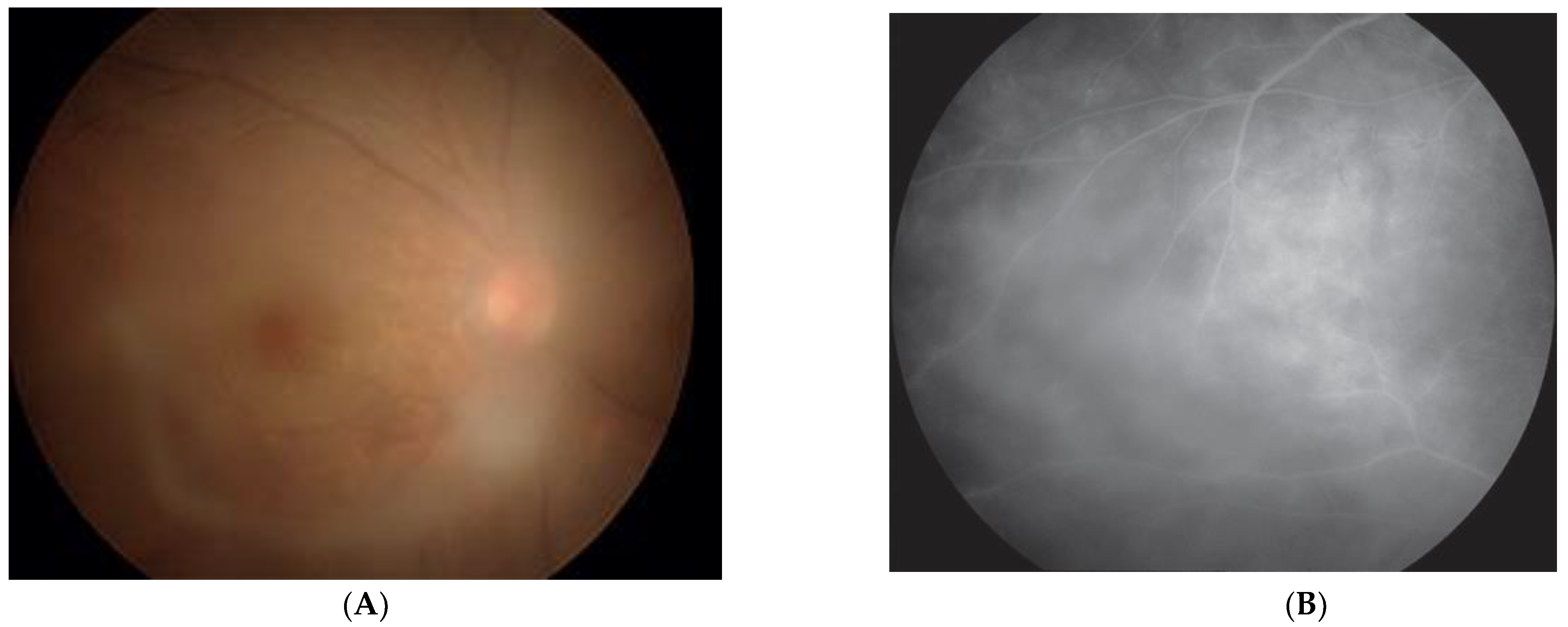
Through virologic analysis of HU patients, along with systemic investigations, we found that severe HU occurred in young HTLV-1 carriers after the onset of Graves’ disease and subsequent administration of methimazole, even though the provirus load was low [19] (Figure 2).
This phenomenon implies that the acceleration of the development of HU occurs in HTLV-1-infected patients who develop Graves’ disease and receive methimazole treatment despite a low provirus load and short latency period. This observation suggests the need for physicians to be cautious when Graves’ disease patients complain of blurry vision and that physicians, including endocrinologists and ophthalmologists, should rule out HTLV-1 infection in the differential diagnoses in such patients.
A possible mechanism for this association between HU and Grave’s disease is that thyroid hormones and methimazole might stimulate a small number of circulating HTLV-1-infected cells, which might contribute to the breakdown of the blood–ocular barrier, resulting in severe ocular inflammation. However, further analysis is needed to clarify this phenomenon.

6. Safety of Biologics in Terms of the Eye in HTLV-1 Infection

Biologics, such as tumor necrosis factor (TNF)-α antibody, were introduced in the late 1990s as a molecularly targeted therapy for inflammation [48,49,50,51,52]. Biologics have been proven to be effective in suppressing inflammation and are currently used for a wide range of inflammatory diseases [53]. However, a paradoxical response or exacerbation of inflammation is also well known during anti-TNF-α therapy [54]. In particular, the occurrence of uveitis (ocular inflammation) has been reported as a major paradoxical effect [55].
It is estimated that there is a large number of patients with inflammatory diseases, such as rheumatoid arthritis, who need to receive biologics despite having HTLV-1 infection [56]. However, patients with rheumatoid arthritis and HTLV-1 infection are susceptible to developing exacerbation of HTLV-1-associated conditions, such as HTLV-1-associated myopathy and HU, after the use of biological agents, such as tocilizumab, as was previously described in a case report [57]. However, none of the existing rheumatology, gastroenterology, or ophthalmology-related international guidelines recommend assessments for HTLV-1 infection before the administration of biologics.
In the field of ophthalmology, no evidence has been found for prescribing biologics to HTLV-1 carriers. In fact, our nationwide survey determined that the measurement of HTLV-1 antibodies is not performed in 50.0% of facilities prescribing biologics, and the reason commonly stated for this was a lack of knowledge regarding whether or not the measurement should be performed [20].
In response to this survey, we started investigating the effects of biologics on the eye under HTLV-1 infectious conditions in vitro. At first, we focused on infliximab, the first biologic for inflammatory diseases [58], and found that infliximab does not exacerbate HTLV-1-associated inflammatory conditions in the eye [59]. Next, we assessed adalimumab, the most frequently used biologic [53], and observed that adalimumab also does not exacerbate HTLV-1-associated inflammation in the eye [60].
Although these reports provided new evidence regarding the ocular safety of the use of biologics for inflammatory diseases in HTLV-1 carriers, which would be clinically useful for ophthalmologists, further analysis, including evaluation of other biologics, is needed in the future.

7. Ocular Manifestations of HTLV-1-Associated Diseases

Although HU is the most well-known and most studied ocular manifestation of HTLV-1, the other HTLV-1-associated disease entities might also present ocular symptoms. Several surveys that enrolled a large number of patients found that HAM sometimes occurs along with HU [10,26,36]. However, information on ATL-related ocular manifestations is scant, and only sporadic cases have been reported because of their rarity. Therefore, we performed a nationwide survey of the ocular manifestations of ATL and found that the most frequent manifestation is intraocular infiltration, followed by opportunistic infection (with all cases involving cytomegalovirus (CMV) retinitis), keratoconjunctivitis sicca, and scleritis [61] (Table 3).
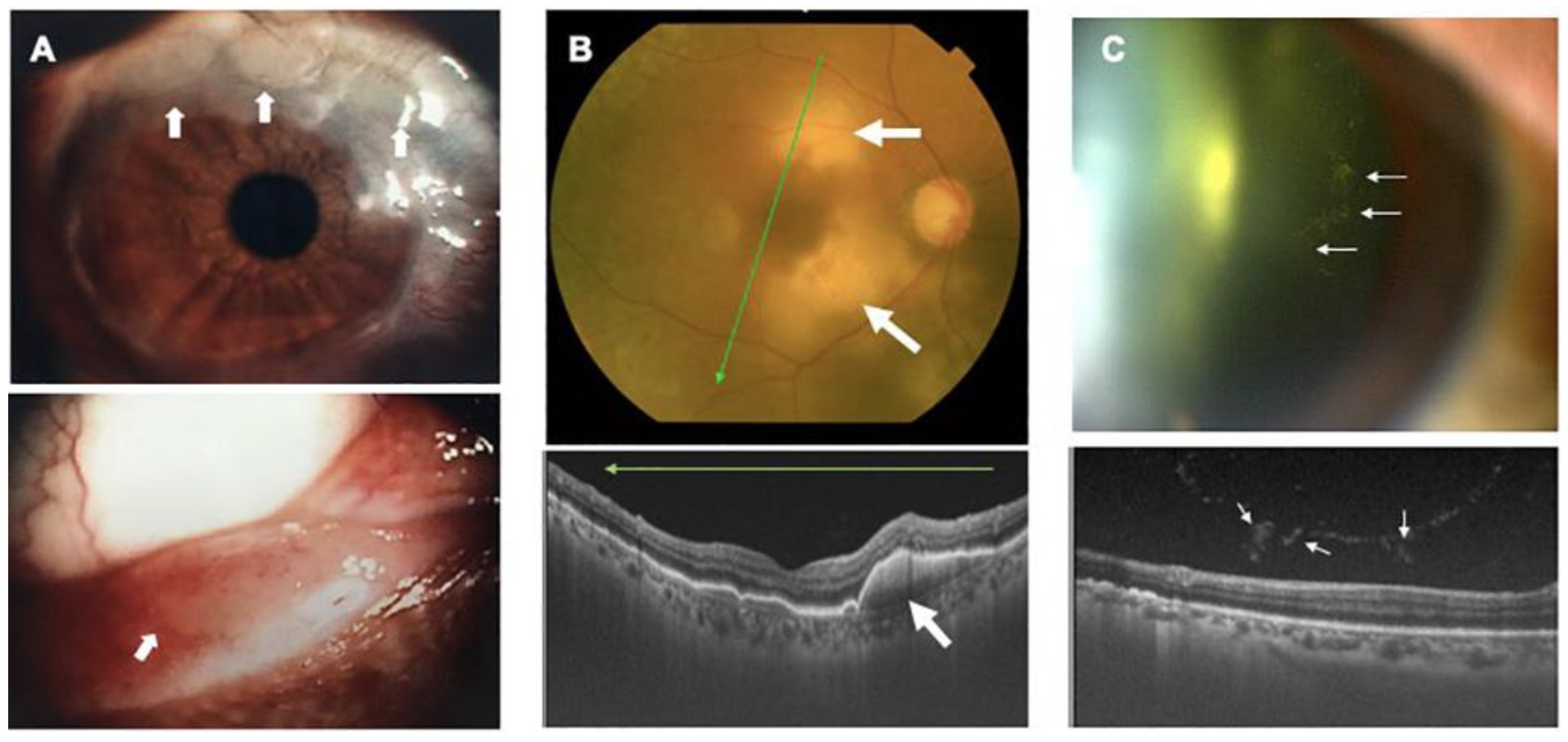
The most frequently seen manifestation is intraocular infiltration of ATL cells. We recently found that infiltration of ATL cells in the eye produces a specific sign, which we termed the “knob-like ATL cell multiple ocular infiltration” (KAMOI) sign, from the ocular surface to the retina [19,62,63] (Figure 3). This was identified using the recently developed imaging technology of ocular coherence tomography. The diagnosis of infiltration was facilitated by using a multiplex and broad-range polymerase chain reaction (PCR) system developed by us, which offers a precise differential diagnosis [64,65,66,67,68]. The therapy for ocular infiltration previously mainly relied on systemic ATL therapy, although the efficacy of topical treatment with the intra-vitreous injection of methotrexate in combination with focal radiation has been recently reported [19,63].
The second-most frequent ocular manifestation in ATL patients is CMV retinitis [61] (Table 3). CMV is well established as the most frequent pathogen underlying opportunistic infections in ATL patients [69,70], which also causes eye conditions. For diagnosis, multiplex and broad-range PCR of ocular samples are useful because ATL patients are typically immunocompromised, and other types of opportunistic infections (such as fungal infection) should also be excluded. In terms of other ATL-specific features, ocular inflammation developing simultaneously with ATL was previously reported [36,71], which correlated with an increase in ATL cells in the peripheral blood. This type of ocular inflammation is called ATL cell-induced uveitis [71].
Recently, a new ocular finding was reported in the eye during the course of ATL. ATL is a life-threatening hematological disease with a poor prognosis that was previously reported to have a poor survival rate despite chemotherapy [72]. Recently, allogenic hematopoietic stem-cell transplantation (allo-HSCT) has been introduced for the treatment of ATL, which has resulted in an improvement in the overall survival of ATL patients [73]. However, a series of allo-HSCT procedures might disrupt immune homeostasis, resulting in systemic inflammation [74]. Our routine observation of the eyes of ATL patients before and after allo-HSCT with long-term follow up demonstrated that the activation of the immune system after allogeneic HSCT might cause intraocular inflammation, resulting in frosted branch angiitis, which might precede inflammation at other sites and organs [75] (Figure 4). This implies that appropriate monitoring of the intraocular condition should be considered after allogeneic HSCT for ATL.

8. Conclusions

Recently, several long-standing unresolved issues related to HU, such as its transmission route and factors associated with its development, have been clarified. Frequent and new ocular manifestations of HTLV-1-associated ATL have also been identified. In addition, a nationwide survey raised new issues about HU and addressed several concerns, such as the safety of biologics in terms of the eye in the presence of HTLV-1 infections. However, information on HU is still insufficient compared to that on ATL and HAM. Further clinical and basic investigations of HU are needed to prevent vision loss among HTLV-1-infected individuals.

Funding

Funding from the Grant on Rare and Intractable Diseases from the Ministry of Health, Labour and Welfare of Japan (grant no. 19FC1007) and the Research Program on Emerging and Re-emerging Infectious Diseases from the Japan Agency for Medical Research and Development, AMED (grant no. JP21fk0108124j) is gratefully acknowledged.

Institutional Review Board Statement

Not applicable.

Informed Consent Statement

Not applicable.

Data Availability Statement

All data related to this study are presented and published here.

Conflicts of Interest

The authors declare no conflict of interest.

References

  1. Coffin, J.M. The discovery of HTLV-1, the first pathogenic human retrovirus. Proc. Natl. Acad. Sci. USA 2015, 112, 15525–15529. [Google Scholar] [CrossRef] [PubMed] [Green Version]
  2. Watanabe, T. HTLV-1-associated diseases. Int. J. Hematol. 1997, 66, 257–278. [Google Scholar] [CrossRef]
  3. WHO. Human T-Lymphotropic Virus Type 1: Technical Report. 2021. Available online: https://www.who.int/publications/i/item/9789240020221 (accessed on 25 February 2022).
  4. Gessain, A.; Cassar, O. Epidemiological Aspects and World Distribution of HTLV-1 Infection. Front. Microbiol. 2012, 3, 388. [Google Scholar] [CrossRef] [PubMed] [Green Version]
  5. Poiesz, B.J.; Ruscetti, F.W.; Gazdar, A.F.; Bunn, P.A.; Minna, J.D.; Gallo, R.C. Detection and isolation of type C retrovirus particles from fresh and cultured lymphocytes of a patient with cutaneous T-cell lymphoma. Proc. Natl. Acad. Sci. USA 1980, 77, 7415–7419. [Google Scholar] [CrossRef] [PubMed] [Green Version]
  6. Hinuma, Y.; Nagata, K.; Hanaoka, M.; Nakai, M.; Matsumoto, T.; Kinoshita, K.I.; Shirakawa, S.; Miyoshi, I. Adult T-cell leukemia: Antigen in an ATL cell line and detection of antibodies to the antigen in human sera. Proc. Natl. Acad. Sci. USA 1981, 78, 6476–6480. [Google Scholar] [CrossRef] [Green Version]
  7. Yoshida, M.; Seiki, M.; Yamaguchi, K.; Takatsuki, K. Monoclonal integration of human T-cell leukemia provirus in all primary tumors of adult T-cell leukemia suggests causative role of human T-cell leukemia virus in the disease. Proc. Natl. Acad. Sci. USA 1984, 81, 2534–2537. [Google Scholar] [CrossRef] [Green Version]
  8. Gessain, A.; Barin, F.; Vernant, J.C.; Gout, O.; Maurs, L.; Calender, A.; de The, G. Antibodies to human T-lymphotropic virus type-I in patients with tropical spastic paraparesis. Lancet 1985, 2, 407–410. [Google Scholar] [CrossRef]
  9. Osame, M.; Usuku, K.; Izumo, S.; Ijichi, N.; Amitani, H.; Igata, A.; Matsumoto, M.; Tara, M. HTLV-I associated myelopathy, a new clinical entity. Lancet 1986, 1, 1031–1032. [Google Scholar] [CrossRef]
  10. Ohba, N.; Matsumoto, M.; Sameshima, M.; Kabayama, Y.; Nakao, K.; Unoki, K.; Uehara, F.; Kawano, K.; Maruyama, I.; Osame, M. Ocular manifestations in patients infected with human T-lymphotropic virus type I. Jpn. J. Ophthalmol. 1989, 33, 1–12. [Google Scholar]
  11. Nakao, K.; Matsumoto, M.; Ohba, N. Seroprevalence of antibodies to HTLV-I in patients with ocular disorders. Br. J. Ophthalmol. 1991, 75, 76–78. [Google Scholar] [CrossRef] [Green Version]
  12. Mochizuki, M.; Watanabe, T.; Yamaguchi, K.; Takatsuki, K.; Yoshimura, K.; Shirao, M.; Nakashima, S.; Mori, S.; Araki, S.; Miyata, N. HTLV-I uveitis: A distinct clinical entity caused by HTLV-I. Jpn. J. Cancer Res. 1992, 83, 236–239. [Google Scholar] [CrossRef] [PubMed]
  13. Mochizuki, M.; Yamaguchi, K.; Takatsuki, K.; Watanabe, T.; Mori, S.; Tajima, K. HTLV-I and uveitis. Lancet 1992, 339, 1110. [Google Scholar] [CrossRef]
  14. Iwanaga, M. Epidemiology of HTLV-1 Infection and ATL in Japan: An Update. Front. Microbiol. 2020, 11, 1124. [Google Scholar] [CrossRef]
  15. Ikeda, E.; Ono, A.; Hikita, N.; Arima, K.; Mochizuki, M.; Yamaguchi, K.; Tajima, K.; Kiyokawa, H. Estimated Prevalence Rate of HTLV-I Uveitis in Chikugo. J. Jpn. Ophthalmol. Soc. 1998, 102, 327–332. [Google Scholar]
  16. Ijichi, S.; Osame, M. Human T lymphotropic virus type I (HTLV-I)-associated myelopathy/tropical spastic paraparesis (HAM/TSP): Recent perspectives. Intern. Med. 1995, 34, 713–721. [Google Scholar] [CrossRef] [PubMed] [Green Version]
  17. Kamoi, K.; Mochizuki, M. HTLV-1 uveitis. Front. Microbiol. 2012, 3, 270. [Google Scholar] [CrossRef] [PubMed] [Green Version]
  18. Kamoi, K.; Mochizuki, M. HTLV infection and the eye. Curr. Opin. Ophthalmol. 2012, 23, 557–561. [Google Scholar] [CrossRef]
  19. Kamoi, K. HTLV-1 in Ophthalmology. Front. Microbiol. 2020, 11, 388. [Google Scholar] [CrossRef] [Green Version]
  20. Kamoi, K.; Okayama, A.; Izumo, S.; Hamaguchi, I.; Uchimaru, K.; Tojo, A.; Watanabe, T.; Ohno-Matsui, K. Tackling HTLV-1 infection in ophthalmology: A nationwide survey of ophthalmic care in an endemic country, Japan. Br. J. Ophthalmol. 2020, 104, 1647–1651. [Google Scholar] [CrossRef]
  21. Beilke, M.A. Human T-cell lymphotropic virus type I-associated uveitis in an African American. Am. J. Ophthalmol. 1995, 119, 104–105. [Google Scholar] [CrossRef]
  22. Merle, H.; Smadja, D.; Le Hoang, P.; Bera, O.; Cabre, P.; Landau, M.; Vernant, J.C. Ocular manifestations in patients with HTLV-I associated infection--a clinical study of 93 cases. Jpn. J. Ophthalmol. 1996, 40, 260–270. [Google Scholar] [PubMed]
  23. Sandy, G.J.; Taylor, J.; Steiger, M.; Wearne, M.; Weber, J.N. HTLV-I associated uveitis: A report of two cases. Eye 1996, 10, 521–522. [Google Scholar] [CrossRef] [PubMed]
  24. Yamamoto, J.H.; Segurado, A.A.; Hirata, C.E.; Sampaio, M.W.; Souza, E.C.; Nukui, Y.; Cliquet, M.; Saez-Alquezar, A.; Olivalves, E.; Mochizuki, M. Human T-cell lymphotropic virus type 1 infection and ocular manifestations in Sao Paulo, Brazil. Arch. Ophthalmol. 1999, 117, 513–517. [Google Scholar] [CrossRef] [PubMed]
  25. Chew, R.; Henderson, T.; Aujla, J.; Whist, E.; Einsiedel, L. Turning a blind eye: HTLV-1-associated uveitis in Indigenous adults from Central Australia. Int. Ophthalmol. 2018, 38, 2159–2162. [Google Scholar] [CrossRef] [PubMed]
  26. Terada, Y.; Kamoi, K.; Komizo, T.; Miyata, K.; Mochizuki, M. Human T Cell Leukemia Virus Type 1 and Eye Diseases. J. Ocul. Pharmacol. Ther. 2017, 33, 216–223. [Google Scholar] [CrossRef]
  27. Sonoda, K.H.; Hasegawa, E.; Namba, K.; Okada, A.A.; Ohguro, N.; Goto, H.; Group, J.U.S.W. Epidemiology of uveitis in Japan: A 2016 retrospective nationwide survey. Jpn. J. Ophthalmol. 2021, 65, 184–190. [Google Scholar] [CrossRef]
  28. Mochizuki, M.; Sugita, S.; Kamoi, K. Immunological homeostasis of the eye. Prog. Retin. Eye Res. 2013, 33, 10–27. [Google Scholar] [CrossRef]
  29. Kamoi, K.; Mochizuki, M. HTLV-1 Uveitis. In Intraocular Inflammation; Zierhut, M., Pavesio, C., Ohno, S., Orefice, F., Rao, N.A., Eds.; Springer: Berlin/Heidelberg, Germany, 2016; pp. 1197–1201. [Google Scholar]
  30. Kamoi, K.; Mochizuki, M. Human T-Cell Leukemia Virus Type 1. In Emerging Infectious Uveitis; Chee, S.-P., Khairallah, M., Eds.; Springer International Publishing: Cham, Switzerland, 2017; pp. 143–148. [Google Scholar]
  31. Nakao, K.; Ohba, N. Clinical features of HTLV-I associated uveitis. Br. J. Ophthalmol. 1993, 77, 274–279. [Google Scholar] [CrossRef]
  32. Takahashi, T.; Takase, H.; Urano, T.; Sugita, S.; Miyata, K.; Miyata, N.; Mochizuki, M. Clinical features of human T-lymphotropic virus type 1 uveitis: A long-term follow-up. Ocul. Immunol. Inflamm. 2000, 8, 235–241. [Google Scholar] [CrossRef]
  33. Ohba, N.; Nakao, K.; Isashiki, Y.; Kaminagayoshi, T.; Sonoda, S.; Yashiki, S.; Osame, M. Clinical features of HTLV-I associated uveitis determined in multicenter collaborative study. Study Group for HTLV-I Associated Ocular Diseases. Jpn. J. Ophthalmol. 1994, 38, 168–174. [Google Scholar]
  34. Yamaguchi, K.; Mochizuki, M.; Watanabe, T.; Yoshimura, K.; Shirao, M.; Araki, S.; Miyata, N.; Mori, S.; Kiyokawa, T.; Takatsuki, K. Human T lymphotropic virus type 1 uveitis after Graves’ disease. Br. J. Ophthalmol. 1994, 78, 163–166. [Google Scholar] [CrossRef] [PubMed] [Green Version]
  35. Miyanaga, M.; Shimizu, K.; Kawaguchi, T.; Miyata, K.; Mochizuki, M. A clinical survey of uveitis in HTLV-1 endemic region. Ocul. Immunol. Inflamm. 2009, 17, 335–341. [Google Scholar] [CrossRef] [PubMed]
  36. Nakao, K.; Abematsu, N.; Sakamoto, T. Systemic diseases in patients with HTLV-1-associated uveitis. Br. J. Ophthalmol. 2018, 102, 373–376. [Google Scholar] [CrossRef] [PubMed]
  37. Watanabe, T. Current status of HTLV-1 infection. Int. J. Hematol. 2011, 94, 430–434. [Google Scholar] [CrossRef] [PubMed]
  38. Iwanaga, M.; Chiyoda, S.; Kusaba, E.; Kamihira, S. Trends in the seroprevalence of HTLV-1 in Japanese blood donors in Nagasaki Prefecture, 2000–2006. Int. J. Hematol. 2009, 90, 186–190. [Google Scholar] [CrossRef] [Green Version]
  39. Nishijima, T.; Shimada, S.; Noda, H.; Miyake, K. Towards the elimination of HTLV-1 infection in Japan. Lancet Infect. Dis. 2019, 19, 15–16. [Google Scholar] [CrossRef]
  40. Satake, M.; Iwanaga, M.; Sagara, Y.; Watanabe, T.; Okuma, K.; Hamaguchi, I. Incidence of human T-lymphotropic virus 1 infection in adolescent and adult blood donors in Japan: A nationwide retrospective cohort analysis. Lancet Infect. Dis. 2016, 16, 1246–1254. [Google Scholar] [CrossRef]
  41. Goncalves, D.U.; Proietti, F.A.; Ribas, J.G.; Araujo, M.G.; Pinheiro, S.R.; Guedes, A.C.; Carneiro-Proietti, A.B. Epidemiology, treatment, and prevention of human T-cell leukemia virus type 1-associated diseases. Clin. Microbiol. Rev. 2010, 23, 577–589. [Google Scholar] [CrossRef] [Green Version]
  42. Rosadas, C.; Taylor, G.P. Mother-to-Child HTLV-1 Transmission: Unmet Research Needs. Front. Microbiol. 2019, 10, 999. [Google Scholar] [CrossRef] [Green Version]
  43. Kamoi, K.; Uchimaru, K.; Tojo, A.; Watanabe, T.; Ohno-Matsui, K. HTLV-1 uveitis and Graves’ disease presenting with sudden onset of blurred vision. Lancet 2022, 399, 60. [Google Scholar] [CrossRef]
  44. Kamoi, K.; Horiguchi, N.; Kurozumi-Karube, H.; Hamaguchi, I.; Yamano, Y.; Uchimaru, K.; Tojo, A.; Watanabe, T.; Ohno-Matsui, K. Horizontal transmission of HTLV-1 causing uveitis. Lancet Infect. Dis. 2021, 21, 578. [Google Scholar] [CrossRef]
  45. Bangham, C.R.; Cook, L.B.; Melamed, A. HTLV-1 clonality in adult T-cell leukaemia and non-malignant HTLV-1 infection. Semin. Cancer Biol. 2014, 26, 89–98. [Google Scholar] [CrossRef] [PubMed] [Green Version]
  46. Akbarin, M.M.; Rahimi, H.; Hassannia, T.; Shoja Razavi, G.; Sabet, F.; Shirdel, A. Comparison of HTLV-I Proviral Load in Adult T Cell Leukemia/Lymphoma (ATL), HTLV-I-Associated Myelopathy (HAM-TSP) and Healthy Carriers. Iran. J. Basic. Med. Sci. 2013, 16, 208–212. [Google Scholar] [PubMed]
  47. Iwanaga, M.; Watanabe, T.; Utsunomiya, A.; Okayama, A.; Uchimaru, K.; Koh, K.R.; Ogata, M.; Kikuchi, H.; Sagara, Y.; Uozumi, K.; et al. Human T-cell leukemia virus type I (HTLV-1) proviral load and disease progression in asymptomatic HTLV-1 carriers: A nationwide prospective study in Japan. Blood 2010, 116, 1211–1219. [Google Scholar] [CrossRef] [Green Version]
  48. Bathon, J.M.; Martin, R.W.; Fleischmann, R.M.; Tesser, J.R.; Schiff, M.H.; Keystone, E.C.; Genovese, M.C.; Wasko, M.C.; Moreland, L.W.; Weaver, A.L.; et al. A comparison of etanercept and methotrexate in patients with early rheumatoid arthritis. N. Engl. J. Med. 2000, 343, 1586–1593. [Google Scholar] [CrossRef]
  49. Leonardi, C.L.; Powers, J.L.; Matheson, R.T.; Goffe, B.S.; Zitnik, R.; Wang, A.; Gottlieb, A.B.; Etanercept Psoriasis Study, G. Etanercept as monotherapy in patients with psoriasis. N. Engl. J. Med. 2003, 349, 2014–2022. [Google Scholar] [CrossRef] [Green Version]
  50. Braun, J.; Baraliakos, X.; Brandt, J.; Listing, J.; Zink, A.; Alten, R.; Burmester, G.; Gromnica-Ihle, E.; Kellner, H.; Schneider, M.; et al. Persistent clinical response to the anti-TNF-alpha antibody infliximab in patients with ankylosing spondylitis over 3 years. Rheumatology 2005, 44, 670–676. [Google Scholar] [CrossRef] [Green Version]
  51. Rutgeerts, P.; Van Assche, G.; Vermeire, S. Optimizing anti-TNF treatment in inflammatory bowel disease. Gastroenterology 2004, 126, 1593–1610. [Google Scholar] [CrossRef] [Green Version]
  52. Rutgeerts, P.; Sandborn, W.J.; Feagan, B.G.; Reinisch, W.; Olson, A.; Johanns, J.; Travers, S.; Rachmilewitz, D.; Hanauer, S.B.; Lichtenstein, G.R.; et al. Infliximab for induction and maintenance therapy for ulcerative colitis. N. Engl. J. Med. 2005, 353, 2462–2476. [Google Scholar] [CrossRef] [Green Version]
  53. Mullard, A. Top product sales forecasts for 2018. Nat. Rev. Drug Discov. 2018, 17, 86. [Google Scholar] [CrossRef]
  54. Toussirot, E.; Aubin, F. Paradoxical reactions under TNF-alpha blocking agents and other biological agents given for chronic immune-mediated diseases: An analytical and comprehensive overview. RMD Open. 2016, 2, e000239. [Google Scholar] [CrossRef] [PubMed] [Green Version]
  55. Wendling, D.; Prati, C. Paradoxical effects of anti-TNF-alpha agents in inflammatory diseases. Expert Rev. Clin. Immunol. 2014, 10, 159–169. [Google Scholar] [CrossRef] [PubMed]
  56. Umekita, K.; Okayama, A. HTLV-1 Infection and Rheumatic Diseases. Front. Microbiol. 2020, 11, 152. [Google Scholar] [CrossRef]
  57. Terada, Y.; Kamoi, K.; Ohno-Matsui, K.; Miyata, K.; Yamano, C.; Coler-Reilly, A.; Yamano, Y. Treatment of rheumatoid arthritis with biologics may exacerbate HTLV-1-associated conditions: A case report. Medicine 2017, 96, e6021. [Google Scholar] [CrossRef] [PubMed]
  58. Melsheimer, R.; Geldhof, A.; Apaolaza, I.; Schaible, T. Remicade((R)) (infliximab): 20 years of contributions to science and medicine. Biologics 2019, 13, 139–178. [Google Scholar] [PubMed] [Green Version]
  59. Uchida, M.; Kamoi, K.; Ando, N.; Wei, C.; Karube, H.; Ohno-Matsui, K. Safety of Infliximab for the Eye Under Human T-Cell Leukemia Virus Type 1 Infectious Conditions in vitro. Front. Microbiol. 2019, 10, 2148. [Google Scholar] [CrossRef]
  60. Kurozumi-Karube, H.; Kamoi, K.; Ando, N.; Uchida, M.; Hamaguchi, I.; Ohno-Matsui, K. In Vitro Evaluation of the Safety of Adalimumab for the Eye Under HTLV-1 Infection Status: A Preliminary Study. Front Microbiol. 2020, 11, 522579. [Google Scholar] [CrossRef]
  61. Kamoi, K.; Okayama, A.; Izumo, S.; Hamaguchi, I.; Uchimaru, K.; Tojo, A.; Ohno-Matsui, K. Adult T-Cell Leukemia/Lymphoma-Related Ocular Manifestations: Analysis of the First Large-Scale Nationwide Survey. Front. Microbiol. 2018, 9, 3240. [Google Scholar] [CrossRef]
  62. Kamoi, K.; Nagata, Y.; Mochizuki, M.; Kobayashi, D.; Ohno, N.; Uchimaru, K.; Tojo, A.; Ohno-Matsui, K. Formation of Segmental Rounded Nodules During Infiltration of Adult T-Cell Leukemia Cells Into the Ocular Mucous Membrane. Cornea 2016, 35, 137–139. [Google Scholar] [CrossRef]
  63. Kamoi, K.; Ohno-Matsui, K. Intraocular Infiltration. Am. J. Trop. Med. Hyg. 2020, 102, 7–8. [Google Scholar] [CrossRef] [Green Version]
  64. Sugita, S.; Shimizu, N.; Watanabe, K.; Mizukami, M.; Morio, T.; Sugamoto, Y.; Mochizuki, M. Use of multiplex PCR and real-time PCR to detect human herpes virus genome in ocular fluids of patients with uveitis. Br. J. Ophthalmol. 2008, 92, 928–932. [Google Scholar] [CrossRef] [PubMed] [Green Version]
  65. Sugita, S.; Shimizu, N.; Watanabe, K.; Katayama, M.; Horie, S.; Ogawa, M.; Takase, H.; Sugamoto, Y.; Mochizuki, M. Diagnosis of bacterial endophthalmitis by broad-range quantitative PCR. Br. J. Ophthalmol. 2011, 95, 345–349. [Google Scholar] [CrossRef] [PubMed] [Green Version]
  66. Sugita, S.; Kamoi, K.; Ogawa, M.; Watanabe, K.; Shimizu, N.; Mochizuki, M. Detection of Candida and Aspergillus species DNA using broad-range real-time PCR for fungal endophthalmitis. Graefes. Arch. Clin. Exp. Ophthalmol. 2012, 250, 391–398. [Google Scholar] [CrossRef] [PubMed]
  67. Sugita, S.; Ogawa, M.; Shimizu, N.; Morio, T.; Ohguro, N.; Nakai, K.; Maruyama, K.; Nagata, K.; Takeda, A.; Usui, Y.; et al. Use of a comprehensive polymerase chain reaction system for diagnosis of ocular infectious diseases. Ophthalmology 2013, 120, 1761–1768. [Google Scholar] [CrossRef]
  68. Mochizuki, M.; Sugita, S.; Kamoi, K.; Takase, H. A new era of uveitis: Impact of polymerase chain reaction in intraocular inflammatory diseases. Jpn. J. Ophthalmol. 2017, 61, 1–20. [Google Scholar] [CrossRef] [Green Version]
  69. Suzumiya, J.; Marutsuka, K.; Nabeshima, K.; Nawa, Y.; Koono, M.; Tamura, K.; Kimura, N.; Hisano, S.; Tachibana, N.; Inoue, S.; et al. Autopsy findings in 47 cases of adult T-cell leukemia/lymphoma in Miyazaki prefecture, Japan. Leuk Lymphoma 1993, 11, 281–286. [Google Scholar] [CrossRef]
  70. Maeda, T.; Babazono, A.; Nishi, T.; Yasui, M.; Matsuda, S.; Fushimi, K.; Fujimori, K. The Impact of Opportunistic Infections on Clinical Outcome and Healthcare Resource Uses for Adult T Cell Leukaemia. PLoS ONE 2015, 10, e0135042. [Google Scholar] [CrossRef]
  71. Hirano, M.; Ohno, N.; Tanosaki, R.; Mochizuki, M.; Ohno-Matsui, K.; Uchimaru, K.; Tojo, A.; Kamoi, K. Adult T-cell leukemia cell-induced uveitis: Rapid increase in adult T-cell leukemia cells disrupts the blood-ocular barrier. Int. J. Hematol. 2017, 106, 842–846. [Google Scholar] [CrossRef]
  72. Taylor, G.P.; Matsuoka, M. Natural history of adult T-cell leukemia/lymphoma and approaches to therapy. Oncogene 2005, 24, 6047–6057. [Google Scholar] [CrossRef] [Green Version]
  73. Utsunomiya, A.; Choi, I.; Chihara, D.; Seto, M. Recent advances in the treatment of adult T-cell leukemia-lymphomas. Cancer Sci. 2015, 106, 344–351. [Google Scholar] [CrossRef]
  74. Copelan, E.A. Hematopoietic stem-cell transplantation. N. Engl. J. Med. 2006, 354, 1813–1826. [Google Scholar] [CrossRef] [PubMed]
  75. Kamoi, K.; Kato, S.; Uchimaru, K.; Tojo, A.; Ohno-Matsui, K. Frosted branch angiitis after allogeneic haematopoietic stem cell transplantation in adult T-cell leukaemia-lymphoma. Lancet Haematol. 2020, 7, e772. [Google Scholar] [CrossRef]
Figure 1. Horizontal transmission of HTLV-1 is responsible for HTLV-1 uveitis. (A) Vitreous opacity is seen in the right eye. (B) Severe and persistent ocular inflammation of horizontally transmitted HU resulted in retinal detachment (Reproduced with permission from Kamoi et al., Lancet Infect. Dis. 2021 [44]).
Viruses 14 00794 g001
Figure 2. HU occurred after methimazole treatment following the onset of Graves’ disease. (A) Vitreous opacity is seen in the right eye. (B) Retinal vasculitis can be seen by fluorescein angiography (Reproduced with permission from Kamoi et al., Lancet 2022 [43]).
Viruses 14 00794 g002
Figure 3. Ocular infiltration in ATL. Multiple knob-like ATL cell ocular infiltrates are seen (KAMOI sign). The KAMOI sign (A,B; white arrows) can be seen at the bulbar conjunctiva around the corneal limbus and at the palpebral conjunctiva around the lacrimal punctum (A) and in the retina (B). Multiple infiltrating ATL cells in the vitreous tend to form clusters (C) (Reproduced with permission from Kamoi et al., Cornea 2016 [62]; Kamoi. Front. Microbiol. 2020 [19]).
Viruses 14 00794 g003
Figure 4. Frosted branch angiitis after allogenic hematopoietic stem-cell transplantation (Reproduced with permission from Kamoi et al., Lancet Haematol. 2020 [75]).
Viruses 14 00794 g004
Table 1. Timing of testing for HTLV-1 antibodies.
Timing of Testing for HTLV-1 AntibodiesNorthern
(n = 4)
Central/Metropolitan (n = 40)Southern (n = 16)Total
Hokkaido/Tohoku (n = 4)Kanto (n = 22)Chubu (n = 7)Kinki (n = 11)Chugoku/Shikoku (n = 7)Kyushu (n = 9)
Carried out routinely in considering differential diagnoses for uveitis50.0%52.5%84.6%58.3%
50.0%50.0%57.1%54.5%75.0%88.9%
When ocular manifestation such as HAU, ATL, or HAM is suspected50.0%47.5%25.0%41.7%
50.0%50.0%42.9%45.5%42.9%11.1%
When requested by
Patient
0.0%7.5%0.0%5.0%
0.0%4.5%0.0%18.2%0.0%0.0%
When administering
Steroid
25.0%2.5%0.0%3.3%
25.0%0.0%0.0%9.1%0.0%0.0%
When administering
Immunosuppressant
25.0%2.5%0.0%3.3%
25.0%0.0%0.0%9.1%0.0%0.0%
When administering
biological product
20.0%0.0%0.0%1.7%
20.0%0.0%0.0%0.0%0.0%0.0%
When performing
Surgery
0.0%5.0%0.0%3.3%
0.0%0.0%14.3%9.1%0.0%0.0%
Others0.0%10.0%0.0%6.7%
0.0%18.2%0.0%0.0%0.0%0.0%
Our nationwide survey identified that testing for HTLV-1 antibodies was routinely performed when considering the differential diagnosis of uveitis among 58.3% of facilities in Japan (Reproduced with permission from Kamoi et al., Br. J. Ophthalmol. 2020 [20]).
Table 2. Testing for HTLV-1 antibodies when an HTLV-1-related ocular manifestation is suspected.
Test for HTLV-1 Antibodies in Considering Differential Diagnoses for UveitisNorthern (n = 5) Central/Metropolitan (n = 47)Southern (n = 16)Total
Hokkaido/Tohoku (n = 5)Kanto (n = 26)Chubu (n = 9)Kinki (n = 12)Chugoku/ Shikoku (n = 7)Kyushu (n = 9)
Yes80.0%82.9%100.0%86.8%
80.0%80.1%77.8%66.7%100.0%100.0%
No/Un-identified20.0%17.1%0.0%13.2%
20.0%19.9%22.2%33.3%0.0%0.0%
Our nationwide survey identified that testing for HTLV-1 antibodies was performed in 86.8% of facilities in Japan when an HTLV-1-related ocular manifestation was suspected (Reproduced with permission from Kamoi et al., Br. J. Ophthalmol. 2020 [20]).
Table 3. Nationwide survey of ATL-related ocular manifestations.
ATL-Related Ocular ManifestationsNumber%
Intraocular infiltration2245.8
Opportunistic infection1939.6
Cytomegalovirus (19)(100.0)
Herpesvirus(2)(10.5)
Toxoplasma(1)(5.3)
Dry eye36.3
Scleritis24.2
Uveitis12.1
Anemic retinopathy12.1
Total48100
Intraocular infiltration is the most frequent manifestation, followed by opportunistic infection (Reproduced with permission from Kamoi et al., Front. Microbiol. 2018 [61]).
Publisher’s Note: MDPI stays neutral with regard to jurisdictional claims in published maps and institutional affiliations.