Article

A Pilot Study Investigating the Dynamics of Pigeon Circovirus Recombination in Domesticated Pigeons Housed in a Single Loft

1
Biodesign Center for Fundamental and Applied Microbiomics, Center for Evolution and Medicine, School of Life Sciences, Arizona State University, Tempe, AZ 85287, USA
2
Department of Poultry Diseases, Faculty of Veterinary Medicine, University of Warmia and Mazury, 10-719 Olsztyn, Poland
3
Structural Biology Research Unit, Department of Integrative Biomedical Sciences, University of Cape Town, Observatory, Cape Town 7701, South Africa
*
Authors to whom correspondence should be addressed.
Viruses 2021, 13(6), 964; https://doi.org/10.3390/v13060964
Submission received: 21 March 2021 / Revised: 17 May 2021 / Accepted: 20 May 2021 / Published: 22 May 2021
(This article belongs to the Special Issue Recombination as an Evolutionary Force in Animal Viruses)

Abstract

:
Pigeon circovirus (PiCV) infects pigeon populations worldwide and has been associated with immunosuppression in younger pigeons. Recombination is a common mechanism of evolution that has previously been shown in various members of the Circoviridae family, including PiCV. In this study, three groups of pigeons acquired from separate lofts were screened for PiCV, and their genome sequence was determined. Following this, they were housed in a single loft for 22 days, during which blood and cloacal swab samples were taken. From these blood and cloacal swabs, PiCV genomes were determined with the aim to study the spread and recombination dynamics of PiCV in the birds. Genome sequences of PiCV were determined from seven pigeons (seven tested PiCV positive) before they were housed together in a loft (n = 58 sequences) and thereafter from the ten pigeons from blood and cloacal swabs (n = 120). These 178 PiCV genome sequences represent seven genotypes (98% pairwise identity genotype demarcation), and they share >88% genome-wide pairwise identity. Recombination analysis revealed 13 recombination events, and a recombination hotspot spanning the 3′ prime region, the replication-associated protein (rep) gene and the intergenic region. A cold spot in the capsid protein-coding region of the genome was also identified. The majority of the recombinant regions were identified in the rep coding region. This study provides insights into the evolutionary dynamics of PiCV in pigeons kept under closed rearing systems.

1. Introduction

The domestic pigeon (Columba livia f. domestica), a species in the Columbidae family, is strongly associated with larger human populations [1]. Wild ancestors of domestic pigeons—the rock pigeon (Columba livia) can flock in groups of up to 400 individuals, and their range can span several kilometers. Similar behavior of living in groups is also observed in the feral domestic pigeon population inhabiting cities around the world, which are referred to as urban pigeons [2]. Nowadays, numerous pigeon breeds are known, and some are used for either meat production, such as ornamental birds, or for pigeon sports (carrier pigeons) [3]. The carrier pigeons are used extensively for racing, and various studies have explored the influence of genetics and dietary parameters as well as infections on race performance [4,5,6,7]. The races have a very high physical burden on racing pigeons as they are held every week for 4 consecutive months with racing distancing ranging from 100 to 1000 km [8]. During preparations for pigeon races and training, the practice of long communal transporting (sometimes for a period of 2 days) of all pigeons taking part in a certain competition is very common [9]. During the transportation stops, the practice of joint feeding and watering birds from disparate lofts is a standard procedure and can create ideal conditions for the transmission and long-distance dissemination of various pathogens, especially viruses [10]. Multiple viruses have been associated and shown to infect pigeons, including those in the families Adenoviridae, Anelloviridae, Circoviridae, Herpesviridae, Paramyxoviruses, Parvoviridae, Picornaviridae and Reoviridae [11,12,13,14,15,16,17,18].
Pigeon circovirus (PiCV, family Circoviridae), also referred to as columbid circovirus, has a single-stranded circular DNA genome of ~2 kb that encodes two bidirectionally transcribed genes, a replication-associated protein (rep) and capsid protein (cp) genes [19,20,21]. PiCV is found globally in both feral and domesticated pigeon populations, and its prevalence can reach up to 70% depending on age and overall health status [5,22,23,24,25,26,27,28,29]. Studies have shown that pigeon circovirus can be transmitted both horizontally via the fecal-oral route and vertically [30,31,32]. Although PiCV has not been shown to directly cause any major disease in pigeons, studies have associated it with immunosuppression in younger pigeons (<4 months) [33,34].
The phylogeny of PiCV sequences does not show any clear geographic structuring [35]. However, not all PiCV variants are widely distributed. The most common PiCV variant has been detected in various European countries, United States, China, Brazil and Australia, whereas the second common variant in Europe, China, Australia and Brazil [26,27,35]. The rest of the variants are less frequent, with most identified in birds in Europe and China [26,27,35]. Perhaps the absence of geographic clustering of PiCV sequence variants could be due to the role recombination, and nucleotide substitution plays in its evolution as well as broader animal movements. Previous studies have shown a recombination hotspot within the intergenic region between cp and rep genes and near the origin of replication [27,35].
In recent years, a popular pigeon sport called “one loft races” (OLR) involving young pigeons has become popular. In this type of competition, all young birds supplied by various breeders are identically housed, fed and trained before the racing season in a single loft. This procedure usually takes 2–3 months with an aim to exclude additional factors affecting the result of the race, such as the location of the loft of origin, care and nutrition, which means that the race can be won by the best individual. However, OLR likely violates all biosecurity measures and can lead to the transmission of various pathogens. Thus, the morbidity of pigeons in OLR can be very high, and if proper veterinary care is not provided, the mortality rates can also be very high. The prevalence of PiCV in young racing pigeons is close to ~80% [30]. Taking together the sequence diversity of PiCV and the high prevalence of infections in young pigeons, it is certain that numerous PiCV variants are introduced and spread at OLRs. Furthermore, OLRs could facilitate the emergence of new recombinant PiCV variants.
In this pilot study, we investigate the recombination dynamics of PiCV in young domesticated pigeons sourced from different facilities and then housed in a common loft.

2. Materials and Methods

2.1. Ethical Statement

The research protocol was approved by the Local Ethics Committee on Animal Experimentation of the University of Warmia and Mazury in Olsztyn (resolution No 41/2019, issued 28 May 2019, valid through 1 October 2023). The researchers made every effort to minimize the suffering of birds.

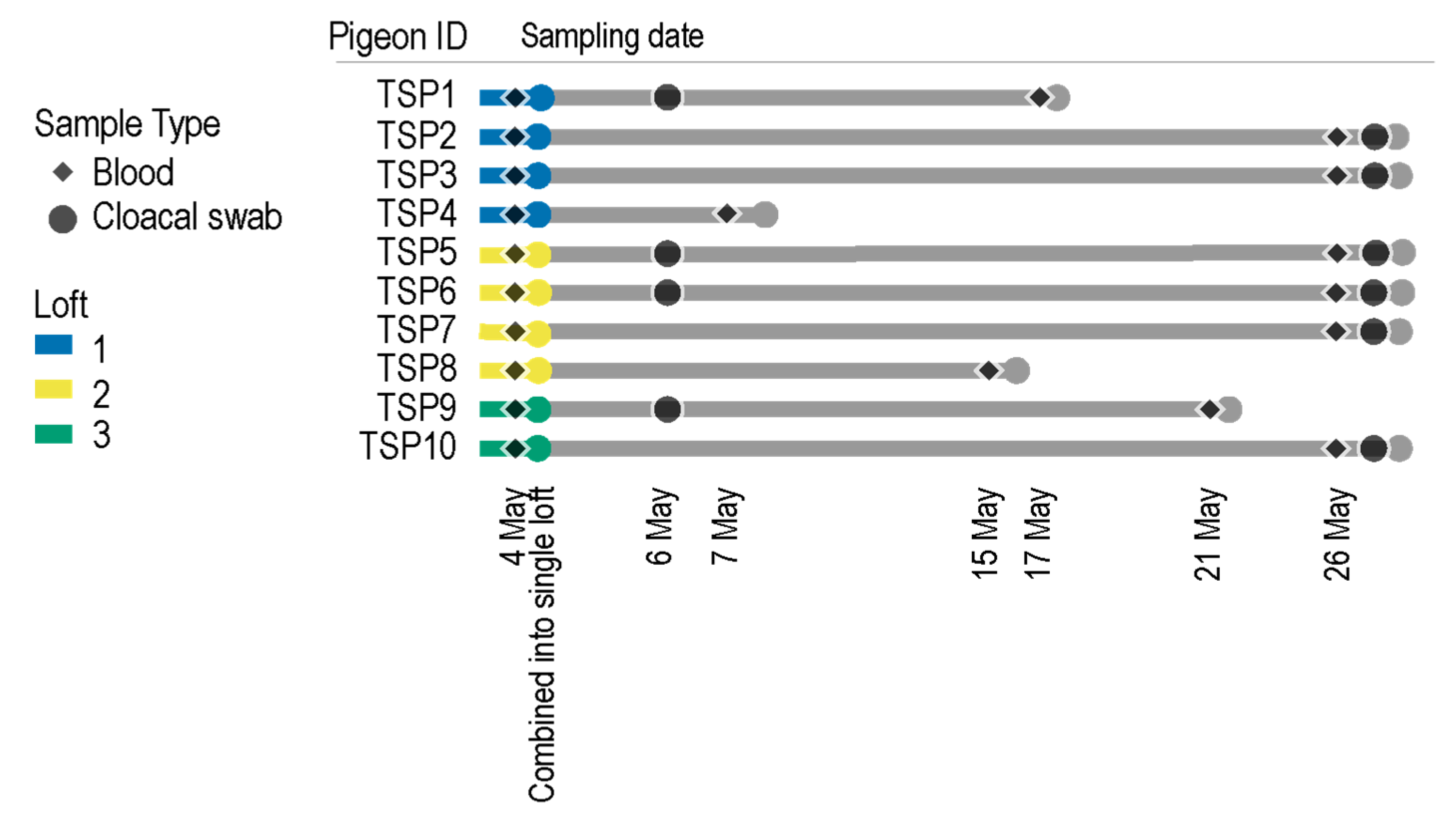
2.2. Pigeon Lofts and Sample Collection

Eight-week-old pigeons (n = 10) were randomly selected and bought from three different pigeon lofts of ca. 50 birds located in Warmińsko-Mazurskie voivodeship in Poland. The lofts where the pigeons were sourced from were not connected in any way, so the passive transfer of viruses via equipment, humans or food was unlikely. In all of the pigeon lofts, PiCV was detected in the past during routine checks, but there were no clinical symptoms of any disease in the animals on the day of purchase. The young birds purchased had not been vaccinated against common viral and bacterial pathogens.
Birds TSP1-4 originated from loft 1 located in Olsztyn (racing pigeons), birds TSP5-8 originated from loft 2 located in Bartoszyce (ornamental pigeons), and birds TSP9-10 originated from the loft 3 located in Ruś (racing pigeons). Blood samples were collected on 4 May 2020 from each of these purchased pigeons. Following this, the ten pigeons (TSP1-10) were housed in a single loft. Four of the birds died during the experiment because of severe dehydration and fibrinous aerosacculitis, probably as a result of stress and mixed infection with bacteria and immunosuppressive PiCV. However, no detailed laboratory examination of deceased pigeons was performed. For the deceased pigeons, the final blood collection date was taken when they died, whereas the second date for the rest of the birds is 26 May 2020 (Figure 1). Cloacal swabs were also collected from some of the birds opportunistically to determine the viruses being shed in fecal samples (Figure 1). Cloacal swabs were not part of the main study design but proved useful in identifying PiCVs as well as a phapecoctavirus (family Myoviridae) [36].

2.3. Viral DNA Extraction and Recovery of PiCV Genomes

DNA was extracted from 10 µL of blood samples using DNeasy Blood & Tissue Kit (Qiagen, Hilden, Germany) in accordance with the manufacturer’s instructions. For cloacal swabs, viral DNA was isolated from 200 µL of UTM (Copan Diagnostics, Murrieta, CA, USA) using the High Pure viral nucleic acid kit (Roche Diagnostics, Basel, Switzerland) according to the manufacturer’s instructions.
The DNA from the individual blood and cloacal swab samples was amplified using rolling circle amplification (RCA) with TempliPhi 2000 kit (GE Healthcare, Chicago, IL, USA). Abutting primers (PICV_NF: 5’-VCG TGA CTT CAA AAC GGA AGT CAT C-3’, PICV_NR: 5’-GGM TGC TGA CCA ATC AGC AGC TT-3’) were designed in a conserved region of PiCV based on an alignment of all PiCV genome sequences available in GenBank (downloaded 14 December 2020). This primer pair was used to amplify the PiCV genomes from the RCA products using polymerase chain reaction (PCR) with Kapa HiFi DNA polymerase (Kapa Biosystems, Wilmington, MA, USA) with the following thermal cycling condition: initial denaturation 95 °C for 3 min followed by 30 cycles of 98 °C for 20 s, 55 °C for 15 s, 72 °C for 2 min and a final extension at 72 °C for 2 min followed by cooling to 4 °C for 10 min. The PCR amplicons were resolved on a 0.7% agarose gel, and ~2 kb amplicons were excised from the agarose gel and purified. The purified amplicons were ligated into the pJET 1.2 vector (Thermo Fisher Scientific, Waltham, MA, USA), and the recombinant plasmids were transformed into Escherichia coli XL blue competent cells. Ten clones were selected from the blood, and 5 from each swab positive sample for Sanger sequencing of the PiCV-cloned genomes. The recombinant plasmids were purified using Fast DNA-spin Plasmid DNA Purification Kit (iNtRON Biotechnology, Seongnam, Gyeonggi, Korea) and Sanger sequenced at Macrogen Inc. (Seoul, Korea) by primer walking. The sequences were assembled and annotated using Geneious v11.0.3. 178 (Biomatters Ltd., Auckland New Zealand). Poorly sequenced clones were not included in downstream analysis. The assembled sequences from high-quality Sanger-sequenced clones were deposited in GenBank (accession # are listed in Table 1).

2.4. Bioinformatic Analyses of PiCV Genome Sequences

Complete PiCV genomes determined in this study together with those available in GenBank (downloaded on 15 February 2021) and beak and feather disease virus sequence as an outgroup were aligned using MUSCLE [37]. This alignment was used to infer a Neighbor-Joining phylogenetic tree using Mega X [38] with the Jukes-Cantor substitution model and 1000 bootstrap replicates. Branches with <60% bootstrap support were collapsed using TreeGraph2 [39]. Genome-wide pairwise identities were determined using SDT 1.2 [40].
Recombination analysis was performed using RDP5 [41] with default settings, and only recombination events that were detected by more than three methods with a p-value < 0.05 were accepted as credible.
The alignment of the sequences, with recombinant regions removed, was used to infer a Maximum Likelihood phylogenetic tree using PhyML [42] with K2 + G + I nucleotide substitution model and 1000 bootstrap replicates. Branches with <60% bootstrap support were collapsed using TreeGraph2 [39].

3. Results and Discussion

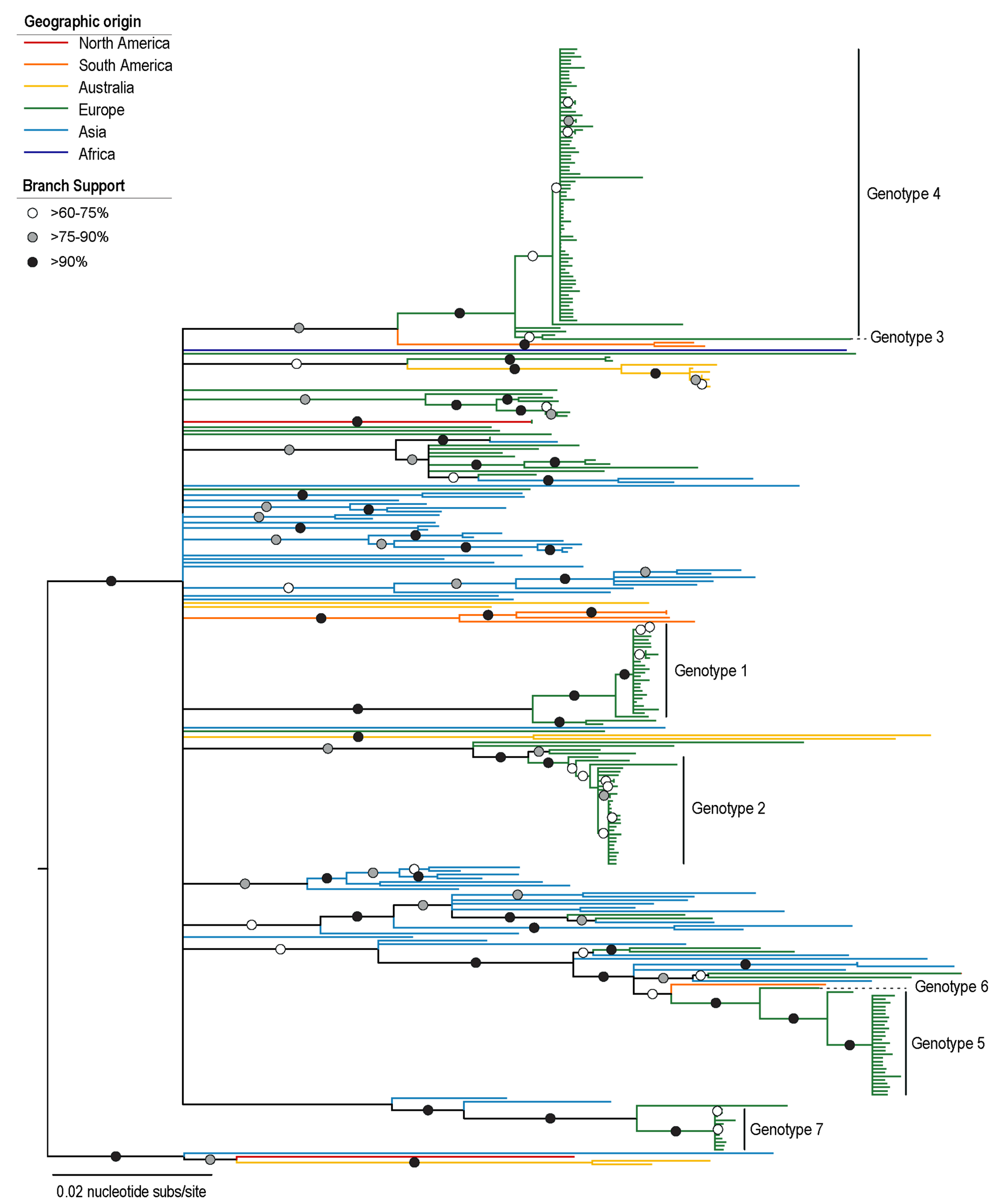
3.1. Characterization of PiCV Genotypes

Ten birds were purchased from three different lofts, and blood samples were collected from each animal. Thereafter, they were all housed in a single loft for 22 days, and blood and cloacal swab samples were collected as summarized in Figure 1 and Figure 2. A total of 58 genome sequences were determined from seven pigeons before they were housed together in a loft and 120 thereafter (Table 1, Figure 2). Pigeons TSP1, TSP2 and TSP4, were identified as PiCV negative prior to grouping them in one loft.
The 178 PiCV genome sequences from this study were analyzed together with all PiCV full genomes available in GenBank (n = 127). Pairwise identity analyses showed the all PiCVs (those available in GenBank and from this study) share >83% genome-wide identity. Based on the genome-wide pairwise identities, we identified seven genotypes for PiCV genome sequences based on the criteria we set for the purpose of this study, which was that viral sequences that share >98% genome-wide identity are part of the same genotype (Figure 2). Additionally, 95 genotypes could represent all other PiCV sequences publicly available (Supplementary Data S1).
PiCV genotypes 1, 2, 3, 4, 5 and 7 were present in the pigeons from the three lofts (Table 1, Figure 2). In the case of TSP3, from loft 1, only genotype 4 was initially identified. Once the pigeons from loft 1 (TSP1-4) were moved to a single loft, PiCV genotypes 2 and 4 were detected in TSP1 and TSP2, and genotype 2 in TSP4, suggesting that these animals became infected subsequent to being moved to a single loft. PiCV genotypes 1, 2, 3, 4, 5 and 7 were identified in TSP5-8 from loft 2; however, after moving them to a single loft, we identified genotypes 2, 4, 5, 6 and 7 and not genotypes 1, 3 and 5. It is interesting to note that animal TSP6 was found harboring four PiCV genotypes (Figure 2). PiCV genotype 3 was detected in animal TSP7 initially, and this is the only animal to have this genotype. PiCV genotype 6 was identified only in animal TSP5 once all of the birds were housed in a single loft. In the two animals from loft 3, PiCV genotypes 1 and 4 were identified. Once moved to a single loft, there is a shift in genotypes to 1 and 2 (Figure 2).
In general, genotype 4 was highly prevalent, making up 37.9% (22/58) of the PiCV sequences in five pigeons in the three lofts prior to being housed in a single loft. Once all the animals were housed in the same loft, this genotype was found in seven pigeons, which accounts for 47.5% (57/120) (Figure 2).

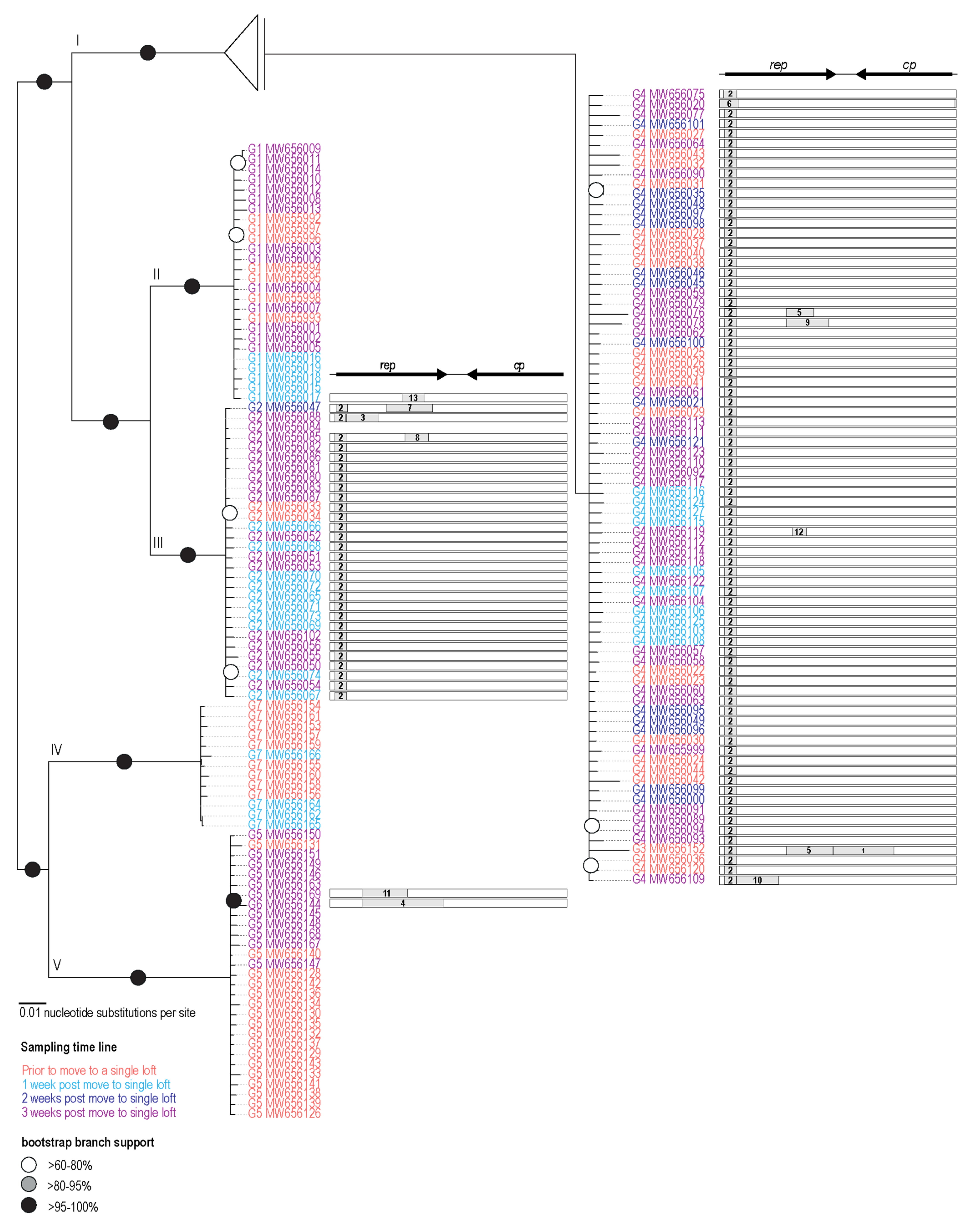
3.2. Phylogenetic Analysis of PiCV Genomes

PiCV genomes available in GenBank (download 15 February 2021) are those identified in pigeons from Australia (n = 13), Belgium (n = 6), Brazil (n = 7), China (n = 63), France (n = 1), Japan (n = 1), Nigeria (n = 1), Poland (n = 30), United Kingdom (n = 2) and United States of America (n = 3). Global phylogenetic analysis of the PiCV recovered from 10 pigeons in this study (n = 178) together with those PiCV genomes available in GenBank was undertaken (Figure 3). The phylogenetic analysis coupled with genotype assignment shows that PiCV sequences from this study are distributed within seven distinct clades according to our genotype designation based on >98% nucleotide pairwise identity threshold. Genotypes 3, 4, 5 and 6 form clades that are most closely related to PiCV sequences identified in a pigeon from Brazil and those from China and Poland. Genotypes 1 and 2 sequences form clades with sequences most closely related to PiCVs from birds sampled in Poland. Lastly, genotype 7 sequences cluster with two PiCV sequences from China. The PiCV genotypes identified in this study are distributed throughout the phylogenetic tree, clearly highlighting diverse PiCVs circulating in at least three of the lofts from which the pigeons were acquired. Furthermore, we detected multiple genotypes within a single animal (Figure 2).

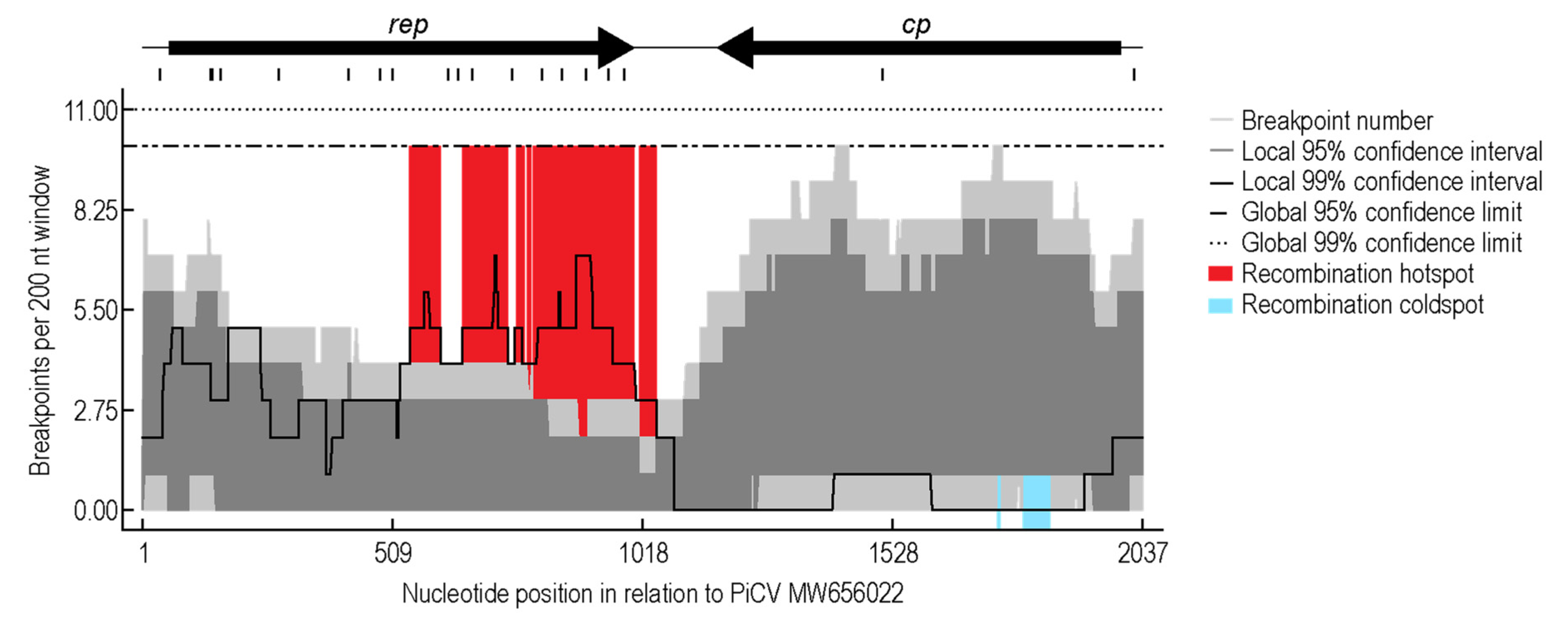
3.3. Recombination Analysis

To investigate PiCV recombination dynamics, a pilot simulation of “housing together” naturally infected and non-infected animals originated from different maternal lofts was undertaken. The recombination analysis of the PiCV sequences from this study revealed 13 detectable recombination events. The recombination event detected in a large portion of the sequences was event 2, occurring in 109 genomes (Figure 4), and this recombinant region spans a ~100 nt region located at the 5’ portion of rep. Twelve of the recombination events detected are within the rep gene (Figure 4). We identified a recombination hotspot spanning the 3′ region of rep and the intergenic region, whereas a cold spot was identified near the 5′ region of the cp (Figure 5). These results differ from the previous hotspot detected in PiCV [35], which shows hotspots towards the 3′ end of the rep and cp, as well as the intergenic region. The differences in recombination patterns could be due to the variation in samples, i.e., global datasets with “circulating” recombinants as opposed to the ones in this study that address recombination in a short time frame within a small set of animals in a loft. The cold spot located in the cp, along with low amounts of recombination events, shows a higher region of conservation and importance of the cp likely for transmission and infectivity. Phylogenetic analysis of the genomes without recombination shows five clades present in the population (Figure 4) in contrast to the seven genotypes present in the Neighbor-Joining phylogenetic analysis (Figure 3).
There is no evidence of recombination in sequences present in Clade II and IV (Figure 4). Clade IV (Figure 4) is mainly composed of sequences determined from animals prior to being in a single loft and those after being in a single loft for a week. PiCV sequence MW656152 from animal TSP7 had three recombinant regions accounting for 42% of the genome, but this was from a sample taken from the animal while in maternal loft 2, and this represents PiCV recombination within the loft of origin (Figure 4; Table 1 and Table 2). PiCV sequence MW656144 from TSP5 has a recombinant region, accounting for 34% of the genome. Both PiCV sequences MW656152 and MW656144 are singletons forming their own genotypes 3 and 6, respectively (Figure 3, Supplementary Data S1).
Only three recombination events were identified in sequences derived from the animals while in the three separate lofts of origin, whereas ten recombination events were detected in sequences derived from animals once they were housed in the same loft.

4. Concluding Remarks

In this study, we undertook a pilot experiment to simulate the viral dynamics at play when infected and non-infected pigeons are acquired from different sources and then housed together in a single loft. We show that genotypes of PiCV spread to non-infected birds and also to already infected birds, either resulting in co-infection or displacement of the previous genotype, e.g., the genotype 4 in animal TSP7 that appears to have “out competed” genotypes 1, 2 and 7 (Figure 2). We also demonstrate that recombination plays a role in virus evolution.
This study demonstrates the PiCV spreads in animals within a loft in a short time frame, in which the animals can harbor multiple genotypes and dominant genotypes possibly outcompete previous variants. Pigeon circovirus is known as an immunosuppressive factor in pigeons. A previous study performed on 30 PiCV full genomes recovered from pigeons of various breeds and different health statuses did not find any correlation between genotype, sequence mutation of circovirus strain and health status of its host [18]. There is also no information concerning a similar correlation between the certain/dominant genotype and immunosuppression of the pigeons. However, this phenomenon cannot be excluded at this stage.

Supplementary Materials

The following are available online at https://www.mdpi.com/article/10.3390/v13060964/s1, Supplementary Data S1: Nucleotide pairwise identity analysis of all PiCV sequences (including those from this study).

Author Contributions

Conceptualization, A.V. and T.S.; methodology, A.K., S.K., D.D., A.V. and T.S.; validation, A.K., S.K. and A.V.; formal analysis A.K., S.K., D.D., A.V. and T.S.; investigation, A.K., S.K., D.D., A.V. and T.S.; resources, A.V. and T.S.; data curation, A.K., S.K. and A.V.; writing—original draft preparation, A.K., S.K., A.V. and T.S.; writing—review and editing, A.K., S.K., D.D., A.V. and T.S.; visualization, A.K., S.K. and A.V.; supervision, A.K., S.K., A.V. and T.S.; project administration, A.V. and T.S.; funding acquisition, A.V. and T.S. All authors have read and agreed to the published version of the manuscript.

Funding

Project financially co-supported by Minister of Science and Higher Education in the range of the program entitled “Regional Initiative of Excellence” for the years 2019–2022, Project No. 010/RID/2018/19, amount of funding 12.000.000 PLN”.

Institutional Review Board Statement

The research protocol was approved by the Local Ethics Committee on Animal Experimentation of the University of Warmia and Mazury in Olsztyn (resolution No 41/2019, issued 28 May 2019, valid through 1 October 2023).

Data Availability Statement

Sequence determined as part of this study have been deposited in GenBank under accession #s MW655992 - MW656169.

Conflicts of Interest

The authors declare no conflict of interest. The funders had no role in the design of the study; in the collection, analyses, or interpretation of data; in the writing of the manuscript, or in the decision to publish the results.

References

  1. Haag-Wackernagel, D.; Moch, H. Health hazards posed by feral pigeons. J. Infect. 2004, 48, 307–313. [Google Scholar] [CrossRef] [PubMed]
  2. Giunchi, D.; Mucci, N.; Bigi, D.; Mengoni, C.; Baldaccini, N.E. Feral pigeon populations: Their gene pool and links with local domestic breeds. Zoology (Jena) 2020, 142, 125817. [Google Scholar] [CrossRef] [PubMed]
  3. Pacheco, G.; van Grouw, H.; Shapiro, M.D.; Gilbert, M.T.P.; Vieira, F.G. Darwin’s Fancy Revised: An Updated Understanding of the Genomic Constitution of Pigeon Breeds. Genome Biol. Evol. 2020, 12, 136–150. [Google Scholar] [CrossRef] [PubMed]
  4. Freick, M.; Muller, H.; Raue, R. Rapid detection of pigeon herpesvirus, fowl adenovirus and pigeon circovirus in young racing pigeons by multiplex PCR. J. Virol. Methods 2008, 148, 226–231. [Google Scholar] [CrossRef]
  5. Huang, Y.L.; Castaneda, O.A.; Thongchan, D.; Khatri-Chhetri, R.; Tsai, S.S.; Wu, H.Y. Pigeon circovirus infection in disqualified racing pigeons from Taiwan. Avian Pathol. 2017, 46, 359–366. [Google Scholar] [CrossRef] [Green Version]
  6. Omar, M.A.; Hassan, F.A.; Shahin, S.E. Economic Assessment of Various Levels of Protein and Energy in Pigeon Squabs Diet. Zagazig Vet. J. 2017, 45, 20–30. [Google Scholar] [CrossRef] [Green Version]
  7. Proskura, W.S.; Kustosz, J.; Dybus, A.; Lanckriet, R. Polymorphism in dopamine receptor D4 gene is associated with pigeon racing performance. Anim. Genet. 2015, 46, 586–587. [Google Scholar] [CrossRef]
  8. Shao, Y.; Tian, H.Y.; Zhang, J.J.; Kharrati-Koopaee, H.; Guo, X.; Zhuang, X.L.; Li, M.L.; Nanaie, H.A.; Dehghani Tafti, E.; Shojaei, B.; et al. Genomic and Phenotypic Analyses Reveal Mechanisms Underlying Homing Ability in Pigeon. Mol. Biol. Evol. 2020, 37, 134–148. [Google Scholar] [CrossRef]
  9. Ye, M.; Xu, M.; Chen, C.; He, Y.; Ding, M.; Ding, X.; Wei, W.; Yang, S.; Zhou, B. Expression analyses of candidate genes related to meat quality traits in squabs from two breeds of meat-type pigeon. J. Anim. Physiol. Anim. Nutr. 2018, 102, 727–735. [Google Scholar] [CrossRef]
  10. Geraldy, K. Transport of racing pigeons and animal welfare. Dtsch. Tierarztl. Wochenschr. 2007, 114, 114–115. [Google Scholar]
  11. Kaleta, E.F.; Baldauf, C. Newcastle disease in free-living and pet birds. In Newcastle Disease; Springer: Berlin/Heidelberg, Germany, 1988; pp. 197–246. [Google Scholar]
  12. Phan, T.G.; Vo, N.P.; Boros, A.; Pankovics, P.; Reuter, G.; Li, O.T.; Wang, C.; Deng, X.; Poon, L.L.; Delwart, E. The viruses of wild pigeon droppings. PLoS ONE 2013, 8, e72787. [Google Scholar] [CrossRef] [Green Version]
  13. McCowan, C.; Crameri, S.; Kocak, A.; Shan, S.; Fegan, M.; Forshaw, D.; Rubbenstroth, D.; Chen, H.; Holmes, C.; Harper, J.; et al. A novel group A rotavirus associated with acute illness and hepatic necrosis in pigeons (Columba livia), in Australia. PLoS ONE 2018, 13, e0203853. [Google Scholar] [CrossRef]
  14. Zhang, Z.; Dai, W.; Dai, D. Molecular characterization of pigeon torque teno virus (PTTV) in Jiangsu province. Comput. Biol. Chem. 2017, 69, 10–18. [Google Scholar] [CrossRef]
  15. Agnihotri, K.; Smith, C.; Oakey, J.; Storie, G. Pigeon adenovirus and pigeon torque teno virus associated with acute multifocal hepatic necrosis in pigeons in Queensland, Australia. Arch. Virol. 2021. [Google Scholar] [CrossRef]
  16. Teske, L.; Rubbenstroth, D.; Meixner, M.; Liere, K.; Bartels, H.; Rautenschlein, S. Identification of a novel aviadenovirus, designated pigeon adenovirus 2 in domestic pigeons (Columba livia). Virus Res. 2017, 227, 15–22. [Google Scholar] [CrossRef]
  17. Wan, C.; Chen, C.; Cheng, L.; Shi, S.; Fu, G.; Liu, R.; Chen, H.; Fu, Q.; Huang, Y. Detection of novel adenovirus in sick pigeons. J. Vet. Med. Sci. 2018, 80, 1025–1028. [Google Scholar] [CrossRef] [Green Version]
  18. Rubbenstroth, D.; Peus, E.; Schramm, E.; Kottmann, D.; Bartels, H.; McCowan, C.; Schulze, C.; Akimkin, V.; Fischer, N.; Wylezich, C.; et al. Identification of a novel clade of group A rotaviruses in fatally diseased domestic pigeons in Europe. Transbound. Emerg. Dis. 2019, 66, 552–561. [Google Scholar] [CrossRef] [PubMed]
  19. Mankertz, A.; Hattermann, K.; Ehlers, B.; Soike, D. Cloning and sequencing of columbid circovirus (coCV), a new circovirus from pigeons. Arch. Virol. 2000, 145, 2469–2479. [Google Scholar] [CrossRef]
  20. Todd, D.; Fringuelli, E.; Scott, A.N.; Borghmans, B.J.; Duchatel, J.P.; Shivaprasad, H.L.; Raidal, S.R.; Abadie, J.X.; Franciosini, M.P.; Smyth, J.A. Sequence comparison of pigeon circoviruses. Res. Vet. Sci. 2008, 84, 311–319. [Google Scholar] [CrossRef] [PubMed]
  21. Todd, D.; Weston, J.H.; Soike, D.; Smyth, J.A. Genome sequence determinations and analyses of novel circoviruses from goose and pigeon. Virology 2001, 286, 354–362. [Google Scholar] [CrossRef]
  22. Coletti, M.; Franciosini, M.P.; Asdrubali, G.; Passamonti, F. Atrophy of the primary lymphoid organs of meat pigeons in Italy associated with circoviruslike particles in the bursa of Fabricius. Avian Dis. 2000, 44, 454–459. [Google Scholar] [CrossRef]
  23. Woods, L.W.; Latimer, K.S.; Niagro, F.D.; Riddell, C.; Crowley, A.M.; Anderson, M.L.; Daft, B.M.; Moore, J.D.; Campagnoli, R.P.; Nordhausen, R.W. A retrospective study of circovirus infection in pigeons: Nine cases (1986–1993). J. Vet. Diagn. Investig. 1994, 6, 156–164. [Google Scholar] [CrossRef] [Green Version]
  24. Stenzel, T.A.; Pestka, D.; Tykalowski, B.; Smialek, M.; Koncicki, A. Epidemiological investigation of selected pigeon viral infections in Poland. Vet. Rec. 2012, 171, 562. [Google Scholar] [CrossRef]
  25. Ledwoń, A.; Bailey, T.; O’Donovan, D.; Mckeown, S.; Lloyd, C.; Więckowski, T.; Kinne, J.; Silvanose, C.; Szeleszczuk, P.; Wernery, U. Prevalence of circovirus and adenovirus in pigeons in Dubai. Med. Weter. 2011, 67, 752–756. [Google Scholar]
  26. Loiko, M.R.; Junqueira, D.M.; Varela, A.P.M.; Tochetto, C.; Scheffer, C.M.; Lima, D.A.; Morel, A.P.; Cerva, C.; Paim, W.P.; Mayer, F.Q.; et al. Columbid circoviruses detected in free ranging pigeons from Southern Brazil: Insights on PiCV evolution. Arch. Virol. 2018, 163, 3083–3090. [Google Scholar] [CrossRef]
  27. Sarker, S.; Das, S.; Ghorashi, S.A.; Forwood, J.K.; Raidal, S.R. Pigeon circoviruses from feral pigeons in Australia demonstrate extensive recombination and genetic admixture with other circoviruses. Avian Pathol. 2019, 48, 512–520. [Google Scholar] [CrossRef]
  28. Stenzel, T.; Pestka, D. Occurrence and genetic diversity of pigeon circovirus strains in Poland. Acta Vet. Hung. 2014, 62, 274–283. [Google Scholar] [CrossRef] [Green Version]
  29. Wang, K.C.; Zhuang, Q.Y.; Qiu, Y.; Wang, T.; Chen, J.M. Genome sequence characterization of pigeon circoviruses in China. Virus Res. 2017, 233, 1–7. [Google Scholar] [CrossRef]
  30. Duchatel, J.P.; Todd, D.; Curry, A.; Smyth, J.A.; Bustin, J.C.; Vindevogel, H. New data on the transmission of pigeon circovirus. Vet. Rec. 2005, 157, 413–415. [Google Scholar] [CrossRef]
  31. Duchatel, J.P.; Todd, D.; Smyth, J.A.; Bustin, J.C.; Vindevogel, H. Observations on detection, excretion and transmission of pigeon circovirus in adult, young and embryonic pigeons. Avian Pathol. 2006, 35, 30–34. [Google Scholar] [CrossRef]
  32. Franciosini, M.P.; Fringuelli, E.; Tarhuni, O.; Guelfi, G.; Todd, D.; Casagrande Proietti, P.; Falocci, N.; Asdrubali, G. Development of a polymerase chain reaction-based in vivo method in the diagnosis of subclinical pigeon circovirus infection. Avian Dis. 2005, 49, 340–343. [Google Scholar] [CrossRef]
  33. Abadie, J.; Nguyen, F.; Groizeleau, C.; Amenna, N.; Fernandez, B.; Guereaud, C.; Guigand, L.; Robart, P.; Lefebvre, B.; Wyers, M. Pigeon circovirus infection: Pathological observations and suggested pathogenesis. Avian Pathol. 2001, 30, 149–158. [Google Scholar] [CrossRef]
  34. Stenzel, T.; Dziewulska, D.; Tykalowski, B.; Koncicki, A. The Clinical Infection with Pigeon Circovirus (PiCV) Leads to Lymphocyte B Apoptosis But Has No Effect on Lymphocyte T Subpopulation. Pathogens 2020, 9, 632. [Google Scholar] [CrossRef]
  35. Stenzel, T.; Piasecki, T.; Chrząstek, K.; Julian, L.; Muhire, B.M.; Golden, M.; Martin, D.P.; Varsani, A. Pigeon circoviruses display patterns of recombination, genomic secondary structure and selection similar to those of beak and feather disease viruses. J. Gen. Virol. 2014, 95, 1338–1351. [Google Scholar] [CrossRef] [PubMed] [Green Version]
  36. Khalifeh, A.; Kraberger, S.; Dziewulska, D.; Stenzel, T.; Varsani, A. Complete Genome Sequence of a Phapecoctavirus Isolated from a Pigeon Cloacal Swab Sample. Microbiol. Resour. Announc. 2021, 10. [Google Scholar] [CrossRef]
  37. Edgar, R.C. MUSCLE: Multiple sequence alignment with high accuracy and high throughput. Nucleic Acids Res. 2004, 32, 1792–1797. [Google Scholar] [CrossRef] [Green Version]
  38. Kumar, S.; Stecher, G.; Li, M.; Knyaz, C.; Tamura, K. MEGA X: Molecular Evolutionary Genetics Analysis across Computing Platforms. Mol. Biol. Evol. 2018, 35, 1547–1549. [Google Scholar] [CrossRef] [PubMed]
  39. Stover, B.C.; Muller, K.F. TreeGraph 2: Combining and visualizing evidence from different phylogenetic analyses. BMC Bioinform. 2010, 11, 7. [Google Scholar] [CrossRef] [Green Version]
  40. Muhire, B.M.; Varsani, A.; Martin, D.P. SDT: A virus classification tool based on pairwise sequence alignment and identity calculation. PLoS ONE 2014, 9, e108277. [Google Scholar] [CrossRef] [PubMed]
  41. Martin, D.P.; Varsani, A.; Roumagnac, P.; Botha, G.; Maslamoney, S.; Schwab, T.; Kelz, Z.; Kumar, V.; Murrell, B. RDP5: A computer program for analyzing recombination in, and removing signals of recombination from, nucleotide sequence datasets. Virus Evol. 2021, 7, veaa087. [Google Scholar] [CrossRef]
  42. Guindon, S.; Dufayard, J.F.; Lefort, V.; Anisimova, M.; Hordijk, W.; Gascuel, O. New algorithms and methods to estimate maximum-likelihood phylogenies: Assessing the performance of PhyML 3.0. Syst. Biol. 2010, 59, 307–321. [Google Scholar] [CrossRef] [PubMed] [Green Version]
Figure 1. A summary of the timeline of blood and cloacal swab sampling of the pigeons (TSP1-10) for the study.
Viruses 13 00964 g001
Figure 2. A summary of the PiCV genotypes identified in the study in either the blood or cloacal swab samples. The pigeons were sourced from three separate lofts (loft 1–3) and then house in a single loft. Blood samples were taken when sourced, and thereafter, blood and cloacal swabs were taken at different time points as outlined in Figure 1. Genotypes identified in animals in each of the three lofts and also once they are housed in a single loft are color-coded, and the number in the circles indicates the number of genomes determined that are assigned to the genotype.
Viruses 13 00964 g002
Figure 3. Neighbor-Joining phylogenetic tree of the PiCV genome sequences from this study together with those available in GenBank. Branches are color-coded based on the geographical sampling of the PiCV. The genotypes 1–7 represent sequences from this study.
Viruses 13 00964 g003
Figure 4. Maximum Likelihood phylogenetic tree of PiCV sequences recovered from study with recombinant regions removed. Recombinant regions identified in the genomes are shown next to the accession numbers. The five main clades are labeled in roman numerals (I–V). Genotypes (G1–G7) are labeled next to all the accession numbers.
Viruses 13 00964 g004
Figure 5. Recombination break point analysis for PiCV genomes from this study. Hot spots and cold spots are highlighted. Genome organization based on PiCV sequence MW656022 as a representative.
Viruses 13 00964 g005
Table 1. The collection date and sampling data of PiCV genome sequences from the study. PiCV sequence accession numbers and the number of isolates from samples are listed.
PigeonDateBloodCloacal SwabAccession #s
TSP14 May 2020x negative
13 May 2020x MW656045, MW656046, MW656047, MW656048, MW656049
6 May 2020 xMW656103, MW656106, MW656107, MW656108, MW656125
TSP24 May 2020x negative
26 May 2020x MW656050, MW656051, MW656052, MW656053, MW656054, MW656055, MW656056
26 May 2020 xMW656109, MW656110, MW656111, MW656104, MW656123
TSP34 May 2020x MW656022, MW656023, MW656024, MW656025, MW656026, MW656027, MW656028, MW656029, MW656030
26 May 2020x MW656057, MW656058, MW656059, MW656060, MW656061, MW656062, MW656063, MW656064
26 May 2020 xMW656112, MW656113, MW656114, MW656122
TSP44 May 2020x negative
7 May 2020x MW656065, MW656066, MW656067, MW656068, MW656069, MW656070, MW656071, MW656072, MW656073, MW656074
TSP504 May 2020x MW656128, MW656129, MW656130, MW656131, MW656132, MW656133, MW656134, MW656126, MW656135, MW656136
6 May 2020 xMW656105, MW656115, MW656116, MW656124, MW656127
26 May 2020x MW655999, MW656079, MW656075, MW656076, MW656077, MW656078, MW656020, MW656144
26 May 2020 xMW656145, MW656149, MW656150, MW656151, MW656168
TSP64 May 2020x MW656153, MW656154, MW656159, MW656137, MW656155, MW656156, MW656157, MW656158
6 May 2020 xMW656162, MW656164, MW656165, MW656166
26 May 2020x MW656080, MW656081, MW656082, MW656083, MW656084, MW656085, MW656086, MW656087, MW656088
26 May 2020 xMW656118, MW656119, MW656146, MW656169
TSP74 May 2020x MW656138, MW656143, MW656152, MW656139, MW656140, MW656031, MW656141, MW656120, MW656142
26 May 2020x MW656089, MW656090, MW656091, MW656092, MW656093, MW656094
26 May 2020 xMW656147, MW656148, MW656117, MW656163, MW656167
TSP84 May 2020x MW655992, MW656032, MW656033, MW656034, MW656160, MW656161,
15 May 2020x MW656000, MW656021, MW656095, MW656096, MW656097, MW656098, MW656099, MW656100, MW656101, MW656121
TSP94 May 2020x MW655993, MW655994, MW655995, MW655996, MW655997, MW656036, MW655998, MW656035
06 May 2020 xMW656015, MW656016, MW656017, MW656018, MW656019
21 May 2020x MW656001, MW656002, MW656003, MW656004, MW656005, MW656006, MW656007
TSP104 May 2020x MW656037, MW656038, MW656039, MW656040, MW656041, MW656042, MW656043, MW656044
26 May 2020x MW656008, MW656009, MW656010, MW656011, MW656102, MW656012, MW656013, MW656014
x = sampled.
Table 2. A summary of the recombination events detected in the genomes of PiCV from this study. The methods used to detect recombination are RDP (R), GENCONV (G), BOOTSCAN (B), MAXCHI (M), CHIMERA (C), SISCAN (S) and 3SEQ (T). For each recombination event, the method with the highest p-value is in bold font and underlined. [P] denotes partial evidence of recombination.
Event # Recombinant RegionMinor Parental Sequence(s)Major Parental
Sequence(s)
Methodp-Value
Recombinant Sequence(s)BeginEnd
1Genotype 39831512Genotype 6 (all)
Genotype 5 (MW656131, MW65613, MW656150, MW656169)
Genotype 4 (all)GBMCST8.88 × 10−47
2Genotype 4 (all)
Genotype 2 (all)
Genotype 3
38140Genotype 5 (all)Genotype 1 (all)GBMCS6.04 × 10−20
3Genotype 2 (MW656088)142418Genotype 7 (all)
Genotype 5 (MW656159)
Genotype 2 (expect MW656088)GMCST2.77 × 10−17
4Genotype 6275983Genotype 4 (MW656020)Genotype 5 (all)GMCST4.69 × 10−16
5Genotype 4 (MW656076)
Genotype 3 (MW656152)[P]
577 *816Genotype 5 (all)Genotype 4 (MW656020, MW656093, MW656106, MW656109, MW656120)GMCST1.52 × 10−13
6Genotype 4 (MW656020)2018158Genotype 1 (all)Genotype 2 (all)GMCST2.46 × 10−13
7Genotype 2 (MW656047)484904Genotype 4 (MW656020)Genotype 2 (MW656085)GBMCST2.59 × 10−13
8Genotype 2 (MW656085)644856Genotype 7 (all)Genotype 2 (except MW656085)GBMCST4.99 × 10−12
9Genotype 4 (MW656078)577*948Genotype 1 (all)Genotype 4 (except MW656078)GBMCST5.69 × 10−11
10Genotype 4 (MW656109)142*508Genotype 5 (all)Genotype 4 (MW656093, MW656119, MW656020)
Genotype 3
GBMCST8.88 × 10−22
11Genotype 5 (MW656169)277676Genotype 4 (all)Genotype 5 (except MW656169)GBMCST3.20 × 10−17
12Genotype 4 (MW656119)624752Genotype 5 (all)Genotype 4 (except MW656119)
Genotype 2 (all)
GBT2.32 × 10−9
13Genotype 1 (MW656017)621813Genotype 5 (all)Genotype 1 (except MW656017)GBT1.29 × 10−8
Publisher’s Note: MDPI stays neutral with regard to jurisdictional claims in published maps and institutional affiliations.

