Article

Cloning and Expression of the Chitinase Encoded by ChiKJ406136 from Streptomyces Sampsonii (Millard & Burr) Waksman KJ40 and Its Antifungal Effect

1
College of Forestry, Sichuan Agricultural University, No. 211, Huimin Road, Wenjiang District, Chengdu 611130, Sichuan, China
2
College of Landscape Architecture, Sichuan Agricultural University, No. 211, Huimin Road, Wenjiang District, Chengdu 611130, Sichuan, China
*
Author to whom correspondence should be addressed.
Forests 2018, 9(11), 699; https://doi.org/10.3390/f9110699
Submission received: 9 October 2018 / Revised: 2 November 2018 / Accepted: 9 November 2018 / Published: 11 November 2018
(This article belongs to the Section Forest Ecophysiology and Biology)

Abstract

:
The present study demonstrated that the chitinase gene ChiKJ406136 of Streptomyces sampsonii (Millard & Burr) Waksman KJ40 could be cloned using a PCR protocol and expressed in Escherichia coli (Migula) Castellani & Chalmers BL21 (DE3), and the recombinant protein had antifungal effect on four forest pathogens (Cylindrocladium scoparium Morgan, Cryphonectria parasitica (Murrill) Barr, Neofusicoccum parvum Crous, and Fusarium oxysporum Schl.) and also had the biological control effects on Eucalyptus robusta Smith leaf blight, Castanea mollissima BL. blight, Juglans regia L. blight and J. regia root rot. The results showed that ChiKJ406136 was efficiently expressed and a 48 kilodalton (kDa) recombinant protein was obtained. No significant change in protein production was observed in the presence of different concentrations of IPTG (isopropyl-b-D-thio-galactoside). The purified protein yield was greatest in the 150 mmol/L imidazole elution fraction, and the chitinase activities of the crude protein and purified protein solutions were 0.045 and 0.033 U/mL, respectively. The antifungal effects indicated that mycelial cells of the four fungi were disrupted, and the control effects of the chitinase on four forest diseases showed significant differences among the undiluted 10- and 20-fold dilutions and the control. The undiluted solution exhibited best effect. The results of this study provide a foundation for the use of S. sampsonii as a biocontrol agent and provides a new source for the chitinase gene, providing a theoretical basis for its application.

1. Introduction

Streptomyces Waksman & Henrici, the largest genus of the phylum Actinobacteria, consists of a group of Gram-positive, aerobic, non-motile, catalase positive, and non-acid-fastbacteria with a filamentous form that resembles fungi [1,2,3]. The members of Streptomyces are well-known for their ability to produce a variety of bioactive compounds with different bioactivities such as antibacterial [4,5,6,7,8,9], antifungal [10], antiviral [11], immunosuppressive [12], anticancer, and antioxidant properties [13,14,15]. Thereinto, Streptomyces sampsonii is widely distributed in nature, having been isolated from Maytenus aquifolia Mart. [16], soil [17,18,19,20,21,22], marine sediment [23,24], and medicinal plants [25]. In particular, numerous isolates have been obtained from soil, such as garden soil [18], alpine soil from the Himalayan glacier region [19], fertile soil in Korea [26], and rhizosphere soil of healthy poplar trees [20]. S. sampsonii possesses antagonistic activity against fungal pathogens, including those of the genera Alternaria Nees and Phomopsis Sacc. & Roum. [19], and the species Candida albicans (C.P.Robin) Berkhout, Aspergillus niger van Tieghem, Microsporum gypseum (E. Bodin) Guiart & Grigoraki, Trichophyton sp. (Castell.) Sabour. [18] and Rhizoctonia violacea (Tul.) Pat. [20]. Previous studies demonstrated that the bioactive compounds of S. sampsonii have important applications in various fields [3]. For example, crude extracts showed antitumor activity against glioblastoma multiforme (GBM) cells, inhibiting cell growth by 70.04% [25], and the supernatant of a S. sampsonii culture showed biological activity against the root-knot nematode [26]. In the purified components, soil isolates of S. sampsonii can produce heptaene polyene antibiotics [18,27], In addition, S. sampsonii has been shown to produce hydrolytic enzymes, such as amylase, chitinase, protease, and lipase [19]. Studies at the molecular level have focused on strain identification and the phylogenesis of related species [28,29,30,31,32,33,34,35]. The complete genome sequence of Streptomyces sampsonii KJ40 was recently described by our lab [36], resulting in the discovery of a large number of gene encoding chitinases and enzymes involved in secondary metabolite production. However, little is known regarding the metabolic pathways and genetic regulation in this strain, limiting its practical application.
Chitin, a linear polymer of β-1,4-glucosidicosamine (GlcNAC), is the second most abundant polysaccharide in nature. Chitin can be degraded by chitinolytic enzymes, that is chitinase. Chitinases (EC 3.2.1.14) are widely present in a great variety of organisms, including insects, fungi, yeast, higher plants, vertebrates, arthropods and humans [37]. Due to the functions of degrading chitin, their antitumor activities, and antihypertensive activities, chitinases have been widely exploited in various fields, such as industrial, agricultural and medicinal applications [38,39]. As it is well-known, control of plant pests and diseases by application of biological environmentally friendly agents has received great attention. Chitinases, as an alternative to the use of chemical, have been holding great promise in control of fungal and insect pathogens of plants. The functions of chitinases in various organisms are diverse, as well as their mechanisms in biotechnological applications. In fungi, previous studies have demonstrated chitinases can inhibit the growth of fungi (such as Verticillium dahliae Kleb. [40], Colletotrichum gloeosporioides (Stoneman) Spauld. & H. Schrenk, Fusarium graminearum (Schwein.) Petch, Fusarium oxysporum (Schlecht.) Snyder & Hansen, Alternaria alternata (Fr.) Keissl. [41], and Rhizoctonia solani Kühn [42,43]) by impacting their synthesis of cell wall, apical growth and morphogenesis of fungal hyphae. In insects, it was reported by Gadelhak et al. [44] that the chitinases produced by Actinoplanes philippinensis Couch, A. missouriensis Couch, and Streptomyces clavuligerus Higgens & Kastner could inhibit the emergence of Drosophila melanogaster Meigen adults from pupae. In plants, Prasad et al. [45] showed that transgenic peanuts plants enhanced their sustained resistance to fungal diseases by over-expressing chitinase genes. Additional transgenic plants expressing chitinases have been successfully created including lemon tree [46], wheat [47] and carrots [48], and so on, this suggests the possibility that to control fungal or insect disease with chitinase transgenic plants or chitinase, and its potential applications is becoming the future.
Streptomyces sampsonii KJ40 was previously isolated from the rhizosphere soil of a poplar plantation in China. Field tests demonstrated that the fermentation filtrate of this bacterium can help to control poplar purple root rot and reduce tree morbidity [20]. Extracellular secondary metabolites or hydrolytic enzymes, including chitinase, can significantly inhibit fungal growth [19,23,26]. At present, the studies of chitinases from S. sampsonii primarily focus on the biological activity of the fermentation liquid. However, the chitinase gene from this strain has not been studied, and the activity of the protein is unknown. In this study, we identified the chitinase gene from the complete genome sequence of S. sampsonii KJ40, which was subsequently cloned into a prokaryotic expression vector, to express and purify the recombinant chitinase protein. In particular, we analyzed the antifungal effect of the purified chitinase on the mycelial morphology of four pathogenic fungi and its ability to control four plant diseases. The goal of study was to characterize the recombinant S. sampsonii KJ40 chitinase for future large-scale industrial production and further accelerated application in agriculture, industry and medicine.

2. Materials and Methods

2.1. Strains, Plasmids and Plant Samples

The strain S. sampsonii KJ40 (accession number: LORI00000000) was isolated from the rhizosphere soil of Populus szechuanica Schneid. and was provided by the Key Laboratory of Forest Protection in the Sichuan Province, which was preserved in the Chinese General Microbiological Culture Collection Center (CGMCC No.5996). The complete genome sequence length of S. sampsonii KJ40 was 7261502 bp, containing 6605 genes, 1260 tandem repeat sequences, 804 minisatellite DNAs, 67 microsatellite DNAs, 90 tRNAs, 9 rRNAs, and 19 sRNAs, the average GC content was 73.41% [36]. The strains were grown at 37 °C on Gause’s No. 1 synthetic medium with ampicillin (Sigma-Aldrich, Llc., Shanghai, China) (100 µg/mL).
The pathogenic fungi (Cylindrocladium scoparium Morgan, Cryphonectria parasitica (Murrill) Barr, Neofusicoccum parvum Crous, and Fusarium oxysporum Schl.) were provided by the Key Laboratory of Forest Protection in Sichuan Province. The Escherichia coli strains Trans5α and BL21 (DE3) were purchased from Beijing TransGen Biotech Co., Ltd. (China). The plasmids pMD19-T vector and pET-32a vector were purchased from Dalian TaKaRa Bio Inc. (China).
For the plant samples, healthy, one-year-old Eucalyptus robusta Smith (twenty leaves per plant), Castanea mollissima BL. (five twigs per plant), and Juglans regia L. (five twigs per plant) seedlings were used. The plant samples were planted in a greenhouse (Temperature 25–28 °C, humidity 60–70%) as the Sichuan Agricultural University in Chengdu, Sichuan Province, China (elevation 503 m, 30°97′01.1″ N, 103°81′46.1″ E).

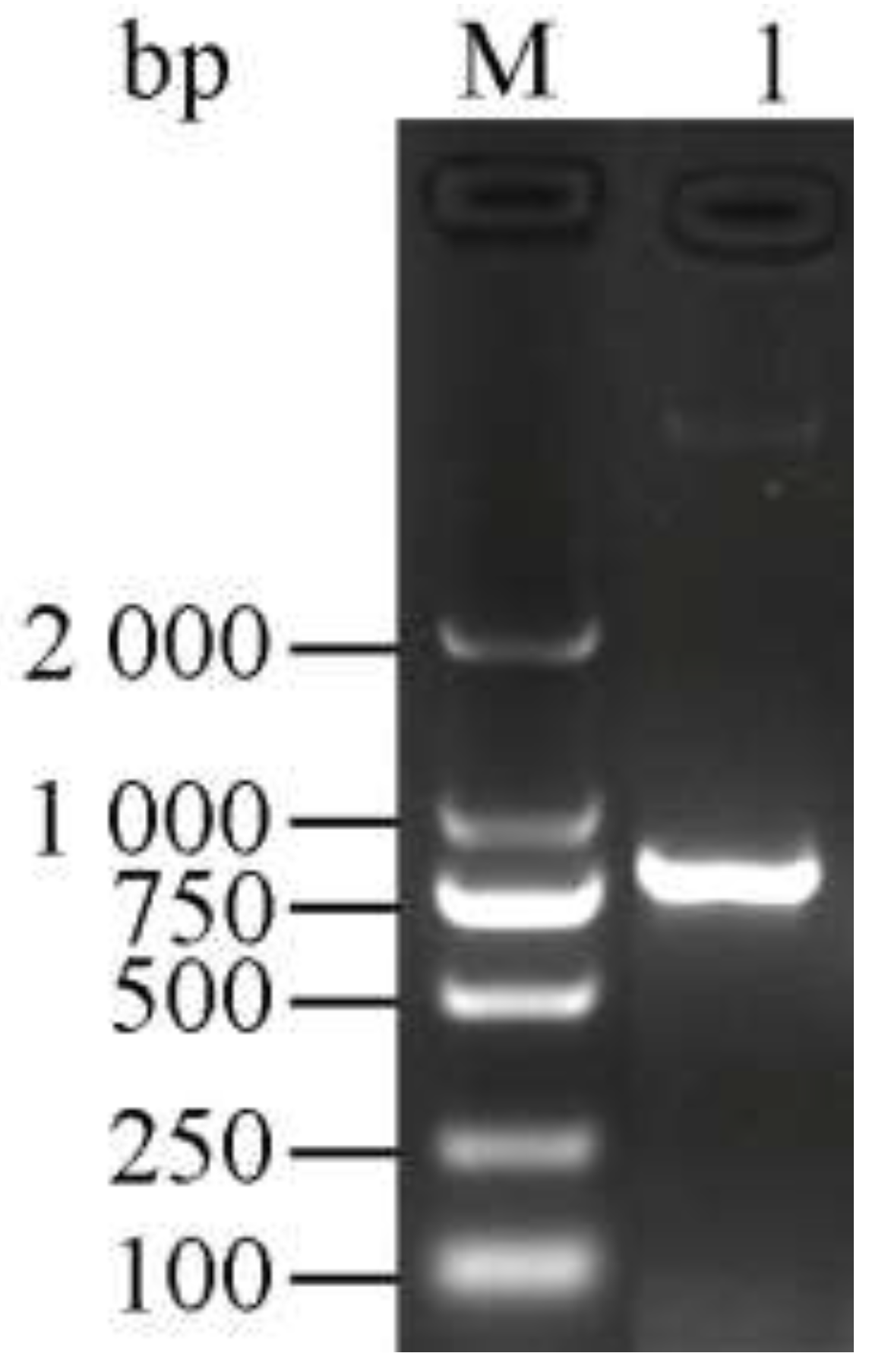
2.2. PCR Amplification of the Chitinase Gene

Based on an analysis of the complete genome sequence of S. sampsonii KJ40 and function prediction and a functional prediction analysis, one chitinase-encoding gene was identified and was named ChiKJ406136. Using Premier 5.0, the primers PL (5′-ATGCGTACCCGTCTGATCG-3′) and PR (5′-TCAGCAGCTGAGGTTGTCG-3′) were designed to amplify the ChiKJ406136 gene. Genomic DNA was prepared from strain KJ40 using a TIANamp Bacteria DNA Kit (Tiangen Biotech Co., Ltd., Beijing, China). All PCR reactions were performed in a total volume of 25 µL and contained 10 µL of ddH2O, 12.5 µL of 2× TransTaq High Fidelity (HiFi) PCR SuperMix I, 0.5 µL of Primer STAR HS DNA Polymerase, and 1 µL of each primer pair (10 µmol/L). The following PCR thermo-cycling conditions were used: 35 cycles of 94 °C for 3 min, 57.8 °C for 30 s, 72 °C for 1 min, and a final extension of 72 °C for 5 min. The PCR fragments were separated on a 1% agarose gel, purified using a TIANgel Midi Purification Kit (Tiangen Biotech Co., Ltd., Beijing, China) and sequenced by Invitrogen (ThermoFisher Scientific Co., Ltd., Shanghai, China).

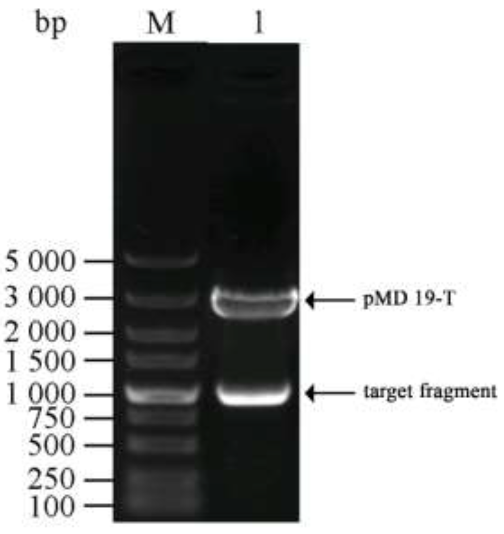
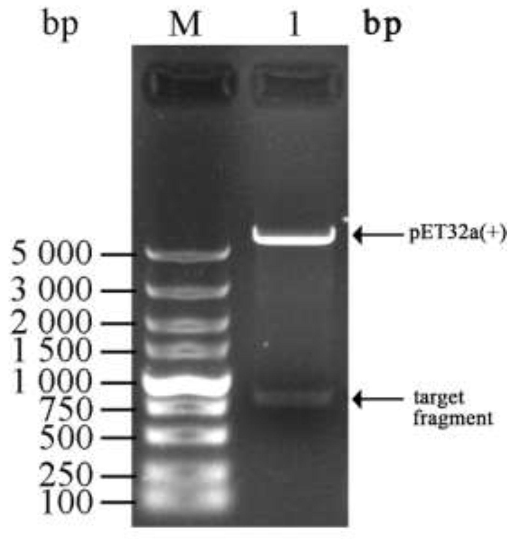
2.3. Construction of a Cloning Vector Harboring the Chitinase Gene

The PCR fragments and the pMD 19-T vector were firstly digested for 1 h in a 10 µL reaction mixture that contained 1 µL of pMD 19-T vector, 2 µL of PCR fragment, 2 µL ddH2O and 5 µL of solution I. Next 10 µL of the ligation products was added to 100 µL of E. coli Trans5α competent cells, incubated without shaking and performed transformation following manufacturer’s instruction. Then the transformed white bacterial colonies were isolated, pipetted, spread and cultured on a LB/X-Gal/Amp medium plate. The plate was cultured overnight at 37 °C. Subsequently, the positive recombinants were identified by PCR. PCR was performed with 1 µL of isolated bacterial cells, 1 µL each of the forward and reverse primers, 12.5 µL of 2× TransTaq High Fidelity (HiFi) PCR SuperMix I and 9.5 µL of ddH2O. The positive recombinants were then sequenced by Invitrogen (ThermoFisher Scientific Co., Ltd., Shanghai, China), of which the colony with correct insertions were cultured, and the plasmids were extracted using a Plasmid Midi Kit (Omega Bio-Tek Inc., Norcross, GA, USA). The cloning vector plasmids pMD19-T-ChiKJ406136-1 was double digested in a 50 µL reaction mixture containing 16 µL of DNA, 1 µL of HindⅢ, 1 µL of BamH I, 5 µL of 10× NEBuffer and 27 µL of ddH2O. Finally, the plasmids were stored at −20 °C, and the recombinant strains were stored at −80 °C.

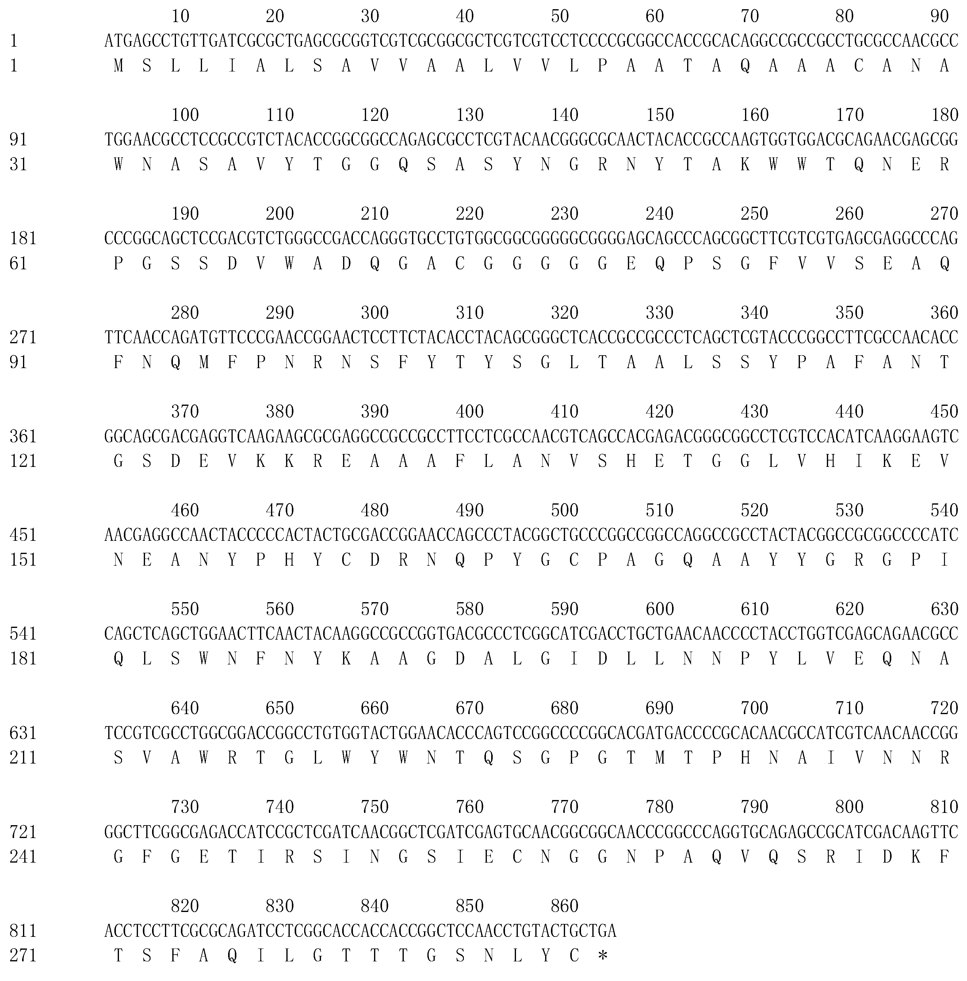
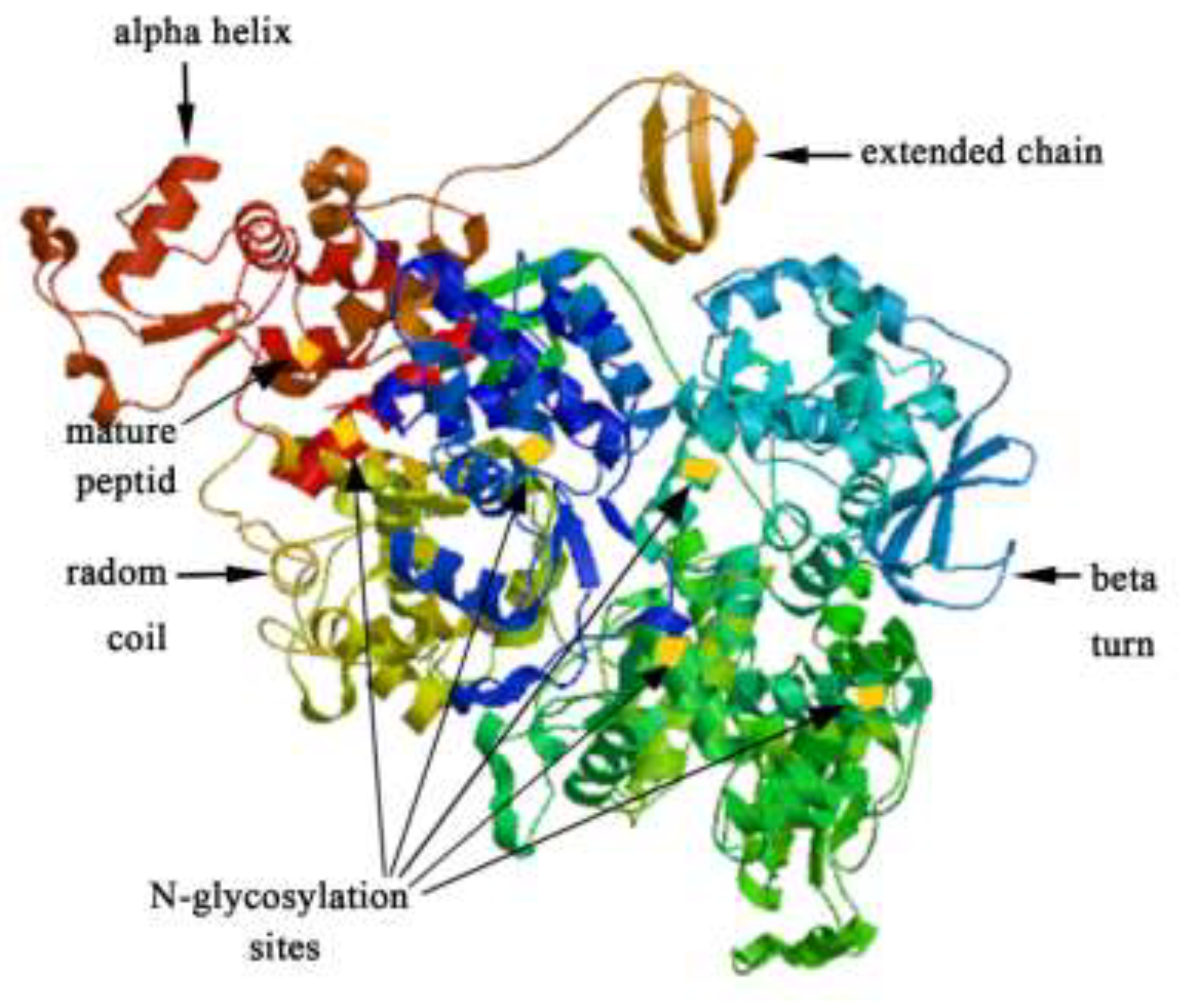
2.4. Gene Sequence and Chitinase Protein Identification

NCBI BLAST (Basic Local Alignment Search Tool) was used to generate a nucleotide sequence alignment, and the sequence and open reading frames (ORFs) were assemblied using DNAMAN software (Lynnon Biosoft, Quebec, QC, Canada). Then, the amino acid sequences were determined using BLAST and the theoretical molecular weight and isoelectric point (pl) of the protein was calculated with the ExPasy Compute pl/Mw and ProtParam tool. Later, for the prediction of the protein’s local hydrophobicity, transmembrane regions and signal peptide, ProtScale, TMHMM Server v. 2.0 and SignalP 4.1 Server (DTU Bioinformatics, Kemitorvet, Lyngby, Denmark) were applied, respectively. At end, NPS@SOPMA and SWISS-MODEL tools were used to predict secondary structure and tertiary structure of the protein.

2.5. Construction of the Chitinase Expression Vector

First, PCR with flanking restriction sites and without the signal peptide sequence from the cloning plasmid pMD19-T-ChiKJ406136-1 as template, was exploited to amplify chitinase gene ChiKJ406136. The forward primer PL (5′-CGCGGATCCGACACCCGCGCCGCCGCCG-3′) and reversed primer PR (5′-CCGCTCGAGTCAGCAGCTGAGGTTGTCG-3′) were used in the amplification and the resulting plasmid was named as pMD19-T-ChiKJ406136-2. Later, the positive recombinants were mixed with the pMD19-T-ChiKJ406136-2 plasmid and E. coli Trans5α. After that, a second PCR was exploited with pMD19-T-ChiKJ406136-2 plasmid as a template. The resulting amplicon and the pET32a (+) plasmid were double digested with two restriction enzymes BamHI and XhoI, after which the products were purified with a TIANgel Midi Purification Kit (Tiangen Biotech Co., Ltd., Beijing, China). Next, a 20 µL recombination reaction system, consisting of 6 µL of chitinase gene fragments, 2 µL of pET32a (+) enzyme-digested products, 2 µL of 10×T4 DNA Ligase Buffer, 9 µL of ddH2O and 1 µL of T4 DNA ligase, was generated, mixed with 100 µL of E. coli Trans5α competent cells, incubated on ice for 30 min and heat shocked at 42 °C for 90 s. The purified product was sequenced by Invitrogen (ThermoFisher Scientific Co., Ltd., Shanghai, China). After adding 900 µL of Gause’s No. 1 synthetic medium and incubating at 37 °C for 1 h, all transformed cells were inoculated onto Gause’s No. 1 synthetic medium containing ampicillin (100 µg/mL) and incubated at 37 °C for 12 h. Finally, the plasmids were extracted from the culture liquid and detected by double digestion with restriction enzymes BamHI and XhoI.

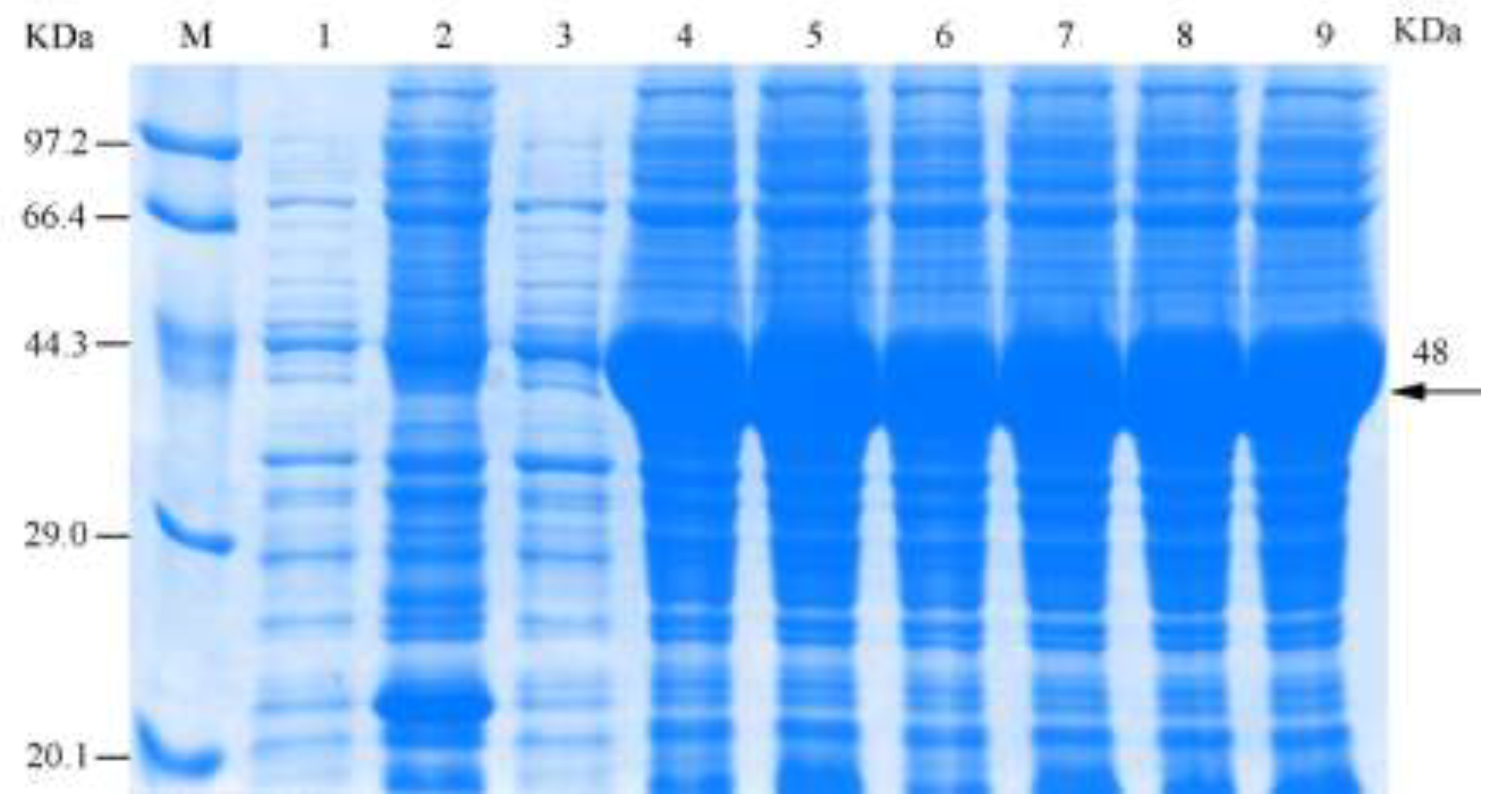
2.6. Induced Expression of the Recombinant Chitinase Gene ChiKJ406136

Recombinant expression plasmids were transformed into BL21 (DE3) competent cells, and a single colony was picked and inoculated into 10 mL of Gause’s No. 1 synthetic medium containing 100 µg/mL ampicillin, which was cultured in a shaker set at 200 rpm and at 37 °C for 12 h. Next, 300 µL of the culture liquid was mixed with 30 mL of Gause’s No. 1 synthetic medium containing 100 µg/mL ampicillin and cultured in a shaker set at 200 rpm and at 37 °C until the culture reached the logarithmic phase (OD 600 (optical density) = 0.6–0.8). Eighteen milliliters of the culture were then distributed into six sterile and dry cuvettes, after which isopropyl-b-D-thio-galactoside (IPTG, Merck) was added to the cultures at final concentrations of 0.2, 0.4, 0.6, 0.8, 1.0, and 1.2 mmol/L. The controls consisted of a strain harboring an empty vector that was induced or uninduced with IPTG (1 mmol/L). Approximately 1 mL of culture was withdrawn after 3 h of induction, which was then centrifuged at 12,000 rpm for 1 min, and the supernatant was discarded. The cell pellets were re-suspended in 1 mL of PBS (phosphate buffered saline) and centrifuged at 12,000 rpm for 1 min, followed by a second wash with PBS. Next, the cells were lysed, and the proteins were denatured in 20 µL of 5× denaturing buffer (60 mmol/L Tris–HCl, 25% glycerol, 2% sodium dodecyl sulfate (SDS), 0.1% bromophenol blue (Sigma-Aldrich Llc., Shanghai, China)) and 60 µL of PBS in a boiling water bath for 10 min. The samples were then centrifuged at 10,000 rpm for 10 min, and 4.5 µL of the supernatant was loaded onto a 12.5% SDS–polyacrylamide gel (SDS–PAGE) run at a constant voltages of (90 and 180 V) through stacking and separating gels, respectively, using a Bio-Rad Mini-PROTEIN Tetra Electrophoresis system (Bio-Rad Co., Ltd., Beijing, China).

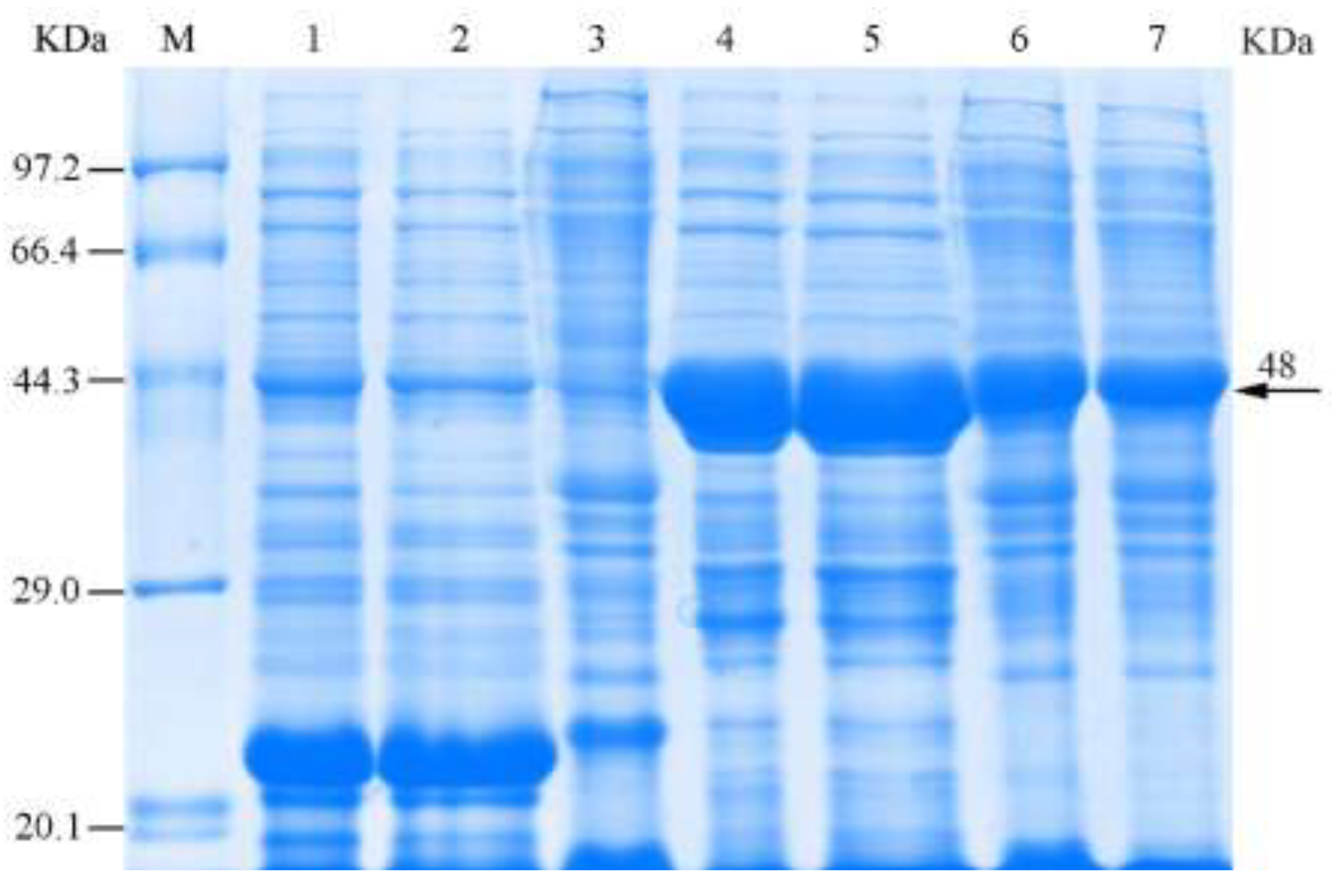
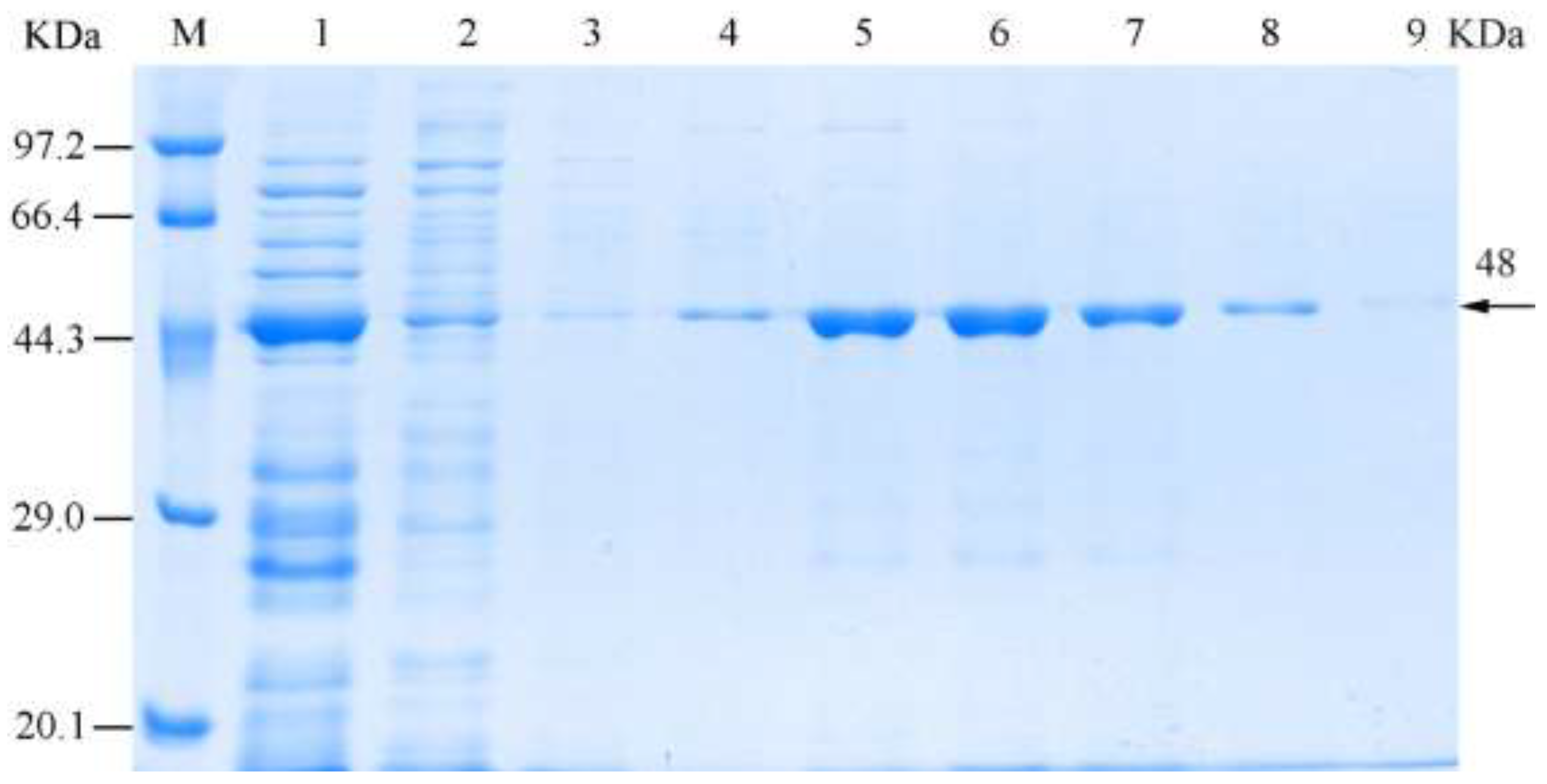
2.7. Dissolubility Determination and Purification of the Recombinant Chitinase

Approximately 1 mL of culture induced with IPTG (16 °C, overnight) was centrifuged at 12,000 rpm for 1 min. The cell pellet was gently re-suspended in 1 mL of PBS and then centrifuged at 12,000 rpm for 1 min, followed by a second wash with 60 µL of PBS. The final pellet was then lysed by 10 successive freeze-thaw cycles in liquid nitrogen and was subsequently centrifuged at 12,000 rpm for 5 min. The collected precipitates were re-suspended in 60 µL of 8 mol/L carbamide, and then was left to stand for 30 min. Next, the 60 µL treated precipitates and supernatant samples were mixed with 20 μL of 4× Protein SDS-PAGE loading buffer and incubated in a boiling water bath for 10 min. The samples were then centrifuged at 12,000 rpm for 10 min, and the supernatants were loaded onto a 12.5% SDS–polyacrylamide gel and run to determine the contents of expression products of the supernatants and precipitates, for confirming whether the expression protein existed in soluble form or in inclusion body form. The recombinant chitinase was purified using a One-Stop His-Tagged Protein Miniprep Pack (Tiandz, Inc., Beijing, China).

2.8. Determination of the Concentration and Activity of the Recombinant Chitinase

The concentrations of the crude extract and purified recombinant chitincase fractions were determined by using the Bradford Protein Quantitative Assay Kit (Solarbio, Inc., Beijing, China). Chitinase activities were quantitated by using 1% of colloidal chitin as substrate, the assay was processed as following steps: (1) add 6 g of chitin powder to 60 mL hydrochloric acid, stir for 24 h on a magnetic plate, then dilute with t distilled water to a volume of 1 L; (2) centrifuge for 10 min and collect white colloidal chitin precipitate; (3) wash and suspend the pellet repeat times till pH around 6.5 with 100 mL of distilled water; (4) suspend Pipette 10 mL of 6% colloidal chitin mother liquor to 50 mL distilled water, then add 1.2 g of agar, mix, sterilize pour into culture dishes, and cool to room temperature. The colloidal chitin mediums were drilled with a 5 mm diameter perforator, and 50 µL of the crude and purified recombinant chitinase were injected into the hole, respectively, the supernatant of cell lysate from a strain with pET32a (+) was as control. The activity was checked after five days of incubation at 30 °C. The quantitative estimation of chitinase activity was determined by using the Chitinase Kit (Nanjing Jiancheng Bioengineering Institute, Nanjing, China).

2.9. Effects of the Recombinant Chitinase on Pathogenic Fungi

Antifungal activity of recombinant chitinase was determined by a hyphal growth inhibition assay against four different pathogenic fungi (C. scoparium, C. parasitica, N. parvum, and F. oxysporum). The four fungi were cultured on potato dextrose agar (PDA) culture medium for several days until their mycelia covered the entire plate. Next, 10 µL of purified enzyme was aliquoted onto a glass slide, then a few mycelia were transferred from the plates with sterile tweezers and placed onto the enzyme solution for 20 h. The mycelial morphology was later observed every 2 h with an electron microscope.

2.10. Determination of Biological Control Effects in a Pot Experiment

Five mycelial sections (Φ = 8 mm) of each assayed pathogenic fungus (C. scoparium, C. parasitica, N. parvum, F. oxysporum) were obtained by the punch method and added to each bottle of PDA liquid medium (potato 200 g, glucose 20 g, agar 15–20 g, distilled water 1000 mL, pH 7.0); pathogen spores were at a concentration of 105/mL after 72 h.
Fifty healthy annual seedlings including E. robusta, C. mollissima, and one hundred annual J. regia seedlings, were treated as follows: (1) plants were inoculated by wound inoculation [49] with 10 mL of pathogenic fungal suspension, including the leaves of E. robusta, the twigs of C. mollissima and J. regia, and the roots of J. regia; (2) after 15 days, 10 mL of the undiluted recombinant chitinase solution and the 10-, 20-fold dilutions were sprayed onto the plants [50], with sterile water serving as the control; and (3) after 20 days, the disease symptoms were evaluated. Each treatment was repeated ten times. Disease was scored as follows: (I) No disease observed on the plant; (II) withering twigs/leaves/roots less than 25%; (III) withering twigs/leaves/roots 25–50%; (IV) withering twigs/leaves/roots 51–75%; and (V) withering twigs/leaves/roots more than 75%.
Statistical analyses: All data were subjected to one-way analysis of variance to determine the significance of individual differences at the p < 0.05 level. Significant means were compared using the least significant difference (LSD) test. All statistical analyses were conducted using the SPSS commercial statistical package (SPSS, Version 17.0 for Windows, SPSS Inc., Chicago, IL, USA).

3. Results

3.1. Cloning and Identification of the Chitinase Gene ChiKJ406136

The amplified chitinase gene PCR fragment was 864 bp and appeared as a single band (Figure 1). The cloning vector plasmids pMD19-T-ChiKJ406136-1 was double digested with the restriction enzymes BamHI and XhoI (Figure 2), resulting in the production of two clear bands of approximately 900 bp (target fragment: ChiKJ406136-1) and 2890 bp (vector plasmid pMD19-T) in size. This result demonstrated the successful cloning of the ChiKJ406136, which was submitted to NCBI under accession number MG323510.

3.2. Analysis of the ChiKJ406136 Gene Sequence and Protein Bioinformatics

The nucleotide sequence of the chitinase gene ChiKJ406136 was 99% similar to that of the sequence of the 864 bp chitinase genes from S. albus SM254 and Streptomyces sp. FR-008. The fragment contained a 864-bp open reading frame (ORF) encoding a 287 amino acid protein (Figure 3), which contained a conserved region in the glycoside hydrolases family 19 domain. The amino acid sequence of ChiKJ406136 was 100% identical to that of the chitinase genes from Streptomyces sp. SM8, Streptomyces wadayamensis, Streptomyces sp. FR-008, S. griseus subsp. griseus, Streptomyces sp. ScaeMP-6W, Streptomyces sp. IgraMP-1, and Streptomyces sp. BvitLS-983, as well as the chitinase A from S. albus.
The protein encoded by ChiKJ406136 was calculated to contain one signal peptide, with the cleavage site predicted to be located between amino acids 24 and 25, such that the mature peptide started at amino acid 25. In addition, the chitinase was predicted to contain five N-glycosylation sites (aa32, NASA; aa49, NYTA; aa136, NVSH; aa209, NASV; and aa250, NGSI). The secondary structure prediction of the protein revealed the following: the number of the alpha helices was 90 (31.36%); the number of extended chains was 66 (23%); the number of random coils was 100 (34.84%), the number of β-turns was 31 (10.80%). The results showed that the alpha helix and random coil features were dominant in the protein. The tertiary structure prediction results of the chitinase are shown in Figure 4.

3.3. Induction of Recombinant Chitinase Expression

The recombinant expression plasmid pMD19-T-ChiKJ406136-2 was digested with restriction enzymes BamHI and XhoI (Figure 5), yielding one 800-bp band (target fragment: ChiKJ406136-2) that was identical to the predicted size of the SSKJ-6136 gene with the signal peptide removed, demonstrating the successful construction of the recombinant expression vector. SDS-PAGE analysis (Figure 6) showed that the expression of ChiKJ406136 in E. coli BL21 (DE3) cells containing pET32a (+)-ChiKJ406136-2 was robust and increased with the time of induction. In contrast, cells cultured in a similar manner and harboring the empty vector or pET32a (+)-ChiKJ406136-2 without induction did not overexpress any particular protein, while cells harboring the empty vector that were induced with 1 mmol/L IPTG (isopropyl-b-D-thio-galactoside) expressed a 20.4 kilodalton (kDa) protein. The induction of pET32a (+)-ChiKJ406136 expression in the presence of all different concentrations of IPTG showed the presence of a 48 kDa protein band, where the molecular weight included the tagged protein and the removed signal peptide. This result also demonstrated that the concentration of IPTG had no effect on the molecular weight of the resulting recombinant protein.

3.4. Solubility and Purification of the Recombinant Chitinase

After an overnight induction at 16 °C, the recombinant protein was primarily insoluble, as the eluted fractions contained precipitated in the form of inclusion bodies (Figure 7). Imidazole concentrations of 10, 50, 100, 150, 200, 250, and 300 mmol/L were used to elute the recombinant protein from the column, resulting in protein being eluted at all concentrations, with the highest amount eluted using 150 mmol/L imidazole (Figure 8).

3.5. Properties of the Recombinant Chitinase

Both the crude and purified proteins exhibited chitin hydeolysis activity (Figure 9), the diameters of chitin hydrolysis circles of crude and purified proteins were 1.6 cm and 1.8 cm, respectively. After being diluted 8-fold, the OD (optical density) values of the crude and purified recombinant proteins were 0.732 and 0.02, and had concentrations of 2.06 and 0.07 mg/mL, respectively (Table 1). In addition, the crude and purified proteins exhibited chitinase activities of 0.045 and 0.033 U/mL and the specific activities of 0.022 and 0.471 U/g, respectively. The recovery rates of the enzyme activities were 100% and 73.33% for the crude and purified recombinant chitinase fractions, respectively.

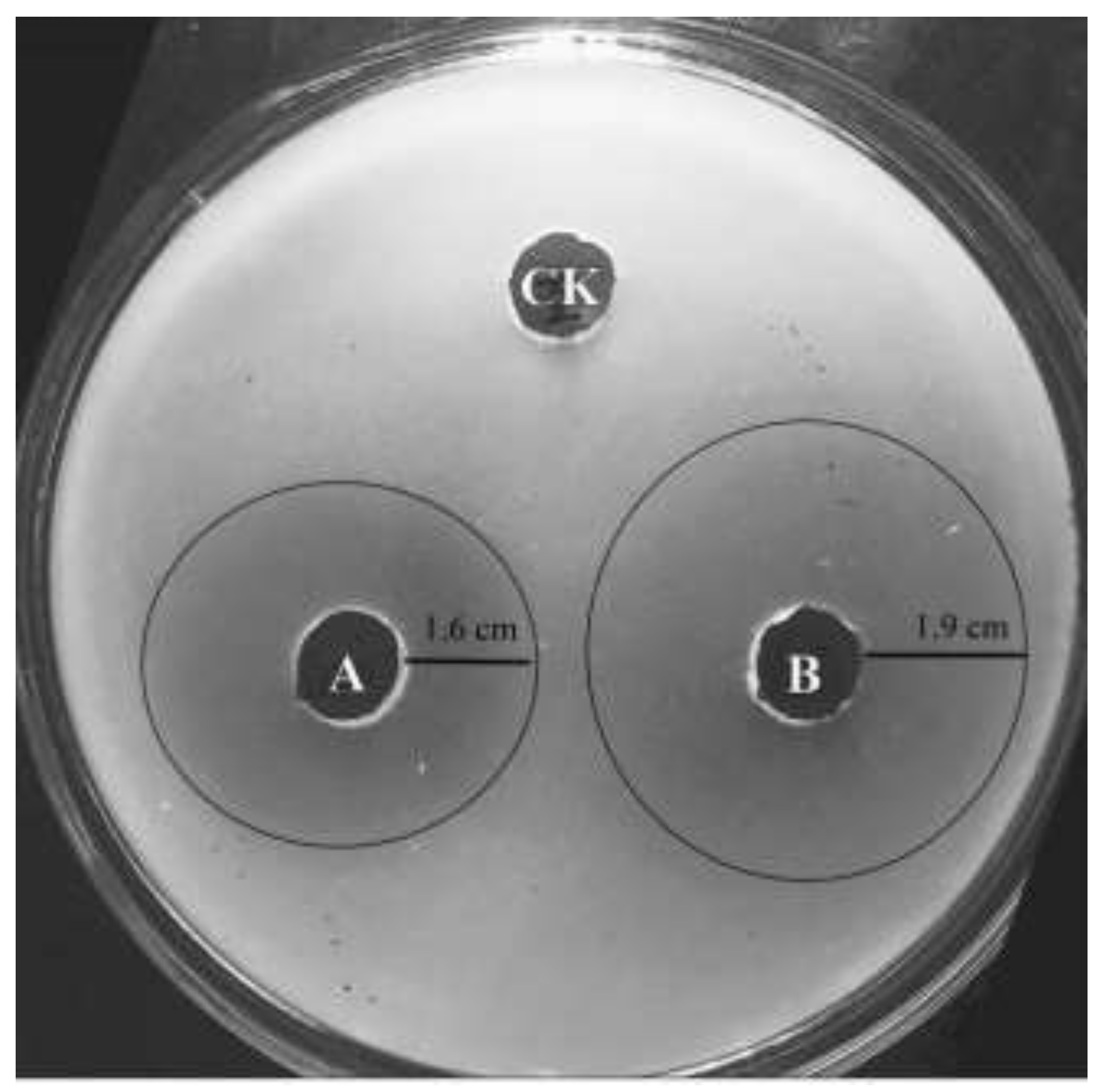
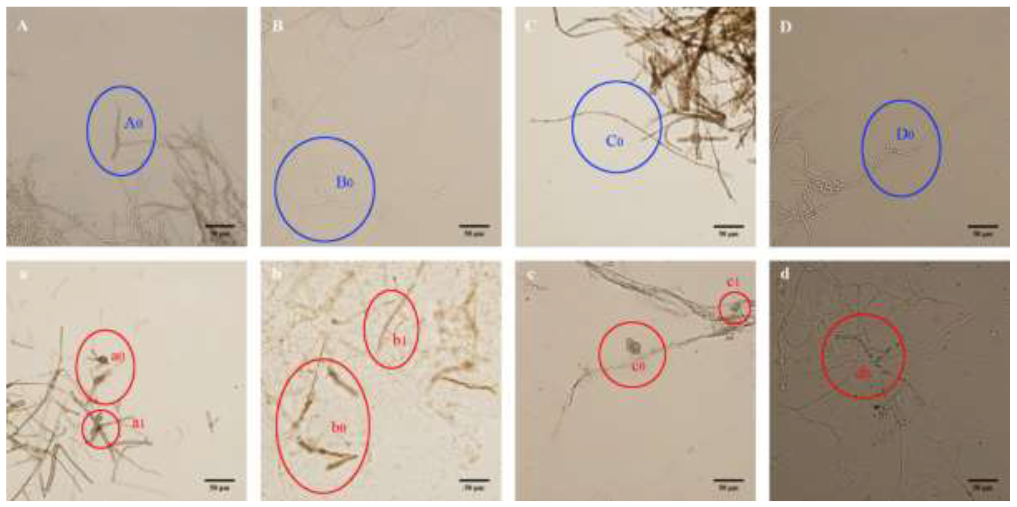
3.6. Effect of the Recombinant Chitinase on Pathogenic Fungi

After being treated with the recombinant chitinase for 2 h, the mycelial morphology of four pathogenic fungi (C. scoparium, C. parasitica, N. parvum and F. oxysporum) that are known to be affected by S. sampsonii KJ40 was observed. Interestingly, the mycelial morphology of all four fungi was altered by the chitinase treatment (Figure 10), as the mycelia were mixed and partly broken down into chunks.Specifically, the merogenesis of the cells increased, inflating the mycelia of N. parvum (Figure 10c), and the mycelia of F. oxysporum were broken and distorted (Figure 10d).

3.7. Biological Control Effects of Chitinase on Potted Plants

The biological control effects of the recombinant chitinase over the course of 20 days in pot experiments are shown in Table 2. The incidences of four diseases were as high as 85% in the control plants, while the recombinant chitinase solutions had different control effects at different dilutions. The biological control effects decreased at higher chitinase dilutions, with the undiluted solution exhibiting the best effect. The control effects of the 20-fold dilution was significantly reduced (below 55%).

4. Discussion

Chitinases have been reported to improve the disease resistance and have been used as insect and biological fungicides and pesticides in agroforestry. Historically, studies have focused on using chitinase-producing microorganisms to directly control plant fungal diseases, which is based on the ability of these microorganisms to decompose the chitin that is present in the fungal cell walls [51]. Baek et al. [42] and Kerrn et al. [43] demonstrated that Trichoderma virens Pers. Gv29-8 and Metarhizium anisopliae (Metchnikoff) Sorokin can produce chitinase, respectively, and that they could inhibit the mycelial growth of Rhizoctonia solani, Alternaria alternata, Fusarium oxysporum, Penicillium aculeatum Peyronel, Trichoderma harzianum Rifai, Rhizopus sp. Ehrenb. and Botrytis cinerea Pers. Although chitinase have great potential for use in a number of applications, the activity observed for these enzymes has been low. For the commercial production of chitinase, previous studies focused that produced high levels of chitinase or on increasing the enzyme production by changing fermentation conditions [52,53,54]. Nevertheless, due to complex regulatory mechanisms, fermentation separation and immature extraction processes, the successful industrial production of chitinase is rare. The development of molecular biology techniques provides a new way to solve this problem, by cloning a chitinase gene into an appropriate vector for efficient expression to meet the needs of large-scale production and research and applications.
The protein encoded by the S. sampsonii ChiKJ406136 gene has a domain belonging to the glycoside hydrolase family 19, and Streptomyces enzymes belongs to this chitinase family [55]. Family 19 chitinases were initially identified in higher plants, while only family 18 chitinases were observed in microorganisms. In a study by Ohno et al. [56], family 19 chitinase-encoding genes were identified from Streptomyces griseus (Krainsky) Waksman & Henrici HUT6037, outside higher plants, and family 19 members were also found in other actinomycetes [57,58,59,60]. Nevertheless, Family 19 chitinases are not frequently found in bacteria and never in S. sampsonii. The tertiary structure of chitinase family 19 is similar to that of lysozyme. The molecular weight of the chitinase from different biological sources varies greatly, from 20 to 90 kDa, with bacterial chitinases varying in size from 20 to 60 kDa, similar to plant chitinases (25–40 kDa) and smaller than insect chitinases (40 to 85 kDa) [61]. Similarly, the molecular weight of the ChiKJ406136-encoded protein expressed in E. coil was 30.6 kDa, and the weight of protein after removing the signal peptide was 28.3 kDa in our study. However, from the SDS-PAGE analysis of protein bands, the expressed protein bands were observed to be close to 48 kDa. The primary reason for this result is that the total molecular weight of the protein expressed in the pET-32a (+) vector is 20.4 kDa, which contains Trx-tags size of 12 kDa. Taking into account the His-tags (0.8 kDa) and S-tag (1.7 kDa), the remaining 5.1 kDa were derived from 54 amino acids between these tags and the termination codon.
The advantages of using an E. coli expression system are its well-characterized genetic background, fast reproduction rate, low cost, and high level of expression; in addition, there are numerous expression vectors, hosts and purification systems [62]. Exogenetic proteins are easily degraded by the host proteases or form inclusion bodies when expressed at high levels. Currently, many studies have investigated protein re-naturation in vitro, but the process of protein re-naturation is often time-consuming. Therefore, exploring the soluble expression of exogenous proteins in E. coli has potentially, wide applications [63]. By lowering the culture temperature, the protein synthesis rate and the concentration of polymeric intermediates are reduced, avoiding the formation of inclusion bodies [64]. The ChiKJ406136 gene was highly expressed at 16 °C from the pET-32a vector. The recombinant protein primarily existed in the supernatant in the soluble form. We easily cloned the chitinase-encoding gene into the histidine tag-containing pET expression vector and identified the expressed protein. In addition, the recombinant ChiKJ406136-encoded protein was eluted with different concentrations of an imidazole solution. The elution efficiency of the recombinant ChiKJ406136-encoded protein was high, and the recovery rate of enzyme activity was as high as 73.33%. The specific activity of the purified ChiKJ406136 protein was 0.471 U/g, higher than the reported in a study by García-Fraga et al. [65], where the activity of the purified recombinant Ptchi19p-encoded protein from Pseudoalteromonas tunicata Holmström was 0.228 U/g.
Similar to the previous studies of antifungal activity, for the four assayed fungi used in our study, we observed complete destruction of the mycelial morphology when all four pathogenic fungi were treated with the purified chitinase from S. sampsonii KJ40. Such a breakdown is likely due to the hydrolysis of chitin that exists in the fungal cell walls. Chi18H8, which encodes a chitinase from Bacillus thuringiensis Berliner BUPM255, was cloned and expressed by Hjort et al. [41]. The purified enzyme could inhibit the mycelial growth of Colletotrichum gloeosporioideschus (Stoneman) Spauld. & H. Schrenk, F. graminearum, F. oxysporum and A. alternata. The recombinant protein encoded by Ptchi19p from P. tunicate could also inhibit the mycelial growth of F. oxysporum and Aspergillus niger [65]. Through in vitro testing, Reyes-Ramírez et al. [66] demonstrated that a Bacillus thuringiensis chitinase had biocontrol potential against Sclerotium rolfsii (Curzi) C.C. Tu & Kimbr., Aspergillus terreus Thom, Aspergillus Flavus Link, Nigrospora sp. Mason with 25–82% inhibitory effects. Kirubakaran and Sakthivel [67] reported that a 35 kDa chitinase had significant effects on B. cinerea and F. oxysporum using a concentration of 1.2 U/g. Lee et al. [68] showed that a Bacillus licheniformis Carl. chitinase could inhibit 50% of the hyphal growth of A. terreus. In addition, some studies demonstrated the antifungal effect in pot experiments in greenhouses. For example, Viterbo et al. [69] reported that a 36 kDa endochitinase from T. harrzianum had some efficacy, but it did not reach significance for the disease caused by B. cinerea and R. solani. An antifungal chitinase of approximately 30kDa was isolated from Sorghum bicolor (L.) Moench using chromatographic techniques and showed a broad-spectrum antifungal activity toward devastating fungal pathogens that attack rice, tobacco, tea and clover at concentration of 18–36 µg/mL [70]. Those studies have been seen that the chitinases from either plants or bacteria display antifungal activity, which might make them a great alternative to the use of chemical products for the biological control of pests [59,61,71]. But as each enzyme may present its own characteristics that make it useful for a specific function, the search for new chitinases from different organisms is of interest. Hence, compared to the above results, the results of our study revealed an excellent ability to control some local plant diseases using lower concentrations of the purified chitinase. Interestingly, the effects of purified chitinases were better than S. sampsonii KJ40 using in disease control caused by F. oxysporum with the incidence, disease index and control effect of 60.67%, 52.88%, 21.25%, respectively [72]. Specifically, the ability of the chitinase to control plant diseases, including E. robusta leaf blight, C. mollissima blight, J. regia blight and J. regia root rot, were first reported by us.

5. Conclusions

In conclusion, we showed that a chitinase from S. sampsonii can be expressed and purified in high amounts with ease. The purified enzyme exhibits antifungal activity against plant pathogenic fungi and inhibits the diseases these fungi cause. As studies of the chitinase and the encoding gene from S. sampsonii were previously lacking, our results can improve and supply a biocontrol protein, providing a new resource for chitinase genes. Furthermore, our results may have potential application for the biocontrol of other phytopathogenic fungi and transgenic resistance breeding in plants, which may be used for commercial purposes. However, the N-terminal amino acid sequence and the mechanism of S. sampsonii chitinase action require further investigation.

Author Contributions

Conceptualization, S.L.; Methodology, B.Z., T.Z; Validation, S.L., B.Z., H.Z., T.Z.; Formal Analysis, B.Z.; Investigation, H.Z.; Resources, T.Z.; Data Curation, S.L., T.Z.; Writing-Original Draft Preparation, S.L., B.Z.; Writing-Review & Editing, H.Z., T.Z.; Visualization, B.Z., H.Z..; Supervision, T.Z.; Project Administration, S.L.; Funding Acquisition, S.L.

Funding

This research was funded by [National Natural Science Foundation of China] grant number [31700568] and [China Postdoctoral Science Foundation] grant number [2016M602705].

Acknowledgments

S.L. and T.Z. conceived and designed the experiments; B.Z. performed the experiments; S.L and H.Z. analyzed the data; T.Z. contributed reagents/materials/analysis tools; S.L. wrote the paper.

Conflicts of Interest

The authors declare no conflict of interest.

References

  1. Flärdh, K.; Buttner, M.J. Streptomyces morphogenetics: Dissecting differentiation in a filamentous bacterium. Nat. Rev. Microbiol. 2009, 7, 36–49. [Google Scholar] [CrossRef] [PubMed]
  2. Hasani, A.; Kariminik, A.; Issazadeh, K. Streptomycetes: Characteristics and their antimicrobial activities. Int. J. Adv. Biol. Biomed. Res. 2014, 2, 63–75. [Google Scholar]
  3. Law, J.W.F.; Ser, H.L.; Khan, T.M.; Chuah, L.H.; Pusparajah, P.; Chan, K.G.; Goh, B.H.; Lee, L.H. The potential of Streptomyces as biocontrol agents against the rice blast fungus, Magnaporthe oryzae (Pyricularia oryzae). Front. Microbiol. 2017, 8, 3. [Google Scholar] [CrossRef] [PubMed]
  4. Schumacher, R.W.; Talmage, S.C.; Miller, S.A.; Sarris, K.E.; Davidson, B.S.; Goldberg, A. Isolation and structure determination of an antimicrobial ester from a marine sediment-derived bacterium. J. Nat. Prod. 2003, 66, 1291–1293. [Google Scholar] [CrossRef] [PubMed]
  5. Ramesh, S.; Mathivanan, N. Screening of marine actinomycetes isolated from the Bay of Bengal, India for antimicrobial activity and industrial enzymes. World J. Microbiol. Biotechnol. 2009, 25, 2103–2111. [Google Scholar] [CrossRef]
  6. De Lima Procópio, R.E.; da Silva, I.R.; Martins, M.K.; de Azevedo, J.L.; de Araújo, J.M. Antibiotics produced by Streptomyces. Braz. J. Infect. Dis. 2012, 16, 466–471. [Google Scholar] [CrossRef] [PubMed]
  7. Kumar, P.S.; Duraipandiyan, V.; Ignacimuthu, S. Isolation, screening and partial purification of antimicrobial antibiotics from soil Streptomyces sp. SCA 7. Kaohsiung J. Med. Sci. 2014, 30, 435–446. [Google Scholar] [CrossRef] [PubMed]
  8. Lee, L.H.; Zainal, N.; Azman, A.S.; Eng, S.K.; Ab Mutalib, N.S.; Yin, W.F.; Chan, K.G. Streptomyces pluripotens sp. nov., a bacteriocin-producing streptomycete that inhibits meticillin-resistant Staphylococcus aureus. Int. J. Syst. Evol. Microbiol. 2014, 64, 3297–3306. [Google Scholar] [CrossRef] [PubMed]
  9. Ser, H.L.; Law, J.W.F.; Chaiyakunapruk, N.; Jacob, S.A.; Palanisamy, U.D.; Chan, K.G.; Goh, B.H.; Lee, L.H. Fermentation conditions that affect clavulanic acid production in Streptomyces clavuligerus: A systematic review. Front. Microbiol. 2016, 7, 522. [Google Scholar] [CrossRef] [PubMed]
  10. Lam, K.S. Discovery of novel metabolites from marine actinomycetes. Curr. Opin. Microbiol. 2006, 9, 245–251. [Google Scholar] [CrossRef] [PubMed]
  11. Ara, I.; Bukhari, N.A.; Aref, N.; Shinwari, M.M.; Bakir, M. Antiviral activities of streptomycetes against tobacco mosaic virus (TMV) in Datura plant: Evaluation of different organic compounds in their metabolites. Afr. J. Biotechnol. 2012, 11, 2130–2138. [Google Scholar] [CrossRef]
  12. Kino, T.; Hatanaka, H.; Hashimoto, M.; Nishiyama, M.; Goto, T.; Okuhara, M.; Kohsaka, M.; Aoki, H. FK-506, a novel immunosuppressant isolated from a Streptomyces. fermentation, isolation, and physico-chemical and biological characteristics. J. Antibiot. 1987, 40, 1249–1255. [Google Scholar] [CrossRef] [PubMed]
  13. Ser, H.L.; Ab Mutalib, N.S.; Yin, W.F.; Chan, K.G.; Goh, B.H.; Lee, L.H. Evaluation of antioxidative and cytotoxic activities of Streptomyces pluripotens MUSC 137 isolated from mangrove soil in Malaysia. Front. Microbiol. 2015, 6, 1398. [Google Scholar] [CrossRef] [PubMed]
  14. Tan, L.T.H.; Ser, H.L.; Yin, W.F.; Chan, K.G.; Lee, L.H.; Goh, B.H. Investigation of antioxidative and anticancer potentials of Streptomyces sp. MUM256 isolated from Malaysia mangrove soil. Front. Microbiol. 2015, 6, 1316. [Google Scholar] [CrossRef] [PubMed]
  15. Tan, L.T.H.; Chan, K.G.; Lee, L.H.; Goh, B.H. Streptomyces bacteria as potential probiotics in aquaculture. Front. Microbiol. 2016, 7, 79. [Google Scholar] [CrossRef] [PubMed]
  16. Pullen, C.; Schmit, P.; Meurer, K.; Bamberg, D.D.V.; Lohmann, S.; França, S.C.; Groth, I.; Schlegel, B.; Mőllmann, U.; Gollmick, F.; et al. New and bioactive compounds from Streptomyces strains residing in the wood of Celastraceae. Planta 2002, 216, 162–167. [Google Scholar] [CrossRef] [PubMed]
  17. Imamura, N.; Nishijima, M.; Adachi, K.; Sano, H. Novel antimycin antibiotics, urauchimycins A. and B., produced by marine actinomycete. J. Antibiot. 1993, 46, 241–246. [Google Scholar] [CrossRef] [PubMed]
  18. Jain, P.K.; Jain, P.C. Isolation, characterization and antifungal activity of Streptomyces sampsonii GS 1322. Indian J. Exp. Biol. 2007, 45, 203–206. [Google Scholar] [PubMed]
  19. Malviya, M.K.; Pandey, A.; Trivedi, P.; Gupta, G.; Kumar, B. Chitinolytic activity of cold tolerant antagonistic species of Streptomyces isolated from glacial sites of Indian Himalaya. Curr Microbiol. 2009, 59, 502–508. [Google Scholar] [CrossRef] [PubMed]
  20. Li, S.J.; Zhu, T.H.; Peng, Y.; Lei, M.Y.; Han, S. Characteristics of chitinase-produced by Streptomycete sampsonii with antimicrobial activity and its biocontrol to Rhizoctonia violacea. J. Northeast. For. Univ. 2014, 42, 116–121. [Google Scholar]
  21. Charousová, I.; Medo, J.; Halenárová, E.; Maková, J.; Javoreková, S. Effect of fertilization on biological activity of community of soil streptomycetes. J. Cent. Eur. Agr. 2016, 17, 1134–1139. [Google Scholar] [CrossRef]
  22. Jain, R.; Jain, A.; Rawat, N.; Nair, M.; Gumashta, R. Feather hydrolysate from Streptomyces sampsonii GS 1322: A potential low cost soil amendment. J. Biosci. Bioeng. 2016, 121, 672–677. [Google Scholar] [CrossRef] [PubMed]
  23. Deepthi, A.R.P. Actinomycete isolates from Arabian Sea and Bay of Bengal: Biochemical, Molecular and Functional Characterization. Ph.D. Thesis, Cochin University of Science and Technology, Kochi, Kerala, India, May 2014. [Google Scholar]
  24. Li, Y.D.; Wang, K.Y.; Fan, S.; Yang, Z.Y.; Tian, X.F.; Xu, L.X.; Jin, Y.Y. Isolation of strain producing D-hydantoinase from marine and its simulation analysis of the enzyme catalytic chanel. J. Arg. Biot. 2016, 24, 729–737. [Google Scholar] [CrossRef]
  25. Savi, D.C.; Shaaban, K.A.; Vargas, N.; Ponomarena, L.V.; Possiede, Y.M.; Thorson, J.S.; Glienke, C.; Rohr, J. Microbispora sp. LGMB259 endophytic actinomycete isolated from Vochysia divergens (Pantanal, Brazil) producing β-carbolines and indoles with biological activity. Curr. Microbiol. 2015, 70, 345–354. [Google Scholar] [CrossRef] [PubMed]
  26. Kim, S.S.; Kang, S.I.; Kim, J.S.; Lee, Y.S.; Hong, S.H.; Naing, K.W.; Kim, K.Y. Biological control of root-knot nematode by Streptomyces sampsonii KK1024. Korean J. Soil. Sci. Fert. 2011, 44, 1150–1157. [Google Scholar] [CrossRef]
  27. Du, H.; Hu, L.; Xu, Y. Influence of geosmin-producing Streptomyces on the growth and volatile metabolites of yeasts during Chinese liquor fermentation. J. Agric. Food Chem. 2015, 63, 290–296. [Google Scholar] [CrossRef] [PubMed]
  28. Jin, F.; Ding, Y.Q.; Ding, W.; Reddy, M.S.; Fernando, W.G.D.; Du, B.H. Genetic diversity and phylogeny of antagonistic bacteria against Phytophthora nicotianae isolated from tobacco rhizosphere. Int. J. Mol. Sci. 2011, 12, 3055–3071. [Google Scholar] [CrossRef] [PubMed]
  29. Dalisay, D.S.; Williams, D.E.; Wang, X.L.; Centko, R.; Chen, J.; Andersen, R.J. Marine sediment-derived Streptomyces bacteria from British Columbia, Canada are a promising microbiota resource for the discovery of antimicrobial natural products. PLoS ONE 2013, 8, e77078. [Google Scholar] [CrossRef] [PubMed]
  30. Pesic, A.; Baumann, H.I.; Kleinschmidt, K.; Ensle, P.; Wiese, J.; Süssmuth, R.D.; Imhoff, J.F. Champacyclin, a new cyclic octapeptide from Streptomyces strain C42 isolated from the Baltic sea. Mar. Drugs 2013, 11, 4834–4857. [Google Scholar] [CrossRef] [PubMed] [Green Version]
  31. El Baz, S.; Baz, M.; Barakate, M.; Hassani, L.; El Gharmali, A.; Imziln, B. Resistance to and accumulation of heavy metals by actinobacteria isolated from abandoned mining areas. Sci. World J. 2015, 1–14. [Google Scholar] [CrossRef] [PubMed]
  32. Lobova, T.I.; Yemelyanova, E.; Andreeva, I.S.; Puchkova, L.I.; Repin, V.Y. Antimicrobial resistance and plasmid profile of bacterial strains isolated from the urbanized eltsovka-1 river (Russia). Microb. Drug Resist. 2015, 21, 477–490. [Google Scholar] [CrossRef] [PubMed]
  33. Rasimus-Sahari, S.; Mikkola, R.; Andersson, M.A.; Jestoi, M.N.; Salkinoja-Salonen, M. Streptomyces strains producing mitochondriotoxic antimycin A found in cereal grains. Int. J. Food Microbiol. 2016, 218, 78–85. [Google Scholar] [CrossRef] [PubMed]
  34. Santos, M.C.D.L.; Taulé, C.; Mareque, C.; Beracochea, M.; Battistoni, F. Identification and characterization of the part of the bacterial community associated with field-grown tall fescue (Festuca arundinacea) cv. SFRO Don Tomás in Uruguay. Ann. Microbiol. 2016, 66, 329–342. [Google Scholar] [CrossRef]
  35. Xu, L.; Liang, K.; Duan, B.; Yu, M.D.; Meng, W.; Wang, Q.G.; Yu, Q. A novel insecticidal peptide SLP1 produced by Streptomyces laindensis H008 against Lipaphis erysimi. Molecules 2016, 21, 1101. [Google Scholar] [CrossRef] [PubMed]
  36. Zhang, B.Y.; Zhu, T.H.; Han, S.; Wang, Y.; Li, S.J.; Qiao, T.M. Whole-genome sequencing and analysis of Streptomyces sampsonii KJ40. Microbiol. China 2018, 45, 805–818. [Google Scholar] [CrossRef]
  37. Kurita, K. Controlled functionalization of the polysaccharide chitin. Prog. Polym. Sci. 2001, 26, 1921–1971. [Google Scholar] [CrossRef]
  38. Purwani, E.Y.; Suhartono, M.T.; Rukayadi, Y.; Hwang, J.K.; Pyun, Y.R. Characteristics of thermostable chitinase enzymes from the indonesian Bacillus sp. 13.26. Enzym. Microb. Tech. 2004, 35, 147–153. [Google Scholar] [CrossRef]
  39. Mathivanan, N.; Kabilan, V.; Murugesan, K. Purification, characterization, and antifungal activity of chitinase from Fusarium chlamydosporum, a mycoparasite to groundnut rust, Puccinia arachidis. Can. J. Microbiol. 1998, 44, 646–651. [Google Scholar] [CrossRef] [PubMed]
  40. Li, J.G.; Jiang, Z.Q.; Xu, L.P.; Sun, F.F.; Guo, J.H. Characterization of chitinase secreted by Bacillus cereus strain CH2 and evaluation of its efficacy against Verticillium wilt of eggplant. BioControl 2008, 53, 931–944. [Google Scholar] [CrossRef]
  41. Hjort, K.; Presti, I.; Elväng, A.; Marinelli, F.; Sjőling, S. Bacterial chitinase with phytopathogen control capacity from suppressive soil revealed by functional metagenomics. Appl. Microbiol. Biotechnol. 2014, 98, 2819–2828. [Google Scholar] [CrossRef] [PubMed]
  42. Baek, J.M.; Howell, C.R.; Kenerley, C.M. The role of an extracellular chitinase from Trichoderma virens Gv29-8 in the biocontrol of Rhizoctonia solani. Curr. Genet. 1999, 35, 41–50. [Google Scholar] [CrossRef] [PubMed]
  43. Kerrn, M.F.; Maraschin, S.D.F.; Endt, D.V.; Schrank, A. Expression of a chitinase gene from Metarhizium anisopliae in tobacco plants confers resistance against Rhizoctonia solani. Appl. Biochem. Biotechnol. 2010, 160, 1933–1946. [Google Scholar] [CrossRef] [PubMed]
  44. Gadelhak, G.G.; El-Tarabily, K.A.; Al-Kaabi, F.K. Insect control using chitinolytic soil actinomycetes as bio control agents. Int. J. Agric. Biol. 2005, 7, 627–633. [Google Scholar]
  45. Prasad, K.; Bhatnagar-mathur, P.; Waliyar, F.; Sharma, K.K. Overexpression of a chitinase gene in transgenic peanut confers enhanced resistance to major soil borne and foliar fungal pathogens. J. Plant Biochem. Biot. 2013, 22, 222–233. [Google Scholar] [CrossRef] [Green Version]
  46. Gentile, A.; Deng, Z.; La, M.S.; Distefano, G.; Vitale, A.; Polizzi, G.; Lorito, M. Enhanced resistance to Phoma tracheiphila and Botrytis cinerea in transgenic lemon plants expressing a Trichoderma harzianum chitinase gene. Plant Breed. 2007, 126, 146–151. [Google Scholar] [CrossRef]
  47. Shin, S.; Mackintosh, C.A.; Lewis, J.; Heinen, S.J.; Radmer, L.; Dill-Macky, R.; Baldridge, G.D.; Zeyen, R.J.; Muehlbauer, G.J. Transgenic wheat expressing a barley class II chitinase gene has enhanced resistance against Fusarium graminearum. J. Exp. Bot. 2008, 59, 2371–2378. [Google Scholar] [CrossRef] [PubMed]
  48. Owen, W.; Jayaraman, J.; Zamir, P. Comparative resistance to foliar fungal pathogens in transgenic carrot plants expressing genes encoding for chitinase, β-1,3-glucanase and peroxidise. Eur. J. Plant Pathol. 2009, 123, 331–342. [Google Scholar] [CrossRef]
  49. Ho, S.H.; Koh, L.; Ma, Y.; Huang, Y.; Sim, K.Y. The oil of garlic, Allium sativum L. (Amaryllidaceae), as a potential grain protectant against Tribolium castaneum (Herbst) and Sitophilus zeamais Motsch. Postharvest Biol. Tec. 1996, 9, 41–48. [Google Scholar] [CrossRef]
  50. Sørensen, C.K.; Thach, T.; Hovmøller, M.S. Evaluation of spray and point inoculation methods for the phenotyping of Puccinia striiformis on wheat. Plant Dis. 2016, 100, 1064–1070. [Google Scholar] [CrossRef]
  51. Adams, J. The proteasome: A suitable antineoplastic target. Nat. Rev. Cancer 2004, 4, 349–360. [Google Scholar] [CrossRef] [PubMed]
  52. Saito, A.; Fujii, T.; Shinya, T.; Shibuya, N.; Ando, A.; Miyashita, K. The msiK gene, encoding the ATP-hydrolysing component of N, N’-diacetylchitobiose ABC transporters, is essential for induction of chitinase production in Streptomyces coelicolor A3(2). Microbiology 2008, 154, 3358–3365. [Google Scholar] [CrossRef] [PubMed]
  53. Hirano, T.; Kadokura, K.; Ikegami, T.; Shigeta, Y.; Kumaki, Y.; Hakamata, W.; Oku, T.; Nishio, T. Heterodisaccharide 4-O-(N-acetyl-β-D-glucosaminyl)-D-glucosamine is a specific inducer of chitinolytic enzyme production in Vibrios harboring chitin oligosaccharide deacetylase genes. Glycobiology 2009, 19, 1046–1053. [Google Scholar] [CrossRef] [PubMed]
  54. Nothaft, H.; Rigali, S.; Boomsma, B.; Swiatek, M.; McDowall, K.J.; van Wezel, G.P.; Titgemeyer, F. The permease gene nagE2 is the key to N-acetylglucosamine sensing and utilization in Streptomyces coelicolor and is subject to multi-level control. Mol. Microbiol. 2010, 75, 1133–1144. [Google Scholar] [CrossRef] [PubMed]
  55. Hamid, R.; Khan, M.A.; Ahmad, M.; Ahmad, M.M.; Abdin, M.Z.; Musarrat, J.; Javed, S. Chitinases: An update. J. Pharm. Bioallied. Sci. 2013, 5, 21–29. [Google Scholar] [CrossRef] [PubMed]
  56. Ohno, T.; Armand, S.; Hata, T.; Nikaidou, N.; Henrissat, B.; Mitsutomi, M.; Watanabe, T. A modular family 19 chitinase found in the prokaryotic organism Streptomyces griseus HUT 6037. J. Bacteriol. 1996, 178, 5065–5070. [Google Scholar] [CrossRef] [PubMed]
  57. Watanabe, T.; Kanai, R.; Kawase, T.; Tanabe, T.; Mitsutomi, M.; Sakuda, S.; Miyashita, K. Family 19 chitinases of Streptomyces species: Characterization and distribution. Microbiology 1999, 145, 3353–3363. [Google Scholar] [CrossRef] [PubMed]
  58. Kawase, T.; Saito, A.; Sato, T.; Kanai, R.; Fujii, T.; Nikaidou, N.; Miyashita, K.; Watanabe, T. Distribution and phylogenetic analysis of family 19 chitinases in Actinobacteria. Appl. Environ. Microbiol. 2004, 70, 1135–1144. [Google Scholar] [CrossRef] [PubMed]
  59. Hoell, I.A.; Dalhus, B.; Heggset, E.B.; Aspmo, S.I.; Eijsink, V.G.H. Crystal structure and enzymatic properties of a bacterial family 19 chitinase reveal differences from plant enzymes. FEBS J. 2006, 273, 4889–4900. [Google Scholar] [CrossRef] [PubMed]
  60. Huang, L.; Garbulewska, E.; Sato, K.; Kato, Y.; Nogawa, M.; Taguchi, G.; Shimosaka, M. Isolation of genes coding for chitin-degrading enzymes in the novel chitinolytic bacterium, Chitiniphilus shinanonensis, and characterization of a gene coding for a family 19 chitinase. J. Biosci. Bioeng. 2012, 113, 293–299. [Google Scholar] [CrossRef] [PubMed]
  61. Bhattacharya, D.; Nagpure, A.; Gupta, R.K. Bacterial chitinases: Properties and potential. Crit. Rev. Biotechnol. 2007, 27, 21–28. [Google Scholar] [CrossRef] [PubMed]
  62. Terpe, K. Overview of bacterial expression systems for heterologous protein production: From molecular and biochemical fundamentals to commercial systems. Appl. Microbiol. Biot. 2006, 72, 211–222. [Google Scholar] [CrossRef] [PubMed]
  63. Ignatova, Z.; Mahsunah, A.; Georgieva, M.; Kasche, V. Improvement of posttranslational bottlenecks in the production of penicillin amidase in recombinant Escherichia coli strains. Appl. Environ. Microbiol. 2003, 69, 1237–1245. [Google Scholar] [CrossRef] [PubMed]
  64. Urban, A.; Aasmant, I.; Motorin, Y. Optimisation of expression and purification of the recombinant Yol066 (Rib2) protein from Saccharomyces cerevisiae. J. Chromatogr. B. 2003, 786, 187–195. [Google Scholar] [CrossRef]
  65. García-Fraga, B.; Silva, A.F.D.; Lopez-Seijas, J.; Sieiro, C. A novel family 19 chitinase from the marine-derived Pseudoalteromonas tunicata CCUG 44952T: Heterologous expression, characterization and antifungal activity. Biochem. Eng. J. 2015, 93, 84–93. [Google Scholar] [CrossRef]
  66. Reyes-Ramírez, A.; Escudero-Abarca, B.I.; Aguilar-Uscanga, G.; Hayward-Jones, P.M.; Barboza-Corona, J.E. Antifungal activity of Bacillus thuringiensis chitinase and its potential for the biocontrol of phytopathogenic fungi in soybean seeds. J. Food Sci. 2004, 69, 131–134. [Google Scholar] [CrossRef]
  67. Kirubakaran, S.I.; Sakthivel, N. Cloning and overexpression of antifngal barley chitinase gene in Escherichia coil. Protein Expres. Purif. 2007, 52, 159–166. [Google Scholar] [CrossRef] [PubMed]
  68. Lee, K.Y.; Heo, K.R.; Choi, K.H.; Kong, H.G.; Nam, J.; Yi, Y.B.; Park, S.H.; Lee, S.W.; Moon, B.J. Characterization of a chitinase gene exhibiting antifungal activity from a biocontrol bacterium Bacillus licheniformis N1. Plant Pathol. J. 2009, 25, 344–351. [Google Scholar] [CrossRef]
  69. Viterbo, A.; Haran, S.; Friesem, D.; Ramot, O.; Chet, I. Antifungal activity of a novel endochitinase gene (chit36) from Trichoderma harzianum Rifai TM. FEMS Microbiol. Lett. 2001, 200, 169–174. [Google Scholar] [CrossRef] [PubMed]
  70. Mincoff, P.C.; Cortez, D.A.G.; Ueda-Nakamura, T.; Nakamura, C.V.; Filho, B.P.D. Isolation and characterization of a 30 kDa antifungal protein from seeds of Sorghum bicolor. Res. Microbio. 2006, 157, 326–332. [Google Scholar] [CrossRef] [PubMed]
  71. Adrangi, S.; Faramarzi, M.A. From bacteria to human: A journey into the world of chitinases. Biotechnol. Adv. 2013, 31, 1786–1795. [Google Scholar] [CrossRef] [PubMed]
  72. Li, S.J.; Zhu, T.H.; Yu, Q.; Qiao, T.M.; Han, S. Antifungal activity of mutant strains of Streptomyces sampsonii and their control effects on poplar purple root disease in potted experiment. Plant Prot. 2015, 41, 61–68. [Google Scholar] [CrossRef]
Figure 1. The electrophoresis results of the PCR products of the ChiKJ406136 gene PCR product M: DL2000 DNA Marker; 1: The PCR products of the ChiKJ406136 gene products.
Forests 09 00699 g001
Figure 2. Confirmation the cloning vector by restriction enzyme digestion M: DL5000 DNA Marker; 1: The products of pMD19-T-ChiKJ406136-1 vector restriction enzyme digestion.
Forests 09 00699 g002
Figure 3. The sequence of ChiKJ406136 gene and its encoded amino acid sequence.
Forests 09 00699 g003
Figure 4. Prediction of proteintertiary structure.
Forests 09 00699 g004
Figure 5. Confirmation of the expression vector by restriction enzyme digestion M: DL5000 DNA marker; 1: The products of the pET32a (+)-ChiKJ406136 vector by restriction enzyme digestion.
Forests 09 00699 g005
Figure 6. SDS (sodium dodecyl sulfate)-PAGE (polyacrylamide gel) analysis of pET32a (+)-ChiKJ406136 expression induced by IPTG (isopropyl-b-D-thio-galactoside) M: Protein marker (low molecular weight standard);1: pET-32a (+) without induction; 2: pET-32a (+) induced by IPTG (1.0 mmol/L); 3-9: pET32a (+)-ChiKJ406136 induced by IPTG, at concentrations of IPTG was 0, 0.2, 0.4, 0.6, 0.8, 1.0, and 1.2 mmol/L.
Forests 09 00699 g006
Figure 7. The detection of soluble recombinant proteins induced at 16 °C M: Protein marker (low molecular weight standard); 1, 2: Supernatant of cell lysate from a strain harboring pET32a (+); 3: Sediment of cell lysate from a strain harboring pET32a (+); 4, 5: Supernatant of cell lysate from a strain harboring the recombinant vector; 6: Sediment of cell lysate from a strain harboring the recombinant vector.
Forests 09 00699 g007
Figure 8. Purification and detection of recombinant protein M: Protein marker (low molecular weight standard); 1: Supernatant of cell lysate from the recombinant vector; 2: Effluent by gravity; 3–9: Fractions eluted with 10, 50, 100, 150, 200, 250 and 300 mmol/L imidazole.
Forests 09 00699 g008
Figure 9. Detection of chitin hydolysis by recombinant proteins on the colloidal chitin medium (1% chitin) CK: 50 µL supernatant of cell lysate from a strain harboring pET32a (+); A: 50 µL crude recombinant protein ChiKJ406136; B: 50 µL purified recombinant protein ChiKJ406136. Culturing for 5 d, the diameters of chitin hydrolysis circles were measured. Black circles indicate the area of chitin hydrolysis.
Forests 09 00699 g009
Figure 10. Hyphal morphology of pathogenic fungi treated with ChiKJ406136 A–D: The typical morphology of mycelia from C. scoparium, C. parasitica, N. parvum, and F. oxysporum (400×), A0, B0, C0, D0: normal hyphal; ad: The morphology of mycelia from C. scoparium, C. parasitica, N. parvum, F. oxysporum treated with the recombinant chitinase (400×), a0, a1-mycelia were mixed, b0, b1-mycelia were broken down, c0, c1-mycelia were inflated, d0-mycelia were broken and distorted.
Forests 09 00699 g010
Table 1. Quantitative comparison between crude recombinant protein and purified recombinant protein a.
Purification
Procedure
Concentration of Protein (mg/mL)Chitinase Activity (U/mL)Specific
Activity
(U/mg)
Purification
Fold
Recovery Rate of Enzyme Activity
(%)
ChiKJ406136-crude protein2.06 ± 0.04 a0.045 ± 0.010 a0.022 ± 0.008 b1 b100 a
ChiKJ406136-purified protein0.07 ± 0.01 b0.033 ± 0.009 a0.471 ± 0.050 a21.41 a73.33 b
a Data are presented as the means ± SD (standard deviation) (n = 3). Lowercase letters after the same column indicate a significant difference between crude protein and purified protein at p < 0.05 by the LSD (least significant difference) test.
Table 2. Biological control effect of the recombinant chitinase against Eucalyptus robusta Smith leaf blight, Castanea mollissima BL. blight, Juglans regia L. blight and J. regia root rot in pot experiment a.
Dilutied SolutionDiseases on the Plant Samples
E. Robusta Leaf BlightC. Mollissima BlightJ. Regia BlightJ. Regia Root Rot
Incidence (%) bDisease Index cControl Effect (%) dIncidence (%)Disease IndexControl Effect (%)Incidence (%)Disease IndexControl Effect (%)Incidence (%)Disease IndexControl Effect (%)
9.0 ± 1.0 d6.8 ± 0.5 d92.7 ± 1.5 a12.0 ± 2.0 d13.3 ± 1.1 d86.1 ± 1.7 a8.0 ± 1.5 d6.4 ± 0.4 d93.5 ± 3.1 a10.0 ± 1.0 d12.4 ± 0.9 d90.4 ± 2.5 a
10-fold21.0 ± 2.0 c15.9 ± 1.1 c79.0 ± 2.3 b30.0 ± 2.0 c20.2 ± 2.1 c70.7 ± 2.0 b19.0 ± 1.0 c12.3 ± 1.5 c80.6 ± 2.8 b22.0 ± 1.0 c19.0 ± 1.2 c75.2 ± 2.2 b
20-fold44.0 ± 3.0 b33.6 ± 1.6 b50.5 ± 2.5 c55.0 ± 3.0 b41.7 ± 2.4 b42.6 ± 2.8 c42.0 ± 2.0 b31.8 ± 1.9 b52.4 ± 2.0 c46.5 ± 2.5 b38.3 ± 2.5 b46.1 ± 1.8 c
Control92.0 ± 2.0 a66.7 ± 2.1 a90 ± 2.0 a70.5 ± 2.3 a85 ± 2.0 a64.9 ± 2.4 a90 ± 2.0 a65.8 ± 2.2 a
a Data are presented as the means ± SD (n = 10). Lowercase letters after the same column indicate a significant difference among bacterial concentrations at p < 0.05 by the LSD (least significant difference) test; b Incidence (%) = (number of infected plants/total number of inoculated plants) × 100; c Disease index = (Σ(numerical value of each disease rating × number of twigs for each disease rating)/(total twigs × most serious disease rating)) × 100; d Control effect (%) = (Σ(control disease index-treatment disease index)/control disease index) × 100.

