Article

Characteristics of Hendersonia on Pinus mugo Needles Primarily Infected with Lophodermella sulcigena in the Tatra Mountains


Department of Forest Ecosystems Protection, University of Agriculture in Krakow, Al. 29 Listopada 46, 31-425 Krakow, Poland
*
Author to whom correspondence should be addressed.
Forests 2026, 17(9), 1102; https://doi.org/10.3390/f17091102
Submission received: 13 August 2026 / Revised: 12 September 2026 / Accepted: 14 September 2026 / Published: 16 September 2026
(This article belongs to the Section Forest Health)

Abstract

For several years, an intense needle disease of Pinus mugo, caused by Lophodermella sulcigena, has been occurring in the Polish Tatra Mountains. These needles are secondarily colonized by a fungus of the genus Hendersonia. Phylogenetic analyses placed the Polish isolates together with North American sequences deposited in GenBank as Hendersonia pinicola. However, a major limitation of this comparison is the current lack of an authentic sequence of H. acicola, a sequence from the holotype of H. pinicola, and adequate morphological documentation for the published accessions. To date, no fungus species with morphological characteristics typical of H. pinicola has been documented in Europe. In the current study, detailed morphological and extended molecular analyses were undertaken on Hendersonia colonizing P. mugo needles in the Tatra Mts. For eight representative in vivo samples and eight in vitro isolates, conidia size was measured, the proportion of conidia was calculated according to the number of transverse septa, and the presence of constrictions at the septa was recorded. The conidia of the analyzed Hendersonia reached a wide range of dimensions from 9–23 × 3–7 (14.6 × 4.9) µm, but the proportion of conidia above 15 µm was sparse. Conidia produced zero to five transverse septa, but conidia with two septa predominated (69.0%). Most conidia showed more or less pronounced constrictions at the septa. The morphological characteristics of this fungus do not provide any basis for its classification as Hendersonia pinicola Wehm. Most morphological characteristics support affiliation Polish isolates to Hendersonia acicola Münch & Tubeuf. The nucleotide sequences for four gene fragments from six representative H. acicola cultures were determined and deposited in GenBank (18S; ITS, 28S rRNA, and TEF1). A preliminary in vitro dual culture assay was used to assess the antagonistic properties of six H. acicola isolates against ten fungal species isolated from P. mugo needles. The most common finding was an inhibition zone between the colonies (81.7% of dual cultures). The dual culture assays indicate antagonistic activity of H. acicola against several needle-associated fungi and justify further testing of its interactions with pine needle pathogens under more natural conditions.

1. Introduction

The genus Lophodermella v. Höhn. (Rhytismataceae, Rhytismatales) comprises nine species worldwide [1]. Their greatest diversity is observed in North America, where six species occur [2]. In Europe, two species are known, L. conjuncta (Darker) Darker and L. sulcigena v. Höhn. [3,4,5,6,7,8,9]. Some Lophodermella species are aggressive pathogens that cause severe needle cast disease in natural pine forests [2,6,8,9,10,11,12,13,14,15,16].
Needles primarily infected by Lophodermella spp. are quite often colonized by certain secondary fungi. For some, this relationship is rather specific, while other fungi follow more than one rhytismaceous parasitic species [2,6,12,17,18,19]. These secondary fungi may play a crucial role because, by exploiting food resources, they prevent primary fungi from fruiting, thus acting as natural biological control agents. Among the more widely known secondary fungi of this kind are representatives of the genus Hendersonia [2,6,12,15,17,19,20].
In Europe, the main fungus known for this activity is Hendersonia acicola Münch & Tubeuf, which was described at the beginning of the last century on P. sylvestris L. needles in Germany [21,22]. Its occurrence has been confirmed in most European countries. This fungus colonizes P. mugo Turra, P. nigra Arn., and P. sylvestris needles primarily infected by L. conjuncta and L. sulcigena [6,13,18,19,23,24,25,26]. Hendersonia acicola intensively exploits cytoplasmic contents in needles primarily infected by Lophodermella, causing the pathogen to have insufficient reserves to form hysterothecia, as a result of which Lophodermella cannot complete its life cycle [18]. Its antagonistic role is greater if it colonizes the needle lesion caused by L. sulcigena relatively early [24]. Pathogenicity tests did not show this species as able to infect and cause disease of living needles [6,24]. This was also confirmed by Millar [19], who observed that H. acicola conidia did not germinate on attached green needles but did so within a few hours when the needles were detached. Outside Europe, H. acicola has been reported in the USA on the needles of P. elliottii Engelm. and P. taeda L. in association with L. cerina [11] and in Asia on diseased yellow needles of P. sylvestris var. mongolica, without symptoms of infection by Lophodermella species [27]. In Europe, needles infected by L. sulcigena and L. conjuncta are also secondarily colonized by Hendersonia montana Vuill. This species has been reported on pine needles in France, the Czech Republic, Slovakia, and Italy [28,29,30].
In North America, the first appearance of Hendersonia sp. on needles of Pinus contorta Dougl. ex Loud., primarily infected by L. montivaga Petr., was reported by Darker in 1932 [31]. Much later, Staley and Bynum [32] reported an undescribed species of Hendersonia as a secondary colonizer of P. ponderosa Laws. and P. attenuata Lemm. needles infected with L. morbida Staley & Bynum. In 1946, Wehmeyer [33] described a new fungal species, Hendersonia pinicola Wehm., on needles of Pinus contorta var. murrayana (as P. murrayana Balf.) infected by L. concolor (Dearn.) Darker [33]. According to this author, spores of H. pinicola are slightly larger, not constricted at the septa and four-celled when mature compared to H. acicola known from Europe [33]. Numerous cases of secondary infection by H. pinicola of pine needles primarily attacked by L. concolor or L. montivaga have also been reported by other authors [12,15,17,20,34,35,36,37,38]. It has also been observed that H. pinicola occurred on symptomatic needles, without association with primary pathogens. This led the study authors to conclude that H. pinicola may be the primary cause of needle blight of P. contorta [2,35,37]. In symptomatic needles of P. strobus L., the fungus H. pinicola was found along with pathogenic fungi such as Lecanosticta acicola (von Thümen) Sydow, Lophophacidium dooksi Corlett & Shoem., and Bifusella linearis (Peck) v. Höhn. However, its role in the disease process of these needles was not indicated [39]. In P. taeda L., H. pinicola occurred on needles infected with pathogens such as Coleosporium sp., Lecanosticta acicola, and Lophodermium spp. [40].
For several years, an intense needle disease of P. mugo, caused by L. sulcigena, has been observed in the Polish Tatra Mts., and such needles are often secondarily colonized by a fungus of the genus Hendersonia [10,41]. Its morphological characteristics were largely consistent with those reported for H. acicola by Münch and Tubeuf [21,22]. However, molecular ITS-rDNA sequences cluster with accessions labeled H. pinicola originating from P. strobus from the USA [39]. This was a significant surprise, as no one in Europe had previously documented the occurrence of Hendersonia with morphological feature characteristic of H. pinicola. Moreover, the phenomenon of needles attacked by L. sulcigena being secondarily infested by H. acicola has been known in Europe for over a hundred years [6,13,18,19,23,25]. Therefore, studies were undertaken with the following objectives: (i) to perform a morphological characterization of Hendersonia on P. mugo needles in the Polish Tatra Mts., with particular attention to the features that, according to the literature, distinguish H. acicola from H. pinicola; (ii) to perform a molecular characterization of Hendersonia on P. mugo needles, which is important because no H. acicola sequences have been deposited in GenBank so far; and (iii) to determine the inhibitory effect of Polish isolates of Hendersonia towards selected pine needle fungi in dual cultures in vitro. The results of the obtained studies should constitute a good basis for comparative analyses of Hendersonia spp. colonizing Pinus needles in other European countries and other regions of the world.

2. Materials and Methods

2.1. Sampling and Microscopic Analyses

This study used plant material collected in 2016 during research on P. mugo needle disease in the Tatra Mountains (Tatra National Park) [10,41]. Over 80 previous year’s needles showing symptoms of Lophodermella sulcigena infection were separated from branches collected in July and September 2016 from 21 P. mugo bushes on seven plots located in the Chochołowska Valley [41]. These were dead needles, with a living base (0.3–1.5 cm long) or, less frequently, completely dead. The basics of L. sulcigena identification were presented by Kowalski et al. [10]. Needles were stored at 5 °C and analyzed stepwise to characterize the fungus from the genus Hendersonia. Observations of symptoms on the needles were made using the Zeiss Discovery V12 stereomicroscope (Zeiss, Göttingen, Germany). Measurements of morphological structures were made using the Zeiss Axiophot light microscope with DIC (differential interference contrast) illumination. For microscopic analysis, 1–3 slides of the selected fungus structure, immersed in water, were prepared. Numerous cross-sections of conidiomata were also performed.
In addition, 30 Hendersonia cultures were collected, isolated from various P. mugo needles with symptoms of infection by L. sulcigena, and also obtained directly from spores produced in pycnidia on dead parts of needles [10]. Hendersonia colonies were cultivated on malt extract agar (MEA; 20 g L−1 malt extract (Difco; Sparks, MD, USA), 15 g L−1 Difco agar supplemented with 100 mg L−1 streptomycin sulfate) in Petri dishes (diam. 9 cm). Colonies were characterized after four weeks of incubation at 20 °C in the dark, but observations were also made in younger and older cultures. To account for the diversity of morphological features of conidia, they were analyzed in more detail for four variants of their occurrence (Aa, Ab, Bc, Bd):
Variant A—conidia produced in pycnidia on P. mugo needles in vivo: (a) conidia collected from pycnidia embedded in needle tissue, (b) conidia collected from black crusts above the pycnidial ostiolum on the needle surface.
Variant B—conidia produced in pycnidia in cultures on MEA: (c) conidia collected from young (up to 6 weeks old) colonies, (d) conidia collected from older colonies (3–4 months old).
In each variant, four P. mugo needles and four cultures were randomly selected for analysis. For each sample or isolate: (1) the percentage of conidia with 0, 1, 2, 3, 4, or 5 transversal septa was calculated (n = 100; 5 different locations × 20 conidia), and (2) the size of the conidia (length × width, under 1000× enlargement) was measured. All conidia with 0, 1, 4, or 5 transversal septa, 20 conidia with 2 septa, and 20 conidia with 3 septa were measured (regardless of the percentage share). Furthermore, the presence of constrictions at the septa was observed.
For microscopic analyses and comparative purposes, appropriate mycological keys and monographs were used [12,17,42,43].
The conidia germination process was also observed. Conidia collected from an approximately 4-week-old Pm441E culture were transferred to a few drops of sterile distilled water in three new Petri dishes and spread on the MEA surface (density of 3 to 12 conidia per 0.1 mm2). Incubation took place at 20 °C in the dark for 48 h. Morphological changes in the germinating spores were assessed, and germ hyphae were characterized. The percentage of germinating conidia was determined based on microscopic evaluation of 100 conidia in each dish.

2.2. DNA Extraction, PCR, Sequencing and Phylogenetic Analyses

In the current study, the analysis covered 6 isolates of fungi classified on the basis of morphology as Hendersonia acicola (Table 1). They represented different groups depending on the origin of the isolates and characterized in terms of the number of septa and conidia dimensions.
To corroborate the morphology-based identification, representative isolates were subjected to molecular identification based on nucleotide sequence analysis of four genomic regions: the 18S rRNA gene, the internal transcribed spacer (ITS) region, including ITS1, the 5.8S rRNA gene, and ITS2, the 28S rRNA gene, and the translation elongation factor 1-α (TEF1) gene. Genomic DNA extraction, PCR amplification, and sequencing were performed as described by Bilański et al. [44]. The 18S rRNA, ITS, 28S rRNA, and TEF1 regions were amplified using the primer pairs described in references [45,46,47,48,49], which are listed in the Supplementary Materials (Table S1).
The resulting ITS and 28S sequences were assembled into a single consensus sequence, as the respective amplicons contained overlapping regions. All nucleotide sequences generated in this study were deposited in GenBank, and the corresponding accession numbers are provided in Table 1.
Sequences obtained from representative isolates were used as queries against the GenBank database (http://www.ncbi.nlm.nih.gov; accessed 9 June 2026) using the MegaBLAST algorithm [50,51]. Highly similar sequences representing closely related taxa were retrieved for subsequent phylogenetic analyses.
To clarify the phylogenetic position and assess the phyletic status of various species historically assigned to the genus Hendersonia, a concatenated 18S-ITS-28S-TEF1 dataset was constructed. The dataset comprised representatives of Massarinaceae and several related families, with Pseudophaeophleospora phormii and Hendersonia sacchari designated as outgroup taxa. It included sequences generated in the present study together with corresponding reference sequences retrieved from GenBank. In addition, all Hendersonia species represented by at least one gene sequence in GenBank were included, supplemented with sequences from previously published phylogenetic datasets of Massarinaceae [52,53,54] and Phaeosphaeriaceae [55].
Only a single sequence was available for the Hendersonia pinicola strains CTM-311 and 3598_958.
The 18S, ITS, 28S, and TEF1 gene regions were combined and analyzed as a concatenated dataset because phylogenetic analyses of the individual gene regions revealed no significant topological conflicts. A topological conflict was operationally defined as the presence of mutually exclusive clades at the same node that received significant statistical support in different single-gene trees. Significant support was defined as maximum likelihood (ML) bootstrap support ≥ 75%, maximum parsimony (MP) bootstrap support ≥ 75%, and Bayesian inference (BI) posterior probability ≥ 0.95.
The datasets were assembled and edited using BioEdit v. 7.2.6.1 [56] and aligned with the online version of MAFFT ver. 7 [57] using the default settings. The resulting alignments were manually inspected in BioEdit v. 7.2.6.1 [56] and compared with reference sequences to ensure the correct alignment of intronic and exonic regions.
Phylogenetic analyses were performed using three complementary approaches: maximum likelihood (ML), maximum parsimony (MP), and Bayesian inference (BI). For each dataset, the best-fitting nucleotide substitution model for the ML and BI analyses was selected based on the corrected Akaike information criterion (AIC) using jModelTest v. 2.1.10 [58,59]. For the combined 18S-ITS-28S-TEF1 dataset of Massarinaceae, the GTR+I+G model was selected as the best-fitting model.
ML analyses were performed using PhyML 3.0 [60], with 1000 bootstrap replicates used to assess branch support. BI was conducted using a Markov chain Monte Carlo (MCMC) approach implemented in MrBayes v. 3.1.2 [61]. Two parallel runs were performed for 20 million generations using the selected substitution model, with trees sampled every 100 generations. This resulted in a total of 200,001 trees from both runs. The first 25% of the sampled trees were discarded as burn-in, and the remaining trees were used to construct a majority-rule consensus tree and calculate posterior probability values for the individual nodes. Convergence of the MCMC runs was confirmed by an average standard deviation of split frequencies of 0.009285, potential scale reduction factors (PSRF) of 1.000 (maximum 1.008), and estimated sample sizes (ESS) well above the threshold of 200 for all parameters (minimum ESS = 8630.28). MP analysis was performed using with PAUP* 4.0b10 [62], with gaps treated as missing data. Node support within the inferred tree topologies was evaluated using 1000 bootstrap pseudoreplicates. The resulting phylogenetic trees were combined and visualized in TreeGraph 2.15.0-887 beta [63] and FigTree v1.4.0 [64]. All alignments and phylogenetic trees were deposited in the TreeBASE repository (http://purl.org/phylo/treebase/phylows/study/TB2:S32729, accessed on 26 July 2026).

2.3. Antagonism of Hendersonia Isolates Against Pine Needle Fungi In Vitro

Six isolates of Hendersonia were screened using in vitro dual culture assays on MEA for their ability to suppress the mycelial growth of the following P. mugo needle-colonizing fungi: Aureobasidium pullulans (De Bary) Arnaud, Biscogniauxia nummularia (Bull.) Kuntze, Botrytis cinerea Pers., Epicoccum nigrum Link, Lophodermium conigenum (Brun.) Hilitz., Lophodermium corconticum Koukol et al., Lophodermium sp., Nemania serpens (Pers.) Gray, Sydowia polyspora (Bref. & Tavel) E. Müll., and Xylaria sp. These fungi were isolated by us from P. mugo needles in the Tatra Mountains. Their ITS sequences have been deposited in NCBI GenBank. Lophodermella sulcigena could not be included in this assay due to its very limited in vitro growth. Dual culture assays were carried out in a very similar way as for ash dieback pathogen, Hymenoscyphus fraxineus (Kowalski) Baral et al., and ash leaves mycobiota [65]. However, these were currently preliminary observations that were made in 60 dual in vitro cultures (6 H. acicola isolates × 10 tested fungal species, n = 1). Plugs (diam. 8 mm) excised from 3-week-old cultures were placed at distance of 4 cm from each other on Petri dishes with MEA. After 21 days incubation at 20 °C in the darkness, the interactions between the dual culture partners were assessed and growth measurements were taken. Any earlier or later (after 5 weeks) changes in the cultures were also recorded [66].
The growth parameters measured included the radius of the Hendersonia colony and the colony of the tested fungus, measured along the axis joining the centers of their plugs (Ri), and the longest radius of the colony (Rm), measured in the upper and lower parts of the colony (average value was used for calculations). The percentage inhibition of radial growth was calculated according to the equation (RmRi)/Rm × 100 [67]. The value calculated using this formula is not equivalent to the inhibition index relative to an independent monoculture control. All the growth measurements were made with a ruler to an accuracy of 1 mm.
Four types of interactions were considered: (A) direct contact of the counterpart colonies without an inhibition zone, (B) occurrence of an inhibition zone, (C) overgrowth of the tested fungus by Hendersonia, and (D) overgrowth of the Hendersonia colony by the tested fungus. The inhibition zone width (mm) was measured along the axis joining the plugs used to inoculate the co-partners. The following four-step scale was used for the specification of the width of the inhibition zone: Bs (up to 5 mm), Bm (6–10 mm), Bw (11–15 mm), Bv (16–21 mm). Significant changes in the structure and color of the colonies as well as the medium pigmentation were recorded. Microscopic observations of mycelia at the interaction zone were made to determine the presence of morphological deformations compared to the control colonies in monocultures.

2.4. Statistical Analyses

Statistical analyses were performed using R version 4.6.1 [68]. Length and width of conidia were analyzed separately using linear mixed-effects models (LMM) fitted with the lme4 package, with significance testing performed using lmerTest and Satterthwaite’s approximation for denominator degrees of freedom. The models included Origin of conidia, Type (2 and 3 septa), and their interaction as fixed effects, whereas Strain nested within Origin was treated as a random intercept.
Conidia with 2 and 3 septa were selected for the mixed-effects model because they represent the dominant septation categories in the population, allowing for a strictly balanced design (20 measurements per type for each of the 16 biological units, n = 640 in total). Other septation classes (0, 1, 4, and 5 septa) were excluded from the LMM due to their scarcity and complete absence in many isolates, which would have led to an unbalanced dataset and model non-convergence.
Descriptive statistics (mean and standard deviation) were calculated for each Origin × Type combination. Normality and homogeneity of variance were initially evaluated using the Shapiro–Wilk and Levene’s tests. In cases where these assumptions were violated, the suitability of the linear mixed-effects models was assessed by visual examination of diagnostic plots (Q–Q plots and residual-versus-fitted plots), following current recommendations for mixed-model diagnostics.
Pairwise comparisons among estimated marginal means were performed using the emmeans package with Tukey’s adjustment for multiple comparisons [68].
Principal component analysis (PCA) based on standardized variables was used for graphical visualization of relationships among groups.
The frequencies of conidia with different numbers of septa were compared among conidial origins using the χ2 test, and significant pairwise differences in proportions were identified with the Marascuilo post hoc procedure.
Correspondence analysis (CA) was performed using PAST 4.17 [69] to visualize the relationships between the studied groups and the categories of spore septation. The coordinates of the first two CA axes were used for the interpretation of the results.
To determine whether type 2 septate spores occurred significantly more frequently than type 3 septate spores, a two-stage statistical analysis was performed in R version 4.6.1 [68]. In the first stage, each biological unit was analyzed separately. For each of the 16 units, a one-sided exact binomial test was performed, with the null hypothesis assuming an equal probability of occurrence of the two spore types (p = 0.5). The one-sided alternative hypothesis specified that the proportion of type 2 spores was greater than 0.5. To account for multiple comparisons across the 16 independent tests, one for each unit, p-values were adjusted using the Benjamini–Hochberg (BH) procedure to control the false discovery rate (FDR). In the second stage, a generalized linear mixed-effects model (GLMM) with a binomial error distribution and a logit link was fitted to assess whether the relative frequency of type 2 versus type 3 spores differed among unit-origin groups (Aa, Ab, Bc, and Bd). The hierarchical data structure comprised 16 independent biological units nested within the four origin groups, with four units per group (Aa: units 1–4; Ab: units 5–8; Bc: units 9–12; Bd: units 13–16). Group was included as a fixed effect, with group Aa specified as the reference level. The response variable was specified as the joint binomial count of type 2 versus type 3 conidia for each unit. To account for the hierarchical structure and non-independence of conidia within units, Unit was included as a random intercept. Because each biological unit contributed a single aggregate binomial count, this random intercept served as an observation-level random effect (OLRE), directly controlling for potential overdispersion among units. The statistical significance of the fixed effect of Group was evaluated using a likelihood ratio test (LRT) comparing the full model to a null model containing only the random intercept. Pairwise comparisons among groups were conducted using estimated fixed-effect contrasts. Unless otherwise stated, differences were considered statistically significant at p < 0.05.

3. Results

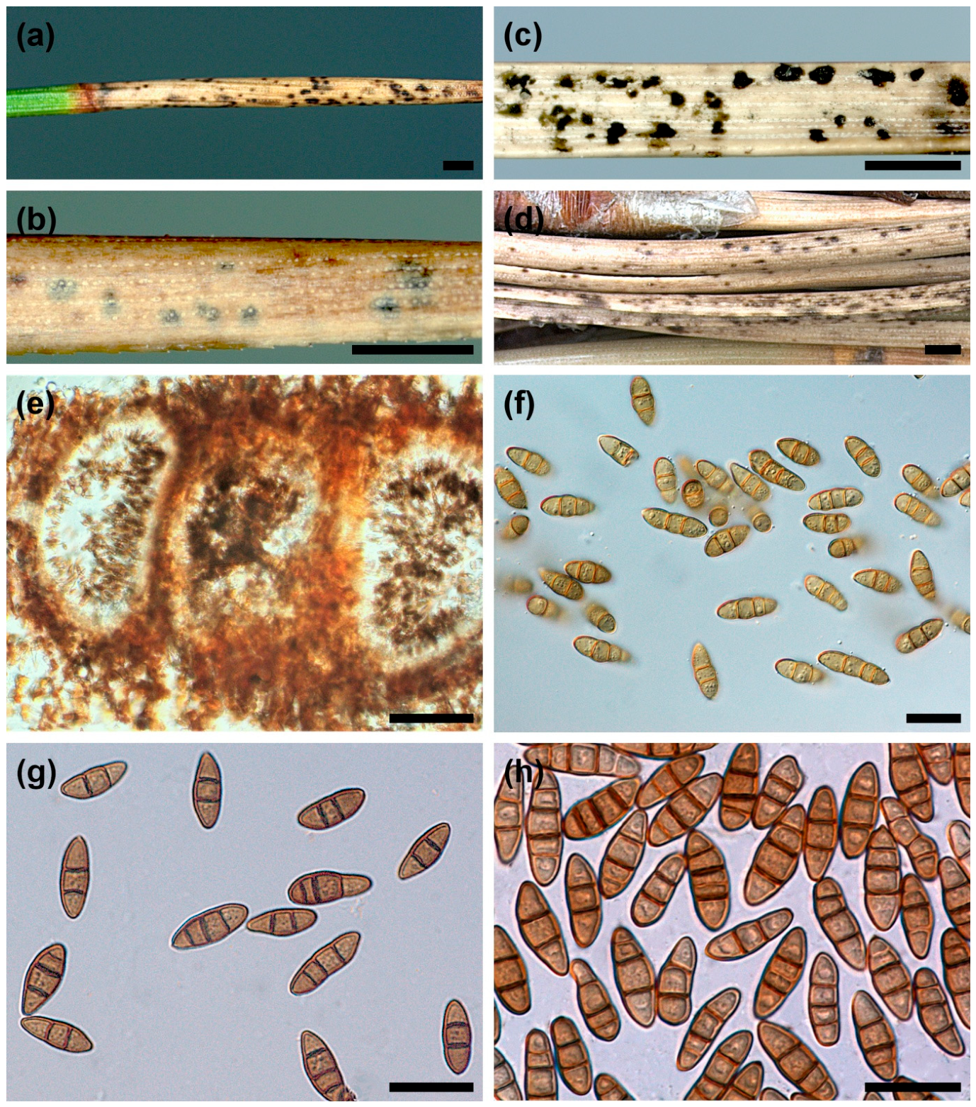
The presence of Hendersonia sp. was confirmed by pycnidia developed on needles in vivo with symptoms of primary infection by L. sulcigena and by isolation from such needles on MEA. On infected needles, a green basal part and dead distal parts could be distinguished (Figure 1a). Hendersonia pycnidia were formed only on dead needle tissue. On most needles, pycnidia were poorly visible due to their location within the substrate (Figure 1b). Only after cutting off the peripheral tissues did they become clearly visible as black circular formations. However, the places where pycnidia occurred could be easily spotted when spore masses, visible as black crusts, accumulated above the ostiole (Figure 1c). In humid conditions, spores spread around the ostiole, forming black, diffuse spots (Figure 1d).

3.1. Morphology of Hendersonia Colonizing the Needles of Pinus mugo

In vivo: Pycnidia were globose or subglobose, 120–300 µm in diameter, with a minute ostiole measuring 25–35 µm in diameter, immersed in needle tissue. Pycnidia usually occurred singly and were separated from each other. When grouped, their shape was deformed (Figure 1e). The pycnidia wall was grayish-brown, 8–15 µm thick, and consisted of two to four layers of cells with a prismatic to angular texture, measuring 5–7 × 2.5–4 µm. Conidiophores were frequently reduced to conidiogenous cells formed from the inner cells of the pycnidial wall. Conidiogenous cells were ampulliform to flask-shaped, hyaline, 5–8 × 4–6 µm. Conidia were thick-walled, smooth, narrowly ellipsoid, obovoid, cylindrical, or pyriform, pale or medium olivaceous, brown to rusty brown, with 0–5 (most often 2) transversal septa measuring 10–19 (22) × 4–6 µm (Table 2). The average length of conidia with two septa was 12.9 to 14.9 µm, and of conidia with three septa was 15.4 to 16.9 µm (Table 2). Significant variation was observed in conidia size (Table 2), septa number (Supplementary Table S2), and the size of the constrictions at the septa (Figure 1f–h). Within the pycnidia in vivo, almost three times more conidia with two septa were present than those with three septa, and they exhibited more or less pronounced constrictions at the septa (Supplementary Table S2, Figure 1f,g). However, in crusts formed on needles (Figure 1c), the proportion of conidia with three septa was very similar, and sometimes slightly greater than the proportion of conidia with two septa (Supplementary Table S2, Figure 1h). Conidia clustered in crusts generally exhibited less pronounced constrictions at the septa (Figure 1h).
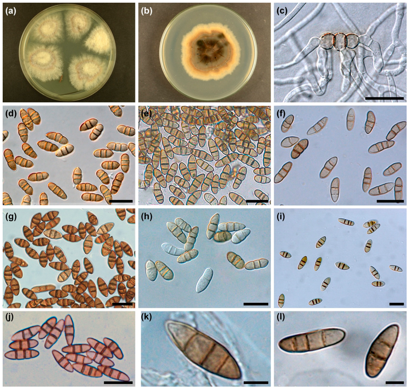
In vitro: Colonies growing on agar medium from P. mugo needles were initially whitish, later becoming olive-gray, quite compact and cottony in the center, and looser at the periphery, with an uneven, slightly sinuate margin (Figure 2a). Aerial mycelium hyphae were initially hyaline, later olive-gray, 1.5–3.0 (4.0) μm, smooth, septate, with a small number of single or chain swollen cells, 5–14 × 5–7.0 μm. More numerous swellings were produced in the mycelium substrate. The reverse was grayish-brown, hazy, with numerous black spots at the center, sometimes confluent (Figure 2b). Colonies after transplantation to new plates showed a more compact structure and variable growth rates, reaching a diameter of 3.6 to 6.2 cm after 4 weeks at 20 °C. Conidia in pycnidia in vitro produced, similarly to in vivo, zero to five transverse septa (Supplementary Table S2). However, conidia with two septa clearly predominated. In young colonies (up to 6 weeks old), conidia with two septa were more than seven times more frequent than those with three septa. In older colonies (3 to 4 months old), conidia with two septa were more than three times more common (Supplementary Table S2). During in vitro analyses, not a single pycnidium was found in which the proportion of conidia with three septa was greater than the proportion of conidia with two septa. Mature conidia in vitro were light brown to rusty brown (Figure 2c–l). Only young pycnidia contained immature hyaline conidia (Figure 2h). Conidia in vitro measured (9)10–18(23) × 4–6(7) μm. The average length of conidia with two septa was 12.9 to 14.0 μm, and of conidia with three septa was 14.2 to 16.1 μm (Table 2). In most conidia, constrictions at the septa were noticeable, although in some cultures they were distinct and in others indistinct (Figure 2d–i). This was not a consistent feature. This variation was noticeable even among pycnidia within the same fungal colony. For example, pycnidia produced conidia with distinct constrictions on the colony surface, whereas pycnidia completely embedded in agar medium produced abnormally swollen conidia with no constrictions at all (Figure 2g). This may also have depended on the age of the cultures. Some young colonies produced conidia with distinct constrictions (Figure 2h), while in the same, but in older colonies were only conidia with slight constrictions (Figure 2i). It was also observed that among conidia from the same pycnidia exhibiting at least mild constrictions, for few conidia no constrictions were present (Figure 2j–l). The test showed that after 48 h at 20 °C, in Petri dishes, 94%, 95% and 97% of conidia germinated, respectively. Individual conidia were visibly swollen to 6–8 μm in diameter, and constrictions at the septa became very pronounced (Figure 2c). In some cells, additional oblique or longitudinal septa were formed. One to six germ hyphae arose from a single conidium. These were septate, cylindrical, 2–5 μm in diameter, hyaline, some light olive or olive brown with delicate surface papillae. Sometimes these hyphae were undulate or spirally twisted at a distance.

3.2. Statistical Analysis of Conidia Size and Septation Variation

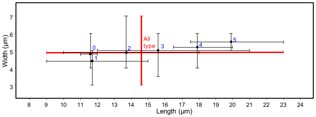
As the number of septa increases (from zero to five), a clear, almost linear increase in the average spore length (Length) was observed—from 11.6 µm for type 0 to 19.9 µm for type 5 (total range: 9.0–23.0 µm). Spore width showed significantly smaller variation between types. The average width fell within a narrow range from 4.4 µm (type 1) to 5.5 µm (type 5), with the range of extreme values for the entire dataset being 3.0 to 7.0 µm. The largest range of conidia widths was observed for type 2 (4.0–7.0 µm). Overall, the conidia of Hendersonia reached sizes of 9–23 × 3–7 (14.6 × 4.9) µm (Figure 3).
A linear mixed-effects model revealed a significant main effect of conidia type (Type) on conidium length (F1,620 = 299.20, p < 0.001), whereas the main effect of Origin was not significant (F3,12 = 2.00, p = 0.168). A significant Origin × Type interaction was detected (F3,620 = 4.90, p = 0.002), indicating that differences between conidia types varied depending on their origin. Tukey-adjusted post hoc comparisons revealed a significant difference in length only between origins Ab and Bc within Type 3 (p = 0.014); all other pairwise comparisons were non-significant.
For conidium width, the mixed model similarly identified significant main effects of Type (F1,620 = 6.83, p = 0.009) and the Origin × Type interaction (F3,620 = 5.77, p < 0.001), while the main effect of Origin remained non-significant (F3,12 = 1.16, p = 0.364).
Diagnostic checks showed that Levene’s test confirmed homogeneity of variances for length (p = 0.41), but indicated heterogeneity for width (p < 0.001). Nevertheless, graphical residual diagnostics revealed no severe deviations from model assumptions, and given the large, balanced dataset, the mixed-effects model results were considered robust and reliable (Figures S1 and S2).
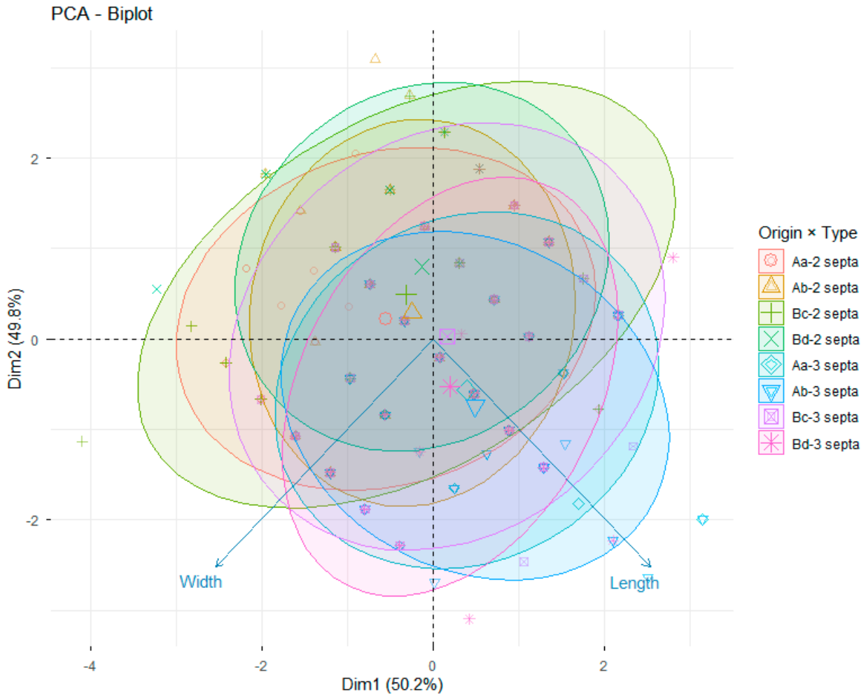
Principal component analysis (PCA) revealed a partial separation between two-septate and three-septate spores, whereas the eight Origin × Type combinations showed substantial overlap. The first two principal components explained 50.2% and 49.8% of the total variance, respectively. Samples representing three-septate spores tended to be shifted towards positive PC1 values, reflecting variation primarily associated with spore length. Despite this trend, the extensive overlap of the confidence ellipses indicates considerable within-group variability and limited discrimination among origin groups (Figure 4). This pattern is consistent with the mixed-effects models, which identified a strong effect of spore type and a significant Origin × Type interaction, but no significant main effect of origin.
Overall, conidium morphometry is primarily determined by conidia type, with length and width differing significantly between two- and three-septate conidia. Origin alone does not exert a direct main effect on morphometry when accounting for unit-level variation; however, its significant interaction with conidium type indicates that origin-dependent differences manifest only within specific conidial types; e.g., the length difference observed between origins Ab and Bc in three-septate conidia.
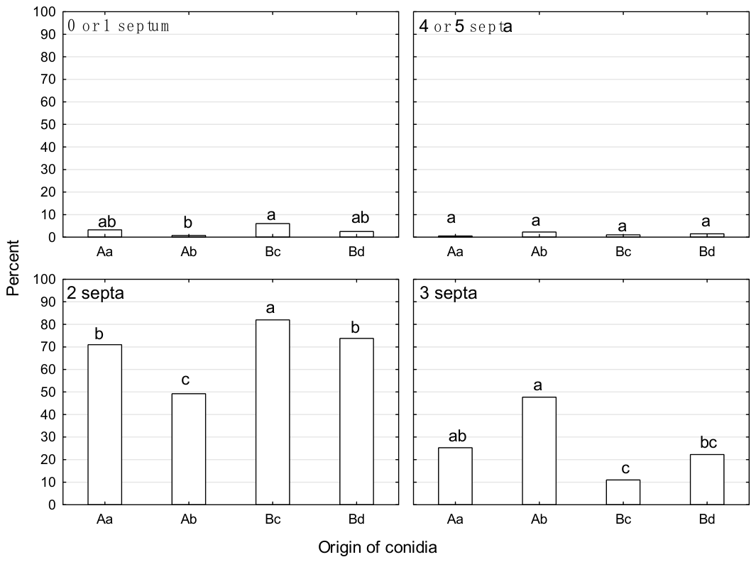
The distribution of spore septa differed significantly between spores of different origins in the categories of zero–one, two and three septa, while no significant differences were found for spores with four–five septa. Spores with zero or one septum were relatively rare in all groups. The highest proportion was observed in young in vitro colonies (Bc), which differed significantly from spores in the crusts on the surface of the needles (Ab). Spores from pycnidia embedded in needles (Aa) and older in vitro colonies (Bd) showed intermediate values and did not differ significantly from either Bc or Ab. Spores with two septa constituted the dominant septation type in all groups of origin (Supplementary Table S2) Their proportion was significantly highest in young in vitro colonies (Bc) and significantly lowest in spores within the crusts (Ab). The proportions observed in embedded pycnidia (Aa) and older colonies (Bd) were intermediate and differed significantly from both Bc and Ab, but not from one another. In the case of three-septate spores, the opposite pattern was observed. The highest proportion was found in the conidial crusts (Ab) and was significantly higher than in young in vitro colonies (Bc), which had the lowest proportion. Conidia from submerged pycnidia (Aa) and older colonies (Bd) had intermediate frequencies; Aa did not differ significantly from either Ab or Bd, while Bd did not differ significantly from either Aa or Bc (Figure 5). However, spores with four or five septa were rare across all groups categorized by origin and age, and no statistically significant differences were observed among the four groups (Figure 5).
Overall, these results show that the development of septa is significantly influenced by their origin and age. Young in vitro colonies were characterized by a predominance of two-septate spores, while spores accumulating in the crusts above the ostioles of pycnidia contained a significantly higher proportion of three-septate spores (Figure 5).
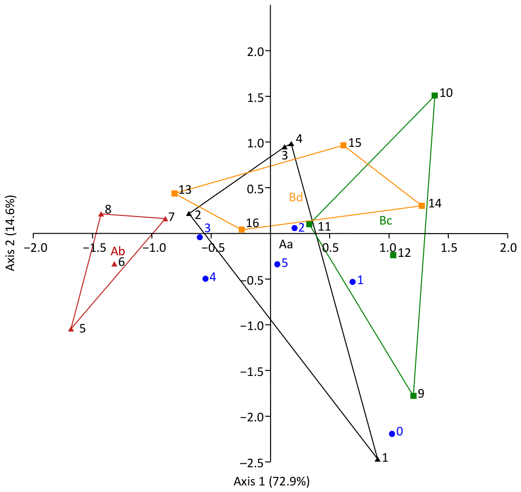
Correspondence analysis (CA) showed that the first two axes together explained 87.5% of the total variance in spore septation. The CA results confirmed the main findings of the χ2 test. Spores of the samples in group Ab formed a distinct cluster, consistent with their low within-group variability in septation. In contrast, spores from groups Aa, Bc, and Bd were relatively similar to one another. However, group Aa exhibited the greatest variability in spore morphology with respect to the number of septa (Figure 6).
Individual analysis showed that the predominance of type 2 spores over type 3 spores is not a consistent feature across all the biological units studied. After applying the Benjamini–Hochberg correction (FDR), a significantly higher abundance of type 2 spores was observed in 12 of the 16 samples analyzed. No significant differences were found only in the case of units T1g87, T1e49, Pm440E and T1e69, belonging to group Ab (Table S2).
The generalized linear mixed-effects model (GLMM) (glmer(cbind(type 2, type 3) ~ Origin + (1|Sample), family = binomial(link = “logit”), data = data)) showed that unit origin had a highly significant effect on the ratio of type 2 to type 3 spores (χ23 = 15.76, p = 0.0013).
Relative to group Aa (model predicted proportion of type 2 = 74.7%), group Ab showed a significantly lower proportion of type 2 spores (type 2 = 50.7%; Z = −2.65, p = 0.008), consistent with an absence of any excess of type 2 over type 3 in this group. Group Bc showed a significantly higher proportion of type 2 than Aa (type 2 = 89.4%; Z = 2.50, p = 0.012), while group Bd did not differ significantly from Aa (type 2 = 79.0%; Z = 0.60, p = 0.551).
Supplementary Table S3 summarizes the most important morphological features of conidia for three Hendersonia species known from pine needles. Comparison of the results of the present analyses with data from other authors indicates that there are clear grounds for identifying the fungus on P. mugo needles in the Tatra Mountains as Hendersonia acicola Münch & Tubeuf. However, greater variation in conidia length and conidiomata size should be noted compared to the original description [21,22]. On the other hand, there is no morphological basis for classifying this species on P. mugo as Hendersonia pinicola Wehm.
Figure 6. Correspondence analysis (CA) of Hendersonia conidia septation (blue number and points) profiles across the studied biological units (1–16, listed according to the row order in Table 3), grouped into four categories based on their origin (Aa, Ab, Bc, Bd). Abbreviations are as defined in Figure 5.
Forests 17 01102 g006
Table 3. Interaction type of six isolates of Hendersonia acicola with fungi isolated from Pinus mugo needles assessed in three weeks old dual cultures.
TaxonRepresentative Strain 1Interaction Type with H. acicola Isolate 2Inhibition Zone for Type B (mm)
Mean (Min–Max)
Pm 379EPm 421EPm 436EPm 439EPm 440EPm 441E
Aureobasidium pullulansPZ734645BmBwBwBmBmBm10.0 (7–13)
Biscogniauxia nummulariaPZ734646BmBmBmDDD8.7 (8–9)
Botrytis cinereaPZ734647BmBmBwBmBmA7.0 (6–11)
Epicoccum nigrumPZ734650BwBmBwBmBmBm9.3 (7–15)
Lophodermium conigenumPZ734652BwBwBwBmBwBw10.5 (7–12)
Lophodermium corconticumPZ734666BwBwBvBwBmBm13.7 (7–21)
Lophodermium sp.PZ734671AAAAAA
Nemania serpensPZ734672BvBvBvBwBmBm13.3 (8–16)
Sydowia polysporaPZ734673BwBwBwBsCBm10.8 (5–15)
Xylaria sp.PZ734674BmBsBmBsBsBs5.0 (2–10)
1 GenBank number. 2 A—physical contact between colonies; B—inhibition zone width: Bs (up to 5 mm), Bm (6–10 mm), Bw (11–15 mm), Bv (16–21 mm); C—overgrowth of the test fungus colony by H. acicola; D—overgrowth of the H. acicola colony by the test fungus.

3.3. DNA Sequence Data and Phylogenetic Analysis

The alignments of the 18S, ITS, 28S, and TEF1 sequences comprised 1756, 854, 860, and 979 characters, respectively, including gaps. The datasets used for phylogenetic analyses contained different numbers of variable characters: 165 for 18S, 469 for ITS, 285 for 28S, and 237 for TEF1. Of these, 98, 314, 154, and 179 characters, respectively, were parsimony-informative. The aligned TEF1 region comprised a fragment of exon 8.
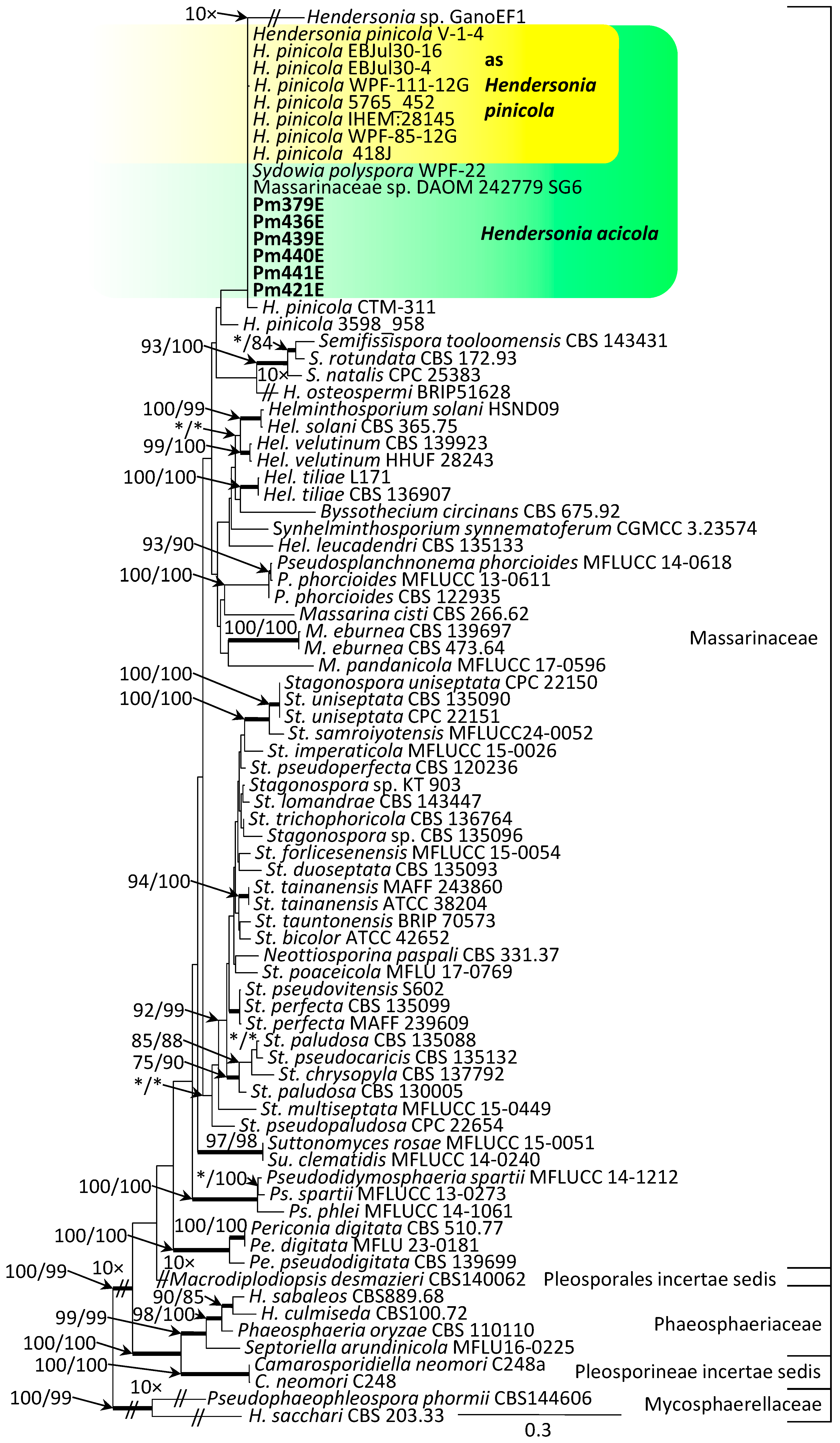
The results of the phylogenetic analysis indicate that the genus Hendersonia is highly polyphyletic (Figure 7).
The isolates obtained in the present study, morphologically identified as Hendersonia acicola, form one clade together with several GenBank reference isolates currently labeled as Hendersonia pinicola. This clade also includes the isolates Massarinaceae sp. DAOM 242779 SG6 and Sydowia polyspora WPF-22. In contrast, the isolate designated H. pinicola CTM-311 forms a distinct sister lineage to the main H. acicola–H. pinicola clade. This lineage falls outside the core clade and may represent a misidentified accession or a distinct taxon (Figure 7).
Similarly, the isolate H. pinicola 3598_958 forms a separate lineage outside the main H. acicolaH. pinicola clade, suggesting that it may likewise represent a misidentified accession or a separate taxon (Figure 7).
Overall, the phylogenetic reconstruction indicates that Hendersonia acicola, Hendersonia pinicola, the distinct lineages represented by isolates CTM-311 and 3598_958, and Hendersonia osteospermi belong to the family Massarinaceae. In contrast, Hendersonia sacchari clusters with Pseudophaeophleospora phormii within the family Mycosphaerellaceae, whereas Hendersonia sabaleos forms a well-supported clade with its sister species Hendersonia culmiseda within the family Phaeosphaeriaceae (Figure 7).

3.4. Dual Culture Assays

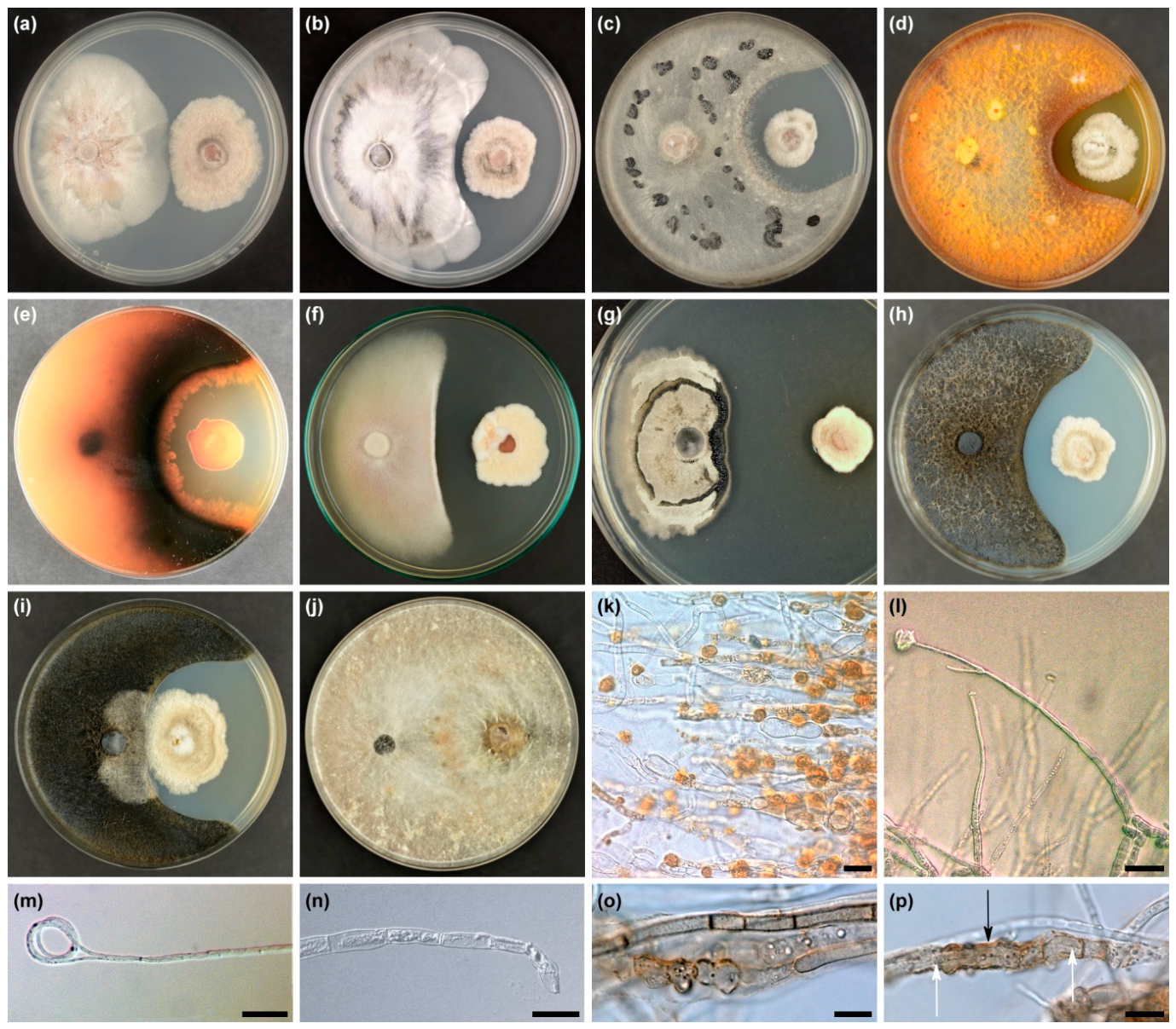
Observations were made in 60 dual cultures in vitro (6 H. acicola isolates × 10 tested fungal species). At the evaluation time (after three weeks) four different types of interactions were distinguished: A—physical colony contact, B—presence of an inhibition zone between the colonies, C—H. acicola overgrowth of copartner colonies, and D—the H. acicola colony became overgrown by the copartner colony (Table 3, Figure 8a–k). The frequency of these types varied significantly. The physical contact of the colonies of both partners (type A) was observed in seven (11.7%) of the dual cultures. This was the case for all isolates of H. acicola and Lophodermium sp. (Figure 8a) and in one dual culture of H. acicola Pm441E with B. cinerea (Table 3), in which a 6 mm wide inhibition zone was first formed, which was relatively quickly overgrown by B. cinerea hyphae. An inhibition zone (type B) was the most common type of interaction between colonies. It was observed in 49 (81.7%) of the dual cultures (Table 3, Figure 8b–h). The width of the inhibition zone ranged from 2 to 21 mm (Table 3). The widest average inhibition zone (13.7 mm) formed between H. acicola and L. corconticum, while the smallest (5.0 mm) was formed when the copartner was Xylaria sp. (Table 3). Type C interaction was observed in only one (1.6%) of the dual cultures (Figure 8i). H. acicola Pm440E overgrown a colony of S. polyspora (Table 3). After 5 weeks, other H. acicola isolates also began to overgrow colonies of S. polyspora and L. corconticum. A type D interaction was observed in three (5.0%) dual cultures. H. acicola was overgrown by B. nummularia mycelia, which began to form a cord-like pattern at this point (Table 3, Figure 8j). Furthermore, after 5 weeks, Xylaria sp. was observed in three Petri dishes to have crossed the inhibition zone and begun to overgrow H. acicola (type D) colonies. This was achieved using cord-like mycelia.
Colonies of all 10 fungal species in dual cultures with H. acicola showed inhibition of growth. The reduction in their radius (Ri) toward the copartner ranged from 6.3 to 70.6% (Supplementary Table S4). On average, the greatest reduction was observed for B. cinerea (60.3%), while the smallest was observed for Lophodermium sp. (16.0%) (Supplementary Table S4).
However, the H. acicola colony in 16 dual cultures showed no changes in growth; in two dual cultures (with N. serpens and S. polyspora), growth toward the copartner was greater, while in the remaining 42 dual cultures, a reduction in growth was observed. The greatest reduction in H. acicola growth was observed in dual cultures with E. nigrum (Supplementary Table S4). H. acicola in dual cultures, as in monocultures, showed some variation in colony structure and color (Figure 8a–j).
Various morphological and structural changes were observed in the colonies of some fungi tested in dual cultures. L. concorticum always produced larger accumulations of black stromatic structures on the side of H. acicola interaction (Figure 8g). The hyphae of the A. pullulans turned dark, which resulted in the formation of a black zone. Biscogniauxia nummularia produced an intense blackish-brown pigment (Figure 8e). Epicoccum nigrum, on the other hand, produced numerous crystal-like angular secretions on the inhibition zone side. Under the influence of H. acicola, various morphophysiological deformations of the hyphae of tested fungi in the dual cultures, compared to the control colonies, were observed (Figure 8k–p). Apical and intercalary cytoplasmic extrusions were the most common (Figure 8k,l). Their high density was particularly visible in E. nigrum (Figure 8k). Conidia were not produced on conidiophores of B. cinerea with cytoplasmic extrusion (Figure 8l). Coiling of the apical part of the S. polyspora hyphae was associated with a lack of further linear growth (Figure 8m). In B. nummularia hyphae, decomposition of the internal cell contents occurred, which also resulted in growth inhibition (Figure 8n). In a colony of S. polyspora, covered by H. acicola, the hyphae showed symptoms of disruption via lysis (Figure 8o). H. acicola hyphae were found inside the hyphae of S. polyspora (Figure 8p). This was accompanied by disappearance of the dark pigment in the affected hyphae of S. polyspora (Figure 8o,p).

4. Discussion

4.1. Morphological Aspects of Hendersonia acicola on P. mugo Needles

Three Hendersonia species are known to occur on pine needles, primarily invaded by Lophodermella: H. acicola, H. montana, and H. pinicola. H. montana (Supplementary Table S3) is the easiest to distinguish morphologically, as it produces relatively long and narrow conidia [28,29,30]. Morphological features that could be used to distinguish the other two species are less clear. Wehmeyer [33], when describing H. pinicola as a new species, stated that it was very similar to H. acicola, described much earlier from Europe [22]. It differs, however, in that the conidia are slightly larger (14–20 × 5–7 µm), one-celled at first, becoming four-celled, and are not constricted at the septa [33]. Electron microscopy studies have shown that the conidium wall of H. pinicola is composed of three layers, with only the inner layer involved in septum formation [34]. According to Wehmeyer [33], H. pinicola was easily visible because conidia emerged from spherical pycnidia immersed in the leaf mesophyll and remained on the needle surface in irregular, black, paint-like masses. Wehmeyer [33] also pointed out as an additional difference the fact that H. pinicola is related to L. concolor, while H. acicola accompanies L. sulcigena.
The basic morphological characteristics reported for H. pinicola by Wehmeyer [33] have been confirmed by other authors (Supplementary Table S3). However, James [35] observed slight constrictions of conidia from black crusts on P. contorta needles at each septum. Chao [34] reported that the conidia are navicular, rounded at the base and acute at the apex, whereas according to Wehmeyer [33], they are fusoid-ellipsoid to clavate. Furthermore, Stahl et al. [37] found a sparse proportion of both smaller and larger conidia, the presence of 1–6 septa, and the presence of muriform conidia, which may indicate morphological diversity within H. pinicola or the presence of different but similar fungal species. In Europe, H. acicola has been known for over a hundred years [21,22,24,42]. Hendersonia acicola colonizes P. sylvestris, P. nigra, and P. mugo needles primarily attacked by L. sulcigena or L. conjuncta [5,6,18,19,23,24,25,26]. However, the number of publications presenting in more detail the morphological features of H. acicola spores is small. They result in the following three characteristic features for the spores of this species: (i) the length of the conidia is in the range of 11–15 µm, (ii) the conidia may contain one–three transverse septa, but most often there are two septa, (iii) the conidia are constricted at the septa.
The morphological features of conidia found in Hendersonia on P. mugo needles in the Tatra Mountains mostly corresponded to those reported for H. acicola [22,23,24,27,42,43]. However, in the population studied now, it is noteworthy that the length of the vast majority of conidia was in the range of 11–15 µm, while some spores, especially those with three septa, were slightly longer [22,23,24,42,43]. At the same time, the current analyses provided some data indicating that the variability of conidia characteristics may be influenced by factors such as the length of the in vitro cultivation period, the site of pycnidia production (on the colony surface, embedded in agar medium), and the concentration of conidia in crusts on the surface of needles in situ. Despite the currently observed significant morphological diversity of conidia, there is no basis for identifying any sample as H. pinicola [33]. No sample was found without conidia less than 14 µm in length, and conidia with three septa clearly predominated [33]. Available data indicate, for example, that in H. pinicola in North America, the proportion of spores with three septa ranges from approximately 70 to 90% [12,35,38].

4.2. Phylogenetical Aspects

So far, no ITS sequences of H. acicola are available in GenBank. There is also no ITS sequence of the holotype of H. pinicola. However, sequences of six strains of H. pinicola from North America are deposited in GenBank. One of them (MT994893) concerns an H. pinicola strain isolated from symptomatic needles of P. taeda in the southeastern United States, infected with Lecanosticta acicola and other pathogens, but there were no representatives of Lophodermella. However, there is no morphological characterization of this H. pinicola strain [40]. Five H. pinicola sequences (KT000152, KT000169, KT000174, KT000191, KT000192) refer to strains isolated from symptomatic needles of P. strobus in the northeastern United States, which were infected by Lecanosticta acicola, Lophophacidium dooksii, and Bifusella linearis [39]. These authors also did not present morphological characteristics of the strains deposited as H. pinicola. This limits the possibility of comparative analysis with H. acicola strains from Europe. Hendersonia found on P. strobus requires particularly thorough analysis. In 1928, Hendersonula pinicola Dearn. was described and, after mycological analysis, renamed Hendersonia dearnessii [17,70]. This species occurred only on P. strobus needles originally infected with Bifusella linearis. This situation is therefore partially similar to that described by Broders et al. [39].
The current phylogenetic analysis suggests that traditional morphological characters used to define the genus Hendersonia have resulted in an artificial assemblage of unrelated taxa, making it a classic example of a wastebasket taxon. Consequently, the genus requires comprehensive taxonomic revision, as also suggested by other authors [39,71].
Within the GenBank accessions currently labeled as H. pinicola, the phylogenetic analysis revealed multiple distinct evolutionary lineages: the main H. pinicola–H. acicola clade and two separate divergent lineages represented by CTM-311 and 3598_958. These results indicate that the sequences currently designated as H. pinicola in GenBank are highly polyphyletic, representing a complex of divergent sequence lineages rather than a single uniform taxon. However, because sequence data from authentic or type material of H. pinicola is lacking, and several North American GenBank isolates lack morphological characterization, it remains unclear which lineage represents H. pinicola sensu stricto. Thus, our phylogeny demonstrates the polyphyly of these GenBank accessions, but cannot definitively prove that H. pinicola itself consists cryptic species.
The inability to distinguish H. pinicola and H. acicola from one another and from other species in the phylogenetic analysis can be attributed to two factors. Firstly, the preliminary or provisional morphological identification of these isolates may not have fully corresponded to the description provided by Wehmeyer [33]. Secondly, there is currently a lack of data covering multiple loci, such as the 18S, 28S or TEF1 sequences, which could potentially help to identify genetic differences between H. pinicola and H. acicola.
The limitations of relying solely on ITS sequence analysis to distinguish species within the family Massarinaceae are well exemplified by the case of Helminthosporium italicum and H. velutinum [52]. The phylogenetic reconstruction further indicates that most species currently assigned to Hendersonia belong to the family Massarinaceae. In contrast, several taxa are phylogenetically distant from the core clade, suggesting they may eventually require formal transfer to other genera. However, such nomenclatural reclassifications require comprehensive taxonomic work based on type specimens and are outside the scope of the present study. For example, H. culmiseda and H. sabaleos are placed within the Phaeosphaeriaceae [72], while H. sacchari and Pseudophaeophleospora phormii (epitype of H. phormii) are allied with the Mycosphaerellaceae. Together, these findings provide robust evidence that the traditional circumscription of Hendersonia represents a polyphyletic assemblage of unrelated taxa.

4.3. Antagonistic Activity of Polish Isolates of Hendersonia acicola In Vitro

Lophodermella sulcigena infects newly developing pine needles in early summer. If development is undisturbed, its hysterothecia mature in early summer the following year, and the shed ascospores infect new needles [19,24]. If H. acicola invades the current year’s needles secondarily in late summer, this fungus deleteriously affects the life cycle of the pathogenic L. sulcigena, which may inhibit its fruiting [6,18,24,25]. According to Jalkanen and Laakso [6], colonies of L. sulcigena and H. acicola grow in vitro on agar without forming barriers to each other. Ten fungal species were used to conduct the current in vitro dual test. They were isolated from P. mugo needles exhibiting various types of disease symptoms, and some of them also from symptomless needles [41]. Unfortunately, L. sulcigena could not be used in the test due to its very limited growth on MEA in vitro [10]. The fungi used in the test play different ecological roles. Aureobasidium pullulans and Epicoccum nigrum are ubiquitous saprotrophs and endophytes that exhibit antagonistic effects on numerous plant pathogens [66,73,74,75]. Until recently, Biscogniauxia nummularia was found primarily in southern Europe as an endophyte and saprotroph on angiosperm, and also as a cause of strip-cankers on Fagus sylvatica L. [76,77]. In recent years, it has been increasingly reported in central Europe, mostly in coniferous trees [78]. Botrytis cinerea is a known worldwide cause of gray mold, leading to significant losses in agriculture and forest nurseries. In young pines and other forest tree species, it often colonizes various types of necrotic tissue, from where it attacks living tissue [79,80]. Lophodermium conigenum and L. corconticum are among the species most frequently found on P. mugo in mountainous areas in Poland [41,81]. They are isolated from both symptomless needles and living needles with necrotic spots. On dead needles, they often produce ascomata and asexual morphs. However, their pathogenicity towards P. mugo has not been confirmed so far [41,81]. Sydowia polyspora is also among the fungi frequently found on symptomatic P. mugo needles [41]. It is a fungus commonly found on various coniferous species worldwide. It can inhabit living tissue as an endophyte and also colonize dying needles, shoots, and stems as a saprotroph or weak pathogen [2,12,82]. Nemania serpens and Xylaria species are common endophytes and saprotrophs on conifers and deciduous trees [41,44,66,74].
The present dual culture assay showed that H. acicola exerted mainly an antagonistic effect on various pine needle fungi. After three weeks, 81.7% of dual colonies developed an inhibition zone 2–21 mm wide. Despite the lack of contact between mycelia, cytoplasmic extrusion and lysis of hyphae occurred in some pine needle fungi. This seems to indicate that H. acicola produces bioactive compounds, also observed in many other fungi [7,83,84,85]. These results are consistent with the results of Mitchell et al. [4], who observed in pine needles in vivo, that cells of L. sulcigena were vacuolated in the vicinity of H. acicola and can even lose their cytoplasmic contents, which indicates an antagonistic effect. Interesting in this respect are the results of research on the fungus identified as H. pinicola, isolated from living symptomless needles of Pinus strobus in Canada [39,86,87]. From their culture filtrate extracts, four dihydrobenzofurans and two xanthenes were known. Individually tested, each metabolite displayed in vitro bioactivity against the biotrophic pathogen Microbotryum violaceum (Pers.) G. Deml & Oberw. and the Gram-positive bacterium Bacillus subtilis (Ehrenb.) Cohn [86].
Although infection of pine needles by H. acicola is dependent on their prior infection by L. sulcigena, hyperparasitism of H. acicola against L. sulcigena was not observed [88]. However, the present dual culture assay showed that some H. acicola strains were capable of overgrowing of other pine needle fungi, leading to the disintegration of their hyphae. The observed interactions between H. acicola and needle fungi were largely similar to the in vitro interactions between mycobiota colonizing Fraxinus excelsior L. petioles and the ash dieback pathogen Hymenoscyphus fraxineus [65,74]. However, it may be surprising that species such as Nemania serpens or Xylaria sp., which were able to strongly limit the development of H. fraxineus were now strongly inhibited by H. acicola. This may indicate the high bioactive capacity of metabolites produced by H. acicola. Currently, significant growth reduction of H. acicola has been reported in dual cultures with E. nigrum. This fungus is known to produce flavipin, epicorazine, and epipyrone, which enable its antifungal activities. Furthermore, this species secretes cell-wall degrading enzymes that may contribute to lysis of hyphae [89,90]. The observed in vitro antagonism suggests that further studies could evaluate whether H. acicola contributes to natural suppression of selected pine needle pathogens. This would be particularly important in areas where chemical treatments are not possible, for example in national parks. Studies show that H. acicola is harmless to healthy green needles [6,17,19]. H. acicola occurs only sporadically as an endophyte in living symptomless needles of P. mugo and P. sylvestris [25,41,91]. High humidity may favor increasing the inoculum reservoir of H. acicola on dead needles [6,19,27]. The preliminary dual culture test conducted recently indicates a strong inhibitory effect of H. acicola on numerous pine needle fungi. This is a new finding, as previous studies indicated that H. acicola causes intensive needle exploitation, resulting in the pathogen L. sulcigena having insufficient reserves to form ascocarps, preventing it from completing its life cycle [18,88]. This interaction is a result of competition for substrate. Studies in Finland show that, due to the activity of H. acicola, the endemic L. sulcigena on P. sylvestris did not last longer than 3–4 years [6,24].

5. Conclusions

The current study was undertaken as a result of high intensity of P. mugo needle disease caused by the ascomycetous fungus Lophodermella sulcigena in the Polish Tatra Mountains. In Europe, the often-important secondary fungus Hendersonia acicola has been identified on such needles for over a hundred years based on morphological characteristics. However, multi-locus molecular analyses of four gene fragments (18S, ITS, 28S, and TEF1) conducted in the current study indicated their compatibility with reference isolates of Hendersonia pinicola originating from Pinus strobus from the USA, whose ITS sequences are deposited in GenBank. The current study revealed that there is no basis for identifying the secondary fungus on P. mugo needles in Poland as H. pinicola. Most morphological features are consistent with data for H. acicola. Due to the relatively wide range of variability observed in spore morphological features, and varied pycnidial size, further studies using reference strains from Europe and North America are necessary for comparative purposes. Further studies should also include assessment of H. acicola across Europe, identification of potential cryptic species, and studies on produced metabolites. Dual culture assays demonstrated in vitro antagonistic activity H. acicola against ten fungal species isolated from P. mugo needles, and identified a basis for further biological control research. The pathogen L. sulcigena could not be tested in vitro and further studies on needles or plants in field trials are necessary.

Supplementary Materials

The following supporting information can be downloaded at https://www.mdpi.com/article/10.3390/f17091102/s1, Table S1: Primers used in this study to amplify and sequence barcode regions in molecular identification.; Table S2: Presence of septa in Hendersonia conidia produced on Pinus mugo needles in situ in pycnidia (A) and in vitro on agar medium (B), according to their location or age. (A) a—conidia within pycnidia embedded in P. mugo needles; b—conidia aggregated in a “crust” above the ostiole on the surface of P. mugo needles. (B) c—conidia produced in young colonies (up to 6 weeks old); d—conidia produced in older colonies (3–4 months old).; Table S3: Selected morphological characteristics of pycnidia and conidia for three Hendersonia species on pine (Pinus sp.) needles.; Table S4: Colony radius reduction of Hendersonia acicola and fungi isolated from Pinus mugo needles assessed in three weeks old dual cultures.; Figure S1: Residual diagnostic plots for the linear mixed-effects model of Hendersonia sp. conidium length (n = 640): (a) normal quantile–quantile (Q–Q) plot of the model residuals, evaluating the assumption of normality; (b) residuals-versus-fitted values plot, assessing the homogeneity of residual variance (homoscedasticity) and model linearity.; Figure S2: Residual diagnostic plots for the linear mixed-effects model of Hendersonia sp. conidium width (n = 640): (a) normal quantile–quantile (Q–Q) plot of the model residuals, evaluating the assumption of normality; (b) residuals-versus-fitted values plot, assessing the homogeneity of residual variance (homoscedasticity) and model linearity.

Author Contributions

Conceptualization, T.K. and P.B.; methodology, T.K. (mycological aspects) and P.B. (molecular and statistical aspects); investigation, T.K. and P.B.; formal analysis, T.K. and P.B.; data curation, T.K. and P.B.; writing—review and editing, T.K. and P.B.; software and visualization, P.B. All authors have read and agreed to the published version of the manuscript.

Funding

This research received no external funding.

Data Availability Statement

The data presented in this study are available in Supplementary Material, GenBank and TreeBase.

Acknowledgments

We would like to thank the anonymous reviewers for their very valuable suggestions and comments on the current version of the manuscript.

Conflicts of Interest

The authors declare no conflicts of interest.

References

  1. Lumbsch, H.T.; Huhndorf, S.M. Myconet Volume 14. Part One. Outline of Ascomycota—2009. Part Two. Notes on Ascomycete Systematics. Nos. 4751–5113. Fieldiana Life Earth Sci. 2010, 1, 1–64. [Google Scholar] [CrossRef] [Scilit]
  2. Sinclair, W.A.; Lyon, H.H. Diseases of Trees and Shrubs, 2nd ed.; Cornell University Press: Ithaca, NY, USA, 2005. [Google Scholar]
  3. Terrier, C. Über zwei in der Schweiz bisher wenig bekannte Schüttepilze der Kiefern: Hypodermella sulcigena (Rostr.) v.Tub. und Hypodermella conjuncta Darker. Phytopathol. Z. 1944, 14, 442–449. [Google Scholar]
  4. Mitchell, C.P.; Millar, C.S.; Williamson, B. The biology of Lophodermella conjuncta Darker on Corsican pine needles. For. Pathol. 1978, 8, 108–118. [Google Scholar] [CrossRef] [Scilit]
  5. Millar, C.S.; Minter, D.W. Lophodermella conjuncta. Descr. Fungi Bact. 1980, 66, Sheet 658. [Google Scholar] [CrossRef] [Scilit]
  6. Jalkanen, R.; Laakso, R. Hendersonia acicola in an epidemic caused by Lophodermella sulcigena with special reference to biological control. Karstenia 1986, 26, 49–56. [Google Scholar] [CrossRef] [Scilit]
  7. Elvira-Recuenco, M.; Cacciola, S.O.; Sanz-Ros, A.V.; Garbelotto, M.; Aguayo, J.; Solla, A.; Mullett, M.; Drenkhan, T.; Oskay, F.; Aday Kaya, A.G.; et al. Potential Interactions between Invasive Fusarium circinatum and Other Pine Pathogens in Europe. Forests 2019, 11, 7. [Google Scholar] [CrossRef] [Scilit]
  8. Ata, J.P.; Burns, K.S.; Marchetti, S.; Munck, I.A.; Beenken, L.; Worrall, J.J.; Stewart, J.E. Molecular characterization and phylogenetic analyses of Lophodermella needle pathogens (Rhytismataceae) on Pinus species in the USA and Europe. PeerJ 2021, 9, e11435. [Google Scholar] [CrossRef] [Scilit] [PubMed]
  9. Dubach, V.; Queloz, V.; Stroheker, S. Needle and Shoot Diseases of Pine; WSL Fact Sheet 70; Swiss Federal Institute WSL: Birmensdorf, Switzerland, 2022; 12p. [Google Scholar]
  10. Kowalski, T.; Bartnik, C.; Bilański, P. Involvement of Lophodermella sulcigena in Endemic Disease of Pinus mugo Needles in the Polish Tatra Mountains. Forests 2024, 15, 422. [Google Scholar] [CrossRef] [Scilit]
  11. Czabator, F.J.; Staley, J.M.; Snow, G.A. Extensive southern Pine needle blight during 1970–1971, and associated fungi. Plant Dis. Report. 1971, 55, 764–766. [Google Scholar]
  12. Funk, A. Foliar Fungi of Western Trees. Information Report BC-X-265; Canadian Forestry Service: Victoria, BC, Canada, 1985. [Google Scholar]
  13. Kowalski, T. Erstnachweis von Lophodermella sulcigena in Polen. Eur. J. For. Pathol. 1988, 18, 445–447. [Google Scholar] [CrossRef] [Scilit]
  14. Minter, D.W.; Millar, C.S. IMI descriptions of fungi and bacteria No. 1146: Lophodermella concolor. Mycopathologia 1993, 121, 53–54. [Google Scholar]
  15. Worrall, J.; Marchetti, S.; Mask, R. An Epidemic of Needle Cast on Lodgepole Pine in Colorado; Biological Evaluation R2-12-01; USDA Forest Service, Rocky Mountain Region, Forest Health Protection: Denver, CO, USA, 2012; 16p.
  16. Beenken, L. Lophodermella-Nadelschütte. In Waldschutzüberblick 2018; Queloz, V., Forster, B., Beenken, L., Stroheker, S., Odermatt, O., Hölling, D., Meyer, J., Dubach, V., Eds.; Forschungsanstalt WSL: Birmensdorf, Switzerland, 2019; pp. 18–19. [Google Scholar]
  17. Darker, G.D. A revision of the genera of the Hypodermataceae. Can. J. Bot. 1967, 45, 1399–1444. [Google Scholar] [CrossRef] [Scilit]
  18. Mitchell, C.P.; Williamson, B.; Millar, C.S. Hendersonia acicola on pine needles infected by Lophodermella sulcigena. For. Pathol. 1976, 6, 92–102. [Google Scholar] [CrossRef] [Scilit]
  19. Millar, C.S. Lophodermella species on pines. In Proceedings of the Recent Research on Conifer Needle Diseases; Peterson, G.W., Ed.; General Technical Report GTR-WO 50; USDA Forest Service: Gulfport, MS, USA, 1984; pp. 45–55. [Google Scholar]
  20. Hunt, R. Common pine needle casts and blights in the Pacific Region. In Forest Pest Leaflet; Pacific Forestry Centre, Canadian Forest Service: Victoria, BC, Canada, 1995; pp. 1–7. [Google Scholar]
  21. Münch, E.; von Tubeuf, C. Eine neue Nadelkrankheit der Kiefer, Pinus silvestris. Naturwissenschaftliche Z. Für Forst-Und Landwirtsch. 1911, 9, 20–25. [Google Scholar]
  22. Münch, E.; von Tubeuf, C. Eine neue Nadel-Krankheit der Kiefer, Pinus silvestris. Naturwissenschaftliche Z. Für Forst-Und Landwirtsch. 1910, 8, 39–44. [Google Scholar]
  23. Lagerberg, T. Om grabarrsjukan host allen, dess orsak och verkningar. Medd. Från Statens Skogsförsöksanst. 1910, 7, 127–174. [Google Scholar]
  24. Jalkanen, R. The occurrence and importance of Lophodermella sulcigena and Hendersonia acicola on Scots pine in Finland. Karstenia 1985, 25, 53–61. [Google Scholar] [CrossRef] [Scilit][Green Version]
  25. Kowalski, T.; Krygier, J. Mycological study on symptomless and diseased needles in pine stand attacked by Lophodermella sulcigena. Phytopathol. Pol. 1996, 11, 159–168. [Google Scholar]
  26. Bachinger, M. Lophodermella sulcigena an Latsche und Spirke. Forstsch. Aktuell 1991, 8, 7. [Google Scholar]
  27. Bingzhang, H.; Yujie, C.; Guoxin, Q.; Lixin, Y. Study on Biological Characteristics of Hendersonia acicola. J. Northeast For. Univ. 1994, 5, 37–40. [Google Scholar] [CrossRef] [Scilit]
  28. Saccardo, P.A.; Sydow, P. Supplementum Universale, Pars IV. Sylloge Fungorum 1899, 14, 1–1316. [Google Scholar]
  29. Kalandra, A. Nová sypavka u nás způsobená houbou Hypodermella sulcigena (Rostr.) Tub. na borovici obecné a kleči v Tatrách a na Šumavě. Ochr. Rostl. 1938, 14, 38–46. [Google Scholar]
  30. Moriondo, F. La diffusione della ruggine curvatrice nelle pinete italiane. Ann. Accad. It. Sci. 1963, 11, 247–263. [Google Scholar]
  31. Darker, G.D. The Hypodermataceae of Conifers; Jamaica Plain, Mass, The Arnold Arboretum of Harvard University: Boston, MA, USA, 1932; Volume 1. [Google Scholar]
  32. Staley, J.M.; Bynum, H.H. A New Lophodermella on Pinus ponderosa and P. attenuata. Mycologia 1972, 64, 722. [Google Scholar] [CrossRef] [Scilit]
  33. Wehmeyer, L.E. Studies on some fungi from North-western Wyoming. II. Mycologia 1946, 38, 306–330. [Google Scholar] [CrossRef] [Scilit]
  34. Chao, R. Conidium Morphology and Ontogeny in Species of Leptomelanconium, Gloeocoryneum and Hendersonia. Master’s Thesis, The University of Manitoba, Winnipeg, MB, Canada, 1969. [Google Scholar]
  35. James, R. Hendersonia Blight of Lodgepole Pine in Idaho; Nursey Disease Notes No. 9; USDA Forest Service Northern Region: Missoula, MT, USA, 1984; pp. 2–8.
  36. Minter, D.W. Some members of the Rhytismataceae (Ascomycetes) on conifer needles from central and north America. In Proceedings of the IUFRO Working Party Conference: Recent Research on Needle Diseases, Gulfport, MS, USA, 14–18 October 1984; pp. 71–106. [Google Scholar]
  37. Stahl, S.A.; Rogers, J.D.; Adams, M.J. Observations on Hendersonia pinicola and the needle blight of Pinus contorta. Mycotaxon 1988, 31, 323–337. [Google Scholar] [CrossRef] [Scilit]
  38. Munck, I.; Burns, B.; Ostrofsky, W.; Lombard, K.; Weimer, J. Eastern White Pine Needle Damage Survey, 2011. In Maine, New Hampshire, and Vermont; Durham Field Office, USDA Forest Service: Durham, NH, USA, 2012; pp. 1–12. [Google Scholar]
  39. Broders, K.; Munck, I.; Wyka, S.; Iriarte, G.; Beaudoin, E. Characterization of Fungal Pathogens Associated with White Pine Needle Damage (WPND) in Northeastern North America. Forests 2015, 6, 4088–4104. [Google Scholar] [CrossRef] [Scilit]
  40. Datta, D. Identification and Distribution of Fungal Pathogens Associated with Loblolly Pine Defoliation and Tree Mortality in the Southeastern United States; Auburn University: Auburn, AL, USA, 2021. [Google Scholar]
  41. Bartnik, C.; Kowalski, T.; Bilański, P.; Zwijacz-Kozica, T. Fungi associated with disease symptoms on Pinus mugo needles in the Polish Tatra Mountains. Plant Fungal Syst. 2021, 66, 53–65. [Google Scholar] [CrossRef] [Scilit]
  42. Kujala, V. Über die Kleinpilze der Koniferen in Finnland. Ascomycetes, Fungi Imperfecti, Uredinales. In Communicationes Instituti Forestalis Fenniae; Finnish Forest Research Institute: Vantaa, Finland, 1950; Volume 38, pp. 1–121. [Google Scholar]
  43. Ellis, M.B.; Ellis, J.P. Microfungi on Land Plants. An Identification Handbook; Macmillan Publishing Co.: New York, NY, USA, 1985. [Google Scholar]
  44. Bilański, P.; Grad, B.; Kowalski, T. Pyrenochaeta fraxinina as colonizer of ash and sycamore petioles, its morphology, ecology, and phylogenetic connections. Mycol. Prog. 2022, 21, 74. [Google Scholar] [CrossRef] [Scilit]
  45. White, T.J.; Bruns, T.; Lee, S.; Taylor, J.W. Amplification and direct sequencing of fungal ribosomal RNA genes for phylogenetics. In PCR Protocols: A Guide to Methods and Applications; Innis, M.A., Gelfand, D.H., Sninsky, J.J., White, T.J., Eds.; Academic Press Inc.: New York, NY, USA, 1990; pp. 315–322. [Google Scholar]
  46. Vilgalys, R.; Hester, M. Rapid genetic identification and mapping of enzymatically amplified ribosomal DNA from several Cryptococcus species. J. Bacteriol. 1990, 172, 4238–4246. [Google Scholar] [CrossRef] [Scilit] [PubMed]
  47. Rehner, S.A.; Samuels, G.J. Taxonomy and phylogeny of Gliocladium analysed from nuclear large subunit ribosomal DNA sequences. Mycol. Res. 1994, 98, 625–634. [Google Scholar] [CrossRef] [Scilit]
  48. Rehner, S.A.; Buckley, E. A Beauveria phylogeny inferred from nuclear ITS and EF1-α sequences: Evidence for cryptic diversification and links to Cordyceps teleomorphs. Mycologia 2005, 97, 84–98. [Google Scholar] [CrossRef] [Scilit]
  49. Gardes, M.; Bruns, T.D. ITS primers with enhanced specificity for basidiomycetes-application to the identification of mycorrhizae and rusts. Mol. Ecol. 1993, 2, 113–118. [Google Scholar] [CrossRef] [Scilit] [PubMed]
  50. Zhang, Z.; Schwartz, S.; Wagner, L.; Miller, W. A Greedy Algorithm for Aligning DNA Sequences. J. Comput. Biol. 2000, 7, 203–214. [Google Scholar] [CrossRef] [Scilit] [PubMed]
  51. Morgulis, A.; Coulouris, G.; Raytselis, Y.; Madden, T.L.; Agarwala, R.; Schäffer, A.A. Database indexing for production MegaBLAST searches. Bioinformatics 2008, 24, 1757–1764. [Google Scholar] [CrossRef] [Scilit] [PubMed]
  52. Konta, S.; Hyde, K.D.; Karunarathna, S.C.; Mapook, A.; Senwanna, C.; Dauner, L.A.P.; Nanayakkara, C.M.; Xu, J.; Tibpromma, S.; Lumyong, S. Multi-Gene Phylogeny and Morphology Reveal Haplohelminthosporium gen. nov. and Helminthosporiella gen. nov. Associated with Palms in Thailand and A Checklist for Helminthosporium Reported Worldwide. Life 2021, 11, 454. [Google Scholar] [CrossRef] [Scilit] [PubMed]
  53. Bhagya, A.T.; Phukhamsakda, C.; Tanaka, K.; Jones, E.B.G. Morphology and multigene phylogeny reveal a novel Stagonospora species (Massarinaceae, Dothideomycetes) from Thailand. Phytotaxa 2024, 644, 281–293. [Google Scholar] [CrossRef] [Scilit]
  54. Tian, W.-H.; Jin, Y.; Liao, Y.-C.; Faraj, T.K.; Guo, X.-Y.; Maharachchikumbura, S.S.N. Phylogenetic Insights Reveal New Taxa in Thyridariaceae and Massarinaceae. J. Fungi 2024, 10, 542. [Google Scholar] [CrossRef] [Scilit] [PubMed]
  55. Wanasinghe, D.N.; Maharachchikumbura, S.S.N. Exploring the Diversity and Systematics of Phaeosphaeriaceae: Taxonomic Novelties from Ecologically Diverse Habitats and Their Phylogenetic Resolution. J. Fungi 2023, 9, 853. [Google Scholar] [CrossRef] [Scilit] [PubMed]
  56. Hall, T.A. BioEdit: A user-friendly biological sequence alignment editor and analysis program for Windows 95/98/NT. Nucleic Acids Symp. Ser. 1999, 41, 95–98. [Google Scholar]
  57. Katoh, K.; Rozewicki, J.; Yamada, K.D. MAFFT online service: Multiple sequence alignment, interactive sequence choice and visualization. Brief. Bioinform. 2019, 20, 1160–1166. [Google Scholar] [CrossRef] [Scilit] [PubMed]
  58. Guindon, S.; Gascuel, O. A Simple, Fast, and Accurate Algorithm to Estimate Large Phylogenies by Maximum Likelihood. Syst. Biol. 2003, 52, 696–704. [Google Scholar] [CrossRef] [Scilit] [PubMed]
  59. Darriba, D.; Taboada, G.L.; Doallo, R.; Posada, D. jModelTest 2: More models, new heuristics and parallel computing. Nat. Methods 2012, 9, 772. [Google Scholar] [CrossRef] [Scilit] [PubMed]
  60. Guindon, S.; Dufayard, J.-F.; Lefort, V.; Anisimova, M.; Hordijk, W.; Gascuel, O. New Algorithms and Methods to Estimate Maximum-Likelihood Phylogenies: Assessing the Performance of PhyML 3.0. Syst. Biol. 2010, 59, 307–321. [Google Scholar] [CrossRef] [Scilit] [PubMed]
  61. Ronquist, F.; Huelsenbeck, J.P. MrBayes 3: Bayesian phylogenetic inference under mixed models. Bioinformatics 2003, 19, 1572–1574. [Google Scholar] [CrossRef] [Scilit] [PubMed]
  62. Swofford, D.L. PAUP* 4.0. Phylogenetic Analysis Using Parsimony (*and Other Methods); Sinauer Associates: Sunderland, MA, USA, 2003. [Google Scholar]
  63. Stöver, B.C.; Müller, K.F. TreeGraph 2: Combining and visualizing evidence from different phylogenetic analyses. BMC Bioinform. 2010, 11, 7. [Google Scholar] [CrossRef] [Scilit] [PubMed]
  64. Rambaut, A. FigTree. Tree Figure Drawing Tool Version 1.4.0; Institute of Evolutionary Biology, University of Edinburgh: Edinburgh, UK, 2006. [Google Scholar]
  65. Kowalski, T.; Bilański, P. Fungi Detected in the Previous Year’s Leaf Petioles of Fraxinus excelsior and Their Antagonistic Potential against Hymenoscyphus fraxineus. Forests 2021, 12, 1412. [Google Scholar] [CrossRef] [Scilit]
  66. Bilański, P.; Kowalski, T. Fungal endophytes in Fraxinus excelsior petioles and their in vitro antagonistic potential against the ash dieback pathogen Hymenoscyphus fraxineus. Microbiol. Res. 2022, 257, 126961. [Google Scholar] [CrossRef] [Scilit] [PubMed]
  67. Martínez-Álvarez, P.; Fernández-González, R.A.; Sanz-Ros, A.V.; Pando, V.; Diez, J.J. Two fungal endophytes reduce the severity of pitch canker disease in Pinus radiata seedlings. Biol. Control 2016, 94, 1–10. [Google Scholar] [CrossRef] [Scilit]
  68. R Core Team. R: A Language and Environment for Statistical Computing; R Core Team: Vienna, Austria, 2026. [Google Scholar]
  69. Hammer, Ø.; Harper, D.A.T.; Ryan, P.D. Past: Paleontological statistics software package for education and data analysis. Palaeontol. Electron. 2001, 4, 9. [Google Scholar]
  70. Dearness, J. New and Noteworthy Fungi: V. Mycologia 1928, 20, 235. [Google Scholar] [CrossRef] [Scilit]
  71. Hyde, K.; McKenzie, E.; KoKo, T. Towards incorporating anamorphic fungi in a natural classification—Checklist and notes for 2010. Mycosphere 2011, 2, 1–88. [Google Scholar] [CrossRef] [Scilit]
  72. Crous, P.W.; Schumacher, R.K.; Akulov, A.; Thangavel, R.; Hernández-Restrepo, M.; Carnegie, A.J.; Cheewangkoon, R.; Wingfield, M.J.; Summerell, B.A.; Quaedvlieg, W.; et al. New and Interesting Fungi. 2. Fungal Syst. Evol. 2019, 3, 57–134. [Google Scholar] [CrossRef] [Scilit] [PubMed]
  73. Yurlova, N.A.; De Hoog, G.S.; Gerrits van den Ende, A.H.G. Taxonomy of Aureobasidium and allied genera. Stud. Mycol. 1999, 43, 63–69. [Google Scholar]
  74. Bakys, R.; Bajerkevičienė, G.; Pliūra, A.; Marčiulynas, A.; Marčiulynienė, D.; Lynikienė, J.; Mishcherikova, V.; Menkis, A. Fungal Communities in Re-Emerging Fraxinus excelsior Sites in Lithuania and Their Antagonistic Potential against Hymenoscyphus fraxineus. Microorganisms 2022, 10, 1940. [Google Scholar] [CrossRef] [Scilit] [PubMed]
  75. Czachura, P.; Piątek, M. The Genus Aureobasidium From Sooty Mould Communities in Poland, Including A. epipinicola sp. nov. and A. insectorum, New to Europe. Plant Pathol. 2025, 74, 1593–1611. [Google Scholar] [CrossRef] [Scilit]
  76. Granata, G.; Sidoti, A. Biscogniauxia nummularia: Pathogenic agent of a beech decline. For. Pathol. 2004, 34, 363–367. [Google Scholar] [CrossRef] [Scilit]
  77. Nugent, L.K.; Sihanonth, P.; Thienhirun, S.; Whalley, A.J.S. Biscogniauxia: A genus of latent invaders. Mycologist 2005, 19, 40–43. [Google Scholar] [CrossRef] [Scilit]
  78. Patejuk, K.; Baturo-Cieśniewska, A.; Pusz, W.; Kaczmarek-Pieńczewska, A. Biscogniauxia Charcoal Canker—A New Potential Threat for Mid-European Forests as an Effect of Climate Change. Forests 2022, 13, 89. [Google Scholar] [CrossRef] [Scilit]
  79. Domański, S.; Kowalski, T. Untypical die-back of the current season’s shoots of Pinus sylvestris in Poland. Eur. J. For. Pathol. 1988, 18, 157–160. [Google Scholar] [CrossRef] [Scilit]
  80. Xie, J.; Li, B.; Li, J.; Zhang, K.; Ran, L.; Ge, B. Effect of Combining Wuyiencin and Pyrimethanil on Controlling Grape Gray Mold and Delaying Resistance Development in Botrytis cinerea. Microorganisms 2024, 12, 1383. [Google Scholar] [CrossRef] [Scilit] [PubMed]
  81. Koukol, O.; Pusz, W.; Minter, D. A new species of Lophodermium on needles of mountain pine (Pinus mugo) from the Giant Mountains in Poland. Mycol. Prog. 2015, 14, 23. [Google Scholar] [CrossRef] [Scilit]
  82. Butin, H. Krankheiten der Wald-und Parkbäume; Ulmer Verlag: Stuttgart, Germany, 2011. [Google Scholar]
  83. Madrigal, C.; Tadeo, J.L.; Melgarejo, P. Relationship between flavipin production by Epicoccum nigrum and antagonism against Monilinia laxa. Mycol. Res. 1991, 95, 1375–1381. [Google Scholar] [CrossRef] [Scilit]
  84. Schulz, B.; Sucker, J.; Aust, H.J.; Krohn, K.; Ludewig, K.; Jones, P.G.; Döring, D. Biologically active secondary metabolites of endophytic Pezicula species. Mycol. Res. 1995, 99, 1007–1015. [Google Scholar] [CrossRef] [Scilit]
  85. Nawrot-Chorabik, K.; Grad, B.; Kowalski, T. Interactions between callus cultures of Pinus silvestris and pine fungi with different trophic properties. For. Pathol. 2016, 46, 179–186. [Google Scholar] [CrossRef] [Scilit]
  86. Richardson, S.N.; Nsiama, T.K.; Walker, A.K.; McMullin, D.R.; Miller, J.D. Antimicrobial dihydrobenzofurans and xanthenes from a foliar endophyte of Pinus strobus. Phytochemistry 2015, 117, 436–443. [Google Scholar] [CrossRef] [Scilit] [PubMed]
  87. Tanney, J.B.; McMullin, D.R.; Miller, J.D. Toxigenic foliar endophytes from the Acadian Forest. In Endophytes of Forest Trees: Biology and Applications; Pirttilä, A.M., Frank, A.C., Eds.; Springer International Publishing AG: Cham, Switzerland, 2018; pp. 343–381. [Google Scholar]
  88. Williamson, B.; Mitchell, C.P.; Millar, C.S. Histochemistry of Corsican Pine Needles Infected by Lophodermella sulcigena (Rostr.) v.Höhn. Ann. Bot. 1976, 40, 281–288. [Google Scholar] [CrossRef] [Scilit]
  89. Brown, A.E.; Finlay, R.; Ward, J.S. Antifungal compounds produced by Epicoccum purpurascens against soil-borne plant pathogenic fungi. Soil Biol. Biochem. 1987, 19, 657–664. [Google Scholar] [CrossRef] [Scilit]
  90. Lee, A.J.; Cadelis, M.M.; Kim, S.H.; Swift, S.; Copp, B.R.; Villas-Boas, S.G. Epipyrone A, a Broad-Spectrum Antifungal Compound Produced by Epicoccum nigrum ICMP 19927. Molecules 2020, 25, 5997. [Google Scholar] [CrossRef] [Scilit] [PubMed]
  91. Sieber, T.N.; Ryś, J.; Holdenrieder, O. Mycobiota in symptomless needles of Pinus mugo ssp. uncinata. Mycol. Res. 1999, 103, 306–310. [Google Scholar] [CrossRef] [Scilit]
Figure 1. Hendersonia acicola on Pinus mugo needles in situ: (a) needle with Lophodermella sulcigena hysterothecia, black spots and H. acicola pycnidia, (b) submerged, barely visible pycnidia, without spore tendrils, (c) black crusts of H. acicola conidia around the ostioles, (d) blurred dark spots in place of conidia crusts, (e) cross-section through a group of three pycnidia, (f,g) conidia from the interior of pycnidia with variously developed constrictions at the septa, (h) conidia from black crusts on the surface of the needles. Scale bars: (ad) = 2 mm, (e) = 100 µm, (fh) = 15 µm.
Forests 17 01102 g001
Figure 2. Hendersonia acicola in vitro: (a) colonies of H. acicola growing from needles, 3 weeks, MEA, 20 °C, (b) reverse colony, 6 weeks, MEA, 20 °C, (c) germinating conidium, (d,e) conidia with distinct constrictions at the septa, (f) conidia with slight constrictions at the septa, (g) swollen conidia without constrictions from pycnidia completely embedded in agar medium, (h,i) conidia of H. acicola Pm441E from a young (h) and 3 months old colony (i), (jl) conidia without visible constrictions at the septa. Scale bars: (cj) = 15 µm, (k,l) = 5 µm.
Forests 17 01102 g002
Figure 3. Spore dimensions of Hendersonia in relation to the degree of septation (number of septa indicated by blue numbers, 0–5). Black or red dots represent mean spore dimensions, while horizontal and vertical bars indicate the full ranges (Min–Max) of spore width and length, respectively.
Forests 17 01102 g003
Figure 4. Principal component analysis (PCA) of spore length and width in samples and isolates representing combinations of four origins (Ab, Aa, Bd, Bc) and two spore types (two-septate and three-septate) Hendersonia. The first two principal components explained 50.2% (PC1) and 49.8% (PC2) of the total variance, respectively. Arrows indicate the vectors (contributions) of the measured variables, whereas ellipses denote the 95% confidence intervals around group centroids.
Forests 17 01102 g004
Figure 5. Percentage of conidia with specified septa counts by origin. Presence of septa in Hendersonia conidia produced on Pinus mugo needles in situ in pycnidia (A) and in vitro on agar medium (B), according to their location or age: (A) a—conidia within pycnidia embedded in P. mugo needles; b—conidia aggregated in a crust above the ostiole on the surface of P. mugo needles; (B) c—conidia produced in young colonies (up to 6 weeks old); d—conidia produced in older colonies (3–4 months old). Different lowercase letters indicate statistically significant differences between groups (p < 0.05), χ2 test with Marascuilo post hoc procedure).
Forests 17 01102 g005
Figure 7. Phylogram based on maximum likelihood (ML) analysis of the combined 18S-ITS-28S-TEF1 dataset for Massarinaceae. Sequences generated in this study are shown in bold. Bootstrap support values ≥ 75% from ML and maximum parsimony (MP) analyses are indicated at the nodes as ML/MP. Bold branches indicate Bayesian posterior probabilities ≥ 0.95 obtained from Bayesian inference (BI) analysis. Asterisks (*) indicate bootstrap support values < 75%. Double slashes (//10×) indicate branches that were shortened 10-fold to facilitate visualization. The tree is drawn to scale, with branch lengths representing the number of substitutions per site. Pseudophaeophleospora phormii and Hendersonia sacchari represent the outgroup.
Forests 17 01102 g007
Figure 8. Various types of interactions observed in dual cultures (aj), (MEA, 3 weeks, 20 °C) and morphological deformations (kp); tested pine needles fungi from the left, Hendersonia acicola from the right: (a) type A interaction, Lophodermium sp., (bh) type B interaction: (b) Xylaria sp., (c) Botrytis cinerea, (d) Epicoccum nigrum, (e) Biscogniauxia nummularia and H. acicola Pm436E, reverse, (f) Lophodermium conigenum, (g) Lophodermium corconticum, (h) Sydowia polyspora and H. acicola Pm441E, (i) type C interaction: Sydowia polyspora and H. acicola Pm440E, (j) type D interaction: Biscogniauxia nummularia and H. acicola Pm439E, (kp) morphological deformations in hyphae of pine needles fungi in dual cultures with H. acicola: (k) Epicoccum nigrum—intercalary cytoplasmic extrusion, (l) conidiophore of Botrytis cinerea—apical cytoplasmic extrusion, (m) coiled hypha of Sydowia polyspora, (n) lysis and disintegration of Biscogniauxia nummularia hypha, copartner H. acicola Pm436E, (o,p) lysis and disintegration of pigmented Sydowia polyspora hyphae; (p) black arrow—Sydowia polyspora, white arrows—internal hypha of Hendersonia acicola. Scale bars: (k) = 15 µm, (l,m) = 30 µm, (n) = 15 µm, (o,p) = 10 µm.
Forests 17 01102 g008
Table 1. Hendersonia acicola isolates obtained from Pinus mugo needles and used in phylogenetic analyses.
Isolate NumberOriginCollection DateGenBank Accession Numbers
18SITS-28STEF1
Pm379ENeedles infected primarily with L. sulcigenaAugust-2016PZ735680PZ735686PZ740569
Pm421EAugust-2016PZ735681PZ735687PZ740570
Pm436EAugust-2016PZ735682PZ735688PZ740571
Pm439ESeptember-2016PZ735683PZ735689PZ740572
Pm440ESeptember-2016PZ735684PZ735690PZ740573
Pm441ESeptember-2016PZ735685PZ735691PZ740574
Table 2. Dimensions (µm) of Hendersonia conidia produced on Pinus mugo needles in situ in pycnidia (A) and in vitro on agar medium (B), according to their location or age. (A) a—conidia within pycnidia embedded in P. mugo needles; b—conidia aggregated in a crust above the ostiole on the surface of P. mugo needles. (B) c—conidia produced in young colonies (up to 6 weeks old); d—conidia produced in older colonies (3–4 months old).
Origin of ConidiaIsolate NumberConidia with Number of Septa
012345
AaT1e6811–12 × 4–4.5 (11.7 × 4.3)11–13 × 4.5–5 (12 × 4.9)11–14 × 5–6 (12.9 × 5.5)13–17 × 4.5–6 (15.4 × 5.1)17 × 619 × 6
AaPm436E 10–12 × 4–4.5 (11 × 4.3)13–18 × 4–6 (14.9 × 4.9)14–21 × 4–6 (16.9 × 4.8)
AaT1e84 12 × 4.511–15 × 4.5–6 (13.8 × 5.2)14–19 × 4–6 (15.6 × 5.1)
AaT1h1 12 × 411–16 × 4–5.5 (13.3 × 4.8)14–17 × 4–6 (15.6 × 5.1)
Subtotal Aa11–12 × 4–4.5 (11.7 × 4.3)10–13 × 4–5 (11.8 × 4.6)11–18 × 4–6 (13.7 × 5.1)13–21 × 4–6 (15.8 × 5)17 × 619 × 6
AbT1g87 12–13 × 4–5 (12.5 × 4.5)10–17 × 4–6 (13.6 × 5)14–21 × 4–6 (16.8 × 5)18–20 × 4.5–6 (18.5 × 5.3)
AbT1e49 11–15 × 4–6 (14 × 4.9)14–20 × 4.5–6 (16.4 × 5.2)17–20 × 5–6 (19.2 × 5.4)22 × 5.5
AbPm440E 12 × 410–17 × 4–6 (14.2 × 4.9)14–18 × 4–6 (15.8 × 5.1)17 × 5
AbT1e69 12–16 × 4–5.5 (14.2 × 4.9)13–18 × 4–5 (15.9 × 4.8)
Subtotal Ab 12–13 × 4–5 (12.3 × 4.3)10–17 × 4–6 (14 × 4.9)13–21 × 4–6 (16.2 × 5)17–20 × 4.5–6 (18.6 × 5.3)22–22 × 5.5–5.5 (22 × 5.5)
BcPm441E11–12 × 5–6 (11.5 × 5.5)11–12 × 4.5–5 (11.5 × 4.9)10–15 × 4–7 (12.9 × 5.1)12–17 × 4–6 (14.2 × 5)17 × 5
BcT1g71 12–13 × 4–5 (12.5 × 4.5)11–18 × 4–6 (13.9 × 4.8)12–19 × 4–6 (15.2 × 4.8)
BcT1g74 12–15 × 4–5 (13.5 × 4.5)11–18 × 4–6 (13.9 × 4.8)12–19 × 4–6 (15.2 × 4.8)17 × 4.518 × 5
BcPm439E 10–14 × 4–5 (12.1 × 4.3)11–16 × 4–6 (14 × 4.8)12–17 × 4–6 (14.7 × 4.9)17 × 4
Subtotal Bc11–12 × 5–6 (11.5 × 5.5)10–15 × 4–5 (12.2 × 4.6)10–18 × 4–7 (13.7 × 4.9)12–19 × 4–6 (14.8 × 4.9)17–17 × 4–5 (17 × 4.5)18 × 5
BdPm379E 10–14 × 4–6 (13 × 4.8)13–18 × 4–6 (14.9 × 4.9)17 × 5
BdPm421E 9–11 × 3–4 (10 × 3.6)12–15 × 4–5 (13.5 × 4.5)14–17 × 3.5–6 (15.4 × 4.9)
BdT2a71 13.5 × 510–16 × 4–6 (13.8 × 4.7)13–19 × 4–6 (16 × 5)16.5 × 517.5 × 5
BdT1s1 10–11 × 3.5–4 (10.5 × 3.8)12–18 × 4–5 (13.8 × 4.8)15–20 × 5–6 (16.1 × 5.5)17–18 × 5.5–6 (17.5 × 5.8)23 × 6
Subtotal Bd 9–13.5 × 3–5 (10.6 × 3.8)10–18 × 4–6 (13.5 × 4.7)13–20 × 3.5–6 (15.6 × 5.1)16.5–18 × 5–6 (17.1 × 5.4)17.5–23 × 5–6 (20.3 × 5.5)
Disclaimer/Publisher’s Note: The statements, opinions and data contained in all publications are solely those of the individual author(s) and contributor(s) and not of MDPI and/or the editor(s). MDPI and/or the editor(s) disclaim responsibility for any injury to people or property resulting from any ideas, methods, instructions or products referred to in the content.