


Abstract
As a Chinese national key protected medicinal fern naturally occurring in forest understories, Cibotium barometz faces severe threats of wild population degradation, while standardized large-scale artificial breeding technology for conservation purposes remains immature. To establish an efficient spore-based conservation propagation system for this endangered forest fern, this study quantified the independent and interactive effects of spore storage temperature, storage duration and sowing density on spore germination, gametophyte growth and sporophyte seedling establishment. Spores were preserved under four gradient temperature treatments with sequential sampling at multiple storage durations, followed by sowing trials with a series of density gradients; germination rate, seedling establishment rate and gametophyte–sporophyte conversion rate were dynamically recorded and statistically analyzed. The results demonstrated that appropriately extended storage significantly shortened the germination phase and simultaneously elevated both spore germination and sporophyte seedling formation rates. Among all temperature treatments, storage at −4 °C achieved the maximum germination and seedling establishment capacity, whereas ultra-low-temperature cryopreservation at −196 °C greatly promoted gametophyte–sporophyte conversion rate. The optimal sowing density balancing growth space and survival rate was determined to be 30 spores per cm2. The complete dynamic developmental traits covering the full spore propagation life cycle of C. barometz were systematically summarized in this work. Our findings supply reliable technical parameters to standardize spore breeding protocols, and offer critical support for ex situ conservation, wild forest population restoration and sustainable resource utilization of C. barometz.
1. Introduction
Ferns are one of the most ancient groups of vascular plants on Earth [1], playing a critical role in ecosystem stability and human utilization [2]. The history of human utilization of ferns dates back thousands of years [3], and their modern applications cover fields such as food, medicine, ornamentation, and industry [4,5]. Most ferns are exploited mainly through wild collection, with only a very small number having been artificially cultivated [6], such as the edible Pteridium aquilinum [7] and the ornamental Davallia trichomanoides [8]. Propagation methods mainly include spore sowing, tissue culture, and division propagation. A large number of fern species, such as Cibotium barometz and Alsophila spinulosa, lack accumulated cultivation techniques, and their long-term exploitation has led to the threat of overharvesting.
Cibotium barometz is a well-documented medicinal fern with a long history of ethnopharmacological application, and it has attracted widespread research attention in contemporary phytochemistry and pharmacology [9]. This species is commonly prescribed to relieve clinical symptoms including lumbus and knee soreness, as well as numbness of the hands and feet [10]. Phytochemical investigations have isolated more than 100 secondary metabolites from this species, covering phenylpropanoids, polyphenols, flavonoids, terpenoids, steroids, polysaccharides and other constituents. Contemporary pharmacological research further verifies that C. barometz possesses multiple bioactivities, including anti-osteoporotic, antimicrobial, anti-osteoarthritic, anticancer, hepatoprotective, and myoprotective effects, alongside ameliorative effects against Alzheimer’s disease [11]. In addition, the rhizome hairs are a good source of bioactive compounds with antioxidant and antimicrobial activities [12].
In China, it is the original plant of the traditional Chinese medicinal herb “Gouji”, with its rhizome used for medicinal purposes, which has the effects of dispelling wind-dampness, tonifying the liver and kidney, and strengthening the waist and knees [13]. In Southeast Asian countries such as Vietnam, Thailand, and Malaysia, C. barometz is used in folk medicine to treat rheumatic arthralgia and traumatic bleeding. It is also employed in traditional Kampo medicine in Japan and appears in similar compatibility formulas in Ayurvedic medicine in India [11]. Approximately more than 120 kinds of Chinese patent medicines and over 70 kinds of prescriptions in China rely on its compatibility, resulting in a large market demand [14].
C. barometz is mainly distributed in provinces and regions of China including Chongqing, Sichuan, Zhejiang, Guangdong, Guangxi, Yunnan, Taiwan and Fujian, as well as in the low-altitude tropical rainforests and monsoon forests of Vietnam, Laos, Cambodia, Thailand and Myanmar. It is suitable for growing in acidic soil with high air humidity and high canopy density [15]. Due to factors such as low natural reproduction rate, slow growth and excessive harvesting [16], its wild resources have been endangered. As a result, it has been listed in Appendix II of the Convention on International Trade in Endangered Species of Wild Fauna and Flora (CITES) and classified as a national second-class protected plant in China [17]. It requires 5–30 years of grow after spore sowing to develop rhizomes that meet medicinal harvest standards, which restricts large-scale sustainable supply of wild medicinal materials.
Conducting research on seedling propagation of C. barometz constitutes an important means of addressing the seedling source for artificial cultivation, thereby achieving the conservation of its wild resources and resolving the conflict between wild plant protection and medicinal material supply. At present, the artificial propagation methods for C. barometz include division propagation [18], spore sowing [19], and spore tissue culture [20,21].
Division propagation is simple and easy to operate, yet this method is characterized by a low propagation coefficient and slow breeding speed, making it impossible to obtain a large numbers of new plants in a short period [22]. Spore sowing involves uniformly sowing mature spores on the surface of a substrate, which can germinate and grow under suitable environmental conditions with relatively low requirements for facilities [23]. Spore tissue culture refers to inoculating sterilized mature spores onto a germination medium, followed by multiple subcultures to develop into spore seedlings, which imposes high requirements on both facilities and technologies [24].
Compared with division propagation, spore sowing features a higher propagation coefficient; in contrast to spore tissue culture, it is more cost-effective. Therefore, spore sowing should be regarded as the key research direction for the seedling propagation of C. barometz.
In recent years, a large Numbers of studies on the propagation of C. barometz have been carried out, most of which focus on spore tissue culture and the spore germination stage [18]. However, the research results are relatively independent and difficult to apply to guide production in seedling propagation. It is urgent to carry out research on the key technical nodes of seedling propagation production, so that the research results can be directly applied to seedling propagation production.
This study takes the key technologies in spore sowing and propagation as the research objects. By recording the spore germination process in detail and clarifying the morphological characteristics of each stage of seedling propagation, it provides a reference for morphological observation at each stage of seedling propagation and formulation of production plans. Through the research on the effects of spore storage duration, storage temperature and sowing density on spore germination, it provides technical support for the spore storage of C. barometz. In view of the phenomenon that the broadcast sowing method is mostly adopted in spore sowing in previous studies, the spore germination data is counted based on random microscopic fields of view, and there is a lack of sowing quantity and sowing density data, this study sets up quantifiable sowing density and sowing methods, so as to provide basic support for large-scale seedling propagation and a reference for production units to carry out seedling propagation work.
2. Materials and Methods
2.1. Materials and Equipment
Spores of C. barometz were collected from Pu’er City, Yunnan Province, by a legally registered institution following standardized and compliant collection protocols (Permit No. 20230013097). The original plant was identified as C. barometz by Associate Research Fellow Qiuling Wang from the Institute of Medicinal Plant Development, Chinese Academy of Medical Sciences. At the time of collection, the sporangia were slightly yellow with a small number dehiscing. The sporophylls bearing sporangial clusters were cut off, placed in sealed bags, and transported back to the laboratory. Subsequently, they were transferred to kraft paper bags and kept in a well-ventilated and dry indoor location. After one week, the shed spores were collected, sieved through a 200-mesh sieve, and stored in centrifuge tubes.
In this experiment, the cultivation substrate was commercial peat moss with a fiber fraction of 0–10 mm and a pH value ranging from 5.5 to 6.0 (Pindstrup Mosebrug A/S, Ryomgård, Denmark). The artificial climate incubator Model was PQX-450B-22H (Ningbo Laifu Technology Co., Ltd., Ningbo, China). The scanning electron microscope was JSM-6510LV (JEOL Ltd., Tokyo, Japan), and the optical microscope was BX63 (Olympus Corporation, Japan).
2.2. Research Methods for Spore Storage Conditions
The storage experiment for C. barometz spores was initiated in December 2023. Four storage durations were established: 3 months, 6 months, 9 months, and 12 months. Six temperature treatments were implemented, including −196 °C (liquid nitrogen cryopreservation), −80 °C (ultra-low temperature preservation), −18 °C (long-term cold storage), −10 °C (medium-term cold storage), 10 °C (short-term cold storage), and room temperature (22 ± 3 °C; average daily temperature of the laboratory storage environment, recorded by an automatic temperature data logger). Each replicate contained 40 μL of spores sealed in a 0.5 mL centrifuge tube.
For the liquid nitrogen cryopreservation and ultra-low temperature preservation groups, the centrifuge tubes were placed in ultra-low temperature storage boxes before being transferred to their respective storage facilities. For the long-term, medium-term, and short-term storage bank groups, the centrifuge tubes were sealed in aluminum foil bags and then stored in their corresponding storage banks. For the room temperature storage group, the centrifuge tubes were placed in aluminum boxes and kept at room temperature. One day before sowing, the centrifuge tubes containing spores were removed from the liquid nitrogen, ultra-low temperature, long-term, medium-term, and short-term storage facilities, and then placed in insulated foam boxes for slow thawing. The spores stored at room temperature were also taken out at the same time.
2.3. Sowing Method
Before sowing, the peat was subjected to autoclaving at 120 °C for 120 min, followed by cooling for 8–10 h.
The peat substrate was autoclaved to eliminate inherent contaminants including algae, bryophyte spores and miscellaneous fungi naturally contained within peat materials. These competing organisms would occupy the substrate surface, compete for water and nutrients, and inhibit the germination and gametophyte development of C. barometz spores. This cultivation system was not operated under strict axenic sterile conditions, as all culture containers were placed in a common light incubator rather than an ultra-clean workbench. Therefore, only the solid peat medium was sterilized to remove dominant competitive propagules, while distilled water was not autoclaved. Distilled water was only stored in sealed plastic bottles to reduce exogenous microbial contamination without additional high-temperature sterilization treatment.
Then it was placed into covered seedling-raising containers and fully moistened. The cross-sectional area of each seedling-raising container was 125 cm2. Prior to sowing, the spores in the centrifuge tube (40 μL per sample) were poured into a volumetric flask. The centrifuge tube was rinsed with distilled water 3–5 times, and the volume was made up to 45 mL with distilled water. After thorough shaking, 200 μL of the spore suspension was transferred into a spray vial, with an appropriate amount of distilled water added. During sowing, the suspension was evenly sprayed into the seedling-raising containers, and the spray vial was rinsed twice, with all the rinsate also sprayed into the containers. The container lids were then closed, and the containers were placed in an incubator. The light incubator was set as follows: a 14 h light period with a light intensity of 2800 lux and a temperature of 26 °C, alternating with a 10 h dark period with a temperature of 24 °C, relative humidity was maintained at 85%.
2.4. Research Methods for Sowing Density
2.4.1. Spore Quantity Calculation
One scoop of spore powder (40 μL per scoop) was weighed using a volumetric spoon and transferred into a volumetric flask. Distilled water was added to bring the volume up to 50 mL, followed by thorough shaking. A 2 μL aliquot of the suspension was sampled to prepare a microscopic slide and then counted under a microscope. The total number of spores contained in 40 μL of spore powder was calculated based on the average count of the 2 μL spore suspension.
2.4.2. Spore Sowing Density
A total of four sowing densities were set, namely 5, 10, 20, and 30 spores/cm2. Spores stored at room temperature for 6 months were sown in seedling-raising containers with a cross-sectional area of 125 cm2. The method for obtaining spores of different densities was as follows: 40 μL of spores were added to 45 mL of distilled water and shaken thoroughly. A 200 μL aliquot of the suspension was taken for sowing, resulting in a density of ap-proximately 30 spores/cm2. Then, 200 μL of the above suspension was mixed with 100 μL of distilled water and shaken well. A 200 μL aliquot of the diluted suspension was used for sowing, giving a density of about 20 spores/cm2; a 100 μL aliquot was used for sowing at a density of roughly 10 spores/cm2; and a 50 μL aliquot was used for sowing at a density of around 5 spores/cm2.
2.5. Morphological Observation Method
The spore morphology was observed under a scanning electron microscope. Starting from the 5th day after spore sowing, the surface substrate was sampled to prepare microscopic slides, and the morphological characteristics of each growth and development stage were observed under an optical microscope and recorded by photography. The observation of spore germination was initiated on the 20th day after sowing, and the time when spores became visible to the naked eye was recorded as the germination time.
2.6. Data Statistics
The spore germination rate and sporophyte seedling formation rate were calculated per square centimeter by counting germinated spores and mature sporophytes separately. Each treatment was replicated three times.
The gametophyte-to-sporophyte conversion rate was defined as the percentage of gametophytes that successfully developed into sporophytes, calculated via the formula:
Conversion rate = (Numbers of sporophytes/Number of gametophytes) × 100%
All experimental datasets were organized and computed in Microsoft Excel 2019. One-way analysis of variance (ANOVA) was adopted to detect significant differences in spore germination rate, gametophyte formation rate and sporophyte conversion rate across varying storage temperatures and sowing densities. Duncan’s multiple range test was applied for post hoc mean separation at a significance threshold of p < 0.05. All statistical analyses were performed using SPSS 27.0.1 (IBM, Armonk, NY, USA).
3. Results
3.1. Life History of Spore Propagation
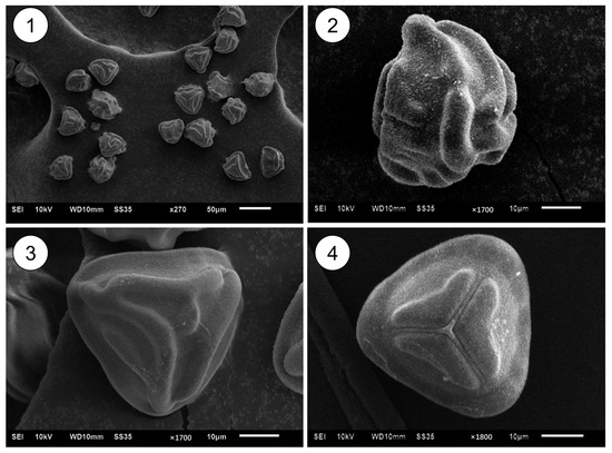
The spores of C. barometz are sub-tetrahedral and radially symmetrical, with an equatorial surface ornamented by an annulate pattern of rounded ribs. In the equatorial view, the proximal face is slightly convex while the distal face is hemispherical. The proximal face appears triangular, with three fissures that are approximately two-thirds the length of the spore radius, and the distal face is also triangular, featuring a distinct ornamentation of raised rounded ribs (Figure 1).
Figure 1.
Spore micrograph. 1. Multi-directional views of spores; 2. equatorial surface of spores; 3. distal face of spores; 4. proximal face of spores.
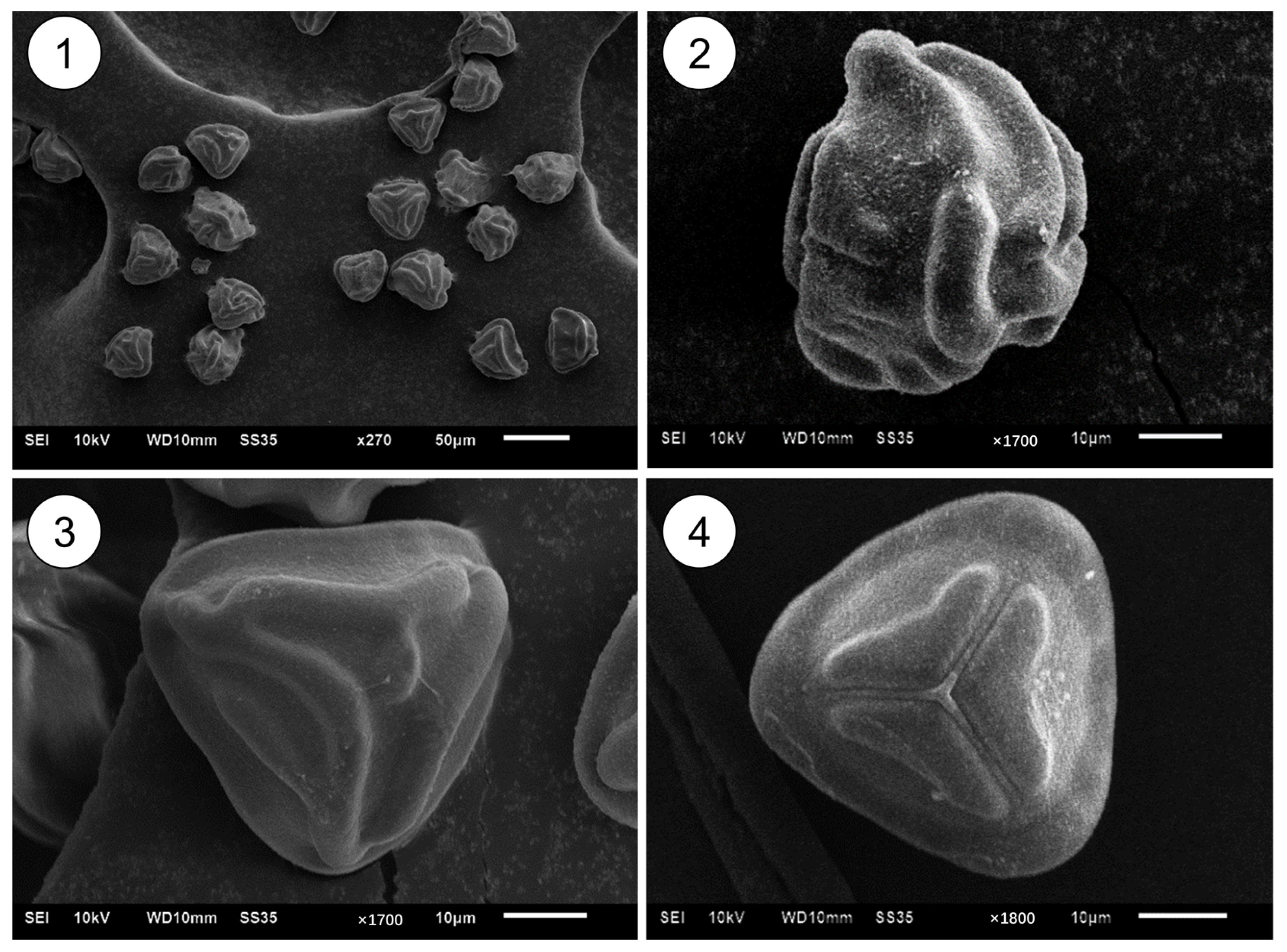
Based on the observations of the developmental process from spores to sporophytes combined with field observations during spore sampling, the life cycle diagram of C. barometz was drawn (Figure 2). According to the observations, most spores turned green and germinated successively within 7–15 days after sowing. Filamentous bodies formed at 9–20 days, prothallial plate developed at 15–35 days, and prothalli emerged at 28–55 days. The prothalli were visible to the naked eye in the early stage and could persist for up to 180 days. Antheridia formed at 40–70 days and archegonia developed at 55–80 days, with the latter able to persist for up to 90 days. The initial germination time of spores varied considerably: when the early-germinating spores developed into prothalli, the germination of newly emerging spores could also be observed in the substrate at the same time.
Figure 2.
Life Cycle of C. barometz. All time points indicated in the figure are calculated after spore sowing. 7–15 d: spores turn green and germinate within 7–15 days post-sowing; 9–20 d: filaments form within 9–20 days post-sowing; 5–30 y: t takes approximately 5 years for spores to develop into mature plants; the rhizomes reach the harvestable size for medicinal use in 5–30 years post-sowing.
3.1.1. Germination Stage of Spore Propagation
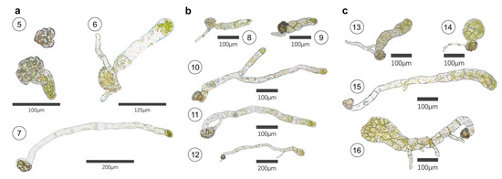
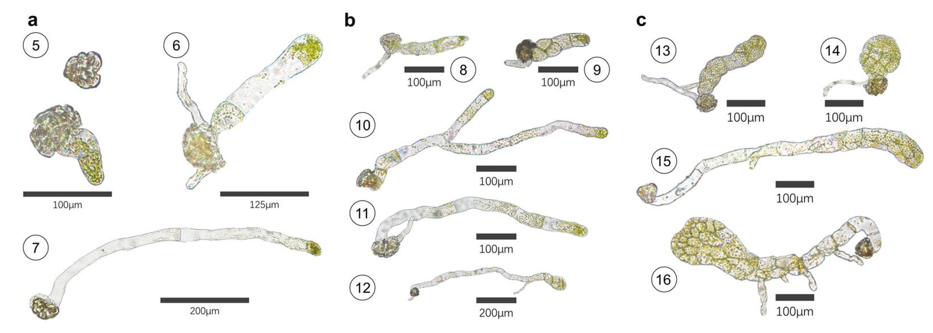
Before germination, the spore cells absorb water and turn green, with their interior filled with chloroplasts. Subsequently, a primary prothallial cell emerges from the trilete fissure on the proximal face of the spore, and 1–2 primary rhizoids may grow out simultaneously. Some spores, however, do not produce primary rhizoids from this site. The rhizoids contain a small amount of chloroplasts (Figure 3a).
Figure 3.
Micrograph of spore germination stage (a). Germination stage of spore: 5. Spore greening and germination; 6. Germination with primary rhizoids; 7. Germination without primary rhizoids. (b) Filamentous stage: 8. Uniseriate short filaments; 9. Multiseriate filaments; 10. Branched filaments; 11. Long filaments; 12. Filaments with rhizoids; (c) Prothallial plate stage: 13. Apical cell division of shorter filaments; 14. Shorter prothallial plate; 15. Apical cell division of longer filaments; 16. Longer prothallial plate.
3.1.2. Filamentous Stage of Spore Propagation
The primary prothallial cells undergo continuous division to form uniseriate or multiseriate filaments composed of 2–8 cells, and branched filaments can also be observed. The filaments vary greatly in length: the shorter ones measure 150–200 μm, while the longer ones can reach 600–650 μm. Rhizoids can differentiate from the filament cells (Figure 3b).
3.1.3. Prothallial Plate Stage of Spore Propagation
Filament cells undergo transverse division to form prothallial plate. The size of the prothallial plate is determined by that of the filaments: prothallial plate developed from shorter filaments are relatively small, whereas those developed from longer filaments are comparatively large (Figure 3c).
3.1.4. Green Villous Body Stage of Spore Propagation
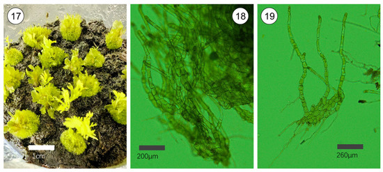
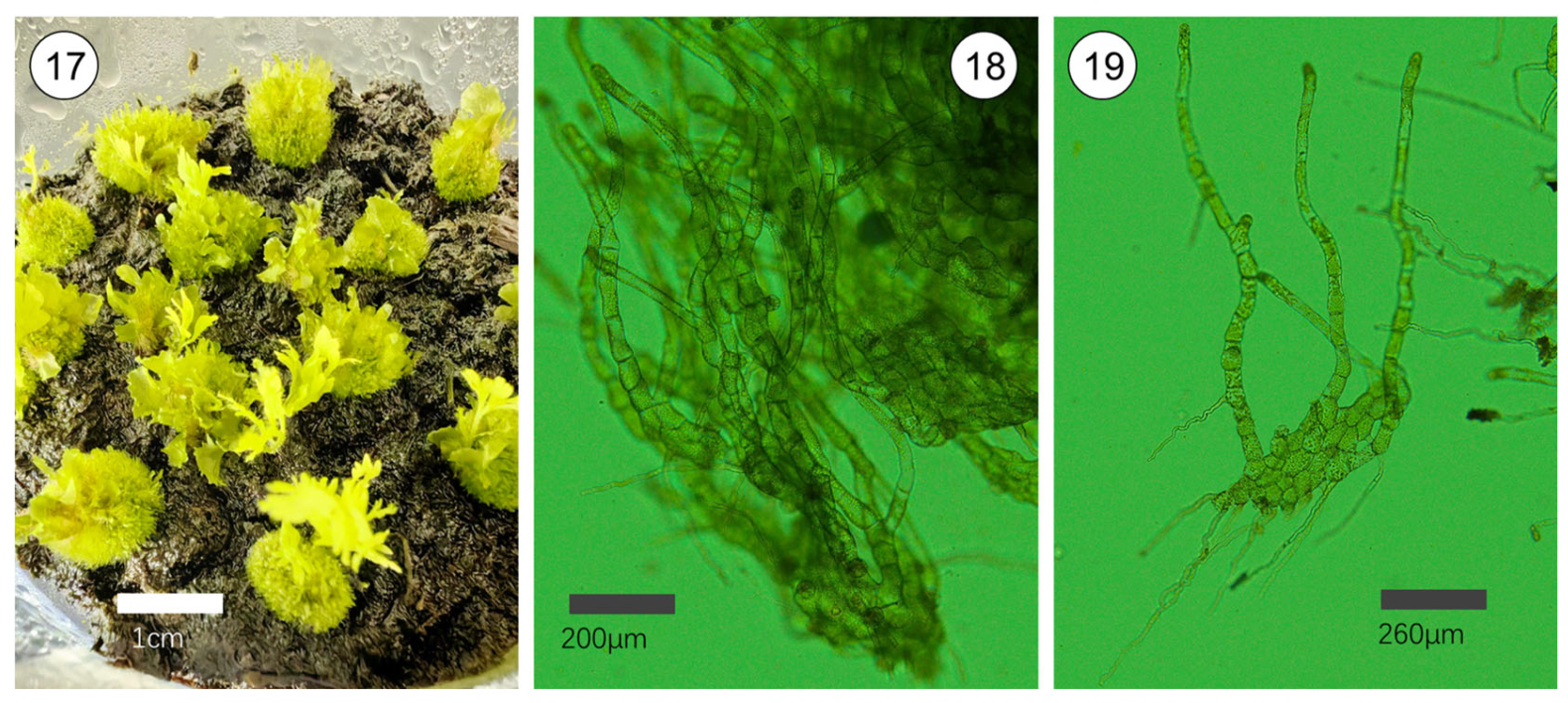
Another morphological characteristic was observed after spore germination. Visible to the naked eye as small, green villous hemispherical bodies, they can gradually increase in size and further develop into numerous prothalli and sporophytes. When dissected and observed under a microscope, these bodies are found to be aggregated structures formed by a large number of filamentous tissues produced via cell division.
Due to their morphological differences from the green globular bodies (GGB) induced from explants [25], they are tentatively named green villous bodies (Figure 4). Observations indicated that they can also develop into prothalli and sporophytes in subsequent stages.
Figure 4.
Green villous body stage of spore propagation. 17. Photo of green villous bodies; 18. Partial micrograph of green villous bodies; 19. Dissected micrograph of green villous bodies.
3.1.5. Gametophyte (Prothallus) Stage of Spore Propagation
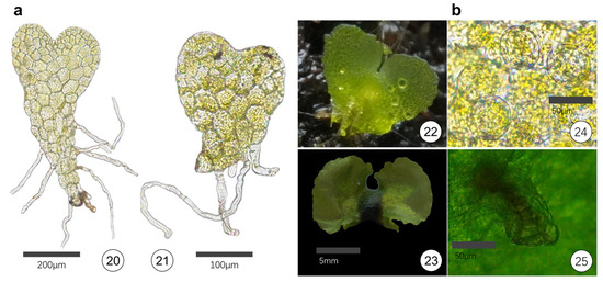
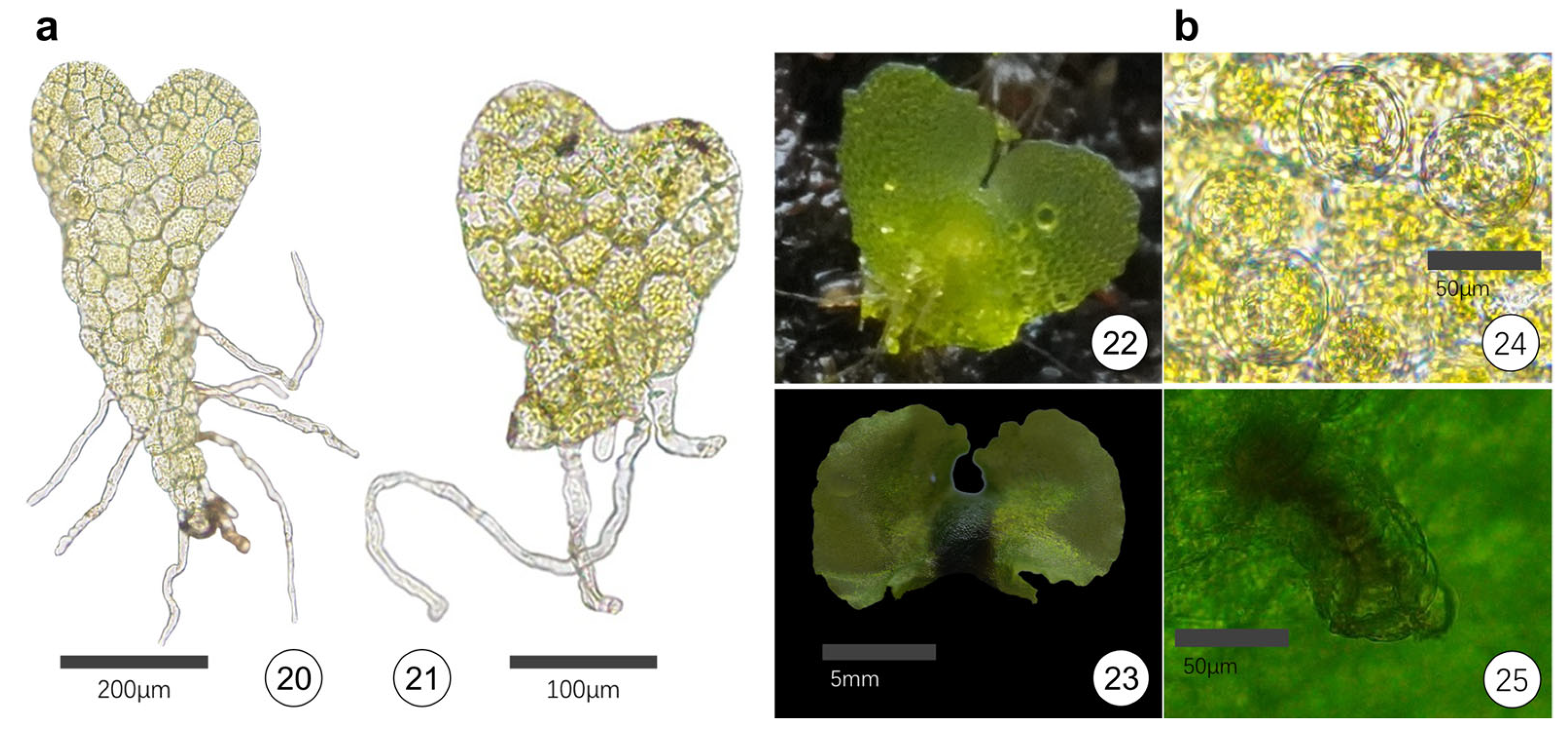
As the thallus cells undergo increased division towards both sides, the anterior end of the thallus becomes depressed, and the overall morphology takes on a cordate shape, marking the entry into the young prothallus stage. Rhizoids grow attached to the peripheral cells at the lower part of the prothallus (Figure 5a). With continued growth, most prothallus cells undergo further division on both sides, and the depressed area at the apex becomes increasingly prominent, forming symmetrical cordate or long-cordate prothalli; a small number develop into symmetrical narrow linear prothalli. The margin of the prothallus gradually forms folds, with a maximum width of approximately 0.8–1.2 cm, and the rhizoids are distributed near the axis of symmetry.
Figure 5.
Gametophyte (Prothallus) stage of spore propagation. (a) Gametophyte stage: 20–21. Prothallus micrographs; 22. Early stage prothalli; 23. Late-stage prothalli. (b) Sexual organs: 24. Antheridia; 25. Archegonia.
Numerous antheridia can be observed on the prothallus. The antheridia are spherical and visible in a wide range near the rhizoids and on both sides of the central axis of the prothallus. Ten to twenty days after the formation of the antheridia, archegonia appear on the abaxial side of the prothallus, located near the apex on both sides of the central axis (Figure 5b).
3.1.6. Sporophyte Stage of Spore Propagation
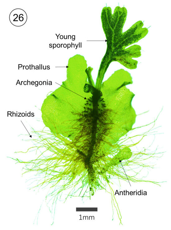
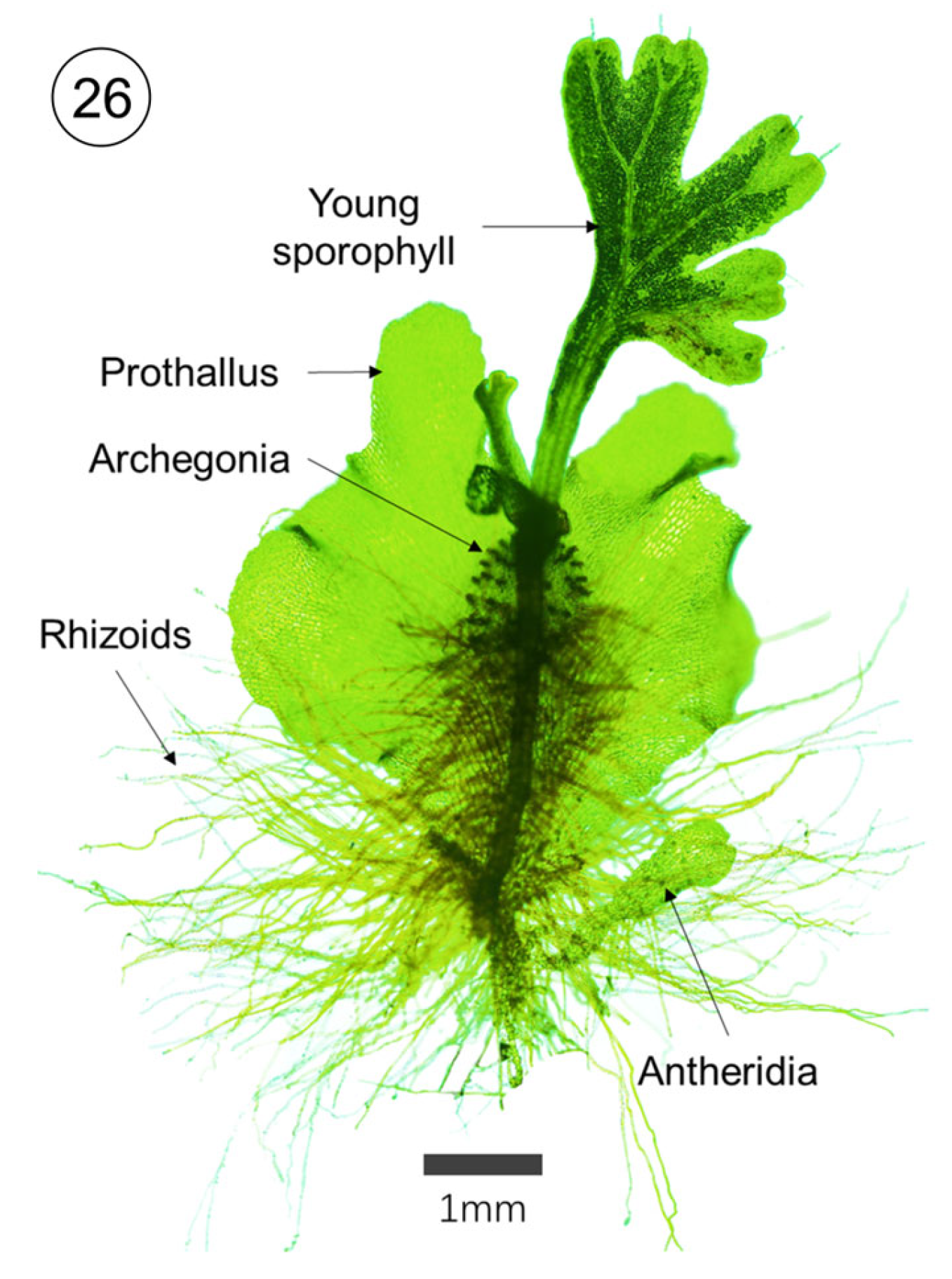
After the archegonia complete fertilization, they develop into young sporophytes, which produce sporophylls and root systems (Figure 6). A small number of prothalli can develop 2–3 sporophytes on the same individual.
Figure 6.
Sporophyte stage of spore propagation. 26. Young sporophyte.
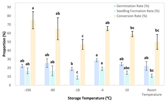
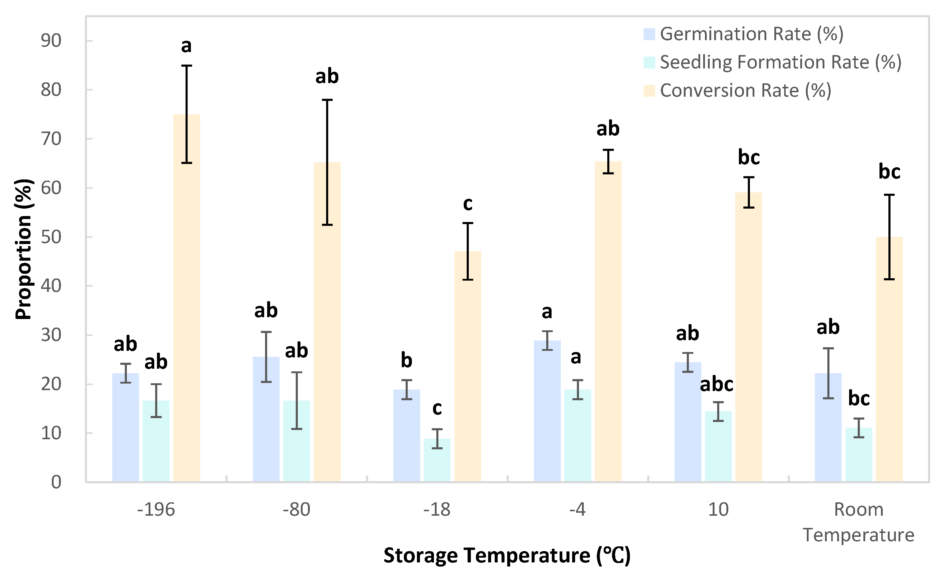
3.2. Effects of Spore Storage Temperature on Germination and Seedling Formation Rate
The results showed that the spore germination rate reached the highest value of 28.89% at a storage temperature of −4 °C, which was significantly higher than the 18.89% recorded at −18 °C, but showed no significant difference compared with that under other storage temperatures.
The spore seedling formation rate peaked at 18.89% when stored at −4 °C, which was significantly higher than the rates of 8.89% at −18 °C and 11.11% at room temperature, while no significant differences were observed in comparison with other storage temperatures.
The conversion rate was the highest at 75% at a storage temperature of −196 °C, which was significantly higher than those at −18 °C, 10 °C and room temperature, but exhibited no significant differences from the rates at −80 °C and −4 °C (Figure 7).
Figure 7.
Effects of storage temperature on spore germination rate, sporophyte seedling formation rate and gametophyte–sporophyte conversion rate. Different lowercase letters denote significant differences among treatments, determined via one-way ANOVA followed by Duncan’s multiple range test (p < 0.05). Lowercase letters were labeled separately for germination rate, seedling formation rate, and conversion rate.
In the temperature gradient where the spore storage temperature was gradually increased from −196 °C, −4 °C served as an important temperature node. When the storage temperature was higher than −4 °C, the germination rate and seedling formation rate decreased progressively. When the storage temperature was lower than −4 °C, the germination rate and seedling formation rate first decreased and then showed an increasing trend. The spore germination rate, seedling formation rate and conversion rate were all the lowest when stored at −18 °C.
3.3. Effects of Spore Storage Duration on Germination and Seedling Formation Rate
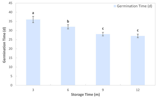
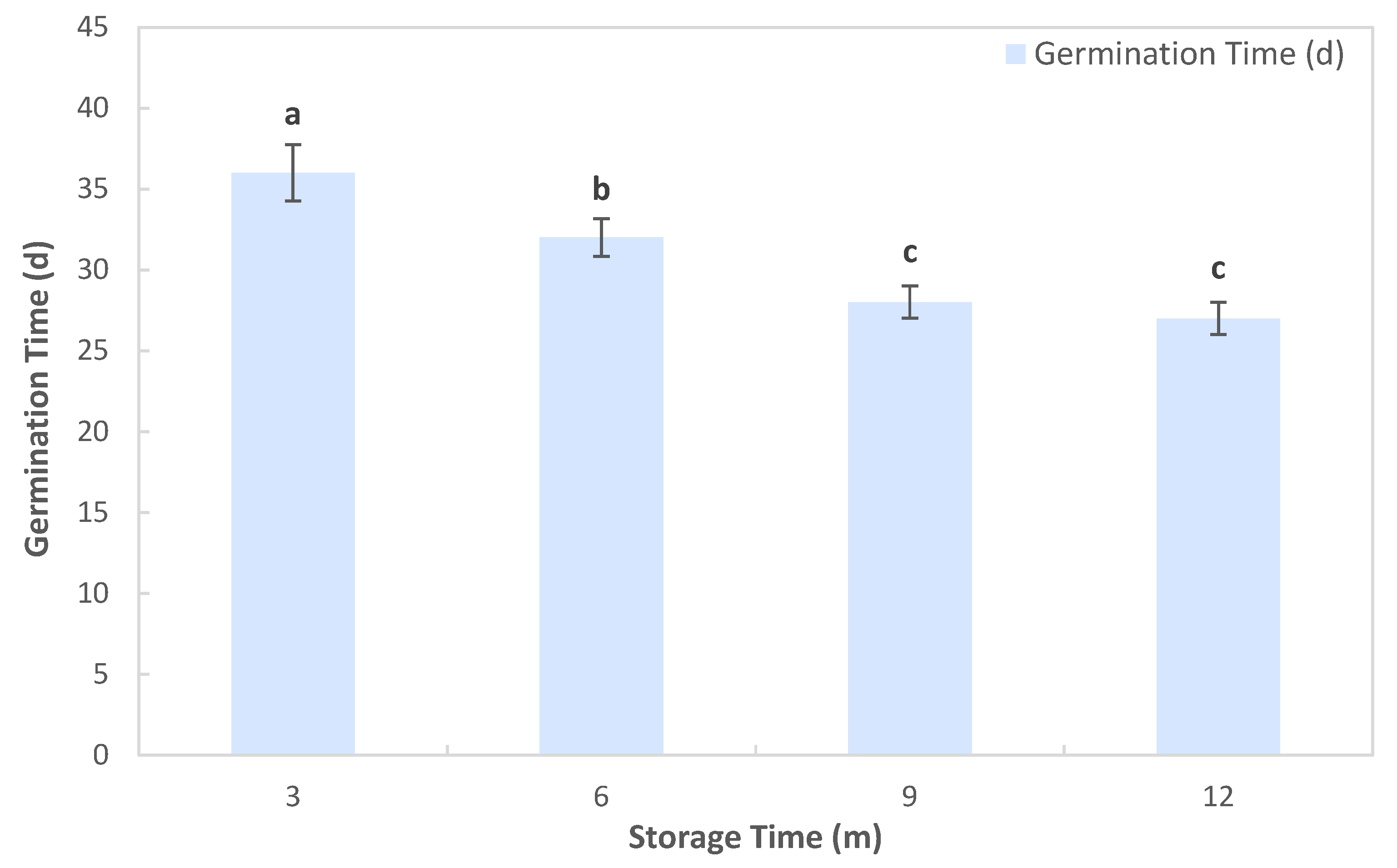
With the extension of spore storage duration, the time required for spore germination decreased significantly from an average of 36 days for spores stored for 3 months, to 32 days for those stored for 6 months, and then to 28 days for those stored for 9 months. There was no significant difference between spores stored for 12 months and those stored for 9 months, with the germination time reaching the minimum of 27 days (Figure 8).
Figure 8.
Effects of storage duration on germination time. Different lowercase letters denote significant differences among treatments, determined via one-way ANOVA followed by Duncan’s multiple range test (p < 0.05).
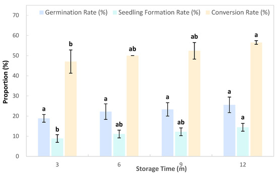
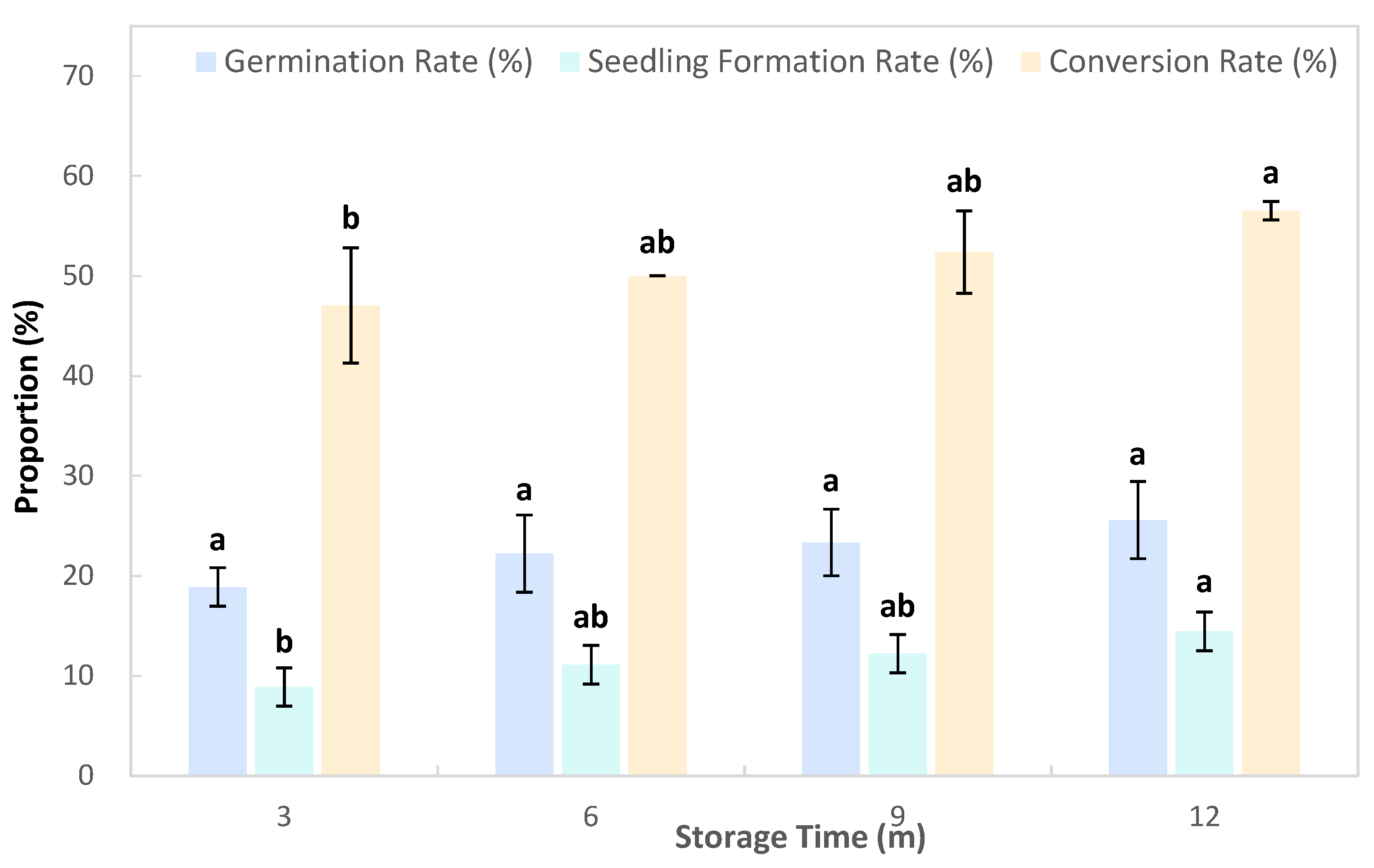
The spore germination rate, seedling formation rate, and conversion rate from gametophytes to sporophytes increased gradually with the extension of storage duration. The germination rate, seedling formation rate, and conversion rate were the lowest when spores were stored for 3 months, at 18.89%, 8.89%, and 47.06%, respectively. These indices peaked when spores were stored for 12 months, at 25.56%, 14.44%, and 56.52%, respectively, which were significantly higher than the seedling formation rate and conversion rate of spores stored for 3 months (Figure 9).
Figure 9.
Effects of storage duration on spore germination rate, sporophyte seedling formation rate and gametophyte–sporophyte conversion rate. Different lowercase letters denote significant differences among treatments, determined via one-way ANOVA followed by Duncan’s multiple range test (p < 0.05). Lowercase letters were labeled separately for germination rate, seedling formation rate, and conversion rate.
3.4. Effects of Spore Sowing Density on Germination and Seedling Formation Rate
3.4.1. Relationship Between Spore Quantity and Volume
According to the spore counting method, the number of spores in a 2 μL spore suspension was counted with seven replications, and the average value was 33.57 spores. Based on this calculation, the number of spores in a 40 μL volume was 839,250 spores.
3.4.2. Effects of Sowing Density on Germination and Seedling Formation Rate
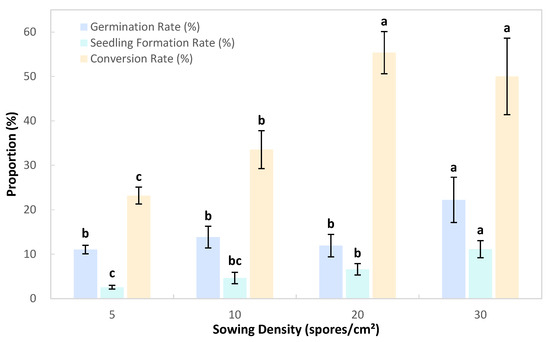
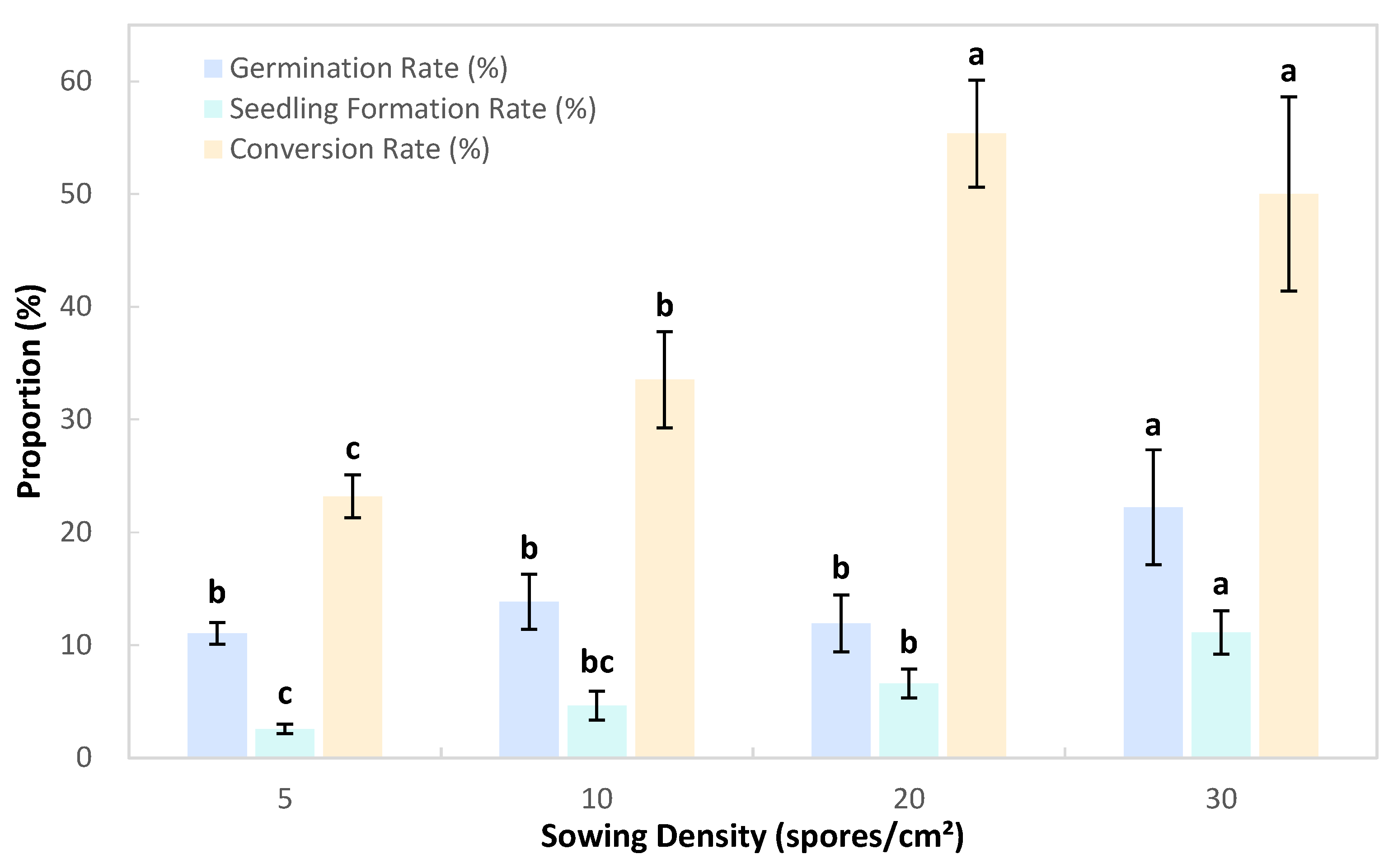
When the spore sowing density was 30 spores/cm2, the spore germination rate and seedling formation rate reached their peak values, at 22.22% and 11.11%, respectively, which were significantly higher than those at other sowing densities. The conversion rate was the highest (55.37%) at a sowing density of 20 spores/cm2, which was significantly higher than the rates at 5 spores/cm2 and 10 spores/cm2, but showed no significant difference from the rate at 30 spores/cm2 (Figure 10).
Figure 10.
Effects of sowing density on spore germination rate, sporophyte seedling formation rate and gametophyte–sporophyte conversion rate. Different lowercase letters denote significant differences among treatments, determined via one-way ANOVA followed by Duncan’s multiple range test (p < 0.05). Lowercase letters were labeled separately for germination rate, seedling formation rate, and conversion rate.
4. Discussion
This study focused on the key large-scale seedling propagation technologies for the protected medicinal fern C. barometz. The full dynamic life cycle of spore-based propagation characterized in this work provides quantitative temporal parameters to standardize commercial seedling breeding schedules. Our investigation of spore storage temperature and duration clarifies the critical thermal and time thresholds for maintaining spore viability, which establishes reliable baseline parameters for standardized long-term spore preservation and laying a foundation for year-round seedling breeding. The sowing density trial preliminarily identified the optimal seeding dosage for nursery cultivation practice: the spore germination and sporophyte formation rates peaked at 30 spores per cm2. We also note that further gradient trials with higher seeding densities are required to verify whether a more productive upper threshold exists without inhibiting gametophyte growth.
Most previous research on C. barometz ferns has predominantly focused on in vitro tissue culture systems [22], including protoplast induction [23], culture medium screening [24], and plant growth regulator regulation of gametophyte development [26]. These sterile culture protocols yield high propagation efficiency [25] but demand strict ultra-clean laboratory facilities, precise medium formulation, and sustained technical operation, which substantially raise upfront construction and running costs for seedling producers. In contrast, the substrate-based spore sowing system established in the present study operates under conventional non-axenic incubator conditions, with low requirements for specialized laboratory infrastructure and complicated chemical medium preparation. This cost-effective feature greatly lowers the technical and financial barriers for forest conservation nurseries and medicinal plant enterprises to implement large-scale artificial breeding and ex situ population restoration of wild C. barometz resources.
The results demonstrated that appropriately prolonged cold storage continuously shortened the germination lag phase and simultaneously elevated final germination and sporophyte seedling formation rates. This trend can be attributed to slow metabolic dormancy induced by low temperature, which alleviates oxidative damage to spore cytoplasm and maintains intact cell membrane structures to preserve long-term viability. Nevertheless, the maximum storage duration tested in this experiment was limited to 12 months. Comparatively, Peng et al. [27] reported that spore germination capacity of another understory fern peaked after 630 days of cold preservation, indicating that fern spore viability can be sustained over far longer timeframes under optimized low-temperature regimes. Therefore, extended multi-year storage trials covering continuous temperature gradients are needed to clarify the long-term dynamic changes in spore vitality, gametophyte development and sporophyte conversion efficiency across prolonged preservation periods.
A unique developmental phenomenon observed in this culture system was the formation of multiple independent sporophytes originating from a single prothallus, which confirms that numerous archegonia distributed on one gametophyte can independently complete fertilization after sufficient antheridia sperm release. For further improvement of propagation efficiency, hybrid protocols combining simplified tissue culture operations with substrate cultivation are worthy of exploration. As documented in [20], young prothalli can be homogenized in half-strength MS liquid medium and inoculated onto solid medium or sterilized peat substrate; this fragmentation approach drastically multiplies viable gametophyte material and accelerates the entire sporophyte formation cycle.
When individual intact gametophytes were separated and transplanted into independent seedling containers, partial isolated prothalli successfully developed into mature sporophytes, verifying that single gametophytes possess independent sporophyte regeneration potential. Fine-tuning microenvironmental conditions (light intensity, substrate humidity, ambient temperature) during the vulnerable gametophyte stage and optimizing routine moisture management regimes will further lift the overall seedling formation efficiency. We also detected a clear positive correlation between sowing density and sporophyte yield, accompanied by obvious variation in germination synchronization across density treatments. This phenomenon can be mechanistically explained by elevated inter-gametophyte contact frequency under higher seeding densities, which facilitates sperm migration and cross-fertilization between genetically distinct gametophytes, thereby boosting the probability of successful fertilization and subsequent sporophyte generation.
A distinctive aggregated filamentous structure termed the “green villiform structure” was identified throughout the developmental process. Antheridia and archegonia were directly differentiated on the surface of these filamentous clusters, and immature prothalli and gametophytes continuously emerged from branching villi filaments. We infer that fertilization events may occur directly on the villiform filaments prior to sporophyte initiation. Morphologically, this structure originates from repeatedly branched primary spore germ filaments; continuous filament branching drives the aggregation and formation of green villiform clusters, and selected lateral branches differentiate into typical heart-shaped prothalli to enter subsequent sexual development stages. Additional cytological and morphological observation experiments are required to confirm whether this green villiform aggregation is functionally equivalent to the green globular body (GGB) [23], a common propagule structure widely reported in other fern micropropagation studies.
From the perspective of industrial medicinal cultivation, the full production cycle of C. barometz needs systematic sorting based on our spore propagation system. Under the indoor non-axenic spore sowing conditions established in this study, viable sporophyte seedlings suitable for field transplantation can be obtained within 8–10 months after spore seeding. After outdoor transplanting, seedlings enter slow rhizome expansion growth; rhizomes reach the medicinal harvest specification after 5–30 years of continuous cultivation, with growth rate largely controlled by shade, soil moisture and substrate nutrient supply. This long growth cycle is a universal bottleneck limiting commercial planting of tree fern medicinal materials.
Compared with rhizome division propagation that directly harvests mature wild individuals and shortens the seedling stage, spore seedling propagation sacrifices short-term harvest speed but achieves zero damage to wild germplasm resources. Combined with the low-cost and easy-operating advantages of our spore sowing technology, nursery enterprises can build seedling banks for batch seedling breeding in advance to alleviate the long-cycle pressure of medicinal cultivation.
Three mainstream propagation pathways for C. barometz have been recorded: rhizome clump division, sterile tissue culture and substrate-based spore sowing established in this research, and their practical industrial application characteristics differ drastically in seedling cycle, facility input, germplasm protection and production cost.
Rhizome clump division: The operation relies on mature wild or artificially grown rhizome blocks, and transplantable seedlings can be formed within 2–3 months. However, this method cannot expand population quantity on a large scale, and wild rhizome excavation will further endanger wild populations, which conflicts with wild plant conservation policies; it is only applicable to small-scale individual reproduction in botanical gardens.
In vitro tissue culture: The technique realizes high-efficiency mass proliferation of gametophytes and sporophytes, but relies on constant-temperature ultra-clean laboratories, high-purity culture medium, regular subculture operation and strict sterile management. The early construction and daily operation costs are extremely high for small and medium-sized medicinal planting enterprises; the technical threshold also limits widespread promotion in mountain rural nurseries.
Non-axenic spore sowing in this study: Seedlings can be produced using common seedling incubators and peat substrate without sterile workshop construction. The overall facility investment and daily technical management costs are reduced by more than 60% compared with tissue culture. Although the time required to obtain transplantable sporophytes (8–10 months) is longer than rhizome division, spores can be collected from artificially conserved maternal plants to realize unlimited batch seedling breeding without damaging wild populations, balancing conservation demands and medicinal material production needs.
For actual cultivation promotion, transplanting of sporophyte seedlings is recommended in late spring (April–May) after stable temperatures above 20 °C, with shading net coverage of 70%–80% to simulate understory wild habitat light environment. The suitable cultivation substrate is loose, humus-rich acidic peat loam with good drainage; mountain underwood land with scattered light, high air humidity and gentle slope is the optimal field planting location, avoiding open fields with strong direct sunlight and waterlogged lowlands.
In terms of harvest management, partial artificial planted individuals can begin to carry out rhizome picking after 5 years of growth; full rhizome harvesting for complete medicinal raw material output is generally arranged after 15–30 years of cultivation. Combined with the low-cost seedling breeding system in this paper, growers can build layered seedling and adult plant cultivation plots to form a continuous production chain of seedling supply, young plant cultivation and medicinal rhizome harvest, significantly improving the economic and ecological comprehensive benefits of C. barometz planting.
5. Conclusions
When sown after 1 year of storage, the spores germinated earlier and achieved a higher seedling formation rate. The highest seedling formation rate of 18.89% was obtained for spores stored at −4 °C, while the maximum seedling formation rate of 11.11% was recorded at a sowing density of 30 spores/cm2. The morphological characteristics and the time required for each stage of spore germination provide a basis for judging the germination stages in seedling breeding and production. The research results on spore storage conditions and sowing density offer important basic data for formulating spore propagation plans.
Author Contributions
Conceptualization, S.Z.; Data curation, S.Z., T.L. and Y.J.; Formal analysis, S.Z.; Funding acquisition, J.W.; Investigation, J.Y., S.H. and K.L.; Methodology, S.Z.; Project administration, Q.W., J.W.; Resources, J.Y., S.H. and K.L.; Supervision, Q.W.; Validation, J.Y.; Visualization, S.Z.; Writing—original draft, S.Z. and Y.J.; Writing—review and editing, Q.W. All authors have read and agreed to the published version of the manuscript.
Funding
CAMS Innovation Fund for Medical Sciences (CIFMS) (Grant No. 2023-I2M-2-006); State Administration of Traditional Chinese Medicine of the People’s Republic of China (GZY-KJS-2023-008); Yunnan Provincial Jianhe Wei Expert Workstation Project under the Science and Technology Talents and Platform Program (Academician and Expert Workstation) (Project No. 202405AF140129).
Data Availability Statement
Data are contained within the article.
Acknowledgments
We thank all the participants in this study.
Conflicts of Interest
Author Shuwen He was employed by Suijiang County Yaofu Modern Agriculture Development Co., Ltd. Author Ke Li was employed by Pu’er Liangpin Yikang Pharmaceutical Co., Ltd. The remaining authors declare no commercial or financial relationships that may constitute potential conflicts of interest.
References
- Bateman, D.B. The Origin and Early Diversification of Land Plants: A Cladistic Study.—Paul Kenrick and Peter Crane. Syst. Biol. 2001, 50, 753–754. [Google Scholar] [CrossRef] [Scilit]
- Li, J.; Chen, X.; Wu, P.; Niklas, K.J.; Lu, Y.; Zhong, Q.; Hu, D.; Cheng, L.; Cheng, D. The fern economics spectrum is unaffected by the environment. Plant Cell Environ. 2022, 45, 3205–3218. [Google Scholar] [CrossRef] [Scilit] [PubMed]
- Zhang, Y.M. A Preliminary Investigation of the History of Pteridophyte Eating in China. J. Southwest Agric. Univ. (Soc. Sci. Ed.) 2007, 5, 103–106. (In Chinese) [Google Scholar]
- Wang, X.; Liu, T.T.; Sun, H. Physiological Characteristics and Application Value of Ferns. Border Econ. Cult. 2012, 5, 171–172. (In Chinese) [Google Scholar]
- Zhang, J.; Cai, J.R.; Xu, J.X.; Liu, Z.W.; Shen, Y.H.; Qian, T.H.; Liu, J.H. Research Progress on Application of Ferns in Ecological Restoration. J. Anhui Agric. Sci. 2017, 45, 69–71, 109. (In Chinese) [Google Scholar]
- Han, Y.L.; Ren, Y.; Cao, Y.; Xiao, L.Y.; Li, H.; Wang, Y.; Li, X.W. Research Progress on Resource Status and Artificial Cultivation of Medicinal Ferns. Chin. Tradit. Herb. Drugs 2026, 2. (In Chinese) [Google Scholar]
- Cao, Y.F.; Xue, Y.H. Analysis of Domestic Cultivation Technology of Ferns in Changbai Mountain. Mod. Hortic. 2013, 2, 41. (In Chinese) [Google Scholar]
- Xiang, C.C.; Yang, M.; Yu, Q.L.; Zhou, P.C.; Xiao, D.; Wei, K.H. Research Progress on Davallia trichomanoides. South. Agric. 2024, 15. (In Chinese) [Google Scholar]
- Wen, Z.S.; Li, X.R.; Qi, L.T.; Li, J.C.; Ma, D.L.; Zheng, Y.G. Textual research on the variety and habitat changes of Cibotium barometz in Chinese herbal medicine. China Pharm. 2019, 30, 553–555. [Google Scholar]
- Ye, H.H.; Dong, Y.; Han, J.Y.; Ren, Q.F.; Yuan, M.Q.; Zhang, M.; Huang, F.Y. Research Progress on Chemical Constituents, Pharmacological Effects and Quality Control of Cibotium barometz. Guizhou Agric. Sci. 2020, 38, 28–31. [Google Scholar]
- Xu, X.R.; Wang, J.Y.; Wang, X.Y.; Bi, H.Z.; Bai, Q.X.; Wang, M. Botany, traditional uses, phytochemistry, pharmacology, processing, and applications of Cibotium barometz (L.) J. Sm.: A review. Fitoterapia 2025, 186, 106843. [Google Scholar] [CrossRef] [Scilit] [PubMed]
- Heng, Y.W.; Ban, J.J.; Khoo, K.S.; Sit, N.W. Biological activities and phytochemical content of the rhizome hairs of Cibotium barometz (Cibotiaceae). Ind. Crops Prod. 2020, 153, 112612. [Google Scholar] [CrossRef] [Scilit]
- Pharmacopoeia Commission of the People’s Republic of China. Pharmacopoeia of the People’s Republic of China (2020 Edition) Volume I; China Medical Science and Technology Press: Beijing, China, 2020. [Google Scholar]
- Yang, C.Z.; Liu, X.F.; Cai, D.L.; Fan, S.M. Resource investigation and quality evaluation of Cibotium barometz. China J. Chin. Mater. Med. 2015, 40, 1919–1924. [Google Scholar]
- Liu, G.H. Study on Gametophyte Development, Population and Community Structure of the Protected Plant Cibotium barometz. Master’s Thesis, Southwest University, Chongqing, China, 2007. [Google Scholar]
- Chen, D.L.; Zhong, C.; Mi, K.; Jian, S.F.; Miao, J.H.; Wei, K.H.; Li, L.X.; Guo, X.Y. Research progress on resource status and conservation of the endangered medicinal plant Cibotium barometz. Guizhou Agric. Sci. 2022, 50, 122–129. [Google Scholar]
- Ming, Y. Official announcement of the revised National Key Protected Wild Plants List. Green China 2021, 581, 74–79. [Google Scholar]
- Su, Y.Q.; Mo, Y.L. Study on seedling breeding of Cibotium barometz. Mod. Agric. Sci. Technol. 2019, 8, 47–49. [Google Scholar]
- Zhang, Z.R.; Zhang, S.B. Preliminary study on spore propagation technology of Cibotium barometz, a national second-class protected medicinal and ornamental plant. North. Hortic. 2010, 13, 203–206. [Google Scholar]
- Li, X.; Ye, Q.M.; Zhan, Q.C.; Kang, B.L.; Jian, L.G.; Huang, M.L. Establishment of in vitro culture and plant regeneration system of Cibotium barometz. North. Hortic. 2010, 6, 152–155. [Google Scholar]
- Yu, Q.; Lin, W.; Yu, X.; Qin, B.; Liang, Y.; Wei, F.; Qin, S.; Wei, K. Research on the rapid tissue breeding technology of the endangered plant Cibotium barometz (L.) J. Sm. Plant Cell Tissue Organ Cult. 2024, 158, 38. [Google Scholar] [CrossRef] [Scilit]
- Zhao, L.T.; Lin, D.X.; Li, B. Artificial breeding and high-efficiency cultivation technology of Cibotium barometz. For. Sci. Technol. Commun. 2025, 1, 63–66. [Google Scholar] [CrossRef]
- Liu, X.; Xue, M.; Xu, L.; Liang, Y.M.; Shi, Y.T.; Tang, Q.H.; Wang, W.Q. Research progress on spore propagation technology of medicinal ferns in traditional Chinese medicine. Asia-Pac. Tradit. Med. 2018, 14, 80–83. (In Chinese) [Google Scholar]
- Pu, L.L. Study on Breeding Technology of Cibotium barometz. Master’s Thesis, Guangzhou University of Chinese Medicine, Guangzhou, China, 2015. [Google Scholar]
- Yu, R.; Zhang, G.; Li, H.; Cao, H.; Mo, X.; Gui, M.; Zhou, X.; Jiang, Y.; Li, S.; Wang, J. In vitro propagation of the endangered tree fern Cibotium barometz through formation of green globular bodies. Plant Cell Tissue Organ Cult. 2016, 128, 369–379. [Google Scholar] [CrossRef] [Scilit]
- Chen, X.D.; Zhou, Y.L.; Chang, C.Y.; Li, Y.L. Scanning electron microscope observation on spores of three key protected ferns in China. J. Beijing Norm. Univ. (Nat. Sci. Ed.) 2002, 2, 250–254. [Google Scholar]
- Peng, L.L. Study on the Effects of Environmental Factors on Spore Germination and Gametophyte Development of Cibotium barometz. Master’s Thesis, Southwest University, Chongqing, China, 2013. [Google Scholar]
Disclaimer/Publisher’s Note: The statements, opinions and data contained in all publications are solely those of the individual author(s) and contributor(s) and not of MDPI and/or the editor(s). MDPI and/or the editor(s) disclaim responsibility for any injury to people or property resulting from any ideas, methods, instructions or products referred to in the content. |
© 2026 by the authors. Licensee MDPI, Basel, Switzerland. This article is an open access article distributed under the terms and conditions of the Creative Commons Attribution (CC BY) license.