Article

Genome-Wide Identification of the PRP Gene Family Members of the Dove Tree (Davidia involucrata Baill.)

1
College of Life Sciences and Technology, Central South University of Forestry and Technology, Changsha 410004, China
2
Coconut Research Institute, Chinese Academy of Tropical Agricultural Sciences, Wenchang 571339, China
3
Yuelushan Laboratory Carbon Sinks Forests Variety Innovation Center, Changsha 410004, China
*
Author to whom correspondence should be addressed.
Forests 2025, 16(9), 1425; https://doi.org/10.3390/f16091425
Submission received: 2 June 2025 / Revised: 1 September 2025 / Accepted: 3 September 2025 / Published: 5 September 2025
(This article belongs to the Special Issue Latest Progress in Research on Forest Tree Genomics)

Abstract

The large, white paired bract is a unique trait, as well as the most intriguing feature of the dove tree (Davidia involucrata). However, the mechanisms underlying bract development remain unclear. Our previous comparative transcriptome analysis concerning Davidia bracts at different developmental stages has identified a number of bract-specific genes. Among these, the genes encoding PRPs (proline-rich proteins) show dramatic expression variation, indicating the participation of this gene family in bract development. In this study, we screened the whole Davidia genome and identified twelve Davidia PRP (DiPRP) genes, showing obvious expression variation during bract development, with some upregulated up to 100-fold at the fast-developing stage. These PRP genes are evenly distributed on seven Davidia chromosomes. The cis-element composition of the promoter regions of the DiPRPs demonstrates that these genes might be controlled by phytohormones (especially ABA, GA, and MeJA), light, and the circadian clock, which is consistent with the environmental cues during Davidia bract development. Synteny analysis indicated that the PRP genes from the Davidia genome have higher collinearity with naturally bracted plants, such as Antirhonum majus and Bougainvillea glabra, but lower collinearity with non-bracted species. Our results suggest that high expression of certain PRP genes, specifically in bracts, might be critical for leaf metamorphosis.

1. Introduction

Floral organs are critical for the sexual reproduction of plants. Various morphological features of floral organs attract the attention of both ornamental applications and developmental biology studies. A perfect flower usually contains four types of floral organs, including sepal, petal, stamen (male reproductive organ), and carpel (female reproductive organ) [1]. However, there are many plant species that possess imperfect flowers lacking one or more of the above floral organs. For instance, sepals and petals are absent in the flowers of Spathiphyllum lanceifolium, Rheum nobile, Bougainvillea spectabilis, and Davidia involucrata. Alternatively, the functions of sepals and petals in these species are partially substituted by a bract, an organ generated through leaf metamorphosis. Bracts are not real floral organs under the strict definition. However, they only emerge with floral development by closely subtending the inflorescences. Therefore, bracts can be considered as “extended floral organs”. Bracts from different plant species usually exhibit distinctive appearances, such as white spathes in Zantedeschia aethiopica, flower-shaped bracts in Houttuynia cordata, and yellow bracts in Musella lasiocarpa. In addition to their ornamental value, bracts also play a critical role in the reproductive growth of these species. For instance, a number of studies focusing on several bracted plant species have demonstrated that the bright color or color variation of bracts attracts more pollinators, improves the frequency of pollinator visitation, and helps them find pollen sooner [2]. On the contrary, the seed setting rate is greatly reduced if the bracts are artificially removed from these plants [3,4,5]. In addition, some bracts have been proposed to protect pollen from heavy rain or improve the tolerance of floral organs against abiotic stress [6,7,8].
The dove tree (Davidia involucrata Baill., Davidia here after) is an ancient bracted species famous as a “living fossil” [9]. The inflorescence of Davidia is a capitulum type, which lacks sepals and petals but is subtended by a pair of white, large, and asymmetrical bracts. Davidia bracts undergo a color changing process (from light green to pure white) during development and finally exhibit a unique “dove-like” appearance. Based on the morphological characteristics, we have divided the period of bract development into four stages, B1–B4 (Figure 1). At the B1 stage, the bracts and leaves are located in the mixed bud, with minimal phenotypic differences. The stamens on the inflorescences are tightly arranged and wrap around the pistils at this stage. At the B2 stage, the bracts unfold, and the green color fades slightly, entering a rapid growth phase, and the surface tends to be smooth. At the B3 stage, the bracts are completely white with a paper texture. Growth enters a stagnant period, and the stamens are unfolded. At the B4 stage, the stamens are shedding, leaving only green pistils. The edge of the bracts turns yellow and wilted and soon falls off [10]. Microscopic observation demonstrated that the cell architecture of Davidia bract is puzzle-shaped and obviously different from that in leaves. However, no cone cells, the typical cell of petals, were found in Davidia bracts, indicating that they are not real floral organs despite reproductive functions [11]. In brief, the bract is generated through leaf metamorphosis, which is supposed to be triggered by flowering signals.
There are several studies providing some molecular evidence regarding the development mechanism of Davidia bracts. Li et al. (2002) [12] constructed a Davidia cDNA library and identified some differentially expressed genes (DEGs) between bracts and leaves using suppression subtractive hybridization. They found that some stress response-related genes, including LTP (liquid transfer protein) and ASR (abscisic acid, stress and ripening protein), showed significantly higher expression in bracts [12]. In addition, three MADS-box genes, DiSOC1, DiMADS-1, and DiMADS-2, were identified to be dominantly expressed in Davidia bracts. However, overexpression of these genes in Arabidopsis altered the flowering time but did not change floral development [13,14]. These results indicate that there is a distinctive regulatory system governing bract development in Davidia. Although some genes involved in bract development have been identified, the molecular mechanism underlying this process is still largely unclear.
To explore the regulatory mechanisms underlying bract development, we performed transcriptome analysis across different developmental stages of Davidia bracts. Among the DEGs, most of the genes encoding PRPs (proline-rich proteins) showed bract-specific expression and were significantly upregulated during bract development [15]. The first PRP containing tandem repeats of a hexapeptide in its N-terminal was identified in maize (Zea mays) endosperm [16]. More PRPs have been gradually identified in various plant species [17]. PRPs are usually cell wall proteins and can be classified into three types, including type I containing a proline-rich domain and N-signal peptide, type II containing an extra Cys domain, and type III containing pentapeptide repeats but lacking an N-signal peptide. Type II PRPs can be further classified into the 8-CM and LTP families depending on the Cys domain [18,19]. Based on the arrangement of proline in the sequence, the PRP proteins are further divided into three subtypes: (1) proline enrichment occurs throughout the entire sequence; (2) proline enrichment is irregularly distributed; (3) the sequence lacks proline-enriched repeats and contains only small proline-rich motifs [20].
The functions of PRPs have been extensively verified in different plants. They mainly regulate plant growth and development, as well as participate in various stress responses [21,22]. However, whether the PRPs play a role in bract development in Davidia or other bracted species is challenging. In this study, we screened the Davidia PRP genes genome-wide to identify potential members of this family involved in Davidia bract development. This exploration will facilitate a better understanding of the mystery of a unique trait of this ancient species.

2. Materials and Methods

2.1. Plant Materials and Treatments

Samples of Davidia were collected from the Badagong Mountain Nature Reserve, Sangzhi County, Zhangjiajie City, Hunan Province (110°5′30″ E, 28°46′60″ N, 1383 m altitude). All samples were collected from the same tree. The root samples were collected from the non-lignified part at the end. The leaf samples were fully developed mature leaves, and the flower samples were the part of the flower bud without bracts. The samples of leaves, flowers, roots, and bracts were collected in April 2024. The bract samples at four developmental stages are shown in Figure 1B. All samples were immediately preserved in a stabilizer (Accurate Biotechnology (Hunan) Co., Ltd., Changsha, China) after collection, stored at 4 °C for 24 h, and then transferred to −80 °C. Three independent biological replicates were collected for each sample.

2.2. Identification of the DiPRP Genes and Predicted Subcellular Localization

Davidia genome data were obtained from the National Center for Biological Information (https://ngdc.cncb.ac.cn/gwh/PRJCA001721, accessed on 22 March 2023) [23], and the Davidia transcriptome profiles have been deposited in the National Genomics Data Center under accession number PRJCA008410 (https://ngdc.cncb.ac.cn/bioproject/browse/PRJCA008410, accessed on 22 March 2023) [24].
Based on the following criteria, we conducted a sequence search in the Davidia genome to identify the DiPRP genes: (1) sequences with an 8-CM domain at the C-terminus, containing six or eight cysteine residues arranged in a specific pattern, representing the most typical class of hybrid proline-rich proteins; (2) sequences containing multiple repeated polypeptide motifs were analyzed through searches based on previously identified proline-rich repeat sequences. Through this approach, we discovered novel proline-rich repeat sequences that differ from those reported earlier; (3) sequences were searched using a small motif consisting of two proline residues as the criterion. The obtained PRP sequences were submitted to the SMART website for further confirmation of the candidate DiPRPs.
Information such as pI and molecular weight of the DiPRPs was calculated using the online tool Expasy [25] (https://web.expasy.org/protparam/, accessed on 21 July 2023). The subcellular localization of the DiPRPs was predicted using the online website WoLF PSORT (https://wolfpsort.hgc.jp/, accessed on 21 July 2023).

2.3. Analysis of the Expression Patterns of the DiPRPs

The FPKM values of the DiPRPs were extracted from the Davidia transcriptome data regarding the young leaves and bracts at different developmental stages. The expression pattern heatmaps were drawn using TB Tools-II software (v2.326) [26].

2.4. Chromosome Localization

The chromosomal position information of the DiPRPs were obtained from the Davidia genome GFF annotation file, and the chromosome localization map was drawn using TB Tools-II software [26].

2.5. Inter-Species Collinearity Analysis

The genome information for Antirrhinum majus, Malus domestica, Oryza sativa, and Bougainvillea spectabilis were obtained from the National Genomics Data Center (https://ngdc.cncb.ac.cn/gwh/). The Nyssa sinensis genome was obtained from the CoGe database (https://genomevolution.org/coge/). The Arabidopsis genome was obtained from the TAIR database (https://www.arabidopsis.org/). Collinearity analysis between these genomes and the Davidia genome was analyzed using TB Tools-II software [26].

2.6. Identification of Conserved Motifs and Cis-Elements in the Promoter Regions of the DiPRP Genes

Analysis of the conserved motifs of the DiPRPs was conducted using the MEME [27] (https://meme-suite.org/meme/, v5.5.8) online database. The 2000 bp sequence upstream of each DiPRP gene was extracted using TB Tools-II software [26], and the cis-acting elements located in the promoter regions of the DiPRP genes were analyzed using the online tool PlantCARE [28] (https://bioinformatics.psb.ugent.be/webtools/plantcare/html/, accessed on 20 February 2024).

2.7. RNA Extraction and qRT-PCR

The total RNA of different Davidia tissues was extracted using the Steady Pure RNA Extraction Kit (Accurate Biotechnology (Hunan) Co., Ltd., Changsha, China). The cDNA was synthesized using the Evo M-MLV Reverse Transcription Kit (Accurate Biotechnology (Hunan) Co., Ltd., Changsha, China). Primer sequences were designed using Primer 5.0 software, and the primers were synthesized by Shanghai Sangyo Biologicals. The Davidia β-Actin7 gene was used as a reference gene for data normalization [29]. The qRT-PCR reactions were performed on an ABI Step One fluorescence quantifier, and relative expression levels were calculated by the 2−ΔΔct method [30]. The primers used in RT-qPCR are listed in Supplementary Table S1.

2.8. Vector Construction and Arabidopsis Transformation

The pCAMBIA1300-35S-GFP vector was linearized using the BamHI restriction enzyme. The coding sequence of Dinv18207.t1 was obtained from the Davidia transcriptome and cloned to obtain the full-length fragment. This CDS (coding sequence) fragment was inserted into the linearized pCAMBIA1300-35S-GFP vector at the BamHI recognition site and using the In-Fusion Cloning Kit (Vazyme (Jiangsu) Co., Ltd., Nanjing, China) to generate the fusion vector containing Dinv18207.t1.
Arabidopsis thaliana genetic transformation was performed using the floral dip method [31]. Transgenic plants were selected on MS agar plates supplemented with 50 mg/L kanamycin and 25 mg/L hygromycin B. The transgenic lines were further confirmed by qPCR analysis.
Cellular morphology was examined using paraffin-embedded tissue sections [32].

2.9. Statistical Analysis

qPCR analysis was performed using three independent biological replicates and at least three internal technical replicates of each biological replicate. All data were analyzed using a one-way ANOVA, and differences between means were assessed using Tukey’s multiple range tests (p < 0.05). All the statistical analysis was implemented using GraphPad Prism software version 8.0.2 (GraphPad Software, Boston, MA, USA).

3. Result

3.1. The PRP Genes in Davidia Genome

A total of 621 PRP or PRP-like genes were identified through screening of the Davidia genome. Among these, twelve DiPRPs exhibit significantly different expression patterns during bract development according to the transcriptome data.
During different stages of bract development, the selected DiPRPs exhibit distinct expression patterns (Figure 1). There were seven DiPRPs (Dinv28567.t1, Dinv39119.t1, Dinv39122.t1, Dinv21761.t1, Dinv29707.t1, Dinv18207.t1, and Dinv04967.t1) exclusively expressed in the bracts. Two of these (Dinv28567.t1 and Dinv39119.t1) were only expressed at the early stage (B1), while the remaining five genes were expressed at the later stages, especially the B3 stage. Five genes were expressed in both leaves and bracts, with two of them (Dinv36477.t1 and Dinv41473.t1) upregulated as bract development progressed. Most DiPRPs were either undetected or largely downregulated at the B4 stage (Figure 1). These results suggest that DiPRPs function in bract development mainly at B2 and B3 stages, when the Davidia bracts are rapidly growing and their color changes are most prominent.
The sequences of the DiPRP genes and their encoding products were analyzed. Among these 12 DiPRPs, the smallest (129 amino acids) and largest (497 amino acids) product was encoded by Dinv18207.t1 and Dinv04967.t1, respectively. The average length of the DiPRP proteins was 242 amino acids, with molecular weights ranging from 14.02 to 55.31 kDa. Subcellular localization prediction showed that eight, two, one, and one DiPRPs were located in the chloroplast, plasma membrane, cell wall, and cytoskeleton, respectively.
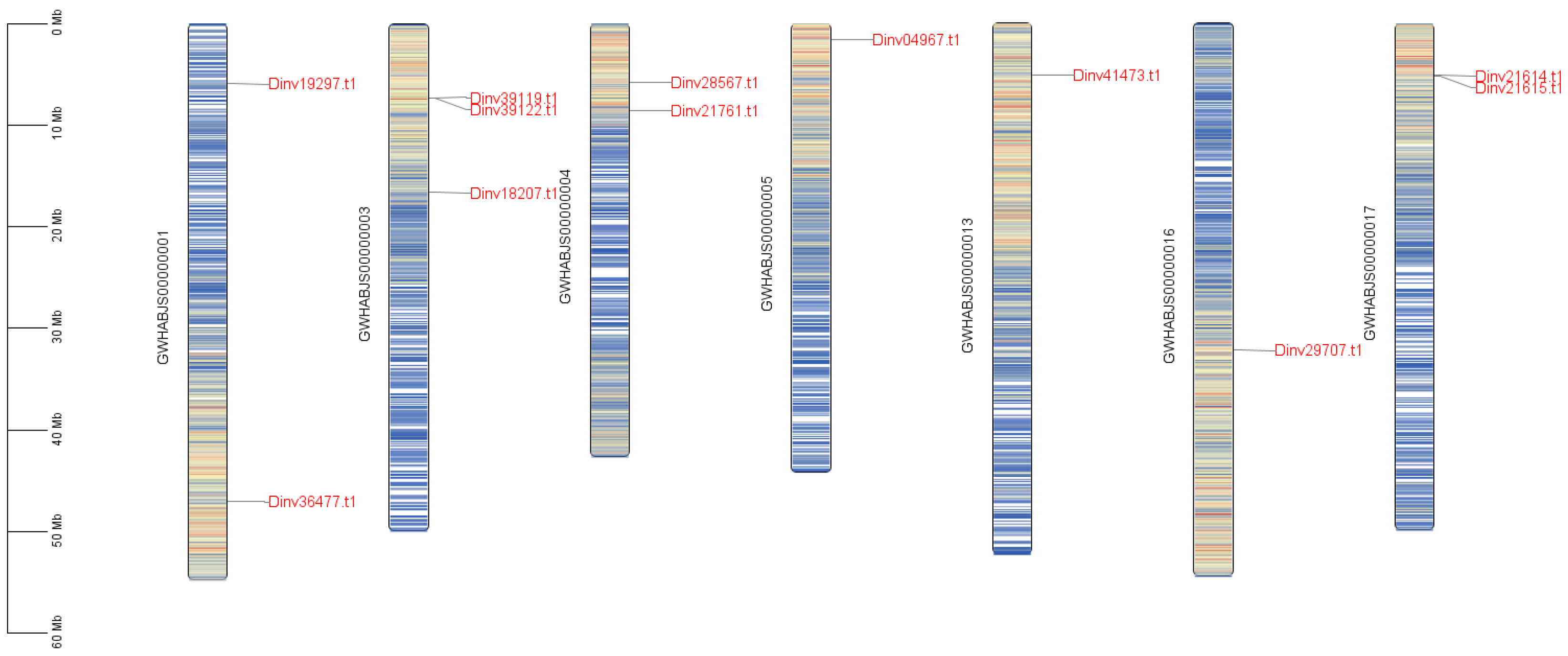
Based on chromosomal localization data, a gene family member distribution map was constructed to visualize their genomic positions. The analysis identified gene cluster distribution patterns and assessed potential gene duplication events. Among these, the 12 DiPRP genes associated with bract development were mapped to chromosomes 1, 3, 4, 5, 13, 16, and 17 in the Davidia genome (Figure 2). The results demonstrated that there were two gene clusters of DiPRPs separately distributed on chromosome 3 (Dinv39119.t1 and Dinv39122.t1) and chromosome 17 (Dinv21614.t1 and Dinv21615.t1).

3.2. Analysis of Collinearity of the PRPs Between Davidia and Other Species

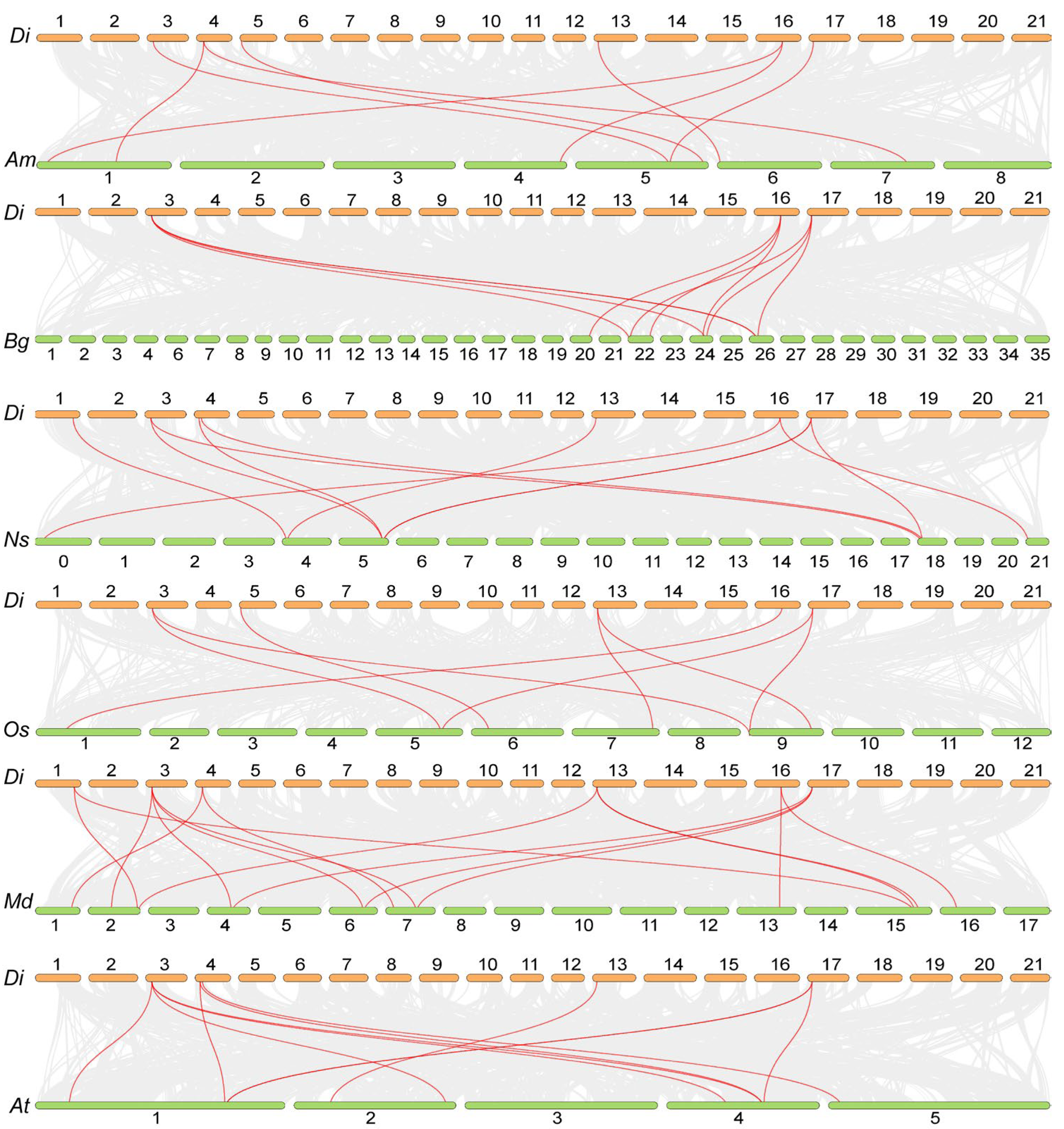
Collinearity analysis was performed to further reveal the evolution of the DiPRPs. We compared the DiPRPs to their homologues from the model plants Arabidopsis thaliana and Oryza sativa, with three other bracted species (Nyssa sinensis, Bougainvillea spectabilis, and Antirrhinum majus) and a non-bracted plant (Malus domestica). The results demonstrated that the DiPRPs exhibited the highest similarity with those of Nyssa sinensis, while showing the lowest similarity with those of Arabidopsis thaliana. The PRP genes from bracted species all showed high synteny with those of Davidia. However, the PRPs from non-bracted plants (Arabidopsis thaliana and Malus domestica) showed lower synteny with Davidia, although 14 (At) and 16 (Md) pairs of syntenic genes were identified between them (Figure 3).
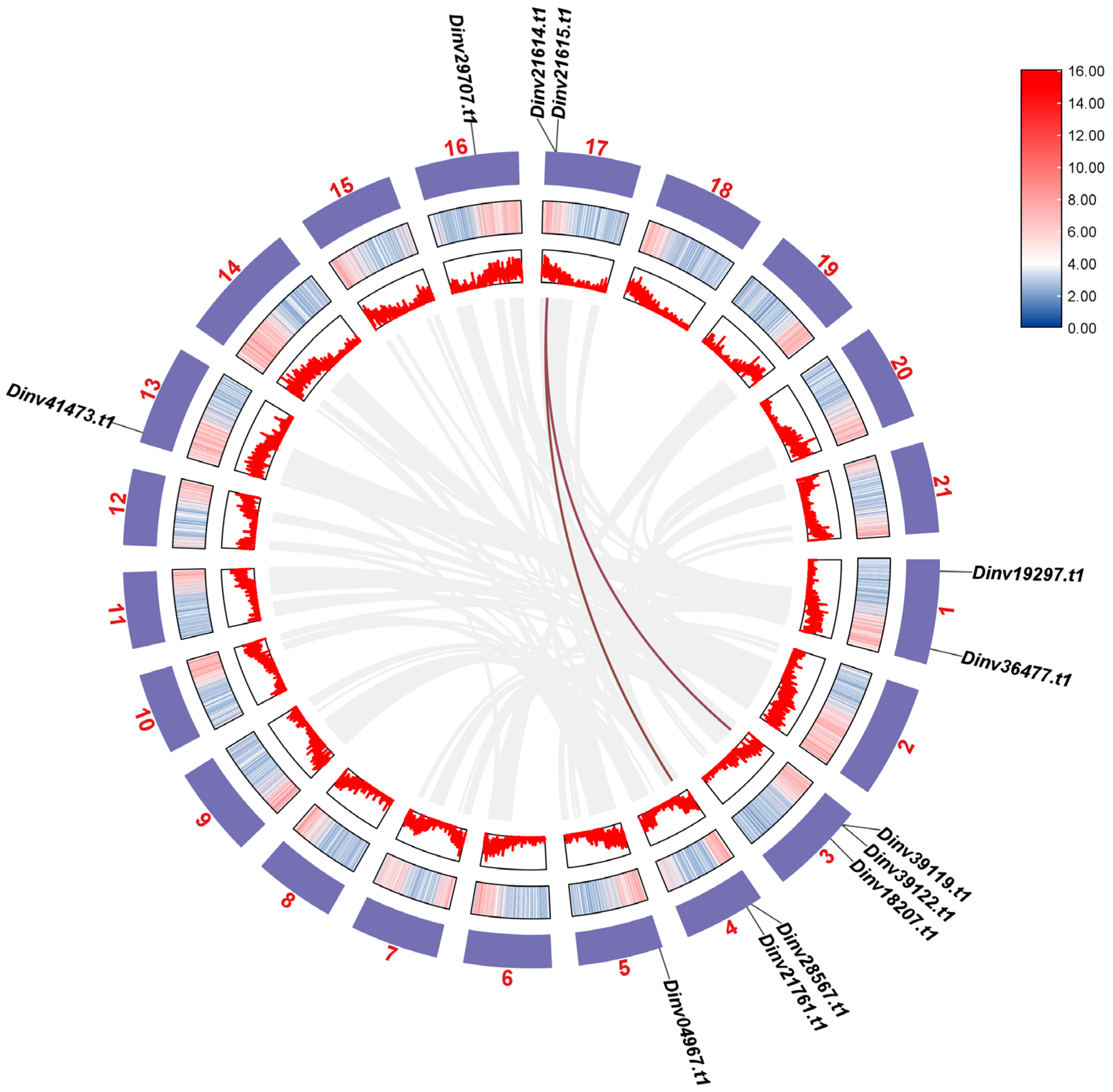
In addition, within the DiPRP gene family, two paralogous gene pairs arose via segmental duplication events. These genes were distributed across chromosomes 3, 4, 17, and 20, with the duplicated pairs being Dinv28567.t1/Dinv21614.t1 and Dinv21615.t1/Dinv39119.t1 (Figure 4). This result indicates that gene duplications of the DiPRPs might have occurred in the Davidia genome during evolution.

3.3. Conserved Motifs of DiPRPs and Cis-Elements of ProDiPRPs

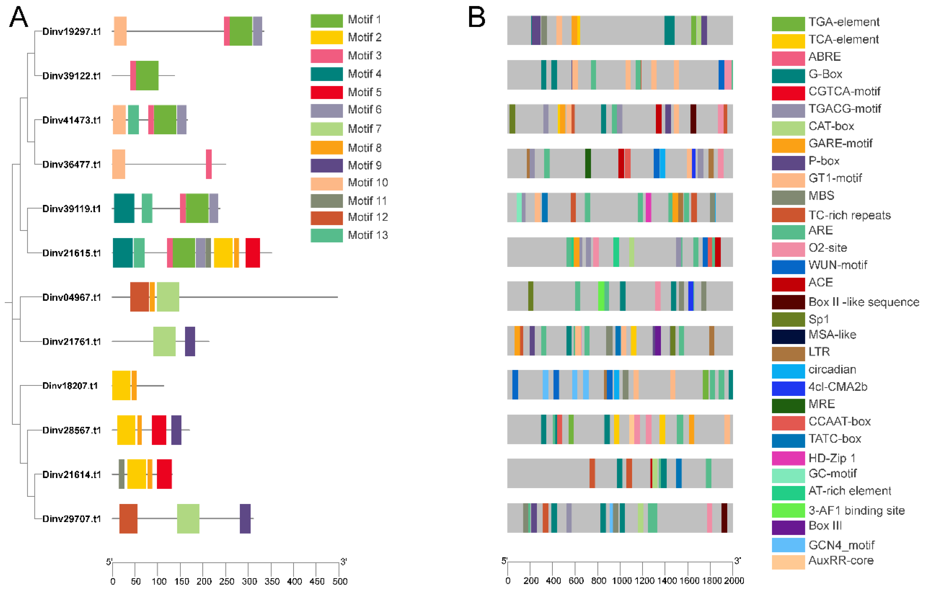
Thirteen conserved motifs were identified among twelve DiPRPs. Most DiPRP genes whose encoding products contained motif 1 and motif 3 showed decreased expression in Davidia bracts (Figure 5A).
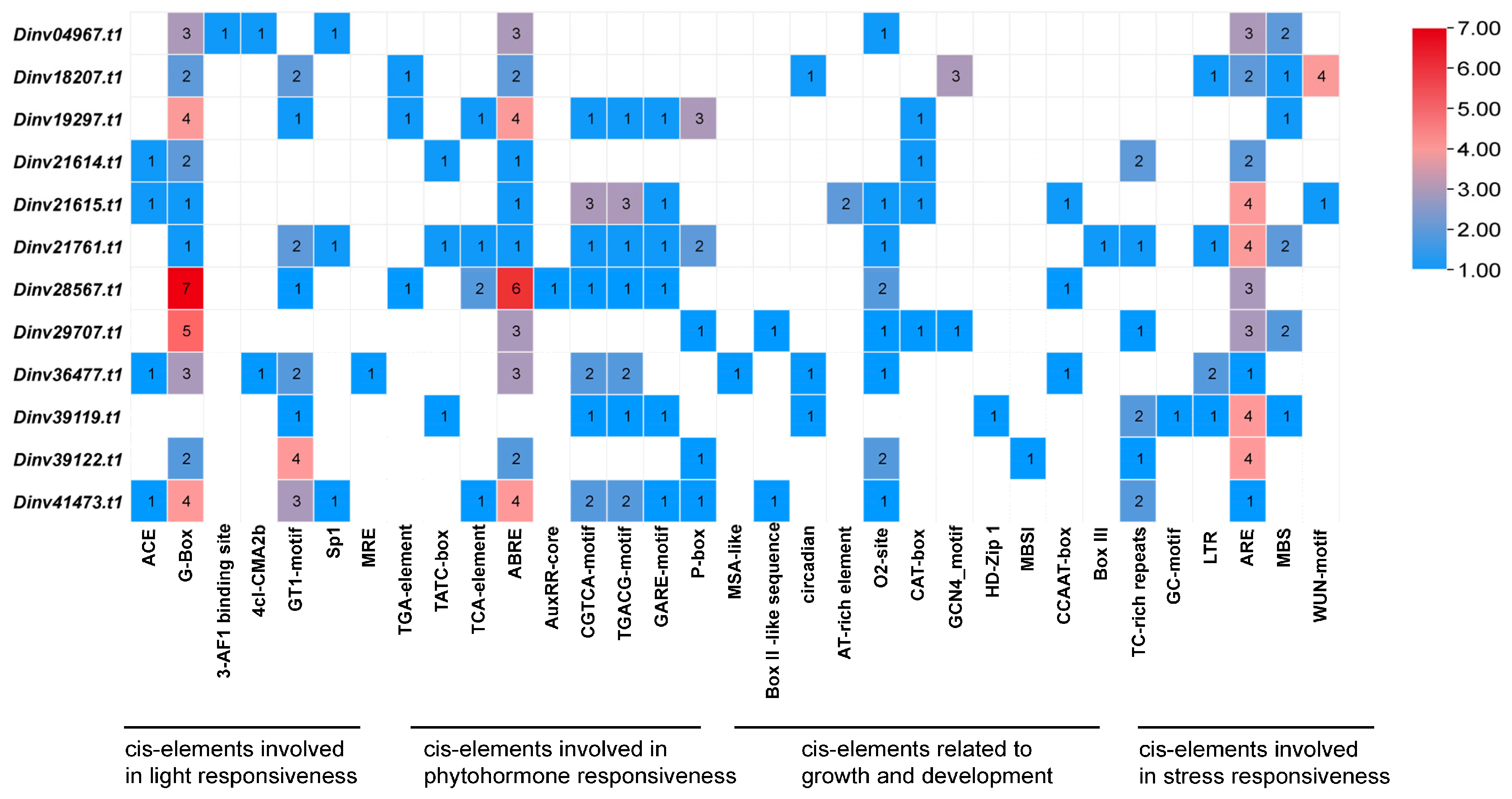
The 2000 bp upstream sequences of each DiPRP were extracted and considered as promoter regions for cis-element identification. A total of 32 types including 231 cis-elements were identified from the ProDiPRPs sequences. Among these, cis-elements associated with light responsiveness, phytohormone responsiveness, growth and development, and stress responsiveness were identified in most promoter regions (Figure 5B).
There were 61 cis-elements involved in light responsiveness (Figure 6), including ACE, G-Box, 3-AF binding site, 4cl-CAM2b, GT1-motif, Sp1, and MRE. Nearly all ProDiPRPs contained a G-Box, the number of which ranged from one to seven. There were 78 cis-elements involved in phytohormone responsiveness, including TGA-element, AuxRR-core, TATA-box, GARE-motif, P-box, TCA-element, ABRE, CGTCA-motif, and TGACG-motif. Almost all ProDiPRPs contained an ABRE, with counts ranging from one to six. There were 32 cis-elements involved in growth and development, including MSA-like, AT-rich element, circadian, O2-site, CAT-box, GCN4-motif, and HD-Zip1. There were 60 cis-elements involved in stress responsiveness, including TC-rich repeats, LTR, ARE, GC-motif, MBS, and WUN-motif. Notably, almost all ProDiPRPs contained an ARE element, the number of which ranged from one to four.
In brief, promoter analysis indicates that the expression of most DiPRPs was regulated by light, abscisic acid (ABA), and abiotic stress, which aligns with the inducing signals for Davidia bract development.

3.4. The Expression Patterns of DiPRPs During Bract Development

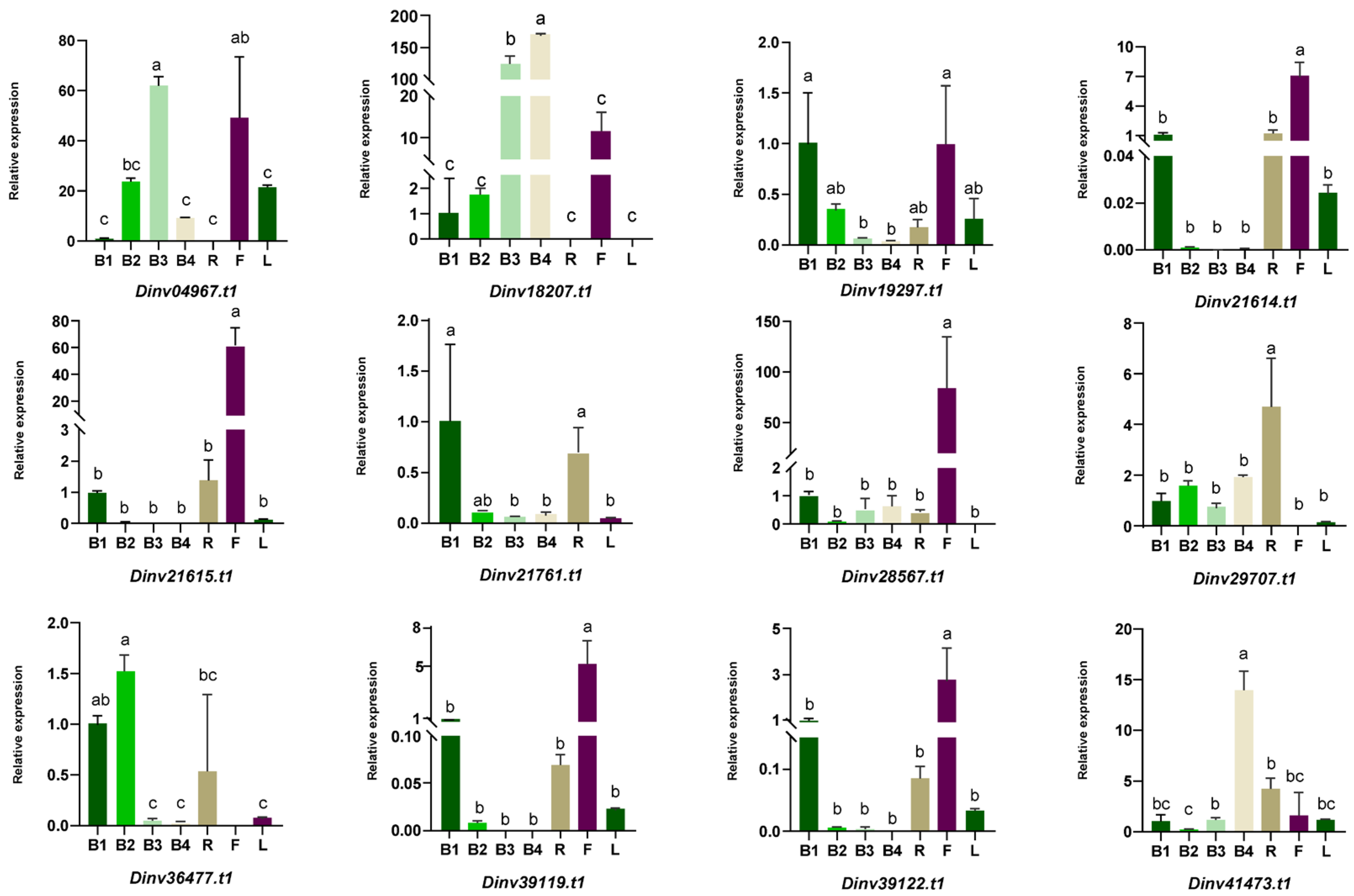
To further investigate the expression patterns of the DiPRPs, expression analysis was conducted in the roots, leaves, capitula, and bracts at different developmental stages. To evaluate the reliability of Davidia samples and the qRT-PCR amplification system, three previously reported genes, DiSOC1, DiMADS1, and DiMADS2, were employed [13,14]. The expression pattern was consistent with existing results (Figure 7).
Most DiPRPs were dominantly expressed in reproductive organs but showed different expression patterns across various organs (Figure 8).
No expression was detected in flowers for Dinv21761.t1 and Dinv29707.t1 (Figure 8). The genes Dinv19297.t1, Dinv21614.t1, Dinv21761.t1, and Dinv36477.t1 showed a significant decrease during bract development (Figure 8). In particular, Dinv21615.t1 (Figure 8) was not detected in bracts at the B3 and B4 stages, while its expression was approximately 60-fold higher at the B1 stage than that at B2 stage. The expression of Dinv04967.t1 and Dinv41473.t1 (Figure 8) showed a considerable boost during bract development, peaking at the B3 stage before slightly decreasing at the B4 stage. Dinv18207.t1 exhibited a significant upregulation trend during bract development, with its expression peaking at the B4 stage (Figure 8). Notably, the relative expression level at stage B4 was approximately 100-fold higher than that at stage B1. In addition, the expression levels of genes, such as Dinv21614.t1, Dinv21615.t1, Dinv28567.t1, Dinv39119.t1, and Dinv39122.t1, in flowers were significantly higher than those in other tissues. Overall, most DiPRPs were expressed more abundantly in floral organs (flowers and bracts) than in vegetative organs (roots and leaves).

3.5. Functional Validation of a Dinv18207.t1 Gene

Among the 12 DiPRPs, the expression level of the Dinv18207.t1 gene underwent significant changes across the four stages of bract development. Moreover, these expression changes temporally correlated with the variations in bract size. Therefore, it is hypothesized that this gene plays a role in the rapid growth of bracts. To further investigate the function of Dinv18207.t1, its ectopic overexpression was performed in Arabidopsis thaliana. Eleven independent transgenic lines were generated. The expression level of Dinv18207.t1 was assessed in these overexpression lines by qPCR (Figure S2). Based on this analysis, three lines exhibiting relatively high expression levels (OE-6, OE-7, and OE-8) were selected for further characterization.
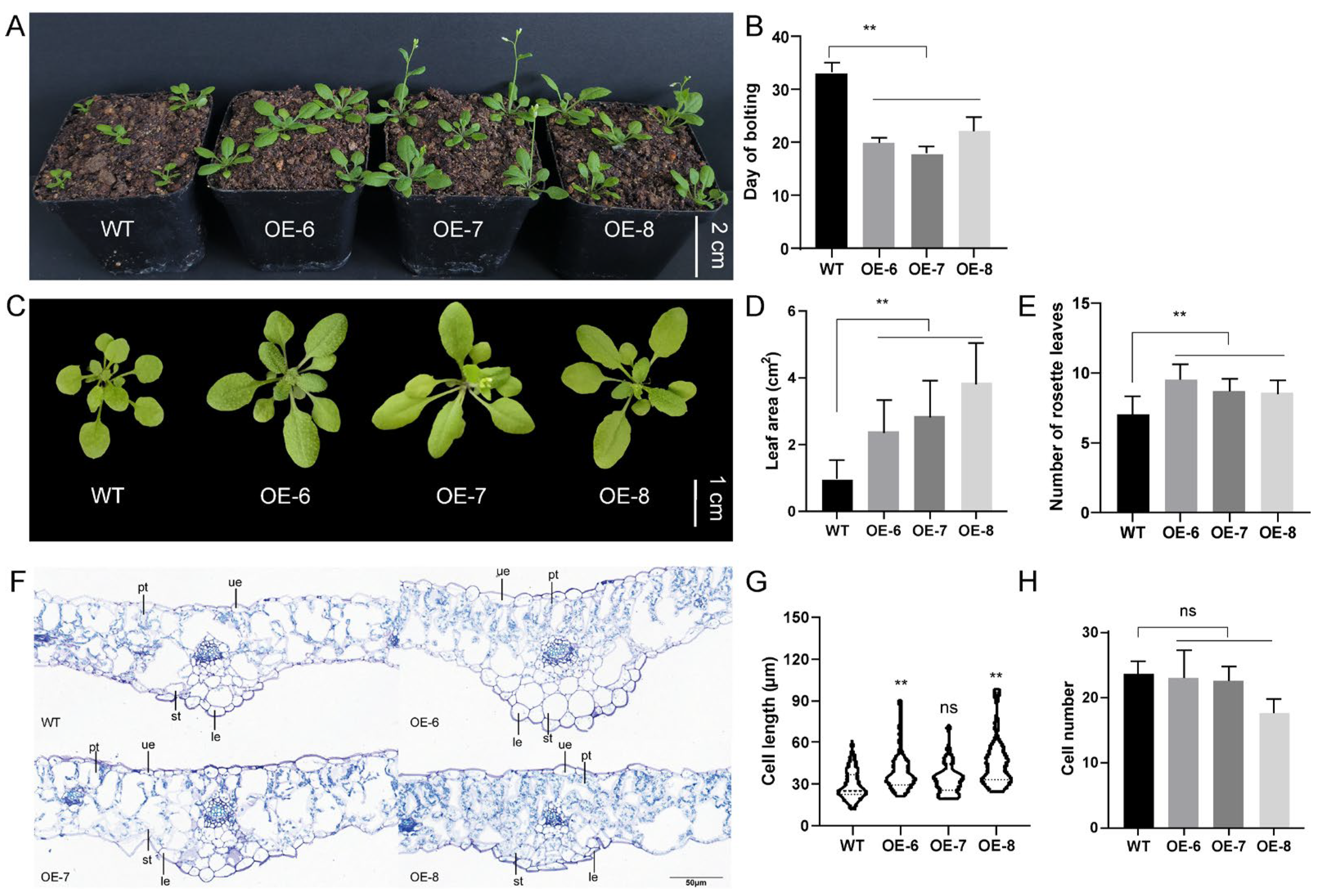
Compared to the wild type (WT), the transgenic lines exhibited earlier bolting (Figure 9A). The average bolting times of transgenic lines OE-6, OE-7, and OE-8 were 22, 18, and 20 days, respectively. In contrast, the average bolting time of WT plants was approximately 33 days, indicating that the transgenic lines bolted about 11–15 days earlier (Figure 9B). Further examination of rosette leaves revealed distinct morphological differences. The rosette leaves of WT Arabidopsis were relatively round, whereas those of the transgenic lines were elongated and oval-shaped. Additionally, the petioles and laminae of the transgenic rosette leaves formed a spoon-shaped structure (Figure 9C). Moreover, the transgenic lines produced a significantly greater number of rosette leaves and exhibited substantially larger leaf areas compared to the WT (Figure 9D,E).
Unlike WT Arabidopsis, the transgenic lines exhibited significant phenotypic differences in their rosette leaves. To further investigate the effects of the Dinv18207.t1 gene on rosette leaf development, we conducted microscopic structural observations of rosette leaves of both WT and transgenic lines. The results revealed that, compared to the WT, epidermal cells in the rosette leaves of transgenic lines were noticeably enlarged or longitudinally elongated (Figure 9F). Within the area shown in the longitudinal sections of rosette leaves, the number of epidermal cells in transgenic Arabidopsis did not differ significantly from that of the WT. However, cell length was significantly increased in the transgenic lines (Figure 9G,H).

4. Discussion

In most angiosperms, flowers develop from axillary meristematic tissues and are located in mixed buds alongside leaves and bracts. However, visible bracts are not observed in some flowering plants, possibly due to the inhibition of bracts during early development stages [33].
Using grasses as an example, the Tsh1 (TASSEL SHEATH 1) gene in maize (Zea mays) inhibits the growth of bracts. In the tsh1 mutant, which has a deletion, bracts develop at the base of both female and male flowers. The Trd (THIRD OUTER GLUME) of Hordeum vulgare L. is a homologous gene of Tsh1 in maize. The spike-shaped inflorescence of barley consists of individual small spikes. In situ hybridization has shown that Trd is expressed in the suppressed bracts at the base of the spikelet, while its expression is not observed in the vegetative meristem. Meanwhile, decreased spikelets on the main inflorescence of the trd mutant [34]. The homologous gene of maize Tsh1 also exists in rice and is named NL1 (NECK LEAF 1). Research has shown that the nl1 deficiency mutant has short inflorescences and excessive bract growth at the base of spikelets. The bracts disappear, and the number of spikelets increase in the complementary plant [35].
In crops, the growth of bracts may affect the growth of vegetative tissues at the axillary meristem, leading to a decreased in reproductive branching and affecting crop yield. But in Davidia, the presence of bracts has a unique function.
The Davidia bracts undergo an approximately six-month developmental period (from October–November of the previous year to March–April of the current year) [14]. The Davidia bracts enlarge their original size dozens of times in 20–30 days. Meanwhile, the bract color fades from bright green to pure white, and the bracts wilt and soon fall off. The unique growth and development processes of Davidia bracts suggest the special expression of certain genes in bracts to trigger and govern these events.
In this study, we identified a class of genes encoding proline-rich proteins (PRPs) that are predominantly expressed in bracts, based on transcriptome data. A total of twelve DiPRP genes were subsequently identified from the Davidia genome. Previous reports have suggested that the expression pattern of PRPs is tissue-specific or influenced by the developmental stage. In Arabidopsis thaliana, the AtPRP1 and AtPRP3 genes are specifically expressed in roots, and AtPRP3 is only detected in root hairs. The AtPRP2 and AtPRP4 genes are both expressed in the hypocotyl, rosette, and cauline, but only AtPRP4 has been detected in the stigma [36]. Tissue-specific expression patterns of the PRP genes have also been observed in soybean (Glycine max) and potato (Solanum tuberosum) [37,38], indicating the fine division of this family. Differently, most PRP genes selectively expressed in floral organs have been reported to promote reproductive growth. For instance, tobacco gene NtProRP1 is only expressed in the anther, pollen tube, and zygote. The germination and elongation of the pollen tube were severely inhibited when NtProRP1 was suppressed [39]. These findings suggest that the PRPs also participate in regulating floral development. The expression pattern of DiPRP in various tissues of Davidia suggests that PRPs may be involved in multiple physiological and biochemical processes within Davidia. Interestingly, members of the DiPRP family in Davidia exhibit contrasting expression patterns in bracts. Dinv18207.t1 shows an increasing expression trend during bract development, peaking at the B4 stage, whereas Dinv19297.t1 shows its highest expression at the B1 stage and declines thereafter. This observation aligns with previous studies where PRP family members have also shown different expression trends in the same tissue [40,41]. It means that there is perhaps some interaction of family members that together regulate the growth and development of Davidia bracts.
The analysis of promoter regions revealed the presence of multiple cis-acting elements related to hormone response, stress response, and the regulation of growth and development. These findings suggest that endogenous phytohormone levels and environmental cues may influence the expression of bract-related DiPRPs. Observations of Davidia in its natural habitat showed that low temperature or rainfall causes Davidia bract development to enter a stagnant phase, and when temperatures rise or rain ceases, the bracts growth resumes rapidly. This indicates that abiotic stress may somehow affect DiPRP expression and thereby inhibit bract development. Similar results have been reported in other species. For instance, the expression of OsHyPRP14, OsHyPRP16, and OsHyPRP40 is largely upregulated during abiotic stress in the blast resistance lines of rice [42]. The CcHyPRP gene in pigeon pea (Cajanus cajan) consistently showed elevated expression levels after being treated with cold, heat, high salt, and ABA [43]. In soybean, the upregulation of CmPRP expression is induced by environmental factors and signaling molecules [44]. When MdPRP6 in apples is subjected to high salt stress, the protein abundance decreases. Under stress conditions, RNAi plants have a more complete chloroplast structure and higher chlorophyll content. Indicating that MdPRP6 enhances tolerance by protecting chloroplasts [45].
Except for Dinv39119.t1, eleven DiPRP genes contain the light-responsive G-Box element. It has demonstrated that G-Box cis-elements are enriched in the promoter regions of genes involved in the early senescence process of rice sword leaves [46]. Therefore, the G-Box is often considered a binding site for transcription factors, such as bZIP, bHLH, and NAC [47]. The HY5 (LONG HYPOCOTYL5) transcription factor, which belongs to the bZIP family, binds to the G-Box in the promoter regions of target genes in Arabidopsis, and overexpression of GLKs (GOLDEN2-LIKE2) resulted in a significant increase in the chlorophyll content in roots only in the presence of AtHY5. In addition, the G-Box and GLK recognition sequences are consistently enriched in the promoter regions of seventeen genes strongly associated with the co-expression of the CHLH, CHLP, and CHL27 genes, which encode key enzymes for chlorophyll synthesis. These results imply an interaction between these two cis-elements and chloroplast development [48,49,50].
All DiPRPs contained the ABA-responsive element ABRE, except for Dinv19297.t1. Members of the bZIP family specifically bind to the ABA response element ABRE [51]. ABA is widely present in plants and participates in various processes, such as senescence and stress resistance. MdbHLH93 significantly promotes the transcription of SAGs, and the expression levels of SAGs further increase following ABA treatment, thereby accelerating leaf senescence [52]. These results indicate a close relationship between ABA and bract development. As another piece of evidence, the ASR (abscisic acid, stress, and ripening), another gene family of plant transcription factors, is predominantly expressed in Davidia bracts. The expression level of a Davidia ASR gene, DiASR1, was elevated more than 5-fold after 24 h of exogenous ABA application. Remarkably, the overexpression of DiASR1 in Arabidopsis caused albino plants and the appearance of “bract-like” leaves [53]. These results imply that fast growth and chloroplast degeneration in the bracts are mediated by ABA.
In addition to ABRE, 22 methyl-jasmonate (MeJA)-responsive elements were also identified in the PRP promoter regions. The type IIIe bHLH transcription factors MYC2\3\4 activate MeJA-mediated senescence by binding to the G-Box of SAGs, thereby inducing their expression [54]. Overexpression of these MYCs enhances the transcriptional activity of AtPAO and increases the expression of NYC1 and NYE1/SGR1, accelerating chlorophyll degradation. Similarly, aNACs, which act downstream of MYCs, promote the expression of NYE1/SGR1, NYE2/SGR2, and NYC1 [55]. It is hypothesized that Davidia bracts participate in their own growth and developmental processes in response to biotic or abiotic stimuli, with these transcription factors potentially acting upstream by directly or indirectly binding to the PRP gene promoters to activate or repress their expression.
Proline-rich proteins (PRPs) are structural components of the cell wall and are involved in its construction, and plants respond to stress by modifying the structure or composition of the cell wall [56,57,58]. Previous studies have demonstrated that the PRP proteins are primarily localized at the cell wall or plasma membrane [59,60,61]. Populus PdPRP is localized at the plasma membrane [22], multiple MdPRPs in apples are found in the cell membrane [40], and Gossypium barbadense GbHyPRP1 is localized in the cell wall [62]. Eight of the DiPRPs identified in this study were localized at chloroplasts. DiPRPs may play an important function in chloroplast degradation or chlorophyll degradation, rather than being mainly involved in plant adversity response as previously determined. The degradation of chlorophyll marks the onset of senescence [63]. Overexpression of OsNAC2 increases the expression levels of chlorophyll degradation-related genes OsSGR, OsNYC3, OsNOL, OsPAO, and OsRCCR1. OsNAC2 can bind to the promoters of OsSGR and OsNYC3 to directly activate their expression [64].
Analysis of covariance showed higher collinearity between Davidia and the naturally bracted plants Antirrhinum majus and Bougainvillea and lower covariance with the model plant Arabidopsis thaliana. This is similar to the results of phylogenetic analyses, in which most PRPs of Davidia are in the same branch with those in rice. According to the GenBank database annotation, the PRPs in rice are mostly found in floral organs and seeds. A gene from rice (Oryza sativa), OsPRP3, has been reported to be highly expressed in flowers at later stages, and knockdown of OsPRP3 caused low vitality of pollen, anther defects, and flower abortion [65]. Studies indicate that the transition in leaf shape in Arabidopsis serves as a marker for the shift from the juvenile to the adult phase during vegetative growth [66,67]. It is hypothesized that the overexpression of Dinv18207.t1 accelerates plant growth during this stage by shortening the juvenile phase, thereby promoting an earlier transition from vegetative to reproductive growth in transgenic lines. Compared to the wild type, the Dinv18207.t1 overexpression lines exhibited significantly enhanced growth and development. Paraffin section analysis revealed that this enhancement resulted from changes in cell morphology. It is hypothesized that Dinv18207.t1 may be involved in cell wall remodeling. This observation aligns with previous results. For instance, overexpression of PdPRP in poplar increased the radial width of the xylem and phloem, whereas its downregulation resulted in slower growth and reduced radial width of these tissues [22]. Similar effects have also been observed in tobacco [39,68]. Notably, although overexpression of Dinv18207.t1 accelerated growth in Arabidopsis, the transgenic plants exhibited no apparent growth defects or abnormalities.

5. Conclusions

In this study, a comprehensive bioinformatics analysis of the DiPRP family was carried out based on the whole-genome and transcriptome data of Davidia. A total of twelve DiPRP genes were identified in the Davidia genome. Predictions of subcellular localization indicated that the DiPRPs encoded by eight members were localized in chloroplasts. A variety of cis-acting elements were identified in DiPRP promoter regions, with the highest proportion of hormone-responsive elements. The overexpression lines of Dinv18207.t1 were generated to verify its function. It was observed that overexpressing this gene significantly shortened the juvenile stage of the plants and promoted the expansion and elongation of epidermal cells in Arabidopsis rosette leaves, thereby facilitating the rapid growth of the transgenic lines. Our findings indicate that some bract-specific PRPs play a critical role in leaf metamorphosis.

Supplementary Materials

The following supporting information can be downloaded at: https://www.mdpi.com/article/10.3390/f16091425/s1, Figure S1. Sequencing of the fusion plasmid containing Dinv18207.t1. pCAMBIA1300-35S-GFP represent empty plasmid. Figure S2. The relative expression of Dinv18207.t1 in transgenic lines. Table S1. Primers used for qRT-PCR.

Author Contributions

Conceptualization, Y.F. and M.L.; Formal analysis, Y.F., X.Z., and Y.L.; Validation, Y.F. and J.L.; Visualization, Y.F., X.Z., and J.N.; Writing—original draft, Y.F. and M.L.; Writing—review & editing, Y.F. and M.L. All authors have read and agreed to the published version of the manuscript.

Funding

This research was supported by grants from the Natural Science Foundation of Hunan Province (2024JJ5634), the Postgraduate Scientific and Technological Innovation Project of CSUFT (2023CX02058), and the Natural Science Foundation of Changsha (kq2402251).

Institutional Review Board Statement

The authority responsible for the Davidia resources is the Badagong Mountain Nature Reserve Management Division, who provided permission to collect the samples for our scientific research.

Data Availability Statement

Data are contained within the article and Supplementary Materials.

Acknowledgments

We are grateful to the managers of Badagong Mountain Nature Reserve for their support of this study.

Conflicts of Interest

The authors declare that they have no conflicts of interest.

References

  1. Causier, B.; Schwarz-Sommer, Z.; Davies, B. Floral organ identity: 20 years of ABCs. Semin. Cell Dev. Biol. 2010, 21, 73–79. [Google Scholar] [CrossRef]
  2. Sun, J.; Gong, Y.; Renner, S.S.; Huang, S. Multifunctional Bracts in the Dove Tree Davidia involucrata (Nyssaceae: Cornales): Rain Protection and Pollinator Attraction. Am. Nat. 2008, 171, 119–124. [Google Scholar] [CrossRef] [PubMed]
  3. Armbruster, W.S.; Antonsen, L.; Pélabon, C. Phenotypic selection on Dalechampia blossoms: Honest signaling affects pollination success. Ecology 2005, 86, 3323–3333. [Google Scholar] [CrossRef]
  4. Keasar, T.; Sadeh, A.; Gerchman, Y.; Shmida, A. The signaling function of an extra-floral display: What selects for signal development? Oikos 2009, 118, 1752–1759. [Google Scholar] [CrossRef]
  5. Higginson, A.D.; Gilbert, F.S.; Reader, T.; Barnard, C.J. Reduction of visitation rates by honeybees (Apis mellifera) to individual inflorescences of lavender (Lavandula stoechas) upon removal of coloured accessory bracts (Hymenoptera: Apidae). Èntomol. Gen. 2007, 29, 165–178. [Google Scholar] [CrossRef]
  6. Song, B.; Zhang, Z.-Q.; Stöcklin, J.; Yang, Y.; Niu, Y.; Chen, J.-G.; Sun, H. Multifunctional bracts enhance plant fitness during flowering and seed development in Rheum nobile (Polygonaceae), a giant herb endemic to the high Himalayas. Oecologia 2013, 172, 359–370. [Google Scholar] [CrossRef]
  7. Omori, Y.; Ohba, H. Pollen development of Rheum nobile Hook.f. & Thomson (Polygonaceae), with reference to its sterility induced by bract removal. Bot. J. Linn. Soc. 1996, 122, 269–278. [Google Scholar] [CrossRef]
  8. Song, B.; Gao, Y.; Stöcklin, J.; Song, M.; Sun, L.; Sun, H. Ultraviolet screening increases with elevation in translucent bracts of Rheum nobile (Polygonaceae), an alpine ‘glasshouse’ plant from the high Himalayas. Bot. J. Linn. Soc. 2020, 193, 276–286. [Google Scholar] [CrossRef]
  9. He, J.; Lin, J.; Chen, W. The current status of endemic and endangered species Davidia involucrata and the preserving strategies. Biodivers. Sci. 1995, 3, 213–221. [Google Scholar] [CrossRef]
  10. Xu, W.; Cao, F.; Li, M.; Liu, Z.; Wu, Y.; Wu, X.; Zhang, X. Research on the changes of endogenous hormones in the development of Davidia involucrata bracts. J. Cent. S. Univ. For. Technol. 2017, 37, 103–109. [Google Scholar] [CrossRef]
  11. Vekemans, D.; Viaene, T.; Caris, P.; Geuten, K. Transference of function shapes organ identity in the dove tree inflorescence. New Phytol. 2011, 193, 216–228. [Google Scholar] [CrossRef]
  12. Li, Y. Cloning and Analyzing of Bract Differentially Expressed Genes of Davidia involucrata Baill. Ph.D. Thesis, Sichuan University, Chengdu, China, 2002. [Google Scholar]
  13. Li, G.; Cao, C.; Yang, H.; Wang, J.; Wei, W.; Zhu, D.; Gao, P.; Zhao, Y. Molecular cloning and potential role of DiSOC1s in flowering regulation in Davidia involucrata Baill. Plant Physiol. Biochem. 2020, 157, 453–459. [Google Scholar] [CrossRef]
  14. Sun, M.; Zhu, L.; Zeng, L.; Yang, H.; Cao, C.; Wang, R.; Zhao, Y.; Wei, W. Analysis and Characterization of MADS-box Genes from Davidia involucrata Baill. and Regulation of Flowering Time in Arabidopsis. Russ. J. Plant Physiol. 2022, 69, 62. [Google Scholar] [CrossRef]
  15. Niu, J. Identification of Key Genes Involved in Bract Development and Functional Validation of a DiPRP4 Gene in the Dove Tree (Davidia involucrata Baill.). Master’s Thesis, Center South University of Foresty and Technolgy, Changsha, China, 2024. [Google Scholar]
  16. Esen, A.; Bietz, J.A.; Paulis, J.W.; Wall, J.S. Tandem repeats in the N-terminal sequence of a proline-rich protein from corn endosperm. Nature 1982, 296, 678–679. [Google Scholar] [CrossRef]
  17. Chen, J.; Varner, J.E. Isolation and characterization of cDNA clones for carrot extensin and a proline-rich 33-kDa protein. Proc. Natl. Acad. Sci. USA 1985, 82, 4399–4403. [Google Scholar] [CrossRef]
  18. Josè-Estanyol, M.; Puigdomènech, P. Plant cell wall glycoproteins and their genes. Plant Physiol. Biochem. 2000, 38, 97–108. [Google Scholar] [CrossRef]
  19. José-Estanyol, M.; Gomis-Rüth, F.; Puigdomènech, P. The eight-cysteine motif, a versatile structure in plant proteins. Plant Physiol. Biochem. 2004, 42, 355–365. [Google Scholar] [CrossRef]
  20. Josè, M.; Puigdomènech, P. Structure and expression of genes coding for structural proteins of the plant cell wall. New Phytol. 1993, 125, 259–282. [Google Scholar] [CrossRef]
  21. Su, L.; Li, W.; Chen, X.; Wang, P.; Liu, D. Proline-rich protein PRPL1 enhances Panax notoginseng defence against Fusarium solani by regulating reactive oxygen species balance and strengthening the cell wall barrier. Plant Cell Environ. 2024, 47, 2375–2393. [Google Scholar] [CrossRef]
  22. Li, S.; Zhang, Y.; Ding, C.; Gao, X.; Wang, R.; Mo, W.; Lv, F.; Wang, S.; Liu, L.; Tang, Z.; et al. Proline-rich protein gene PdPRP regulates secondary wall formation in poplar. J. Plant Physiol. 2019, 233, 58–72. [Google Scholar] [CrossRef]
  23. Chen, Y.; Ma, T.; Zhang, L.; Kang, M.; Zhang, Z.; Zheng, Z.; Sun, P.; Shrestha, N.; Liu, J.; Yang, Y. Genomic analyses of a “living fossil”: The endangered dove-tree. Mol. Ecol. Resour. 2020, 20, 756–769. [Google Scholar] [CrossRef]
  24. Niu, J. “Transcriptome of Davidia involucrata Baill.” the National Center for Biological Information. Available online: https://ngdc.cncb.ac.cn/bioproject/browse/PRJCA008410 (accessed on 15 July 2025).
  25. Gabella, C.; Duvaud, S.; Durinx, C. Managing the life cycle of a portfolio of open data resources at the SIB Swiss Institute of Bioinformatics. Brief. Bioinform. 2021, 23, 1477–4054. [Google Scholar] [CrossRef]
  26. Chen, C.; Wu, Y.; Li, J.; Wang, X.; Zeng, Z.; Xu, J.; Liu, Y.; Feng, J.; Chen, H.; He, Y.; et al. TBtools-II: A “one for all, all for one” bioinformatics platform for biological big-data mining. Mol. Plant 2023, 16, 1733–1742. [Google Scholar] [CrossRef]
  27. Bailey, T.L.; Johnson, J.; Grant, C.E.; Noble, W.S. The MEME Suite. Nucleic Acids Res. 2015, 43, W39–W49. [Google Scholar] [CrossRef]
  28. Lescot, M.; Déhais, P.; Thijs, G.; Marchal, K.; Moreau, Y.; Van de Peer, Y.; Rouzé, P.; Rombauts, S. PlantCARE, a database of plant cis-acting regulatory elements and a portal to tools for in silico analysis of promoter sequences. Nucleic Acids Res. 2002, 30, 325–327. [Google Scholar] [CrossRef]
  29. Ren, R.; Dai, P.H.; Li, M.; Liu, Z.; Cao, F. Selection and stability evaluation of reference genes for real-time quantitative PCR in dove tree (Davidia involucrata). Plant Physiol. J. 2016, 52, 1565–1575. [Google Scholar] [CrossRef]
  30. Livak, K.J.; Schmittgen, T.D. Analysis of Relative Gene Expression Data Using Real-Time Quantitative PCR and the 2−ΔΔCT Method. Methods 2001, 25, 402–408. [Google Scholar] [CrossRef]
  31. Zhang, X.; Henriques, R.; Lin, S.-S.; Niu, Q.-W.; Chua, N.-H. Agrobacterium-mediated transformation of Arabidopsis thaliana using the floral dip method. Nat. Protoc. 2006, 1, 641–646. [Google Scholar] [CrossRef]
  32. Yang, J. Improvement of traditional paraffin section preparation methods. J. Biol. 2006, 1, 45–46. [Google Scholar] [CrossRef]
  33. Baum, D.A.; Day, C.D. Cryptic Bracts Exposed: Insights into the Regulation of Leaf Expansion. Dev. Cell 2004, 6, 318–319. [Google Scholar] [CrossRef]
  34. Whipple, C.J.; Hall, D.H.; DeBlasio, S.; Taguchi-Shiobara, F.; Schmidt, R.J.; Jackson, D.P. A Conserved Mechanism of Bract Suppression in the Grass Family. Plant Cell 2010, 22, 565–578. [Google Scholar] [CrossRef]
  35. Wang, L.; Yin, H.; Qian, Q.; Yang, J.; Huang, C.; Hu, X.; Luo, D. NECK LEAF 1, a GATA type transcription factor, modulates organogenesis by regulating the expression of multiple regulatory genes during reproductive development in rice. Cell Res. 2009, 19, 598–611. [Google Scholar] [CrossRef] [PubMed]
  36. Fowler, T.J.; Bernhardt, C.; Tierney, M.L. Characterization and Expression of Four Proline-Rich Cell Wall Protein Genes in Arabidopsis Encoding Two Distinct Subsets of Multiple Domain Proteins. Plant Physiol. 1999, 121, 1081–1091. [Google Scholar] [CrossRef] [PubMed]
  37. Wyatt, R.E.; Nagao, R.T.; Key, J.L. Patterns of soybean proline-rich protein gene expression. Plant Cell 1992, 4, 99–110. [Google Scholar] [CrossRef] [PubMed]
  38. Menke, U.; Renault, N.; Mueller-Roeber, B. StGCPRP, a Potato Gene Strongly Expressed in Stomatal Guard Cells, Defines a Novel Type of Repetitive Proline-Rich Proteins. Plant Physiol. 2000, 122, 677–686. [Google Scholar] [CrossRef]
  39. Chen, J.; Zhao, J.; Ning, J.; Liu, Y.; Xu, J.; Tian, S.; Zhang, L.; Sun, M. NtProRP1, a novel proline-rich protein, is an osmotic stress-responsive factor and specifically functions in pollen tube growth and early embryogenesis in Nicotiana tabacum. Plant Cell Environ. 2014, 37, 499–511. [Google Scholar] [CrossRef]
  40. Zhang, X.; Gong, X.; Li, D.; Yue, H.; Qin, Y.; Liu, Z.; Li, M.; Ma, F. Genome-Wide Identification of PRP Genes in Apple Genome and the Role of MdPRP6 in Response to Heat Stress. Int. J. Mol. Sci. 2021, 22, 5942. [Google Scholar] [CrossRef]
  41. Rajasheker, G.; Nagaraju, M.; Varghese, R.P.; Jalaja, N.; Somanaboina, A.K.; Singam, P.; Ramakrishna, C.; Penna, S.; Sreenivasulu, N.; Kishor, P.B.K. Identification and analysis of proline-rich proteins and hybrid proline-rich proteins super family genes from Sorghum bicolor and their expression patterns to abiotic stress and zinc stimuli. Front. Plant Sci. 2022, 13, 952732. [Google Scholar] [CrossRef]
  42. Kapoor, R.; Kumar, G.; Arya, P.; Jaswal, R.; Jain, P.; Singh, K.; Sharma, T.R. Genome-Wide Analysis and Expression Profiling of Rice Hybrid Proline-Rich Proteins in Response to Biotic and Abiotic Stresses, and Hormone Treatment. Plants 2019, 8, 343. [Google Scholar] [CrossRef]
  43. Priyanka, B.; Sekhar, K.; Reddy, V.D.; Rao, K.V. Expression of pigeonpea hybrid-proline-rich protein encoding gene (CcHyPRP) in yeast and Arabidopsis affords multiple abiotic stress tolerance. Plant Biotechnol. J. 2010, 8, 76–87. [Google Scholar] [CrossRef]
  44. Zhai, Y.; Lei, T.-T.; Yan, F.; Huang, K.-M.; Li, X.-W.; Zhang, Q.-L.; Zhang, H.-J.; Su, L.-T.; Sun, X.; Wang, Y.; et al. Cloning and Expression of a Stress-induced GmPRP Gene in Soybean (Glycine max). Acta Agron. Sin. 2011, 37, 2152–2157. [Google Scholar] [CrossRef]
  45. Zhang, X.; Gong, X.; Yu, H.; Su, X.; Cheng, S.; Huang, J.; Lei, Z.; Li, M.; Ma, F. The proline-rich protein MdPRP6 confers tolerance to salt stress in transgenic apple (Malus domestica). Sci. Hortic. 2022, 308, 111581. [Google Scholar] [CrossRef]
  46. Liu, L.; Xu, W.; Hu, X.; Liu, H.; Lin, Y. W-box and G-box elements play important roles in early senescence of rice flag leaf. Sci. Rep. 2016, 6, 20881. [Google Scholar] [CrossRef]
  47. Toledo-Ortiz, G.; Huq, E.; Quail, P.H. The Arabidopsis Basic/Helix-Loop-Helix Transcription Factor Family. Plant Cell 2003, 15, 1749–1770. [Google Scholar] [CrossRef]
  48. Kobayashi, K.; Obayashi, T.; Masuda, T. Role of the G-box element in regulation of chlorophyll biosynthesis in Arabidopsis roots. Plant Signal. Behav. 2012, 7, 922–926. [Google Scholar] [CrossRef] [PubMed]
  49. Lee, J.; He, K.; Stolc, V.; Lee, H.; Figueroa, P.; Gao, Y.; Tongprasit, W.; Zhao, H.; Lee, I.; Deng, X.W. Analysis of Transcription Factor HY5 Genomic Binding Sites Revealed Its Hierarchical Role in Light Regulation of Development. Plant Cell 2007, 19, 731–749. [Google Scholar] [CrossRef] [PubMed]
  50. Waters, M.T.; Wang, P.; Korkaric, M.; Capper, R.G.; Saunders, N.J.; Langdale, J.A. GLK Transcription Factors Coordinate Expression of the Photosynthetic Apparatus in Arabidopsis. Plant Cell 2009, 21, 1109–1128. [Google Scholar] [CrossRef] [PubMed]
  51. Uno, Y.; Furihata, T.; Abe, H.; Yoshida, R.; Shinozaki, K.; Yamaguchi-Shinozaki, K. Arabidopsis basic leucine zipper transcription factors involved in an abscisic acid-dependent signal transduction pathway under drought and high-salinity conditions. Proc. Natl. Acad. Sci. USA 2000, 97, 11632–11637. [Google Scholar] [CrossRef]
  52. An, J.; Zhang, X.; Bi, S.; You, C.; Wang, X.; Hao, Y. MdbHLH93, an apple activator regulating leaf senescence, is regulated by ABA and MdBT2 in antagonistic ways. New Phytol. 2019, 222, 735–751. [Google Scholar] [CrossRef]
  53. Wu, X.; Gao, R.; Mao, R.; Lin, Y.; Yang, Z.; Li, J.; Cao, F.; Li, M. Inducing bract-like leaves in Arabidopsis through ectopically expressing an ASR gene from the dove tree. Ind. Crop. Prod. 2022, 180, 114796. [Google Scholar] [CrossRef]
  54. Qi, T.; Wang, J.; Huang, H.; Liu, B.; Gao, H.; Liu, Y.; Song, S.; Xie, D. Regulation of Jasmonate-Induced Leaf Senescence by Antagonism between bHLH Subgroup IIIe and IIId Factors in Arabidopsis. Plant Cell 2015, 27, 1634–1649. [Google Scholar] [CrossRef]
  55. Zhu, X.; Chen, J.; Xie, Z.; Gao, J.; Ren, G.; Gao, S.; Zhou, X.; Kuai, B. Jasmonic acid promotes degreening via MYC2/3/4- and ANAC019/055/072-mediated regulation of major chlorophyll catabolic genes. Plant J. 2015, 84, 597–610. [Google Scholar] [CrossRef] [PubMed]
  56. Lin, W.; Tang, W.; Pan, X.; Huang, A.; Gao, X.; Anderson, C.T.; Yang, Z. Arabidopsis pavement cell morphogenesis requires FERONIA binding to pectin for activation of ROP GTPase signaling. Curr. Biol. 2022, 32, 497–507.e4. [Google Scholar] [CrossRef] [PubMed]
  57. Wang, D.; Kanyuka, K.; Papp-Rupar, M. Pectin: A critical component in cell-wall-mediated immunity. Trends Plant Sci. 2023, 28, 10–13. [Google Scholar] [CrossRef] [PubMed]
  58. Xiao, Y.; Sun, G.; Yu, Q.; Gao, T.; Zhu, Q.; Wang, R.; Huang, S.; Han, Z.; Cervone, F.; Yin, H.; et al. A plant mechanism of hijacking pathogen virulence factors to trigger innate immunity. Science 2024, 383, 732–739. [Google Scholar] [CrossRef]
  59. Seo, S.-G.; Jeon, S.B.; Kim, J.-S.; Shin, J.-M.; Kim, J.-B.; Kang, S.-W.; Lee, G.-P.; Kim, S.-H. Characterization and expression pattern of IbPRP1 and IbPRP2 stress-related genes from sweetpotato. Genes Genom. 2010, 32, 487–497. [Google Scholar] [CrossRef]
  60. Ma, Y.; Wu, T.; Zhong, G.; Zheng, Y. Overexpression of a new proline-rich protein encoding Gene CsPRP4 increases starch accumulation in Citrus. Sci. Hortic. 2020, 260, 108744. [Google Scholar] [CrossRef]
  61. Liu, D.Q.; Han, Q.; Shah, T.; Chen, C.Y.; Wang, Q.; Tang, B.F.; Ge, F. A hybrid proline-rich cell-wall protein gene JsPRP1 from Juglans sigillata Dode confers both biotic and abiotic stresses in transgenic tobacco plants. Trees 2018, 32, 1199–1209. [Google Scholar] [CrossRef]
  62. Yang, J.; Zhang, Y.; Wang, X.; Wang, W.; Li, Z.; Wu, J.; Wang, G.; Wu, L.; Zhang, G.; Ma, Z. HyPRP1 performs a role in negatively regulating cotton resistance to V. dahliae via the thickening of cell walls and ROS accumulation. BMC Plant Biol. 2018, 18, 339. [Google Scholar] [CrossRef]
  63. Hörtensteiner, S. Stay-green regulates chlorophyll and chlorophyll-binding protein degradation during senescence. Trends Plant Sci. 2009, 14, 155–162. [Google Scholar] [CrossRef]
  64. Mao, C.; Lu, S.; Lv, B.; Zhang, B.; Shen, J.; He, J.; Luo, L.; Xi, D.; Chen, X.; Ming, F. A Rice NAC Transcription Factor Promotes Leaf Senescence via ABA Biosynthesis. Plant Physiol. 2017, 174, 1747–1763. [Google Scholar] [CrossRef]
  65. Gothandam, K.M.; Nalini, E.; Karthikeyan, S.; Shin, J.S. OsPRP3, a flower specific proline-rich protein of rice, determines extracellular matrix structure of floral organs and its overexpression confers cold-tolerance. Plant Mol. Biol. 2010, 72, 125–135. [Google Scholar] [CrossRef] [PubMed]
  66. Telfer, A.; Bollman, K.M.; Poethig, R.S. Phase change and the regulation of trichome distribution in Arabidopsis thaliana. Development 1997, 124, 645–654. [Google Scholar] [CrossRef]
  67. Chien, J.C.; Sussex, I.M. Differential regulation of trichome formation on the adaxial and abaxial leaf surfaces by gibberellins and photoperiod in Arabidopsis thaliana (L.) Heynh. Plant Physiol. 1996, 111, 1321–1328. [Google Scholar] [CrossRef]
  68. Dvořáková, L.; Srba, M.; Opatrny, Z.; Fischer, L. Hybrid proline-rich proteins: Novel players in plant cell elongation? Ann. Bot. 2011, 109, 453–462. [Google Scholar] [CrossRef]
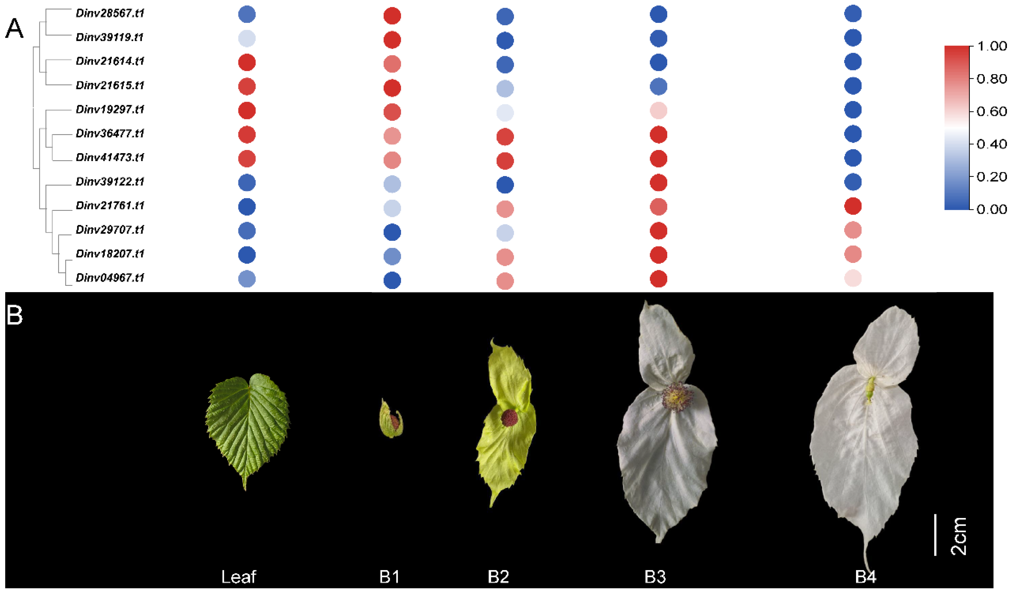
Figure 1. The expression patterns of DiPRP genes in Davidia bracts at different developmental stages. (A) The expression patterns of DiPRPs in Davidia bracts at different developmental stages; (B) leaf, contemporaneous leaf at the B2 stage; B1–B4, Davidia bracts at four developmental stages.
Forests 16 01425 g001
Figure 2. Chromosomal mapping of the DiPRP genes. Chromosome numbers are shown on the left side of each chromosome. The scale represents the length of Davidia chromosomes.
Forests 16 01425 g002
Figure 3. Analysis of collinearity among Davidia and other bracted and non-bracted plants. Am, Antirrhinum majus; Bg, Bougainvillea spectabilis; Ns, Nyssa sinensis; Os, Oryza sativa; Md, Malus domestica; At, Arabidopsis thaliana. The gray lines represent syntenic gene pairs across different species, while the red lines indicate syntenic gene pairs of the DiPRPs among these species.
Forests 16 01425 g003
Figure 4. The covariance of the DiPRPs in the Davidia genome. The numbers represent chromosomes. The color bar on the right represents the gene density on the chromosome; red indicates regions with a higher gene density, while blue represents regions with a lower gene density. Inner colored lines indicate the presence of tandem duplications in the DiPRPs.
Forests 16 01425 g004
Figure 5. The motif (A) of the DiPRPs and cis-acting elements in the promoter regions of the DiPRP genes (B). DiPRP IDs are shown on the left side. Motifs 1–13 represent a specific sequence found across the PRP proteins.
Forests 16 01425 g005
Figure 6. Distribution of cis-elements in the promoter regions of DiPRP genes. The boxes represent the cis-acting elements present in the PRP promoter region, and the number represents the quantity of corresponding elements. Each digit represents the number of a type of cis-element existing in the promoter region of the corresponding gene. The color bar on the right illustrates the quantity of cis-acting elements, where red denotes a greater number, and blue signifies a lesser number. The DiPRPs ID are shown on the left side of the figure.
Forests 16 01425 g006
Figure 7. Relative expression levels of the DiMADS and DiSOC1 genes in Davidia. B1–B4, four developmental stages of bracts; R, roots; F, flowers; L, leaves; the experiment was performed with three independent biological replicates with technical repeats. Different letters above the bars stand for significant differences (the Tukey’s multiple range tests, p < 0.05). The bars represent the standard deviation.
Forests 16 01425 g007
Figure 8. Expression patterns of the DiPRPs in different Davidia tissues. B1–B4, four developmental stages of bracts; R, roots; F, flowers; L, leaves; the experiment was performed with three independent biological replicates with technical repeats. Different letters above the bars stand for significant differences (the Tukey’s multiple range tests, p < 0.05). The bars represent the standard deviation.
Forests 16 01425 g008
Figure 9. Overexpression of the Dinv18207.t1 gene altered leaf growth and development in Arabidopsis. (A) Phenotypes of three-week-old seedlings of wild-type (WT) and transgenic Arabidopsis. (B) Bolting time of WT and transgenic lines. (C) Rosette leaf phenotypes of three-week-old seedlings of WT and transgenic lines. (D) Rosette leaf area and (E) rosette leaf number of WT and transgenic lines. (F) Cell morphology of rosette leaves in WT and transgenic lines; pt, palisade tissue; st, sponge tissue; ue, upper epidermal cells; le, lower epidermal cells. (G) Cell length and (H) cell number in longitudinal sections of rosette leaves. The bars represent the standard deviation. ** represent significant differences at p < 0.01, according to Tukey’s multiple range tests; ns represent no significant difference.
Forests 16 01425 g009
Disclaimer/Publisher’s Note: The statements, opinions and data contained in all publications are solely those of the individual author(s) and contributor(s) and not of MDPI and/or the editor(s). MDPI and/or the editor(s) disclaim responsibility for any injury to people or property resulting from any ideas, methods, instructions or products referred to in the content.