Article

First Assessment of the Biodiversity of True Slime Molds in Swamp Forest Stands of the Knyszyn Forest (Northeast Poland) Using the Moist Chambers Detection Method

by
Tomasz Pawłowicz
1,*,
Igor Żebrowski
1,
Gabriel Michał Micewicz
1,
Monika Puchlik
1,
Konrad Wilamowski
1,
Krzysztof Sztabkowski
2 and
Tomasz Oszako
3
1
Institute of Forest Sciences, Faculty of Civil Engineering and Environmental Sciences, Białystok University of Technology, Ul. Wiejska 45E, 15-351 Białystok, Poland
2
Laboratory of Environmental Chemistry, Forest Research Institute, Sękocin Stary, Ul. Braci Leśnej 3, 05-090 Raszyn, Poland
3
Forest Protection Department, Forest Research Institute, Ul. Braci Leśnej 3, 05-090 Raszyn, Poland
*
Author to whom correspondence should be addressed.
Forests 2025, 16(8), 1259; https://doi.org/10.3390/f16081259 (registering DOI)
Submission received: 10 June 2025 / Revised: 15 July 2025 / Accepted: 30 July 2025 / Published: 1 August 2025

Abstract

True slime molds (Eumycetozoa) remain under-explored globally, particularly in water-logged forest habitats. Despite evidence suggesting a high biodiversity potential in the Knyszyn Forest of north-eastern Poland, no systematic effort had previously been undertaken there. In the present survey, plant substrates from eight swampy sub-compartments were incubated for over four months, resulting in the detection of fifteen slime mold species. Four of these taxa are newly reported for northern and north-eastern Poland, while several have been recorded only a handful of times in the global literature. These findings underscore how damp, nutrient-rich conditions foster Eumycetozoa and demonstrate the effectiveness of moist-chamber culturing in revealing rare or overlooked taxa. Current evidence shows that, although slime molds may occasionally colonize living plant or fungal tissues, their influence on crop productivity and tree vitality is negligible; they are therefore better regarded as biodiversity indicators than as pathogens or pests. By establishing a replicable framework for studying water-logged environments worldwide, this work highlights the ecological importance of swamp forests in sustaining microbial and slime mold diversity.

1. Introduction

True slime molds (Eumycetozoa) constitute a monophyletic group within the phylum Amoebozoa, comprising three main lineages—Myxogastria, Dictyostelia, and Protosporangiida [1]. Historically, the term “slime molds” encompassed diverse eukaryotic organisms spread across multiple supergroups, but molecular analyses have clarified that only Eumycetozoa are the “true” representatives, firmly placed within Amoebozoa [2,3,4]. These organisms display a wide ecological spectrum, from macrovisible plasmodia in Myxogastria to microscopic stages inhabiting soil and decomposing wood [3,5]. Moisture availability is pivotal for each stage of their life cycle, driving spore germination and plasmodial development, and often localizing them to habitats rich in water-saturated substrates such as decaying logs or partially submerged debris [6,7]. The critical role of water is especially evident in swamp and bog forest ecosystems, where Eumycetozoa benefit from consistently high humidity levels and abundant microbial prey in decaying organic matter [8,9]. These waterlogged environments enable the trophic stages to thrive, while also promoting the formation of sporangia capable of dispersing spores to new moist niches.
Within such wetland habitats, true slime molds constitute a conspicuous and functionally relevant component of the microorganismal consortia colonizing lignocellulosic substrates. Their plasmodia and amoeboflagellate stages actively graze on bacteria, fungi and other micro-organisms that enzymatically decompose woody debris, thereby modulating the pace and trajectory of nutrient release [10,11]. High humidity not only facilitates plasmodial migration through detritus but also fosters the proliferation of microbial prey that sustain these organisms [3,4].
Swamp and bog forests, in particular, provide a near-ideal setting for these interactions: standing water and saturated soils create microhabitats favored by many myxomycete species, and they support abundant bacterial and fungal biofilms on submerged woody debris [7,9,10,12]. Through intensive bacterivory and fungivory, members of the Eumycetozoa indirectly accelerate nutrient mineralization and reinforce detrital food-web resilience rather than decomposing lignocellulose directly [7,9,10]. Despite their ecological importance, these taxa remain understudied because of their inconspicuous microscopic stages and transient, often minute sporocarps. Within hydric forest habitats, true slime molds make a substantive contribution to the breakdown of lignocellulosic substrates, thereby accelerating nutrient turnover and influencing soil organic-matter dynamics [6,9]. Their activity sits at the nexus of microbial interactions, soil chemistry, and hydrological fluxes: persistent humidity not only permits plasmodial migration through water-soaked detritus but also promotes the proliferation of bacterial and fungal prey upon which these protists depend [3,4]. Swamp and bog forests are thus near-optimal arenas for slime-mold development, as standing water and saturated soils provide the microhabitats required for both trophic expansion and sporocarp formation. The resulting decomposition processes support diverse saprotrophic guilds, ultimately strengthening ecosystem resilience through enhanced nutrient availability [7,9,12]. Although a few studies have documented the transient colonization of live seedlings or fungal sporocarps, such events do not translate into measurable crop losses or forest decline [13]; slime molds are therefore excluded from lists of economically significant pathogens and pests, and their ecological value is more appropriately framed around the diversification of microbial and invertebrate communities that underpin stand-level robustness. Despite this importance, slime molds remain understudied owing to their largely microscopic trophic stages and the fleeting appearance of often diminutive fruiting bodies.
Investigating the diversity of slime molds in such moist and inaccessible habitats is aided by the moist chambers method, a widely employed technique for detecting and culturing plasmodial forms [14,15]. Although meticulous monitoring is required to prevent contamination and to maintain optimal moisture conditions, the moist chambers method remains one of the most efficient tools for uncovering cryptic myxogastrid diversity in wetlands and other saturated forest environments [16,17]. In northeastern Poland, Knyszyn Forest (Puszcza Knyszyńska) represents a prime example of a large, hydrologically complex woodland that is expected to harbor a rich assemblage of slime molds. Extending over 1050 km2, this forest is characterized by a mosaic of coniferous, mixed, and swampy stands [18,19]. Situated in a region that has historically straddled the border between Poland and Lithuania, Knyszyn Forest encompasses a variety of geomorphological features shaped by subboreal and subcontinental climatic influences [18,19,20]. Dominant canopy species such as Pinus sylvestris, Picea abies, and Betula spp. form distinct forest associations that transition into alder swamps and transitional bog woodlands, reflecting the region’s abundant groundwater seepages and nutrient fluxes [18]. In these wetland forests, partially decayed wood and water-soaked soil create microhabitats supportive of the moisture-dependent life cycles of Eumycetozoa.
Despite the recognized ecological importance of this subboreal woodland and the substantial research on its vascular flora and fauna, systematic investigations of slime molds remain scarce [18]. Given Knyszyn Forest’s expansive old-growth pockets, persistent humidity, and extensive network of boggy depressions, it offers an exceptional setting in which to survey for true slime molds. The wetland stands are particularly noteworthy, as the presence of standing water and saturated substrates provides a near-optimal environment for trophic development and sporocarp formation. Therefore, a systematic study utilizing the moist chambers method has significant potential to reveal undiscovered or regionally unique taxa, contributing substantially to the understanding of protistan biodiversity in European swamp forests.
Consequently, the objective of this research is to conduct a pioneering assessment of the true diversity of slime molds in Knyszyn Forest swamp forests using the moist chamber method. By focusing on these highly water-rich habitats, the study aims to clarify the distribution and ecological roles of Eumycetozoa in a subboreal region largely overlooked by previous surveys. Through detailed inventories of plasmodial development and fruiting body formation, the findings will expand the existing knowledge of forest microbial and protistan communities, highlighting the importance of conserving decaying organic matter and intact hydrological conditions to maintain biodiversity at multiple trophic levels.

2. Materials and Methods

2.1. Study Area

All research was carried out in the Knyszyn Forest in northeastern Poland (Figure 1A,B), where no investigations on the diversity of true slime molds (Eumycetozoa) had previously been conducted. The choice of this region was motivated by the scarcity of systematic surveys on Eumycetozoa in this part of the country, with only a few related reports available from nearby protected areas. Sampling sites did not include nature reserves or other strictly protected localities, in compliance with local regulations. All field collections were conducted under the permission of the appropriate local Forest District (Nadleśnictwo).

2.2. Field Sampling

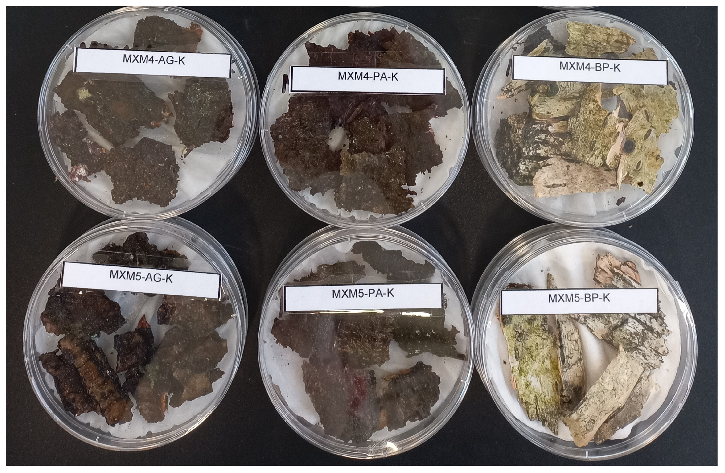
Plant material used for moist-chamber cultures was collected on 10 December 2024. Eight forest sub-compartments were selected, representing three forest associations of high European and global conservation value: Sphagno girgensohnii–Piceetum (corresponding to 91D0* Bog woodland in the Natura 2000 framework and G3.D Boreal bog conifer woodland in the EUNIS system), Ribeso nigri–Alnetum (91E0* Alluvial forests with Alnus glutinosa and Fraxinus excelsior in Natura 2000, EUNIS G1.13 Southern Alnus and Betula galleries), and Fraxino–Alnetum (also 91E0* in Natura 2000, assigned to EUNIS G1.13). Each of these habitat types is recognized as a priority habitat within the Natura 2000 network, underscoring their significance at both European and international levels. Specifically, compartments 124D, 125D, and 138a correspond to Sphagno girgensohnii–Piceetum, compartments 75G and 76G to Ribeso nigri–Alnetum, and compartments 8I, 31f, and 7a to Fraxino–Alnetum (Figure 1C). Three tree species, Alnus glutinosa, Betula pubescens, and Picea abies, served as sources for four types of dead organic matter: wood, bark, fallen leaves, and twigs. From each tree we collected one representative sample of every substrate type. Each sample of 5–10 fragments of bark, dead wood, leaf/needle litter, or fine twigs, was harvested from five spatially distinct micro-sites (1–10 m apart) within the same forest compartment in order to maximize micro-habitat heterogeneity. The fragments were pooled and trimmed to a uniform volume of approximately 60–70 cm3, which is sufficient to cover the base of a sterile polystyrene Petri dish (NOEX, Ø 90 mm × 14.2 mm, aseptic) while still permitting the lid to close hermetically and act as a self-contained moist chamber. In total, 96 such moist-chamber cultures were prepared (Figure 2). Each dish was labelled with the sampling location, date, host tree species, and substrate type, and then transported to the laboratories at the Institute of Forest Sciences, Białystok University of Technology for incubation and subsequent observation.

2.3. Establishment and Maintenance of Moist Chamber Cultures

Following standardized moist-chamber protocols for Eumycetozoa [15,21], field-collected substrates (decaying wood fragments and senescent leaf litter) were arranged on a double layer of sterilized cellulose tissue in 90 mm plastic Petri dishes and completely covered with distilled water. This initial flooding recreates the saturated conditions of water-logged microsites, thereby initiating spore germination, amoebal fusion, and plasmodial growth; under subsequent incubation at a moderate temperature and controlled illumination, the life cycle proceeds to sporocarp formation, enabling a comprehensive inventory of both conspicuous and minute taxa that are frequently overlooked [12,22,23].
Moist-chamber cultures were set up on 10 December 2024 (MXM1–MXM5) and 19 December 2024 (MXM6–MXM8), the only precipitation-free days suitable for fieldwork. After 12–24 h the excess water was decanted, leaving the substrates moist but not submerged. Because the indigenous bacterial and fungal communities constitute an essential food source for developing slime molds, the soaking water was neither autoclaved nor amended with antibiotics. The dishes were sealed, assigned unique identifiers, and incubated under diffuse natural daylight (7 h 42 min on 10 December 2024 increasing to 12 h 25 min on 25 March 2025) at 18–24 °C. Once active plasmodia appeared, dishes were transferred to a south-facing windowsill to stimulate sporulation and were checked daily. Moisture was monitored visually, and distilled water was added as needed to keep the substrates moist but not saturated. All cultures were maintained until 25 March 2025, providing a 105-day incubation period sufficient for the full maturation of every observed taxon. These cultures were monitored regularly under a stereomicroscope. During the first two weeks, observations were carried out every three days, allowing the early detection of plasmodia and young fruiting bodies; thereafter, checks were made weekly. Any visible plasmodia were relocated closer to a light source to exploit their positive phototaxis and promote sporulation [15].
Upon maturation, fruiting bodies were carefully collected with scalpels or pin tips, air-dried, labeled with metadata (habitat, sampling date, sporulation date), and stored in small cardboard boxes (4 × 6 cm, 6 × 8 cm, or 10 × 10 cm, chosen according to sporocarp size) to preserve their morphological and genetic integrity [16,23,24]. Established procedures were followed throughout this period to maintain humidity levels, minimize contamination, and document developmental stages [21]. Where necessary, distilled water was added sparingly, ensuring neither desiccation nor uncontrolled fungal growth. All plasmodia and fruiting bodies were photographed with an Opta-tech LAB 40 compound microscope and an Opta-tech SK binocular microscope for record-keeping, and dried sporophores were archived for subsequent studies. This moist-chamber strategy, widely adopted in mycological and protistological research [15,21], provided a low-cost, efficient method to reveal the otherwise hidden diversity of Eumycetozoa in the Knyszyn Forest.

2.4. Morphological Character Assessment and Checklist Verification

Diagnostic identification of the myxomycete material relied exclusively on directly observable characteristics of the fructifications. Particular attention was paid to the habit, pigmentation, surface sculpturing, and dimensions of sporocarps, together with the presence, length, and external topology of any stalk. We additionally recorded the color and thickness of the hypothallus, noting any basal reddening, and documented the peridium architecture and its mode of dehiscence. Structural attributes of the capillitium or elaters—branching pattern, wall thickness, degree of spiral banding, and apical ornamentation—were scored, as were key spore traits including color, outline, wall ornamentation, and diameter. Terminology follows the standard myxomycete glossaries cited in the Introduction, and diagnostic thresholds correspond to those adopted in contemporary European monographs.
All identified taxa were cross-checked against regional, national, and international inventories to determine whether they had previously been reported from the Knyszyn region, from Poland, or from Europe. Published checklists and floristic papers consulted for this purpose included the sources listed in references [7,25,26,27,28,29,30,31,32,33,34,35,36,37,38,39]. This comparative approach enabled an unambiguous determination of whether each record constituted a new finding for the study area, for the country, or for the broader European slime-mold biota.
A comprehensive overview of field sites, culture codes, host tree species, substrate types, and dates of culture maintenance is provided in Table A1. This documentation reflects the systematic approach adopted and illustrates the geographic extent and ecological variation encompassed by the sampling strategy.

2.5. Statistical Analysis

All quantitative analyses were carried out in R (v. 4.3.2; R Core Team 2024). Data preparation followed a reproducible workflow based on tidyverse (v. 2.0.0) and janitor (v. 2.2.0). Differences in culture success among the twelve host–substrate classes were examined with Pearson’s chi-square test (stats::chisq.test); pairwise contrasts were adjusted using the Benjamini–Hochberg procedure. Variation in species richness per positive culture was assessed with the Kruskal–Wallis rank-sum test, and post-hoc Dunn contrasts were obtained via FSA::dunnTest. Species-accumulation and sample-based rarefaction curves were generated with the vegan package (v. 2.6-4; functions specaccum and rarecurve). Incidence-based richness estimates (Chao1) and sample coverage were calculated using iNEXT (v. 3.0.0). Graphical outputs, including the heat-map of species incidence, were produced in ggplot2 (v. 3.5.1) with the perceptually uniform viridis palette.

3. Results

Fifteen species of true slime molds were recovered from ninety-six moist-chamber cultures prepared from the dead wood, bark, leaves, and branches of Alnus glutinosa, Betula pubescens, and Picea abies collected on eight swamp-forest plots in the Knyszyn Forest. After a period of more than four months of regular cultivation with systematic monitoring, sporocarps developed on their native substrates and species were delimited using macro- and microscopic characteristics, such as sporocarp habit, peridial persistence, capillitium architecture, and spore ornamentation. In the following subsections, each detected taxon is presented in detail, together with notes on its morphology and the provenance of the cultures in which it appeared.

3.1. Taxonomy

3.1.1. Arcyria cinerea (Bull.) Pers., Synopsis Methodica Fungorum 1: 724 (1801) [40]

Classification: Phylum Amoebozoa; Infraphylum Eumycetozoa; Class Myxogastria; Order Trichiales; Family Arcyriaceae; Genus Arcyria.
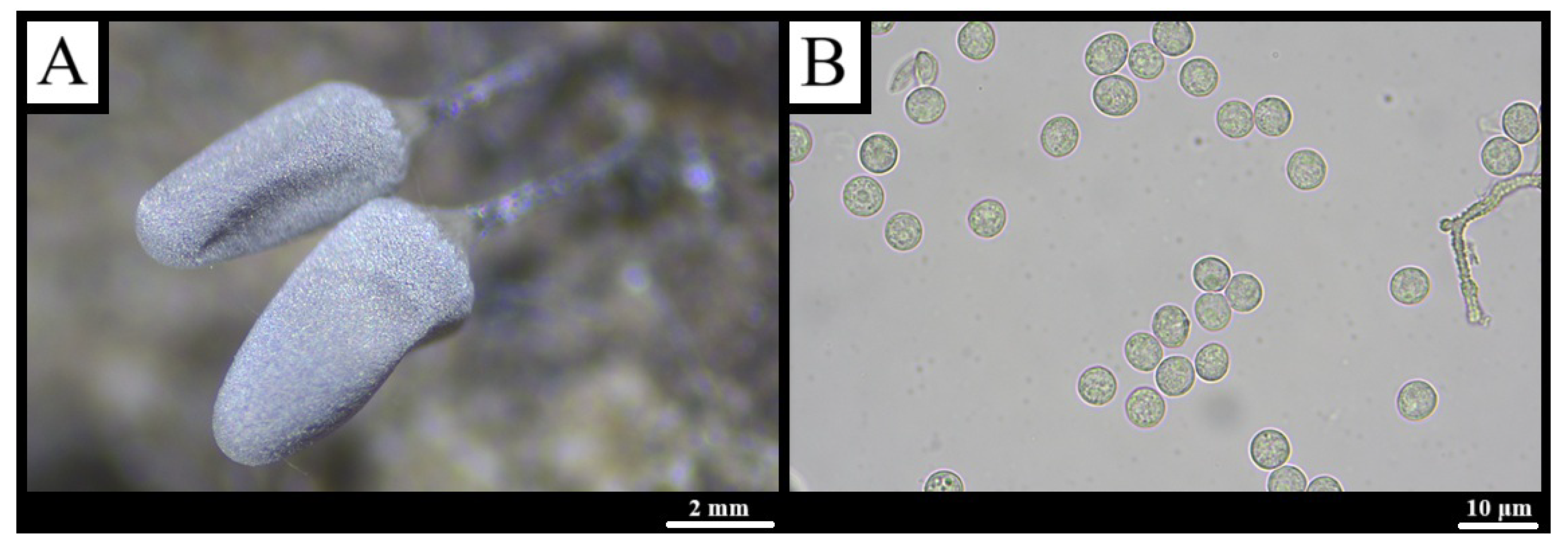
Description: Sporocarps stalked, solitary or loosely clustered, conical to short-cylindrical, 0.1–0.9 mm wide × 1–4 mm high, whitish to pale grey-beige with occasional pink or green hues (Figure 3A). A thin, common, disciform hypothallus is translucent to light brown. The rugose stalk, 0.2–1.5 mm long, is pale reddish-brown in transmitted light. Peridium soon evanescent, leaving a small, radially folded calyculus. Capillitium slightly elastic, forming a fine net of 4–7 μm threads with sparse 6–8 μm wart clusters, hyaline to faint brown. Spores beige to very pale grey en masse, nearly colorless in transmitted light, finely warted, 1.5–4 μm diam (Figure 3B) [41,42,43,44].
Habitat and geographical distribution: Specimens originated from moist-chamber cultures MXM8-BP-K, MXM4-PA-K, MXM3-BP-K, MXM2-BP-K, MXM2-PA-K, MXM1-BP-K, MXM7-PA-D, and MXM7-PA-K, established from bark and wood of Betula pubescens or Picea abies collected in various sub-compartments of Knyszyn Forest (see Appendix A).

3.1.2. Arcyria pomiformis (Leers) Rostaf., Pamiętn. Towarz. Nauk Ścisłych w Paryżu 6(1): 271 (1875) [45]

Classification: Phylum Amoebozoa; Infraphylum Eumycetozoa; Class Myxogastria; Order Trichiales; Family Arcyriaceae; Genus Arcyria.
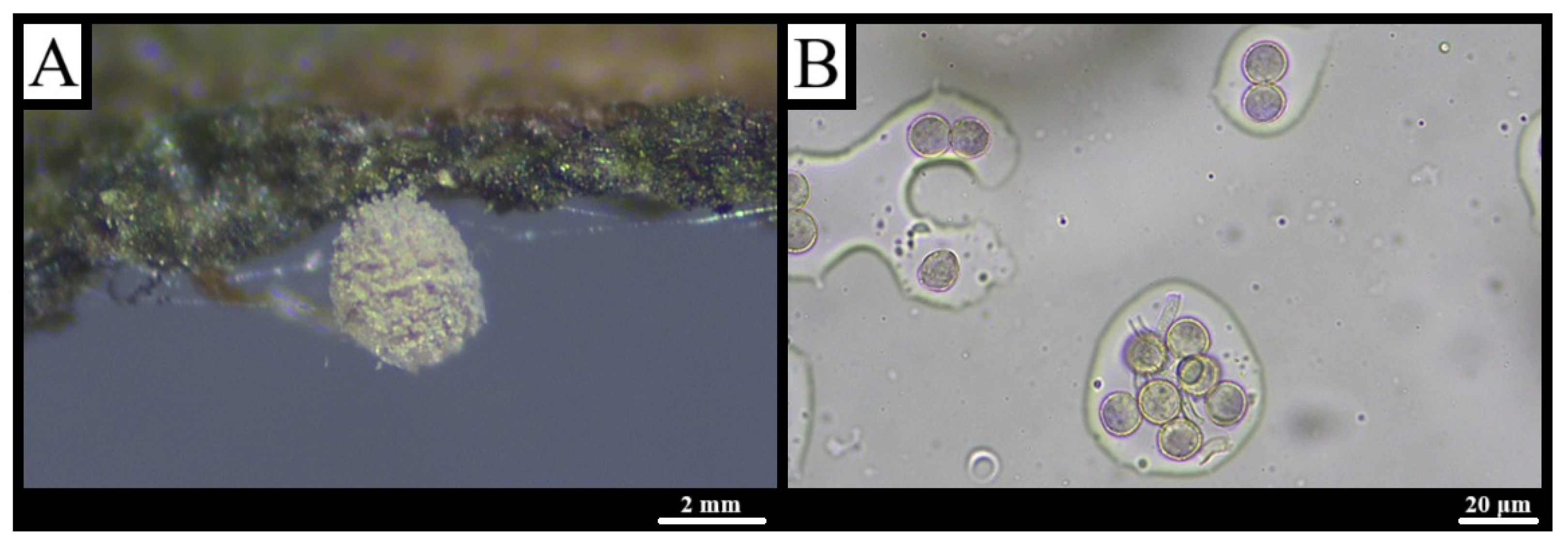
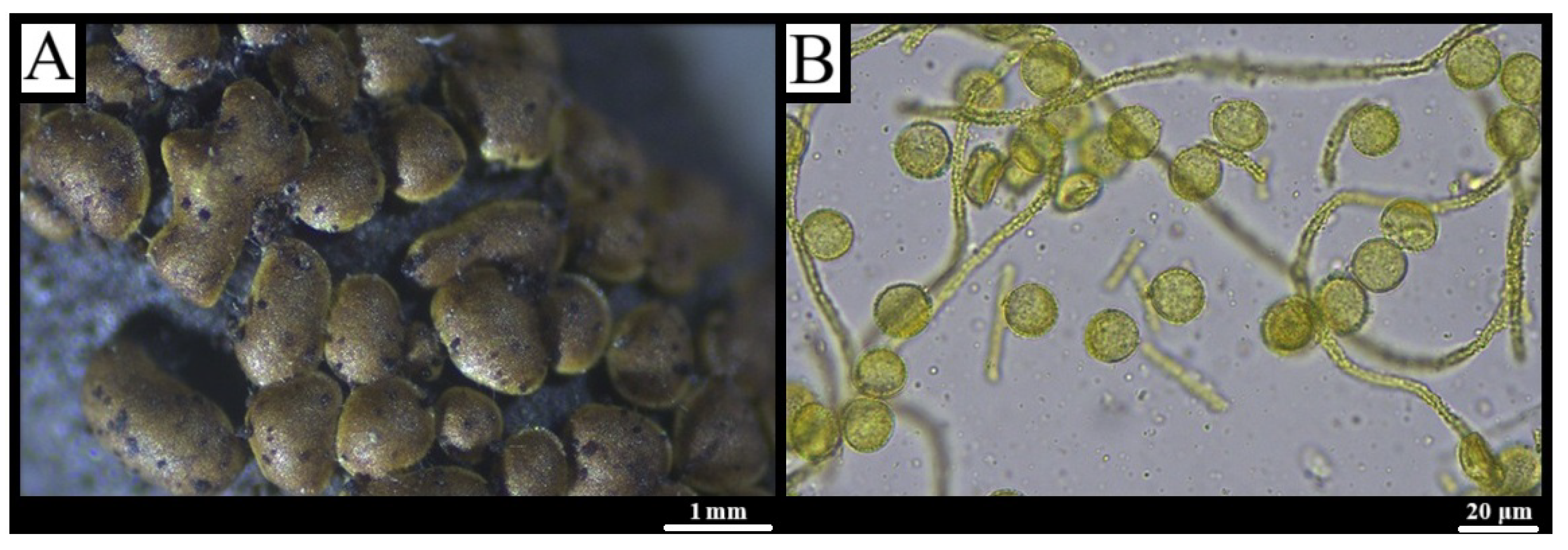
Description: Stipitate sporocarps, scattered to gregarious, globose to broadly ovoid, 0.1–0.5 mm in diameter, 0.3–1.2 mm high when closed, expanding to ca. 1 mm × 2.5 mm; vivid yellow to olive-yellow, maturing pale yellow-brown to dark olive (Figure 4A). A discoid, shining hypothallus is common to adjoining fructifications. Stalk 0.2–0.5 mm, cream to dark brown, packed with spore-like cysts. Peridium fugacious, persisting as an irregular basal disc, yellow-green in transmitted light. Capillitium elastic, with wide irregular meshes; threads 2–6 μm, pale brown-grey to yellow-green, bearing half-rings, rings and scattered warts. Spores yellow to pale yellow in mass, light olive to hyaline in transmitted light, 7–11 μm, uniformly finely warted with occasional larger clusters (Figure 4B) [41,42].
Habitat and geographical distribution: Detected in moist-chamber cultures MXM6-BP-K, MXM8-PA-K, MXM5-PA-K, MXM4-PA-D, and MXM2-BP-K (Appendix A), prepared from bark and wood of Betula pubescens or Picea abies collected in several sub-compartments of Knyszyn Forest; sporocarps developed on the original substrates.

3.1.3. Arcyria denudata (L.) Wettst., Verh. Zool.-Bot. Ges. Wien 35: 353 (1886) [46]

Classification: Phylum Amoebozoa; Infraphylum Eumycetozoa; Class Myxogastria; Order Trichiales; Family Arcyriaceae; Genus Arcyria.
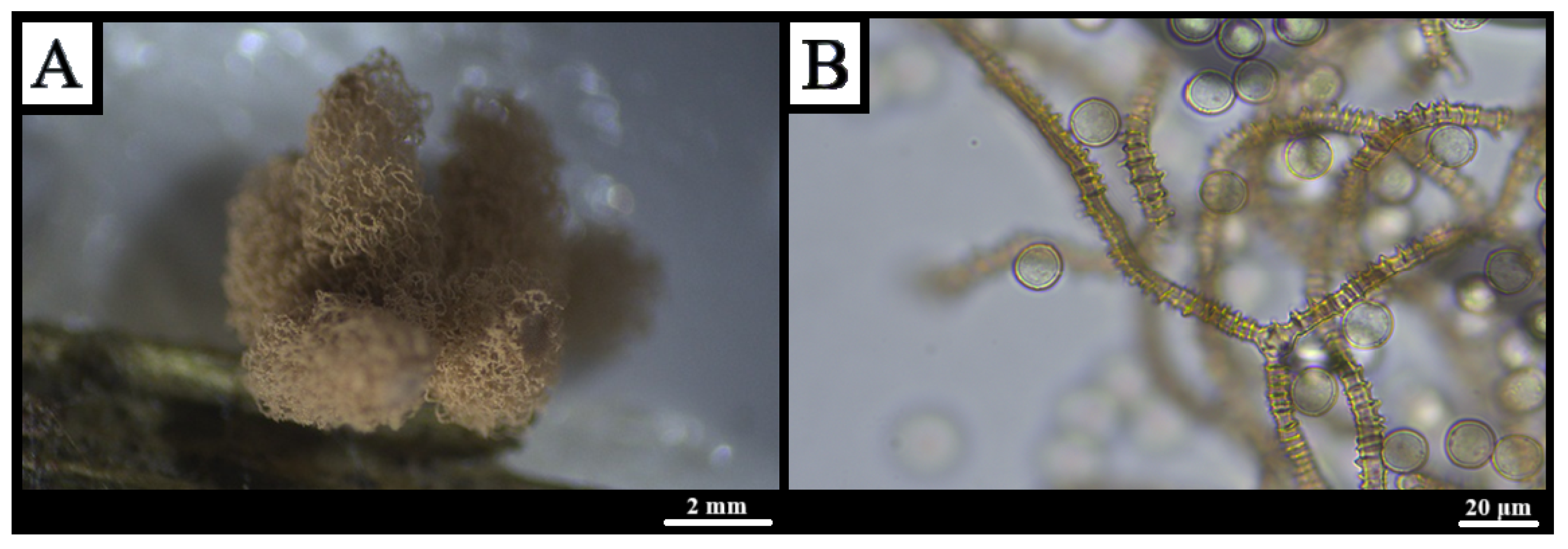
Description: Stipitate sporocarps crowded to gregarious, ovoid to short-cylindrical, red to dark brown, 0.5 mm wide × 1–3 mm high when closed, expanding to 1.2 mm × 6 mm. Disc-shaped hypothallus inconspicuous, pale yellow to yellow-brown (Figure 5A). Stalk 0.2–2 mm, striate, red-brown, containing 10–20 μm cysts. Peridium reduced to a shallow glossy cup with fine papillae and narrow ridges. Capillitium moderately elastic, dense, lacking free ends; rings, half-rings and blunt warts linked by low ridges; threads 2–5 μm, nearly colorless to rust-brown. Spores red to dark brown in mass, bluish-rust to almost colorless in transmitted light, 6–8 μm, finely warted with scattered larger clusters (Figure 5B) [41,42].
Habitat and geographical distribution: Observed in moist-chamber cultures MXM5-PA-D and MXM7-PA-D (Appendix A) derived from Picea abies wood in sub-compartments 01-28-1-06-7a and 01-08-2-05-125D of Knyszyn Forest, corresponding respectively to the Fraxino–Alnetum and Sphagno girgensohnii–Piceetum associations.

3.1.4. Arcyodes incarnata (Alb. & Schwein.) O.F. Cook, Science 15: 651 (1902) [47]

Classification: Phylum Amoebozoa; Infraphylum Eumycetozoa; Class Myxogastria; Order Trichiales; Family Arcyriaceae; Genus Arcyodes.
Description: Sporocarps nearly globose to pyriform, often coalescing, scarlet to copper, later ochre to olive, 0.5–1.5 mm high × 0.4–0.8 mm wide (Figure 6A). Hypothallus thin, commonly shared. Peridium membranous, splitting apically but persisting basally, colorless to light ochre, minutely granulate with short, irregular lines forming partial nets. Capillitium inelastic, wide-meshed, tubes 2–8 μm, sparsely warted or granular; free ends rounded; ochre to olive in mass, pale brown-pink in transmitted light, adnate to peridium. Spores pale pink, turning ochre-yellowish; nearly colorless in transmitted light, 6–10 μm, finely warted to almost smooth with scattered larger warts (Figure 6B) [30,41,48].
Habitat and geographical distribution: This record constitutes the first documented occurrence of A. incarnata in north-eastern Poland [25,26,27,28,29] and the second confirmed record for the country overall, the species having been last reported in Polish literature in 1972 [28,49]. Specimens originated from moist-chamber culture MXM6-AG-G (Appendix A) prepared from twigs of Alnus glutinosa collected in sub-compartment 01-08-2-02-75D (Ribeso nigri–Alnetum, 53.8789° N, 21.2489° E) of Knyszyn Forest, where sporocarps subsequently formed.

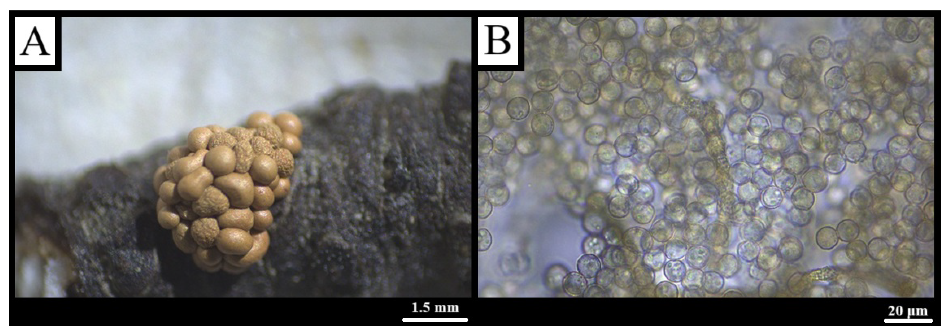
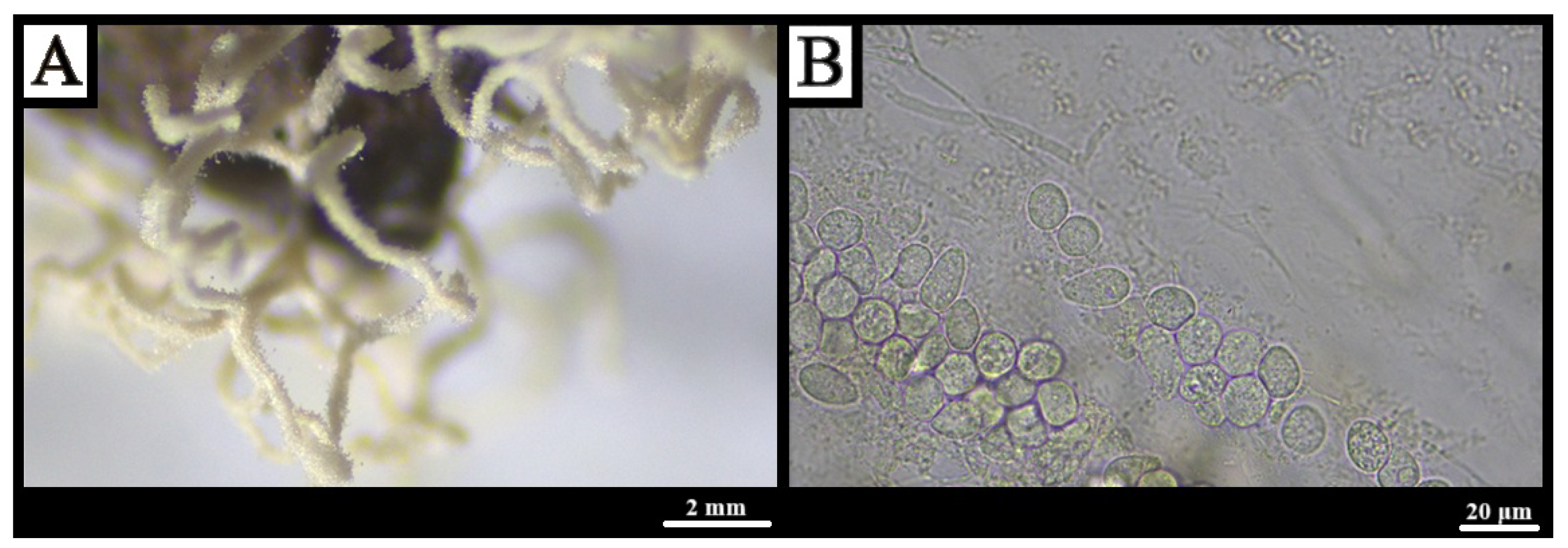
3.1.5. Ceratiomyxa fruticulosa var. flexuosa (Lister) G. Lister, Monograph of the Mycetozoa, ed. 2: 26 (1911) [50]

Classification: Phylum Amoebozoa; Infraphylum Eumycetozoa; Class Protosteliomycetes; Order Ceratiomyxales; Family Ceratiomyxaceae; Genus Ceratiomyxa.
Description: Sporophores arise as droplets on decaying wood, elongating into white, dichotomously branched dendroid columns; basal regions coarsely reticulate, apices smooth and finely granular, sides sheathed by a reticulate layer; tips capped by a perforate membrane with 0.35–0.55 μm calcareous granules (Figure 7A). A colony-wide white hypothallus cracks into irregular fields upon drying. Spores borne singly on stalks 7–20 μm × 1.5–3 μm; spores broadly elliptic to globose, hyaline to faint green-yellow, with granular contents and a basal calcareous granule, 8–15 × 6–10 μm (Figure 7B) [41,51,52].
Habitat and geographical distribution: Specimens were documented in moist-chamber cultures MXM7-BP-D and MXM8-AG-D (Appendix A) derived from wood of Betula pubescens and Alnus glutinosa, respectively, collected in adjacent sub-compartments 01-08-2-05-125D and 01-08-2-05-124D of Knyszyn Forest within a Sphagno girgensohnii–Piceetum association. The finding represents the second record of the subspecies in Poland and the first for the north-eastern part of the country [25,26,27,28,29].

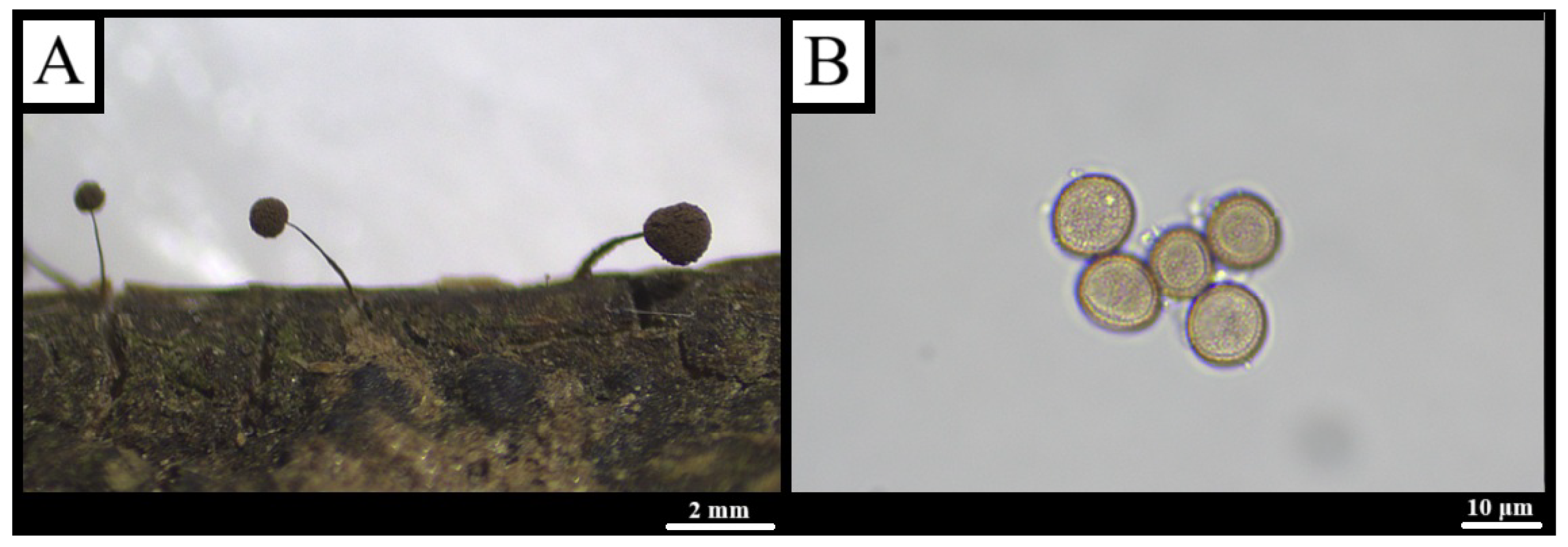
3.1.6. Comatricha nigra (Pers.) J. Schröt., Krypt.-Fl. Schlesien 3(1): 118 (1885) [53]

Classification: Phylum Amoebozoa; Infraphylum Eumycetozoa; Class Myxogastria; Order Stemonitidales; Family Stemonitidaceae; Genus Comatricha.
Description: Sporocarps in dense to loose rows, long-stalked, globose to ovoid, 0.3–1 mm in diameter, total height 1.5–8 mm, dark brown. Disciform hypothallus brown to reddish-brown, pale to colorless in transmitted light (Figure 8A). Stalk 1–7 mm, glossy black, tapering upward. Peridium very fugacious. Columella reaches almost to apex, merging into capillitium. Capillitium dense, wavy, peripheral meshes tortuous with short free ends; threads smooth to slightly knotted, reddish-brown in transmitted light. Spores dark brown to blackish in mass, brown-violet in transmitted light, finely warted, 8–11 μm (Figure 8B) [28,54,55].
Habitat and geographical distribution: C. nigra was recovered from eight moist-chamber cultures—MXM1-AG-G, MXM2-AG-K, MXM2-PA-G, MXM3-PA-K, MXM5-PA-K, MXM6-AG-K, MXM6-AG-G and MXM8-AG-K (Appendix A)—originating from bark and twigs of Alnus glutinosa or Picea abies. The substrates were collected across several sub-compartments of Knyszyn Forest representing Sphagno girgensohnii–Piceetum, Ribeso nigri–Alnetum, and Fraxino–Alnetum associations, where sporocarps eventually developed.

3.1.7. Cribraria microcarpa (Schrad.) Pers., Synopsis Methodica Fungorum 1: 190 (1801) [40]

Classification: Phylum Amoebozoa; Infraphylum Eumycetozoa; Class Myxogastria; Order Cribrariales; Family Cribrariaceae; Genus Cribraria.
Description: Solitary to crowded, nodding sporocarps, globose, ochre- to yellow-brown, 0.2–0.3 mm in diameter, on 1–3.5 mm dark red-brown stalks (Figure 9A). Discoid hypothallus red-brown. Peridium reduced to short ribs and a basal disc, forming a delicate net with thickened incurved nodes 10–20 μm (rarely to 35 μm); free ends scarce. Dark brown granules, 1–2 μm, densely coating nodes and disc. Spores ochre-yellow to reddish ochre in mass, pale reddish-brown in transmitted light, finely warted, 6–8 μm (Figure 9B) [41,56].
Habitat and geographical distribution: During the present study, C. microcarpa was detected in moist-chamber cultures MXM5-PA-K, MXM6-PA-K, and MXM3-AG-K (Appendix A), each prepared from bark of Picea abies or Alnus glutinosa collected in several sub-compartments of Knyszyn Forest.

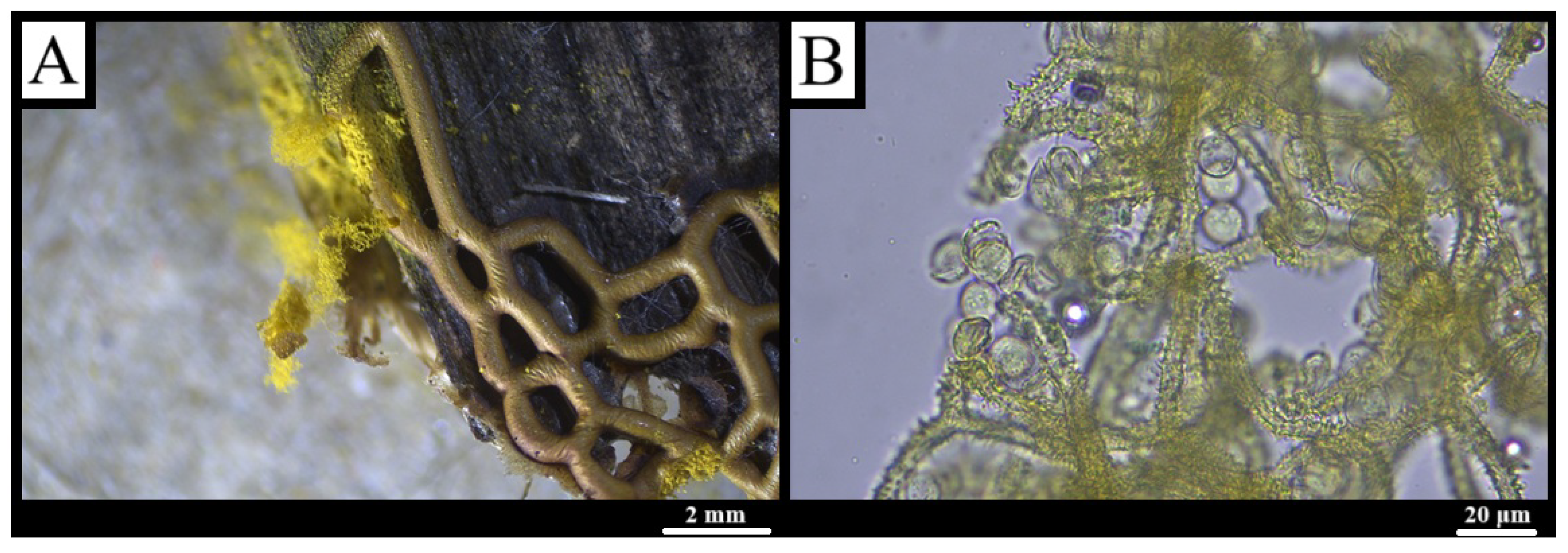
3.1.8. Hemitrichia serpula (Scop.) Rostaf., Versuch eines Systems der Mycetozoen: 14 (1873) [57]

Classification: Phylum Amoebozoa; Infraphylum Eumycetozoa; Class Myxogastria; Order Trichiales; Family Trichiaceae; Genus Hemitrichia.
Description: Plasmodiocarps form a red-brown to yellow-brown network to 1 dm2; meshes 0.3–5 mm, strands 0.4–0.6 mm thick, sessile. Brown hypothallus lies beneath. Peridium ruptures irregularly and vanishes, leaving light-yellow, vein-marked remnants (Figure 10A). Capillitium golden yellow to orange, highly elastic; threads 5–7 μm, with 3–4 tight spiral ridges and occasional 3–7 μm colorless spines; free ends short and tapered. Spores golden yellow in mass, pale yellow in transmitted light, with large irregular reticulate meshes and a 1–1.5 μm marginal zone, 10–14 μm (Figure 10B) [41,58,59].
Habitat and geographical distribution: Slime mold H. serpula occurred in moist-chamber cultures MXM2-AG-D, MXM3-PA-D, and MXM6-AG-D (Appendix A) established from wood of Alnus glutinosa or Picea abies collected across three distinct sub-compartments of Knyszyn Forest.

3.1.9. Lamproderma scintillans (Berk. & Broome) Morgan, Journal of the Cincinnati Society of Natural History 16: 131 (1894) [60]

Classification: Phylum Amoebozoa; Infraphylum Eumycetozoa; Class Myxogastria; Order Stemonitidales; Family Stemonitidaceae; Genus Lamproderma.
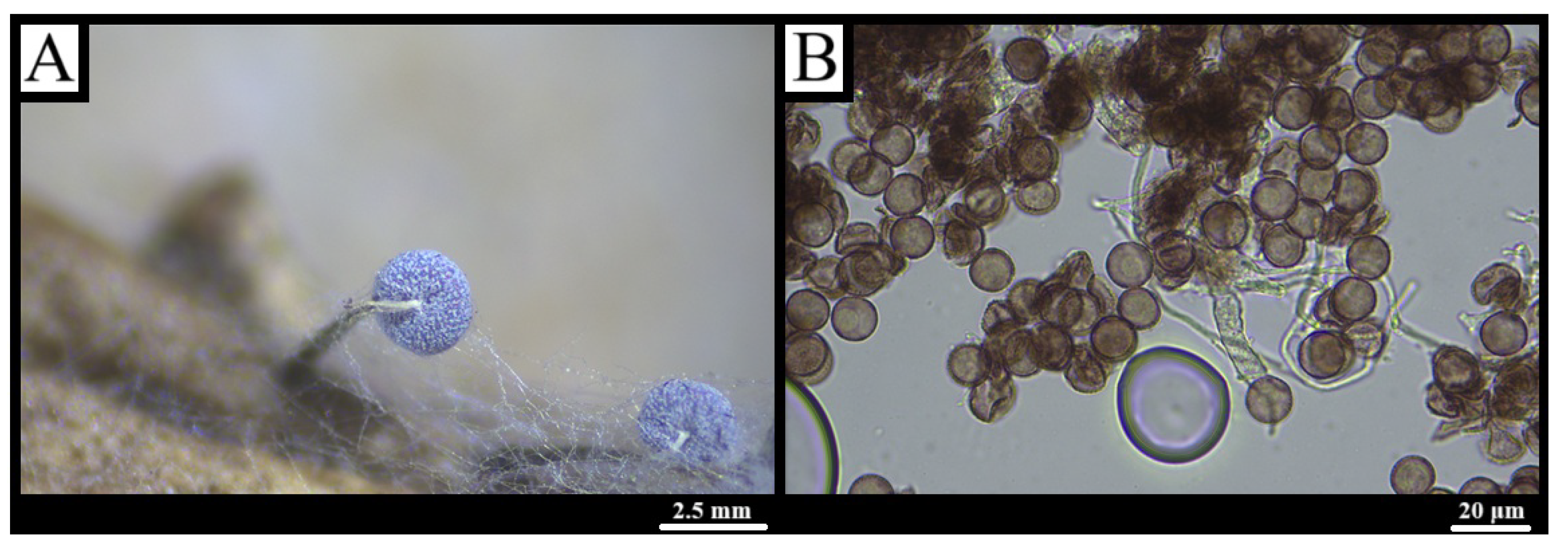
Description: Stalked sporocarps, scattered to gregarious, globose, 0.2–0.6 mm in diameter and 0.5–2 mm tall, iridescent blue to violet-bronze. Disciform hypothallus light to reddish-brown, darker toward the stalk (Figure 11A). Stalk 0.5–1.3 mm, glossy black, indistinctly striate. Peridium delicate, slightly roughened, sometimes with crystalline needles, persisting as collar-like remnants; colorless in transmitted light, apex reddish-brown. Columella cylindrical, reaching mid-sporocarp, dark brown to black. Capillitium a loose, sparsely branched net arising from the columella; threads pale proximally, dark distally, tapering to colorless tips. Spores brown in mass, pale grey-brown in transmitted light, with dark, widely spaced warts, 7–9.5 μm in diameter (Figure 11B) [54,61,62].
Habitat and geographical distribution: A single record of L. scintillans derived from culture MXM1-AG-L (Appendix A), initiated from Alnus glutinosa leaves gathered in sub-compartment 01-08-2-05-138a of the Knyszyn Forest (53.237284° N, 23.151192° E), within a Sphagno girgensohnii–Piceetum association.

3.1.10. Metatrichia vesparium (Batsch) Nann.-Bremek., Proc. Kon. Ned. Akad. Wetensch. C 69: 348 (1966) [63]

Classification: Phylum Amoebozoa; Infraphylum Eumycetozoa; Class Myxogastria; Order Trichiales; Family Trichiaceae; Genus Metatrichia.
Description: Fructifications solitary, clustered or pseudoaethaloid, dark red-brown, obovoid to cylindroid, 0.2–0.7 mm wide × 0.4–1.2 mm high, on a ribbon-like wrinkled stalk to 2 mm (rarely sessile) (Figure 12A). Hypothallus yellowish-white, reddening basally. Peridium bilayered: inner thin, pellucid honey-colored; outer thick, opaque red-brown, usually dehiscing by a sharply delimited convex lid. Capillitium of highly elastic, sparsely branched elaters 4–7 μm thick, with 3–5 moderate spiral bands and short spines; tips pointed. Spores brown-red to rust in mass, yellow- to honey-brown in transmitted light, finely warted, subglobose to broadly ellipsoid, 9–13 μm (ellipsoids 8.5 × 9.5–11 × 12.5 μm) (Figure 12B) [54,64].
Habitat and geographical distribution: Slime mold M. vesparium was represented by a single culture, MXM3-AG-D (Appendix A), obtained from A. glutinosa wood collected in sub-compartment 01-08-2-02-8i of the Knyszyn Forest (53.272998° N, 23.116182° E), belonging to a Ribeso nigri–Alnetum association.

3.1.11. Oligonema flavidum (Peck) Peck, Ann. Rep. N.Y. State Mus. Nat. Hist. 31: 42 (1878) [65]

Classification: Phylum Amoebozoa; Infraphylum Eumycetozoa; Class Myxogastria; Order Trichiales; Family Trichiaceae; Genus Oligonema.
Description: Sessile sporocarps densely clustered or solitary, pale glossy yellow, 0.2–0.5 mm wide × 0.5–0.8 mm high; no hypothallus (Figure 13A). Peridium thin, translucent, inner surface finely warted with fern-like markings, rupturing irregularly above. Capillitium simple to branched; threads 3–5 μm, finely warted with indistinct spirals, swollen at intervals, ends rounded. Spores yellow in mass and transmitted light, 13–16 μm in diameter, with complete reticulate ornamentation and a 1.5 μm marginal zone (Figure 13B) [30,41,66,67].
Habitat and geographical distribution: O. flavidum emerged in culture MXM8-AG-K (Appendix A), prepared from bark of A. glutinosa collected in sub-compartment 01-08-2-05-124D of the Knyszyn Forest (53.851152° N, 21.207244° E), within a Sphagno girgensohnii–Piceetum association. This finding constitutes the second confirmed record of the species for Poland and the first documented occurrence in north-eastern Poland [25,26,27,28,29,30].

3.1.12. Perichaena depressa (Libert) Rostaf., Versuch eines Systems der Mycetozoen: 112 (1873) [57]

Classification: Phylum Amoebozoa; Infraphylum Eumycetozoa; Class Myxogastria; Order Trichiales; Family Perichaenaceae; Genus Perichaena.
Description: Sesile sporocarps flattened to hemispherical, often compressed into pseudoaethaloid masses or short comma-like plasmodiocarps; red- to dark brown, 0.2–1.5 mm in diameter, <0.3 mm high (Figure 14A). Common hypothallus dirty grey-brown. Peridium double: outer red-brown layer densely granulate; inner pale yellow, dehiscing by an upward-rolling lid or rarely by irregular scaling. Capillitium golden-yellow, elastic, simple or branched, 1.5–4 μm, with fine warts, spines, and occasional annular swellings. Spores golden-yellow in mass and transmitted light, finely warted, 8.5–11 μm in diameter (Figure 14B) [41,68,69,70].
Habitat and geographical distribution: During our investigation, P. depressa was detected in moist-chamber culture MXM8-AG-K (Appendix A), prepared from the bark of Alnus glutinosa collected in sub-compartment 01-08-2-05-124D of the Knyszyn Forest (53.851152° N, 21.207244° E) within a Sphagno girgensohnii–Piceetum association. This record represents the third documented occurrence of the species in Poland and the first for north-eastern Poland [25,26,27,28,29].

3.1.13. Physarum album (Bull.) Chevall., Flore Générale des Environs de Paris 1: 345 (1826) [71]

Classification: Phylum Amoebozoa; Infraphylum Eumycetozoa; Class Myxogastria; Order Physarales; Family Physaraceae; Genus Physarum.
Description: Gregarious sporangia erect to slightly nodding, subglobose to lenticular, 0.4–0.7 mm broad on distinct stalks, total height 1–1.5 mm; white to grey-white, faintly iridescent where lime is sparse; wall studded with white lime granules (Figure 15A). Stalk subulate, wrinkled, grey to olivaceous-black, upper part translucent, base sometimes white with wall-bound lime. Hypothallus continuous, black. Peridium lime-crusted; columella absent after dehiscence. Capillitium a network of colorless, acutely branching threads with few flattened nodes and scattered lime particles. Spores black in mass, brown-violet in transmitted light, 8–11 μm in diameter, minutely spinulose to nearly smooth (Figure 15B) [72,73,74,75].
Habitat and geographical distribution: P. album was identified in moist-chamber cultures MXM2-PA-G, MXM3-BP-K, MXM3-PA-G, MXM4-BP-K, MXM6-BP-D, MXM6-BP-K, MXM7-BP-G, MXM8-BP-D, and MXM8-BP-K (Appendix A), established from twigs of Picea abies and bark and wood of Betula pubescens collected across the Knyszyn Forest, where sporocarps subsequently developed on the respective substrates.

3.1.14. Physarum bivalve Pers., Neues Magazin für die Botanik 1: 89 (1794) [76]

Classification: Phylum Amoebozoa; Infraphylum Eumycetozoa; Class Myxogastria; Order Physarales; Family Physaraceae; Genus Physarum.
Description: Plasmodiocarps irregularly winding, laterally compressed, <7 mm long, 1 mm high, 0.3 mm wide, white to light grey; occasional loose globose sporocarps 0.4–0.8 mm in diameter (Figure 16A). Fructifications sessile or shortly stalked, sometimes with a scarlet base. Hypothallus thin, colorless to light brown, common beneath the group. Peridium double: outer granular to wrinkled, lime-rich white to grey, basal zone light brown; inner hyaline, opening by a broad longitudinal slit. Columella absent. Capillitium of white lime nodes joined by short hyaline threads, colorless in transmitted light. Spores black-brown in mass, light to violet-brown singly, uniformly finely warted, 8–10 μm in diameter (Figure 16B) [72,77,78].
Habitat and geographical distribution: A single occurrence of P. bivalve was confirmed in moist-chamber culture MXM1-PA-L (Appendix A), derived from needles of Picea abies collected in sub-compartment 01-08-2-05-138a of the Knyszyn Forest (53.237284° N, 23.151192° E) within a Sphagno girgensohnii–Piceetum association, where sporocarps developed on the leaf substrate.

3.2. Statistical Analysis

3.2.1. Performance of Moist-Chamber Cultures

Of the 96 moist-chamber preparations, 35 produced sporocarps, giving an overall success rate of 36.5% (Table 1). Culture outcome differed significantly among the 12 host–substrate classes ( χ 11 2 = 27.5 , p = 0.004 ). Bark consistently outperformed all other matrices: success reached 87.5% for Picea abies bark and 75.0% for Betula pubescens bark, whereas the foliage of every host was largely unproductive, including a complete failure for B. pubescens leaves. Mean species richness per positive culture was universally low (grand mean 1.3 ± 0.6 , median = 1) and did not vary across substrates (Kruskal–Wallis H 10 = 5.06 , p = 0.89 ). Hence, sporulating cultures rarely harbored more than one myxomycete taxon.

3.2.2. Species Incidence Across Host–Substrate Combinations

The incidence matrix (Table 2) documents 46 species–culture occurrences representing 14 morphologically verified species in 11 genera and 5 orders. Three taxa—Physarum album, Arcyria cinerea, and Comatricha nigra—were dominant, accounting jointly for 54% of all occurrences (9, 8, and 8 records, respectively). Each of the remaining eleven species appeared in five or fewer cultures.
Substrate identity structured the assemblage most strongly. Bark yielded 25 occurrences (54%) and supported seven species; wood also hosted 7 species but only 12 occurrences. Twigs contributed seven occurrences of three species, whereas leaves produced two single-species records. Incidence patterns are visualized in a heat-map (Figure 17), which highlights the concentration of sporulation on bark and wood and the near absence of activity on foliar substrates. Host tree influences were secondary yet discernible. Alnus glutinosa supported the highest observed richness (9 species) but a moderate incidence of 14 occurrences. P. abies generated the greatest overall activity (18 occurrences) and 8 species, while B. pubescens was both numerically and compositionally the poorest host (14 occurrences, 4 species).

3.2.3. Host-Specific Richness and Sampling Coverage

Observed and estimated richness values for each host tree are summarized in Table 3. Chao1 estimates indicate substantial unseen diversity for A. glutinosa (33.5 ± 31.1 species; sampling coverage = 26.9%), whereas the inventories for B. pubescens (4.5 ± 1.3 species; 88.9% coverage) and P. abies (8.7 ± 1.3 species; 92.3% coverage) are nearing completeness.

3.2.4. Sampling Completeness and Substrate-Specific Accumulation

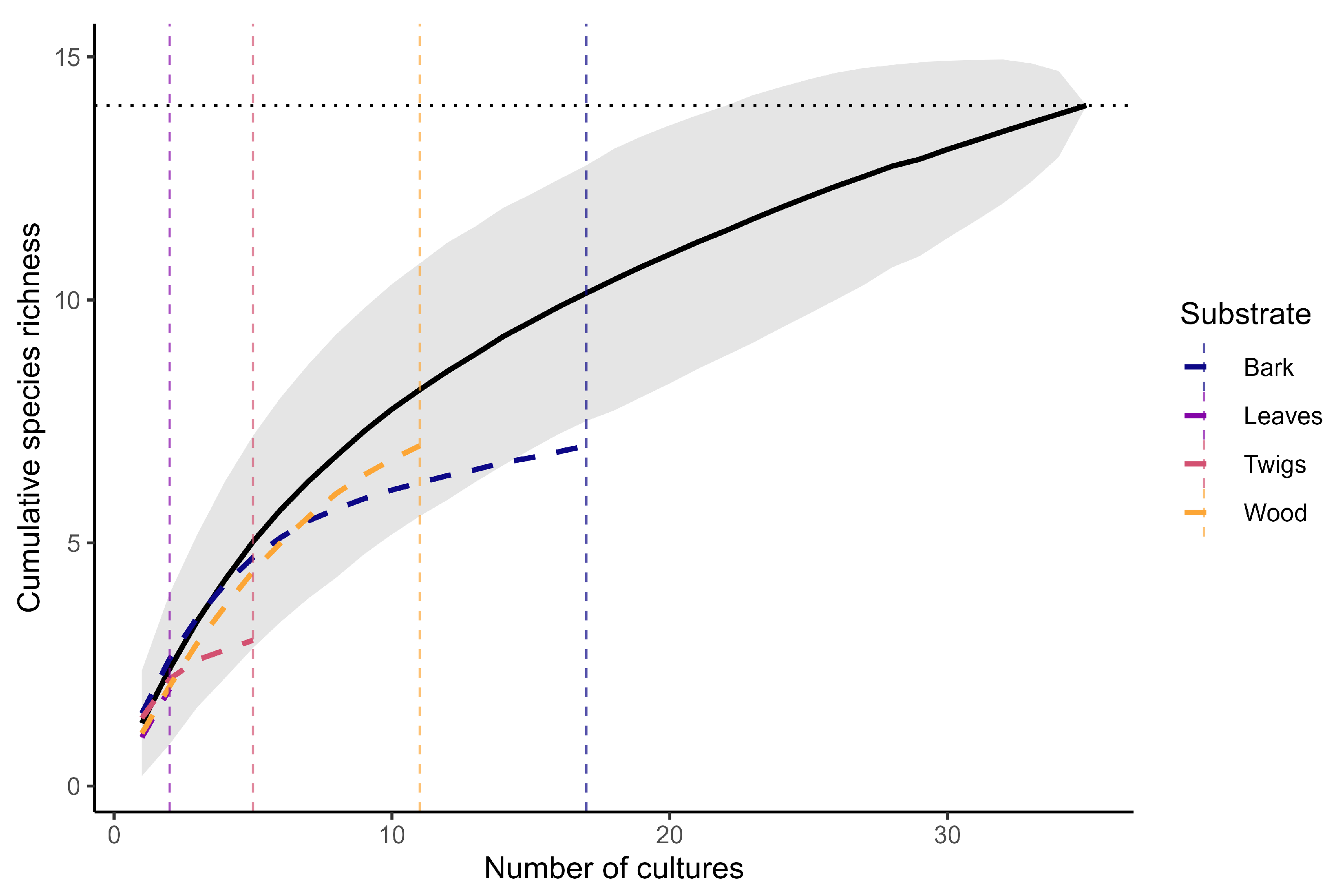
The pooled species-accumulation curve (Figure 18) leveled off after approximately 30 positive cultures and approached an asymptote at 14 species. The final five cultures added on average fewer than 0.2 species, and the 95% confidence interval narrowed to ±0.7 species, suggesting that more than 90% of the recoverable richness was captured by the study design. Substrate-specific curves diverged markedly. Bark and wood both plateaued at seven species after 10–12 sporulating cultures, whereas trajectories for twigs (three species from five cultures) and leaves (two species from two cultures) were still rising when sampling ended, indicating an incomplete characterization of these microhabitats.

3.2.5. Incidence Patterns

A total of 46 sporulation events involving 14 myxomycete species were recorded. Three taxa—Physarum album, Arcyria cinerea, and Comatricha nigra—contributed 54 % of all positives, whereas the remaining eleven species appeared in <5 cultures each. Species richness peaked on bark and wood of all hosts (seven species each), declined on twigs (three), and was minimal on foliar substrates (two). Alnus glutinosa hosted nine species, Picea abies eight, and Betula pubescens four, underscoring bark as the principal microhabitat structuring the swamp-forest myxobiota.

4. Discussion

The present survey demonstrates that swamp-forest ecosystems of the Knyszyn Forest harbor a diverse community of true slime molds (Eumycetozoa), as revealed by moist-chamber cultures. Such in vitro methods have long been recognized as an effective approach for detecting myxomycetes with cryptic life cycles or inconspicuous sporocarps, especially in consistently humid conditions [15,21]. The standing water, saturated soils, and substantial volumes of decaying plant material characteristic of boggy stands promote stable moisture and plentiful microbial prey, thus favoring the proliferation of numerous Eumycetozoa [8,79].
The statistical evaluation of culture performance corroborates these qualitative observations. Pearson’s χ 11 2 = 27.5 ( p = 0.004 ) revealed a highly significant association between substrate category and sporulation success, with bark—particularly that of Picea abies and Betula pubescens—yielding up to 87.5% positive cultures, whereas foliar substrates never exceeded 12.5%. This sharp gradient confirms that the physicochemical properties of bark (coarser texture, greater water-holding capacity, and richer endophytic propagule load) act as primary filters governing myxomycete fructification in swamp-forest settings.
Notably, several rarely reported species were recovered, underscoring the under-explored potential of swamp-forest complexes in Poland. The rediscovery of Arcyodes incarnata after its last national report in 1972 [49] highlights the importance of targeted sampling in overlooked habitats. Similarly, other taxa newly recorded for northeastern Poland, including Oligonema flavidum and Perichaena depressa, are scarcely documented at both the national and continental scales [25,26,27,28,29]. Their presence aligns with similar findings in wet woodland localities, where specialized conditions often sustain populations of uncommon slime molds [4]. Overall, this study underscores the value of combining systematic moist-chamber protocols with fine-scale habitat selection in swampy forests, which remain comparatively understudied in myxomycete research [15,22]. Set against the spectrum of inventories conducted in structurally comparable temperate forests, the richness documented here occupies an unequivocally mid-range position, yet it augments that baseline with an assemblage of corticolous micro-species that are rarely encountered during plot-based fruiting surveys. These results reinforce the consensus that only a dual strategy—large-scale moist-chamber culturing coupled with systematic in-field searches—can approximate the true community composition of forest myxobiota, and they provide a rare quantitative benchmark for calibrating the efficiency of the moist chambers approach under swamp-forest conditions.
Although substrate identity dictated culture success, the Kruskal–Wallis test detected no significant differences in the number of species developing per positive culture ( H 10 = 5.06 , p = 0.89 ). Once sporulation commenced, community assembly quickly converged on one—occasionally two—dominant taxa, a pattern consistent with competitive exclusion and priority effects previously documented for temperate forests.
Sample-based rarefaction curves (Figure 18) approached an asymptote after circa 30 positive cultures, indicating that the experimental design captured >90% of the recoverable species pool. Nonetheless, tree-specific Chao1 estimates exposed unequal sampling completeness: coverage was high for B. pubescens (88.9%) and P. abies (92.3%), but markedly lower for A. glutinosa (26.9%). The wide standard error ( ± 31.1 ) around the alder estimate implies a substantial reservoir of undetected, low-frequency specialists on this host, warranting intensified future sampling.
Methodologically, the overall culture success rate of 36.5% mirrors values reported from other cool-temperate regions and validates moist-chamber assays as a robust tool even under highly water-logged conditions. From a practical standpoint, the steep decline in success from bark/wood to leaves strongly advocates for prioritizing cortico- and xylophilous substrates when resources are limited.
Finally, host-tree effects proved secondary but ecologically informative. Alder substrates supported the broadest, albeit patchily expressed, species pool; spruce and birch, by contrast, were dominated by cosmopolitan bark specialists such as Arcyria cinerea and Physarum album. These contrasts illustrate how microhabitat heterogeneity, nested within a single swamp-forest stand, can sustain both widespread r-strategists and narrowly distributed k-strategists, thereby enhancing overall myxobiota diversity.
The quantitative insights derived from chi-square tests, non-parametric richness comparisons, and rarefaction modeling refine our ecological understanding of slime-mold assemblages in peat landscapes and offer a statistically grounded framework for optimizing future biodiversity inventories in analogous wet-forest mosaics.
Building on the material amassed here, forthcoming work will employ multilocus DNA barcoding and phylogenomic analyses to corroborate morphological identifications and reveal cryptic diversity within these communities. Integrating such molecular data with the present ecological framework will enable more precise delimitations of species and lineages, thereby strengthening future assessments of slime mold biodiversity in various ecosystems.

5. Conclusions

Based on the findings of this study, the following conclusions can be drawn:
  • Moist-chamber cultures proved highly effective for uncovering Eumycetozoa in swamp-forest habitats, detecting numerous slime-mold species despite waterlogged substrates and challenging field conditions. The protocol yielded a high proportion of sporulating preparations, sufficient for the species-accumulation curve to approach an asymptote and broadly comparable with success rates reported from other temperate European forests.
  • Several taxa—Arcyodes incarnata, Ceratiomyxa fruticulosa var. flexuosa, Oligonema flavidum, and Perichaena depressa—constitute new records for northeastern Poland, highlighting peatland and swamp forests as refugia for regionally rare or under-explored myxomycetes.
  • A. incarnata has not been recorded in Poland for several decades, emphasizing the importance of targeted sampling that can reveal taxa assumed to be absent or exceedingly rare in the current myxobiota.
  • The overall rarity of newly documented species, both nationally and continentally, underlines the urgent need for expanded inventories and systematic culturing studies in wet forest habitats. Such investigations will likely continue to uncover additional, previously unrecorded slime molds, further enriching our knowledge of Eumycetozoa diversity in Poland and throughout Europe.
  • A chi-square test confirmed a significant association between the substrate type and culture success, with bark markedly outperforming other microhabitats, whereas leaves and needles were least suitable.
  • By contrast, once sporulation was triggered, the number of species per culture remained similar across substrates, indicating that competitive assembly processes quickly narrow the community composition regardless of the microhabitat.
  • Rarefaction modeling showed that the current sampling effort captured almost the entire resident species pool overall, but alder substrates in particular still harbor an appreciable fraction of undiscovered diversity, warranting further attention.
  • The complementary distribution of taxa across bark, wood, twigs, and leaves underscores the necessity of multi-substrate sampling in any future monitoring program.

Author Contributions

Conceptualization, T.P., I.Ż. and G.M.M.; methodology, T.P., I.Ż. and G.M.M.; validation, T.P. and I.Ż.; formal analysis, T.P.; investigation, T.P., I.Ż. and G.M.M.; resources, T.P., I.Ż., G.M.M., M.P. and K.W.; writing—original draft preparation, T.P.; writing—review and editing, T.P., I.Ż., G.M.M., M.P., K.W., K.S. and T.O.; visualization, T.P. and I.Ż.; supervision, M.P., K.W., K.S. and T.O.; project administration, T.P.; funding acquisition, T.O. All authors have read and agreed to the published version of the manuscript.

Funding

This research received no external funding.

Data Availability Statement

Data are contained within the article.

Conflicts of Interest

The authors declare no conflicts of interest.

Appendix A. Detailed Inventory of Moist-Chamber Cultures Established from Knyszyn Forest Samples

Table A1. Compendium of all moist-chamber cultures established from the Knyszyn Forest (NE Poland). Listing for each sample: culture code, sampling locality, forest sub-compartment (Polish Forest Data Bank), plant association, geographic coordinates, date of field sampling, date of culture initiation, culture maintenance end date, host-tree species, and substrate type (dead organic matter).
Culture CodeSampling LocalityForest Sub-CompartmentPlant AssociationCoordinatesField Sampling DateCulture Start DateCulture Maintenance End DateHost TreeSubstrate
MXM1-AG-DKnyszyn Forest01-08-2-05-138aSphagno girgensohnii–Piceetum53.237284, 23.15119210 Dec 202410 Dec 202425 Mar 2025Alnus glutinosaWood
MXM1-AG-KKnyszyn Forest01-08-2-05-138aSphagno girgensohnii–Piceetum53.237284, 23.15119210 Dec 202410 Dec 202425 Mar 2025Alnus glutinosaBark
MXM1-AG-LKnyszyn Forest01-08-2-05-138aSphagno girgensohnii–Piceetum53.237284, 23.15119210 Dec 202410 Dec 202425 Mar 2025Alnus glutinosaLeaves
MXM1-AG-GKnyszyn Forest01-08-2-05-138aSphagno girgensohnii–Piceetum53.237284, 23.15119210 Dec 202410 Dec 202425 Mar 2025Alnus glutinosaTwigs
MXM1-BP-DKnyszyn Forest01-08-2-05-138aSphagno girgensohnii–Piceetum53.237284, 23.15119210 Dec 202410 Dec 202425 Mar 2025Betula pubescensWood
MXM1-BP-KKnyszyn Forest01-08-2-05-138aSphagno girgensohnii–Piceetum53.237284, 23.15119210 Dec 202410 Dec 202425 Mar 2025Betula pubescensBark
MXM1-BP-LKnyszyn Forest01-08-2-05-138aSphagno girgensohnii–Piceetum53.237284, 23.15119210 Dec 202410 Dec 202425 Mar 2025Betula pubescensLeaves
MXM1-BP-GKnyszyn Forest01-08-2-05-138aSphagno girgensohnii–Piceetum53.237284, 23.15119210 Dec 202410 Dec 202425 Mar 2025Betula pubescensTwigs
MXM1-PA-DKnyszyn Forest01-08-2-05-138aSphagno girgensohnii–Piceetum53.237284, 23.15119210 Dec 202410 Dec 202425 Mar 2025Picea abiesWood
MXM1-PA-KKnyszyn Forest01-08-2-05-138aSphagno girgensohnii–Piceetum53.237284, 23.15119210 Dec 202410 Dec 202425 Mar 2025Picea abiesBark
MXM1-PA-LKnyszyn Forest01-08-2-05-138aSphagno girgensohnii–Piceetum53.237284, 23.15119210 Dec 202410 Dec 202425 Mar 2025Picea abiesLeaves
MXM1-PA-GKnyszyn Forest01-08-2-05-138aSphagno girgensohnii–Piceetum53.237284, 23.15119210 Dec 202410 Dec 202425 Mar 2025Picea abiesTwigs
MXM2-AG-DKnyszyn Forest01-08-2-02-76gSphagno girgensohnii–Piceetum53.254823, 23.14325310 Dec 202410 Dec 202425 Mar 2025Alnus glutinosaWood
MXM2-AG-KKnyszyn Forest01-08-2-02-76gSphagno girgensohnii–Piceetum53.254823, 23.14325310 Dec 202410 Dec 202425 Mar 2025Alnus glutinosaBark
MXM2-AG-LKnyszyn Forest01-08-2-02-76gSphagno girgensohnii–Piceetum53.254823, 23.14325310 Dec 202410 Dec 202425 Mar 2025Alnus glutinosaLeaves
MXM2-AG-GKnyszyn Forest01-08-2-02-76gSphagno girgensohnii–Piceetum53.254823, 23.14325310 Dec 202410 Dec 202425 Mar 2025Alnus glutinosaTwigs
MXM2-BP-DKnyszyn Forest01-08-2-02-76gSphagno girgensohnii–Piceetum53.254823, 23.14325310 Dec 202410 Dec 202425 Mar 2025Betula pubescensWood
MXM2-BP-KKnyszyn Forest01-08-2-02-76gSphagno girgensohnii–Piceetum53.254823, 23.14325310 Dec 202410 Dec 202425 Mar 2025Betula pubescensBark
MXM2-BP-LKnyszyn Forest01-08-2-02-76gSphagno girgensohnii–Piceetum53.254823, 23.14325310 Dec 202410 Dec 202425 Mar 2025Betula pubescensLeaves
MXM2-BP-GKnyszyn Forest01-08-2-02-76gSphagno girgensohnii–Piceetum53.254823, 23.14325310 Dec 202410 Dec 202425 Mar 2025Betula pubescensTwigs
MXM2-PA-DKnyszyn Forest01-08-2-02-76gSphagno girgensohnii–Piceetum53.254823, 23.14325310 Dec 202410 Dec 202425 Mar 2025Picea abiesWood
MXM2-PA-KKnyszyn Forest01-08-2-02-76gSphagno girgensohnii–Piceetum53.254823, 23.14325310 Dec 202410 Dec 202425 Mar 2025Picea abiesBark
MXM2-PA-LKnyszyn Forest01-08-2-02-76gSphagno girgensohnii–Piceetum53.254823, 23.14325310 Dec 202410 Dec 202425 Mar 2025Picea abiesLeaves
MXM2-PA-GKnyszyn Forest01-08-2-02-8iRibeso nigri–Alnetum53.272998, 23.11618210 Dec 202410 Dec 202425 Mar 2025Picea abiesTwigs
MXM3-AG-DKnyszyn Forest01-08-2-02-8iRibeso nigri–Alnetum53.272998, 23.11618210 Dec 202410 Dec 202425 Mar 2025Alnus glutinosaWood
MXM3-AG-KKnyszyn Forest01-08-2-02-8iRibeso nigri–Alnetum53.272998, 23.11618210 Dec 202410 Dec 202425 Mar 2025Alnus glutinosaBark
MXM3-AG-LKnyszyn Forest01-08-2-02-8iRibeso nigri–Alnetum53.272998, 23.11618210 Dec 202410 Dec 202425 Mar 2025Alnus glutinosaLeaves
MXM3-AG-GKnyszyn Forest01-08-2-02-8iRibeso nigri–Alnetum53.272998, 23.11618210 Dec 202410 Dec 202425 Mar 2025Alnus glutinosaTwigs
MXM3-BP-DKnyszyn Forest01-08-2-02-8iRibeso nigri–Alnetum53.272998, 23.11618210 Dec 202410 Dec 202425 Mar 2025Betula pubescensWood
MXM3-BP-KKnyszyn Forest01-08-2-02-8iRibeso nigri–Alnetum53.272998, 23.11618210 Dec 202410 Dec 202425 Mar 2025Betula pubescensBark
MXM3-BP-LKnyszyn Forest01-08-2-02-8iRibeso nigri–Alnetum53.272998, 23.11618210 Dec 202410 Dec 202425 Mar 2025Betula pubescensLeaves
MXM3-BP-GKnyszyn Forest01-08-2-02-8iRibeso nigri–Alnetum53.272998, 23.11618210 Dec 202410 Dec 202425 Mar 2025Betula pubescensTwigs
MXM3-PA-DKnyszyn Forest01-08-2-02-8iRibeso nigri–Alnetum53.272998, 23.11618210 Dec 202410 Dec 202425 Mar 2025Picea abiesWood
MXM3-PA-KKnyszyn Forest01-08-2-02-8iRibeso nigri–Alnetum53.272998, 23.11618210 Dec 202410 Dec 202425 Mar 2025Picea abiesBark
MXM3-PA-LKnyszyn Forest01-08-2-02-8iRibeso nigri–Alnetum53.272998, 23.11618210 Dec 202410 Dec 202425 Mar 2025Picea abiesLeaves
MXM3-PA-GKnyszyn Forest01-08-2-02-8iRibeso nigri–Alnetum53.272998, 23.11618210 Dec 202410 Dec 202425 Mar 2025Picea abiesTwigs
MXM4-AG-DKnyszyn Forest01-28-1-05-31fFraxino–Alnetum53.278592, 23.13575210 Dec 202410 Dec 202425 Mar 2025Alnus glutinosaWood
MXM4-AG-KKnyszyn Forest01-28-1-05-31fFraxino–Alnetum53.278592, 23.13575210 Dec 202410 Dec 202425 Mar 2025Alnus glutinosaBark
MXM4-AG-LKnyszyn Forest01-28-1-05-31fFraxino–Alnetum53.278592, 23.13575210 Dec 202410 Dec 202425 Mar 2025Alnus glutinosaLeaves
MXM4-AG-GKnyszyn Forest01-28-1-05-31fFraxino–Alnetum53.278592, 23.13575210 Dec 202410 Dec 202425 Mar 2025Alnus glutinosaTwigs
MXM4-BP-DKnyszyn Forest01-28-1-05-31fFraxino–Alnetum53.278592, 23.13575210 Dec 202410 Dec 202425 Mar 2025Betula pubescensWood
MXM4-BP-KKnyszyn Forest01-28-1-05-31fFraxino–Alnetum53.278592, 23.13575210 Dec 202410 Dec 202425 Mar 2025Betula pubescensBark
MXM4-BP-LKnyszyn Forest01-28-1-05-31fFraxino–Alnetum53.278592, 23.13575210 Dec 202410 Dec 202425 Mar 2025Betula pubescensLeaves
MXM4-BP-GKnyszyn Forest01-28-1-05-31fFraxino–Alnetum53.278592, 23.13575210 Dec 202410 Dec 202425 Mar 2025Betula pubescensTwigs
MXM4-PA-DKnyszyn Forest01-28-1-05-31fFraxino–Alnetum53.278592, 23.13575210 Dec 202410 Dec 202425 Mar 2025Picea abiesWood
MXM4-PA-KKnyszyn Forest01-28-1-05-31fFraxino–Alnetum53.278592, 23.13575210 Dec 202410 Dec 202425 Mar 2025Picea abiesBark
MXM4-PA-LKnyszyn Forest01-28-1-05-31fFraxino–Alnetum53.278592, 23.13575210 Dec 202410 Dec 202425 Mar 2025Picea abiesLeaves
MXM4-PA-GKnyszyn Forest01-28-1-05-31fFraxino–Alnetum53.278592, 23.13575210 Dec 202410 Dec 202425 Mar 2025Picea abiesTwigs
MXM5-AG-DKnyszyn Forest01-28-1-06-7aFraxino–Alnetum53.274692, 23.15549310 Dec 202410 Dec 202425 Mar 2025Alnus glutinosaWood
MXM5-AG-KKnyszyn Forest01-28-1-06-7aFraxino–Alnetum53.274692, 23.15549310 Dec 202410 Dec 202425 Mar 2025Alnus glutinosaBark
MXM5-AG-LKnyszyn Forest01-28-1-06-7aFraxino–Alnetum53.274692, 23.15549310 Dec 202410 Dec 202425 Mar 2025Alnus glutinosaLeaves
MXM5-AG-GKnyszyn Forest01-28-1-06-7aFraxino–Alnetum53.274692, 23.15549310 Dec 202410 Dec 202425 Mar 2025Alnus glutinosaTwigs
MXM5-BP-DKnyszyn Forest01-28-1-06-7aFraxino–Alnetum53.274692, 23.15549310 Dec 202410 Dec 202425 Mar 2025Betula pubescensWood
MXM5-BP-KKnyszyn Forest01-28-1-06-7aFraxino–Alnetum53.274692, 23.15549310 Dec 202410 Dec 202425 Mar 2025Betula pubescensBark
MXM5-BP-LKnyszyn Forest01-28-1-06-7aFraxino–Alnetum53.274692, 23.15549310 Dec 202410 Dec 202425 Mar 2025Betula pubescensLeaves
MXM5-BP-GKnyszyn Forest01-28-1-06-7aFraxino–Alnetum53.274692, 23.15549310 Dec 202410 Dec 202425 Mar 2025Betula pubescensTwigs
MXM5-PA-DKnyszyn Forest01-28-1-06-7aFraxino–Alnetum53.274692, 23.15549310 Dec 202410 Dec 202425 Mar 2025Picea abiesWood
MXM5-PA-KKnyszyn Forest01-28-1-06-7aFraxino–Alnetum53.274692, 23.15549310 Dec 202410 Dec 202425 Mar 2025Picea abiesBark
MXM5-PA-LKnyszyn Forest01-28-1-06-7aFraxino–Alnetum53.274692, 23.15549310 Dec 202410 Dec 202425 Mar 2025Picea abiesLeaves
MXM5-PA-GKnyszyn Forest01-28-1-06-7aFraxino–Alnetum53.274692, 23.15549310 Dec 202410 Dec 202425 Mar 2025Picea abiesTwigs
MXM6-AG-DKnyszyn Forest01-08-2-02-75DRibeso nigri–Alnetum53.878910, 21.24889319 Dec 202419 Dec 202425 Mar 2025Alnus glutinosaWood
MXM6-AG-KKnyszyn Forest01-08-2-02-75DRibeso nigri–Alnetum53.878910, 21.24889319 Dec 202419 Dec 202425 Mar 2025Alnus glutinosaBark
MXM6-AG-LKnyszyn Forest01-08-2-02-75DRibeso nigri–Alnetum53.878910, 21.24889319 Dec 202419 Dec 202425 Mar 2025Alnus glutinosaLeaves
MXM6-AG-GKnyszyn Forest01-08-2-02-75DRibeso nigri–Alnetum53.878910, 21.24889319 Dec 202419 Dec 202425 Mar 2025Alnus glutinosaTwigs
MXM6-BP-DKnyszyn Forest01-08-2-02-75DRibeso nigri–Alnetum53.878910, 21.24889319 Dec 202419 Dec 202425 Mar 2025Betula pubescensWood
MXM6-BP-KKnyszyn Forest01-08-2-02-75DRibeso nigri–Alnetum53.878910, 21.24889319 Dec 202419 Dec 202425 Mar 2025Betula pubescensBark
MXM6-BP-LKnyszyn Forest01-08-2-02-75DRibeso nigri–Alnetum53.878910, 21.24889319 Dec 202419 Dec 202425 Mar 2025Betula pubescensLeaves
MXM6-BP-GKnyszyn Forest01-08-2-02-75DRibeso nigri–Alnetum53.878910, 21.24889319 Dec 202419 Dec 202425 Mar 2025Betula pubescensTwigs
MXM6-PA-DKnyszyn Forest01-08-2-02-75DRibeso nigri–Alnetum53.878910, 21.24889319 Dec 202419 Dec 202425 Mar 2025Picea abiesWood
MXM6-PA-KKnyszyn Forest01-08-2-02-75DRibeso nigri–Alnetum53.878910, 21.24889319 Dec 202419 Dec 202425 Mar 2025Picea abiesBark
MXM6-PA-LKnyszyn Forest01-08-2-02-75DRibeso nigri–Alnetum53.878910, 21.24889319 Dec 202419 Dec 202425 Mar 2025Picea abiesLeaves
MXM6-PA-GKnyszyn Forest01-08-2-02-75DRibeso nigri–Alnetum53.878910, 21.24889319 Dec 202419 Dec 202425 Mar 2025Picea abiesTwigs
MXM7-AG-DKnyszyn Forest01-08-2-05-125DSphagno girgensohnii–Piceetum53.878205, 21.20252419 Dec 202419 Dec 202425 Mar 2025Alnus glutinosaWood
MXM7-AG-KKnyszyn Forest01-08-2-05-125DSphagno girgensohnii–Piceetum53.878205, 21.20252419 Dec 202419 Dec 202425 Mar 2025Alnus glutinosaBark
MXM7-AG-LKnyszyn Forest01-08-2-05-125DSphagno girgensohnii–Piceetum53.878205, 21.20252419 Dec 202419 Dec 202425 Mar 2025Alnus glutinosaLeaves
MXM7-AG-GKnyszyn Forest01-08-2-05-125DSphagno girgensohnii–Piceetum53.878205, 21.20252419 Dec 202419 Dec 202425 Mar 2025Alnus glutinosaTwigs
MXM7-BP-DKnyszyn Forest01-08-2-05-125DSphagno girgensohnii–Piceetum53.878205, 21.20252419 Dec 202419 Dec 202425 Mar 2025Betula pubescensWood
MXM7-BP-KKnyszyn Forest01-08-2-05-125DSphagno girgensohnii–Piceetum53.878205, 21.20252419 Dec 202419 Dec 202425 Mar 2025Betula pubescensBark
MXM7-BP-LKnyszyn Forest01-08-2-05-125DSphagno girgensohnii–Piceetum53.878205, 21.20252419 Dec 202419 Dec 202425 Mar 2025Betula pubescensLeaves
MXM7-BP-GKnyszyn Forest01-08-2-05-125DSphagno girgensohnii–Piceetum53.878205, 21.20252419 Dec 202419 Dec 202425 Mar 2025Betula pubescensTwigs
MXM7-PA-DKnyszyn Forest01-08-2-05-125DSphagno girgensohnii–Piceetum53.878205, 21.20252419 Dec 202419 Dec 202425 Mar 2025Picea abiesWood
MXM7-PA-KKnyszyn Forest01-08-2-05-125DSphagno girgensohnii–Piceetum53.878205, 21.20252419 Dec 202419 Dec 202425 Mar 2025Picea abiesBark
MXM7-PA-LKnyszyn Forest01-08-2-05-125DSphagno girgensohnii–Piceetum53.878205, 21.20252419 Dec 202419 Dec 202425 Mar 2025Picea abiesLeaves
MXM7-PA-GKnyszyn Forest01-08-2-05-125DSphagno girgensohnii–Piceetum53.878205, 21.20252419 Dec 202419 Dec 202425 Mar 2025Picea abiesTwigs
MXM8-AG-DKnyszyn Forest01-08-2-05-124DSphagno girgensohnii–Piceetum53.851152, 21.20724419 Dec 202419 Dec 202425 Mar 2025Alnus glutinosaWood
MXM8-AG-KKnyszyn Forest01-08-2-05-124DSphagno girgensohnii–Piceetum53.851152, 21.20724419 Dec 202419 Dec 202425 Mar 2025Alnus glutinosaBark
MXM8-AG-LKnyszyn Forest01-08-2-05-124DSphagno girgensohnii–Piceetum53.851152, 21.20724419 Dec 202419 Dec 202425 Mar 2025Alnus glutinosaLeaves
MXM8-AG-GKnyszyn Forest01-08-2-05-124DSphagno girgensohnii–Piceetum53.851152, 21.20724419 Dec 202419 Dec 202425 Mar 2025Alnus glutinosaTwigs
MXM8-BP-DKnyszyn Forest01-08-2-05-124DSphagno girgensohnii–Piceetum53.851152, 21.20724419 Dec 202419 Dec 202425 Mar 2025Betula pubescensWood
MXM8-BP-KKnyszyn Forest01-08-2-05-124DSphagno girgensohnii–Piceetum53.851152, 21.20724419 Dec 202419 Dec 202425 Mar 2025Betula pubescensBark
MXM8-BP-LKnyszyn Forest01-08-2-05-124DSphagno girgensohnii–Piceetum53.851152, 21.20724419 Dec 202419 Dec 202425 Mar 2025Betula pubescensLeaves
MXM8-BP-GKnyszyn Forest01-08-2-05-124DSphagno girgensohnii–Piceetum53.851152, 21.20724419 Dec 202419 Dec 202425 Mar 2025Betula pubescensTwigs
MXM8-PA-DKnyszyn Forest01-08-2-05-124DSphagno girgensohnii–Piceetum53.851152, 21.20724419 Dec 202419 Dec 202425 Mar 2025Picea abiesWood
MXM8-PA-KKnyszyn Forest01-08-2-05-124DSphagno girgensohnii–Piceetum53.851152, 21.20724419 Dec 202419 Dec 202425 Mar 2025Picea abiesBark
MXM8-PA-LKnyszyn Forest01-08-2-05-124DSphagno girgensohnii–Piceetum53.851152, 21.20724419 Dec 202419 Dec 202425 Mar 2025Picea abiesLeaves
MXM8-PA-GKnyszyn Forest01-08-2-05-124DSphagno girgensohnii–Piceetum53.851152, 21.20724419 Dec 202419 Dec 202425 Mar 2025Picea abiesTwigs

References

  1. Tekle, Y.I.; Wang, F.; Wood, F.C.; Anderson, O.R.; Smirnov, A. New insights on the evolutionary relationships between the major lineages of Amoebozoa. Sci. Rep. 2022, 12, 11173. [Google Scholar] [CrossRef]
  2. Baldauf, S.L. A Search for the Origins of Animals and Fungi: Comparing and Combining Molecular Data. Am. Nat. 1999, 154, S178–S188. [Google Scholar] [CrossRef]
  3. Stephenson, S.L.; Rojas, C.E. Myxomycetes: Biology, Systematics, Biogeography and Ecology, 1st ed.; Academic Press: Cambridge, MA, USA, 2017. [Google Scholar]
  4. Lado, C.; Eliasson, U. Taxonomy and Systematics: Current Knowledge and Approaches on the Taxonomic Treatment of Myxomycetes: Updated Version. In Myxomycetes; Elsevier: Amsterdam, The Netherlands, 2022; Chapter 8; pp. 269–324. [Google Scholar] [CrossRef]
  5. Keller, H.W.; Everhart, S.E.; Kilgore, C.M. The Myxomycetes: Introduction, Basic Biology, Life Cycles, Genetics, and Reproduction. In Myxomycetes; Elsevier: Amsterdam, The Netherlands, 2022; pp. 1–45. [Google Scholar] [CrossRef]
  6. Stephenson, S.L. Past and Ongoing Field-Based Studies of Myxomycetes. Microorganisms 2023, 11, 2283. [Google Scholar] [CrossRef] [PubMed]
  7. Paul, W.; Janik, P.; Ronikier, A. Checklist of Myxomycetes (Amoebozoa) of the Polish Tatra Mts. Acta Mycol. 2023, 58, 1–13. [Google Scholar] [CrossRef]
  8. Clark, J.D.; Haskins, E.F. Reproductive systems in the myxomycetes: A review. Mycosphere 2010, 1, 337–353. [Google Scholar]
  9. Baba, H.; Sevindik, M.; Dogan, M.; Akgül, H. Antioxidant, antimicrobial activities and heavy metal contents of some Myxomycetes. Fresenius Environ. Bull. 2020, 29, 7840–7846. [Google Scholar]
  10. Stephenson, S.L. From morphological to molecular: Studies of myxomycetes since the publication of the Martin and Alexopoulos (1969) monograph. Fungal Divers. 2011, 50, 21–34. [Google Scholar] [CrossRef]
  11. Stephenson, S.L.; Feest, A. Ecology of Soil Eumycetozoans. Acta Protozool. 2012, 51, 201–208. [Google Scholar] [CrossRef]
  12. Pawłowicz, T.; Wilamowski, K.; Puchlik, M.; Żebrowski, I.; Micewicz, G.M.; Gabrysiak, K.A.; Borowik, P.; Malewski, T.; Zapora, E.; Wołkowycki, M.; et al. Biologically Active Compounds in True Slime Molds and Their Prospects for Sustainable Pest and Pathogen Control. Int. J. Mol. Sci. 2025, 26, 1951. [Google Scholar] [CrossRef]
  13. Zhang, Z.; Zhai, C.; Li, Y.; Stephenson, S.L.; Liu, P. Slime molds (Myxomycetes) causing a “disease” in crop plants and cultivated mushrooms. Front. Plant Sci. 2024, 15, 1411231. [Google Scholar] [CrossRef]
  14. Keller, H.W.; Brooks, T.E. Corticolous Myxomycetes I: Two New Species of Didymium. Mycologia 1973, 65, 286–294. [Google Scholar] [CrossRef]
  15. de Basanta, D.W.; Estrada-Torres, A. Techniques for Recording and Isolating Myxomycetes. In Myxomycetes; Academic Press: Cambridge, MA, USA, 2017; pp. 333–363. [Google Scholar] [CrossRef]
  16. Rollins, A.W.; Stephenson, S.L. Myxomycete Assemblages Recovered from Experimental Grass and Forb Microhabitats Placed Out and Then Recollected in the Tallgrass Prairie Preserve, OK. Southeast. Nat. 2016, 15, 681–688. [Google Scholar] [CrossRef]
  17. Bordelon, A.P.; Keller, H.W.; Scarborough, A.R. An Inexpensive Moist Chamber Culture Technique for Finding Microbiota on Live Tree Bark. Appl. Plant Sci. 2024, 12, e11578. [Google Scholar] [CrossRef]
  18. Czerwiński, A. (Ed.) Puszcza Knyszyńska. Monografia Przyrodnicza; Zespół Parków Krajobrazowych w Supraślu: Supraśl, Poland, 1995. [Google Scholar]
  19. Wołkowycki, D.; Łaska, G. Puszcza Knyszyńska. In Śladami Mistrzów. Miejsca Fascynacji Prekursorów Polskiej Geobotaniki; Polskie Towarzystwo Botaniczne i Wydawnictwo SGGW: Warszawa, Poland, 2024; pp. 351–360. [Google Scholar] [CrossRef]
  20. Kołos, A. Park Krajobrazowy Puszczy Knyszyńskiej. Przyr. Pol. 1989, 1, 10–12. [Google Scholar]
  21. Stephenson, S.L.; Schnittler, M.; Novozhilov, Y.K. Myxomycete Diversity and Distribution from the Fossil Record to the Present. Biodivers. Conserv. 2008, 17, 285–301. [Google Scholar] [CrossRef]
  22. Stephenson, S.L.; Stempen, H. Myxomycetes: A Handbook of Slime; Timber Press, Inc.: Portland, OR, USA, 2000. [Google Scholar]
  23. Baba, H.; Sevindik, M. Myxomycetes (Myxogastria) of Türkiye: A checklist 2023. Mycopath 2023, 21, 53–67. [Google Scholar]
  24. Keller, H.W.; Kilgore, C.M.; Everhart, S.E.; Carmack, G.J.; Crabtree, C.D.; Scarborough, A.R. Myxomycete Plasmodia and Fruiting Bodies: Unusual Occurrences and User-Friendly Study Techniques. Fungi 2008, 1, 24–37. [Google Scholar]
  25. Ślusarczyk, D.M. First Record of Slime Molds in Biebrza National Park (NE Poland). Acta Mycol. 2021, 56. [Google Scholar] [CrossRef]
  26. Krzysztofiak, L. Śluzowce Myxomycetes, Grzyby Fungi i Mszaki Bryophyta Wigierskiego Parku Narodowego; Stowarzyszenie, Człowiek i Przyroda”: Suwałki, Poland, 2010. [Google Scholar]
  27. Bochynek, A. First Polish Records of Myxomycetes Rare in Europe. Acta Soc. Bot. Pol. 2015, 84, 443–448. [Google Scholar] [CrossRef]
  28. Drozdowicz, A.; Ronikier, A.; Stojanowska, W.; Panek, E. Myxomycetes of Poland—A checklist: Krytyczna Lista Śluzowców Polski; Biodiversity of Poland; W. Szafer Institute of Botany, Polish Academy of Sciences: Kraków, Poland, 2003; Volume 10, p. 103. [Google Scholar]
  29. Slusarczyk, D. Some Observations of Slime Moulds on Wood and Litter in Beech Forests. Acta Mycol. 2010, 45, 239–246. [Google Scholar] [CrossRef]
  30. Salamaga, A. Oligonema Flavidum (Myxomycetes): A Species New Poland. Pol. Bot. J. 2013, 58, 747–749. [Google Scholar] [CrossRef]
  31. Drozdowicz, A.; Szolc, P.; Bochynek, A.; Salamaga, A. Myxomycetes of the Lipówka Reserve in the Niepołomice Old Growth Forest (S Poland). Acta Mycol. 2012, 47, 97–107. [Google Scholar] [CrossRef]
  32. Lawrynowicz, M.; Slusarczyk, D.; Salamaga, A. Revised Data on the Occurrence of Myxomycetes in Central Poland. Acta Mycol. 2011, 46, 223–232. [Google Scholar] [CrossRef]
  33. Bochynek, A.; Drozdowicz, A. Biota Śluzowców (Myxomycetes) Lasu Użytkowanego Gospodarczo w Okolicy Przysiółka Wyrchczadeczka (Beskid Śląski). Sylwan 2012, 156, 57–63. [Google Scholar]
  34. Gierczyk, B.; Kujawa, A. Contribution to the Knowledge of Mycobiota of the Wielkopolski National Park (W Poland). Acta Mycol. 2020, 55, 5515. [Google Scholar] [CrossRef]
  35. Salamaga, A.M. The Myxobiota of the Łagiewnicki Forest in Łódź (Central Poland). Acta Mycol. 2021, 56, 561. [Google Scholar] [CrossRef]
  36. Narkiewicz, C.; Kita, W.; Pusz, W.; Panek, E. Grzyby i Śluzowce. In Przyroda Karkonoskiego Parku Narodowego; Knapik, R., Migoń, P., Raj, A., Eds.; Karkonoski Park Narodowy: Jelenia Góra, Poland, 2019; pp. 339–358. [Google Scholar]
  37. Wilga, M.S.; Ciechanowski, M. Ostoja Grzybów Wielkoowocnikowych i Śluzowców w Lasach Oliwskich (Trójmiejski Park Krajobrazowy). Chrońmy Przyr. Ojczystą 2007, 63, 82–101. [Google Scholar]
  38. Bochynek, A.; Drozdowicz, A. Martwe Drewno jako Mikrosiedlisko Śluzowców w Wybranych Zbiorowiskach Leśnych w Polskich Karpatach. Rocz. Bieszczadzkie 2011, 19, 165–179. [Google Scholar]
  39. Ronikier, A.; Ronikier, M.; Drozdowicz, A. Diversity of Nivicolous Myxomycetes in the Gorce Mountains—A Low-Elevation Massif of the Western Carpathians. Mycotaxon 2008, 103, 337–352. [Google Scholar] [CrossRef]
  40. Persoon, C.H. Synopsis Methodica Fungorum; Henricum Dieterich: Göttingen, Germany, 1801; Volume 1, p. 190. [Google Scholar] [CrossRef]
  41. Neubert, H.; Nowotny, W.; Baumann, K. Die Myxomyceten, Band 1: Ceratiomyxales, Echinosteliales, Liceales, Trichiales; Die Myxomyceten; Karlheinz Baumann Verlag: Gomaringen, Germany, 1993; Volume 1, p. 344. [Google Scholar]
  42. Kryvomaz, T.I.; Michaud, A.; Minter, D.W. Arcyria denudata . Descr. Fungi Bact. 2012, 192, 1–7. [Google Scholar] [CrossRef]
  43. Kryvomaz, T.I.; Michaud, A.; Minter, D.W. Arcyria cinerea . Descr. Fungi Bact. 2020, 222, 1–11. [Google Scholar] [CrossRef]
  44. Farr, M.L. Arcyria cinerea and A. Pomiformis Revised. Mycologia 1962, 54, 516–520. [Google Scholar] [CrossRef]
  45. Rostafiński, J. Arcyria pomiformis. Pamiętnik Tow. Nauk Ścisłych W Paryżu 1875, 6, 271. [Google Scholar] [CrossRef]
  46. Wettstein, R.v. Arcyria denudata. Verhandlungen Der Zool.-Bot. Ges. Wien 1886, 35, 353. [Google Scholar]
  47. Cook, O.F. Arcyodes, a new genus of Myxomycetes. Science 1902, 15, 651. [Google Scholar]
  48. Kunttu, P.; Varis, E.; Kunttu, S. New Records of Myxomycetes to the Åland Islands. Memo. Soc. Pro Fauna Et Flora Fenn. 2014, 90, 1–4. [Google Scholar]
  49. Stojanowska, W. Slime Moulds Flora of the Kaczawskie Mts. on the Background of Silesian Myxomycetes. Acta Univ. Wratislav. Pr. Bot. 1972, 171, 9–75. [Google Scholar]
  50. Lister, A.; Lister, G. A Monograph of the Mycetozoa, 2nd ed.; British Museum: London, UK, 1911. [Google Scholar] [CrossRef]
  51. Silva, C.F.D.; Cavalcanti, L.D.H. Myxobiota of the Brazilian Atlantic Forest: Species on Oil Palm Tree (Elaeis Guineensis, Arecaceae). Rodriguésia 2010, 61, 575–583. [Google Scholar] [CrossRef]
  52. Scheetz, R.W.; Nelson, R.K.; Carlson, E.C. Scanning Electron Microscopy of Ceratiomyxa Fruticulosa. Can. J. Bot. 1980, 58, 392–400. [Google Scholar] [CrossRef]
  53. Schröter, J. Kryptogamen-Flora von Schlesien. Die Pilze Schlesiens; J. U. Kern’s Verlag: Breslau, Poland, 1885; Volume 3, p. 118. [Google Scholar] [CrossRef]
  54. Neubert, H.; Nowotny, W.; Baumann, K.; Marx, H. Die Myxomyceten, Band 3: Stemonitales; Die Myxomyceten; Karlheinz Baumann Verlag: Gomaringen, Germany, 2000; Volume 3, p. 391. [Google Scholar]
  55. Goodwin, D.C. Morphogenesis of the Sporangium of Comatricha. Am. J. Bot. 1961, 48, 148–154. [Google Scholar] [CrossRef]
  56. Liu, C.H.; Chang, J.H.; Chen, Y.F.; Yang, F.H. Myxomycetes of Taiwan (XVII): Four New Records of Cribraria. Taiwania 2006, 51, 214–218. [Google Scholar] [CrossRef]
  57. Rostafiński, J. Versuch Eines Systems der Mycetozoen; F. Wolff: Strassburg, France, 1873. [Google Scholar]
  58. Phate, P.; Mishra, R.L. Culture of Hemitrichia Serpula Wide Range Agar Mediu. Int. J. Curr. Microbiol. Appl. Sci. 2014, 3, 480–488. [Google Scholar]
  59. Kryvomaz, T.I.; Michaud, A.; Minter, D.W. Hemitrichia serpula. Descr. Fungi Bact. 2020, 222. [Google Scholar] [CrossRef]
  60. Morgan, A.P. The Myxomycetes of the Miami Valley, Ohio. J. Cincinnati Soc. Nat. Hist. 1894, 16, 131. [Google Scholar]
  61. Kowalski, D.T. The Species of Lamproderma. Mycologia 1970, 62, 621–672. [Google Scholar] [CrossRef]
  62. Dagamac, N.H.A.; Rea-Maminta, M.A.D.; Batungbacal, N.S.; Jung, S.H.; Bulang, C.R.T.; Cayago, A.G.R.; dela Cruz, T.E.E. Diversity of Plasmodial Slime Molds (Myxomycetes) in Coastal, Mountain, and Community Forests of Puerto Galera, Oriental Mindoro, the Philippines. J. Asia-Pac. Biodivers. 2015, 8, 322–329. [Google Scholar] [CrossRef]
  63. Nannenga-Bremekamp, N.E. Notes on Myxomycetes XI. Proc. K. Ned. Akad. Van Wet. Ser. C 1966, 69, 348. [Google Scholar]
  64. Kryvomaz, T.I.; Michaud, A.; Minter, D.W. Metatrichia vesparium. Descr. Fungi Bact. 2012, 192, 1916. [Google Scholar] [CrossRef]
  65. Peck, C.H. New species of Myxomycetes. Annu. Rep. N. Y. State Mus. Nat. Hist. 1878, 31, 42. [Google Scholar]
  66. de Haan, M.; De Pauw, S.; Bogaerts, A. A Study of the Genus Oligonema (Myxomycota) in Belgium. Syst. Geogr. Plants 2004, 74, 251–260. [Google Scholar] [CrossRef]
  67. de Holanda Cavalcanti, L.; Costa, A.A.A.; Barbosa, D.I.; de Almeida Neves Nepomuceno Agra, L.; Bezerra, A.C.C. Distribution and Occurrence of Oligonema (Trichiales, Myxomycetes) in Brazil. Braz. J. Bot. 2015, 38, 187–191. [Google Scholar] [CrossRef]
  68. Keller, H.W.; Brooks, T.E. A New Species of Perichaena Decaying Leaves. Mycologia 1971, 63, 657–663. [Google Scholar] [CrossRef]
  69. Keller, H.W. The Genus Perichaena (Myxomycetes): A Taxonomic and Cultural Study. Ph.D. Thesis, The University of Iowa, Iowa City, IA, USA, 1971. [Google Scholar]
  70. Stephenson, S.L.; Urban, L.A.; Rojas, C.; McDonald, M.S. Myxomycetes Associated with Woody Twigs. Rev. Mex. De Micol. 2008, 27, 21–28. [Google Scholar]
  71. Chevallier, F.F. Flore Générale des Environs de Paris; Ferra Jeune: Paris, France, 1826; Volume 1, p. 345. [Google Scholar] [CrossRef]
  72. Neubert, H.; Nowotny, W.; Baumann, K.; Marx, H. Die Myxomyceten, Band 2: Physarales; Die Myxomyceten; Karlheinz Baumann Verlag: Gomaringen, Germany, 1995; Volume 2, p. 368. [Google Scholar]
  73. Gao, Y.; Tao, W.; Yan, S.Z.; Chen, S.L. The Life Cycle of Didymium Laxifilum Physarum Album Oat Agar Cult. J. Eukaryot. Microbiol. 2017, 64, 457–463. [Google Scholar] [CrossRef]
  74. Eroğlu, G. A New Species of Physarum (Physaraceae) Turkey. Phytotaxa 2023, 618, 99–102. [Google Scholar] [CrossRef]
  75. Sevindik, M.; Baba, H.; Bal, C.; Colak, O.F.; Akgül, H. Antioxidant, Oxidant and Antimicrobial Capacities of Physarum album. J. Bacteriol. Mycol. Open Access 2018, 6, 317–320. [Google Scholar] [CrossRef]
  76. Persoon, C.H. Dispositio Methodica Fungorum. Neues Mag. Für Die Bot. 1794, 1, 89. [Google Scholar] [CrossRef]
  77. Moreno, G.; López-Villalba, Á.; Castillo, A.; Deschamps, J.R. Critical Revision of Some Myxomycetes in the Argentinian Herbaria BAFC and LIL – 5. Mycotaxon 2020, 135, 729–751. [Google Scholar] [CrossRef]
  78. García-Martín, J.M.; Mosquera, J.; Lado, C. Morphological and Molecular Characterization of a New Succulenticolous Physarum (Myxomycetes, Amoebozoa) Unique Polyg. Spores Linked Chain. Eur. J. Protistol. 2018, 63, 13–25. [Google Scholar] [CrossRef]
  79. Baba, H.; Sevindik, M. The roles of myxomycetes in ecosystems. J. Bacteriol. Mycol. Open Access 2018, 6, 165–166. [Google Scholar] [CrossRef]
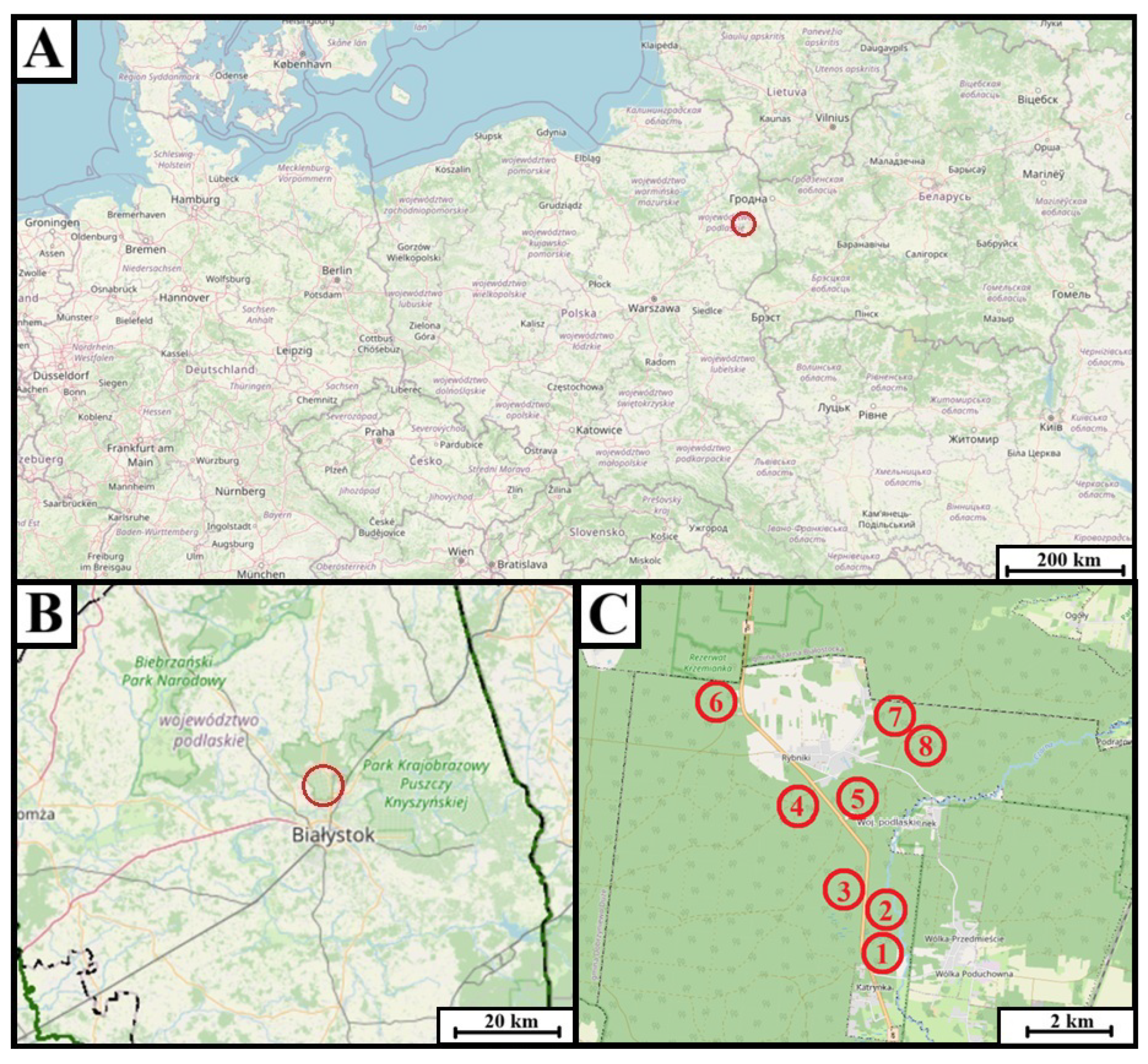
Figure 1. Geographical setting of the survey sites. (A) Position of the Knyszyn Forest study area (red circle) within Central Europe. (B) Location of the study area (red circle) within Podlaskie Voivodeship, northeastern Poland. (C) Detailed map of the Knyszyn Forest showing the eight forest sub-compartments investigated: 1—138a, 2—124D, 3—125D, 4—76G, 5—75G, 6—8I, 7—31f, and 8—7a. Map prepared by Tomasz Pawłowicz; base map data © OpenStreetMap contributors (2025).
Forests 16 01259 g001
Figure 2. Example of moist chamber cultures prepared from various forest sites and substrates encompassing three species of deadwood: Alnus glutinosa (AG), Picea abies (PA), and Betula pubescens (BP). Photograph by Tomasz Pawłowicz.
Forests 16 01259 g002
Figure 3. Morphology of the slime mold Arcyria cinerea. (A) Macroscopic view showing sporocarps with conical to cylindrical habit and whitish-to-grey pigmentation; (B) microscopic view depicting fine-meshed capillitium and finely warted spores of A. cinerea.
Forests 16 01259 g003
Figure 4. Morphology of Arcyria pomiformis. (A) Macroscopic view displaying globose to broadly ovoid, pale-yellow sporocarps; (B) microscopic view illustrating capillitium filaments and uniformly finely warted, pale-yellow spores of A. pomiformis.
Forests 16 01259 g004
Figure 5. Morphology of Arcyria denudata. (A) Macroscopic view of bright red to, stipitate sporocarps occurring in dense clusters; (B) microscopic view showing reticulate capillitium with rings and blunt warts, together with pale-brown, finely warted spores of A. denudata.
Forests 16 01259 g005
Figure 6. Photographic documentation of slime mold Arcoydes incarnata. (A) Macroscopic view showing sessile, copper-toned sporocarps; (B) microscopic view revealing wide-meshed capillitium adorned with fine granules ochre, sparsely warted spores of A. incarnata.
Forests 16 01259 g006
Figure 7. Morphological features of Ceratiomyxa fruticulosa var. flexuosa. (A) Macroscopic view exhibiting protoplasmic droplets elongating into dendroid columns; (B) microscopic view showing globose spores.
Forests 16 01259 g007
Figure 8. Morphological features of Comatricha nigra. (A) Macroscopic view displaying scattered, long-stalked sporocarps with a dark brown coloration; (B) microscopic observation of dark brown spores bearing fine warts.
Forests 16 01259 g008
Figure 9. Photographic documentation of slime mold Cribraria microcarpa. (A) Macroscopic view illustrating long-stipitate sporocarps with yellow-brown globose head supported by a dark, longitudinally wrinkled stalk; (B) microscopic depiction of a delicate peridial net showing kidney-shaped nodes and sparsely warted spores.
Forests 16 01259 g009
Figure 10. Morphological features of Hemitrichia serpula. (A) Macroscopic presentation of plasmodiocarps forming a reticulate network, sessile, and yellow-brown; (B) microscopic view of spirally ridged capillitium and pale-yellow spores bearing a large, reticulate pattern.
Forests 16 01259 g010
Figure 11. Composite morphological documentation of Lamproderma scintillans. (A) Stalked, metallic blue sporocarps at mature, globose stage; (B) loose reticulate capillitium with tapering filaments and brown, coarsely warted spores of L. scintillans.
Forests 16 01259 g011
Figure 12. Representative images illustrating diagnostic macroscopic and microscopic of Metatrichia vesparium. (A) Dark red-brown sporocarps, solitary to pseudoaethaloid aggregates, borne on ribbon-like stalks; (B) brown capillitium of spirally banded and pale-rust colored, finely warted spores of M. vesparium.
Forests 16 01259 g012
Figure 13. Photographic documentation of slime mold Oligonema flavidum. (A) Pale yellow, glossy, sessile sporocarps forming dense clusters; (B) branched capillitium bearing indistinctly spiralled warts and completely reticulate, yellow spores of O. flavidum.
Forests 16 01259 g013
Figure 14. Morphological characteristics of Perichaena depressa. (A) Densely crowded, brown sporocarps compressed into pseudoaethaloid masses; (B) elastic, golden-yellow capillitium with annular thickenings and finely warted spores of P. depressa.
Forests 16 01259 g014
Figure 15. Morphological features of slime mold Physarum album. (A) White to greyish-white, lime-encrusted sporangia on distinct stalks; (B) hyaline, anastomosing capillitium bearing scattered lime nodes and spinulose, brown spores of P. album.
Forests 16 01259 g015
Figure 16. Composite morphological characteristics of Physarum bivalve. (A) Light-grey plasmodiocarps; (B) filamentous capillitium with irregular lime nodes and uniformly finely warted spores of P. bivalve.
Forests 16 01259 g016
Figure 17. Heat-map ( log 10 ( count + 1 ) ) for 14 slime-mold species across 12 tree–substrate classes (AG = Alnus glutinosa, BP = Betula pubescens, PA = Picea abies; D = wood, K = bark, L = leaves/needles, G = twigs). Warm tones mark higher culture frequencies; white indicates absence.
Forests 16 01259 g017
Figure 18. Species-accumulation curves (sample-based rarefaction, n = 1000 ). Black line ± 95 % CI: all substrates combined; colored dashed lines: individual substrates. Horizontal dotted line marks the total observed richness, vertical dashed lines the number of sporulating cultures per substrate.
Forests 16 01259 g018
Table 1. Efficacy of moist-chamber cultures by substrate type. Marked contrasts among host–substrate combinations emphasize that careful substrate choice is critical when assessing moist-chamber performance.
Substrate Type (Tree) N total N positive Success (%)Mean Species ±  SDMedian (IQR)
Bark—Alnus glutinosa8450.01.5 ± 1.01.0 (1–1)
Leaves—A. glutinosa8112.51.0 ± 0.01.0 (1–1)
Twigs—A. glutinosa8225.01.5 ± 0.71.5 (1–1)
Wood—A. glutinosa8450.01.0 ± 0.01.0 (1–1)
Bark—Betula pubescens8675.01.7 ± 0.52.0 (1–2)
Leaves—B. pubescens800.0
Twigs—B. pubescens8112.51.0 ± 0.01.0 (1–1)
Wood—B. pubescens8337.51.0 ± 0.01.0 (1–1)
Bark—Picea abies8787.51.3 ± 0.81.0 (1–1)
Leaves—P. abies8112.51.0 ± 0.01.0 (1–1)
Twigs—P. abies8225.01.5 ± 0.71.5 (1–1)
Wood—P. abies8450.01.2 ± 0.51.0 (1–1)
All substrates combined963536.51.3 ± 0.61.0 (1–1)
Table 2. Incidence of myxomycete species in moist-chamber cultures sorted by tree-species × substrate-type combinations. Zeros indicate absence, facilitating rapid appraisal of ecological structure.
SpeciesAG BarkAG LeavesAG TwigsAG WoodBP BarkBP LeavesBP TwigsBP WoodPA BarkPA LeavesPA TwigsPA WoodRow TotalNon Zero Cols
Arcyodes incarnata00100000000011
Arcyria cinerea00004000300183
Arcyria denudata00000000000221
Arcyria pomiformis00002000200153
Ceratiomyxa fruticulosa var. flexuosa00010001000022
Comatricha nigra30200000201084
Cribraria microcarpa10000000200032
Hemitrichia serpula00020000000132
Lamproderma scintillans01000000000011
Metatrichia vesparium00010000000011
Oligonema flavidum10000000000011
Perichaena depressa10000000000011
Physarum album00004012002094
Physarum bivalve00000000010011
Table 3. Observed and estimated myxomycete richness per host tree species with sampling coverage and the frequency of all detected taxa.
Tree species N cult S obs Chao1 ± SECoverageSpecies (Frequency)
Alnus glutinosa32933.5 ± 31.126.9%Arcyodes incarnata (3.1%), Ceratiomyxa fruticulosa var. flexuosa (3.1%), Comatricha nigra (15.6%), Cribraria microcarpa (3.1%), Hemitrichia serpula (6.3%), Lamproderma scintillans (3.1%), Metatrichia vesparium (3.1%), Oligonema flavidum (3.1%), Perichaena depressa (3.1%)
Betula pubescens3244.5 ± 1.388.9%Arcyria cinerea (12.5%), Arcyria pomiformis (6.3%), Ceratiomyxa fruticulosa var. flexuosa (3.1%), Physarum album (21.9%)
Picea abies3288.7 ± 1.392.3%Arcyria cinerea (12.5%), Arcyria denudata (6.3%), Arcyria pomiformis (9.4%), Comatricha nigra (9.4%), Cribraria microcarpa (6.3%), Hemitrichia serpula (3.1%), Physarum album (6.3%), Physarum bivalve (3.1%)
Disclaimer/Publisher’s Note: The statements, opinions and data contained in all publications are solely those of the individual author(s) and contributor(s) and not of MDPI and/or the editor(s). MDPI and/or the editor(s) disclaim responsibility for any injury to people or property resulting from any ideas, methods, instructions or products referred to in the content.